electrocardiographic changes of left ventricular hypertrophy: effects of antihypertensive treatment
TRANSCRIPT
ELECTROCARDIOGRAPHIC CHANGES OF LEFT VENTRICTKAR HYPERTROPHY: EFFECTS OF ANTIHYPERTENSIVE
TREATMENT
J. G. HELMCKE, M.D.,* ROLANDSCHNECKLOTH,M.D.,ANDA.(I.COKCORAN,~~.I:).
CLEVELAND, OEIIO
INTRODUCTION
C ONGESTIVE heart failure, commonly associated with left ventricular hypertrophy, has been recognized as the most common cause of death in
hypertension.ls2 In recent years, with the introduction of improved methods of treatment of congestive heart failure and the increasing availability of effective antipressor drugs, death due to intractable congestive failure as such (in the absence of evident coronary artery disease) has become an uncommon event.X However, the presence of left ventricular hypertrophy still maintains a consider- able clinical significance. It may influence selection of and response to anti- pressor drugs, e.g., the choice of a ganglion-blocking drug instead of an agent which, like hydralazine, may increase cardiac work; it also determines the prc- scription of low-sodium diets, digitalis, and diuretics.
Hypertrophy of the left ventricle (with which we are most particularI! concerned here) can be proved only by post-mortem examination. However, the diagnosis can be made with some confidence by physical examination or b,- roentgenography. The electrocardiogram contributes greatly to the diagnosis since certain characteristics appearing in standard limb,4 unipolar limb, and pry- cordial5 leads show a distinct association with anatomically proved left ventric.- ular hypertrophy. Scott and co-workers6 evaluated the electrocardiographic criteria of Sokolow and Lyon5 and confirmed the diagnosis anatomically in 8.5 of 100 cases, when the diagnosis was based on the presence of any- single cri- terion of hypertrophy.
We selected certain of these electrocardiographic criteria of left ventricul:cr hypertrophy as the area of this study. The aim was to determine the association\ of these electrocardiographic patterns of hypertrophy with the syndrome of malignant hypertension on the one hand, and with severe essential hypertcnsioll on the other, the association of these abnormal patterns with subsequent ther;i- peutic responses, and the changes which occurred during treatment.
It will be shown (1) that the imposition of the syndrome of malignant hyper- tension only slightly alters the incidence and distribution of electrocardiographic. criteria of hypertrophy from that of severe essential hypertension; (2,) that pretreatment tracings are poor indications of the probability of success in anti- pressor therapy; (3) that, although diminution of electrocardiographic abnor- ____
From the Research Division, The Cleveland Clinic Foundation, and The Frank E. Bunts Educ:t- tional Institute, Cleveland, Ohio.
Received for publication July 9, 1956. *Present address: 114 Rue Capois, Port-au-Prince, Haiti.
549

550 HELMCKE, SCHNECKLOTH, AND CORCORAN
malities are more common in patients with good therapeutic responses than in those with poor responses, there is not a consistent association; (4) that, because of this variability in electrocardiographic response, serial tracings are of little assistance in assessing therapeutic response; and (5) that the scattered incidence of the individual abnormalities taken as criteria of hypertrophy makes it impos- sible to select any one of these as of exclusive or predominant diagnostic value.
MATERIAL AND METHODS
Records of 78 patients with severe essential hypertension or the malignant phase of essential hypertension were ,studied; no patient had evident congestive cardiac failure. The group was composed of fifty men and twenty-seven women, ranging in age from 25 to 70 years, and including one boy of 12 years. The aver- age age of the adults was 45 years.
Patients with electrocardiographic evidence of left bundle branch block or myocardial infarction, or with clinical evidence of chronic valvular heart disease or angina pectoris were not included in this study. Since S-T-T changes due to digitalis and left ventricular hypertrophy cannot easily be differentiated, patients receiving digitalis were also excluded. Multiple lead electrocardiograms were avail- able for every patient; each electrocardiogram had three standard limb, three aug- mented unipolar limb, and three to eight precordial leads. The diagnosis of left ventricular hypertrophy in each patient prior to treatment was based on the presence of one or more of eight electrocardiographic criteria adapted after Sokolow and Lyon ,5 as listed in Table I. Tracings made before and during an adequate trial of one or more antihypertensive agents given alone or in combi- nation were analyzed. The periods of treatment ranged from 1 to 50 months, averaging 21 months. Twelve of the 78 patients died after less than 3 months of observation; 9 of these 12 patients had shown no response to therapy. Forty patients were observed for two or more years.
For convenience in the evaluation of response to treatment, patients were separated into two classes of hypertensive disease: (1) severe essential (35 pa-
TABLE I. CRITERIA FOR DIAGNOSIS OF LEFT VENTRICULAR HYPERTROPHY MODIFIED AFTER S~KOLOW AND LYONS
A. Unipolar Limb Leads 1. Upright T-wave in a\‘a and S-T segment elevated 0.5 mm. or more 2. Voltage of R-wave in aVr, > 12.5 mm. 3. S-T segment depressed > 0.5 mm. in aVr. in case of horizontal heart or in ~VF in
case of vertical heart 4. T-wave flat, diphasic, or inverted, with R-wave 6.0 mm. or more in aVL or ~VF and
with (3)
R. Precordial Leads 5. Voltage of R-wave in VS or Ve plus S-wave in VI > 3.5 mm. 6. S-T segment depressed 0.5 mm. in VS or Vb 7. T-wave flat, diphasic, or inverted in leads 1’6 or Vs, with normal R and small S-
waves and with (6) 8. Onset of intrinsicoid deflection in VG or VS > 0.05 sec.
-_- __-. -__ ~-

ECG CHANGES OF LVH: ANTIHPPEKTENSIVIS TRE.\TMENT 551
tients) defined as a state of persistent diastolic: hypertension, \vith ret inal, cerelrro- vascular, cardiac, and/or renal evidence of advancing hypertensive WJ- cular disease; (2) malignant (43 patients) d&led as a syndrome characterized by papilledema, retinal hemorrhages and exudates, objective evidence of hypcr- tensive heart disease, and proteinuria and hematuria, usually associated with impaired renal function and commonly accompanied by loss of weight.
A composite severity index was assembled for every patient, both before and during therapy, concurrently with each electrocardiogram. This index expresses numerically the over-all severity of the patient’s disease at the time of observation, by assigning a maximum of four points each to estimates of (1.) average daily hospital and home supine diastolic pressure, and of the extent of hypertensive vascular disease, (2) in the heart, (3) in the kidney, and (4) in the l~rain.5
.Antihypertensive treatment consisted of hydralazine (I-hydrazinophth;ll- azine) or ganglion-blocking agents (hexamethonium or pentapyrrolidinium), alo~re or combined, or in combination with reserpine. The majority of patients were treated with hydralazine alone. Two patients with essential hypertension h;ld had bilateral thoracolumbar sympathectom>..
Response to treatment was classified according to change in average diastolic blood pressure. If the average diastolic pressure fell below 110 mm. Hg the pa- tient was considered a responder, and if it did not, as a nonresponder. ‘I‘went).- nine of the 43 patients with malignant hypertension were classified as responders to treatment and 14 as nonresponders; 20 of the 35 patients with severe essential hypertension were responders and 15 were nonresponders.
RESULTS
1. Pretreatment Electrocardiograms.- a. Incidence of criteria of left ventricular hypertrophy (Table II): In the
whole hypertensive group, the mean number of electrocardiographic criteria of
TABLE II. INCIDENCE OF ELECTROCARDIOGRAPHIC CRITERIA FOR LEFT VENTRICULAR HYPERTROPHY AND TOTAL SEVERITY INDEX (T.S.I.) IN 78 HYPERTENSIVE PATIENTS
- ____
1 / BEFORE TREATMENT
NUMBER ’ I
OF I AVERAGE I PATIENTS NO. AVERAGE
CRITEKIA T.S.I. -__ __-.-- ---.- -.---__
Hypertensive Patients 78 ~ 5.0 8.1 Responders 49 7.6 Nonresponders 29 / 8 5
Malignant Hypertension Responders Nonresponders
Essential Hypertension :i / 4.8 69 Responders ~ 4.2 6 3 Nonresponders 15 1 5.4 ~ 7.4
I
AFTER TREATMEKT
AVERAGE NO. AVlTKA(~l.
CRITERIA ‘I’.)r.l.
5.1 j 0 -0 4.2 ; 5.8 5.9 ; Y.0
4.5 ~ 5 0 3.2 .z 0 5.8
1
I 6.4
552 HELMCKE, SCHNECKLOTH, AND CORCORAN Am. Heart J.
Alxil, 1957
left ventricular hypertrophy present in tracings prior to treatment was 5.0 (range 1 to 8). The mean incidence was somewhat greater (5.6) in patients who did not respond to treatment than in those who did (4.4)) and in this respect corresponded to the respective estimates of severity of the hypertensive disease, which was greater in the nonresponders. In comparing the diagnostic groups, the criteria of hypertrophy were on the average more frequent and severity indices higher in patients with the malignant syndrome than in those with severe essential hypertension, and in both of these groups, greater in those who did not respond to treatment than in those who did. These differences, however, were small and the ranges of variation wide.
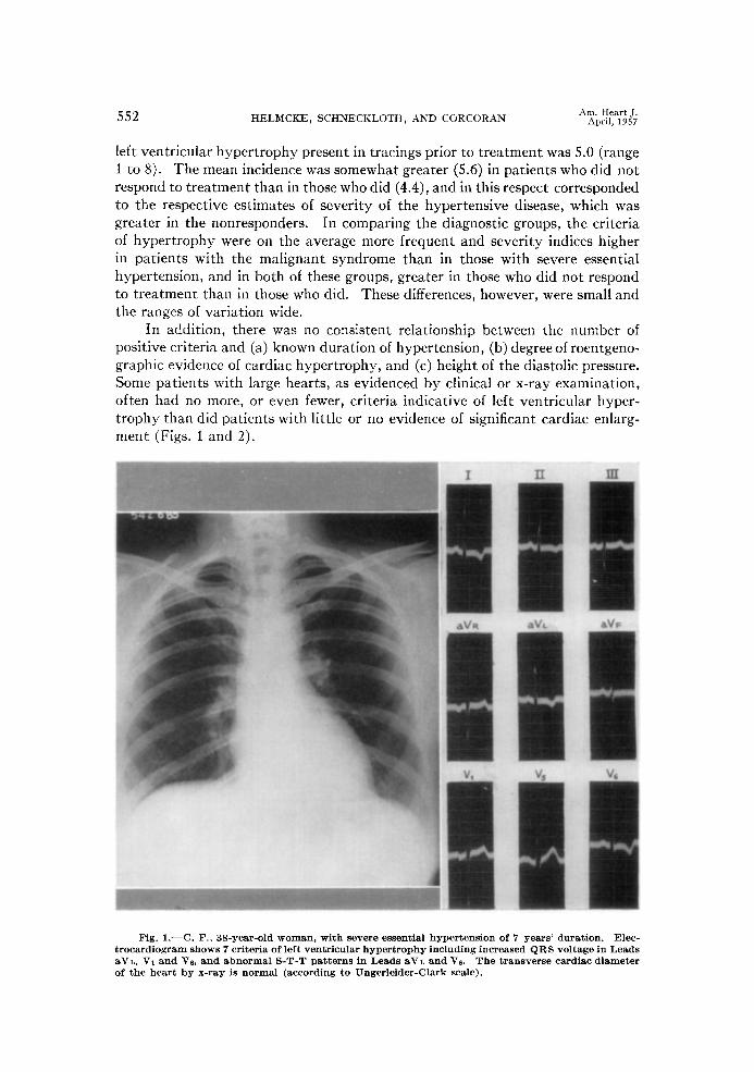
In addition, there was no consistent reIationship between the number of positive criteria and (a) known duration of hypertension, (b) degree of roentgeno- graphic evidence of cardiac hypertrophy, and (c) height of the diastolic pressure. Some patients with large hearts, as evidenced by clinical or x-ray examination, often had no more, or even fewer, criteria indicative of left ventricular hyper- trophy than did patients with little or no evidence of significant cardiac enlarg- ment (Figs. 1 and 2).
Fig. 1.-C. F., 3%year-old woman, with severe essential hypertension of 7 years’ duration. Elec- trocardiogram shows 7 criteria of left ventricular hypertrophy including increased QRS voltage in Leads aVL, VI and VS, and abnormal S-T-T patterns in Leads aVL and VS. The transverse cardiac diameter of the heart by x-ray is normal (according to Ungerleider-Clark scale).
Volume 53 Kumlm 1 ECG CHANGES OF LVH: ANTIHYPERTENSIVE TREATMENT 55.1
b. Distribution of electrocardiographic criteria of left ventricular hyper- trophy (Table III): The most common criteria indicative of left ventricular hypertrophy were increased voltage of QRS complexes in Leads aVL and/or pre- cordial leads. Depressed S-T segments and abnormal T-waves, the so-called “strain pattern,” were a little less frequently observed. All abnormalities, includ- ing delayed intrinsicoid deflections, were somewhat more often observed in trac- ings of patients with malignant hypertension than in patients with severe essen- tial h>.pertension. Abnormal S-T-T patterns or increased voltage of &KS complexes as the sole criterion were present in a minority of patients (26 per cent).
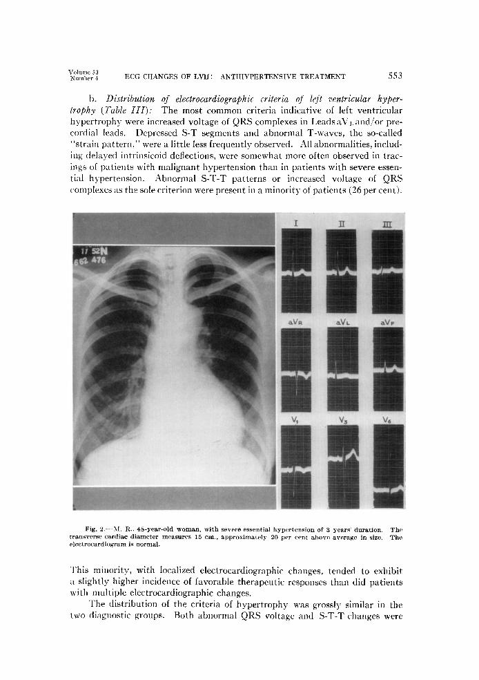
Fig. 2.--M. R.. 4%year-old woman, with severe essential hypertension of 3 years’ duration. The transverse cardiac diameter measures 15 cm., approximately 20 per cent ahove average in size. The electrocardiogram is normal.
This minority, with localized electrocardiographic changes, tended to exhibit a slightly higher incidence of favorable therapeutic responses than did patients with multiple electrocardiographic changes.
The distribution of the criteria of hypertrophy was grossly similar in the two diagnostic groups. Both abnormal QRS voltage and S-T-T changes were
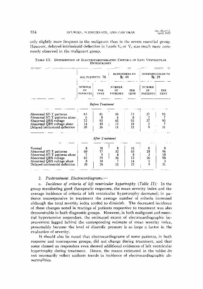
only slightly more frequent in the nlalignan t than in the severe essential groul). However, delayed intrinsicoid deflection in 1,eads V, or V5 was much more com- monly observed in the malignant group.
TABLE III. DISTRIBUTION OF ELECTKOCAKDIOGKAPHIC CRITERIA OF LEFT VENTRICULAR
Abnormal ST-T patterns Abnormal ST-T patterns alone Abnormal QRS voltage Abnormal QRS voltage alone Delayed intrinsicoid deflection
HYPERTROPHY
RESPONDERSTO ALL PATIENTS: 78 3: 49
-__---
NUMBER NUMBER OF PER OF PER
PATIENTS CENT PATIENTS CENT
~~-.
Before Treatment
NONRESPONDERSTO Q: 29
NUMBER OF PER
PATIENTS CENT
27 93 2; 7
93 ; 7
1 31
After Treatment
Normal Abnormal ST-T patterns Abnormal ST-T patterns alone Abnormal QRS voltage Abnormal QRS voltage alone Delayed intrinsicoid deflection
2. Posttreatment Electrocardiograms.-
a. Incidence of criteria of left ventricular hypertrophy (Table II): In the group manifesting good therapeutic responses, the mean severity index and the average incidence of criteria of left ventricular hypertrophy decreased ; in pa- tients nonresponsive to treatment the average number of criteria increased although the total severity index tended to diminish. The decreased incidence of these changes noted in tracings of patients responsive to treatment was also demonstrable in both diagnostic groups. However, in both malignant and essen- tial hypertensive responders, the estimated extent of electrocardiographic im- provement lagged behind the corresponding estimate of mean severity index, presumably because the level of diastolic pressure is so large a factor in the evaluation of severity.
It should also be noted that electrocardiograms of some patients, in both response and nonresponse groups, did not change during treatment, and that some classed as responders even showed additional evidences of left ventricular hypertrophy during treatment. Hence, the means estimated in the tables do not necessarily reflect uniform trends in incidence of electrocardiographic ab- normalities.
Volume 5 :, Number 1 ECG CHANGES OF LVH: ANTIHYPERTENSIVE TREATMENT 55.5
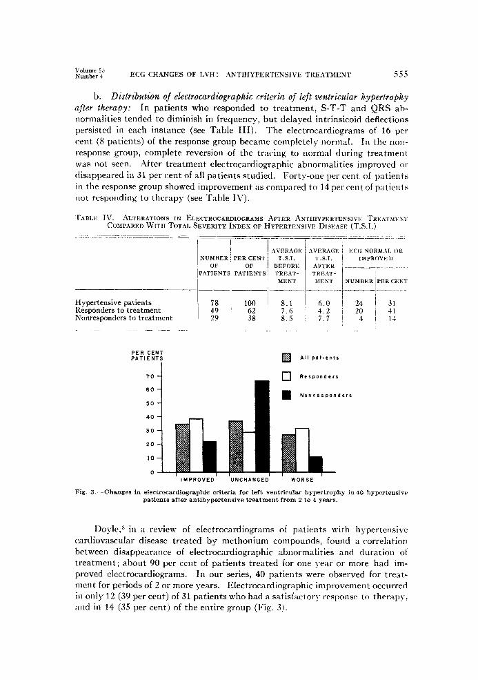
b. Distribution of electrocardiographic criteria of left ventricular hypertrophy after therapy: In patients who responded to treatment, S-T-T and QRS ab- normalities tended to diminish in frequency, but delayed intrinsicoid deflections persisted in each instance (see Table III). The electrocardiograms of 16 per cent (8 patients) of the response group became completely normal. In the non- response group, complete reversion of the tracing to normal during treatment was not seen. After treatment electrocardiographic abnormalities improved or disappeared in 31 per cent of all patients studied. Forty-one per cent of patients in the response group showed improvement as compared to 14 per cent of patient 5 Ilot responding to therapy (see Table IV).
'I‘ABLE Iv. ALTERATIONS IN ELECTROCARDIOGRAMS AFTER ANTIHYPERTENSIVE 'I'REATMEKJ. COMPARED WITH TOTAL SEVERITY INDEXOF HYPERTENSIVE DISEASE (T.S.I.)
NUMBER'PERCENT OF OF
PATIENTS PATIENTS
-___- ---___
Hypertensive patients Responders to treatment Xonresponders to treatment
AVERAGE T.S.I.
BEFORE TREAT-
MENT
8.1 6.0 7.6 4.2 8.5 7.7
AVERAGE T.S.I.
AFTER TREAT-
MENT __
ECG NORMAL OR IMPROVED
___-.-.
NUMBERiPERCENT - ----_
PER CENT PATIENTS
70 J
Nonresponders
20
10
0 IMPROVED UNCHANGED WORSE
Fig. 3.-Changes in electrocardiographic criteria for left ventricular hypertrophy in 40 hypertensivr patients after antihypertensive treatment from 2 to 4 years.
Doyle,” in a review of electrocardiograms of patients with hypertensive cardiovascular disease treated by methonium compounds, found a correlation between disappearance of electrocardiographic abnormalities and duration of treatment; about 90 per cent of patients treated for one year or more had im- proved electrocardiograms. In our series, 40 patients were observed for treat- ment for periods of 2 or more years. Electrocardiographic improvement occurred in only 12 (39 per cent) of 31 patients who had a satisfactory response to therapy, and in 14 (3.5 per cent) of the entire group (Fig. 3).
556 HELMCKE, SCHNECKLOTH, AND CORCORAN -4m. Heart J.
April, 1957
DISCUSSION
The severity of the hypertensive vascular disease in the group of patients studied is reflected in the high percentage of pretreatment tracings which showed S-T-T changes (81 per cent) and abnormal QRS voltages (92 per cent). The frequent concurrence in the tracings of more than one electrocardiographic criterion (average 5.0) can be presumed to enhance the accuracy of electrocardio- graphic diagnosis of left ventricular hypertrophy in this group, and in addition has permitted evaluation of some of these criteria separately as to their incidence and association with clinical status and therapeutic response.
The incidence and type of criteria of left ventricular hypertrophy found in pretreatment electrocardiograms were not guides to subsequent therapeutic response in individual patients, although the average incidence of these criteria was less in the group with adequate therapeutic responses. Eight per cent of the responders showed complete disappearance of abnormal S-T-T patterns, and 19 per cent had a return to normal of the abnormal QRS voltage. This tendency of increased voltage to revert to normal was particularly evident in patients who had essential hypertension and were responsive to treatment (25 per cent). In 8 of the 49 patients who had a satisfactory response to therapy, increased QRS voltage and abnormal S-T-T patterns disappeared completely during treatment.
The relative importance of one electrocardiographic criterion over another in the diagnosis of clinically significant cardiac hypertrophy could not be deter- mined because of a lack of adequate necropsy material, plus the fact that different positive criteria were either present alone or, more frequently, in many and varying combinations. Although improvement was observed predominantly in electrocardiograms of patients responsive to antihypertensive therapy (41 per cent), favorable electrocardiographic changes of similar type and degree were observed as well in some patients nonresponsive to therapy and otherwise exhibit- ing progression of their vascular disease.
Probably because of the many factors other than true hypertrophy that contribute to electrocardiographic abnormalities, the study of serial electro- cardiograms does not appear to be of much help as a means of estimating the success or failure of present-day therapy. From this it seems that the electro- cardiogram considered alone does not accurately reflect the extent of hypertensive heart disease in an individual patient. In general, after treatment with available antipressor agents, electrocardiographic abnormalities which are attributable to left ventricular hypertrophy secondary to hypertension disappear or improve in about 30 per cent of all patients. Such improvement was 3 times more common in patients classed as responders-i.e., those with diastolic pressure averages of less than 110 mm. Hg-than in those listed as nonresponders, although the latter group showed significant improvement in over-all severity index.
SUMMARY AND CONCLUSIONS
Electrocardiograms of 78 patients with severe essential hypertension, includ- ing 43 in the malignant phase, were studied with reference to electrocardiographic evidence of left ventricular hypertrophy, before and during use of one or more antihypertensive drugs. The criteria for left ventricular hypertrophy were
Volume 5 .i Nunhrr 4 ECG CHANGES OF LVH : ,\NTIHYPERTENSIVE TREATMENT 557
adapted from those of Sokolow and Lyon 5; the relative severity of the hyperten- sive disease (severity index) was estimated concurrently with each elertro- cardiogram.
The variable incidence of the separate criteria of hypertrophy (increased QRS voltage, S-T segment and T-wave changes) does not permit the selection of any one of them as having exclusive or predominant diagnostic value. The presence of the syndrome of malignant hypertension only slightly changes the incidence and distribution of electrocardiographic evidence of left ventricular hypertrophy.
I’retreatment electrocardiograms in this group of hypertensive patients had no predictive value as to the success or failure of subsequent therapy. Electro- cardiographic improvement was more common in those patients who had a satisfactory response to therapy, but this was not a consistent association. Be- cause of this variability, routine serial tracings add little to estimates of thera- peutic response.
There was no consistent relationship between height of the blood pressure, total severity indices, size of the heart by teleroentgenogram, and the electro- cardiographic changes attributable to left ventricular hq-pertrophy. Thus, while electrocardiographic estimates follow group trends rather closely, they do not accurateI>. reflect the cardiac or even the over-all status of individual patients with hypertensive heart disease.
\Ve are indebted to William L. Proudfit, M.D., Department of Cardiovascular Disease, 1)ivision of Medicine, The Cleveland Clinic Foundation, for helpful advice and critical ctnnmenrs.
REFERENCES
1. Clawson, B. J.: The Heart in Essential Hypertension, in Hypertension, a Symposium. edited by E. T. Bell, Minneapolis, 1951, University of hlinnesota Press.
2. Goldring, W., and Chasis, H.: Commonwealth Fund.
Hypertension and Hypertensive Disease, New York, 1944,
3. Corcoran, A. C., Page, I. H., Dustan, H. P., and Lewis, L. A.: Cleveland Clin. Quart. 23:115, 19.56.
4. 5.
Gubner, R., and Ungerleider, H. E.: Arch. Int. Med. 72:196, 1943.
6. Sokolow, M., and Lyon, T. P.: AM. HEART J. 37:161, 1949.
7. Scott, R. C., Seiwert, V. J., Simon, D. L., and McGuire, J.: Circulation 11339, 1955.
8. Corcoran, A. C., Dustan, H. P., Taylor, R. D., and Page, I. H.: Am. J. Med. 17:383, 19.54. Doyle, A. E.: AM. HEART J. 45:363, 1953.