ekg interpretation unc emergency medicine medical student lecture series
TRANSCRIPT

EKG Interpretation
UNC Emergency MedicineMedical Student Lecture
Series

Objectives The Basics Interpretation Clinical Pearls Practice
Recognition

The Normal Conduction System

Lead Placement
aVF

All Limb Leads

Precordial Leads

EKG Distributions Anteroseptal: V1, V2, V3,
V4 Anterior: V1–V4 Anterolateral: V4–V6, I,
aVL Lateral: I and aVL Inferior: II, III, and aVF Inferolateral: II, III, aVF,
and V5 and V6

Waveforms
Interpretation Develop a systematic approach to
reading EKGs and use it every time The system we will practice is:
Rate Rhythm (including intervals and
blocks) Axis Hypertrophy Ischemia

Rate Rule of 300- Divide 300 by the
number of boxes between each QRS = rate
Number of big boxes
Rate
1 300
2 150
3 100
4 75
5 60
6 50

Rate HR of 60-100 per minute is normal HR > 100 = tachycardia HR < 60 = bradycardia

Differential Diagnosis of Tachycardia
Tachycardia
Narrow Complex
Wide Complex
Regular STSVTAtrial flutter
ST w/ aberrancySVT w/ aberrancy
VT
Irregular A-fibA-flutter w/ variable conductionMAT
A-fib w/ aberrancy
A-fib w/ WPWVT

What is the heart rate?
(300 / 6) = 50 bpm
www.uptodate.com

Rhythm Sinus
Originating from SA node
P wave before every QRS
P wave in same direction as QRS

What is this rhythm?Normal sinus rhythm

Normal Intervals PR
0.20 sec (less than one large box)
QRS 0.08 – 0.10 sec (1-2
small boxes) QT
450 ms in men, 460 ms in women
Based on sex / heart rate
Half the R-R interval with normal HR

Prolonged QT Normal
Men 450ms Women 460ms
Corrected QT (QTc) QTm/√(R-R)
Causes Drugs (Na channel blockers) Hypocalcemia, hypomagnesemia, hypokalemia Hypothermia AMI Congenital Increased ICP

Blocks AV blocks
First degree block PR interval fixed and > 0.2 sec
Second degree block, Mobitz type 1 PR gradually lengthened, then drop QRS
Second degree block, Mobitz type 2 PR fixed, but drop QRS randomly
Type 3 block PR and QRS dissociated

What is this rhythm?
First degree AV block PR is fixed and longer than 0.2 sec

What is this rhythm?
Type 1 second degree block (Wenckebach)

What is this rhythm?
Type 2 second degree AV block Dropped QRS

What is this rhythm?
3rd degree heart block (complete)

The QRS Axis
Represents the overall direction of the heart’s activity
Axis of –30 to +90 degrees is normal

The Quadrant Approach QRS up in I and up in aVF =
Normal

What is the axis?
Normal- QRS up in I and aVF

Hypertrophy Add the larger S wave of V1 or V2
in mm, to the larger R wave of V5 or V6.
Sum is > 35mm = LVH

Ischemia Usually indicated by ST changes
Elevation = Acute infarction Depression = Ischemia
Can manifest as T wave changes Remote ischemia shown by q
waves

What is the diagnosis?
Acute inferior MI with ST elevation in leads II, III, aVF

What do you see in this EKG?
ST depression II, III, aVF, V3-V6 = ischemia

Let’s PracticeThe sample EKGs were obtained from the following text:

Normal Sinus Rhythm
Mattu, 2003

First Degree Heart Block
PR interval >200ms

Accelerated Idioventricular
Ventricular escape rhythm, 40-110 bpm
Seen in AMI, a marker of reperfusion

Junctional Rhythm
Rate 40-60, no p waves, narrow complex QRS

Hyperkalemia
Tall, narrow and symmetric T waves

Wellen’s Sign
ST elevation and biphasic T wave in V2 and V3Sign of large proximal LAD lesion
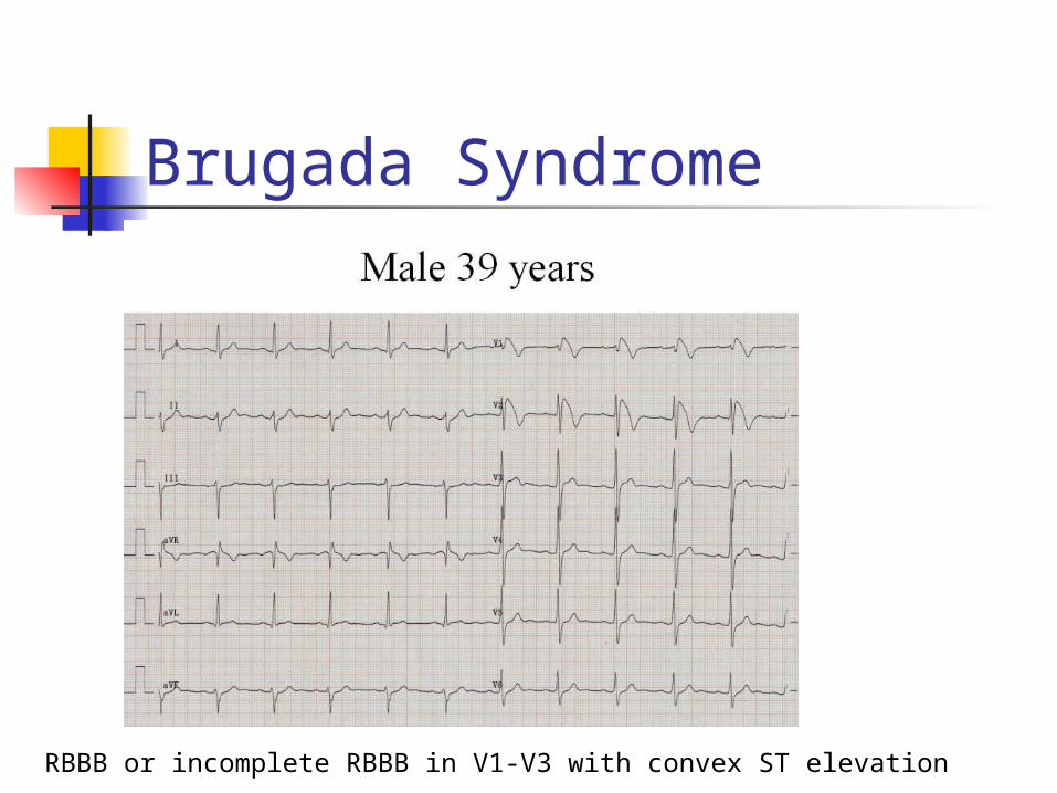
Brugada Syndrome
RBBB or incomplete RBBB in V1-V3 with convex ST elevation
Brugada Syndrome Autosomal dominant genetic
mutation of sodium channels Causes syncope, v-fib, self
terminating VT, and sudden cardiac death
Can be intermittent on EKG Most common in middle-aged males Can be induced in EP lab Need ICD

Premature Atrial Contractions
Trigeminy pattern
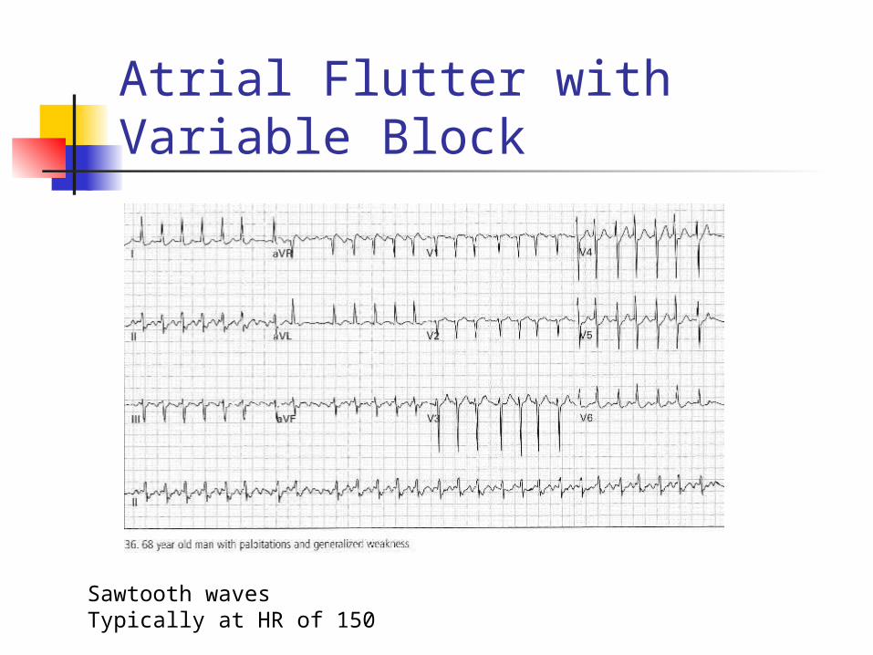
Atrial Flutter with Variable Block
Sawtooth wavesTypically at HR of 150

Torsades de Pointes
Notice twisting pattern
Treatment: Magnesium 2 grams IV

Digitalis
Dubin, 4th ed. 1989

Lateral MI
Reciprocal changes

Inferolateral MI
ST elevation II, III, aVF
ST depression in aVL, V1-V3 are reciprocal changes

Anterolateral / Inferior Ischemia
LVH, AV junctional rhythm, bradycardia

Left Bundle Branch Block
Monophasic R wave in I and V6, QRS > 0.12 secLoss of R wave in precordial leadsQRS T wave discordance I, V1, V6Consider cardiac ischemia if a new finding

Right Bundle Branch Block
V1: RSR prime pattern with inverted T waveV6: Wide deep slurred S wave

First Degree Heart Block, Mobitz Type I (Wenckebach)
PR progressively lengthens until QRS drops
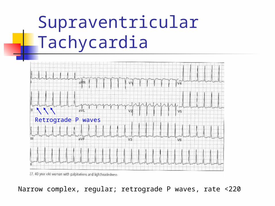
Supraventricular Tachycardia
Narrow complex, regular; retrograde P waves, rate <220
Retrograde P waves

Right Ventricular Myocardial Infarction
Found in 1/3 of patients with inferior MI
Increased morbidity and mortality
ST elevation in V4-V6 of Right-sided EKG
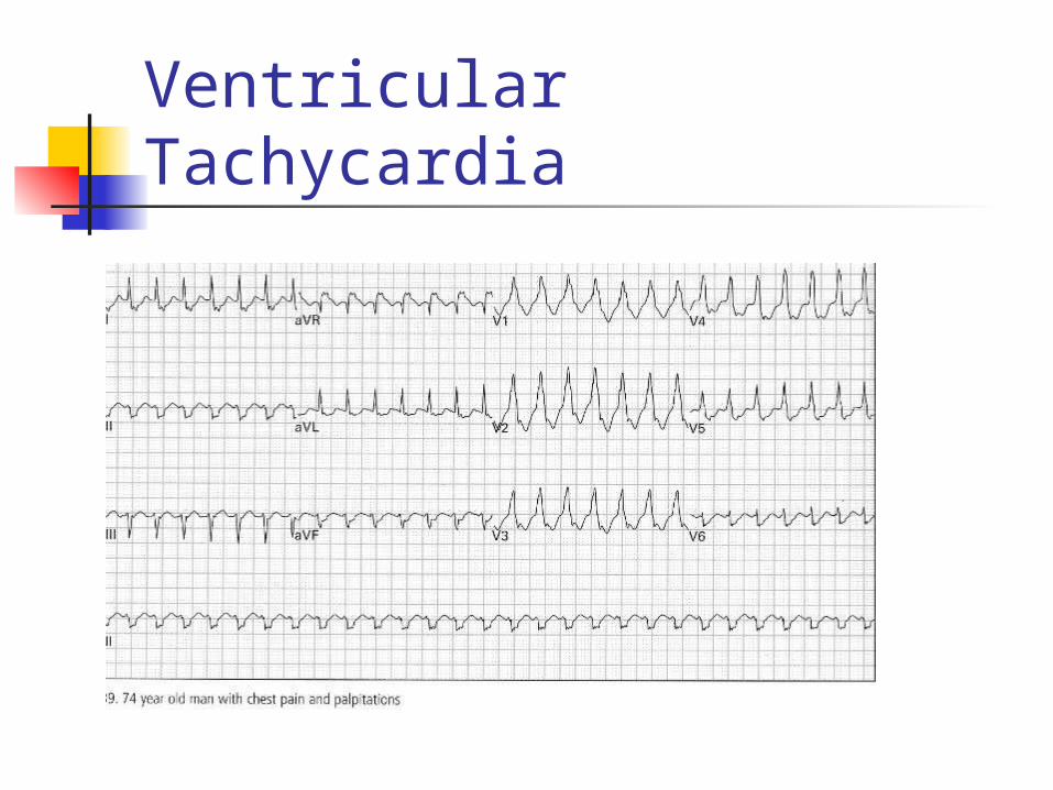
Ventricular Tachycardia

Prolonged QT
QT > 450 ms
Inferior and anterolateral ischemia

Second Degree Heart Block, Mobitz Type II
PR interval fixed, QRS dropped intermittently

Acute Pulmonary Embolism
SIQIIITIII in 10-15%
T-wave inversions, especially occurring in inferior and anteroseptal simultaneously
RAD

Wolff-Parkinson-White Syndrome
Short PR interval <0.12 secProlonged QRS >0.10 secDelta waveCan simulate ventricular hypertrophy, BBB and previous MI
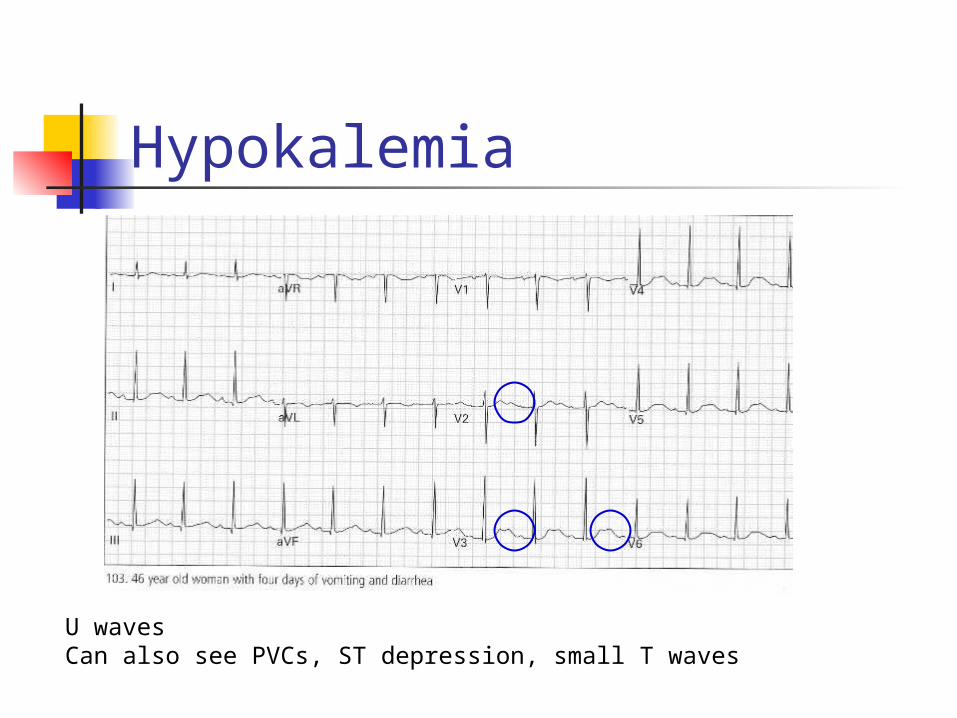
Hypokalemia
U wavesCan also see PVCs, ST depression, small T waves


Thank You
Any Questions?