ekg boot camp: ischemia and infarction - canpcanpweb.org/canp/assets/file/2014 conference...
TRANSCRIPT

EKG Boot Camp: Ischemia and Infarction

Systematic Approach • Heart Rate • Rhythm • Intervals • Axis • Voltage • R Wave Progression • Q Wave • ST segments • T waves • Device Involvement
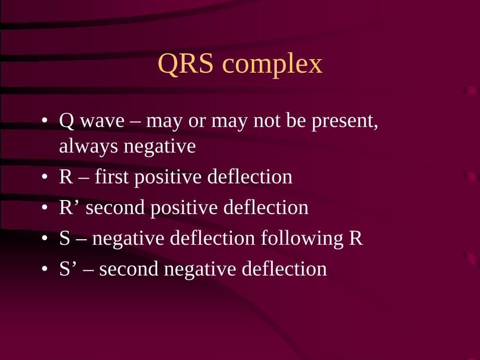
QRS complex
• Q wave – may or may not be present, always negative
• R – first positive deflection • R’ second positive deflection • S – negative deflection following R • S’ – second negative deflection


ST Segment
• In limb leads, the ST segment is isoelectric, may be slightly elevated or depressed by less than 1 mm
• In precordial leads, the ST segment may be elevated 1-2 mm
• The J point is where the QRS complex and the ST segment meet

ST Depression

ST Depression

T waves
• Ventricular repolarization • Usually oriented in the same direction as the
QRS • Slightly asymmetrical • Height less than 5mm in the limb leads • Height less than 10mm in precordial leads

T Wave Inversion • T wave inversion suggestive of ischemia or
injury • T waves should be pos I, II • Inversion is common in V1, may occur III,
aVL aVF and be normal • Inv in V2-V6 suggestive of ischemia/injury • Symmetrical T wave inversion develops
after terminal TWI, common in NSTEMI • Post STEMI, TWI is a sign of reperfusion
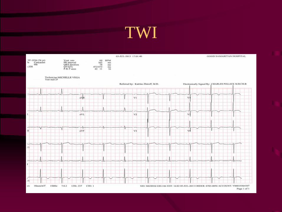
TWI

• Always compare to the patient’s prior EKG

Ischemia • Usually indicated by
ST changes – Elevation = Acute
infarction – Depression = Ischemia
• Can manifest as T wave changes
• Remote ischemia shown by q waves

TWI

ST Segment Elevation Patterns
• Hyperacute T wave – may occur as early as 2 mins post myocardial occlusion
• J Point Elevation • Subtle ST elevation forming broad T wave • If there is elevation in limb leads, I, II, III,
then worry • If there is depression in the V leads, then
worry

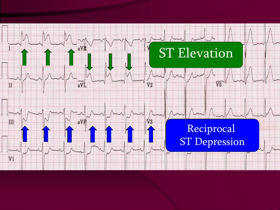
ST Elevation

ST Elevation

ST Elevation

Ischemia and Infarction
• ST segment depression – Ischemia/Subendocardial
• T Wave Inversion – Ischemia/Injury
• ST segment elevation – Injury/Transmural
• Q Wave – Old MI

EKG Distributions
• Septal V1-V2 • Anterior: V3–V4 • Anteroseptal: V1-V4 • Lateral: V4–V6, I, aVL • Inferior: II, III, and aVF • Inferolateral: II, III, aVF, and
V5 and V6

Lead Representation of the Heart

Evolution of MI

Evolution of MI

Evolution of MI

Pathologic Q Waves
• A Q wave is the first negative deflection after the P wave.
• A pathologic Q wave is greater than .04 seconds wide (1 little box on the EKG paper) in 2 contiguous leads excluding V1, III.
• Depth is 1/3 the height of the R wave. • 50% of adults have non-diagnostic Q wave


You are asked to review an ECG from an asymptomatic 73yo male scheduled for
prostate surgery – is there an inferior MI?

Inferior Q wave inconsistency

Coronary Arteries Supply to the Myocardium
• Anterior LV – LAD • Lateral LV – LCx (OM) • Posterior LV – RCA (PDA), LCx (OM) • Inferior LV – RCA (marginal) • Septum – LAD, RCA (PDA) • RV - RCA (marginal/PDA)

Reciprocal Leads
Reciprocal ST Depression
ST Elevation

Reciprocal ST Depression
ST Elevation

aVR Elevation

Posterior MI Criteria
• ST depression in V1-V3 • Prominent R>S in V1-V3 • Upright T waves in V1-V3 • Often coexisting with inferior-lateral MI • True posterior MI involves the left Cx
artery

ST elevation P>R, ST Depression T wave inversion
Recip St depression
Lateral MI ST Elevation
R>S, ST Depression, Upright T Waves

Now compare with a normal ECG


Right Ventricular Infarct
• RVI should be considered in all pts who have an inferior MI (found in 1/3 of pts)
• Right-sided ECG, echo, and invasive hemodynamic monitoring also be helpful in diagnosis
• Elevated JVD, Hypotension and clear lung fields suggests RVI
• Treatment: volume load with NS

Right Ventricular Infarct Criteria
• ST elevation > 1mm in V4R- V6R • Concurrent Inferior and Infero-posterior MI • V5 and V6 are reciprocal to RV MI
*Treatment: fluid loading*

Right Ventricular Myocardial Infarction

Coronary Arteries Supply to the Conduction System
• SA node: RCA (LCx) • AV node: RCA (LCx) • His Bundle: RCA • Bundle Branches: LAD
• Bradyarrhythmias/conduction disturbances are well recognized complications of acute MI

Bradyarrythmias that occur early in the setting of an inferior MI within the first 24 hours may respond to atropine

Conduction Disturbances and Infarct Location
• Inferior MI – Sinus brady most common arrhythmia, but Mobitz I and
CHB also seen – High degree AV block located above the His – Usually resolves
• Anterior MI – Mobitz II, BBB, CHB – High degree AV block located below the AVN – Ppm for pts at high risk for CHB (2 or more: PR
prolongation, 2nd degree AVB, left ant or post fascicular block, LBBB, RBBB

84 yo woman with syncope
ST Elevation
Mobitz II

72 year old man with nausea and dyspnea
Mobitz Type I: Wenckebach PR Prolongation
ST Elevation with Reciprocal Changes

44 year old man with dyspnea
CHB Anteroseptal MI

A few others


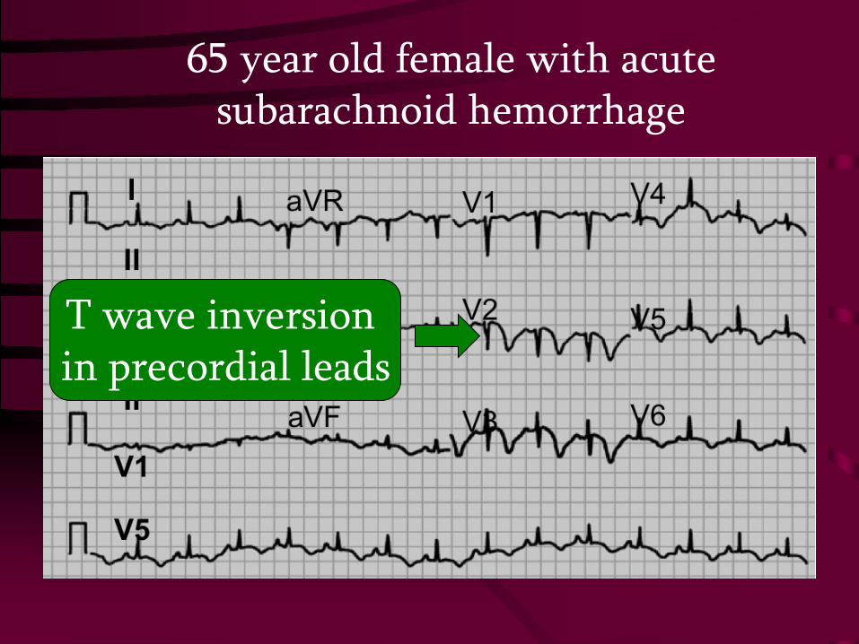
65 year old female with acute subarachnoid hemorrhage
T wave inversion in precordial leads

A 36yo male presents with severe CP – EKG is consistent with: a) early repolarization b) ant MI c) pericarditis d) inf ischemia

A 36yo male presents with severe CP – EKG is consistent with: a) early repolarization b) ant MI c) pericarditis d) inf ischemia


51 yo female with pericardial effusion


Case Studies

89yo female presents with dyspnea and back pain
ST Elevation

46 year old man with chest pain
Acute Pericarditis: Diffuse ST elevation
PR depression

71yo female with nausea, dyspnea and general malaise. ECG changes in V2 are
due to: Ant ischemia or Post injury

71yo female with nausea, dyspnea and general malaise. ECG changes in V2 are
due to: Ant ischemia or Post injury
