eimeria species - springer · e eimeria species heinz mehlhorn institut f€ur zoomorphologie,...
TRANSCRIPT
E
Eimeria Species
Heinz Mehlhorn
Institut f€ur Zoomorphologie, Zellbiologie und
Parasitologie Universitatsstraße 1, D€usseldorf,
Germany
Name
The genus was named honoring Professor Eimer
(1843–1897), T€ubingen, Germany.
Geographic Distributions/Epidemiology
Worldwide; some species may introduce epi-
demics in young hosts (e.g., chickens, cattle)
which are kept very close together in stables.
Morphology/Life Cycle
The life cycle of the Eimeria species (Table 1)
mainly runs inside the intestine of plant feeders
(e.g., chickens, ruminants, and rodents) and in
some few omnivores (e.g., pigs) and is diagram-
matically depicted in Fig. 1. Infectious stages are
the oocysts, which are excreted unsporulated
(Fig. 2a) and which developed outside of the
body, four sporocysts each containing two sporo-
zoites (Figs. 2b and 3). If such “sporulated”
oocysts are ingested by a specific host – the
Eimeria species are extremely host
specific – the sporozoites enter the cells of the
host’s intestine and start within a parasitophorous
vacuole their life cycle (Figs. 4 and 5).
For details, see:
(A) ▶Eimeria species of ruminants
(B) Eimeria suis(C) Eimeria leuckarti of equids
(D) ▶Eimeria species of birds
(E) ▶Eimeria species of rabbits
(F) ▶Eimeria species of fish and relatives
Eimeria Species of Ruminants
Name
The genus name honors Professor Theodor
▶Eimer (1843–1897). Species names are related
to towns (e.g., Auburn), to their shapes (e.g.,
ellipsoidalis), to names of scientists (e.g., Nina
Kohl-Yakimov, Stieda, Danilov, etc.), or to
names of animals (e.g., bos = cattle).
Geographic Distributions/Epidemiology
Worldwide; some species may introduce epi-
demics among young animals in the same stable.
Often very high prevalence rates (~80 %) are
found in farmed animals.
Morphology/Life Cycle
For each host group (cattle, sheep, and goats),
always around 10 specific species have been
# Springer-Verlag Berlin Heidelberg 2015
H. Mehlhorn (ed.), Encyclopedia of Parasitology,DOI 10.1007/978-3-642-27769-6_3833-1

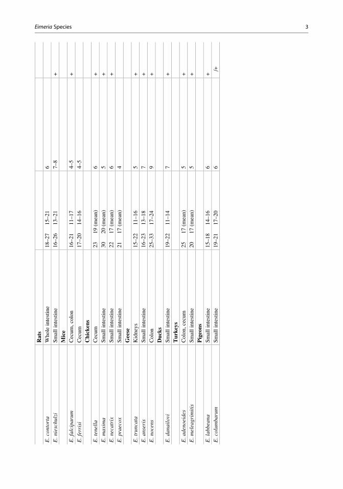
Eim
eriaSpecies,Table
1Im
portantspeciesofthegenusEimeria
Species
Host/habitat
Oocystsize
(mm)
Prepatentperiod(days)
Pathogenicity
Cattle
E.bo
vis
Posteriorsm
allintestine
23–34�
17–23
15–21
+
E.au
burnensis
Smallintestine
36–42�
19–26
17–20
+
E.zuernii
Smallintestine
16–20�
15–18
15–19
+
E.ellipsoidalis
Smallintestine
18–26�
13–18
8–13
+
Sheep
E.fauri
Smallintestine
22–33�
19–24
12–15
+
E.intricata
Smallintestine,cecum
40–56�
30–41
20–27
+
E.ovina
Smallintestine
23–36�
16–24
19
+
E.ovinoida
lis
Colon
17–25�
13–20
10–15
+
Goats
E.arloingi
Intestinalcrypts
25–33�
16–21
14–20
+
E.nina
kohlyakimovae
Intestinalcrypts
16–28�
14–23
11–17
+
E.christenseni
34–41�
23–38
14–23
+
Pigs
E.scabra
Smallintestine
25–45�
17–28
7–10
+
E.suis
Smallintestine
13–20�
11–15
10
+
Horses
E.leuckarti
Smallintestine
70–90�
50–69
31–37
�Rabbits
E.intestinalis
Cecum,colon
23–32�
15–20
10
+
E.perforans
Smallintestine
16–28�
12–16
4–6
+
E.magn
aSmallintestine
28–40�
18–30
7–9
+
E.stiedae
(syn.E.stiedai)
Bileducts
26–40�
16–25
12–16
+
2 Eimeria Species
Rats
E.contorta
Wholeintestine
18–27�
15–21
6�
E.nieschu
lzi
Smallintestine
16–26�
13–21
7–8
+
Mice
E.falciparum
Cecum,colon
16–21�
11–17
4–5
+
E.ferrisi
Cecum
17–20�
14–16
4–5
�Chickens
E.tenella
Cecum
23�
19(m
ean)
6+
E.maxima
Smallintestine
30�
20(m
ean)
5+
E.necatrix
Smallintestine
22�
17(m
ean)
6+
E.praecox
Smallintestine
21�
17(m
ean)
4�
Geese
E.trun
cata
Kidneys
15–22�
11–16
5+
E.an
seris
Smallintestine
16–23�
13–18
7+
E.no
cens
Colon
25–33�
17–24
9+
Ducks
E.dan
ailovi
Smallintestine
19–22�
11–14
7+
Turkeys
E.adenoeides
Colon,cecum
25�
17(m
ean)
5+
E.meleagrimitis
Smallintestine
20�
17(m
ean)
5+
Pigeons
E.labb
eana
Smallintestine
15–18�
14–16
6+
E.columba
rum
Smallintestine
19–21�
17–20
6�/
+
Eimeria Species 3
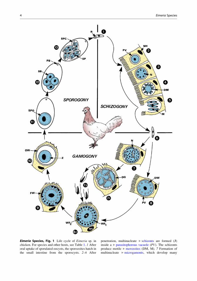
Eimeria Species, Fig. 1 Life cycle of Eimeria sp. in
chicken. For species and other hosts, see Table 1. 1 After
oral uptake of sporulated oocysts, the sporozoites hatch in
the small intestine from the sporocysts. 2–6 After
penetration, multinucleate ▶ schizonts are formed (3)inside a ▶ parasitophorous vacuole (PV). The schizonts
produce motile ▶merozoites (DM, M). 7 Formation of
multinucleate ▶microgamonts, which develop many
4 Eimeria Species
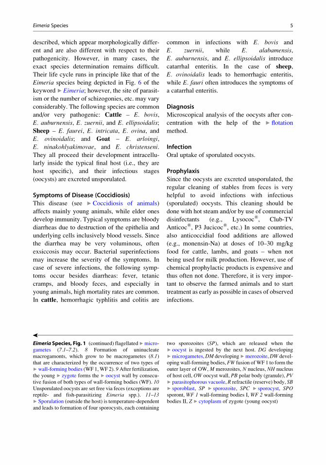
described, which appear morphologically differ-
ent and are also different with respect to their
pathogenicity. However, in many cases, the
exact species determination remains difficult.
Their life cycle runs in principle like that of the
Eimeria species being depicted in Fig. 6 of the
keyword ▶Eimeria; however, the site of parasit-
ism or the number of schizogonies, etc. may vary
considerably. The following species are common
and/or very pathogenic: Cattle – E. bovis,
E. auburnensis, E. zuernii, and E. ellipsoidalis;
Sheep – E. faurei, E. intricata, E. ovina, andE. ovinoidalis; and Goat – E. arloingi,
E. ninakohlyakimovae, and E. christenseni.
They all proceed their development intracellu-
larly inside the typical final host (i.e., they are
host specific), and their infectious stages
(oocysts) are excreted unsporulated.
Symptoms of Disease (Coccidiosis)
This disease (see ▶Coccidiosis of animals)
affects mainly young animals, while elder ones
develop immunity. Typical symptoms are bloody
diarrheas due to destruction of the epithelia and
underlying cells inclusively blood vessels. Since
the diarrhea may be very voluminous, often
exsiccosis may occur. Bacterial superinfections
may increase the severity of the symptoms. In
case of severe infections, the following symp-
toms occur besides diarrheas: fever, tetanic
cramps, and bloody feces, and especially in
young animals, high mortality rates are common.
In cattle, hemorrhagic typhlitis and colitis are
common in infections with E. bovis and
E. zuernii, while E. alabamensis,
E. auburnensis, and E. ellipsoidalis introduce
catarrhal enteritis. In the case of sheep,
E. ovinoidalis leads to hemorrhagic enteritis,
while E. fauri often introduces the symptoms of
a catarrhal enteritis.
Diagnosis
Microscopical analysis of the oocysts after con-
centration with the help of the ▶ flotation
method.
Infection
Oral uptake of sporulated oocysts.
Prophylaxis
Since the oocysts are excreted unsporulated, the
regular cleaning of stables from feces is very
helpful to avoid infections with infectious
(sporulated) oocysts. This cleaning should be
done with hot steam and/or by use of commercial
disinfectants (e.g., Lysococ®, Club-TV
Anticoc®, P3 Jucicoc®, etc.) In some countries,
also anticoccidial food additions are allowed
(e.g., monensin-Na) at doses of 10–30 mg/kg
food for cattle, lambs, and goats – when not
being used for milk production. However, use of
chemical prophylactic products is expensive and
thus often not done. Therefore, it is very impor-
tant to observe the farmed animals and to start
treatment as early as possible in cases of observed
infections.
��
Eimeria Species, Fig. 1 (continued) flagellated▶micro-
gametes (7.1–7.2). 8 Formation of uninucleate
macrogamonts, which grow to be macrogametes (8.1)that are characterized by the occurrence of two types of
▶wall-forming bodies (WF 1,WF 2). 9After fertilization,the young ▶ zygote forms the ▶ oocyst wall by consecu-
tive fusion of both types of wall-forming bodies (WF). 10Unsporulated oocysts are set free via feces (exceptions are
reptile- and fish-parasitizing Eimeria spp.). 11–13▶ Sporulation (outside the host) is temperature-dependent
and leads to formation of four sporocysts, each containing
two sporozoites (SP), which are released when the
▶ oocyst is ingested by the next host. DG developing
▶microgametes,DM developing▶merozoite,DW devel-
oping wall-forming bodies, FW fusion ofWF 1 to form the
outer layer of OW, M merozoites, N nucleus, NH nucleus
of host cell, OW oocyst wall, PB polar body (granule), PV▶ parasitophorous vacuole, R refractile (reserve) body, SB▶ sporoblast, SP ▶ sporozoite, SPC ▶ sporocyst, SPOsporont, WF 1 wall-forming bodies I, WF 2 wall-forming
bodies II, Z ▶ cytoplasm of zygote (young oocyst)
Eimeria Species 5

Incubation Period
The timing is species specific and also depends on
the amount of ingested oocysts; however, symp-
toms of disease mostly start within 1 week after
infection.
Prepatent Period
Species specific: 6–35 days.
Patency
A few weeks.
Therapy
The use of medicaments to control coccidiosis is
regulated by national health authorities. Thus,
many compounds are not available everywhere.
Cattle, sheep, and goats may be treated by
▶ toltrazuril, sulfonamides, or combination prep-
arations (see ▶Coccidiocidal drugs).
Eimeria and Isospora Species of Pigs
See ▶Coccidia of swine.
Eimeria (Synonym, Globidium) leuckartiof Equids
Name
The genus name Eimeria honors Prof.
Dr. Theodor ▶Eimer (1843–1897). The species
name honors Prof. Dr. ▶Leuckart (1822–1898).
Geographic Distributions/Epidemiology
Worldwide, due to horse transportations all over
the world. In Central Europe, up to 80 % of the
foals may be infected.
Morphology/Life Cycle
E. leuckarti is the onlyEimeria species in equids. The
oocysts are rather large (70–90 mm � 50–69 mm)characterized by an extremely thick (8–10 mm)
and outer oocyst wall and a smooth inner one
(Fig. 3). The schizogony and gamogony proceed
in cells of the intestinal wall. Schizogony was
found in epithelial cells, while gamogony was
documented in cells of the lamina propria. Spor-
ulation time outside of the body takes about
2 weeks.
Eimeria Species,Fig. 2 Light micrograph
of unsporulated (a) and a
sporulated (b) oocysts ofEimeria tenella from
chicken
6 Eimeria Species

Symptoms of Disease
Mostly no or only low-grade symptoms of dis-
ease occur. However, young or diseased horses,
camels, etc. may suffer from diarrheas. Foals start
to excrete oocysts from days 28 until 190; after-
ward, only rare cases of disease occur.
Diagnosis
Microscopical demonstration of the typical
oocysts obtained by use of ▶ concentration
methods of feces (▶ flotation; ▶ S.A.F.). Since
these oocysts are very heavy, it is needed to
centrifuge the flotation medium for 7–8 min.
Infection
Oral via contaminated food or drinking water.
Prophylaxis
Regular cleaning of the stables with the help of
hot steam and use of disinfectants (see ▶ cattle).
Incubation Period
Two to three weeks (after experimental
infections).
Prepatent Period
Rather long: 28–37 days in young animals.
Patency
Three to four weeks.
Therapy
Toltrazuril; however, treatment is only needed in
case of symptoms of disease.
Eimeria Species, Fig. 3 Diagrammatic representation of
a sporulated oocyst of the genus Eimeria containing four
sporocysts with two sporozoites each. AW outer layer of
the wall of the oocyst, IW inner layer of the wall of the
oocyst, MK cap of micropyle, N nucleus, PK polar body,
RK refractile body, RO residual body of the oocyst, RSresidual body of the sporocyst, S sporocyst, SP sporozoite,
ST Stieda body, SST substieda body
Eimeria Species, Fig. 4 Light micrograph of the bloody
ceca of a chicken infected with Eimeria tenella
Eimeria Species 7
Eimeria Species and Related Coccidiaof Birds
Name
The genus name Eimeria honors the Swiss-born
Professor Theodor ▶Eimer (1843–1897). The
species names honor scientists, describe host
names, refer to their outer appearance, or are
given according to their location in hosts.
Geographic Distributions/Epidemiology
Worldwide; outbreaks in large breeding facilities
may reach the status of local epidemics.
Morphology/Life Cycle
The life cycle is diagrammatically depicted in
Fig. 1. Some species of farmed animals are very
pathogenic and thus they are of huge economic
importance. A broad spectrum of host-specific
Eimeria species occur in the different birds
(Figs. 4, 5 and 7).
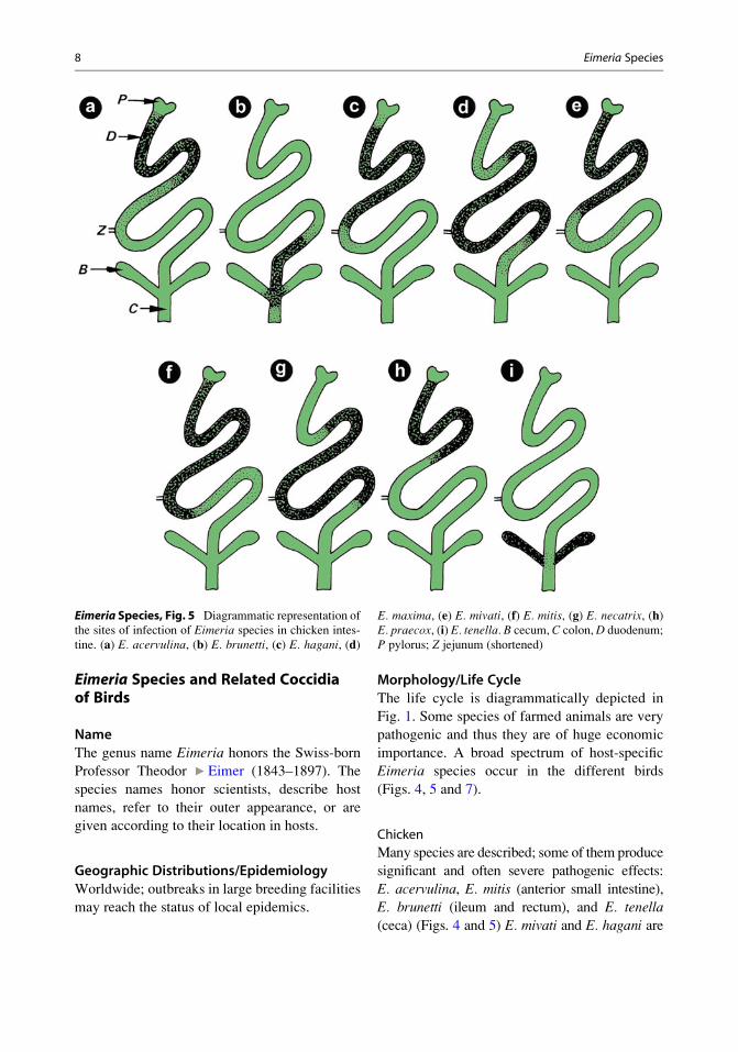
Chicken
Many species are described; some of them produce
significant and often severe pathogenic effects:
E. acervulina, E. mitis (anterior small intestine),
E. brunetti (ileum and rectum), and E. tenella
(ceca) (Figs. 4 and 5) E. mivati and E. hagani are
Eimeria Species, Fig. 5 Diagrammatic representation of
the sites of infection of Eimeria species in chicken intes-
tine. (a) E. acervulina, (b) E. brunetti, (c) E. hagani, (d)
E. maxima, (e) E. mivati, (f) E. mitis, (g) E. necatrix, (h)E. praecox, (i) E. tenella. B cecum, C colon,D duodenum;
P pylorus; Z jejunum (shortened)
8 Eimeria Species
of low pathogenicity and E. praecox is even
described as apathogenic.
Geese
Pathogenic species are E. anseris and E. nocens(small intestine and colon), E. truncata (in kidney
epithelium), and E. kotlani (rectum, cloaca).
Ducks
E. kotlani,E. danailovi, Tyzzeria perniciosa (small
intestine), and E. adenoeides are the most patho-
genic species in the posterior regions of the small
intestine (ileum) but also in the ceca and colon.
Pigeons
E. labbeana and E. columbarum (in the mid
region of the small intestine) are both pathogenic.
Parrots
E. dunsigni (in the small intestine) – this species is
pathogenic, but occurs only rarely in farmed birds.
Symptoms of Disease (Coccidiosis,
Coccidiasis)
Characteristic symptoms of an acute coccidiosis
are severe, sometimes bloody, diarrheas based on
catarrhal or hemorrhagic enteritis. The feces
appear watery fluid, contain slimy or bloody ele-
ments, or appear yellowish-greenish. High mor-
tality rates occur in some species (e.g., E. tenella;
Fig. 4); thus, an outbreak of a coccidiosis may
lead to enormous economic losses (Fig. 8).
Diagnosis
Microscopical determination of oocysts in the
feces after use of ▶ flotation method.
Infection
Oral uptake of sporulated oocysts with contami-
nated food or drinking water.
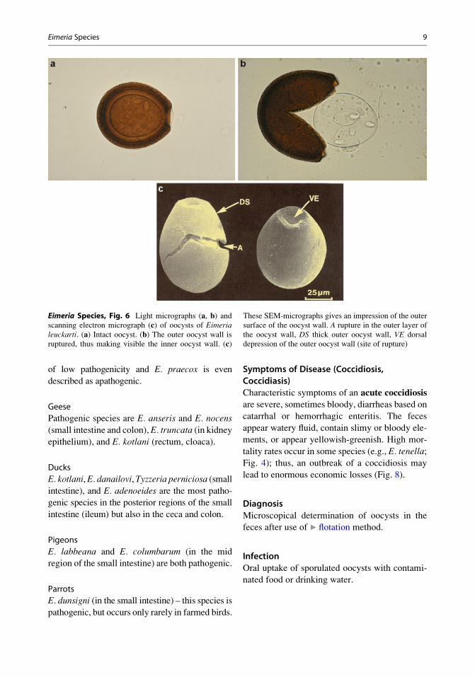
Eimeria Species, Fig. 6 Light micrographs (a, b) andscanning electron micrograph (c) of oocysts of Eimerialeuckarti. (a) Intact oocyst. (b) The outer oocyst wall is
ruptured, thus making visible the inner oocyst wall. (c)
These SEM-micrographs gives an impression of the outer
surface of the oocyst wall. A rupture in the outer layer of
the oocyst wall, DS thick outer oocyst wall, VE dorsal
depression of the outer oocyst wall (site of rupture)
Eimeria Species 9
Prophylaxis
(a) Hygienicmeasurements: Regular cleaning of
cages or soil/floors from feces. Protect
drinking water from fecal contaminations.
Disinfect stables, floors, and cages with the
help of registered disinfectants. Important:
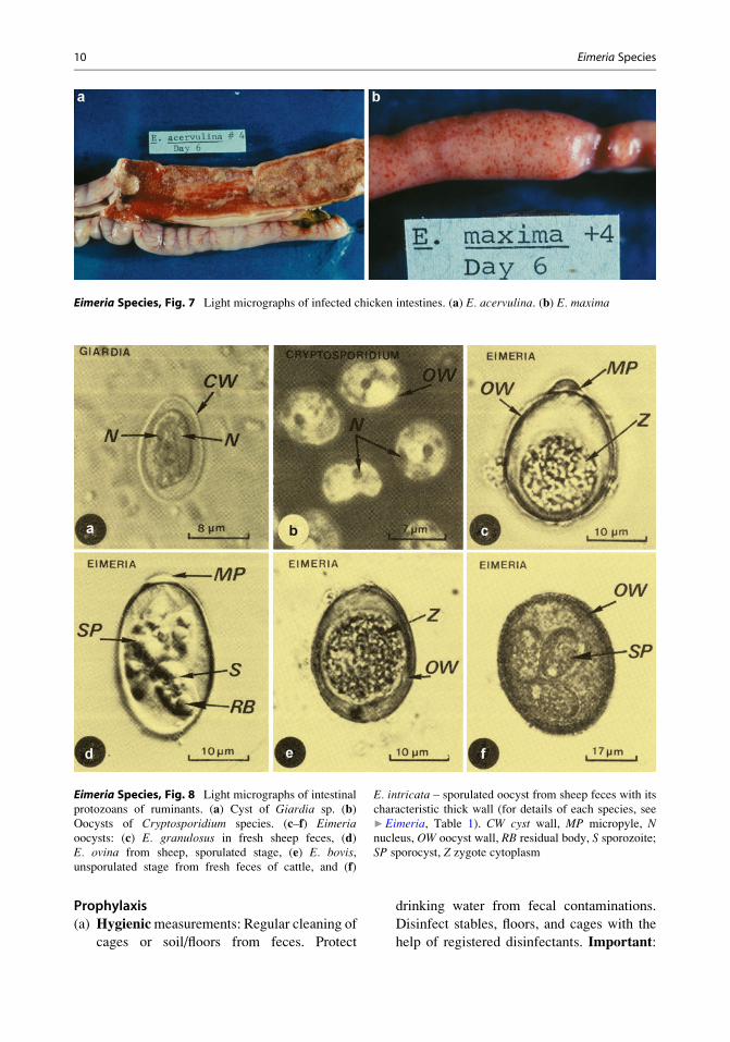
Eimeria Species, Fig. 7 Light micrographs of infected chicken intestines. (a) E. acervulina. (b) E. maxima
Eimeria Species, Fig. 8 Light micrographs of intestinal
protozoans of ruminants. (a) Cyst of Giardia sp. (b)Oocysts of Cryptosporidium species. (c–f) Eimeriaoocysts: (c) E. granulosus in fresh sheep feces, (d)E. ovina from sheep, sporulated stage, (e) E. bovis,unsporulated stage from fresh feces of cattle, and (f)
E. intricata – sporulated oocyst from sheep feces with its
characteristic thick wall (for details of each species, see
▶Eimeria, Table 1). CW cyst wall, MP micropyle, Nnucleus, OW oocyst wall, RB residual body, S sporozoite;SP sporocyst, Z zygote cytoplasm
10 Eimeria Species
Use always the all in-all out principle for
stables with farmed birds, i.e., take always
all animals out of a stable after a period of
egg laying or a period of fattening; clean
equipment and floors and then enter all new
birds at once. Never add single birds to a
stable with healthy animals.
(b) Chemoprophylaxis: The basics of this
method is to feed permanently small, but
efficient amounts of ▶ coccidiocidal drugs.
This method, however, may introduce resis-
tances against several coccidiocidal
medicaments.
(c) Immunization: There exist several vaccines
(containing attenuated, low-grade patho-
genic oocysts); however, many of them are
only registered in a certain number of coun-
tries (e.g., Coccivac®, Paracox®, Livacox®,
etc.). The problem is that these strains may
recover their full pathogenicity after some
periods of use.
Incubation Period
Three to five days depending on the number of
ingested oocysts and the grade of the pathogenic-
ity of the ingested Eimeria species.
Prepatent Period
The different Eimeria species need different
periods until oocysts appear in feces:
(a) Chickens: 4 days (E. praecox, E. acervulina,
E. mitis, E. mivati); 5 days (E. maxima,
E. brunetti, E. tenella); 6 days (E. necatrix).(b) Turkeys: 4 days (E. dispersa); 5 days
(E. meleagrimitis, E. adenoeides).
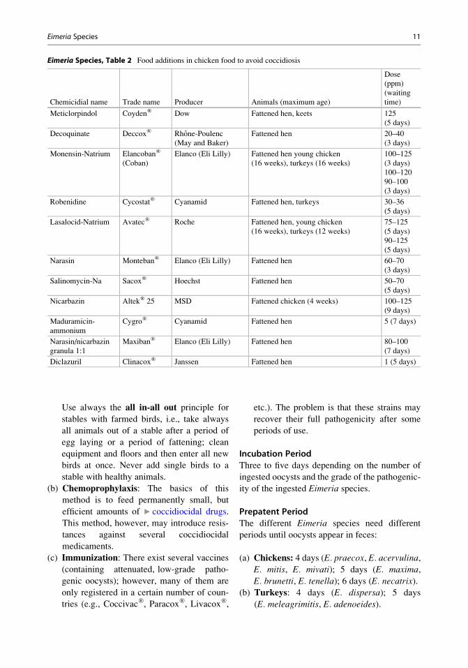
Eimeria Species, Table 2 Food additions in chicken food to avoid coccidiosis
Chemicidial name Trade name Producer Animals (maximum age)
Dose
(ppm)
(waiting
time)
Meticlorpindol Coyden® Dow Fattened hen, keets 125
(5 days)
Decoquinate Deccox® Rhone-Poulenc
(May and Baker)
Fattened hen 20–40
(3 days)
Monensin-Natrium Elancoban®
(Coban)
Elanco (Eli Lilly) Fattened hen young chicken
(16 weeks), turkeys (16 weeks)
100–125
(3 days)
100–120
90–100
(3 days)
Robenidine Cycostat® Cyanamid Fattened hen, turkeys 30–36
(5 days)
Lasalocid-Natrium Avatec® Roche Fattened hen, young chicken
(16 weeks), turkeys (12 weeks)
75–125
(5 days)
90–125
(5 days)
Narasin Monteban® Elanco (Eli Lilly) Fattened hen 60–70
(3 days)
Salinomycin-Na Sacox® Hoechst Fattened hen 50–70
(5 days)
Nicarbazin Altek® 25 MSD Fattened chicken (4 weeks) 100–125
(9 days)
Maduramicin-
ammonium
Cygro® Cyanamid Fattened hen 5 (7 days)
Narasin/nicarbazin
granula 1:1
Maxiban® Elanco (Eli Lilly) Fattened hen 80–100
(7 days)
Diclazuril Clinacox® Janssen Fattened hen 1 (5 days)
Eimeria Species 11

(c) Geese: 5 days (E. truncata); 7 days
(E. anseris); 9 days (E. nocens).
(d) Ducks: 6 days (E. perniciosa); 8 days
(E. danilovi).(e) Pigeons: 6 days (E. labbeana).
(f) Parrots: 5 days (E. dunsigni).
Patency
Mostly rather short (1–3 weeks); however, also
slight infections had been reported to occur for
months (due to repeated self-infections).
Therapy
Against many of the “older” anticoccidial com-
pounds, resistances have been developed. Thus,
the products based on toltrazuril (e.g., Baycox®
15–20 mg/kg bodyweight in drinking water) are
widely used. For further information, see
▶Coccidiocidal Drugs and Table 2.
Eimeria Species of Rabbits
Name
The genus name Eimeria honors the Swiss-born
Prof. Dr. Theodor ▶Eimer (1843–1897). The
species name describes activities (e.g., Latin:
perforans, penetrating) and sites of infections
(e.g., Latin: intestinalis, inside the intestine) or
honors scientists (e.g., Christian Stieda,
1837–1918).
Geographic Distributions/Epidemiology
Worldwide; in farmed animals small epidemics
may occur.
Morphology/Life Cycle
The members of the Lagomorpha are parasitized
by a large number of Eimeria species, which
mostly enter the epithelial cells of the small intes-
tine. However, E. stiedae (Fig. 9) which parasit-
izes in liver cells of rabbits is one of the most
important pathogens. E. intestinalis,E. perforans, and E. magna infect also rabbits
and are very pathogenic. E. contorta and
Eimeria Species, Fig. 9 Light micrographs of unsporulated and sporulated E. stiedae oocysts. MP micropore, OWoocyst wall, SP sporocyst
Eimeria Species, Fig. 10 Liver of an Eimeriastiedae-infected liver
12 Eimeria Species

E. nieschulzi are found in rats, whereas
E. falciparum and E. ferrisi parasitize in mice
(e.g., laboratory mouse: Mus musculus).
Symptoms of Disease (Rabbits)
(a) Intestinal coccidiosis leads to severe diar-
rheas combined with anemia, extreme weak-
ness, and loss of weight. The effects of these
infections are mostly increased by bacterial
superinfections leading to high mortality
rates.
(b) Liver coccidiosis of rabbits is initiated by
Eimeria stiedae. Main symptoms are liver
dysfunctions (loss of activity) and appear-
ance of yellow-whitish nodes in and on the
liver (Fig. 10) and destruction of the parasit-
ized epithelia of the excreting channels of the
gall bladder system.
Diagnosis
Microscopical inspection of the fecally excreted
oocysts with the help of concentration methods
(e.g., ▶ flotation).
Infection
Oral uptake of sporulated oocysts with contami-
nated food or drinking water.
Prophylaxis
Regular cleaning of the cages. Avoidance to add
new animals into an existing system without pre-
vious isolation (= quarantine) of newcomers.
Incubation Period
Mostly less than 1 week.
Prepatent Period
Species specific: 9–12 days (E. intestinalis);
12–14 days (E. stiedae).
Patency
Mostly 2–5 weeks, with highest excretion rates
during the first and second week of patency.
Therapy
Toltrazuril (Baycox®), 25 ppm in drinking water
for 2 days; repetition after 5 days.
Further Reading
Bahat TK et al (1996) Rabbit coccidiosis and its control.
World Rabbit Sci 4:37–41
Beelitz P et al (1994) Eimeria leuckarti. Infektionen bei
Fohlen und ihren Mutterstuten in Oberbayern.
Tierarztl Prax 22:377–381
Daugschies A, Najdrowsk M (2005) Eimeriosis in cattle:
current understanding. J Vet Med B Infect Dis Vet
Public Health 52:417–427
Levine ND (1988) The protozoan phylum Apicomplexa,
vol I and II. CRC Press, Boca Raton
Levine ND, Ivens V (1990) Coccidian parasites of rodents.
CRC Press, Boca Raton
Mehlhorn H (1972–1974) Electron microscopical investi-
gations on the developmental stages of Eimeria max-ima (macrogametes, microgametes, schizonts,
merozoites). Parasitol Res 39, 40:161–182, 151–163,
243–260
Mehlhorn H (2012) The parasites of animals, 7th edn.
Springer Spectrum, Heidelberg
Pellerdy L (1984) Coccidia and coccidiosis, 2nd edn.
Parey, Berlin
Scholtyseck E, Mehlhorn H (1970) Ultrastructural study
of characteristic organelles of Sporozoa. Parasitol Res
34:97–127
von Samson-Himmelstjerna G et al (2006) Clinical and
epidemiological characteristics of Eimeria infections
in first-year grazing cattle. Vet Parasitol 136:215–221
Eimeria Species 13