efficient role of igh 3′ regulatory region deficient b ... · 3′rr-deficient b-cells in the...
TRANSCRIPT

Oncotarget38741www.impactjournals.com/oncotarget
www.impactjournals.com/oncotarget/ Oncotarget, Vol. 7, No. 25
Efficient role of IgH 3′ regulatory region deficient B-cells in the development of oil granulomas
Nour Ghazzaui1, Alexis Saintamand1, Hussein Issaoui1, Faten Saad1, Yves Denizot1
1Department of Immunology, CNRS UMR 7276, CRIBL, Université de Limoges, Limoges, France
Correspondence to: Yves Denizot, email: [email protected]: IgH 3′ regulatory region, knock-out mice, pristine, granulomasReceived: March 25, 2016 Accepted: April 29, 2016 Published: May 25, 2016
ABSTRACT
Functional B-cells are essential for the formation of oil granulomas. The IgH 3′ regulatory region (3′RR) activates important check-points during B-cell maturation. We investigated if 3′RR-deficient B-cells remain efficient to develop oil granulomas in response to pristine. B-cells expressing an IgH 3′RR-deficient allele were similarly recruited to wild type allele expressing B-cells in the granuloma. No differences were observed between 3′RR-deficient mice and control mice for granuloma numbers, cellular composition and ability to express mRNA transcripts for several pro- and anti-inflammatory cytokines. Altogether these results suggest a normal role for 3′RR-deficient B-cells in the development of an acute B-cell-mediated inflammatory response.
INTRODUCTION
The IgH locus undergoes multiple changes along B-cell differentiation, affecting transcription and accessibility to V(D)J recombination, somatic hypermutation (SHM) and class switch recombination (CSR) [1,2]. IgH cis-regulatory regions are major locus regulators. The IgH 3′ regulatory region (3′RR) promotes SHM [3, 4], CSR [5–7], μ transcription [8] but not V(D)J recombination [9]. The 3′RR is also a potent lymphoma oncogene deregulator [10]. Transgenic mice models demonstrated the 3′RR implication in the development of several B-cell lymphomas [11–18]. The absence of the 3′RR influences lymphomagenesis in λ-myc mice toward less mature lymphomas [19]. Until now, the functionality of 3′RR-deficient B-cells in inflammatory response is poorly documented. The lack of 3′RR was only reported to marginally impact the development of a chronic inflammatory ascite formation in response to pristine [20].
The i.p. injection of pristine induces the formation of mesenteric oil granulomas [21, 22]. Pristine-induced granulomas are characterised by clustered cells adhered to the mesentery and other peritoneal tissues. The granuloma formation constitutes a protective inflammatory cellular response of the host against invading pathogens or indigestible substances. Two types of granulomas are reported. Serosal granulomas (SG) are located at the interface of the mesenteric margins and gut. Mesenteric granulomas (MG) are located around the center of the
mesenteric tissue [23, 24]. Oil granulomas are considered as tertiary lymphoid tissues constituted of monocytes, granulocytes, T-cells and B-cells. Their formation is regulated by several cytokines [25]. The absence of functional B-cells blocks SG formation and diminishes MG development in response to pristine [23]. Mesenteric oil granulomas thus appear as an interesting tool to ensure the functional ability of 3′RR-deficient B-cells in the occurrence and/or development of an acute inflammatory response. In this study we investigated the generation of pristine-induced oil granulomas in IgH 3′RR-deficient mice.
RESULTS
Spleen and peritoneal B-cells expressing a 3′RR-deleted allele
Mouse substrains have dissimilar differentiation programs culminating in different B-cell fate, BCR expression and signalling [8]. Pristine-induced oil granuloma generation is different with respect to mouse substrains [24]. Before assessing the influence of an IgH 3′RR-deleted allele vs a wt allele in B-cell recruitment in oil granulomas we firstly investigated heterozygous IgH aΔ3′RR/bwt mice. The presence of a 3′RR-deficient allele and a wt allele was investigated by PCR (Figure 1A). The 3′RR deletion was done in a 129 ES cell line (IgH a allotype) and developed in a 129 background (IgH awt/awt). Heterozygous IgH aΔ3′RR/bwt mice were generated by

Oncotarget38742www.impactjournals.com/oncotarget
Figure 1: Generation of aΔ3′RR/bwt and awt/bwt mice. A. PCR profile for a 3′RR-deficient and a wt IgH allele. B. Backcross for generation of aΔ3′RR/bwt and awt/bwt mice, and expression of either IgMa or IgMb allele by B-cells from F1 mice. B-cell are expected to express IgMa or IgMb at similar frequency, including when the a allele is deleted for the 3′RR, since its deletion does not affect VDJ recombination. If the expression of one of this allele impedes the B-cell development, the equilibrium between IgMa or IgMb expressing B-cell will be disrupted. Lowered number of IgMa expressing B-cells in aΔ3′RR/bwt mice will thus demonstrate that deletion of the 3′RR alters B-cell development or recruitment.

Oncotarget38743www.impactjournals.com/oncotarget
crossing homozygous 3′RR-deficient mice (IgH aΔ3′RR/aΔ3′RR) with C57BL/6 mice bearing an IgH b allotype (IgH bwt/bwt) mice (Figure 1B). Mixed 129 x C57BL/6 mice (IgH awt/bwt) were used as control mice. As previously reported [8], analysis of splenic B-cells with IgM-allotype specific antibodies indicated a lowered (p=0.001, Mann-Whitney U-test) percentage of B-cells expressing an a allotype (IgMa/IgMb ratio: 0.33) in aΔ3′RR/bwt mice (Figure 2A and 2B). A similar decrease (p=0.0006) was also found for peritoneal B-cells (IgMa/IgMb ratio: 0.59) (Figure 2C and 2D). While similar percentages of B-splenocytes expressed either an a or b allotype (IgMa/IgMb ratio: 0.96) in awt/bwt mice (Figure 2A and 2B), elevated number of peritoneal B-cells expressing an a allotype was found (IgMa/IgMb ratio: 1.53) (Figure 2C and 2D). This result might be linked to a differential strength of signalling between IgMa BCR and IgMb BCR for proliferation/survival of peritoneal B-cells. Such specific interactions with IgMa (but not IgMb) determinants have been already reported with the HIV-1 envelope gp41 membrane proximal external region [26]. Furthermore, the phenotype of mature B-cells differs between the various mouse substrains. Notably, BCR signalling has been suggested to be lower in 129 mice than in C57BL/6 [27]. Altogether these results suggest that the 3′RR deletion is not only detrimental for efficient B-cell maturation in spleen but also for B-cells in the peritoneal cavity.
B-cells expressing a 3′RR-deleted allele in oil granulomas
We next compared B-cell recruitment in granulomas from aΔ3′RR/bwt and awt/bwt mice. For granuloma experiments we used mechanical dissociation instead of collagenase dissociation. Collagenase-based intestinal digestion procedure is frequently used to isolate tissue-resident B-cells. However this procedure was recently reported to alter B-cell surface marker expression and thus can impede the correct phenotypic analysis of these B-cells [28]. All granulomas were investigated the same day to ensure similar recovery efficiency. As a positive control, similar percentages of B-cells expressing either a a or b allotype (IgMa/IgMb ratio: 1.00) were found in granulomas of awt/bwt mice (Figure 3A and 3B). Analysis of B-cells in oil granulomas with IgM-allotype specific antibodies indicated a lowered (p=0.0006) percentage of B-cells expressing an a allotype (IgMa/IgMb ratio: 0.47) in aΔ3′RR/bwt mice (Figure 3A and 3B). The 53% of IgMa reduction paralleled the 41% and 67% of IgMa reduction in peritoneal cavity and spleen of IgH aΔ3′RR/bwt mice, respectively. Finally the mean membrane IgMa and IgMb densities were similar in heterozygous aΔ3′RR/bwt and awt/bwt mice (Figure 3C and 3D). Thus, differences in IgMa and IgMb allotypes in oil granulomas in heterozygous aΔ/bwt mice are linked to differences in the percentage of
Figure 2: IgMa and IgMb in spleen and peritoneal cavity of heterozygous IgH aΔ3′RR/bwt and awt/bwt mice. A. Flow cytometry analysis of the percentages of IgMa and IgMb in spleen of aΔ3′RR/bwt and awt/bwt mice. Cells were gated on B220+ cells. One representative experiment out of six aΔ3′RR/bwt mice and nine awt/bwt mice is shown. B. IgMa/IgMb ratio in splenocytes of aΔ3′RR/bwt and awt/bwt mice. Mean ± SEM of six aΔ3′RR/bwt mice and nine awt/bwt mice. Significance was assessed using the Mann-Whitney U-test. C. Flow cytometry analysis of the percentages of IgMa and IgMb in the peritoneal cavity of aΔ3′RR/bwt and awt/bwt mice. Cells were gated on B220+ cells. One representative experiment out of seven for both genotypes is shown. D. IgMa/IgMb ratio in the peritoneal cavity of aΔ3′RR/bwt and awt/bwt mice. Mean ± SEM of seven experiments. Significance was assessed using the Mann-Whitney U-test.

Oncotarget38744www.impactjournals.com/oncotarget
IgMa and IgMb B-cells in mice but not to a defect in B-cell recruitment of 3′RR-deficient B-cells.
Oil granulomas in 3′RR-deficient mice and wt mice
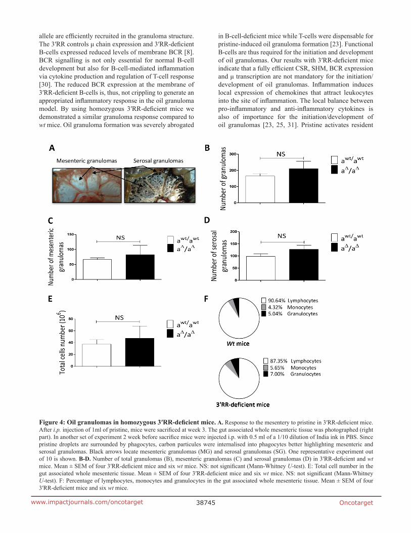
We next compared granuloma formation in 3′RR-deficient mice (IgH locus aΔ3′RR/aΔ3′RR) and wt mice (IgH locus awt/awt). A representative photograph of the gut associated whole mesenteric tissue 3 weeks after i.p. injection of 1ml pristine is reported in Figure 4A. To contrast with the background we labelled phagocytes with India ink by injecting it intraperitoneally into mice at week 1 after pristine (Figure 4A). Arrows indicate locations of MG and SG. For all experiments we counted granulomas on the whole mesenteric tissue. Numbers of total granulomas (Figure 4B), MG (Figure 4C) and SG (Figure 4D) were not significantly different between 3′RR-deficient mice and wt mice. No significant differences were found for the total cell number in the gut associated whole mesenteric tissue (MG + SG) between 3′RR-deficient mice and wt mice (Figure 4E). The percentages of granulocytes, monocytes and lymphocytes (morphological analysis and counts in the CELL-DYN Emerald) were not significantly affected in the gut associated whole mesenteric tissue (MG + SG) of 3′RR-deficient mice (Figure 4F). Finally flow cytometry analysis indicated similar (p=1, Mann-Whitney U-test) percentages of IgMa+ B-cells in granulomas of 3′RR-deficient mice (32.6 ± 5.8 %, mean ± SEM of 3 animals) and wt mice (40.7 ± 13.2 %, mean ± SEM of 3
animals). Taken altogether these results suggest no impact of the 3′RR deficiency for oil granuloma formation.
Inflammatory cytokine network in oil granuloma in 3′RR-deficient mice and wt mice
Cytokines have been reported to regulate the structure and formation of oil granulomas in mice [25, 29]. Several pro- (INF-γ, TNF-α, CXCL2, IL-12, IL-6) and anti-inflammatory (IL-4, IL-10, TGF-β) cytokine transcripts were investigated, by real-time PCR, in granulomas cells of 3′RR-deficient and wt mice. Adherent (monocytes/macrophages) and non-adherent (lymphocytes/granulocytes) cells were investigated to search putative differences between 3′RR-deficient and wt mice. As shown in Table 1, no significant differences were found for INF-γ, TNF-α, CXCL2, IL-12, IL-6, IL-4, IL-10, TGF-β mRNA transcripts between 3′RR-deficient mice and wt mice. These results reinforce the hypothesis of a similar mechanistic/kinetic of granuloma formation in mice with 3′RR-deficient B-cells and wt B-cells.
DISCUSSION
The IgH 3′ regulatory region (3′RR) stimulates numerous important B-cell check-points during B-cell maturation [3–8]. We have investigated the impact of the 3′RR deletion on the in vivo pristine-induced granuloma formation. By using heterozygous aΔ3′RR/bwt mice we demonstrated that B-cells expressing a 3′RR-deficient
Figure 3: IgMa and IgMb in oil granulomas of heterozygous IgH aΔ3′RR/bwt and awt/bwt mice. A. Flow cytometry analysis of the percentages of IgMa and IgMb in granulomas of aΔ3′RR/bwt and awt/bwt mice. Cells were gated on B220+ cells. One representative experiment out of eight aΔ3′RR/bwt mice and nine awt/bwt mice is shown. B. IgMa/IgMb ratio in granulomas of aΔ3′RR/bwt and awt/bwt mice. Mean ± SEM of eight aΔ3′RR/bwt mice and nine awt/bwt mice. Significance was assessed using the Mann-Whitney U-test. C. Mean ± SEM membrane IgMa density in granulomas of seven aΔ3′RR/bwt mice and eight awt/bwt mice. NS: not significant (Mann-Whitney U-test). D. Mean ± SEM membrane IgMb density in granulomas of seven aΔ3′RR/bwt mice and eight awt/bwt mice. NS: not significant (Mann-Whitney U-test).
Oncotarget38745www.impactjournals.com/oncotarget
allele are efficiently recruited in the granuloma structure. The 3′RR controls μ chain expression and 3′RR-deficient B-cells expressed reduced levels of membrane BCR [8]. BCR signalling is not only essential for normal B-cell development but also for B-cell-mediated inflammation via cytokine production and regulation of T-cell response [30]. The reduced BCR expression at the membrane of 3′RR-deficient B-cells is, thus, not crippling to generate an appropriated inflammatory response in the oil granuloma model. By using homozygous 3′RR-deficient mice we demonstrated a similar granuloma response compared to wt mice. Oil granuloma formation was severely abrogated
in B-cell-deficient mice while T-cells were dispensable for pristine-induced oil granuloma formation [23]. Functional B-cells are thus required for the initiation and development of oil granulomas. Our results with 3′RR-deficient mice indicate that a fully efficient CSR, SHM, BCR expression and μ transcription are not mandatory for the initiation/development of oil granulomas. Inflammation induces local expression of chemokines that attract leukocytes into the site of inflammation. The local balance between pro-inflammatory and anti-inflammatory cytokines is also of importance for the initiation/development of oil granulomas [23, 25, 31]. Pristine activates resident
Figure 4: Oil granulomas in homozygous 3′RR-deficient mice. A. Response to the mesentery to pristine in 3′RR-deficient mice. After i.p. injection of 1ml of pristine, mice were sacrificed at week 3. The gut associated whole mesenteric tissue was photographed (right part). In another set of experiment 2 week before sacrifice mice were injected i.p. with 0.5 ml of a 1/10 dilution of India ink in PBS. Since pristine droplets are surrounded by phagocytes, carbon particules were internalised into phagocytes better highlighting mesenteric and serosal granulomas. Black arrows locate mesenteric granulomas (MG) and serosal granulomas (SG). One representative experiment out of 10 is shown. B-D. Number of total granulomas (B), mesenteric granulomas (C) and serosal granulomas (D) in 3′RR-deficient and wt mice. Mean ± SEM of four 3′RR-deficient mice and six wt mice. NS: not significant (Mann-Whitney U-test). E: Total cell number in the gut associated whole mesenteric tissue. Mean ± SEM of four 3′RR-deficient mice and six wt mice. NS: not significant (Mann-Whitney U-test). F: Percentage of lymphocytes, monocytes and granulocytes in the gut associated whole mesenteric tissue. Mean ± SEM of four 3′RR-deficient mice and six wt mice.

Oncotarget38746www.impactjournals.com/oncotarget
peritoneal cells such as B-cells and monocytes/macrophage leading to the secretion of various cytokines. Analysis of several pro-inflammatory (INF-γ, TNF-α, CXCL2, IL-12, IL-6) and anti-inflammatory (IL-4, IL-10, TGF-β) cytokines by granuloma cells did not evidenced any differences between 3′RR-deficient mice and wt mice. These results reinforce the hypothesis of a similar mechanistic/kinetic of granuloma formation in mice with 3′RR-deficient B-cells and wt B-cells.
In conclusion the 3′RR targeting has no significant effect on the acute inflammatory B-cell-mediated oil granuloma model. The 3′RR is a major lymphoma oncogene deregulator [10–19]. The 3′RR might be considered as a potential target for anti-lymphoma pharmacological therapy without significant impact on the normal immune and inflammatory networks [32]. 3′RR activation and transcriptional activity are altered by a diverse range of chemicals, including ones with anti-carcinogenic properties [33]. Histone deacetylase inhibitors (HDACi) might be of interest since 3′RR-induced activation is mediated through activation of specific epigenetic marks [7] and since the hs1,2 enhancer (located in the central palindromic 3′RR structure) is sensitive to HDACi [34]. A limitation of the pristine mouse model is that inflammation is restricted to the peritoneal cavity. Other mice models of inflammatory reactions must be tested before definitive validation of this hypothesis such as the pathogenic role of B-cells in the development of diffuse alveolar hemorrhage induced by pristine [35] and the inflammatory pathology induced by surgical implants [36].
MATERIALS AND METHODS
Animals
Our research has been approved by our local ethics committee review board (Comité Régional d’Ethique sur l’Expérimentation Animale du Limousin, Limoges, France) and carried according the European guidelines for animal experimentation. The 3′RR deletion has been done in a 129 ES cell line and developed in a 129 background [5]. The presence of the 3′RR-deleted allele was verified by PCR. 3′RR-deficient mice (IgH aΔ3′RR/aΔ3′RR) and wt 129 mice (IgH awt/awt) were investigated. Heterozygous IgH aΔ3′RR/bwt mice were generated by crossing homozygous 3′RR-deficient mice (IgH aΔ3′RR/aΔ3′RR) with C57BL/6 mice (IgH bwt/bwt) mice. Mixed Sv/129 x C57BL/6 mice (IgH awt/bwt) were used as control mice.
PCR
PCR experiments for detection of the wt 3′RR allele were carried out with specific forward 5′-CCAAAAATGGCCAGGCCTAGG-3′ and reverse 5′-GA CCCTGTCCTATGGCT GAC-3′ primers. PCR experiments for detection of the deficient 3′RR allele were carried out with specific forward 5′-TCCCTGGACAATCTGCACAT-3′ and reverse 5′-GACCCTGTCCTATGGCTGAC-3′ primers. DNA was denatured 180 sec at 95°C, and then submitted to 35 cycles consisting in 94°C / 30 sec, 60°C / 30 sec and 72°C / 60 sec. Amplification products were analysed on a
Table 1: INF-γ, TNF-α, CXCL2, IL-12, IL-6, IL-4, IL-10 and TGF-β mRNA transcripts in adherent and non-adherent granuloma cells from 3′RR-deficient mice and wt mice
Non-adherent cells Adherent cells
Cytokines Wt mice 3′RR-deficient mice
Significance Wt mice 3′RR-deficient mice
Significance
IL-10 102.2 ± 24.0 170.7 ± 184.6 1.0 18.3 ± 18.2 57 ± 59.5 0.7
IL-4 22.3 ± 10.4 5.5 ± 3.0 0.1 187.6 ± 92.6 58.1 ± 68.8 0.2
TGF-β 10.0 ± 5.7 7.3 ± 4.8 0.8 18182.8 ± 13113.5
17473.4 ± 12895.2 1.0
IL-6 104.6 ± 127.0 265.6 ± 451.4 1.0 12043.4 ± 15219.9
37439.7 ± 57409.8 1.0
TNF-α 21.4 ± 2.8 26.9 ± 32.9 0.4 432.6 ± 493.9 615.2 ± 585.8 0.6
IL-12 736.5 ± 244.3 1448.1 ± 1346.8 0.6 1044.1 ±
1111.52038.1 ± 2411.3 1.0
INF-γ 41.0 ± 12.8 50.7 ± 34.0 0.6 64 ± 13.4 60.06 ± 72.6 1.0
CXCL2 31.1 ± 15.3 43.3 ± 64.6 0.4 612.3 ± 704.4 543.3 ± 612.4 1.0
Gene expression levels were normalized to GAPDH mRNA. Amounts of transcripts were compared to sample with the lowest level of transcripts. Results are reported as Mean ± SEM from 4 independent experiments per group. Significance was assessed with the Mann-Whitney U-test. No significant differences were documented between groups.

Oncotarget38747www.impactjournals.com/oncotarget
1.2% agarose gel. Expected sizes of amplified products were 250 bp and 587 bp for mutated and wt alleles, respectively.
Granulomas induction
To induce inflammatory process, 12-14-weeks-old mice received a single i.p. injection of 1 ml of 2,6,10,14 tetrametyl-pentadecane (pristine) (95%, Sigma). After 3 weeks mice were euthanized.
Flow cytometry analysis
Gut associated whole mesenteric tissue was obtained from each animal. A single-cell suspension was obtained after filtration through a fine nylon mesh. Cells were incubated with monoclonal antibodies for 30 min at 4°C. The following antibodies were used: IgMa-FITC, IgMb-PE and B220-BV510. Labelled cells were analyzed on a Fortessa LSR2 (Beckman Coulter).
Isolation of adherent and non adherent granuloma cells
Animals were sacrificed, gut associated whole mesenteric tissue collected and single cell suspension obtained as described above. Samples (2×10⁶ cells) were cultured in a 6 well plate at 37°C with 5% CO2 for 2 hours. Total RNA was isolated using Tri-Reagent (Sigma) from both adherent and non adherent cells. Samples were stored at −20°C until used.
Cell counts
Single cell suspension of granulomas were counted and characterised in the CELL-DYN Emerald (Abbot), a compact bench-top hematology analyzer that can be used for a three-part white cell differential analysis of human and mouse samples [37].
Real-time PCR analysis
RNA was extracted according to the manufacturer’s instruction. Complementary DNA (cDNA) was synthesized with 1 μg of total RNA using the high capacity cDNA reverse transcription kit from Applied biosystems. Real-time PCR analysis was performed using TaqMan reagents: TNF-α (Mm00443258-m1), IL-6 (Mm00446190-m1), IL-12-P40 (Mm00434189-m1), CXCL2 (Mm00436450-m1), IL-10 (Mm01288386-m1), IL-4 (Mm00445259-m1), INF-γ (Mm00801778-m1), TGF-β (Mm01178820-m1) and GAPDH (Mm99999915-g1). Experiments were performed using the Step One Plus (Applied biosystems). Amounts of various transcripts were compared to sample with the lowest level of transcripts. The relative quantification of gene expression was performed using the comparative CT method (ΔΔCT). Experiments were made in duplicate.
Mean CT values were used in the ΔΔCT calculation by using the “relative quantitation calculation and analysis software for Applied Biosystems real-time PCR systems”.
ACKNOWLEDGMENTS
This work was supported by grants from Conseil Régional du Limousin, Comité d’Orientation de la Recherche Cancérologie du Limousin (CORC:FJA/NP-2015-109), Association pour la Recherche sur le Cancer (PJA 20141201649), Ligue Contre le Cancer (comité de la Corrèze et Haute Vienne) and “Lions Club de la Corrèze, Zone 33 district 103 Sud”. A. Saintamand was supported by a grant from fondation ARC (DOC20150602943). N. Ghazzaui was supported by a grant from Association de Spécialisation et d’Orientation Scientifique (Lebanon) and the municipality of Khiam (Lebanon).
CONFLICTS OF INTEREST
None.
Author contributions
N. Ghazzaui, H. Issaoui, A. Saintamand, F. Saad and Y. Denizot actively participated to the experimental design of the study and participated to the scientific discussion for manuscript writing. Y. Denizot obtained financial grants to perform the study.
REFERENCES
1. Henderson A and Calame K. Transcription regulation during B cell development. Ann Rev Immunol. 1998; 16:163-200.
2. Pinaud E, Marquet M, Fiancette R, Péron S, Vincent-Fabert C, Denizot Y and Cogné M. The IgH locus 3′ regulatory region: pulling the strings from behind. Adv Immunol. 2011; 110:27-70.
3. Rouaud P, Vincent-Fabert C, Saintamand A, Fiancette R, Marquet M, Robert I, Reina-San-Martin B, Pinaud E, Cogné M and Denizot Y. The IgH 3′ regulatory region controls AID-induced somatic hypermutation in germinal centre B-cells in mice. J Exp Med. 2013; 210:1501-1507.
4. Saintamand A, Vincent-Fabert C, Garot A, Rouaud P, Oruc Z, Magnone V, Cogné M and Denizot Y. Deciphering the importance of the palindromic architecture of the immunoglobulin heavy chain 3′ regulatory region. Nature Commun. 2016; 7:10730.
5. Vincent-Fabert C, Fiancette R, Pinaud E, Truffinet V, Cogné N, Cogné M and Denizot Y. Genomic deletion of the whole IgH 3′ regulatory region (hs3a, hs1,2, hs3b, hs4) dramatically affects class switch recombination and Ig secretion to all isotypes. Blood. 2010; 116:1895-1898.

Oncotarget38748www.impactjournals.com/oncotarget
6. Rouaud P, Saintamand A, Saad F, Carrion C, Lecardeur S, Cogné M and Denizot Y. Elucidation of the enigmatic IgD class switch recombination via germ-line deletion of the IgH 3′ regulatory region. J Exp Med. 2014; 211:975-985.
7. Saintamand A, Rouaud P, Saad F, Rios G, Cogné M, Denizot Y. Elucidation of IgH 3′ region regulatory role during class switch recombination via germline deletion. Nature Commun. 2015; 6:7084.
8. Saintamand A, Rouaud P, Garot A, Saad F, Carrion C, Oblet C, Cogné M, Pinaud E and Denizot Y. The IgH 3′ regulatory region governs μ chain transcription in mature B lymphocytes and the B cell fate. Oncotarget. 2015; 6:4845-4852. doi: 10.18632/oncotarget.3010.
9. Rouaud P, Vincent-Fabert C, Fiancette R, Cogné M, Pinaud E and Denizot Y. Enhancers located in heavy chain regulatory region (hs3a, hs1,2, hs3b and hs4) are dispensable for diversity of VDJ recombination. J Biol Chem. 2012; 287:8356-8360.
10. Vincent-Fabert C, Fiancette R, Cogné M, Pinaud E and Denizot Y. The IgH 3′ regulatory region and its implication in lymphomagenesis. Eur J Immunol. 2010; 40:3306-3311.
11. Park SS, Kim JS, Tessarollo L, Owens JD, Peng L, Han SS, Tae Chung S, Torrey TA, Cheung WC, Polakiewicz RD et al. Insertion of c-Myc into Igh induces B-cell and plasma-cell neoplasms in mice. Cancer Res. 2005; 65:1306-1315.
12. Park SS, Shaffer AL, Kim JS, duBois W, Potter M. Staudt LM and Janz S. Insertion of Myc into Igh accelerates peritoneal plasmacytomas in mice. Cancer Res. 2005; 65:7644-7652.
13. Wang J and Boxer LM. Regulatory elements in the immunoglobulin heavy chain gene 3′-enhancers induce c-myc deregulation and lymphomagenesis in murine B cells. J Biol Chem. 2005; 280:12766-12773.
14. Truffinet V, Pinaud E, Cogné N, Petit B, Guglielmi L, Cogné M and Denizot Y. The 3′ IgH locus control region is sufficient to deregulate a c-myc transgene and promote mature B cell malignancies with a predominant Burkitt-like phenotype. J Immunol. 2007; 179:6033-6042.
15. Vincent-Fabert C, Fiancette R, Rouaud P, Baudet C, Truffinet V, Magnone V, Guillaudeau A, Cogné M, Dubus P and Denizot Y. A defect of the INK4-Cdk4 checkpoint and c-myc collaborate in blastoid mantle cell lymphoma (MCL)-like lymphoma formation in mice. Am J Pathol. 2012; 180:1688-1701.
16. Fiancette R, Rouaud P, Vincent-Fabert C, Laffleur B, Magnone V, Cogné M and Denizot Y. A p53 defect sensitizes various stages of B cell development to lymphomagenesis in mice carrying an IgH 3′ regulatory region-driven c-myc transgene. J Immunol. 2011; 187:5772-5782.
17. Rouaud P, Fiancette R, Vincent-Fabert C, Magnone V, Cogné M, Dubus P and Denizot Y. Mantle cell lymphoma-like lymphomas in c-myc-3′RR/p53+/- mice and c-myc-3′RR/Cdk4R24C mice: differential oncogenic mechanisms but similar cellular origin. Oncotarget. 2012; 3:586-593. doi: 10.18632/oncotarget.474.
18. Amin R, Marfak A, Pangault C, Oblet C, Chanut A, Tarte K, Denizot Y and Cogné M. The class-specific BCR tonic signal modulates lymphomagenesis in a c-myc deregulation transgenic model. Oncotarget. 2014; 15:8995-9006. doi: 10.18632/oncotarget.2297.
19. Saad F, Saintamand A, Cogné M, Denizot Y. The IgH 3′ regulatory region influences lymphomagenesis in Igλ-Myc mice. Oncotarget. 2015; 6:20302-20311. doi: 10.18632/oncotarget.3963.
20. Saad F, Saintamand A, Rouaud P and Denizot Y. Targeting the oncogene B lymphoma deregulator IgH 3′ regulatory region does not impede the in vivo inflammatory response in mice. Oncoscience. 2014; 1:591-598. doi: 10.18632/oncoscience.81.
21. Potter M. Neoplastic development in plasma cells. Immunol Rev. 2003; 194:177-195.
22. Byrd LG, McDonald AH, Gold LG and Potter M. Specific pathogen-free balb/cAn mice are refractory to plasmacytoma induction by pristine. J Immunol. 1991; 147:3632-3637.
23. Chen H, Liao D, Holl TM, Snowden P, Ueda Y and Kelsoe G. Genetic regulation of pristine-induced oil granuloma responses. Int J Exp Path. 2010; 91:472-483.
24. Chen H, Liao D, Cain D, McLeod I, Ueda Y, Guan Z, Raetz C and Kelsoe G. Distinct granuloma responses in C57BL/6J and BALB/cByJ mice in response to pristine. Int J Exp Path. 2010; 91:460-471.
25. Brand C, de Costa TP, Bernardes ES, Machado CML, Andrade LR, Chammas R, de Oliveira FL and El-Cheikh MC. Differential development of oil granulomas induced by pristane injection in galectin-3 deficient mice. BMC Immunol. 2015; 16:68.
26. Verkoczy L, Moody MA, Holl TM, Bouton-Verville H, Scearce RM, Hutchinson J, Alam SM, Kelsoe G and Haynes BF. Functional, non-clonal IgMa-restricted B cell receptor interaction with HIV-1 envelope gp41 membrane proximal external region. Plos One. 2009; 4:e7215.
27. Kaminsky DA, Stavneser J. Antibody class switching differs among SJL, C57BL/6 and 129 mice. Int Immunol. 2007; 19:545-556.
28. Shen C, Xu H, Alvarez X, Lackner AA, Veazey RS, Wang X. Reduced expression of CD27 by collagenase treatment: implications for interpreting B cell data in tissues. Plos One. 2015; 10:e0116667.
29. Kim EY, Moudgil KD. Regulation of autoimmune inflammation by pro-inflammatory cytokines. Immunol Lett. 2008; 120:1-5.
30. Puri KD, Di Paolo JA, Gold MR. B-cell receptor signalling inhibitors for treatment of Autoimmune inflammatory diseases and B-cell malignancies. Int Rev Immunol. 2013; 32:397-427.
31. Nacionales DC, Kelly KM, Lee PY, Zhuang H, Li Y, Weinstein JS, Sobel E, Kuroda Y, Akaogi J, Satoh M, Reeves WH. Type I interferon production by tertiary lymphoid tissue developing in response to 2,6,10,14-tetramethyl-pentadecane (pristane). Am J Pathol. 2006; 168:1227-1240.

Oncotarget38749www.impactjournals.com/oncotarget
32. Saintamand A, Saad F, Denizot Y. 3′RR targeting in lymphomagenesis: a promising strategy? Cell Cycle. 2015; 14:789-790.
33. Henseler RA, Romer EJ, Sulentic CE. Diverse chemicals including aryl hydrocarbon receptor ligands modulate transcriptional activity of the 3′ immunoglobulin heavy chain regulatory region. Toxicology. 2009; 261:9-18.
34. Lu ZP, Ju ZL, Shi GY, Zhang JW, Sun J. Histone deacetylase inhibitor trichostatin A reduces anti-DNA autoantibody production and represses IgH gene transcription. Biochem Biophys Res Commun. 2005; 330:204-209.
35. Barker T, Lee PY, Kelly-Scumpia K, Weinstein J, Nacionales DC, Kumagai Y, Akira S, Croker BP, Sobel ES, Reeves WH, Satoh M. Pathogenic role of B-cells in the development of diffuse alveolar hemorrhage induced by pristane. Lab Invest. 2011; 91:1540-1550.
36. Gualini F, Berglundh T. Immunohistochemical characteristics of inflammatory lesions at implants. J Clin Periodontol. 2003; 30:14-18.
37. Khoo TL, Xiros N, Guan F, Orellana D, Holst J, Joshua DE, Rasko JE. Performance evaluation of the Abbott CELL-DYN Emerald for use as a bench-top analyzer in a research setting. Int J Lab Hematol. 2013; 35:447-456.