effects of plant extracts on immune...
TRANSCRIPT

EFFECTS OF PLANT EXTRACTS ON IMMUNE FUNCTION AND DISEASE
RESISTANCE IN PIGS
BY
YANHONG LIU
DISSERTATION
Submitted in partial fulfillment of the requirements
for the degree of Doctor of Philosophy in Animal Sciences
in the Graduate College of the
University of Illinois at Urbana-Champaign, 2011
Urbana, Illinois
Doctoral Committee:
Professor James E. Pettigrew, Chair and Director of Research
Associate Professor Carol W. Maddox
Professor Rodney W. Johnson
David Bravo, Ph.D., Pancosma SA.
ii
ABSTRACT
Plant extracts (PE) are bioactive substances, extracted from some foods or traditional
herbs. It has been known that PE possess antioxidant, antibacterial, anti-inflammatory, and
perhaps immunoregulatory effects. The 3 studies below demonstrate the anti-inflammatory
effects of PE in vitro and the effect of PE on immune function and disease resistance of pigs in
vivo. The first study evaluated the effects of 7 PE (anethol, capsicum oleoresin, carvacrol,
cinnamaldehyde, eugenol, garlic, and turmeric oleoresin) on cell viability and cytokine secretion
of porcine alveolar macrophages (PAM) with or without lipopolysaccharide (LPS) stimulation.
Without LPS stimulation, anethol (35 to 52%) and capsicum oleoresin (39 to 59%) increased cell
viability of PAM, whereas other PE reduced (P < 0.05) it. Anethol (12 to 34%), capsicum
oleoresin (53 to 92%), or carvacrol (46 to 61%) enhanced (P < 0.05) the cell viability of LPS-
treated PAM. Without LPS stimulation, anethol, capsicum oleoresin, cinnamaldehyde, or
turmeric oleoresin stimulated TNF-α secretion from PAM, whereas all PE except eugenol
enhanced IL-1β secretion from PAM. However, all PE suppressed (P < 0.05, 15 to 100%) TNF-α,
and carvacrol, cinnamaldehyde, eugenol, or garlic decreased (P < 0.05, 31 to 95%) IL-1β
secretion from LPS-induced PAM. This study indicates all PE may have potent anti-
inflammatory effects to varying degrees. Based on the in vitro study, three PE (capsicum
oleoresin (CAP), garlic (GAR), and turmeric oleoresin (TUR)) showing diverse effects in vitro
were selected to investigate their effects in vivo with two different disease models, Escherichia
coli (E. coli) and porcine reproductive and respiratory syndrome (PRRS). The second study
evaluated the effects of 3 PE on diarrhea, immune response, intestina morphology, and growth

iii
performance of weaned pigs experimentally infected with a pathogenic F-18 E. coli. The E. coli
infection increased (P < 0.05) white blood cells (WBC), tumor necrosis factor (TNF)-α, and
hapotoglobin (Hp), and reduced overall ADG, G:F, and villi height (VH) of the small intestine as
expected. In the challenged group, the supplementation of 10 mg of CAP, GAR, or TUR/kg diet
reduced average diarrhea score from d 0 to 2 and d 6 to 11 and overall frequency of diarrhea,
decreased (P < 0.05) TNF-α and Hp on d 5 and WBC and NEU on d 11, and increased (P < 0.05)
ileal VH on d 5, and tended (P = 0.10) to increase jejunum VH and villi height:crypt depth
compared with the control diet (CON). In the sham group, the PE treatments reduced (P < 0.05)
average DS from d 3 to 5, overall frequency of diarrhea, and Hp on d 5, compared with the CON.
In addition, the 3 PE tested here showed different influences on the inflammatory mediators. In
conclusion, the 3 PE tested reduced diarrhea, increased the VH of the small intestine, and
affected total WBC, the populations of immune cells, and inflammatory mediators in E. coli-
infected piglets, which may be beneficial to pig health. The third study was conducted to
determine the effects of these 3 PE on growth efficiency and immune responses of pigs
experimentally infected with PRRS virus (PRRSV). Infection of PRRSV reduced pig
performance (P < 0.01), but increased rectal temperature (RT), viral load (VL), and PRRSV
specific antibody titer (AT), and serum inflammatory mediators (P < 0.05). In addition, the
PRRSV infection reduced (P < 0.01) leukocytes on d 7, but increased (P < 0.01) leukocytes on d
14. In the PRRSV challenged group, the PE treatments increased (P < 0.05) growth efficiency,
IL-10, and Hp, but reduced (P < 0.05) viral load, TNF-α, C-reactive protein, and RT on d 4 as
compared to the CON. In the unchallenged group, all piglets were PRRSV negative during the
overall period PI. The CAP increased (P < 0.05) ADFI from d 0 to 7 and overall period PI, and
final weight of piglets compared with the CON. Similar to the second study with E. coli infection,
iv
the 3 PE tested showed diverse effects on growth efficiency and inflammatory mediators of pigs
infected with PRRSV, and TUR appeared to strengthen the immune responses and efficiency of
pigs infected with PRRSV. In summary, PE are potent s in both in vitro and in vivo systems.
Dietary supplementation of different PE for pigs may bring different influences to pigs infected
with a bacterial or viral model. In the E. coli infection model, PE may bring the benefits by
preventing over-stimulation of the immune system, while in the PRRSV infection model, PE
may exhibit the benefits by boosting the host’s disease resistance in the early stage of disease and
maintaining it in the later stage.
Key words: cytokines, Escherichia. coli, immune responses, plant extracts, porcine alveolar
macrophages, porcine reproductive and respiratory syndrome

v
DEDICATION
This dissertation is dedicated with my deepest gratitude to
my parents, Mr. Zhixiu Liu & Mrs. Xiuyun Mao,
my mother-in-law, Mrs. Xia Wu,
my husband, Peng Ji, my future baby in my belly.
This dissertation is also especially dedicated with my deepest gratitude to
Professor James E. Pettigrew & Mrs. Cinda Pettigrew
as they are almost like my parents in U.S.
Without your support and love,
I could not have accomplished the most important journey in my life. I love all of you.

vi
ACKNOWLEDGEMENTS
The more than three and half years’ study at the University of Illinois was a long but very
happy, wonderful, and fruitful journey. When I was writing this section, my heart was
overflowed with gratitude to lots of important people, who supported me during this period.
First and foremost, I would like to express my deepest gratitude to my Ph.D. advisor,
Professor James E. Pettigrew, for giving me the wonderful opportunity to be a graduate student,
for guiding me to learn and understand more in this interesting area, for supporting and
encouraging me to overcome lots of challenges throughout my study. His euthusiasm, efforts,
wisdom, broad insights, and outstanding achievements inspired me having more euthusiasm and
interesting in my research. His excellent guidance and mentoring had led me to be a confident
and independent person and researcher. And his warm heart and careful concern, almost like a
father, helped me to overall all obstacles not only in the research but also in my life. He is one of
most important reasons why I can accomplish this long journey in the U.S.
I would like to sincerely thank all my advisory commette members, Dr. Carol W.
Maddox, Dr. Rodney W. Johnson, and Dr. David Bravo, for their countless time, whole-hearted
instruction, excellent advice as well as persistent assistance during my doctoral program. I am
especially grateful Dr. Maddox for providing unlimited help in my challenge study and enriching
my knowledge in the field of microbiology, Dr. Johnson for unconditional supporting me in my
in vitro study and improving my knowledge in the field of immunology, Dr. Bravo for his
professional suggestions, advices, and broad insights to help me to know more about plant
extracts and to enrich my research and knowledge. I would also like to thank Dr. Williams G.
Van Alstine, for providing villus and lots of help in my research. I’m sincerely grateful to his
steady cooperation, generous patience, kind help, and excellent suggestions. I would also like to

vii
extend an overwhelming thank you to Dr. Hans H. Stein for his encouragement, support and
warm-hearted concern during my Ph.D. research.
I would like to extend my gratitude to laboratory supervisor, JoElla Barnes, and my
laboratory colleagues and friends, Dr. Tung M. Che, Dr. Minho Song, Juliana Soares, Jeongjae
lee, and Dr. Victor Perez for their unconditional supporting, contribution, understanding, and
encouragement in my research as well as in my life. Without you, I could not have completed
this most important journey in my life. We were the best team. A special thank is given to Jing
Chen, from Dr. Johnson lab, and Amy and Christina from Dr. Maddox lab for their kind help and
excellent advices in my research. Lots of thanks are also given to Dr. Vickie Jarrell for her
excellent advice on IACUC revision, animal facility staffs, Glenn, Rick, Scott, Ruth, Diann, Pat,
and Raegan, for their kind help on amimal works, department staffs, Nancy, Allison, HiDee, and
Evonne for their help on official works, and my friends, Xianhui, Fang, Ferdinando, Julan, Pedro,
Younju and Soorin for their help, concern, and encouragement.
Lastly, I would like to express my deepest gratitude to my beloved family, my parents,
Mr. Zhixiu Liu and Mrs. Xiuyun Mao, and my mother-in-law, Mrs. Xia Wu. Without their love,
support, encouragement, understanding, and patience, I could not focus on and complete my
research in the U.S. I also want to give my gratitude to Mrs. Cinda Pettigrew for her love,
understanding, encouragement, and thoughtful concern like my mom. I’m also grateful to my
family and my family in law, for their love, support and encouragement to our small family.
Most importantly, I wish to express my deepest gratitude to my husband, Peng Ji, for his endless
love, unconditional support, and unlimited patience in my research as well as in our lives. He is
not only my husband, but also my closest friend. He gave me the most invaluable and cherished
things in this World, a small family and a baby. I love both of you.

viii
TABLE OF CONTENTS
LIST OF TABLES ...........................................................................................................................x
LIST OF FIGURES ...................................................................................................................... xii
LIST OF ABBREVIATIONS ........................................................................................................xv
CHAPTER 1: LITERATURE REVIEW .........................................................................................1
1.1. The Changes of Swine Nutrition Research ...................................................................1
1.2. Plant Extracts ................................................................................................................3
1.3. The Immune System ...................................................................................................15
1.4. Escherichia coli Infection ...........................................................................................20
1.5. Porcine Reproductive and Respiratory Syndrome ......................................................28
Summary ............................................................................................................................33
Literature Cited ..................................................................................................................34
Tables and Figures .............................................................................................................65
CHAPTER 2: ANTI-INFLAMMATORY EFFECTS OF SEVERAL PLANT EXTRACTS ON
PORCINE ALVEOLAR MACROPHAGES IN VITRO ..............................................................76
Abstract ..............................................................................................................................76
Introduction ........................................................................................................................77
Materials and Methods .......................................................................................................78
Results ................................................................................................................................83
Discussion ..........................................................................................................................85
Literature Cited ..................................................................................................................89
Tables and Figures .............................................................................................................92

ix
CHAPTER 3: EFFECTS OF PLANT EXTRACTS ON DIARRHEA, IMMUNE RESPONSES,
INTESTINAL MORPHOLOGY, AND GROWTH PERFORMANCE OF WEANED PIGS
EXPERIMENTALLY INFECTED WITH A PATHOGENIC ESCHERICHIA COLI ...............100
Abstract ............................................................................................................................100
Introduction ......................................................................................................................101
Materials and Methods .....................................................................................................102
Results ..............................................................................................................................108
Discussion ........................................................................................................................111
Literature Cited ................................................................................................................116
Tables and Figures ...........................................................................................................123
CHPATER 4: PLANT EXTRACTS IMPROVE IMMUNE RESPONSES AND GROWTH
EFFICIENCY OF WEANED PIGS EXPERIMENTALLY INFECTED WITH PORCINE
REPRODUCTIVE AND RESPIRATORY SYNDROME VIRUS .............................................138
Abstract ............................................................................................................................138
Introduction ......................................................................................................................139
Materials and Methods .....................................................................................................140
Results ..............................................................................................................................145
Discussion ........................................................................................................................147
Literature Cited ................................................................................................................152
Tables and Figures ...........................................................................................................158
CHAPTER 5: GENERAL SUMMARY, DISCUSSION, AND CONCLUSION
..........................................................................................................................................172
AUTHOR’S BIOGRAPHY .........................................................................................................177

x
LIST OF TABLES
Table Page
1.1 An incomplete list of potential dietary technologies to improve pig health and productive
performance (Adapted from Pettigrew, 2006) ...................................................................65
1.2 Chemical properties of several essential oil constituents carvacrol, cinnamaldehyde,
eugenol, and anethol ..........................................................................................................66
1.3 Plant extracts and their main components exhibiting antimicrobial activities ..................67
1.4 Plant extracts and their main components exhibiting anti-inflammatory activities ...........68
1.5 Plant extracts and their main components exhibiting anti-oxidant activities ....................69
2.1 The relative cell viability of porcine alveolar macrophages treated with various
concentrations of plant extracts (PE) in the absence or presence of 1 μg LPS/mL ...........92
2.2 Nitric oxide production by porcine alveolar macrophages treated with various
concentrations of plant extracts (PE) in the absence or presence of 1 μg/mL LPS ...........93
3.1 Ingredient composition of basal diet (as-fed basis) .........................................................123
3.2 Effect of plant extracts on growth performance of pigs experimentally infected with a
pathogenic E. coli.............................................................................................................125
3.3 Effect of plant extracts on diarrhea score and frequency of diarrhea of pigs
experimentally infected with a pathogenic E. coli ...........................................................127
3.4 Effect of plant extracts on culture score of feces from pigs experimentally infected with a
pathogenic E. coli.............................................................................................................128
3.5 Effects of plant extracts on total and differential white blood cells of pigs infected with a
pathogenic E. coli.............................................................................................................130
3.6 Effects of plant extracts on red blood cell measures of pigs infected with a pathogenic E.
coli....................................................................................................................................132
3.7 Effects of plant extracts on cytokine and acute phase protein concentrations in the serum
of pigs experimentally infected with a pathogenic E. coli ...............................................134
3.8 Effect of plant extract on histological characteristics of the small intestine of pigs
experimentally infected with a pathogenic E. coli ...........................................................136
4.1 Ingredient composition of basal diet (as-fed basis) .........................................................158

xi
4.2 Effect of plant extracts on growth performance of pigs experimentally infected with
porcine reproductive and respiratory syndrome virus .....................................................160
4.3 Effect of plant extracts on differential leukocyte counts of pigs experimentally infected
with porcine reproductive and respiratory syndrome virus .............................................162
4.4 Effects of plant extracts on red blood cell measures of pigs experimentally infected with
porcine reproductive and respiratory syndrome virus .....................................................164
4.5 Effect of plant extract on cytokine and acute phase protein concentrations in the serum of
pigs experimentally infected with porcine reproductive and respiratory syndrome virus .....
..........................................................................................................................................166

xii
LIST OF FIGURES
Figure Page
1.1 Post-weaning pigs mortality (Adapted from NAHMS, 2008) ...........................................70
1.2 Causes of pig deaths in post-weaning period by producer (Adapted from NAHMS, 2008)
............................................................................................................................................71
1.3 Causes of pig deaths in post-weaning period by veterinarian or laboratory (Adapted from
NAHMS, 2008) ..................................................................................................................72
1.4 The mevalonic acid pathway (Miziorko, 2011) .................................................................73
1.5 The shikimic acid pathway (Herrmann and Weaver, 1999) ..............................................74
1.6 Schematic representation of the signaling pathways activated upon pattern recognition by
TLRs. (Dempsey et al., 2003) ............................................................................................75
2.1 Plant extracts (PE) influence the production of TNF-α from porcine alveolar macrophage
in the absence or presence of LPS. Cells were incubated with various concentrations (0,
25, 50, 100, and 200 μg/mL unless otherwise noted) of each PE in the absence or
presence of LPS (1 µg/mL) for 24 hours. The production of TNF-α by PAM treated with
(A) Anethol, (B) Capsicum oleoresin, (C) Carvacrol, (D) Cinnamaldehyde (0, 2.5, 5, 10,
and 20 μg/mL), (E) Eugenol, (F) Garlic, or (G) Turmeric oleoresin (0, 2.5, 5, 10, and 20
μg/mL) is presented as pg/mL. The results were means of values from 6 pigs. For anethol
(A), LPS: P < 0.001; Level, P = 0.720; Interaction: P = 0.466. For all other PE, LPS: P <
0.001; Level, P < 0.001; Interaction: P < 0.001. *different (P < 0.05) from the negative
control (0% PE) without LPS. †different (P < 0.05) from the positive control (0% PE)
stimulated with 1 μg/mL LPS ............................................................................................94
2.2 Plant extracts (PE) influence the production of IL-1β from porcine alveolar macrophage
in the absence or presence of LPS. Cells were incubated with various concentrations (0,
25, 50, 100, and 200 μg/mL unless otherwise noted) of each PE in the absence or
presence of LPS (1 µg/mL) for 24 hours. The production of IL-1β by PAM treated with
(A) Anethol, (B) Capsicum oleoresin, (C) Carvacrol, (D) Cinnamaldehyde (0, 2.5, 5, 10,
and 20 μg/mL), (E) Eugenol, (F) Garlic, or (G) Turmeric oleoresin (0, 2.5, 5, 10, and 20
μg/mL) is presented as pg/mL. The results were means of values from 6 pigs. For anethol
(A), LPS: P < 0.001; Level, P = 0.016; Interaction: P = 0.349. For capsicum oleoresin
(B), LPS: P < 0.001; Level, P = 0.249; Interaction: P = 0.280. For all other PE, LPS: P <
0.001; Level, P < 0.001; Interaction: P < 0.001. *different (P < 0.05) from the negative
control (0% PE) without LPS. †different (P < 0.05) from the positive control (0% PE)
stimulated with 1 μg/mL LPS ............................................................................................96
2.3. Plant extracts (PE) influence the production of TGF-β from porcine alveolar macrophage
in the absence or presence of LPS. Cells were incubated with various concentrations (0,

xiii
25, 50, 100, and 200 μg/mL unless otherwise noted) of each PE in the absence or
presence of LPS (1 µg/mL) for 24 hours. The production of TGF-β by PAM treated with
(A) Anethol, (B) Capsicum oleoresin, (C) Carvacrol, (D) Cinnamaldehyde (0, 2.5, 5, 10,
and 20 μg/mL), (E) Eugenol, (F) Garlic, or (G) Turmeric oleoresin (0, 2.5, 5, 10, and 20
μg/mL) is presented as pg/mL. The results were means of values from 6 pigs.
Cinnamaldehyde (D), Eugenol (E), Garlic (F), and turmeric oleoresin (G): Level: P <
0.05. No difference was from LPS stimulation. No interaction between LPS and level was
observed. *different (P < 0.05) from the negative control (0% PE) without LPS.
†different (P < 0.05) from the positive control (0% PE) stimulated with 1 μg/mL LPS .......
............................................................................................................................................98
4.1. Rectal temperature (RT) in pigs fed control (CON) or capsicum oleoresin (CAP), garlic
(GAR), or turmeric oleoresin (TUR) diets with or without porcine reproductive and
respiratory syndrome virus (PRRSV) infection. The RT of PRRSV-infected pigs at d 7 (P
< 0.01), 9 (P < 0.01), and 11 (P < 0.05) postinfection (PI) was greater than that of
uninfected pigs. There was a PRRSV×diet interaction (P < 0.05) on RT at d 4 PI, as the
PRRSV-infected pigs fed the 3 PE diets had lower RT than those fed the CON, but no
difference between PE and CON was found in the sham pigs. Value were means ±
pooled SEM, n = 7 or 8. Individual pig was an experimental unit ..................................168
4.2. Microscopic section of lung from pig not infected (left) and infected (right) with porcine
reproductive and respiratory syndrome virus. In the uninfected lung (left panel), there
was no exudate in alveoli (A) or bronchioles (Br) and alveolar septa (circle) were normal
thickness. However, in the infected lung (right panel), there was interstital pneumonia
characterized by a severe septal infiltration with macrophages, lymphocytes, and type II
penumocyte hypertrophy and hyperplasia (circle). Necrotic debris and mixed
inflammatory cells (arrow) accumulated in alveolar spaces (Hemotoxylin and Eosin stain,
200 × magnification) ........................................................................................................169
4.3. Serum viral load in control- or plant extracts (PE)-fed pigs infected with porcine
reproductive and respiratory syndrome virus (PRRSV). ICON: pigs fed control diet in the
PRRSV-infected group; ICAP: pigs fed capsicum oleoresin diet in the PRRSV-infected
group; IGAR: pigs fed garlic diet in the PRRSV-infected group; ITUR: pigs fed turmeric
oleoresin diet in the PRRSV-infected group. Viral load is presented as cycle threshold (Ct)
values, where Ct value is inverse to the amount of virus. Capsicum oleoresin and turmeric
oleoresin increased Ct value at d 7 PI, and turmeric oleoresin increased Ct value at d 14
PI, which indicates these 2 PE reduce the viral load in the serum of PRRSV-infected pigs.
Values were means ± pooled SEM, n = 7 or 8. The individual pig was the experimental
unit ...................................................................................................................................170
4.4. Antibody titer in control- or plant extracts (PE)-fed pigs infected with porcine
reproductive and respiratory syndrome virus (PRRSV). ICON: pigs fed control diet in the
PRRSV-infected group; ICAP: pigs fed capsicum oleoresin diet in the PRRSV-infected
group; IGAR: pigs fed garlic diet in the PRRSV-infected group; ITUR: pigs fed turmeric
oleoresin diet in the PRRSV-infected group. Antibody titer is presented as sample to
positive control (S/P) ratios of all infected pigs, where a ratio of 0.4 or greater is
A

xiv
considered positive. Plant extracts did not affect (P > 0.05) the S/P ratios of the infected
pigs postinfection. Values were means ± pooled SEM, n = 7 or 8. The individual pig was
the experimental unit........................................................................................................171

xv
LIST OF ABBREVIATIONS
× g relative centrifugal force
ADFI average daily feed intake
ADG average daily gain
ANOVA analysis of variation
APC antigen presenting cells
APP acute phase proteins
AT antibody titer
B B lymphocytes
C Carbon
Ca calcium
cAMP cyclic adenosine monophosphate
CAP dietary treatment with 10 mg capsicum oleoresin per kg diet
CD cluster of differentiation
CDH crypt depth
CFTR cystic fibrosis transmembrane conductance regulator
cGMP cyclic guanosine monophosphate
cm centimeter
CNS central nervous system
CO2 carbon dioxide
CoA coenzyme A
CON control
COX-2 cyclooxygenase-2
CRP C-reactive protein
Ct cycle threshold
d day
DM dry matter
DMAPP dimethyylallyl-pyrophosphate

xvi
DMI dry matter intake
DMSO dimethyl sulfoxide
DNA deoxyribonucleic acid
DS diarrhea score
dsRNA double-stranded ribonucleic acid
E. coli Escherichia coli
EDTA ethylenediaminetetraacetic acid
ELISA enzyme-linked immunosorbent assay
EO essential oil
ERK 1/2 mitogen-activated protein kinase
ETEC enterotoxigenic Escherichia coli
F fimbriae
FD frequency of diarrhea
g gram
G:F gain:feed
GAR dietary treatment with 10 mg garlic per kg diet
Gb globotriaosylceramine
GC-C guanylate cyclase
GM1 monosialotetrahexosylganglioside
GPP geranyl purophosphate
h hour
HCl hydrogen chloride
HCO3 bicarbonate
HCT hematocrit
HGB hemoglobin
HMG-CoA 3-hydroxy-3-methylglutaryl-coenzyme A
Hp haptoglobin
IFN interferon

xvii
Ig immunoglobin
IKK IκB kinase
IL interleukin
iNOS inducible nitric oxide synthase
IP-10 interferon gamma-induce protein 10 kDa
IPP isopentenyl-5-pyrophosphate
IU international unit
IκB nuclear factor of kappa light polypeptide gene enhancer in B-cells
inhibitor
K potassium
kDa kilodalton
kg kilogram
L liter
LBP lipopolysaccharide binding protein
LPS lipopolysaccharide
LSMEANS least squares means
LT heat-labile toxin
LYM lymphocytes
MAPKs mitogen-activated protein kinase
mCD14 membrane cluster of differentiation 14
MD2 lymphocyte antigen 96
mg miligram
MHC major histocompatibility complex
MIG monokine induced by gamma interferon
min minutes
mL milliliter
mM millimolar
MONO monocytes
mRNA messenger ribonucleic acid

xviii
MTT 3-[4,5-dimethylthiazol-2-yl]-2,5 diphenyltetrazolium bromide
N nitrogen
Na sodium
NDP NanoZoomer Digital Pathology System
NEU neutrophils
NF- κB nuclear factor kappa-light-chain-enhancer of activated B cells
NK natural killer
NLRs NOD-like receptors
nm nanometer
NO nitric oxide
NOD nucleotide-binding oligomerization domain
NOS nitric oxide synthase
NAHMS National Animal Health Monitoring System
NRC National Research Council
oC degrees celsius
OD optical density
OH hydroxyl
OM organic matter
PAM porcine alveolar macrophage
PAMPs pathogen-associated molecular patterns
PBS phosphate-buffered saline
PE plant extract
PI post inoculation
ppm g/ton
PRR pattern recognition receptors
PRRS porcine reproductive and respiratory syndrome
PRRSV procine reproductive and respiratory syndrome virus
PWD Escherichia coli postweaning diarrhea
xix
qPCR quantitative real time polymerase chain reaction
RBC red blood cells
RelA v-rel reticuloendotheliosis viral oncogene homolog A
RHT ration of β-hemolytic coliforms to total coliforms
RPMI Roswell Park Memorial Institute medium
RT rectal temperature
S/P sample to positive ratio
SAS Statistical Analysis System
sCD14 soluble cluster of differentiation 14
SDS sodium dodecyl sulfate
SLT-2 shiga-like toxin
ssRNA single-stranded ribonucleic acid
ST heat-stable toxin
STaH heat-stable toxin a, human
STaP heat-stable toxin a, porcine
T T lymphocytes
TGF-β transforming growth factor-beta
Th T helper cells
TLR toll-like receptor
TNF-α tumor necrosis factor
TUR dietary treatment with 10 mg turmeric oleoresin per kg diet
USDA United States Department of Agriculture
VFA volatile fatty acids
VH villi height
VIP vasoactive intestinal peptide
VL viral load
WBC white blood cells
wk week

xx
wt weight
XT a mixture of plant extract standardized to 5% (wt/wt) carvacrol, 3%
cinnamaldehyde, and 2% capsicum oleoresin
μg microgram
μL microliter
μm micrometer

1
CHAPTER 1
LITERATURE REVIEW
1.1. The Changes of Swine Nutrition Research
Swine products occupy an important position in the structure of human food consumption.
The need to continue to increase food production with the earth’s limited sources places the onus
squarely on the swine industry to increase both efficiency and production. In swine production,
nutrition, genetics, and management have been largely applied to improve production. But, on
the other side, protecting the health of animals in the future livestock production systems has
been put in an important position to successfully meeting this goal.
Post-weaning is one of the most challenging and critical stages in swine production. Its
effects are many, affecting behavior, environment, disease, immunity and nutrition. In this period,
piglets are immediately subjected to a combination of stressors that predispose them to diarrhea,
which can adversely impact survival at a very early and most vulnerable stage (Moeser et al.,
2007). Weaning is usually associated with low and variable feed intake, resulting in decreased
pig performance and alterations in gut environment and function, making piglets highly sensitive
to digestive disease (Pluske et al., 1997). As the Swine 2006 survey (NAHMS, USDA, 2008)
reported, the mortality of post-weaning pigs (Figure 1.1) increased recently from 2.6% to 2.9%
because of diarrhea, respiratory problem, or CNS/meningitis caused by diarrheic Escherichia
coli (E. coli), porcine reproductive and respiratory syndrome (PRRS), Streptococcus,

2
Haemophilis, or others (Figure 1.2 & 1.3). Thus, it becomes particular important to improve
post-weaning piglet’s health.
Many production technologies that include age segregation, all-in/all-out pig flow,
biosecurity measures, sanitation, vaccination and depopulation/repopulation have been used in
the swine industry to improve disease resistance and keep pigs from disease (Hardy, 2002;
Adjiri-Awere and van Lunen, 2005; NAHMS, USDA, 2008). All of these technologies are
powerful, but they cannot guarantee freedom from disease for pigs.
In addition, scientists focus on several methods to improve the health of weaning pigs.
One of the most important is modulating the microbial ecology in the digestive tract, which plays
an important role in regulating pig performance and health. A second is ensuring the proper
function of the immune system. One good example is in-feed antibiotics that have been widely
used in the pig industry to enhance production efficiency for several decades (Cromwell, 2002).
Antibiotics have been used not only to treat sick animals, but also prevent disease among animals
susceptible to infections. Dierick er al. (2002) stated that growth promotion by in-feed antibiotics
is related and proportional to the inhibition of the total microbial load and microbial metabolism
in the stomach and the jejunum. A reduction in the general bacterial growth in the small intestine
and pathogen proliferation should be major targets to improve animal performance and/or health
(Apajalahti and Kettunen, 2006). However, the potential threat to human health from the use of
antibiotics has led to their ban as growth promoters throughout the European Union since 1
January 2006 (Regulation (EC) No. 1831/2003) and outside the European Union. Risks to human
health include the possibility of antibiotics residues in meat, unapparent carriage of anti-
microbial drug-resistant bacteria, and exchange of plasmids from antibiotic-resistant bacteria of
swine to human pathogens making them resistant to antibiotics (Dewey et al., 1997; Anadon and

3
Martinez-Larrranaga, 1999; Pugh, 2002). But, the initial experience of an in-feed antibiotics ban
in Sweden and Denmark indicated that there was a reduced performance and increased morbidity
in nursery pigs (Stein, 2002). This increases the importance of other reliable alternative strategies
of maintaining pig health.
In the pig industry, many of the feed ingredients and additives now available for use as
“alternatives to antibiotics” either alter microbial populations in the gastrointestinal tract or
influence the immune system. An incomplete list of dietary ingredients and other technologies
that may improve pig health is offered in Table 1.1 (Adapted from Pettigrew, 2006).
1.2. Plant Extracts
Plant extracts (PE) have been largely employed for human nutrition and improvement of
human health. At present, thousands of PE are known, hundreds of which are commercially
important especially for the pharmaceutical, agronomic, food, sanitary, cosmetic and perfume
industries. Plant extracts are of potential interest due to their antiviral (Sökmen et al., 2004),
antimicrobial (Baydar et al., 2004; Sökmen et al., 2004), antioxidant (Dundar et al., 2008), anti-
inflammatory (Sosa et al., 2005), and other biological effects (Lee et al., 2004). This may lead to
the ability to use these PE, instead of antibiotics, in diets to improve performance and health of
animals (Pettigrew, 2006; Stein and Kil, 2006). Based on the literature, many in vitro and in vivo
studies have shown that PE can improve animal health through several mechanisms such as
direct suppression of the proliferation of pathogens, alteration of gut microbial populations, and
enhancement of immune functions. Lee et al. (2004), Calsamiglia et al. (2007), and Bakkali et al.
(2008) have well reviewed essential oils and their biological effects.
4
1.2.1. Description of Plant Extracts
Plant extracts are responsible for the odor and color of plants, and are composed of more
than a hundred individual components. Plant extracts are secondary plant metabolites and can be
obtained naturally from parts of plant materials, such as, flowers, buds, seeds, leaves, twigs, bark,
wood, fruits, and roots. There are 4 commonly used methods to extract PE from plants, steam
distillation, maceration, cold pressing, and solvent extraction (Kerrola, 1995). Otherwise, PE can
be synthesized directly. They are in two different forms, liquid oil and solid powder. Most of oil
formed PE are commonly called essential oils (EO), which are mixed oil compounds with
variable chemical compositions and concentrations of individual compounds depending on the
plants and extraction methods (Lee et al., 2004). Most EO are water-insoluble.
Most PE extracted from plants, vegetables, or flowers are not pure. The chemical
compositions of lots of EO are summarized by Surburg and Panten (2006) and Burt (2004). Plant
extracts can contain about 20-60 components at quite different concentrations. The major
components can constitute up to 85% of the EO, whereas other components are present only as a
trace (Surburg and Panten, 2006). For example, the concentration of thymol from same species
of plant, Origanum vaulgare can vary from trace to 64%, Thymus vulgaris from 10 to 64%.
Another predominant component, carvcarol has been reported to range from trace to 80% in
Origanum vaulgare and 2-11% in Thymus vulgaris (Burt, 2004; Lawrence and Reynolds, 1984).
Cinnamaldehyde, a main component of cinnamon EO, amounts to approximately 60 to 75% of
the total oil (Duke, 1986; Lens-Lisbonne et al., 1987). The chemical properties and biological
activities of several selected EO components are summarized in Table 1.2. Because of the large
variation in composition, the biological effects of different batches of the same EO may differ.
5
The diversity among sources of EO prompted us to select pure principles for evaluating their
possible role as alternatives to antibiotics in livestock production.
The plant extracts basically consist of two classes of compounds, the terpenes and
phenylpropenes (Bakkali et al., 2008). Terpenes are made from combinations of several
isoprenes, 5-carbon-base units. The biosynthesis of the terpenes is mainly through the mevalonic
acid pathway (Miziorko, 2011; Figure 1.4). Briefly, three acetate units form 3-hydroxy-3-
methylglutaryl-CoA (HMG-CoA), which is converted to mevalonic acid by HMG-CoA
reductase. The mevalonic acid can be further converted to isopentenyl-5-pyrophosphate (IPP)
and dimethyylallyl-PP (DMAPP), which then combined in a 1:1 molar ratio to genereate the
precursor of monoterpenes, geranyl pyrophosphate (GPP). The monoterpenes (C10) are the most
representative molecules consistituting 90% of the EO and allow a great variety of strutures.
Thymol and carvacrol are synthesized from GPP and classified as monoterpene products. The
repetitive addition of IPPs to DMAPP can form the precursors of the various classes of terpenes,
such as, C15, C20, C30. Phenylpropenes are synthesized by the shikimic acid pathway
(Herrmann and Weaver, 1999; Figure 1.5), which produces the aromatic amino acid
phenylalanine. Then phenylalanine can be transconfigured to cinnamic acid and p-coumaric acid
(Seigler, 1998; Wilson et al., 1998). Eugenol, trans-cinnamaldehyde, and capsaicin are the most
important phenylpropene compounds.
1.2.2. Biological Effects of Plant Extracts
Antimicrobial effects. The antimicrobial activity of plant extracts has been recognized for
a long time. Various PE have been shown to exhibit a wide spectrum of antibacterial activity
against Gram-negative and Gram-positive bacteria including Escherichia, Salmonella,

6
Staphylococcus, Klebsiella, Proteus, Bacillus, Clostridium, and Mycobacterium (Hammer et al.,
1999; Dorman and Deans, 2000; Wong et al., 2008). Besides antibacterial properties, PE or their
components have been shown to exhibit antifungal (Pinto et al., 2006; Abed, 2007), antiparasitic
(Pandey et al., 2000; Pessoa et al., 2002; Moon et al., 2006), antiviral (Bishop, 1995; Garozzo et
al., 2009), and antitoxigenic (Ultee and Smid, 2001) properties. Several commonly used PE and
their main components displaying antimicrobial activities are shown in Table 1.3.
Considering the large number of different groups of chemical compounds present in PEs,
it is not surprising that several modes of action are involved in the antimicrobial activity of PE.
First, the hydrophobicity of PE enables them to partition into the lipids of the bacterial cell
membrane and mitochondria, disturbing the structures and rendering them more permeable
(Knobloch et al., 1989; Burt, 2004; Xu et al., 2008). The greater permeability of the membrane
results in leakage of critical intracellular materials and finally leads to cell death (Juven et al.,
1994; Helander et al., 1998; Carson et al., 2002). Second, the structural properties, such as the
presence of the functional groups (Farag et al., 1989) and aromaticity (Bowles and Miller, 1993)
are also responsible for the antibacterial activity of PE. The PE possessing the strongest
antibacterial properties, such as carvacol, eugenol and thymol, often contain a high percentage of
phenolic compounds (Farag et al., 1989; Dorman and Deans, 2000; Lambert et al., 2001).
Phenolics are generally considered to disturb the cytoplasmic membrane, disrupting the proton
motive force, electron flow, active transport and coagulation of cell contents (Helander et al.,
1998; Dorman and Deans, 2000; Burt, 2004). Third, plant extracts exert antibacterial activity
through modifying the enzyme systems of bacteria. Ankri and Mireman (1999) indicated that
allicin, the main active component in garlic, can rapidly react with the thiol groups of certain
enzymes in microorganisms and subsequently inhibit their enzymatic activity. The inhibition of

7
thiol-dependent enzymatic systems may block the microbe’s virulence, even be lethal for the
microorganism. Finally, carvacrol can prevent the development of flagella in E. coli O157:H7,
which is critical for bacterial adhesion to the intestinal cell membranes (Burt et al., 2007).
Anti-inflammatory effects. Many in vitro experiments show anti-inflammatory activity of
PE (Aggarwal and Shishodia, 2004; Lang et al., 2004; Tung et al., 2008). Table 1.4 shows
several commonly used PE and their main components displaying anti-inflammatory activities. A
variety of inflammatory mediators, including tumor necrosis factor (TNF)-α and IL-1β, are
involved in the development of inflammatory diseases (Dinarello, 2000). Dung et al. (2009)
concluded that the EO of the C. operculatus buds had potential anti-inflammatory effects due to
inhibition of TNF-α and IL-1β expression and secretion from lipopolysaccharide (LPS)-induced
RAW 264.7 cells. Hart et al. (2000), Lang et al. (2004), and Lee et al. (2007) demonstrated that
eugenol, tea tree oil and garlic extract can inhibit the secretion of both TNF-α and IL-1β.
Another important molecule involved in the immune defense is nitric oxide (NO), which is
produced mainly by macrophages through the activity of nitric oxide synthase (NOS)
(MacMicking et al., 1997). A high concentration of NO is associated with inflammatory diseases.
Previous studies from Lee et al. (2005), Li et al. (2006), and Tung et al. (2008) reported that
cinnamaldehyde and eugenol were able to suppress the NO release and suppress the inducible
NOS expression in LPS-treated murine macrophages. Moreover, Kim et al. (2003), Li et al.
(2006), and Landa et al. (2009) observed carvacrol, eugenol, and cinnamaldehyde suppressed
cyclooxygenase-2 (COX-2) gene expression in LPS-stimulated mouse macrophage cells.
Cyclooxygenase -2 is mainly responsible for the production of prostaglandins, which are
involved in various pathophysiological process including inflammation and carcinogenesis.
Otherwise, Lang et al. (2004) found allicin derived from garlic also can inhibit intestinal

8
epithelial cell secretion of several chemokines, IL-8, interferon gamma-induced protein 10 kDa
(IP-10), and monokine induced by gamma interferon (MIG), which mediate the inflammatory
response by recruitment of various circulating leukocytes into the flamed tissue.
The modes of action for the anti-inflammatory activity of PE are still not clear, but
evidence suggests that these effects are mediated, at least in part, by blocking the nuclear factor
kappa-light-chain-enhancer of activated B cells (NF-κB) pathway. This factor is a key regulator
of various genes involved in immune and inflammatory responses (Xie et al., 1994). In resting
cells, NF-κB exists in an inactive state in the cytoplasm, complexed with an inhibitory protein,
called IκB. Upon activation, IκB undergoes phosphorylation and degradation, and NF-κB is
translocated into the nucleus, where it binds to DNA and activates transcription of various genes,
including TNF-α, IL-1β and iNOS (Rice and Ernst, 1993; Hiscott et al., 1993; Ghosh et al.,
1998). Jobin et al. (1999) found that curcumin can block cytokine-induced NF-κB DNA binding
activity, RelA nuclear translocation, IκBα degradation, IκB serine 32 phosphorylation, and IκB
kinase (IKK) activity, all of which are involved in the upstream NF- κB signaling pathway. Lee
et al. (2005) and Choi et al. (2007) also demonstrated blockade of p50 and p65 translocation,
phosphorylation of ERK 1/2 and p38 kinase, and degradation of I-κBα by cinnamaldehyde and
eugenol. A minireview from Aggarwal and Shishodia (2004) also demonstrated that a large
number of spice-derived phytochemicals can mediate therapeutic effects, possibly through
suppression of the NF-κB activation pathway.
Anti-oxidant effects. Animals in intensive farming systems are frequently exposed to
oxidative stress which can result in damage to proteins, lipids and DNA (McCall and Frei, 1999).
Antioxidants act as radical scavengers, inhibiting lipid peroxidation and other free-radical-
mediated processes, and protecting the animal from oxidative damage caused by free radicals.
9
The antioxidative properties of extracts of oregano, thyme, clove, pepper, lavender, and basil
have been evaluated by many studies in vitro (Economou et al., 1991; Gülçin et al., 2004; Oboh
et al., 2007). Several commonly used PE showing antioxidant activities are summarized in Table
1.5. Otherwise, some in vivo studies also reported the antioxidant properties of some PE.
Slamenova et al. (2008) indicated that carvacrol given in drinking water reduced the level of
DNA lesions induced in freshly isolated hepatocytes and testicular cells by H2O2, which could be
associated with an increase of antioxidant activity of liver and testicular cells in these animals.
Frankič et al. (2010) showed the supplementation of PE to pigs reduced the DNA damage in
lymphocytes, which indicated their potentially beneficial effects on the immune system under
dietary-induced oxidative stress. Botsoglou et al. (2002) found the dietary administration of
oregano oil increased the antioxidative status of broiler meat.
The high correlation between the total phenol content of PE and low-density-lipoprotein
oxidation indicated that the high antioxidant activity of PE is related to their chemical
compositions (Teissedre and Waterhouse, 2000). The presence of phenolic OH groups in thymol,
carvacrol, and other PE act as hydrogen donors to the peroxy radicals produced during the first
step in lipid oxidation, thus retarding the hydroxyl peroxide formation (Farag et al., 1989;
Djeridane et al., 2006).
1.2.3. Plant Extracts on Growth Performance and Animal Health
Swine. Plant extracts have been proposed and reviewed as alternatives for in-feed
antibiotics in the pig industry. Use of phytogenic products as feed additives for swine has been
reviewed by Windisch et al. (2008). The inclusion of PE, especially EO during different periods
of pig production falls within the scope of this discussion.

10
Sows are the physical engines of the swine industry and keeping them healthy and
performing efficiently is a major challenge. Many studies support a beneficial effect of EO on
sow’s performance. Allan and Bilkei (2005) showed that sows fed diets containing 1000 ppm
oregano (dried leaf and flower of Origanum vulgare, consisted of 50% cold-pressed essential oil
of O. vulgare) had lower annual sow mortality rate, lower sow culling rate during lactation,
increased farrowing rate, increased number of liveborn piglets per litter, and decreased stillbirth
rate. Ilsley et al. (2002) reported that sows fed 100 ppm of the combination of several PE (1%
spices capsicum, 1.25% cinnamaldehyde, and 0.85% oregano oil) had greater litter performance,
such as heavier piglets and higher piglet average daily gain. However, no beneficial effect was
observed from feeding sows Yucca shidigera and Quillaja saponaria extract (Ilsley et al., 2002),
and oregano EO (Ariza-Nieto et al., 2011).
The supplementation of PE to the diet has resulted in a large variation in growth
performance of newly-weaned pigs. Simonson (2004) used nursery pigs to identify potential
benefits of several PE (horseradish, mustard, oregano, and cassia) in diets for newly weaned pigs.
They found only mustard and cassia increased ADG, ADFI and G:F. Another study from Sads
and Bilkei (2003) found that weaner piglets fed 1000 ppm of an oregano supplement had higher
weight gain and lower disease incidence compared to the unsupplemented control animals. But,
some other researchers found no beneficial effect of PE on weaned pig performance (Manzanilla
et al., 2004; Neill et al., 2006; Nofrarías et al., 2006).
Although failing to find beneficial effect on productive performance, Manzanilla et al.
(2004) and Nofrarías et al. (2006) suggested PE might improve gut health. They reported that a
mixture of PE (XT) standardized to 5% (wt/wt) carvacrol, 3% cinnamaldehyde, and 2%
capsicum oleoresin (oregano, cinnamon and Mexican pepper), increased stomach contents and

11
percentage of DM, suggesting an increased gastric retention time. In addition, the XT decreased
ileal total microbial mass and increased the lactobacilli:enterobacteria ratio. Otherwise, Michiels
et al. (2010) also indicated that supplementing with 500 ppm carvacrol and thymol reduced the
number of intra-epithelial lymphocytes and increased villus height/crypt depth in the distal small
intestine.
In the grower-finisher period, the application of different levels and different sources of
PE shows some benefits on growth performance. Cullen et al. (2005) and Janz et al. (2007)
reported pigs fed a garlic-treated diet had higher ADG, ADFI, and feed conversion ratio
compared to the control diets. Grela et al. (1998) observed a significant improvement in ADG
and feed conversion ratio with the use of an herb mixture (great nettle, garlic, wheat grass) in the
diet of pigs from 25 to 105 kg. Dunshea et al. (2003) demonstrated an improvement in growth
performance with the inclusion of vanillylnonamide, a capsaicin analogue, in the diets of finisher
pigs. Otherwise, an experiment with growth-retarded low-weight growing-finishing pigs found
that a diet with 3000 ppm commercial oregano containing 60 g carvacrol and 55 g thymol/ kg
improved daily gain and feed conversion rate and reduced mortality of pigs (Walter and Bilkei
2004).
Poultry. Use of plant extracts in poultry has also been reviewed in the papers of Lee et al.
(2004) and Windisch et al. (2008). The observed effects of PE on growth performance in
chickens are either positive (Langhout, 2000; Ertas et al., 2005; Al-Kassie, 2009) or non-
significant (Botsoglou et al., 2002; Hernández et al., 2004). The inclusion levels varied from 20
to 400 ppm. Al-Kassie et al. (2009) showed that chicks fed 200 ppm EO derived from thyme and
cinnamon had significantly higher feed intake, body weight and feed conversion ratio than did
controls. In addition, Ertas et al. (2005) showed that compared with the control group and
12
antibiotic group, the usage of 200 ppm essential oil mix derived from oregano, clove and anise in
the broiler diets increased daily live weight (16% and 8%, respectively) and feed conversion
ratio (12% and 6%, respectively). The authors indicated the improvement of growth performance
might be due to enhanced enzyme activities and digestibility (Langhout, 2000; Hernández et al.,
2004) caused by the active materials (i.e., thymol, carvacrol, cinnamaldehyde, and eugenol) in
these plants (Kamel, 2001). On the other hand, Botsoglou et al. (2002) reported that 50 or 100
ppm of dietary oregano EO fed to rapidly growing broiler chickens for a period of 38 days did
not affect body weight and feed conversion ratio. Hernández et al. (2004) also failed to find
beneficial effects on growth performance when broilers were fed diets containing 200 ppm of an
EO blend from oregano, cinnamon, and pepper.
Some relevant studies were conducted under commercial poultry production conditions.
Alçiçek et al. (2003) observed the effects of supplementation of 24, 48, and 72 ppm essential oil
combinations (EOC) on growth performance of broiler chickens. The results showed that the
inclusion of 48 ppm EOC significantly improved the body weight, feed conversion ratio and
carcass yield of broilers after a growing period of 42 days, but the higher level had no additional
beneficial effect on these production traits. Moreover, Dalkiliç and Güler (2009) reported the
effects of different levels (100, 200, and 400 ppm) of clove extract on performance and nutrient
digestibility in broilers. The 400 ppm clove-supplemented group had the best feed conversion
ratio and the best digestibility.
In addition, supplementation of PE has shown beneficial effects on the disease resistance
of birds. Allen et al. (1997) reported that diets containing camphor or 1,8-cineole (5% Artemisia
anna dried leaf) led to significantly increased weight gains when the birds were infected with
coccidia, but no beneficial effect was found in uninfected birds. Mitsch et al. (2004)

13
demonstrated that PE reduced Clostridium perfringens infection, which causes necrotic enteritis
in broilers. McElroy et al. (1994) and Vicente et al. (2007) stated that the dietary capsaicin
administration (5, 20, or 36 ppm) increased resistance to S. enteritidis colonization and organ
invasion throughout the normal growth period without detrimental effects on growth in broiler
chickens and laying hens. The reduced bacterial infection or colonization might be explained by
increased release of mucin and the creation of a thick layer of mucus on the small intestine of
broilers fed PE (Jamroz et al., 2006) Otherwise, the enhanced local innate immunity may be also
contribute to the greater protective immunity against bacterial infection when feeding dietary PE
to broiler chickens (Lee et al., 2010). However, there is still a need for a systematic approach to
explain the modes of action of these extracts. Kim et al. (2010) investigated the gene expression
changes of intestinal intraepithelial lymphocytes after oral feeding of carvacrol, cinnamaldehyde
or capsicum oleoresin by microarray analysis. They indicated that these phytonutrients exert
significant effects on host immunity, protein and nuclear metabolism, and physiology by
changing the expression of important genes related to these pathways.
Ruminants. Plant extracts have potential benefits on rumen microbial fermentation.
Calsamiglia et al. (2007) extensively reviewed the essential oils as modifers of rumen microbial
fermentation, especially in vitro or in situ. They concluded that some EO reduced ammonia
nitrogen (N), methane, and acetate concentrations and increased propionate and butyrate
concentrations by inhibiting deamination and methanogenesis. But these effects may vary
depending on the different types and doses of EO or combination of EO supplemented, and
different conditions under which these EO are used to modify rumen microbial fermentation. For
example, Castillejos et al. (2006) reported that the addition of 50 mg/L of thymol had no effect
on in vitro rumen microbial fermentation, but 500 mg of thymol/L reduced total VFA and

14
ammonia N and increased acetate-to-propionate ratio. The rumen fluid used in Castillejos et al.
(2006) was from lactating dairy cows fed a 60:40 forage:concentrate diet with pH 6.4. However,
Cardozo et al. (2005) reported reduction of acetate-to-propionate ratio when thymol was
incubated in rumen fluid from cattle fed a 10:90 straw:concentrate diet at pH 5.5. In addition,
many other in vitro studies also indicated that garlic oil, cinnamaldehyde, eugenol, capsicum,
and anethol improved the fermentation profile of rumen microorganisms in continuous culture
(Cardozo et al., 2004; Basquet et al., 2005; 2006).
Shaver and Tassoul (2008) reviewed the effects of plant extracts as dietary supplements
for dairy cows. They summarized that feeding a mixture of natural and synthesized EO,
including thymol, eugenol, vanillin, guaiacol, and limonene, may increase DMI and feed
efficiency, milk yield and composition (fat and protein percentages), ruminal OM and N
digestibility, ruminal pH and total tract ADF digestibility and reduce total VFA based on several
reports (Benchaar et al., 2006; 2007; Yang et al., 2007). Otherwise, Bravo and Doane (2008)
indicated that the supplementation of EO comprised of cinnamaldehyde and eugenol increased
DMI and milk yield based on a meta-analysis of lactating dairy cow trials. However, another
review from Benchaar et al. (2008) summarized the effects of EO in ruminant nutrition and
production, and indicated that more in vitro and in vivo research is needed to clarify the
mechanisms of these effects, and to find more active products.
In summary, several benefits from the use of PE in animals and the beneficial biological
effects of PE have been discussed, but whether immunity of pigs is improved by PE remains
unclear. Understanding of the mechanisms of PE in modulation of immune responses of pigs is
necessary to gain maximum benefit from PE under practical applications. The data from in vitro
studies suggest that PE may directly interact with immune cells and induce changes in

15
expression of molecules involved in immune regulation such as cytokines, chemokines, molecules
in the signaling pathway etc. Thus, the special effects of PE on immune response need to be
evaluated under various conditions.
1.3. The Immune System
Immunity is divided into two parts, the innate immunity and the adaptive immunity,
based on the speed and specificity of the immune reaction, although in practice there is much
interaction between them.
1.3.1. The Innate Immune System
The innate immune system encompasses the elements of the immune system, such as,
neutrophils, monocytes, complement, cytokines, and acute phase proteins, which provide
immediate host defense. Otherwise, this system also includes physical, chemical, and
microbiological barriers (Parkin and Cohen, 2001).
The innate immune system gives protection against a broad variety of pathogens using a
variety of pattern recognition receptors (PRR), which can be expressed on the cell surface, in
intracellular compartments, or secreted into the bloodstream and tissue fluids (Janeway and
Medzhitov, 2002). The PRRs can recognize pathogen-associated molecular patterns (PAMPs),
such as microbial membranes, cell walls, proteins and DNA, and further, stimulate opsonization
and phagocytosis, and activation of the complement system and proinflammatory signaling
pathways. The PRRs include the members of toll-like receptors (TLRs) family and the
nucleotide-binding oligomerization domain proteins (NOD-like receptors, NLRs). The TLRs are
type I transmembrane proteins characterized by an extracellular leucine-rich domain and an
16
intracellular domain. So far, 13 mammalian TLRs have been identified (Albiger et al., 2007).
Toll-like receptor 4 is the most extensively studied PRR and it recognizes a variety of ligands,
such as mannan from yeast, host heat-shock proteins, envelope proteins from viruses, and LPS,
the main component of the outer membrane of gram-negative bacteria. Toll-like receptor 5
recognizes flagellin, the major structural component of bacterial flagella. Toll-like receptor 3
recognizes the double-stranded RNA (dsRNA) that appears in cells after infection by RNA
viruses, whereaa the TLR7 and TLR8 recognized the viral single-stranded RNA (ssRNA).
Numerous studies have successfully mapped out the signaling pathways that are activated within
the cell upon TLR-ligand recognition (O’Neill and Greene, 1998; Medzhitov, 2001). A
simplified model of the current view of TLR signaling is depicted in Figure 1.6. The TLR-
recognition leads finally to activation of the nuclear NF-κB and members of the mitogen-
activated protein kinase (MAPKs) family, which directly regulate gene expression profiles
following TLR stimulation. Jobin et al. (1999), Aggarwal and Shishodia (2004), Lee et al. (2005),
and Choi et al. (2007) demonstrated that several PE can block the activation of NF-κB, and
further suppress the signaling pathway of NF-κB. But the mechanism of action is still unclear.
Innate immune responses typically involve the participation of many different cell types,
including neutrophils, macrophages, monocytes, natural killers (NK) cells, and dendritic cells
(Dempsey et al., 2003). These cells migrate towards the source of the infection. Neutrophils are
the first cells to migrate from the blood to sites of infection. The main functions of neutrophils
include phagocytosis. But, the display of several TLRs and other pattern receptors on their
surface allows neutrophils to directly recognize pathogens, engulf and digest them. In addition,
neutrophils secrete antimicrobial peptides, include defensins and cathelicidins, cationic peptides
with a broad range of antimicrobial activity (Parkin and Cohen, 2001). Macrophages are also

17
phagocytic cells that reside in many tissues and produce high levels of cytokines and chemokines
that function as the “red alert” of infection. Besides killing and clearing pathogens, macrophages
also play a role in the coordination of other cells and tissues of the immune and other supporting
systems by the secretion of cytokines (Bilitewski, 2008). Numerous studies have shown the anti-
inflammatory properties of PE, mainly through suppressing the cytokine production of
macrophages, such as TNF-α, IL-1β, and nitric oxide (Hart et al., 2000; Hodge et al., 2002; Lang
et al., 2004; Lee et al., 2007). In addition, Abe et al. (2003) demonstrated that EO suppressed the
neutrophil adhesion through signal transduction.
1.3.2. The Adaptive Immune System
The innate response is rapid but lacks specificity, so sometimes it can damage normal
tissues. The adaptive response is more precise and has memory, so subsequent exposure leads to
a more vigorous and rapid response, but the development of the adaptive response takes several
days or weeks (Dempsey et al., 2003). The adaptive response includes two stages. First, the
antigen is recognized by antigen-presenting cells (APC) and presented to the antigen specific T
or B cells leading to cell priming, activation, and differentiation, which usually occurs within the
specialized environment of lymphoid tissue. Second, the activated T cells leave the lymphoid
tissue to the disease site and the antibodies are released from activated B cells into blood and
tissue fluids, and then to the infective place. In this immune system, three important components,
APC, T lymphocytes, and B lymphocytes, drive the targeted effector responses (Parkin and
Cohen, 2001).
An APC can be defined as any cell that expresses the major histocompatibility complex
(MHC) that binds antigenic components and can be recognized by T cells (Knight and Stagg,
18
1993; Austyn, 2000). Macrophages, dendritic cells, and B cells are three main APC involved in
the antigen presenting process. There are 2 ways in which APC recognize antigens and load onto
their surface MHC. The antigen may have been produced endogenously within the cell (such as
viral or tumor proteins) and is assembled with MHCI through the intracellular processing
pathway. All nucleated cells can express MHCI, so any such cell can work as an APC.
Alternatively, the exogenous antigen is combined with MHC II and presented to T cells or B
cells (Knight and Stagg, 1993; Austyn, 2000). Antigen-presenting cells can internalize antigens
by phagocytosis, endocytosis, or both. Macrophage and dendritic cells internalize antigens by
both processes, whereas most other APC, like B cells, are poorly phagocytic or not phagocytic
and therefore internalize antigen by receptor-mediated endocytosis.
The T cells can be divided into 2 populations according to their expression of CD4 and
CD8 membrane molecules (Jenkins et al., 2001; Parkin and Cohen, 2001). CD4+ T cells are T
helper (Th) cells, which recognize antigen that is combined with class II MHC, whereas CD8+ T
cells recognize antigen that is combined with MHC I and function largely as cytotoxic cells. The
MHC molecule associated with different T cells determines the type of effector response
generated (Lippolis, 2008). For example, any nucleated cell that is infected with a virus or other
intracellular pathogen, or is producing abnormal tumor antigens can assemble these antigens
with MHCI and active CD8+ T cells, which remove this cell by cytotoxic attack (Williams and
Bevan, 2007). Therefore, the CD8+ T cells responses are highly targeted to the cells they
recognize. However, CD4+ T cells only can be activated by small parts of cells that express
MHCII and the activation can lead to production of cytokines which in turn activate a wide range
of cells around them. On stimulation, precursor Th0 cells become Th1 or Th2 cells, which can
stimulate different immune responses by releasing different cytokines. Th1 cells secrete IFN-γ,
19
IL-2, TNF-α, and TNF-β, which induce mainly a cell-mediated inflammatory response. For
example, IFN-γ activates macrophages to kill intracellular pathogens such as mycobacteria, fungi,
and protozoa and induces natural killer cells to cytotoxicity. Tumor necrosis factor-α is one of
most important pro-inflammatory cytokines that stimulates systemic inflammation and acute
phase reaction. Th2 cells produce the cytokines IL-4, IL-5, IL-6, IL-10, and IL-13, which are
essential for optimal antibody production and for the elimination of extracellular organisms. For
instance, IL-4 induces class-switching in B cells to IgE production and provides positive
feedback to induce further Th2 responses and suppress Th1 differentiation. Thus the Th2
response is associated with allergic disease (Jenkins et al., 2001; Szabo et al., 2003). Otherwise,
IL-6 is an important cytokine that induces the synthesis of acute phase proteins by hepatocytes
(Bode and Heinrich, 2001). The limited data from previous studies indicated that the
supplementation of PE might enhance the immune response through increasing the percentage of
CD4+, CD8+, CD4+CD8+, and MHCII+ in peripheral blood (Walter and Bilkei, 2004).
The B cells can be activated by 2 types of antigens: T cell-independent antigens and T
cell-dependent antigens. The former antigens can activate B cells without the involvement of T
cells. These antigens, such as, polysaccharides, can effectively cross-link surface receptors and
initiate response even though they are not recognized by Th cells. However, the latter antigens
need to bind to the B cells and are internalized into endosomal vesicles, assembled with MHCII,
and then presented to Th cells. The interaction between Th and B cells stimulates B cell clonal
expansion, isotype switching, affinity maturation, and differentiation into memory cells (Parkin
and Cohen, 2001). The activated B cells can produce antibodies, which serve to neutralize toxins,
prevent organism adhering to mucosa surfaces, activate complement, opsonise bacteria for
phagocytosis, and sensitize tumor and infected cells for antibody-dependent cytotoxic attachment
20
by killer cells (Parkin and Cohen, 2001; Carroll, 2008). Thus antibodies also act to enhance
elements of the innate system.
In summary, the interaction between PAMPs and PRRs initiates the innate immune
response, leading to upregulation of both MHC I and MHC II and co-stimulatory molecules, as
well as secretion of inflammatory cytokines. All of these factors drive the activation of T cells
and B cells. The activated adaptive immune cells feed-forward stimulate innate cells to amplify
antigen responses. Th1 cells activate macrophages through IFN-γ secretion and cell-cell contact,
whereas Th2 cells activate B cells secrete antibodies to stimulate the cascade of complement
proteins, phagocytes, NK cells, and mast cells. Therefore, the combination of activation of two
arms of the immune system amplifies the immune responses effectively and efficiently against
abnormal antigens in the host or foreign pathogens (Zhao et al., 2008).
1.4 Escherichia coli Infection
1.4.1. Definition of Escherichia coli
Escherichia coli (E. coli) was first discovered in 1885 by Theodor Escherich, who noted
that E. coli are highly prevalent in the intestinal microflora of healthy individuals and have
potential to cause disease when directly inoculated into extra-intestinal sites (Robins-Browne and
Hartland, 2002). Escherichia coli is a Gram-negative rod-shaped bacterium that is commonly
found in the intestine of animals. Most E. coli strains are harmless, but some others are the
important cause of diarrhea and intestinal infection in both humans and animals. According to
the different adhesion characteristics, virulence genes and mechanisms of pathogenicity,
diarrheagenic E. coli can be divided into six groups: enterotoxigenic E. coli (ETEC),

21
enteropathogenic E. coli, enterohemorrhagic E. coli, enteroinvasive E. coli, diffusely adhering E.
coli and enteroaggregative E. coli (Torres et al., 2005).
Escherichia coli postweaning diarrhea (PWD) is an important cause of death in weaned
pigs. This diarrhea is responsible for economic losses due to mortality, morbidity, decreased
growth performance, and cost of medication (Fairbrother et al., 2005; Nagy and Fekete, 2005).
Enterotoxigenic E. coli are the most predominant types of pathogenic E.coli that cause diarrhea
in both pre-weaning and post-weaning piglets (Hampson, 1994; Nagy and Fekete, 1999).
1.4.2. Clinical Signs
Clinical signs of ETEC infection in pigs include reduced appetite, depression, weakness,
rapid dehydration, watery diarrhea (light orange-colored feces), anorexia, and shock due to
hypovolemia and electrolyte imbalance (Bohl and Cross, 1971; Sarmiento et al., 1988; Nollet et
al., 1999). The rectal temperature is normal. Many pigs show cyanotic discoloration of the tip of
the nose, the ears, and the abdomen. Mild cases may recover spontaneously but several cases
may result in death in about 12 hours, sometimes without external evidence of diarrhea.
Dehydration of the carcass with distension of the small intestine by colorless, slightly mucoid
fluid is characteristic of the infection. Enterotoxigenic Eshcherichia coli infection does not
normally cause gross and histological lesions because the bacteria do not invade the mucosa;
however, with optimal fixation, bacteria may be seen lining the epithelial cells of the intestine or
adhered diffusely to the luminal surface of enterocytes. However, the toxins released by ETEC
may induce physiological changes (Faubert and Drolet, 1992; Berberov et al., 2004).

22
1.4.3. The Pathogenesis of E. coli Infection
The pathogenesis of ETEC infection depends on two main virulence factors. First is the
expression of fimbriae that enable bacteria to adhere to the small intestinal epithelial cells.
Second, the colonized E. coli produce one or more endotoxins, such as heat-labile (LT), heat-
stable (STa and STb) toxins, etc. They induce functional changes in the small intestinal epithelial
cells, resulting in increased secretion of H2O, Na+ and Cl
- and decreased fluid absorption, leading
to dehydration and acidosis (Weikel and Guerrant, 1985; Forte et al., 1992; O’Brien and Holmes,
1996). In this step, other components, including capsular polysaccharides, cell wall LPS and iron
binding proteins may also be involved in the pathogenicity of these bacteria in the host (Gyles,
1993). Lipopolysaccharide and Shiga toxin induce disease through stimulating cytokine release
or directly killing cells and organisms (Sandvig, 2001; Bannerman and Goldblum, 2003).
1.4.4. Fimbriae and Attachment of E. coli
Fimbriae are straight or kinky proteinaceous appendages from the outer membrane of the
bacterial cells, which facilitate the adhesion of ETEC to small intestinal mucosa and facilitate the
colonization of ETEC in the small intestine (Nagy et al., 1976; Isaacson et al, 1978; Morris et al.,
1982). Epidemiological studies have shown that many kinds of fimbria may be expressed in
ETEC isolated from piglets with diarrhea, including F4 (K88), F5 (K99), F6 (987p), F18, and etc.
Among these, F4 (K88) and F18 are the 2 best studied fimbria of animal ETEC.
Fimbriae F4 are typically associated with diarrhea in nursing pigs as well as in weaned
pigs. In neonatal pigs, ETEC carrying F4 fimbriae tend to colonize throughout the whole length
of the small intestine (Nagy et al., 1976). The F4 fimbrial adhesin is a filamentous surface
appendage composed of repeating protein subunits, FaeG, that enable F4+ ETEC to bind to

23
specific receptors on intestinal brush border cells (Van den Broeck et al., 2000; Fairbrother et al.,
2005; Nagy and Fekete, 2005). Three antigenic variants of F4 fimbria have been identified: F4ab,
F4ac, and F4ad. Among these, F4ac variant is the most common one that was found in
pathogenic E.coli isolates from a major pork-producing region of the United States (Fairbrother
et al., 2005; Nagy and Fekete, 2005). The adhesion receptors of F4 fimbriae appear to be
glycoconjugates, including glycoproteins and glycolipids, which have been identified from the
brush borders of epithelial cells, intestinal membranes, and mucosa (Blomberg et al., 1993;
Grange et al., 1999). It is interesting to note that F4ad adhesin appears to preferentially bind to
glycolipids, whereas F4ab and F4ac adhesins preferentially bind to glycoproteins (Erickson et al.,
1994; Grange et al., 1999; Jin and Zhao, 2000).
Fimbriae F18 is associated with E.coli strains isolated from post-weaning diarrhea and
oedema disease in pigs. These fimbriae are long flexible appendages that show a characteristic
zigzag pattern (Nagy et al., 1997). Based on morphological, serological, functional and genetic
characteristics, two antigenic variants of the F18 fimbriae were determined and designated:
F18ab and F18ac (Rippinger et al., 1995). The F18ab-positive strains are usually isolated from
cases of oedema disease, whereas F18ac-positive strains are associated with the cases of post-
weaning diarrhea (Wittig et al., 1995; Nagy et al., 1997). The F18 fimbriae are composed of
protein subunits (FedA) with molecular weights of approximately 15.1 kDa (Imberechts et al.,
1992). Five structural genes (fedA, fedB, fedC, fedE, and fedF) encoded on a plasmid have been
identified (Smeds et al., 2001). Among these genes, the fedE and fedF genes are essential for F18
adhesion and fimbrial length (Imberechts et al., 1996). However, receptors for F18 fimbriae
actually increase with age and have not been detected in newborn pigs (Nagy et al., 1992). This
24
may in part explain the reason why ETEC strains carrying F18 are more prevalent in weaned
pigs.
To effectively colonize a host animal and cause disease, it is necessary for bacterial
pathogens to bind to host cells and tissues. Adhesion is required for several reasons: keeping the
bacteria from being swept away by the natural cleansing mechanism of the host; providing the
pathogens with better access to sources of nutrition; facilitating the delivery of toxins into the
host tissues; and helping the bacteria penetrate into the tissues. Thus, the binding of pathogenic
bacteria to the small intestinal wall is absolutely necessary for the process of pathogenesis. But
we still do not know the detailed mechanism.
1.4.5. Toxin Effects
After adhering to the small intestinal surface, ETEC induce diarrheal disease through
release of enterotoxins which stimulate copious secretion by the mucosa of the small intestine.
The enterotoxins include LT, ST, LPS, and Shiga toxin.
Heat-labile toxins. Heat-labile toxin mainly accumulates in the periplasmic space, only a
little amount of this toxin can be found on the surface of the bacteria. Heat-labile toxin consists
of a single A subunit and five B subunits. The B subunits bind predominantly to the
monosialotetrahexosylganglioside (GM1) ganglioside on the cell surfaces and fix the toxin
(O’Brien and Holmes, 1996). After that, a fragment of A domain will translocate into the cell
and activate the adenylate cyclase system and increase cyclic adenosine monophosphate (cAMP)
levels, which mediate several different changes. First, cAMP stimulates the phosphorylation of
the cystic fibrosis transmembrane conductance regulator (CFTR), which cause chloride secretion
from the apical region of enterocytes (Thiagarajah and Verkman, 2003). Second, cAMP

25
stimulates the activation of an apical chloride channel and a basolateral Na/K/2Cl cotransporter,
which result in the release of prostaglandin E2, release of vasoactive intestinal peptide (VIP), and
loosening of tight junctions (Nataro and Kaper, 1998; de Haan and Hirst, 2004). These activities
all contribute to increased chloride secretion, reduced sodium absorption, and a concomitant
massive loss of water into the intestinal lumen. The effect of LT is irreversible (O’Brien and
Holmes, 1996).
Heat-stable toxin. Heat-stable toxins are further classified as STaH found in human
isolates, STaP found in porcine and bovine isolates, and STb found in porcine isolates (Gyles,
1994; Nataro and Kaper, 1998). Heat-stable toxin a is a small, nonimmunogenic protein with a
molecular weight of approximately 2 kDa (Lallier et al., 1982; Lazure et al., 1983). The major
receptor for STa is a particular transmembrane form of guanylate cyclase (GC-C) (Schulz et al.,
1990; de Sauvage et al., 1991). The biological activity of STa is exerted through stimulation of
the GC-C system, leading to excessive levels of cyclic guanosine monophosphate (cGMP) in
enterocytes. Signals resulting from cGMP accumulation lead to activation of CFTR and elevated
secretion of Cl- and H2O in crypt cells and inhibition of Na
+ and Cl
- absorption from cells at the
tips of villi (Forte et al., 1992). Heat-stable toxin a can induce small intestinal fluid secretion in
newborn but not in weaned pigs, all in contrast to STb which can induce fluid secretion in both
age groups of pigs (Nagy and Fekete, 1999).
Heat-stable toxin b is a 48-amino acid protein with a molecular weight of approximately
5.1kDa, which has no homology to STa (Arriaga et al., 1995). Heat-stable toxin b is antigenically
and genetically unrelated to STa and poorly immunogenic (Dubreuil, 1997). Production of STb
is restricted to porcine ETEC (Gyles, 1994). The mechanism of action and molecular
characteristics of STb are still less known than LT and STa. Heat-stable toxin b does not

26
stimulate an increase in intracellular levels of either cAMP or cGMP, either Na+ or Cl
- (Weikel
et al., 1986), but does stimulate the secretion of HCO3- from intestinal epithelial cells (Argenzio
et al., 1984; Weikel and Guerrant, 1985). Heat-stable toxin b toxin causes mild histological
damage in the intestinal epithelium, including loss of villous epithelial cells and partial villous
atrophy. This damage may be responsible for impaired absorption of fluids (Whipp et al., 1987).
Another proposed mechanism of action is to increase the level of prostaglandin E2 (Harville and
Dreyfus, 1995). Otherwise, it has been found that STb opened a G-protein-linked receptor-
operated calcium channel in the plasma membrane, thus elevating intracellular Ca++
, which
activates the prostaglandin endoperoxidase synthetase system, leading to the formation of
prostaglandins (Dreyfus et al., 1993).
LPS. Lipopolysaccharide is the major surface component of outer membrane of most
Gram-negative bacteria, including ETEC (Alexander and Rietschel, 2001). Lipopolysaccharide
consists of three distinct regions: Lipid A, with low structural variability; a core oligosaccharide
with little variability; and the O-antigen polysaccharide, with high variability. The lipid A
anchors the molecule in the outer membrane and is the primary immunostimulatory center of
LPS due to the highly sensitive recognition of lipid A by numerous cellular signaling pathways
of innate immunity. The receptors that respond to LPS are mainly located on the cells in the
innate immune system, such as macrophages and endothelial cells (Raetz and Whitfield, 2002).
Therefore, LPS not only participates in the physiological membrane functions but also plays an
important role in the pathogenesis of gram-negative bacterial infection (Raetz and Whitfield,
2002). LPS induces pathological effects mainly through stimulating the immunological cells of
the host, resulting in the release of large amounts of cytokines, which play critical role in
regulating the immune system.

27
A prerequisite for the activation of cells by LPS is the binding of LPS to the surface of
LPS-responsive target cells. The CD14 and LPS binding protein (LBP) are involved in this
process. The CD14 are membrane glycoproteins (mCD14) or soluble molecules (sCD14),
expressed on monocytes or macrophages. In the case of LPS binding to the cell surface, LBP and
sCD14 play important roles in facilitating the interaction of LPS and CD14. After binding to
mCD14, LPS is delivered to the other LPS receptors on the cell surface, such as TLR and MD2,
which is a protein physically associated with TLR4. Both TLR4 and MD2 are involved in LPS
signaling. The complex of TLR4 and MD2 transduce intracellular LPS signals via several signal
pathways (Wiese et al., 1999), ultimately resulting in the target cells responding to LPS by
activation of NFκB and subsequently the stimulation of the release of TNF-α, IL-1, and IL-6
(Bannerman and Goldblum, 2003).
Shiga toxin. Some strains of ETEC that cause PWD possess additional genes that encode
Shiga toxin, allowing them to cause edema disease as well (Nagy and Fekete, 1999). Shiga toxin
is a large group of protein toxins, which can kill cells after binding to the cell surface. Similar to
LT, Shiga toxin consists of one A subunit and five B subunits.
Completely different mechanisms are involved in their infection of cells. Shiga toxin
binds to only a few types of cells, which possess the glycolipid receptors for Shiga toxin,
globotriaosylceramine (Gb)3 or Gb4 (Paton and Paton, 1998). In Gb3 and Gb4, both
carbohydrate and lipid components play important roles in regulating the binding of Shiga toxin
to the cell surface (Sandvig and Van Deurs, 1996). After binding to the cell surface, Shiga toxin
is transported to the Golgi apparatus through endocytosis. Then, the Golgi is responsible for
transporting Shiga toxin to the endoplasmic reticulum, in which the subunit A is cleaved into A1
and A2 subunits by trypsin. The A1 subunit is the enzymatically active fragment that is released

28
into the cytosol and exerts its effects on ribosomes (Sandvig, 2001). Shiga toxin has the ability to
inhibit protein synthesis and induce synthesis of cytokines, including IL-1, IL-6, IL-8, and TNF-
α (Hughes et al., 2001; Paton and Paton, 1998). Otherwise, Shiga toxin also induces DNA
degradation and release of the cellular contents, which facilitates proteolytic attack on
neighboring cells and contributes to cell apoptosis and a toxic effect in the whole organism
(Sandvig, 2001).
In summary, postweaning E. coli diarrhea in pigs, characterized as anorexia, depression,
rapid dehydration, decreased growth performance, and increased mortality, remains a major
cause of economic losses for the pig industry. The attachment of E. coli in the small intestine
induces the secretion of one or several toxins, such as LT and ST that cause the imbalance of the
small intestine secretion and diarrhea. Another important molecule on the surface of Gram-
negative bacteria, LPS, stimulates the activation of the innate immune system, and subsequently
the adaptive system.
1.5. Porcine Reproductive and Respiratory Syndrome
Porcine reproductive and respiratory syndrome. Porcine reproductive and respiratory
syndrome (PRRS) is one of the most economically important diseases. The characteristics of this
syndrome are reproductive failure in late-term gestation in sows, respiratory disease in pigs of all
ages, decreased performance, and increased mortality (Hall, 2005). The syndrome was first
recognized in the mid-1980’s as “Mystery disease” or “Blue-ear Disease” in North America and
Europe. However, the causative agent was not clear until 1991 Dutch researchers identified the
PRRS virus (Wensvoort et al., 1991).

29
The PRRS virus (PRRSV) is the causative agent of this disease. Porcine reproductive and
respiratory syndrome virus is an enveloped virus, with a 15 kb single-stranded positive-sense
RNA genome (Meulenberg et al., 1997; Van Breedam et al., 2010). Two genotypes are
recognized (American and European), and both are thought to derive from a common ancestor,
but genetic similarity between these two genotypes is about 55-56% (Meng et al., 1995; Dea et
al., 2000). In addition, genetic diversity of strains within a given genotype is high (Forsberg et al.,
2002; Mateu et al., 2003). Porcine reproductive and respiratory syndrome virus is approximately
50-65 nm in diameter, belongs to the family Arteriviridae, and is grouped along with the
Voronaviridae and the Roniviridae in the order of the Nidovirales (Cho and Lee, 2006). In vivo,
the virus shows a very narrow cell tropism and targets specific subsets of porcine macrophages,
porcine alveolar macrophages (PAM) (Mardassi et al., 1994; Van Breedam et al., 2010).
The attachment and internalization of PRRSV is the first step of PRRSV infection.
Recently, three PRRSV receptors have been identified on PAM: heparin sulphate, sialoadhesin
(Sn; CD169) and CD163 (Delputte et al., 2002; Vanderheijden et al., 2003; Calvert et al., 2007).
The binding of virus will lead to uncoating and release of viral RNA into the cytoplasm, RNA
replication, and infection (Calvert et al., 2007).
Clinical signs. Depending on the class of animal infected, prior exposure history and the
virulence of the strain of PRRSV, the clinical signs vary widely. General signs include
reproductive failure, fever, lethargy, decreased appetite and respiratory distress. However, many
different factors also can affect the clinical signs of PRRS. First, the intensity of the disease
appears to vary among isolates and variation in the pathogenicity of PRRSV virulence. Halbur et
al. (1995 and 1996) found that pigs experimentally infected with 9 different isolates of PRRSV
(from USA) had many differences in clinical disease, rectal temperatures, and lung lesions. In
30
these studies, animals infected with mildly virulent isolates had transient pyrexia, dyspnea, and
tachypnea, whereas infection with highly virulent isolates resulted in pyrexia, lethargy, and
anorexia. Secondly, animal age can influence virus replication and clinical signs. Younger pigs
(4-8 weeks) infected with PRRSV had a longer viremia, and higher excretion rates and
replication rates in macrophages compared to the older pigs (16-24 weeks) (Thanawongnuwech
et al., 1998; van der Linden et al., 2003). In addition, Bacterial co-infection also can influence
PRRSV infection. Certain bacterial agents, e.g. Bordetella bronchiseptica and Mycoplasma
hypneumoniae appeared to enhance the duration and severity of PRRSV-induced pneumonia and
lung lesions (Thacker et al., 1999; Brockmeier et al., 2001). On the other hand, PRRSV infection
also can increase the susceptibility of pigs to Streptococcus suis type 2 infection and enhance the
severity of Salmonella choleraesuis infection (Wills et al., 2000; Feng et al., 2001).
Immune responses. The course of PRRSV infection can be divided into three stages: 1)
acute infection characterized by systemic infection centered in the lung and lymphoid tissues,
viremia, and seroconversion; 2) chronic persistent infection characterized by declining antibody
titers and declining levels of virus replication in lymphoid tissues; and 3) clearance of infectious
virus by immune mechanisms (Molina et al., 2008). Porcine reproductive and respiratory
syndrome virus has a complex interaction with the immune system because the primary targets
of PRRSV are immune cells, such as, macrophages, monocytes, and dendritic cells.
The innate immune responses of the host against PRRSV involve alveolar macrophages,
pulmonary epithelial cells, natural killer cells, dendritic cells and IFN-α/β responses in the lung.
These responses affect the initial virus infection and also regulate adaptive responses. It has been
reported that PRRSV can attenuate innate immune responses, evade the antiviral cytokine (IFN-α)
response, and block IFN-α production in the cytoplasm of infected alveolar macrophages
31
(Borghetti et al., 2011). Many studies reported that macrophages infected with PRRSV
demonstrate reduced phagocytic activity, microbial killing, production of reactive oxygen
species, (e.g., superoxide anion and hydrogen peroxide) and production of antiviral cytokines
(e.g., TNF-α and IFN-α) compared to uninfected macrophages (López-Fuertes et al., 2000;
Borghetti et al., 2011).
Apoptosis is often considered as an innate defense mechanism that limits virus infection
by elimination of infected cells (Everett and McFadden, 1999). Therefore, many viruses inhibit
apoptosis to prevent premature cell death and thus increase viral replication. However, it has
been reported that PRRSV infection induced the apoptosis of infected macrophages as well as
uninfected bystander lymphocytes (Miller and Fox, 2004; Charerntantanakul et al., 2006;
Colsters et al., 2008). In these studies, a high percentage of monocytes, macrophages, and
immature dendritic cells were dead after 48 h of inoculation with PRRSV, and the percentage of
APC death was much higher than the percentage of APCs infected with virus, maybe due to
bystander cell death (Charerntantanakul et al., 2006). The APC apoptosis might be related with
the delayed adaptive immune response in PRRSV-infected pigs.
The poor innate immune responses induced by PRRSV compromise the following onset
and development of the antigen-specific adaptive immune response. The weak adaptive immune
responses are characterized as weak cell-mediated immune responses, delayed appearance of
neutralizing antibody, often prolonged viremia and persistent infection of pigs (Murtaugh et al.,
2002; Miller and Fox, 2004; Mateu and Diaz, 2008). Pertaining to the development of adaptive
immunity, circulating antibodies can be detected as early as 5 to 14 days post infection, but most
of them are specific to non-neutralizing epitopes of the virus (Yoon et al., 1995; Mulupuri et al.,
2007). Neutralizing antibodies to PRRSV appear much later, approximately 4 wk after infection,
32
and have relatively low titers throughout the course of infection (Yoon et al., 1995; Mulupuri et
al., 2007). Anti-PRRSV IgM antibodies appear in serum by 5-7 days post inoculation (PI) and
then decline rapidly to undetectable levels after 2-3 weeks PI (Loemba et al., 1996; Joo et al.,
1997). Anti-PRRSV IgG antibodies are first detected 7-10 PI, peak at 2-4 weeks PI, remain
constant for a period of months, and decrease to low levels by 300 days PI (Yoon et al., 1995;
Loemba et al., 1996; Joo et al., 1997). Anti-PRRSV IgA can be detected in serum at 14 days PI,
reaches a maximum at 25 days PI, and remains detectable until 35 days PI (Labarque et al.,
2000).
The cell-mediated immune response to PRRSV is extremely late compared to other swine
viral agents, which appear within 3 d to 1 week PI (López Fuertes et al., 1999). Porcine
reproductive and respiratory syndrome virus causes a transient decline of CD4+ and CD8
+ T cells
in peripheral blood 3 days PI (Shimizu et al., 1996; Nielsen and Bøtner, 1997). The decrease of
CD4+
T cells lasts at least 2 weeks PI, whereas the decrease of CD8+ T cells lasts 4 weeks after
infection (Shimizu et al., 1996; Lamontagne et al., 2003). Following a decrease, the population
of CD8+ T cells, primarily CD4
-CD8
+, increases significantly in peripheral blood and
bronchoalveolar lavage fluid (Shimizu et al., 1996; Samsom et al., 2000; Lamontagne et al.,
2003). In addition, Charerntantanakul et al., 2006 determined that PRRSV had the ability to
suppress T cell responses, by reducing IFN-γ and TNF-α production. It has been reported that
IFN-γ plays a critical role in cell-mediated immune responses. Interferon-γ not only mediates
protection against viral infections in vivo, but also blocks PRRSV replication in cultured cells in
vitro (Bautista et al., 1999; Schroder et al., 2004).
The poor innate and adaptive immune responses to PRRSV have been suggested to be
attributable at least in part to virus-induced IL-10 production (Suradhat et al., 2003;

33
Charerntantanakul and Kasinrerk, 2010). Porcine reproductive and respiratory syndrome virus
has been reported to upregulate IL-10 production correlated with reduced IFN-γ production in
virus-infected cells (Suradhat et al., 2003; Flores-Mendoza et al., 2008). In pigs, IL-10 has been
demonstrated to suppress IL-2, IL-4 and IFN expression by peripheral blood mononuclear cell
(PBMC), Th1, Th2, and NK cells (Charerntantanakul et al., 2006). Charerntantanakul and
Kasinrerk (2010) also proved the suppression of IL-10 expression in PBMC can increase the
percentage of the IFN-γ+ population in lymphocytes and CD8β+ T cells.
In summary, PRRS is an infectious disease caused by PRRSV and characterized by
failure of reproduction in pregnant sows, respiratory problems, decreased pig performance, and
increased mortality of growing pigs. The primary targets of PRRSV are macrophages, monocytes,
and dendritic cells in the lung, later spread to other lymph organs. It causes leucopenia and
lymphopenia after 14 days PI and a weak initial innate response due to the apoptosis of APC
and decreased pro-inflammatory cytokine production. Porcine reproductive and respiratory
syndrome virus infection also induces delayed and reduced humoral and cell-mediated immune
responses due to the increased expression of the anti-inflammatory cytokine, IL-10 and
decreased pro-inflammatory cytokines, such as TNF-α and IFN-γ. The modified immune
responses might induce prolonged viremia and persistent infection of pigs.
SUMMARY
Weaning is one of the most challenging and critical stages in swine production due to the
abrupt exposure of piglets to a combination of stressors. Weaning is generally related to low and
variable feed intake, decreased performance, and high sensitive to digestive disease, such as E.
coli infection, and respiratory disease, such as PRRS. In recent decades, many strategies, such as,

34
nutrition, genetics, and management have been applied to improve production. In addition, in-
feed antibiotics have been used as growth promotants and for therapeutic treatment of
gastrointestinal diseases in newly weaned piglets. Because of concerns about residues in animal
products and bacterial resistance to antibiotics, alternatives to in-feed antibiotics are needed.
Various feed additives, such as plant extracts have been studied as alternatives to antibiotics in
newly weaned piglets. Plant extracts are secondary plant metabolites and can be obtained
naturally from parts of plant materials. Based on the diversity of sources and extraction methods,
PE may exert different influences in vivo and in vitro. This diversity of PE prompted us to select
pure principles for evaluating their possible role as alternatives to antibiotics in swine production.
Plant extracts have shown potential anti-inflammatory effects on human or rat cells. However,
the potential anti-inflammatory effects of PE on porcine cells remain to be elucidated. Otherwise,
the changes in intestinal microorganisms by PE have been known; however, their effects on
humoral and cell-mediated immunity have not been clearly understood yet. Particularly, there is
little information about the effects of PE on immune responses of weaned pigs reared under
disease challenge conditions. More efforts, therefore, need to be made so as to fully understand
the role of PE involved in regulation of the immune system and in prevention of bacterial and
viral infection. This will help at least to substantially reduce the amount of antibiotics in animal
feeds, and at the same time maintain animal health and performance at the optimal level.
Literature Cited
Abe, S., N. Maruyam, K. Hayama, H. Ishibashi, S. Inoue, H. Oshima, and H. Yamaguchi. 2003.
Suppression of tumor necrosis factor-alpha-induced neutrophil adherence responses by
essential oils. Mediat. Inflamm. 12:323-328.

35
Abed, L. F. 2007. Antimicrobial activity of essential oils of some medicinal plants from Saudi
Arabia. Saudi J. Biol. Sci. 14:53-60.
Adjiri-Awere, A. and T. A. van Lunen. 2005. Subtherapeutic use of antibiotics in pork
production: Risks and alternatives. Can. J. Anim. Sci. 85:117-130.
Aeschbach, R., J. Löliger, B. C. Scott, A. Murcia, J. Butler, B. Halliwell, and O. I. Aruoma. 1994.
Antioxidant actions of thymol, carvacrol, 6-gingerol, zingerone and hydroxytyrosol. Fd.
Chem. Toxic. 32:31-36.
Aggarwal, B. B. and S. Shishodia. 2004. Suppresion of nuclear factor-kappa B activation
pathway by spice-derived phytochemicals: reasoning for seasoning. Ann. N. Y. Acad. Sci.
1030:434-441.
Albiger, B., S. Dahlberg, B. Henriques-Normark, and S. Normark. 2007. Role of the innate
immune system in host defense against bacterial infections: focus on the Toll-like
receptors. J. Intern. Med. 261:511-528.
Alçiçek, A., M. Bozkurt, and M. Çabuk. 2003. The effect of an essential oil combination derived
from selected herbs growing wild in Turkey on broiler performance. South African J.
Anim. Sci. 33:89-94.
Alexander, C. and E. T. Rietschel. 2001. Bacterial lipopolysaccharides and innate immunity. J.
Endotoxin Res. 7:167-202.
Al-Kassie, G. A. M. 2009. Influence of two plant extracts derived from thyme and cinnamon on
broiler performance. Pakistan Vet. J. 29:167-173.
Allan, P. and G. Bilei. 2005. Oregano improves reproductive performance of sows.
Theriogenology. 63:716-721.

36
Anadon, A. and M. R. Martinez-Larranaga. 1999. Residues of antimicrobial drugs and feed
additives in animal products: regulatory aspects. Livest. Prod. Sci. 59:183-198.
Ankri, S. and D. Mirelman. 1999. Antimicrobial properties of allicin from garlic. Microbes
Infect. 2:125-129.
Apajalahti, J. and A. Kettunen. 2006. Rational development of novel microbial modulators. In:
Barug, D., De Jong, J., Kies, A. K., and M.W.A. Verstegen. (Eds), Antimicrobial Growth
Promotors. Where do we go from here? Wageningen Academic Publishers, Wageningen,
pp. 165-181.
Argenzio, R. A., J. Liacos, H. M. Berschneider, S. C. Whipp, and D. C. Robertson. 1984. Effect
of heat-stable enterotoxin of Escherichia coli and theophylline on ion transport in porcine
small intestine. Can. J. Comp. Med. 48:14-22.
Ariza-Nieto, C., M. Bandrick, S. K. Baidoo, L. Anil, T. W. Molitor, and M. R. Hathaway. 2011.
Effect of dietary supplementation of oregano essential oils to sows on colostrum and milk
composition, growth pattern and immune status of suckling pigs. J. Anim. Sci. 89:1079-
1089.
Arriaga, L., B. A. Harville, and L. A. Dreyfus. 1995. Contribution of individual disulfide bonds
to biological action of Escherichia coli heat-stable enterotoxin B. Infect. Immun.
63:4715-4720.
Austyn, J. M. 2000. Antigen-presenting cells. Am. J. Respir. Crit. Care. Med. 162:S146-S150.
Bakkali, F., S. Averbeck, D. Averbeck, and M. Idaomar. 2008. Biological effects of essential oils
– A review. Food Chem. Toxicol. 46:446-475.
Bannerman, D. D. and S. E. Goldblum. 2003. Mechanisms of bacterial lipopolysaccharide-
induced endothelial apoptosis. Am. J. Physiol. Cell Mol. Physiol. 284:L899-L914.

37
Bautista, E. M., P. Suárez, and T. W. Molitor. 1999. T cell responses to the structural
polypeptides of porcine reproductive and respiratory syndrome virus. Arch. Virol.
144:117-134.
Baydar, H., O. Sağdiç, G. Özkan, and T. Karadoğan. 2004. Antibacterial activity and
composition of essential oils from Origanum, Thymbra and Satureja species with
commercial importance in Turkey. Food Control. 15:169-172.
Benchaar, C., T. A. McAllister, and P. Y. Chouinard. 2008. Digestion, ruminal fermentation,
ciliate protozoal populations, and milk production from dairy cows fed cinnamaldehyde,
quebracho condensed tannin, or Yucca schidigera saponin extracts. J. Dairy Sci. 91:4765-
4777.
Benchaar, C., H. V. Petit, R. Berthiaume, D. R. Ouellet, J. Chiquette, and P. Y. Chouinard. 2007.
Effects of addition of essential oils on digestion, ruminal fermentation, rumen microbial
populations, milk production, and milk composition in dairy cows fed alfafa silage or
corn silage. J. Dairy Sci. 90:886-897.
Benchaar, C., H. V. Petit, R. Berthiaume, T. D. Whyte, and P. Y. Chouinard. 2006. Effects of
addition of essential oils and monensin premix on digestion, ruminal fermentation, milk
production, and milk composition in dairy cows. J. Dairy Sci. 89:4352-4364.
Berberov, E. M., Y. Zhou, D. H. Francis, M. A. Scott, S. D. Kachman, and R. A. Moxley. 2004.
Relative importance of heat-labile enterotoxin in the causation of severe diarrheal disease
in the gnotobiotic piglet model by a strain of enterotoxigenic Escherichia coli that
produces multiple entertotoxins. Infect. Immun. 72:3914-3924.

38
Bilitewski, U. 2008. Determination of immunomodulatory effects: focus on functional analysis
of phagocytes as representatives of the innate immune system. Anal. Bioanal. Chem.
391:1545-1554.
Bishop, C. D. 1995. Anti-viral activity of the essential oil of Melaleuca alternifolia. J. Essential
Oil Res. 7:641-644.
Blomberg, L., H. C. Krivan, P. S. Cohen, and P. L. Conway. 1993. Piglet ileal mucus contains
protein and glycolipid (galatosylceramide) receptors specific for Escherichia coli K88
fimbriae. Infect. Immun. 61:2526-2531.
Bode, J. G. and P. C. Heinrich. 2001. Interleukin-6 signaling during the acute-phase response of
the liver. In The Liver: Biology and Pathobiology 4th
ed (ed. Arias I. M., J. L. Boyer, F. V.
Chisari, N. Fausto, D. Schachter, and D. A. Shafritz), pp. 565-580. Philadelphis:
Lippincott Williams.
Bohl, E. and R. F. Gross. 1971. Clinical and pathological differences in enteric infections in pigs
caused by Escherichia coli and by transmissible gastroenteritis virus. Ann. N.Y. Acad.
Sci. 176:150-161.
Borghetti, P., R. Saleri, L. Ferrari, M. Morganti, E. De Angelis, V. Franceschi, E. Bottarelli, and
P. Martelli. 2011. Cytokine expression, glucocorticoid and growth hormone changes after
porcine reproductive and respiratory syndrome virus (PRRSV-1) infection in vaccinated
and unvaccinated naturally exposed pigs. Comp. Immunol. Microb. 34:143-155.
Botsoglou, N. A., P. Florou-Paner, E. Christaki, D. J. Fletouris, and A. B. Spais. 2002. Effect of
dietary oregano essential oil on performance of chickens and on iron-induced lipid
oxidation of breast, thigh and abdominal fat tissues. Br. Poult. Sci. 43:223-230.

39
Bowles, B. L. and A. J. Miller. 1993. Antibotulinal properties of selected aromatic and aliphatic
aldehydes. J. Food Prot. 56:788-794.
Bravo, D. and P. Doane. 2008. Meta-analysis on the effect of a cinnamaldehyde and eugenol
mixture on the performance of lactating dairy cows. J. Dairy Sci. 91(E-Suppl.
1):588(Abstr.).
Brockmeier, S. L., M. V. Palmer, S. R. Bolin, and R. B. Rimler. 2001. Effects of intranasal
inoculation with Bordetella bronchiseptica, porcine reproductive and respiratory
syndrome virus, or a combination of both organisms on subsequent infection with
Pasteurella multocida in pigs. Am. J. Vet. Res. 62:521-525.
Burt, S. 2004. Essential oils: their antibacterial properties and potential applications in foods-a
review. Int. J. Food Microbiol. 94:223-253.
Burt, S. A., R. van der Zee, A. P. Koets, A. M. de Graaff, F. van Knapen, W. Gaastra, H. P.
Haagsman, and E. J. A. Veldhuizen. 2007. Carvacrol induces heat shock protein 60 and
inhibits synthesis of flagellin in Escherichia coli O157:H7. Appl. Env. Microbiol.
73:4484-4490.
Calsamiglia, S., M. Busquet, P. W. Cardozo, L. Castillejos, and A. Ferret. 2007. Invited review:
essential oils as modifers of rumen microbial fermentation. J. Dairy Sci. 90:2580-2595.
Calvert, J. G., D. E. Slade, S. L. Shields, R. Jolie, R. M. Mannan, R. G. Ankenbauer, and S. W.
Welch. 2007. CD163 expression confers susceptibility to porcine reproductive and
respiratory syndrome virus. J. Virol. 81:7371-7379.
Castillejos, L., S. Calsamiglia, and A. Ferret. 2006. Effect of essential oil active compounds on
rumen microbial fermentation and nutrient flow in in vitro systems. J. Dairy Sci.
89:2649-2658.

40
Cardozo, P. W., S. Calsamiglia, A. Ferret, and C. Kamel. 2004. Effects of natural plant extracts
on ruminal protein degradation and fermentation profiles in continuous culture. J. Anim.
Sci. 82:3230-3236.
Cardozo, P. W., S. Calsamiglia, A. Ferret, and C. Kamel. 2005. Screening for the effects of
natural plant extracts at different pH on in vitor rumen microbial fermentation of a high-
concentrate diet for beef cattle. J. Anim. Sci. 83:2572-2579.
Carroll, M. C. 2008. Complement and humoral immunity. Vaccine. 26S:128-133.
Carson, C. F., B. J. Mee, and T. V. Roley. 2002. Mechanism of action of Melaleuca alternifolia
(tea tree) oil on Staphylococcus aureus determined by time-kill, lysis, leakage and salt
tolerance assays and electron microscopy. Antimicrob. Agents Ch. 46:1914-1920.
Chao, L. K., K. Hua, H. Hsu, S. Cheng, I. Lin, C. Chen, S. Chen, and S. Chang. 2008.
Cinnamaldehyde inhibits pro-inflammatory cytokines secretion from
monocytes/macrophages through suppression of intracellular signaling. Food Chem.
Toxicol. 46:220-231.
Charerntantanakul, W. and W. Kasinrerk. 2010. Interleukin-10 antisence oligodeoxynucleotide
suppresses IL-10 expression and effects on proinfammatory cytokine responses to
porcine reproductive and respiratory syndrome virus. Viral Immunol. 23:425-435.
Charerntantanakul, W., R. Platt, and J. A. Roth. 2006. Effects of porcine reproductive and
respiratory syndrome virus-infected antigen-presenting cells on T cell activation and
antiviral cytokine production. Viral Immunol. 19:646-661.

41
Chiang, E., O. Dang, K. Anderson, A. Matsuzawa, H. Ichijo, and M. David. 2006. Chemical
polymorphism and antifungal activity of essential oils from leaves of different
provenances of indigenous cinnamon (Cinnamomum osmophloeum). Bioresour. Technol.
97:306-312.
Cho, J. G. and S. A. Dee. 2006. Porcine reproductive and respiratory syndrome virus.
Theriogenology. 66:655-662.
Choi, C. Y., K. Park, J. Lee, Y. J. Jeon, K. Liu, S. Oh, D. Kim, and S. S. Yea. 2007. Isoeugenol
suppression of inducible nitric oxide synthase expression is mediated by down-regulation
of NF-κB, ERK1/2, and p38 kinase. Eur. J. Pharmacol. 576:151-159.
Colsters, S., D. J. Lefebvre, P. L. Delputte, and H. J. Nauwynck. 2008. Porcine reproductive and
respiratory syndrome virus modulates apoptosis during replication in alveolar
macrophages. Arch. Virol. 153:1453-1465.
Cromwell, G. L. 2002. Why and how antibiotics are used in swine production. Anim. Biotechnol.
13:7-27.
Cullen, S. P., F. J. Monahan, J. J. Callan, and J. V. O’Doherty. 2005. The effect of dietary garlic
and rosemary on grower-finisher pig performance and sensory characteristics of porl.
Irish J. Agr. Food Res. 44:57-67.
Dalkiliç, B. and T. Güler. 2009. The effects of clove extract supplementation on performance
and digestibility of nutrients in broilers. F. Ü. Sağ. Bil. Vet. Derg. 23:161-166.
Daniel, A. N., S. M. Sartoretto, G. Schmidt, S. M. Caparroz-Assef, C. A. Bersani-Amado, and R.
K. N. Cuman. 2009. Anti-inflammatory and antinociceptive activities of eugenol essential
oil in experimental animal models. Brazilian J. Pharmacognosy. 19:212-217.

42
de Haan, L. and T. R. Hirst. 2004. Cholera toxin: A paradigm for multi-functional engagement of
cellular mechanisms (Review). Mol. Membr. Biol. 21:77-92.
de Sauvage, F. J., T. R. Gamerato, and D. V. Goeddel. 1991. Primary structure and functional
expression of the human receptor for Escherichia coli heat-stable enterotoxin. J. Biol.
Chem. 266:17912-17918.
Dea, S., C. A. Gagnon, H. Mardassi, B. Pirzadeh, and D. Rogan. 2000. Current knowledge on the
structural proteins of porcine reproductive and respiratory syndrome (PRRS) virus:
comparison of the North American and European isolates. Arch. Virol. 145:659-688.
Delputte, P. L., N. Banderheijden, H. J. Nauwynck, and M. B. Pansaert. 2002. Involvement of
the matrix protein in attachment of porcine reproductive and respiratory syndrome virus
to a heparinlike receptor on porcine alveolar macrophages. J. Virol. 76:4312-4320.
Dempsey, P. W., S. A. Vaidya, and G. Cheng. 2003. The art of war: Innate and adaptive immune
response. Cell. Mol. Life Sci. 60:2604-2621.
Dewey, C. E., B. D. Cox, B. E. Straw, E. J. Bush, and H. S. Hurd. 1997. Associations between
off-label feed-additives and farm size, veterinary consultant use, and animal age. Prev.
Vet. Med. 31:133-146.
Dierick, N., J. A. Decuypere, K. Molly, E. van Beek, and E. Van-derbeke. 2002. The combined
use of triacylglycerols (TAGs) containing medium chain fatty acids (MCFAs) and
exogenous lipolytic enzymes as an alternative to nutritional antibiotics in piglet nutrition:
II. In vivo release of MCFAs in gastric cannulated and slaughtered piglets by endogenous
and exogenous lipases; effects on the luminal gut flora and growth performance. Livest.
Prod. Sci. 42:1-16.
Dinarello, C. A. 2000. Proinflammatory cytokines. Chest. 118:503-508.

43
Djeridane, A., M. Yousfi, B. Nadjemi, D. Boutassouna, P. Stocker, and N. Vidal. 2006.
Antioxidant activity of some Algerian medicinal plants extracts containing phenolic
compounds. Food Chem. 97:654-660.
Dorman, H. J. D. and S. G. Deans. 2000. Antimicrobial agents from plants: antibacterial activity
of plant volatile oils. J.Appl. Microbiol. 88:308-316.
Dreyfus, L. A., B. Harville, D. E. Howard, R. Shaban, D. M. Beatty, and S. J. Morris. 1993.
Calcium influx mediated by the Escherichia coli heat-stable enterotoxin B (STb). Proc.
Natl. Acad. Sci. 90:3202-3206.
Dubreuil, J. D. 1997. Escherichia coli STb enterotoxin. Microbiol. 143:1783-1795.
Duke, J. A. 1986. CRC handbook of medicinal herbs. CRC press, Florida.
Dundar, E., E. G. Olgun, S. Isiksoy, M. Kurkcuoglu, K. H. C. Baser, and C. Bal. 2008. The
effects of intra-rectal and intra-peritoneal application of Origanum onites L. essential oil
on 2,4,6-trinitrobenzenesulfolic acid-induced colitis in the rat. Exp. Toxical. Pathol.
59:399-408.
Dung, N. T., V. K. Bajpai, J. I. Yoon, and S. C. Kang. 2009. Anti-inflammatory effects of
essential oil isolated from the buds of Cleistocalyx operculatus (Roxb.) Merr and Perry.
Food and Chemical Toxicology. Food Chem. Toxicol.47:449-453.
Dunshea, F. R., D. Suster, D. J. Kerton, and B. J. Leury. 2003. A capsacin analogue improves
growth and dressing rate in pigs, particularly gilts. In J. E. Paterson (Ed.). Manipulating
pig production (Vol. IX, pp. 26). Werribee, Australia: Australasian Pig Science
Association Inc.
Economou, K. D., V. Oreopoulou, and C. D. Thomopoulos. 1991. Antioxidant activity of some
plant extracts of the family Labiatae. J. Am. Oil Chem. Soc. 66:792-799.

44
Erickson, A. K., D. R. Baker, B. T. Bosworth, T. A. Casey, D. A. Benfield, and D. H. Francis.
1994. Characterization of porcine intestinal receptors for the K88ac fimbrial adhesin of
Escherichia coli as mucin-type sialoglycoproteins. Infect. Immun. 62:5404-5410.
Ertas, O. N., T. Güler, M. Çiftçi, B. Dalkiliç, and Ü. G. Simsek. 2005. The effect of an essential
oil mix derived from oregano, clove and anise on broiler performance. Int. J. Poult. Sci.
4:879-884.
Everett, H. and G. McFadden. 1999. Apoptosis: an innate immune response to virus infection.
Trends Microbiol. 7:160-165.
Fairbrother, J. M., É. Nadeau, and C. L. Gyles. 2005. Escherichia coli in postweaning diarrhea in
pigs: an uptake on bacterial types, pathogenesis, and prevention strategies. Anim. Health
Res. Rev. 6:17-39.
Farag, R. S., Z. Y. Daw, F. M. Hewed, and G. S. A. El-Baroty. 1989. Antimicrobial activity of
some Egyptian spice essential oils. J. Food Protec. 52:665-667.
Faubert, C. and R. Drolet. 1992. Hemorrhagic gastroenteritis caused by Escherichia coli in
piglets: Clinical, pathological and microbiological findings. Can. Vet. J. 33:251-256.
Feng, W., S. M. Laster, M. Tompkins, T. Brown, J. –S. Xu, C. Altier, W. Gomez, D. Benfield,
and M. B. McCaw. 2001. In Utero infection by porcine reproductive and respiratory
syndrome virus is sufficient to increase susceptibility of piglets to challenge by
Streptococcus suis type II. J. Virol. 75:4889-4895.
Flores-Mendoza, L., E. Silva-Campa, M. Reséndiz, F. A. Osorio, and J. Hernández. 2008.
Porcine reproductive and respiratory syndrome virus infects mature porcine dendritic
cells and up-regulates interleukin-10 production. Clin. Vaccine Immunol. 15:720-725.

45
Forsberg, R., T. Storgaard, H. S. Nielsen, M. B. Oleksiewica, P. Cordioli, G. Sala, J. Hein, and A.
Bøtnert. 2002. The genetic diversity of European type PRRSV is similar to that of the
North American type but is geographically skewed with Europe. Virol. 299:38-47.
Forte, L. R., P. K. Thorne, S. L. Eber, W. J. Krause, R. H. Freeman, S. H. Francis, and J. D.
Corbin. 1992. Stimulation of intestinal Cl- transport by heat-stable enterotoxin:
Activation of cAMP-dependent protein kinase by cGMP. Am. J. Physiol.-Cell Physiol.
263:c607-c615.
Frankič, T., A. Levart, and J. Salobir. 2010. The effect of vitamin E and plant extract mixture
composed of carvacrol, cinnamaldehyde and capsaicin on oxidative stress induced by
high PUFA load in young pigs. Animal. 4:572-578.
Garozzo, A., R. Timpanaro, B. Bisignano, P. M. Furneri, G. Bisignano, and A. Castro. 2009. In
vitro antiviral activity of Melaleuca alternifolia essential oil. Lett. Appl. Microbiol.
49:806-808.
Ghosh, S., M. J. Mary, and E. B. Kopp. 1998. NF-κB and Rel proteins: evoluationary conserved
mediators of immune responses. Annu. Rev. Immunol. 16:225-260.
Grange, P. A., A. K. Erickson, S. B. Levery, and D. H. Francis. 1999. Identification of an
intestinal neutral glycosphingolipid as a phenotype-specific receptor for the K88ad
fimbrial adhesin of Escherichia coli. Infect. Immun. 67:165-172.
Grela, E. R., R. Krusiński, and J. Matras. 1998. Efficacy of diets with antibiotic and herb mixture
additives in feeding of growing-finishing pigs. J. Anim. Feed Sci. 7:171-175.
Gülçin, Ì., İ. G. Şat, Ş. Beydemir, M. Elmastaş, and Ö. I. Küfrevioğlu. 2004. Comparison of
antioxidant activity of clove (Eugenia caryophylata Thunb) buds and lavender
(Lavandula stoechas L.). Food Chem. 87:393-400.

46
Gyles, C. L. 1993. Escherichia coli. In Pathogenesis of bacterial infections in animals ed. Gyles,
C. L. and Thoen, C. O., pp. 164-187. Iowa State University Press, Ames.
Gyles, C. L. 1994. VT toxemia in animal models, p. 233–240. In M. A. Karmali and A. G.
Goglio (ed.), Recent advances in verocytotoxin-producing Escherichia coli infections.
Elsevier Science B.V., Amsterdam, The Netherlands.
Halbur, P. G., P. S. Paul, M. L. Frey, J. Landgraf, K. Eernisse, X. –J. Meng, M. A. Lum, J. J.
Andrews, and J. A. Rathje. 1995. Comparison of the pathohenicity of two US porcine
reproductive and respiratory syndrome virus isolated with that of the Lelystad virus. Vet.
Pathol. 32:648-660.
Halbur, P. G., P. S. Paul, M. L. Frey, J. Landgraf, K. Eernisse, X. –J. Meng, M. A. Lum, J. J.
Andrews, and J. A. Rathje. 1996. Comparison of the antigen of two US porcine
reproductive and respiratory syndrome virus isolated with that of the Lelystad virus. Vet.
Pathol. 33:159-170.
Hammer, K. A., C. F. Carson, and T. V. Riley. 1999. Antimicrobial activity of essential oils and
other plant extracts. J. Appl. Microbiol. 86:985-990.
Hampson, D. J. 1994. Postweaning Escherichia coli diarrhea in pigs. In Escherichia coli in
Domestic Animals and Humans ed. Gyles, C. L. pp. 171-191. Guildford: CAB
International.
Hardy, B. 2002. The issue of antibiotic use in the livestock industry: What have we learned?.
Anim. Biotechnol. 13:129-147.
47
Hart, P. H., C. Brand, C. F. Carson, T. V. Riley, R. H. Prager, and J. J. Finlay-Jones. 2000.
Terpinen-4-olm the main component of the essential oil of Melaleuca alternifolia (tea tree
oil), suppresses inflammatory mediator production by activated human monocytes.
Inflamm. Res. 49:619-626.
Harville, B. A. and L. A. Dreyfus. 1995. Involvement of 5-hydroxytryptamine and prostaglandin
E2 in the intestinal secretory action of Escherichia coli heat-stable enterotoxin B. Infect.
Immun. 63:745-750.
Helander, I. M., H. –L. Alakomi, K. Latva-Kala, T. Mattila-Sandholm, I. Pol, E. J. Smid, L. G.
M. Gorris, and A. Von Wright. 1998. Characterization of the action of selected essential
oil components on Gram-negative bacteria. J. Agr. Food Chem. 46:3590-3595.
Herrmann, K. M. and L. M. Weaver. 1999. The shikimate pathway. Annu. Rev. Plant Physiol.
Plant Mol. Biol. 50:473-503.
Hernández, F., J. Madrid, V. García, J. Orengo, and M. D. Megías. 2004. Influence of two plant
extracts on broiler performance, digestibility, and digestive organ size. Poult. Sci. 83:167-
174.
Hiscott, J., J. Marois, J. Garoufalis, M. D’Addario, A. Roulston, I. Kwan, N. Pepin, J. Lacoste, H.
Nguyen, G. Bensi, and M. Fenton. 1993. Characterization of a functional NF-κB site in
the human interleukin 1β promotor: evidence for a positive autoregulatory loop. Mol.
Cell. Biol. 13:6231-6240.
Hodge, G., S. Hodge, and P. Han. 2002. Allium sativum (Garlic) suppresses leukocyte
inflammatory cytokine production in vitro: potential therapeutic use in the treatment of
inflammatory bowel disease. Cytometry. 48:209-215.

48
Hughes, A. K., P. K. Stricklett, and D. E. Kohan. 2001. Shiga toxin-1 regulation of cytokine
production by human glomerular epithelial cells. Nephron. 88:14-23.
Imberechts, H., H. D. Greve, and P. Lintermans. 1992. The pathogenesis of edema disease in
pigs. A review. Vet. Microbiol. 31:221-223.
Imberechts, H., P. Wild, G. Charlier, H. D. Greve, P. Lintermans, and P. Pohl. 1996.
Characterization of F18 fimbrial genes fedE and fedF involved in adhesion and length of
enterotoxemic Escherichia coli strain 107/86. Microb. Pathogenesis. 21:183-192.
Isaacson, R. E., P. C. Fusco, C. C. Brinton, and H. W. Moon. 1978. In vitro adhesion of
Escherichia coli to porcine small intestinal epithelial cells: pili as adhesive factors. Infect.
Immun. 21:392-397.
Ilsley, S., H. Miller, H. Greathead, and C. Kamel. 2002. Herbal sow diets boost preweaning
growth. Feed Mix. 10:24-25.
Jamroz, D., T. Wertelecki, M. Houszka, and C. Kamel. 2006. Influence of diet type on the
inclusion of plant origin active substances on morphological and histochemical
characteristics of the stomach and jejunum walls in chicken. J. Anim. Physiol. AN. N.
90:255-268.
Janeway, C. A. and R. Medzhitov. 2002. Innate immune recognition. Annu. Rev. Immunol.
20:197-216.
Janz, J. A. M., P. C. H. Morel, B. H. P. Wilkinson, and R. W. Purchas. 2007. Preliminary
investigation of the effects of low-level dietary inclusion of fragrant essential oils and
oleoresins on pig performance and pork quality. Meat Sci. 75:350-355.

49
Jenkins, M. K., A. Khoruts, E. Ingulli, D. L. Mueller, S. J. McSorley, R. L. Reinhardt, A. Itano,
and K. A. Pape. 2001. In vivo activation of antigen-specific CD4 T cells. Annu. Rev.
Immunol. 19:23-45.
Jin, L. Z. and X. Zhao. 2000. Intestinal receptors for adhesive fimbriae of enterotoxigenic
Escherichia coli (ETEC) K88 in swine – a review. Appl. Microbiol. Biotechnol. 54:311-
318.
Jobin, C., C. A. Bradham, M. P. Russo, B. Juma, A. S. Narula, D. A. Brenner, and R. B. Sartor.
1999. Curcumin blocks cytokine-mediated NF-κB activation and proinflammatory gene
expression by inhibiting inhibitory actor I-κB kinase activity. J. Immunol. 163:3474-3483.
Joo, H. S., B. K. Park, S. A. Dee, and C. Pijoan. 1997. Indirect fluorescent IgM antibody
response of pigs infected with porcine reproductive and respiratory syndrome virus. Vet.
Microbiol. 55:303-307.
Juven, B. J., J. Kanner, F. Schved, and H. Weisslowicz. 1994. Factors that interact with the
antibacterial action of thyme essential oil and its active constituents. J. Appl. Bacteriol.
76:626-631.
Kamel, C. 2001. Tracing modes of action and the roles of plant extracts in non-ruminants. Pages
135-150. In: Recent advances in animal nutrition. Garnsworthy, P. C., and J. Wiseman,
eds, Nottingham University Press, Nottingham.
Kerrola, K. 1995. Literature review: Isolation of essential oils and flavor compounds by dense
carbon dioxide. Food Rev. Int. 11:547-573.
Kim, D. K., H. S. Lillehoj, S. H. Lee, S. I. Jang, and D. Bravo. 2010. High-throughout gene
expression analysis of intestinal intraepithelial lymphocytes after oral feeding of
carvacrol, cinnamaldehyde, or capsicum oleoresin. Poult. Sci. 89:68-81.

50
Kim, S. S., O. Oh, H. Min, E. Park, Y. Kim, H. J. Park, Y. N. Han, and S. K. Lee. 2003. Eugenol
suppresses cyclooxygenase-2 expression in lipopolysaccharide-stimulated mouse
macrophage RAW264.7 cells. Life Sci. 73:337-348.
Knight, S. C. and A. J. Stagg. 1993. Antigen-presenting cell types. Curr. Opin. Immunol. 5:374-
382.
Knobloch, K., A. Pauli, B. Ibertl, H. Weigand, and N. Weis. 1989. Antibacterial and antifungal
properties of essential oil components. J. Essential Oil Res. 1:119-128.
Labarque, G. G., H. J. Nauwynck, K. V. Reeth, and M. B. Pensaert. 2000. Effect of cellular
changes and onset of humoral immunity on the replication of porcine reproductive and
respiratory syndrome virus in the lungs of pigs. J. Gen. Virol. 81:1327-1334.
Lallier, R., F. Bernard, M. Gendreau, C. Lazure, N. G. Seidah, M. Chretien, and S. A. St-Pierre.
1982. Isolation and purification of Escherichia coli heat-stable enterotoxin of porcine
origin. Anal. Biochem. 127:267-275.
Lambert, R. J. W., P. N. Skandamis, P. J. Coote, and G. –J. E. Nychas. 2001. A study of the
minimum inhibitory concentration and mode of action of oregano essential oil, thymol
and carvacrol. J. Appl. Microbiol. 91:453-462.
Lamontagne, L., C. Pagé, R. Larochelle, and R. Magar. 2003. Porcine reproductive and
respiratory syndrome virus persistence in blood, spleen, lymph nodes, and tonsils of
experimentally infected pigs depends on the level of CD8high
T cells. Viral Immunol.
16:395-406.
Landa, P., L. Kokoska, M. Pribylova, T. Vanek, and P. Marsik. 2009. In vitro anti-inflammatory
activity of carvacrol: inhibitory effect on COX-2 catalyzed prostaglandin E2 biosynthesis.
Arch. Pharm. Res. 32:75-78.

51
Lang, A., M. Lahav, E. Sakhnini, I. Barshack, H. H. Fidder, B. Avidan, E. Bardan, R.
Hershkoviz, S. Bar-Meir, and Y. Chowers. 2004. Allicin inhibits spontaneous and TNF-α
induced secretion of proinfammatory cytokines and chemokines from intestinal epithelial
cells. Clin. Nutr. 23:1199-1208.
Langhout, P. 2000. New additives for broiler chickens. World Poult. 16:22-27.
Lawrence, B. M. and R. J. Reynolds. 1984. Progress in essential oils. Perfumer and Flavorist.
9:23-31.
Lazure, C., N. G. Seidah, and M. Chretien. 1983. Primary structure determination of Escherichia
coli heat-stable enterotoxin of porcine origin. Can. J. Biochem. Cell Biol. 61:287-292.
Lee, H., B. Kim, and M. Kim. 2002. Suppresion effect of Cinnamomum cassia bark-derived
component on nitric oxide synthase. J. Agric. Food Chem. 50:7700-7703.
Lee, J., K. Chang, and G. Kim. 2009. Composition and anti-inflammatory activities of
Zanthoxylum schinifolium essential oil: suppression of inducible nitric oxide synthase,
cyclooxygenase-2, cytokines and cellular adhesion. J. Sci. Food Agric. 89:1762-1769.
Lee, K. -W., H. Events, and A. C. Beynen. 2004. Essential oils in broiler nutrition. Int. J. Poult.
Sci. 3:738-752.
Lee, S. H., S. Y. Lee, D. J. Son, H. Lee, H. S. Yoo, S. Song, K. W. Oh, D. C. Han, B. M. K, and
J. T. Hong. 2005. Inhibitory effect of 2’-hydroxycinnamaldehyde on nitric oxide
production through inhibition of NF-κB activation in RAW 264.7 cells. Biochem.
Pharmacol. 69:791-799.
Lee, S. H., H. S. Lillehoj, S. I. Jang, D. K. Kim, C. Ionescu, and D. Bravo. 2010. Effect of
dietary curcuma, capsicum, and lentinus on enhancing local immunity against Eimeria
acervulina infection. J. Poult. Sci. 47:89-95.

52
Lee, Y., S. Hung, S. Pai, Y. Lee, and S. Yang. 2007. Eugenol suppressed and expression of
lipopolysaccharide-induced proinfammatory mediators in human macrophages. J. Endod.
33:698-702.
Lens-Lisbonne, C., A. Cremieux, C. Maillard, and G. Balansard. 1987. Methods for evaluation
of antibacterial activity of essential oils: application to essences of thyme and cinnamon.
J. Pharm. Belg. 42:297-302.
Li, W., R. Tsubouchi, S. Qiao, M. Haneda, K. Murakami, and M. Yoshino. 2006. Inhibitory
action of eugenol compounds on the production of nitric oxide in RAW264.7
macrophages. Biomed. Res. 27:69-74.
Lippolis, J. D. 2008. Immunological signaling networks: Integrating the body’s immune
response. J. Anim. Sci. 86:E53-E63.
Loemba, H. D., S. Mounir, H. Mardassi, D. Archambault, and S. Dea. 1996. Kinetics of humoral
immune response to the major structural proteins of the porcine reproductive and
respiratory syndrome virus. 1996. Arch. Virol. 141:751-761.
López-Fuertes, L., E. Campos, N. Doménech, A. Ezquerra, J. M. Castro, J. Domínguez, and F.
Alonso. 2000. Porcine reproductive and respiratory syndrome (PRRS) virus down-
modulates TNF-α production in infected macrophages. Virus Res. 69:41-46.
López Fuertes, L., N. Doménech, B. Alvarez, A. Ezquerra, J. Domínguez, J. M. Castro, and F.
Alonso. 1999. Analysis of cellular immune response in pigs recovered from porcine
reproductive and respiratory syndrome infection. Virus Res. 64:33-42.
MacMicking, J., Q. Xie, and C. Nathan. 1997. Nitric oxide and macrophage function. Annu. Rev.
Immunol. 15:323-350.

53
Manzanilla, E. G., J. F. Perez, M. Martin, C. Kramel, F. Baucells, and J. Gasa. 2004. Effect of
plant extracts and formic acid on the intestinal equilibrium of early-weaned pigs. J. Anim.
Sci. 82:3210-3218.
Mardassi, H., R. Athanassious, S. Mounir, and S. Dea. 1994. Porcine reproductive and
respiratory syndrome virus: morphologal, biochemical, and serological characteristics of
Quebec isolates associated with acute and chronic outbreaks of porcine reproductive and
respiratory syndrome. Can. J. Vet. Res. 58:55-64.
Mateu, E. and I. Diaz. 2008. The challenge of PRRS immunology. Vet. J. 177:345-351.
Mateu, E., M. Martín, and D. Vidal. 2003. Genetic diversity and phylogenetic analysis of
glycoprotein 5 of European-type porcine reproductive and respiratory virus strains in
Spain. J. Gen. Virol. 84:529-534.
McCall, M. R. and B. Frei. 1999. Can antioxidant vitamins materially reduce oxidative damage
in humans? Free Radical Biol. Med. 26:1034-1053.
McElroy, A. P., J. G. Manning, L. A. Jaeger, M. Taub, J. D. Williams, and B. M. Hargis. 1994.
Effect of prolonged administration of dietary capsaicin on broiler growth and Salmonella
enteritidis susceptibility. Avian Dis. 38:329-333.
Medzhitov, R. 2001. Toll-like receptors and innate immunity. Nat. Rev. Immunol. 1:135-145.
Meng, X. –J., P. S. Paul, P. G. Halbur, and M. A. Lum. 1995. Phylogenetic analyses of the
putative M (ORF 6) and N (ORF 7) genes of porcine reproductive and respiratory
syndrome virus (PRRSV): implication for the existence of two genotypes of PRRSV in
the U.S.A and Europe. Arch. Virol. 140:745-755.

54
Meulenberg, J. J. M., A. P. den Besten, E. de Kluyver, A. van Nieuwstadt, G. Wensvoort, and P.
J. M. Moormann. 1997. Molecular characterization of Lelystad virus. Vet. Microbiol.
55:197-202.
Michiels, J., J. Missotten, A. Van Hoorick, A. Ovyn, D. Fremaut, D. De Smet, and N. Dierick.
2010. Effects of dose and formulation of carvacrol and thymol on bacteria and some
functional traits of the gut in piglets after weaning. Arch. Ani. Nutr. 64:136-154.
Miller, L. C. and J. M. Fox. 2004. Apoptosis and porcine reproductive and respiratory syndrome
virus. Vet Immunol. Immunop. 102:131-142.
Mitsch, P., K. Zitterl-Eglseer, B. Köhler, C. Gabler, R. Losa, and I. Zimpernik. 2004. The effect
of two different blends of essential oil components on the proliferation of Clostridium
perfringens in the intestines of broiler chickens. Poult. Sci. 83:669-675.
Miziorko, H. M. 2011. Enzymes of the mevalonate pathway of isoprenoid biosynthesis. Arch.
Biochem. Biophys. 505:131-143.
Moeser, A. J., K. A. Ryan, P. K. Nighot, and A. T. Blikslager. 2007. Gastrointestinal dysfunction
induced by early weaning is attenuated by delayed weaning and mast cell blockade in
pigs. Am J Physiol Gastrointest. Liver Physiol. 293:G413-G421.
Molina, R. M., S. –H. Cha, W. Chittick, S. Lawson, M. P. Murtaugh, E. A. Nelson, J.
Christopher-Hennings, K. –J. Yoon, R. Evens, R. R. R. Rowland, and J. J. Zimmerman.
Immune response against porcine reproductive and respiratory syndrome virus during
acute and chronic infection. Vet. Immunol. Immunop. 126:283-292.
Moon, T., J. M. Wilkinson, and H. M. A. Cavanagh. 2006. Antiparasitic activity of two
Lavandula essential oils against Giardia duodenalis, Trichomonas vaginalis and
Hexamita inflata. Parasitol. Res. 99:722-728.

55
Morris, J. A., C. Thorns, A. C. Scott, W. J. Soika, and G. A. Wells. 1982. Adhesion in vitro and
in vivo associated with an adhesive antigen (F41) produced by a K99 mutant of the
reference strain Escherichia coli B41. Infect. Immun. 36:1146-1153.
Mulupuri, P., J. J. Zimmerman, J. Hermann, C. R. Johnson, J. P. Cano, W. Yu, S. A. Dee, and M.
P. Murtaugh. 2007. Antigen-specific B-cell responses to porcine reproductive and
respiratory syndrome virus infection. J. Virol. 82:358-370.
Murtaugh, M. P., Z. Xiao, and F. Zuckermann. 2002. Immunological responses of swine to
porcine reproductive and respiratory syndrome virus infection. Viral Immunol. 15:533-
547.
Nagy, B. and P. Z. Fekete. 1999. Enterotoxigenic Escherichia coli (ETEC) in farm animals. Vet.
Res. 130:259-284.
Nagy, B. and P. Z. Fekete. 2005. Enterotoxigenic Escherichia coli in veterinary medicine. Int. J.
Med. Microbiol. 295:443-454.
Nagy, B., T. A. Casey, S. C. Whipp, and H. W. Moon. 1992. Susceptibility of porcine intestine
to pilus-mediated adhesion by some isolates of piliated enterotoxigenic Escherichia coli
increases with age. Infect. Immun. 60:1285-1294.
Nagy, B., H. W. Moon, and R. E. Issacson. 1976. Colonization of porcine small intestine by
Escherichia coli: ileal colonization and adhesion by pig enteropathogens that lack K88
antigen and by some acapsular mutants. Infect. Immun. 13:1214-1220.
Nagy, B., S. C. Whipp, H. Imberechts, H. U. Bertschinger, E. A. Dean-Nystrom, T. A. Casey,
and E. Salajka. 1997. Biological relationship between F18ab and F18ac fimbriae of
enterotoxigenic and verotoxigenic Escherichia coli from weaned pigs with oedema
disease or diarrhea. Microb. Pathogenesis. 22:1-11.

56
National Animal Health Monitoring System (NAHMS), USDA. 2008. Swine 2006, Part IV:
Changes in the U.S. Pork Industry, 1990-2006. Pages 31-45 in Section III. Management
Changes in the U.S. Pork Industry, NAHMS Population Estimates –1990, 1995, 2000,
and 2006. http://www.aphis.usda.gov/vs/ceah/ncahs/nahms/swine2006/Swine2006_PartI
V.pdf Accessed Nov. 12, 2009.
Nataro, J. P. and J. B. Kaper. 1998. Diarrheagenic Escherichia coli. Clin. Microbiol. Rev.
11:142-201.
Neill, C. R., J. L. Nelssen, M. D. Tokach, R. D. Goodband, J. M. DeRouchey, S. S. Dritz, C. N.
Groesbeck, and R. B. Brown. 2006. Effects of oregano oil on growth performance of
nursery pigs. J. Swine Health Prod. 14:312-316.
Nielsen, J. and A. Bøtner. 1997. Hematological and immunological parameters of 4½-month old
pigs infected with PRRS virus. Vet. Microbiol. 55:289-294.
Nofrarías, M., E. G. Manzanilla, J. Pujols, X. Gibert, N. Majó, J. Segalés, and J. Gasa. 2006.
Effects of spray-dried porcine plasma and plant extracts on intestinal morphology and on
leukocyte cell subsets of weaned pigs. J. Anim. Sci. 84:2735-2742.
Nollet, H., P. Deprez, E. Van Driessche, and E. Muylle. 1999. Protection of just weaned pigs
against infection with F18+ Escherichia coli by non-immune plasma powder. Vet.
Microbiol. 65:37-45.
Oboh, G., R. L. Puntel, and J. B. T. Rocha. 2007. Hot pepper (Capsicum annuum, Tepin and
Capsicum chinese, Habanero) prevent Fe2+
-induced lipid peroxidation in brain – in vitro.
Food Chem. 102:178-185.

57
O’Brien, A. D. and R. K. Holmes. 1996. Protein toxins of Escherichia coli and Salmonella. In:
Neidhart, F. C., Curtiss, R., Ingraham, J. L., Lin, E. C. C., Low, K. B., Magasanik, B.,
Reznikoff, W. S., Riley, M., Schaechter, M., Umbarger, H. E. (Eds.), Escherichia coli
and Samonella: Cellular and Molecular Biology. ASM press, Washington, DC, pp. 2788-
2802.
O’Neill, L. A. and C. Greene. 1998. Signal transduction pathways activated by the IL-1 receptor
family: ancient signaling machinery in mammals, insects, and plants. J. Leukoc. Biol.
63:650-657.
Ouattara, B., R. E. Simard, R. A. Holley, G. J. Piette, and A. Bégin. 1997. Antibacterial activity
of selected fatty acids and essential oil against six meat spoilage organisms. Int. J. Food
Microbiol. 37:155-162.
Pandey, R., A. Kalra, S. Tandon, N. Methrotra, H. N. Singh, and S. Kumar. 2000. Essential oils
as potent sources of nematicidal compounds. J. Phytopathology. 148:501-502.
Parkin, J. and B. Cohen. 2001. An overview of the immune system. Lancet. 357:1777-1789.
Paton, J. C. and A. W. Paton. 1998. Pathogenesis and diagnosis of shiga toxin-producing
Eschericha coli infections. Clin. Microbiol. Rev. 11:450-479.
Pessoa, L. M., S. M. Morais, C. M. L. Bevilaqua, and J. H. S. Luciano. Anthelmintic activity of
essential oil of Ocimum gratissimum Linn. and eugenol against Haemonchus contortus.
Vet. Parasitol. 109:59-63.
Pettigrew, J. E. 2006. Reduced use of antibiotic growth promoters in diets fed to weanling pigs:
dietary tools, part 1. Anim. Biotechnol. 17:207-215.

58
Pinto. E., C. Pina-Vaz, L. Salgueiro, M. J. Gonçalves, S. Costa-de-Oliveira, C. Cavaleiro, A.
Palmeira, A. Rodrigues, and J. Martinez-de-Oliveira. 2006. Antifungal activity of the
essential oil of Thymus pulegioides on Canadida, Aspergillu and dermatophyte species. J.
Med. Microbiol. 55:1367-1373.
Pluske, J. R., D. J. Hampson and I. H. Williams. 1997. Factors influencing the structure and
function of the small intestine in the weaned pig: a review. Livest. Prod. Sci. 51:215-236.
Pugh, D. M. 2002. The EU precautionary bands of animal feed additive antibiotics. Toxicol. Lett.
128:35-44.
Raetz, C. R. H. and C. Whitfield. 2002. Lipopolysaccharide endotoxins. Annu. Rev. Biochem. 71:
635-700.
Rice, N. R. and M. K. Ernst. 1993. In vivo control of NF-κB activation by IκBα. The EMBO
Journal. 12:4685-4695.
Rippinger, P., H. U. Bertschinger, H. Imberechts, B. Nagy, I. Sorg, M. Stamm, P. Wild, and W.
Wittig. 1995. Designations F18ab and F18ac for the related fimbrial types F107, 2134P
and 8813 of Escherichia coli isolated from porcine postweaning diarrhoea and from
oedema disease. Vet. Microbial. 45:281-295.
Robins-Browne, R. M. and E. L. Hartland. 2002. Escherichia coli as a cause of diarrhea. J.
Gastroen. Hepatol. 17:467-475.
Sads, O. R. and G. Bilkei. 2003. The effect of oregano and vaccination against Glasser’s disease
and pathogenic Escherichia coli on postweaning performance of pigs. Irish Vet. J.
56:611-615.
59
Samsom, J. N., T. G. M. de Bruin, J. J. M. Voermans, J. J. M. Meulenberg, J. M. A. Pol, and A.
T. J. Bianchi. 2000. Changes of leukocyte phenocyte and function in the broncho-alveolar
lavage fluid of pigs infected with porcine reproductive and respiratory syndrome virus: a
role for CD8+ cells. J. Gen. Virol. 81:497-505.
Sandvig, K. 2001. Shiga toxins. Toxicon. 39:1629-1635.
Sandvig, K. and B. Van Deurs. 1996. Endocytosis, intracellular transport, and cytotoxic action of
shiga toxin and ricin. Physiol. Rev. 76:949-966.
Sarmiento, J. I., T. A. Casey, and H. W. Moon. 1988. Postweaning diarrhea in swine:
experimental model of enterotoxigenic Escherichia coli infection. Am. J. Vet. Res.
49:1154-1159.
Schroder, K., P. J. Hertzog, T. Ravasi, and D. A. Hume. 2004. Interferon-γ: an overview of
signals, mechanisms and functions. J. Leukoc. Biol. 75:163-189.
Schulz, S., C. K. Green, P. S. T. Yuen, and D. L. Garbers. 1990. Guanylyl cyclase is a heat-
stable entertoxin receptor. Cell. 63:941-948.
Seigler, D. S. 1998. Phenylpropanoids. In: Plant secondary metabolism. D. S. Seigler. Ed.
Kluwer Academic Publishers, Boston. pp. 106-129.
Shaver, R. D. and M. D. Tassoul. 2008. Essential oils as dietary supplements for dairy cows.
Mid-Atlantic Nutrition Conference, Baltimore, MD.
Shimizu, M., S. Yamada, K. Kawashima, S. Ohashi, S. Shimizu, and T. Ogawa. 1996. Changes
of lymphocyte subpopulations in pigs infected with porcine reproductive and respiratory
syndrome (PRRS) virus. Vet. Immnol. Immunop. 50:19-27.
Simonson, R. R. 2004. Antimicrobial properties of herbs and spices and their potential use in
diets for pigs. Newport Laboratories, Inc. submitted to CRIS.

60
Sivropoulou, A., E. Papanikolaou, C. Nikolaou, S. Kokkini, T. Lanaras, and M. Arsenakis. 1996.
Antimicrobial and cytotoxic activities of origanum essential oils. J. Agr. Food Chem.
44:1202-1205.
Slamenova, D., E. Horvathova, L. Marsalkova, and L. Wsolova. 2008. Carvacrol given to rats in
drinking water reduces the level of DNA lesions induced in freshly isolated hepatocytes
and testicular cells by H2O2. Neoplasma. 55:394-399.
Smeds, A., K. Hemmann, M. Jakava-Viljanen, S. Pelkonen, H. Imberechts, and A. Palva. 2001.
Characterization of the adhesin of Escherichia coli F18 fimbriae. Infect. Immun.
69:7941-7945.
Sökmen, M., J. Serkedjieva, D. Daferera, M. Gulluce, M. Polissiou, B. Tepe, H. A. Akpulat, F.
Sahin, and A. Sokmen. 2004. In vitro antioxidant, antimicrobial, and antiviral activities of
the essential oil and various extracts from herbal parts and callus cultures of Origanum
acutidens. J. Agric. Food Chem. 52:3309-3312.
Sosa, S., G. Altinier, M. Politi, A. Braca, I. Morelli, and R. D. Loggia. 2005. Extracts and
constituents of Lavandula multifida with topical anti-inflammatory activity.
Phytomedicine. 12:271-277.
Stein, H. H. 2002. Experience of feeding pigs without antibiotics: a European perspective. Anim.
Biotech. 13:85-95.
Stein, H. H. and D. Y. Kil. 2006. Reduced use of antibiotic growth promoters in diets fed to
weanling piglets: dietary tools, part 2. Anim. Biotech. 17:217-231.
Suradhat, S., R. Thanawongnuwech, and Y. Poovorawan. 2003. Upregulation of IL-10 gene
expression in porcine peripheral blood mononuclear cells by porcine reproductive and
respiratory syndrome virus. J. Gen. Virol. 84:453-459.

61
Surburg, H. and J. Panten. 2006. Common fragrance and flavor materials: preparation, properties
and uses. Wiley-VCH, Weinheim, p 289-303.
Szabo, S. J., B. M. Sullivan, S. L. Peng, and L. H. Glimcher. 2003. Molecular mechanisms
regulating Th1 immune responses. Annu. Rev. Immunol. 21:713-758.
Teissedre, P. L. and A. L. Waterhouse. 2000. Inhibition of oxidation of human low-density
lipoproteins by phenolic substances in different essential oils varieties. J. Agric. Food
Chem. 48:3801-3805.
Thacker, E. L., P. G. Halbur, R. F. Ross, R. Thanawongnuwech, and B. J. Thacker. 1999.
Mycoplasma hypneumoniae potentiation of porcine reproductive and respiratory
syndrome virus-induced pneumonia. J. Clin. Microbiol. 37:620-627.
Thanawongnuwech, R., E. L. Thacker, and P. G. Halbur. 1998. Influence of pig age on virus titer
and bactericidal activity of porcine reproductive and respiratory syndrome virus
(PRRSV)-infected pulmonary intravascular macrophages (PIMs). Vet. Microbiol.
63:177-187.
Thiagarajah, J. R. and A. S. Verkman. 2003. CFTR pharmacology and its role in intestinal fluid
secretion. Curr. Opin. Pharmacol. 3:594-599.
Torres, A. G., X. Zhou, and J. B. Kaper. 2005. Adherence of diarrheagenic Escherichia coli
strains to epithelial cells. Infect. Immun. 73:18-29.
Tung, Y., M. Chua, S. Wang, and S. Chang. 2008. Anti-inflammation activities of essential oil
and its constituents from indigenous cinnamon (Cinnamomum osmophloeum) twigs.
Bioresource Technol. 99:3908-3913.
Ultee, A. and E. J. Smid. 2001. Influence of carvacrol on growth and toxin production by
Bacillus cereus. Int. J. Food Microbiol. 64:373-383.

62
Van Breedam, W., P. L. Delputte, H. Van Gorp, G. Misinzo, N. Vanderheijden, X. duan, and H.
J. Nauwynck. 2010. Porcine reproductive and respiratory syndrome virus entry into the
porcine macrophage. J. Gen. Virol. 91:1659-1667.
Van den Broeck, W., E. Cox, B. Oudega, and B. M. Goddeeris. 2000. The F4 fimbrial antigen of
Escherichia coli and its receptors. Vet. Microbiol. 71: 223-244.
Van der Linden, I. F. A., J. J. M. Voermans, E. M. van der Linde-Bril, A. T. J. Bianchi, and P. J.
G. M. Steverink. 2003. Virological kinetics and immunological responses to a porcine
reproductive and respiratory syndrome virus infection of pigs at different ages. Vaccine.
21:1952-1957.
Vanderheijden, N., P. L. Delputte, H. W. Favoreel, J. Vandekerckhove, J. Van Damme, P. A. van
Woensel, and H. J. Nauwynck. 2003. Involvement of sialoadhesin in entry of porcine
reproductive and respiratory syndrome virus into porcine alveolar macrophages. J. Virol.
77:8207-8215.
Walter, B. M. and G. Bilkei. 2004. Immunostimulatory effect of dietary oregano etheric oils on
lymphocytes from growth-retarded, low-weight growing-finishing pigs and productivity.
Tijdschrift voor Diergeneeskunde. 129:178-181.
Weikel, C. S. and R. L. Guerrant. 1985. STb enterotoxin of Escherichia coli: cyclic nucleotide-
independent secretion. Ciba Found. Sym. 112:94-115.
Weikel, C. S., H. N. Nellans, and R. L. Guerrant. 1986. In vivo and in vitro effects of a novel
enterotoxin, STb, produced by Escherichia coli. J. Infect. Dis. 153:893-901.
Wensvoort, G., C. Terpstra, J. M. Pol, E. A. ter Laak, M. Bloemraad, E. P. de Kluyver, C.
Kragten, L. van Buiten, A. den Besten, and F. Wagenaar. 1991. Mystery swine disease in
The Netherlands: the isolation of Lelystad virus. Vet. Q. 13:121-130.

63
Whipp, S. C., E. Kokue, R. W. Morgan, R. Rose, and H. W. Moon. 1987. Functional
significance of histologic alterations induced by Escherichiacoli pig-specific, mouse-
negative, heat-stable enterotoxin (STb). Vet. Res. Commun. 11:41-55.
Wiese, A., K. Brandenburg, A. J. Ulmer, U. Seydel, and S. Müller-Loennies. 1999. The dual role
of lipopolysaccharide as effector and target molecule. Biol. Chem. 380:767-784.
Williams, M. A. and M. J. Bevan. 2007. Effector and memory CTL differentiation. Annu. Rev.
Immunol. 25:171-192.
Wills, R. W., J. T. Gray, P. J. Fedorka-Cray, K. –J. Yoon, S. Ladely, and J. J. Zimmerman. 2000.
Synergism between porcine reproductive and respiratory syndrome virus (PRRSV) and
Salmonella choleraesuis in swine. Vet. Microbiol. 71:177-192.
Wilson, D. J., S. Patton, G. Florova, V. Hale, and K. A. Reynolds. 1998. The shikimic acid
pathway and polyketide biosynthesis. J. In. Microbiol. Biotech. 20:299-303.
Windisch, W., K. Schedle, C. Plitzer, and A. Kroismayr. 2008. Use of phytogenic products as
feed additives for swine and poultry. J. Anim. Sci. 86:E140-E148.
Wittig, W., H. Klie, P. Gallien, S. Lehmann, M. Timm, and H. Tschape. 1995. Prevalence of the
fimbrial antigens F18 and K88 and of enterotoxins and verotoxins among Escherichia
coli isolated from weaned pigs. Zentralblatt für Bakteriologie. 283:95-104.
Wong, S. Y. Y., I. R. Grant, M. Friedman, C. T. Elliott, and C. Situ. 2008. Antibacterial
activities of naturally occurring compounds against Mycobacterium avium subsp.
paratuberculosis. Appl. Env. Microbiol. 74:5986-5990.
Xie, Q., Y. Kashiwabara, and C. Nathan. 1994. Role of transcription factor NF-κB/Rel in
induction of nitric oxide synthase. J. Biol. Chem. 269:4705-4708.

64
Xu, J., F. Zhou, B. –P. Ji, R. –S. Pei, and N. Xu. 2008. The antibacterial mechanism of carvacrol
and thymol against Escherichia coli. Lett. Appl. Microbiol. 47:174-179.
Yang, W. Z., C. Benchaar, B. N. Ametaj, A. V. Chaves, M. L. He, and T. A. McAllister. 2007.
Effects of garlic and juniper berry essential oils on ruminal fermentation and on the site
and extent of digestion in lactating cows. J. Dairy Sci. 90:5671-5681.
Yoon, K., J. J. Zimmerman, S. L. Swenson, M. J. McGinley, K. A. Eernisse, A. Brevik, L. L.
Rhinehart, M. L. Frey, H. T. Hill, and K. B. Platt. 1995. Characterization of the humoral
immune response to porcine reproductive and respiratory syndrome (PRRS) virus
infection. J. Vet. Diagn. Invest. 7:305-312.
Zhao, J., X. Yang, S. L. Auh, K. D. Kim, H. Tang, and Y. Fu. 2008. Do adaptive immune cells
suppress or activate innate immunity? Trends Immunol. 30:8-12.

65
Tables and Figures
Table 1.1. An incomplete list of potential dietary technologies to improve pig health and
productive performance (Adapted from Pettigrew, 2006)
Energy & protein sources Additives Feeding management
Spray-dried plasma Immune egg products Low-protein diets
Milk protein products Mannan oligosaccharides Restricted feeding
Egg products, conventional Fructo-oligosaccharides Fermented liquid feed
Fibrous ingredients Other oligosaccharides
Rice Probiotics
Lactose Essential oils & plant extracts
N-3 fats Acids
Medium-chain fats Zinc oxide
Copper sources
Yeasts/yeast products
Bacteriocins
Bacteriophage
Enzymes
Glutamine/glutamate
Conjugated linoleic acid
Antioxidants

66
Table 1.2. Chemical properties of several essential oil constituents carvacrol, cinnamaldehyde, eugenol, and anethol1
Carvacrol Cinnamaldehyde Eugenol Anethol
Molecular weight 150 C10H14O 132 C9H8O 164 C10H12O2 148 C10H12O
Synonym 2-methyl-5-(1-methylethyl)
phenol 3-phenyl-2-propenal 4-Allyl-2-methoxyphenol
1-methoxy-4-(1-
propenyl)benzene
Found in Oregano (Lamiaceae) Cinnamon (Lauraceae) Clove (Eugenia aromaticum) Anise (Pimpinella anisum)
Appearance Colorless to pale yellow liquid Slightly yellow liquid clear to pale yellow liquid Solid
Odor Thymol-odor like Cinnamon Clove Anisic odor
Boiling point 237 246 256 234
Density, g/mL 0.976 1.048 1.060 0.998
LD50 810 mg/kg, orally rat 2,220 mg/kg, orally rat 1,930 mg/kg, orally rat 2,090 mg/kg, orally rat
Stability Good Fair to poor Good Good
Structure
Biological activity
Antimicrobial
Antiinflammatory
Antimelanomic
Antioxidant
Carminative
Flavor
Antimicrobial
Antiinflammatory
Antipasmodic
Antiurease
Cancer-preventive
Flavor
Hyperglycemic
Antimicrobial
Antiinflammatory
Antioxidant
Antifungal
Flavor
Antimicrobial
Antiinflammatory
Antifungal
Antihelmintic
Flavor
1adapted from Lee et al., 2004a

67
Table 1.3. Plant extracts and their main components exhibiting antimicrobial activities
Scientific name Common name Main components References
Allium saticum Garlic Allicin Ankri and Mirelman, 1999
Origanum vulgare spp. hirtum (Link) (Ietsw.) Origanum onites Origanum minutiflorum
Oregano Carvacrol Sivropoulou et al., 1996; Burt and Reinders,
2003; Baydar et al., 2004; Xu et al., 2008
Capsicum Pepper Capsaicin Tellez et al., 1993; McElroy et al., 1994
Cinnamomum verum J. Presl Cinnamomum osmophloeum
Cinnamon Cinnamaldehyde Ouattara et al., 1997; Chang et al., 2007;
Wong et al., 2008
Funicular vulgare Fennel Anethol
Eugenol Abed, 2007
Zingiber officinale Ginger Curcumin Smith-Palmer et al., 1998
Syzygium aromaticum (L.) Merr. & Perry Eugenia caryophyllata
Cloves Fennel
Eugenol Hammer et al., 1999; Burt and Reinders,
2003; Ozcan et al., 2006
Thymus vulgaris L. Thymbra spicata
Thyme Fennel
Thymol
Aktug and Karapinar, 1986; Marino et al.,
1999; Dorman and Deans, 2000; Burt and
Reinders, 2003; Baydar et al., 2004; Ozcan et
al., 2006; Xu et al., 2008

68
Table 1.4. Plant extracts and their main components exhibiting anti-inflammatory activities
Scientific name Common name Main components Anti-inflammatory effects References
Allium saticum Garlic Allicin
Suppress NF-κB expression; Inhibit IL-1β, IL-8, IP-10, and
MIG production; Inhibit iNOS
activity
Dirsh et al., 1998; Schwartz et
al., 2002; Aggarwal and
Shishodia, 2004; Lang et al.,
2004;
Syzygium aromaticum (L.)
Merr. & Perry Eugenia caryophyllata
Cloves Fennel
Anethol Eugenol
Inhibit TNF-α and IL-1β
secretion; Suppress NF-κB
expression; Reduce PGE2
production; Suppress iNOS and
COX-2 expression
Kim et al., 2003; Aggarwal and
Shishodia, 2004; Li et al., 2006;
Choi et al., 2007; Lee et al.,
2007; Daniel et al., 2009; Dung et al.,
2009 Capsicum Pepper Capsaicin Suppress NF-κB expression Aggarwal and Shishodia, 2004
Origanum vulgare Oregano Thyme
Carvacrol Suppress COX-2 and NF-κB
expression Kroismayr et al., 2008; Landa et
al., 2009
Cinnamomum verum J. Presl Cinnamomum osmophloeum
Cinnamon Cinnamaldehyde Inhibit IL-1β & IL-6 production;
Suppress iNOS and COX-2
expression; Inhibit
lymphoproliferation
Koh et al., 1998; Lee et al., 2002;
Chao et al., 2005; Lee et al.,
2005; Chiang et al., 2006; Chao
et al., 2008; Lin et al., 2008;
Tung et al., 2008 Zanthoxylum schinifolium
Rutaceae
Citronellal β-Phellandrene
Suppress iNOS and COX-2
expression; Inhibit IL-1β
secretion
Lee et al., 2009
Zingiber officinale Ginger Turmeric
Curcumin Gingerol
Suppress NF-κB expression;
Block NF-κB signal pathway Jobin et al., 1999; Aggarwal and
Shishodia, 2004
Punica granatum Pomegranate Ellagic acid Suppress NF-κB expression Aggarwal and Shishodia, 2004
Thymus vulgaris L. Thymbra spicata
Thyme Fennel
Thymol Suppress NF-κB expression Aggarwal and Shishodia, 2004

69
Table 1.5. Plant extracts and their main components exhibiting anti-oxidant activities
Scientific name Common name Main components References
Thymus vulgaris
Thymus caoitatus
Thymus floribundum
Thymus munbyanus
Thymus guyonii
etc.
Thyme Thymol
Carvacrol
Terpinene
Aeschbach et al., 1994; Hazzit et al., 2006; Miguel et al.,
2004; Farag et al., 1989a; Schwarz et al., 1996; Teissedre
and Waterhouse, 2000
Zingiber officinalis
Ginger
Zingerone
Gingerol
Aeschbach et al., 1994
Foeniculum vulgare Fennel Anethole Anwar et al., 2009; Waterhouse, 2000
Capsicum annuum
Capsicum chinese
Pepper Capsaicin Kogure et al., 2002; Oboh et al., 2007
Origanum acutidens
Origanum vulgare
Origanum floribundum
Origanum glandulosum
etc.
Oregano Carvacrol Hazzit et al., 2006; Sökman et al., 2004; Srihari et al., 2008;
Waterhouse, 2000
Eugenia caryophyllus Spreng.
Eugenia caryophylata Thunb
Clove Eugenol Gülçin et al., 2004; Waterhouse, 2000
Cinnamomum verum Cinnamon cinnamaldehyde Frankič et al., 2010; Mathew et al., 2006

70
0.0
0.5
1.0
1.5
2.0
2.5
3.0
3.5
1990 1995 2000 2006
%
year
Figure 1.1. Post-weaning pigs mortality (Adapted from NAHMS, 2008)

71
0 10 20 30 40 50
Other
Starvation
Respiratory
CNS/meningitis
Diarrhea
%
2006
2000
1995
1990
Figure 1.2. Causes of pig deaths in post-weaning period by producer (Adapted from NAHMS,
2008)
* Other includes other known and unknown problems.

72
0 5 10 15 20 25
PRRS
Salmonella
APP
Streptococcus
Mycoplasma
E.coli diarrhea
Straphylococcus
Haemophilus
Swine flu
Roundworms
%
2006
2000
Figure 1.3. Causes of pig deaths in post-weaning period by veterinarian or laboratory (Adapted
from NAHMS, 2008)

73
Figure 1.4. The mevalonic acid pathway (Miziorko, 2011)
Acetyl-CoA
Mevalonic acid
3-hydroxy-3-methylglutaryl-CoA
(HMG-CoA)
Isopentenyl-5-pyrophosphate
(IPP)
Dimethylallyl-PP
(DMAPP)
Geranyl pyrophosphate
(GPP)
Carvacrol
Thymol

74
Figure 1.5. The shikimic acid pathway (Herrmann and Weaver, 1999)
Eugenol Capsicin Cinnamaldehyde

75
Figure 1.6. Schematic representation of the signaling pathways activated upon pattern
recognition by TLRs. (Dempsey et al., 2003)
76
CHAPTER 2
ANTI-INFLAMMATORY EFFECTS OF SEVERAL PLANT EXTRACTS ON PORCINE
ALVEOLAR MACROPHAGES IN VITRO
ABSTRACT
Plant extracts (PE) are bioactive substances of some foods or traditional herbs, known to
possess antioxidant, antibacterial, and perhaps immunoregulatory effects. This study investigated
the in vitro anti-inflammatory effects of 7 PE (anethol, capsicum oleoresin, carvacrol,
cinnamaldehyde, eugenol, garlic, and turmeric oleoresin) on porcine alveolar macrophages
(PAM) collected from weaned pigs (n = 6) by bronchoalveolar lavage. The experimental design
for this assay was a 2 (with or without 1 μg lipopolysaccharide (LPS) /mL) × 5 (5 different levels
of each PE) factorial arrangements in a randomized complete block design. The application
levels of PE were 0, 25 50, 100, and 200 μg/mL, except for cinnamaldehyde and turmeric
oleoresin, which were 0, 2.5, 5, 10, and 20 μg/mL. A colorimetric assay was used to determine
the cell viability of PAM, Griess assay was applied to detect nitric oxide (NO) production from
PAM, and ELISA was used to measure tumor necrosis factor-α (TNF-α), IL-1β, transforming
growth factor β (TGF-β), and IL-10 in the cell culture supernatants of PAM. The LPS
stimulation reduced (69 to 85%) cell viability and increased (P < 0.001) the secretion of TNF-α
and IL-1β. Without LPS stimulation, anethol (35 to 52%) and capsicum oleoresin (39 to 59%)
increased cell viability of PAM, whereas other PE reduced (P < 0.05) it. Anethol (12 to 34%),
capsicum oleoresin (53 to 92%), or carvacrol (46 to 61%) enhanced (P < 0.05) the cell viability
of LPS-treated PAM. Without LPS stimulation, anethol, capsicum oleoresin, cinnamaldehyde,

77
or turmeric oleoresin stimulated TNF-α secretion from PAM, whereas all PE except eugenol
enhanced IL-1β secretion from PAM. However, all PE suppressed (P < 0.05, 15 to 100%) TNF-α,
and carvacrol, cinnamaldehyde, eugenol, or garlic decreased (P < 0.05, 31 to 95%) IL-1β
secretion from LPS-induced PAM. In addition, carvacrol, eugenol, garlic, or turmeric oleoresin
suppressed (P < 0.05) anti-inflammatory cytokine TGF-β secretion from PAM with or without
LPS stimulation. Only low levels of NO were detected in the supernatants of PAM with or
without LPS stimulation, and the anti-inflammatory cytokine IL-10 was not detected in any
supernatants. In conclusion, all PE may have potent anti-inflammatory effects to varying degrees.
KEY WORDS: alveolar macrophage, cell viability, cytokines, plant extracts, weaned pigs
INTRODUCTION
Macrophages are involved in the innate immune response through phagocytosis or
production of a variety of compounds, like cytokines (Dempsey et al., 2003) or nitric oxide (NO).
Among cytokines, tumor necrosis factor-α (TNF-α) and IL-1β are pro-inflammatory molecules
whose secretion can be potently induced by lipopolysaccharide (LPS) (Alexander and Rietschel,
2001). Over-production of these cytokines might cause inflammatory diseases (Ferrero-Miliani
et al., 2006). Macrophages also release anti-inflammatory cytokines, such as IL-10 and
transforming growth factor beta (TGF-β) (Opal and DePalo, 2000). They also secrete NO which
is an intercellular messenger produced through the activation of nitric oxide synthase (NOS;
MacMicking et al., 1997). A high concentration of NO is associated with inflammatory diseases
(Bogdan, 2001).

78
Certain plant extracts (PE) from foods or traditional herbs are considered to have
antioxidant, antibacterial, and perhaps immunoregulatory effects. The active components can be
synthesized in pure form and still be considered PE. Eugenol, garlic, and the PE from C.
operculatus buds each had potential anti-inflammatory effects shown as inhibition of TNF-α and
IL-1β secretion from LPS-induced human or rat cells (Lang et al., 2004; Lee et al., 2007; Dung
et al., 2009). Previous studies from Lee et al. (2005) and Li et al. (2006) reported that
cinnamaldehyde and eugenol can suppress NO release and inducible NOS expression in LPS-
treated murine macrophages. Most of these in vitro experiments have been conducted in human,
mouse, or rat cells. However, the potential anti-inflammatory effects of PE on porcine cells
remain to be elucidated.
The purpose of this study was to investigate the effects of 7 PE on the inflammatory
response in porcine alveolar macrophages (PAM). The results may indicate whether some of the
PE may be useful as immunoregulators in pig diets.
MATERIALS AND METHODS
The protocol for this experiment was reviewed and approved by the Institutional Animal
Care and Use Committee at the University of Illinois at Urbana-Champaign.
Materials
Seven PE (anethol, capsicum oleoresin, carvacrol, cinnamaldehyde, eugenol, garlic, and
turmeric oleoresin) were provided by Pancosma, S. A., Geneva, Switzerland. Anethol, carvacrol,
cinnamaldehyde, and eugenol are essential oils synthetically produced but identical to the natural
compounds, and more than 95% pure. Capsicum and turmeric are extracted oleoresins, which
were standardized to 6% capsaicin and dihydrocapsaicin, and 98% curcuminoides, respectively.

79
Garlic is a botanical extract from garlic, standardized to 40% propyl thiosulfonates. Before
conducting the experiment, all PE were first dissolved in dimethyl sulfoxide (DMSO), and
further diluted with the culture medium RPMI-1640 (Roswell Park Memorial Institute medium,
HyClone Laboratories, Inc., Logan, UT) containing 10% heat-inactivated fetal bovine serum
(FBS; HyClone Laboratories, Inc., Logan, UT) and antibiotics, including 100 IU penicillin/mL
and 100 μg streptomyci/mL n (Mediatech, Inc., Manassa, VA). The final concentration of
DMSO in the medium did not exceed 0.05%. Lipolysaccharide (from Escherichia coli 0111:B4)
was purchased from Sigma Co. (St. Louis, MO). Vybrant® MTT Cell Proliferation Assay Kits
were purchased from Molecular Probes Inc. (Eugene, OR). The Griess Reagent System was
bought from Promega Corp. (Madison, WI). Porcine TNF-α, IL-1β, TGF-β, and IL-10 ELISA
kits were purchased from R&D Systems, Inc. (Minneapolis, MN).
Collection of Porcine Alveolar Macrophages
Eighteen clinically healthy donor pigs (6 weeks old) were used to collect PAM. Each
group of 6 pigs was used to test 2 or 3 PE. Pigs were anesthetized by intramuscular injection of a
1 mL combination of telazol, ketamine, and xylazine (2:1:1) per 23.3 kg body weight (BW). The
final mixture contained 100 mg telazol, 50 mg ketamine, and 50 mg xylazine in 1 mL (Fort
Dodge Animal Health, Fort Dodge, IA). After anesthesia, pigs were euthanized by intracardiac
injection with 78 mg sodium pentobarbital (Sleepaway; Henry Schein, Inc., Indianapolis, IN) per
1 kg of BW.
Porcine alveolar macrophages from lungs were obtained by bronchoalveolar lavage by
the following procedures. Lungs with intact trachea were removed immediately after euthanizing
pigs and 150 mL PBS was poured into them through the trachea. After massaging the lungs for
about 30 - 60 seconds, the lavage fluid was filtered through a double layer of sterile gauze into

80
50 mL conical centrifuge tubes and then pelleted by centrifuging at 400 × g for 15 min at room
temperature. The pelleted cells were washed twice with Hank’s Balanced Salt Solution and re-
suspended in 5 mL of the culture medium RPMI-1640 with FBS and antibiotics. The live cells
were stained by Trypan Blue dye exclusion (Sigma-Aldrich Co., St Louis, MO) and counted
using a hemocytometer (Fisher Scientific Inc., Pittsburgh, PA). The final cell concentration was
adjusted to 1 × 10⁵ cells/mL. The viability of the cells was higher than 97%. In this manuscript,
we use the term “PAM” because the majority (93 to 97%) of bronchoalveolar lavage fluid cells
are macrophages (Dickie et al., 2009).
Cell Culture and Experimental Design
The PAM cells were cultured in 48- or 96-well plastic tissue culture plates at a density of
6 × 10⁴ cells/well in 48-well plate and 1 × 10
4 cells/well in 96-well plate. All plates were
incubated overnight at 37oC in a humidified 5% CO2 incubator to allow PAM attach to the
bottom. The non-adherent cells were washed away with warm Hank’s balanced salt solution.
Then adhered PAM cells were incubated with fresh medium containing different stimulators as
described below. After 24 h more of incubation, the supernatants were collected and stored at -
80oC for cytokine analysis.
This experiment contained 7 individual in vitro assays with the same experimental design,
which was a 2 (without or with 1 μg of LPS/mL) × 5 (5 different levels of each PE) factorial
arrangement in randomized complete block design. Therefore, there were a total of 10 treatments
for each PE. The negative control was the treatment without either PE or LPS, and the positive
control was the treatment without PE, but with LPS. All treatments were conducted with 3 wells,
from which the supernatants were pooled for analysis. The concentrations of anethol, capsicum
oleoresin, carvacrol, eugenol, and garlic tested in this experiment were 0, 25, 50, 100, and 200
81
μg/mL. The doses of cinnamaldehyde used in this experiment were adjusted to 0, 2.5, 5, 10, and
20 μg/mL according to Chao et al. (2008) and a preliminary experiment, in which doses of
cinnamaldehyde of 50 μg/mL or more were found toxic to PAM cell viability. In addition, the
levels of turmeric oleoresin used in this assay were reduced to 0, 2.5, 5, 10, and 20 μg/mL, due to
the difficulty of dissolving turmeric oleoresin in both DMSO and culture medium.
Detection of Cell Viability
To determine the toxicity levels of PE on PAM, the 3-[4,5-dimethylthiazol-2-yl]-2,5
diphenyltetrazolium bromide (MTT) assay was used, which measured the metabolic activity of
cell cultures with a color reaction catalyzed by mitochondrial enzymes, to detect changes in cell
viability. Briefly, after the 24 h incubation of cells in 96-well plates with stimulation and
removal of the supernatants as described above, 100 μL of fresh culture medium RPMI-1640 was
added to each well. Then 10 μL of 12 mM MTT solution was added to each well. After 4 h of
incubation at 37oC, 100 μL of the sodium dodecyl sulfate (SDS)-HCl (1 mg SDS with 10 mL of
0.01 M HCl) solution was added to each well and mixed thoroughly. The plates were incubated
at 37oC for 12 h in a humidified chamber. The optical density (OD) was measured at 570 nm.
The background signal inherent to the plates when no cell was present was subtracted from the
absorbance obtained from each sample. The OD of the cells in the negative control was taken as
the standard and set to 100%. The relative viability was calculated by the formula: (OD of
sample/OD of the control) × 100%.
Test of Nitric Oxide (NO)
The Griess assay was used to measure nitrite formed by the spontaneous oxidation of NO.
Briefly, 50 μL of cell supernatant was added to each well of the 96-well microplate and
incubated with 50 μL of sulfanilamide solution (1% sulfanilamide in 5% phosphoric acid) at
82
room temperature for 5 to 10 min in darkness. Then, 50 μL of 0.1% N-1-napthylethylenediamine
dihydrochloride in water was added to each well and incubated at room temperature for 5 to 10
min in darkness. The optical density was measured at 530 nm. Concentrations were calculated
from a standard sodium nitrite curve. All samples were analyzed in duplicate.
Measurements of Cytokines
Protein levels of TNF-α, IL-1β, TGF-β, and IL-10 in the cell culture supernatants were
measured by enzyme-linked immunosorbent assay (ELISA) according to the manufacturer’s
recommendation. Briefly, standard, control, and samples were added to the wells with coated
monoclonal antibody specific for each cytokine. After incubation for 2 h, the unbound
substances were washed away, and an enzyme-linked polyclonal antibody specific for the
cytokine was added to the wells to sandwich the cytokine immobilized during the first incubation.
A further 2 h of incubation was followed by a wash to remove any unbound antibody-enzyme
reagent, and then a substrate solution was added to the wells and color developed in proportion
to the amount of the cytokine bound in the initial step. The color development was stopped by
adding the stop solution and the intensity of the color was measured at 450 nm with the
correction wavelength set at 530 nm. Concentrations were calculated from a standard curve. All
samples were analyzed in duplicate. The intra-assay and inter-assay coefficients of variation
provided by the kit manufactures were lower than 4.1 and 9.2, respectively.
Statistical Analysis
All data were analyzed by ANOVA using the MIXED procedure of SAS (SAS Inst. Inc.,
Cary, NC). The donor pigs were considered as random blocks and a pool of 3 wells was
considered as an experimental unit. The model included the effects of block, LPS, various levels
of PE, and LPS × PE interaction. The LSMEANS procedure was used to calculate mean values

83
and compare the differences between control and different doses of PE treated in this experiment
with or without LPS. An alpha-value of 0.05 was used to assess significance among means.
RESULTS
Cell Viability
Cell viability of PAM responded differently to different PE, indicating a range of toxicity.
For example, a high dose of capsicum oleoresin or anethol increased PAM cell proliferation (P <
0.05) in the absence of LPS stimulation (Table 2.1). Conversely, cells treated with 200 μg/mL of
carvacrol, more than 100 μg/mL of garlic or even more than 50 μg/mL of eugenol, showed
reduced cell viability (P < 0.05). Lower levels of cinnamaldehyde and turmeric oleoresin were
used in this experiment as described above. Cinnamaldehyde reduced (P < 0.05) the cell viability
without LPS stimulation, but still over 70% of PAM remained viable. Turmeric oleoresin at 20
μg/mL was severely toxic (P < 0.05) to PAM.
The stimulation of LPS inhibited the PAM cell viability (P < 0.05), except in the
carvcarol and capsicum oleoresin group. The effects of PE were generally in the same direction
as without LPS, with anethol, capsicum oleoresin, or carvacrol at some levels increasing (P <
0.05) the number of live cells. However, cinnamaldehyde, eugenol, garlic, and turmeric oleoresin
did not increase cell viability of PAM with LPS stimulation.
Nitric Oxide Production
Very low levels of NO were detected in the supernatants of each treatment (Table 2.2).
Compared with the negative control, at some levels capsicum oleoresin and turmeric oleoresin
increased (P < 0.05) while carvacrol decreased (P < 0.05) NO production from PAM.

84
Lipopolysaccharide stimulation increased NO in the eugenol and garlic group, but no effect in
other PE groups.
Compared with the positive control, high levels of capsicum oleoresin increased (P <
0.05) the secretion of NO, but 50 μg of garlic/mL reduced (P < 0.05) NO production in the
presence of LPS. Anethol, cinnamaldehyde, eugenol, and garlic had no effect on NO production
whether or not LPS was present.
Pro-inflammatory Cytokines
The inclusion of intermediate levels of anethol, capsicum oleoresin, cinnamaldehyde, and
turmeric oleoresin enhanced (P < 0.05) the secretion of TNF-α from PAM in the absence of LPS
(Figure. 2.1A, B, D, and G). However, the higher doses of cinnamaldehyde, eugenol, and garlic
had opposite effects (Figure. 1C, E, and F). Stimulation by LPS tremendously increased (P <
0.001) the secretion of TNF-α from PAM, but all 7 PE tested dose-dependently inhibited (P <
0.05) the secretion of TNF-α from LPS-induced PAM (Figure. 2.1).
Six of the 7 PE tested increased (P < 0.05) the secretion of IL-1β from PAM in the
absence of LPS, eugenol being the exception. The LPS sharply elevated (P < 0.001) the secretion
of IL-1β from PAM. In the presence of LPS, individual PE affected secretion of IL-1β differently.
The treatments with carvacrol, cinnamaldehyde, eugenol, garlic, and turmeric oleoresin
suppressed (P < 0.05) the secretion of IL-1β from LPS-induced PAM in a dose-dependent
manner (Figure. 2.2C, D, E, F, and G). However, anethol and capsicum oleoresin enhanced (P <
0.05) the secretion of IL-1β from LPS-induced PAM (Figure. 2.2A and B).

85
Anti-inflammatory Cytokines
Anti-inflammatory cytokines, IL-10 and TGF-β, were analyzed in this experiment, but
IL-10 was not detectable in any supernatants of PAM treated with different stimulators. The
detection limit of ELISA kit for IL-10 analysis was 1.76 pg/mL.
Compared with the negative control, eugenol, garlic, and turmeric oleoresin decreased (P
< 0.05) the secretion of TGF-β from PAM in the absence of LPS (Figure. 2.3E, F, and G). There
was no effect of LPS on the secretion of TGF-β from PAM. However, carvacrol, eugenol, garlic,
and turmeric oleoresin suppressed (P < 0.05) the secretion of TGF-β from LPS-induced PAM
(Figure. 2.3C, E, F, and G).
DISCUSSION
The present study shows for the first time that the addition of PE alters the secretion of
cytokines by porcine cells with or without LPS stimulation. Notably, all 7 PE tested reduced the
production of a pro-inflammatory cytokine by PAM stimulated by LPS.
Tumor necrosis factor-α and IL-1β are 2 important pro-inflammatory cytokines. The
mediation of inflammation against infection by these pro-inflammatory cytokines is beneficial to
the host, but over-expression of these cytokines might cause inflammatory diseases (Ferrero-
Miliani et al., 2006). Previous studies related to mouse, human, and pig have reported that LPS
stimulated the production of pro-inflammatory cytokines secreted from macrophages (Lee et al.,
2007; Chao et al., 2008; Che et al., 2008). In the present study, it was also found that LPS
stimulated sharply increased secretion of TNF-α and IL-1β from PAM. The results showed that
all 7 PE inhibited the secretion of TNF-α from LPS-induced PAM in a dose dependent manner,
consistent with previous studies in a human cell line model (Lee et al., 2007; Chao et al., 2008).

86
Interestingly, the IL-1β response differed from that of TNF-α in the presence of LPS. The
treatments with carvacrol, cinnamaldehyde, eugenol, and garlic significantly suppressed the
secretion of both TNF-α and IL-1β from LPS-induced PAM, but anethol and capsicum oleoresin
stimulated or did not affect the secretion of IL-1β from LPS-induced PAM. The high dose of
turmeric oleoresin (20 μg/mL) in the presence of LPS reduced both the number of live
macrophages and cytokine secretion, suggesting the primary effect may be cytotoxicity rather
than suppression of cytokine production. On the other side, anethol, capsicum oleoresin,
cinnamaldehyde, garlic, and turmeric oleoresin stimulated the secretion of TNF-α, IL-1β, or both
from PAM in the absence of LPS, which indicates that the PE may have the potential ability to
enhance immune responses in the normal conditions.
One of the important anti-inflammatory cytokines found in the immune response is IL-10.
It can suppress the secretion of pro-inflammatory cytokines from macrophages through several
different ways (Opal and DePalo, 2000). The major part of IL-10 synthesis is stimulated by pro-
inflammatory cytokines, such as TNF-α, and also requires the activation of other protein kinases
or pathways (Wanidworanun and Strober, 1993; Meisel et al., 1996). However, the present study
indicates that PAM failed to synthesize significant amounts of IL-10 in response to LPS
stimulation. The results are consistent with Thomassen et al. (1996), Salez et al. (2000), and
Daniels et al. (2011), who found a lack of IL-10 synthesis by human, murine, or porcine alveolar
macrophages upon LPS stimulation. This failure of IL-10 synthesis was not due to an absence of
PAM activation or to a lack of pro-inflammatory cytokines because TNF-α and IL-1β were
detected as expected. The underlying mechanism accounting for the absence of IL-10 synthesis
is not clear. Salez et al. (2000) suggested that the IL-10 protein expression is regulated at the

87
pretranscriptional level, and some unknown pulmonary environmental factors might suppress IL-
10 mRNA expression by alveolar macrophages.
Transforming growth factor-β is another important cytokine involved in the immune
response. Like many cytokines, TGF-β has both immune-suppressive and immune-enhancing
activities (Opal and DePalo, 2000). As an anti-inflammatory cytokine, TGF-β can suppress the
proliferation and differentiation of T and B cells, and deactivate monocyte/macrophage in a
manner similar to IL-10 (Letterio and Roberts, 1997). However, as a pro-inflammatory cytokine,
TGF-β in the presence of different cytokines can drive the differentiation of diverse T helper
cells, which promote further tissue inflammation (Sanjabi et al., 2009). In the present study, the
treatments with PE did not stimulate the secretion of TGF-β from PAM with or without LPS
stimulation; on the contrary, they suppressed it. This result indicates TGF-β may have little or no
effect on pro-inflammatory cytokine secretion from LPS-treated PAM by PE.
The viability test using the MTT assay was performed in order to make sure the influence
of PE on the inflammatory mediators secreted from PAM resulted from mechanisms other than
direct killing of cells. The high level (200 μg/mL) of carvacrol and garlic were cytotoxic to PAM,
and very low levels of cinnamaldehyde and turmeric oleoresin significantly inhibited cell
viability of PAM. However, anethol and capsicum oleoresin increased cell viability of PAM.
These results indicated different effects of different PE on cell viability of PAM. Based on the
MTT results, the data for the highest level of carvacrol (200 μg/mL), garlic (200 μg/mL), and
turmeric oleoresin (20 μg/mL) were removed here due to the cytotoxic effect on PAM. In
addition, normalization of all data by cell viability did not change the response pattern (data not
shown). The MTT results in the LPS group also indicated that the inhibitory effects of PE on the

88
response to this pro-inflammatory mediator probably resulted from mechanisms other than direct
killing of cells.
Nitric oxide is a very important molecule involved in a wide range of physiologic and
pathologic processes in mammalian systems and its production by macrophages is fundamental
for immune defense (MacMicking et al., 1997). Previous studies from Lee et al. (2002) and Li et
al. (2006) reported cinnamaldehyde and eugenol suppressed NO production from LPS-treated
murine macrophages. However, in the present study, the stimulation by 1 μg of LPS/mL did not
affect the NO production of PAM, confirming the previous findings of Pampusch et al. (1998)
and Zelnickova et al. (2008), who also failed to induce NO production from PAM with LPS
stimulation. The LPS stimulation increased the secretion of pro-inflammatory cytokines,
indicating that inability of PAM to produce NO was not caused by non-reactivity to stimulation
with LPS. The fundamental differences among species in the abilities of macrophages to produce
NO are not clear. In the present study, there were detectable effects of specific PE on NO
production from PAM, but they were all small in magnitude and therefore not of clear
importance.
In conclusion, the present results show the ability of all PE used in this study to inhibit
LPS-induced production of the pro-inflammatory cytokine, TNF-α, by PAM. In addition, several
PE can also suppress IL-1β secretion. These results indicate that all of these PE may have potent
anti-inflammatory effects. Especially, carvacrol, cinnamaldehyde, eugenol and garlic might be
the more powerful candidates because they block the secretion of both of the two pro-
inflammatory cytokines measured, TNF-α and IL-1β. These observations require verification in
vivo.
89
LITERATURE CITED
Alexander, C. and E. T. Rietschel. 2001. Bacterial lipopolysaccharides and innate immunity. J.
Endotoxin Res. 7:167-202.
Bogdan, C. 2001. Nitric oxide and the immune response. Nat. Immunol. 2:907-916.
Chao, L. K., K. Hua, H. Hsu, S. Cheng, I. Lin, C. Chen, S. Chen, and S. Chang. 2008.
Cinnamaldehyde inhibits pro-inflammatory cytokines secretion from
monocytes/macrophages through suppression of intracellular signaling. Food Chem.
Toxicol. 46:220-231.
Che, M. T., R. W. Johnson, K. W. Kelley, and J. E. Pettigrew. 2008. Effects of mannan
oligosaccharide and mannan rich fraction of Saccharomyces cerevisiae on production of
cytokines by alveolar macrophages. J. Anim. Sci. 86(E-Suppl. 2):347.
Daniels, A. M., T. M. Che, and J. E. Pettigrew. 2011. Effects of actigen on cytokine production
by porcine leukocytes. J. Anim. Sci. 89(E-Suppl. 2):128.
Dempsey, P. W., S. A. Vaidya, and G. Cheng. 2003. The art of war: Innate and adaptive immune
response. Cell. Mol. Life Sci. 60:2604-2621.
Dickie, R., D. R. Tasat, E. Fernandez Alanis, V. Delfosse, and A. Tsuda. 2009. Age-dependent
changes in porcine alveolar macrophage function during the postnatal period of
alveolarization. Dev. Comp. Immunol. 33:145-151.
Dung, N. T., V. K. Bajpai, J. I. Yoon, and S. C. Kang. 2009. Anti-inflammatory effects of
essential oil isolated from the buds of Cleistocalyx operculatus (Roxb.) Merr and Perry.
Food Chem. Toxicol.47:449-453.
Ferrero-Miliani, L., J. B. Seidelin, and O. H. Nielsen. 2006. Regulation of cytokine production in
inflammatory bowel disease. Ugeskr. Laeger. 168:1847-1850.
90
Lang, A., M. Lahav, E. Sakhnini, I. Barshack, H. H. Fidder, B. Avidan, E. Bardan, R.
Hershkoviz, S. Bar-Meir, and Y. Chowers. 2004. Allicin inhibits spontaneous and TNF-α
induced secretion of proinfammatory cytokines and chemokines from intestinal epithelial
cells. Clin. Nutr. 23:1199-1208.
Lee. H., B. Kim, and M. Kim. 2002. Suppression effect of Cinnamomum cassia barked-drived
component on nitric oxide synthase. J. Agric. Food Chem. 50:7700-7703.
Lee, S. H., S. Y. Lee, D. J. Son, H. Lee, H. S. Yoo, S. Song, K. W. Oh, D. C. Han, B. M. K, and
J. T. Hong. 2005. Inhibitory effect of 2’-hydroxycinnamaldehyde on nitric oxide
production through inhibition of NF-κB activation in RAW 264.7 cells. Biochem.
Pharmacol. 69:791-799.
Lee, Y., S. Hung, S. Pai, Y. Lee, and S. Yang. 2007. Eugenol suppressed the expression of
lipopolysaccharide-induced proinflammatory mediators in human macrophages. J. Endod.
33:698-702.
Letterio, J. J. and A. B. Roberts. 1997. TGF-β: a critical modulator of immune cell function. Clin.
Immunol. Immunopathol. 84:244-250.
Li, W., R. Tsubouchi, S. Qiao, M. Haneda, K. Murakami, and M. Yoshino. 2006. Inhibitory
action of erugenol compounds on the production of nitric oxide in RAW264.7
macrophages. Biomedical Res. 27:69-74.
MacMicking, J., Q. Xie, and C. Nathan. 1997. Nitric oxide and macrophage function. Annu. Rev.
Immunol. 15:323-350.
Meisel, C., K. Vogt, C. Platzer, F. Randow, C. Liebenthal, and H. Volk. 1996. Differential
regulation of monocytic tumor necrosis factor-α and interleukin-10 expression. Eur. J.
Immunol. 26:1580-1586.

91
Opal, S. M. and V. A. DePalo. 2000. Anti-inflammatory cytokines. Chest. 117:1162-1172.
Pampusch, M., A. M. Bennaars, S. Harsch, and M. P. Murtaugh. 1998. Inducible nitric oxide
synthase expression in porcine immune cells. Vet. Immunol. Immunop. 61:279-289.
Salez, L., M. Singer, V. Balloy, C. Créminon, and M. Chignard. 2000. Lack of IL-10 synthesis
by murine alveolar macrophages upon lipopolysaccharide exposure. Comparison with
peritoneal macrophages. J. Leukoc. Biol. 67:545-552.
Sanjabi, S., L. A. Zenewicz, M. Kamanaka, and R. A. Flavell. 2009. Aanti-inflammatory and
pro-inflammatory roles of TGF-β, IL-10, and IL-22 in immunity and autoimmunity. Curr.
Opin. Pharmacol. 9:447-453.
Thomassen, M. J., L. T. Divis, and C. J. Fisher. 1996. Regulation of human alveolar macrophage
inflammatory cytokine production by interleukin-10. Clin. Immunol. Immunopathol.
80:321-324.
Wanidworanun, C. and W. Strober. 1993. Predominant role of tumor necrosis factor-α in human
monocyte IL-10 synthesis. J. Immunol. 151:6853-6861.
Zelnickova, P., J. Matiasovic, B. Pavlova, H. Kudlackova, F. Kovaru, and M. Faldyna. 2008.
Quantitative nitric oxide production by rat, bovine and porcine macrophages. Nitric oxide.
19:36-41.

92
TABLES AND FIGURES
Table 2.1. The relative cell viability of porcine alveolar macrophages treated with various concentrations of plant extracts (PE)
in the absence or presence of 1 μg LPS/mL.1
Items Without LPS With LPS
SEM LPS PE LPS × PE NC
2 25 50 100 200 PC
2 25 50 100 200
Anethol 100 86 135* 114 154
* 74 69 83
† 69 99
† 6.3 <0.001 <0.001 0.002
Capsicum oleoresin 100 110 139* 107 159
* 86 108
† 132
† 127
† 165
† 6.4 0.818 <0.001 0.012
Carvacrol3 100 101 91 81 - 69 111
† 101
† 84 - 11.1 0.751 <0.001 0.047
Eugenol 100 102 85* 72
* 67
* 85 77 84 79 71
† 4.6 0.034 <0.001 0.002
Garlic3 100 111 91 69
* - 85 87 89 78 - 5.3 0.051 <0.001 0.035
NC 2.5 5 10 20 PC 2.5 5 10 20
Cinnamaldehyde 100 83* 91 73
* 70
* 71 63 73 58
† 74 5.3 <0.001 <0.001 0.006
Turmeric oleoresin3 100 87
* 79
* 68
* - 85 68
† 67
† 65
† - 4.9 <0.001 <0.001 0.384
1 The unit for the cell viability was %, and the unit for the concentration of PE was μg/mL.
2 NC = negative control, with no LPS or PE; PC = positive control, LPS but no PE.
3 The high dose of the PE was toxic to cells and removed here.
* Significantly different (P < 0.05) from the negative control without LPS.
† Significantly different (P < 0.05) from the positive control stimulated with 1 μg/mL LPS.

93
Table 2.2. Nitric oxide production by porcine alveolar macrophages treated with various concentrations of plant extracts (PE)
in the absence or presence of 1 μg/mL LPS.1
Items Without LPS With LPS
SEM LPS PE LPS × PE NC
2 25 50 100 200 PC
2 25 50 100 200
Anethol 1.10 1.04 1.14 0.96 1.04 1.06 1.00 1.16 1.12 1.18 0.060 0.191 0.171 0.241
Capsicum oleoresin 1.31 1.95* 1.97
* 2.61
* 3.14
* 1.39 1.46 2.04
† 2.77
† 3.17
† 0.163 0.773 <0.001 0.105
Carvacrol3 1.31 1.14 1.09
* 1.19 - 1.17 1.32 1.35 1.37 - 0.119 0.120 0.271 0.082
Eugenol 0.81 0.89 0.89 0.92 0.93 0.93 0.94 1.01 1.02 1.04 0.054 0.004 0.208 0.961
Garlic3 0.81 0.79 0.74 0.77 - 0.93 0.84 0.81
† 0.87 - 0.045 0.002 0.003 0.922
NC 2.5 5 10 20 PC 2.5 5 10 20
Cinnamaldehyde 1.15 0.97 1.16 1.04 1.23 0.99 1.11 1.03 1.13 1.16 0.105 0.597 0.333 0.238
Turmeric oleoresin3 0.81 0.94
* 0.93
* 0.86 - 0.93 0.95 0.89 0.90 - 0.057 0.520 0.033 0.492
1 The unit for the cell viability was %, and the unit for the concentration of PE was μg/mL.
2 NC = negative control, with no LPS or PE; PC = positive control, LPS but no PE.
3 The high dose of the PE was toxic to cells and removed here.
* Significantly different (P < 0.05) from the negative control without LPS.
† Significantly different (P < 0.05) from the positive control stimulated with 1 μg/mL LPS.
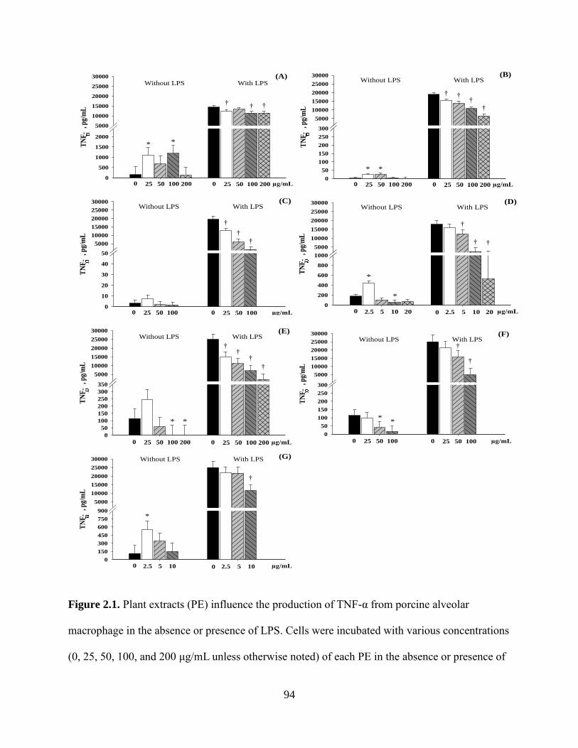
94
TN
F-
, pg/
mL
0
200
400
600
800
1000
5000
10000
15000
20000
25000
30000
*
*
Without LPS With LPS(D)
0 2.5 5 10 20 µg/mL
†
† †
0 2.5 5 10 20
TN
F-
, pg/
mL
0
10
20
30
40
50
5000
10000
15000
20000
25000
30000Without LPS With LPS
(C)
0 25 50 100 0 25 50 100 µg/mL
†
†
†
TN
F-
, pg/
mL
0
50
100
150
200
250
300
5000
10000
15000
20000
25000
30000
* *
Without LPS With LPS(B)
0 25 50 100 200 0 25 50 100 200 µg/mL
† ††
†
TN
F-
, pg/
mL
0
500
1000
1500
2000
5000
10000
15000
20000
25000
30000
Without LPS With LPS(A)
0 25 50 100 200 0 25 50 100 200 µg/mL
* *
†† †
TN
F-
, pg/
mL
0
50
100
150
200
250
300
5000
10000
15000
20000
25000
30000
**
Without LPS With LPS(F)
0 25 50 100 0 25 50 100 µg/mL
†
†
TN
F-
, pg/
mL
0
150
300
450
600
750
900
5000
10000
15000
20000
25000
30000
*
Without LPS With LPS (G)
0 2.5 5 10 µg/mL
†
0 2.5 5 10
TN
F-
, pg/
mL
0
50
100
150
200
250
300
350
5000
10000
15000
20000
25000
30000
* *
Without LPS With LPS(E)
0 25 50 100 200 0 25 50 100 200 µg/mL
††
†
†
Figure 2.1. Plant extracts (PE) influence the production of TNF-α from porcine alveolar
macrophage in the absence or presence of LPS. Cells were incubated with various concentrations
(0, 25, 50, 100, and 200 μg/mL unless otherwise noted) of each PE in the absence or presence of
95
Figure 2.1. (cont.)
LPS (1 µg/mL) for 24 hours. The production of TNF-α by PAM treated with (A) Anethol, (B)
Capsicum oleoresin, (C) Carvacrol, (D) Cinnamaldehyde (0, 2.5, 5, 10, and 20 μg/mL), (E)
Eugenol, (F) Garlic, or (G) Turmeric oleoresin (0, 2.5, 5, 10, and 20 μg/mL) is presented as
pg/mL. The results were means of values from 6 pigs. For anethol (A), LPS: P < 0.001; Level, P
= 0.720; Interaction: P = 0.466. For all other PE, LPS: P < 0.001; Level, P < 0.001; Interaction:
P < 0.001. *different (P < 0.05) from the negative control (0% PE) without LPS. †different (P <
0.05) from the positive control (0% PE) stimulated with 1 μg/mL LPS.

96
Figure 2.2. Plant extracts (PE) influence the production of IL-1β from porcine alveolar
macrophage in the absence or presence of LPS. Cells were incubated with various concentrations
IL-1
, pg/
mL
0
50
100
150
200
1500
3000
4500
6000
7500
9000
* *
Without LPS With LPS(B)
0 25 50 100 200 0 25 50 100 200 µg/mL
† † †
* *
IL-1
, pg/
mL
0
400
800
1200
1600
3000
4500
6000
7500
9000
**
Without LPS With LPS(D)
0 2.5 5 10 20 µg/mL
†
† †
0 2.5 5 10 20
**
IL-1
, pg/
mL
0
200
400
600
800
1000
3000
4500
6000
7500
9000
Without LPS With LPS(A)
0 25 50 100 200 0 25 50 100 200 µg/mL
**
†
*
IL-1
, pg/
mL
0
100
200
300
400
5001500
3000
4500
6000
7500
9000Without LPS With LPS
(E)
0 25 50 100 200 0 25 50 100 200 µg/mL
†
††
IL-1
, pg/
mL
0
100
200
300
400
5001500
3000
4500
6000
7500
9000
*
Without LPS With LPS (G)
0 2.5 5 10 µg/mL 0 2.5 5 10
* *
IL-1
, pg/
mL
0
50
100
150
200
250
300
1500
3000
4500
6000
7500
9000Without LPS With LPS
(C)
0 25 50 100 0 25 50 100 µg/mL
††
†
IL-1
, pg/
mL
0
100
200
300
400
500
6001500
3000
4500
6000
7500
9000
* *
Without LPS With LPS(F)
0 25 50 100 0 25 50 100 µg/mL
†
†
†

97
Figure 2.2. (cont.)
(0, 25, 50, 100, and 200 μg/mL unless otherwise noted) of each PE in the absence or presence of
LPS (1 µg/mL) for 24 hours. The production of IL-1β by PAM treated with (A) Anethol, (B)
Capsicum oleoresin, (C) Carvacrol, (D) Cinnamaldehyde (0, 2.5, 5, 10, and 20 μg/mL), (E)
Eugenol, (F) Garlic, or (G) Turmeric oleoresin (0, 2.5, 5, 10, and 20 μg/mL) is presented as
pg/mL. The results were means of values from 6 pigs. For anethol (A), LPS: P < 0.001; Level, P
= 0.016; Interaction: P = 0.349. For capsicum oleoresin (B), LPS: P < 0.001; Level, P = 0.249;
Interaction: P = 0.280. For all other PE, LPS: P < 0.001; Level, P < 0.001; Interaction: P <
0.001. *different (P < 0.05) from the negative control (0% PE) without LPS. †different (P <
0.05) from the positive control (0% PE) stimulated with 1 μg/mL LPS.

98
Figure 2.3. Plant extracts (PE) influence the production of TGF-β from porcine alveolar
macrophage in the absence or presence of LPS. Cells were incubated with various concentrations
TG
F-
, pg/
mL
0
200
400
600
800
1000
1200
1400
1600
1800
2000Without LPS With LPS
(D)
0 2.5 5 10 20 µg/mL 0 2.5 5 10 20
TG
F-
, pg/
mL
0
200
400
600
800
1000
1200
1400
1600
1800
2000
* *
Without LPS With LPS(F)
0 25 50 100 0 25 50 100 µg/mL
†*
TG
F-
, pg/
mL
0
200
400
600
800
1000
1200
1400
1600
1800
2000Without LPS With LPS
(B)
0 25 50 100 200 0 25 50 100 200 µg/mL
TG
F-
, pg/
mL
0
200
400
600
800
1000
1200
1400
1600
1800
2000
Without LPS With LPS(C)
0 25 50 100 0 25 50 100 µg/mL
† †
TG
F-
, pg/
mL
0
200
400
600
800
1000
1200
1400
1600
1800
2000
Without LPS With LPS(A)
0 25 50 100 200 0 25 50 100 200 µg/mL
TG
F-
, pg/
mL
0
200
400
600
800
1000
1200
1400
1600
1800
2000Without LPS With LPS
(E)
0 25 50 100 200 0 25 50 100 200 µg/mL
† † †* * ** †
TG
F-
, pg/
mL
0
200
400
600
800
1000
1200
1400
1600
1800
2000
*
Without LPS With LPS (G)
0 2.5 5 10 µg/mL
†
0 2.5 5 10
* * †

99
Figure 2.3. (cont.)
(0, 25, 50, 100, and 200 μg/mL unless otherwise noted) of each PE in the absence or presence of
LPS (1 µg/mL) for 24 hours. The production of TGF-β by PAM treated with (A) Anethol, (B)
Capsicum oleoresin, (C) Carvacrol, (D) Cinnamaldehyde (0, 2.5, 5, 10, and 20 μg/mL), (E)
Eugenol, (F) Garlic, or (G) Turmeric oleoresin (0, 2.5, 5, 10, and 20 μg/mL) is presented as
pg/mL. The results were means of values from 6 pigs. Cinnamaldehyde (D), Eugenol (E), Garlic
(F), and turmeric oleoresin (G): Level: P < 0.05. No difference was from LPS stimulation. No
interaction between LPS and level was observed. *different (P < 0.05) from the negative control
(0% PE) without LPS. †different (P < 0.05) from the positive control (0% PE) stimulated with 1
μg/mL LPS.
100
CHAPTER 3
EFFECTS OF PLANT EXTRACTS ON DIARRHEA, IMMUNE RESPONSES,
INTESTINAL MORPHOLOGY, AND GROWTH PERFORMANCE OF WEANED PIGS
EXPERIMENTALLY INFECTED WITH A PATHOGENIC ESCHERICHIA COLI
ABSTRACT
A study was conducted to evaluate the effects of 3 different plant extracts on diarrhea,
immune response, intestinal morphology, and growth performance of weaned pigs
experimentally infected with a pathogenic F-18 E. coli. Weaned pigs (n=64, 6.3 ± 0.2 kg BW, 21
d old) were housed in individual pens in disease-containment chambers for 15 d: 4 d before and
11 d after the first inoculation (d 0). Treatments were in a 2 × 4 factorial arrangement: with or
without an F-18 E. coli challenge (toxins: LT, STb, and SLT-2; 1010
cfu/3 mL oral dose; daily
for 3 d from d 0) and 4 diets (a nursery basal diet (CON), 10 ppm of capsicum oleoresin (CAP),
garlic (GAR), or turmeric oleoresin (TUR)). The growth performance was measured on d 0 to 5,
5 to 11, and 0 to 11. Diarrhea score (DS; 1, normal, to 5, watery diarrhea) was recorded for each
pig daily. Frequency of diarrhea (FD) was the percentage of pig days with DS of 3 or higher.
Feces were collected on d 0, 3, 5, 8, and 11 and plated on blood agar to calculate a ratio (RHT)
of β-hemolytic coliforms to total coliforms by assessing the populations visually using a score (0,
no bacterial growth, to 8, very heavy bacterial growth). Blood was collected on d 0, 5, and 11 to
measure total and differential white blood cell (WBC) counts and serum tumor necrosis factor
(TNF)-α, IL-10, transforming growth factor (TGF)-β, C-reactive protein (CRP), and haptoglobin

101
(Hp). On d 5 and 11, half of the pigs were euthanized to collect jejunum, ileum, and colon to
measure villi height (VH), crypt depth (CDH), and their ratio (VH:CDH). In the challenged
group, the PE treatments reduced (P < 0.05) average DS from d 0 to 2 and d 6 to 11 and FD,
decreased (P < 0.05) TNF-α and Hp on d 5 and WBC, NEU, red blood cell counts, hemoglobin,
and hematocrit on d 11, and increased (P < 0.05) ileal VH on d 5, and tended (P = 0.10) to
increase jejunum VH and VH:CDH compared with the CON, but did not affect growth
performance and RHT. In the unchallenged group, the PE treatments reduced (P < 0.05) average
DS from d 3 to 5, overall FD, and Hp on d 5 compared with the CON. The E. coli infection
increased (P < 0.05) WBC, TNF-α, and Hp, and reduced overall ADG, G:F, and VH of the small
intestine as expected. In conclusion, the 3 PE tested reduced diarrhea, increased the VH of the
small intestine, and affected total WBC, the populations of immune cells, and inflammatory
mediators in E. coli-infected piglets, which may be beneficial to pig health.
KEY WORDS: blood, diarrhea, intestinal morphology, pathogenic E. coli, plant extracts,
weaned pigs
INTRODUCTION
Plant extracts (PE) are secondary plant metabolites, which can be naturally obtained from
plant materials or directly synthesized. Plant extracts have been of potential interest for a long
time due to their antimicrobial (Baydar et al., 2004), anti-inflammatory (Lang et al., 2004),
antioxidant (Dundar et al., 2008), and antiviral effects (Sökmen et al., 2004). Some previous
studies reported that the supplementation of different PE improved growth performance, gut
health, or reduced disease incidence of weaned pigs (Manzanilla et al., 2004; Michiels et al.,

102
2010; Sads and Bilkei, 2003), but other studies did not find beneficial effects from PE (Neill et
al., 2006).
In our previous in vitro study all of 7 different PE tested, including the 3 PE tested here,
had potential anti-inflammatory effects on porcine cells, as all of them inhibited the secretion of
tumor necrosis factor-alpha (TNF-α) from lipopolysaccharide (LPS)-stimulated porcine alveolar
macrophages (Liu et al., 2011c). Another experiment showed feeding 10 ppm of capsicum
oleoresin, garlic, or turmeric oleoresin enhanced immune responses and growth efficiency of
PRRSV-infected weaned pigs (Liu et al., 2011a, b).
Escherichia coli (E. coli) postweaning diarrhea is an important cause of death in weaned
pigs. It is responsible for economic losses due to mortality, morbidity, decreased growth
performance, and cost of medication (Nagy and Fekete, 2005). The toxins secreted from E. coli
affect not only the physiological characteristics of the small intestine (Nagy and Fekete, 2005),
but also the immune system of weaned pigs (Raetz and Whitfield, 2002; Bannerman and
Goldblum, 2003). The objectives of this study were to investigate the effects of feeding PE-
supplemented diets on growth performance and small intestinal morphology of weaned pigs
challenged with a pathogenic E. coli, and to explore the effects of the PE on the disease
resistance and immune responses of weaned pigs after E. coli challenge.
MATERIALS AND METHODS
The protocol for this experiment was reviewed and approved by the Institutional Animal
Care and Use Committee at the University of Illinois at Urbana-Champaign. The experiment was
conducted in the disease containment chambers of the Edwards R. Madigan Laboratory building
at the University of Illinois at Urbana-Champaign.

103
Animals, Housing, Experimental Design, and Diet
A total of 64 weaned piglets (Pig Improvement Company (PIC) line C-22 female × PIC
line 337 male) with same number of gilts and barrows and 6.3 kg of initial BW were selected
from the Swine Research Center of the University of Illinois at Urbana-Champaign. The sows
and piglets used in this experiment did not receive E. coli vaccines, antibiotic injections, or
antibiotics in creep feed. After weaning, all pigs were transferred to the disease containment
chamber and randomly assigned to treatment in a randomized complete block design with weight
within sex as the blocks and pig as the experimental unit. Pigs were housed in the individual pens
for 15 days (4 d before and 11 d after the first E. coli challenge (d 0)). There were a total of 64
individual pens, 4 in each of 16 chambers. There were 2 suites of 8 chambers, and each suite was
used for either E. coli challenged or unchallenged pigs. The piglets had ad libitum access to feed
and water.
The treatments were in a 2 × 4 factorial arrangement (with or without E. coli challenge;
4 dietary treatments). There were 8 replicates per treatment. In the E. coli challenge group, all
pigs were inoculated orally with 3 mL F-18 E. coli/day for 3 consecutive days from d 0 post-
infection (PI). The E. coli strain derived from a field disease outbreak contained heat-labile (LT),
heat-stable (STb), and Shiga-like (SLT-2) toxins, and was provided at 1010
cfu per 3 mL dose in
PBS. This dose has previous caused mild diarrhea (Song et al., 2011). In the unchallenged group,
pigs were inoculated for the 3 consecutive days with 3 mL PBS/day as the sham control (Sham).
The 4 dietary treatments were the complex nursery basal diet (CON) and the addition of 10 ppm
of capsicum oleoresin (CAP), 10 ppm of garlic (GAR), or 10 ppm of turmeric oleoresin (TUR) to
the CON respectively. All 3 plant extracts were obtained from Pancosma S. A. (Geneva,
Switzerland). Capsicum and turmeric are extracted oleoresins, which were standardized to 6%

104
capsaicin and dihydrocapsaicin, and 98% curcuminoides, respectively. Garlic is a botanical
extract from garlic, standardized to 40% propyl thiosulfonates. The CON diet was formulated to
meet or exceed NRC (1998) estimates of requirements of weaned pigs (Table 3.1). Spray-dried
plasma, antibiotics, or zinc oxide were not contained in the CON diet. The experimental diets
were fed to pigs throughout the experiment.
Clinical Observations and Sample Collection
The procedures for this study were adapted from the methods of Song et al. (2011). Prior
to weaning, feces from sows and all their piglets destined for this experiment were collected and
plated on blood and MacConkey agars and verified to be free of detectable β-hemolytic E. coli.
After E. coli inoculation, one pig from the infected control treatment and one pig from the
infected TUR treatment were culled at d 2 and d 4 PI due to severe diarrhea and weight loss.
During the experiment, clinical observations (diarrhea score and alertness score) were recorded
daily from the first day of inoculation (d 0). Diarrhea score of each pig was assessed visually
each day by 2 independent evaluators with a score from 1 to 5 (1 = normal feces, 2 = moist feces,
3 = mild diarrhea, 4 = severe diarrhea, and 5 = watery diarrhea). Frequency of diarrhea was
calculated by counting pig days with diarrhea score of 3 or higher. Alertness score of each pig
was assessed visually with a score from 1 to 3 (1 = normal, 2 = slightly depressed or listless, and
3 = severely depressed or recumbent). Every pig was alert throughout the experiment and was
given an alertness score of 1 (normal) each day, so the data are not reported.
After inoculation, fecal samples were collected from the rectum of each pig using a fecal
loop or cotton swab on d 0, 3, 5, 8, and 11 PI and kept on ice to test for β-hemolytic and total
coliforms. Blood samples were collected in duplicate (Ethylenediaminetetraacetic acid (EDTA)
blood and serum) from the jugular vein of each pig before E. coli challenge (d 0) and d 5 and 11
105
PI. Whole blood samples were used to measure the total and differential white blood cell (WBC)
counts, and cytokines and acute phase proteins (APP) were measured in the sera.
Pigs and feeders were weighed on the day of weaning (d -4), the first inoculation day (d
0), d 5, and d 11. Growth performance (ADG, ADFI, and G:F) was measured for each interval
from d 0 to 5, d 5 to 11, and d 0 to 11.
One-half of the pigs (2 males and 2 females from each treatment) were euthanized on d 5
PI at near the peak of infection and the remainder at the end of experiment (d 11 PI) during
recovery. Before being euthanized, pigs were anesthetized by intramuscular injection of a 1-mL
combination of telazol, ketamine, and xylazine (2:1:1) per 23 kg BW. The final mixture
contained 100 mg telazol, 50 mg ketamine, and 50 mg xylazine in 1 mL (Fort Dodge Animal
Health, For Dodge, IA). After anesthesia, pigs were euthanized by intracardiac injection with 78
mg sodium pentobarbital (Sleepaway) per 1 kg of BW (Henry Schein, Inc., Indianapolis, IN).
Three 3-cm segments from the middle of jejunum, ileum (close to the ileocecal junction), and
middle of colon were collected and fixed in 10% neutral buffered formalin for histological
analysis.
Detection of β-Hemolytic Coliforms
The detection of β-hemolytic and total coliforms was based on Song et al. (2011). Briefly,
fecal samples were plated on blood agar to distinguish β-hemolytic coliforms, which can
complete by digest red blood cell contents surrounding the colony and appear as heavy gray and
shiny colonies from non-hemolytic coliforms which appear as light gray colonies. Meanwhile,
the same fecal samples were plated on MacConkey agar to support that hemolytic colonies on
the blood agar were correctly identified as E. coli colonies, which are lactose fermenting bacteria
and shown as flat pink colonies. All plates were incubated 24 h at 37oC in a humidified 5% CO2

106
incubator. Populations of both total coliforms and β-hemolytic coliforms on blood agar were
assessed visually, with a score from 0 to 8 (0 = no bacterial growth, 8 = very heavy bacterial
growth). The ratio of β-hemolytic coliforms to total coliforms was calculated. The uncertain
colonies were sub-plated on new MacConkey and blood agars to identify whether they were β-
hemolytic. The results of questionable isolate were further verified as the β-hemolytic E. coli
challenge organism using triple sugar iron and lysine iron agars and F-18 E. coli by polymerase
chain reaction.
Intestinal Morphology
The fixed intestinal tissues were embedded in paraffin, sectioned at 5 μm and stained
with hematoxylin and eosin. The slides were scanned by NanoZoomer Digital Pathology System
(Hamamatsu Co., Bridgewater, NJ), and all measurements were conducted in the associated
NDP.view software. Fifteen straight and integrated villi and their associated crypts were selected
for measurement. The intestinal morphological measurements included: villus height and crypt
depth. Only crypt depth was measured in colon due to the lack of villi in this part of intestine.
Mean values of each criteria and villus height:crypt depth ratio within each segment except colon
were calculated for statistical analysis.
Determination of Differential Blood Leukocytes
Blood samples collected in EDTA were used to measure total and differential blood cell
counts by the Veterinary Clinical Pathology Laboratory at the University of Illinois at Urbana-
Champaign. Differential leukocyte proportions and concentrations were analyzed on a
multiparameter, automated hematology analyzer calibrated for porcine blood (CELL-DYN 3700,
Abbott Laboratories, Abbott Park, IL).

107
Measurements of Serum Cytokines and APP
The serum TNF-α, IL-10, and TGF-β concentrations were measured by ELISA according
to the recommendation of the manufacturer (R&D Systems, Inc., Minneapolis, MN, USA).
Briefly, standard, control, and samples were added to the wells with coated monoclonal antibody
specific for each cytokine. After incubation for 2 h, the unbound substances were washed away,
and an enzyme-linked polyclonal antibody specific for the cytokine was added to the wells to
sandwich the cytokine immobilized during the first incubation. A further 2 h of incubation was
followed by a wash to remove any unbound antibody-enzyme reagent, and then a substrate
solution was added to the wells and color developed in proportion to the amount of the cytokine
bound in the initial step. The color development was stopped by adding the stop solution and the
intensity of the color was measured at 450 nm with the correction wavelength set at 530 nm.
Concentrations were calculated from a standard curve. All samples were analyzed in duplicate.
The serum C-reactive protein (CRP), and haptoglobin (Hp) levels were also measured by
commercially available kits (GenWay Biotech, Inc., San Dieo, CA, USA). The procedures were
similar to those described above, except for different incubation times. The intra-assay
coefficients of variation for TNF-α, IL-10, TGF-β, CRP, and Hp were 5.2, 4.4, 3.5, 4.1, and 2.7%,
respectively. The inter-assay coefficients of variation for TNF-α, IL-10, TGF-β, CRP, and Hp
were 7.1, 5.8, 6.0, 5.6, and 6.2%, respectively. The results were expressed in picograms,
nanograms, or micrograms per milliliter based on a standard curve for cytokines and acute phase
proteins.
Statistical Analysis
Data were analyzed using the Proc Mixed procedure of SAS (SAS Inst. Inc, Cary, NC).
Pig was the experimental unit. The statistical model included effects of E. coli challenge, diet,

108
and their interaction as fixed effects. Block was a random effect. Specific contrasts were used to
test comparisons between the control and the PE treatments collectively within each challenge
treatment. Differences among the PE treatments within each challenge treatment were tested by
pair-wise comparisons when the overall main effects were significant. The Chi-square test was
used for the frequency of diarrhea. Probability values of < 0.05 were considered to be significant.
RESULTS
Growth Performance
Several E. coli challenged pigs did not gain BW from d 0 to 5 PI, rendering the G:F
values meaningless, so those data are not presented (Table 3.2). Before E. coli infection, dietary
PE did not affect (P > 0.05) pig performance compared with the sham control (data not shown).
Challenge by E. coli decreased (P < 0.05) the BW of pigs on d 5 and 11 PI, the ADG and G:F
from d 0 to 5 and 0 to 11 PI. The PE treatments did not affect growth rate over the entire
experimental period, but the unchallenged pigs fed the PE treatments grew faster (P < 0.05) than
CON during the early part of the experiment, and more slowly later (P = 0.051). There were no
differences among the PE treatments.
Diarrhea Score and Cultural Score
The E. coli challenge increased (P < 0.05) diarrhea scores from d 2 to 11 PI and increased
(P < 0.05) the frequency of diarrhea (Table 3.3). The PE treatments in the E. coli challenged
group reduced the diarrhea score (P < 0.05) from d 3 to 5 and d 9 to 11 PI, and the frequency of
diarrhea (P < 0.05) for the entire period. In the sham group, the PE treatments reduced (P < 0.05)
the diarrhea score from d 0 to 2 PI and the frequency of diarrhea for the entire period, compared
with the CON. There were no differences among the PE treatments.

109
No treatment effects were found for the ratio of β-hemolytic coliforms to total coliforms
on d 0, so data are not presented. The E. coli challenge increased (P < 0.05) the ratio of β-
hemolytic coliforms to total coliforms on d 3, 5, 8, and 11 PI, compared with the sham group
(Table 3.4). No PE effects were found on the culture score of feces in either the sham or
challenged groups. The fecal β-hemolytic coliforms found in feces from the sham group were not
F-18 E. coli when tested by PCR.
Complete Blood Counts
No treatment effects were found for the total and differential WBC counts and red blood
cells (RBC) on d 0, so data are not shown here. The E. coli challenge increased the number of
total WBC on d 5 (tendency; P = 0.091) and d 11 PI (P < 0.05), but the PE treatments in the E.
coli challenged group decreased (P < 0.05) total WBC (Table 3.5). There were no differences
among the PE treatments. Similarly, the E. coli challenge increased (P < 0.05) the number of
neutrophils on d 11 PI, but the PE treatments in the E. coli challenged group decreased (P < 0.05)
the number of neutrophils. There were no differences among the PE treatments.
The E. coli infection increased (P < 0.05) hemoglobin on d 5 and mean corpuscular
hemoglobin concentration on d 5 and 11 PI (Table 3.6), but the PE treatments had no effect. In
the E. coli challenged group, the supplementation of the PE in diets, reduced (P < 0.05) RBC,
and hemoglobin, and packed cell volume on d 11 PI compared with the CON. No PE effects
were observed in the sham group.
Serum Cytokines and Acute Phase Proteins
A pro-inflammatory cytokine, TNF-α, and 2 anti-inflammatory cytokines, IL-10 and
TGF-β, were detected in the serum of all pigs (Table 3.7). There were no effects of dietary PE
supplementation on serum TNF-α or IL-10 before the E. coli challenge, but PE reduced (P < 0.05)

110
TGF-β compared with the CON. The E. coli challenge increased (P < 0.05) serum TNF-α on d 5
PI, but the PE treatments in the E. coli challenge group reduced (P < 0.05) serum TNF-α as
compared to the CON. There were no differences among the PE treatments. In addition, the E.
coli challenge reduced (P < 0.05) TGF-β on d 11 PI, but the PE treatments did not show any
effect. No differences were detected on IL-10 on either d 5 or 11 PI.
Two acute phase proteins, CRP and Hp were measured in the serum of all pigs (Table
3.7). The E. coli infection increased (P < 0.05) serum Hp levels on d 5 and 11 PI, but the PE
treatments in the E. coli challenge group reduced (P < 0.05) Hp level on d 5 PI, and tended (P
= 0.074) to reduce Hp level on d 11 PI compared with the CON. In the sham group, feeding PE
decreased (P < 0.05) serum Hp concentration on d 5 PI as compared to the CON. The 3 PE tested
here showed different effects on Hp, as pigs fed CAP and GAR had lower (P < 0.05) Hp than
those fed TUR on d 5 PI and pigs fed GAR had lower (P < 0.05) Hp than those fed CAP and
TUR. No differences were detected on CRP on either d 5 or 11 PI.
Intestinal Morphology
The E. coli challenge reduced (P < 0.05) the villi height of jejunum and ileum on d 5 PI,
but the PE treatments in the E. coli challenged group tended to increase (P = 0.093) jejunum villi
height and increased (P < 0.05) ileal villi height, compared with the CON (Table 3.8). In addition,
E. coli infection decreased the villus height:crypt depth ratio of jejunum and ileum and the crypt
depth of the colon on d 5 PI and the villus height:crypt depth ratio of jejunum on d 11 PI, but
increased (P < 0.05) crypt depth of jejunum on d 11 PI. However, there were no PE effects for
these measurements.
111
DISCUSSION
Escherichia coli that express F18 fimbria are a major cause of diarrhea and mortality of
post-weaning pigs for pig producers (Nagy et al., 1997). The results reported here indicate that
feeding PE to weaned pigs challenged with F18 E. coli improved the gut health as indicated by
increasing villus height of the small intestine. In addition, the supplementation of the PE to the E.
coli challenged pigs reduced diarrhea and inflammation caused by the E. coli challenge, as
indicated by reducing WBC, pro-inflammatory cytokines, and acute phase proteins. These
findings are in agreement with previous data showing an improvement of gut health and immune
responses in piglets fed different PE (Manzanilla et al., 2004; Michiels et al., 2010; Wang et al.,
2011).
The present experiment clearly shows a reduction of diarrhea by feeding PE to pigs
challenged with E. coli. These results are supported by observations reported by Gutiérrez et al.
(2007), Mukherjee et al. (1998) and Shoba and Thomas (2001), which indicated many plant
extracts had antidiarrheal activity by inhibiting gastrointestinal mobility and PGE-induced
enteropooling, and stimulating water absorption. Other potential mechanisms might be also
involved in the reduced diarrhea. First, PE may directly affect the activity of pathogenic E. coli
by killing or inhibiting bacterial proliferation or toxin secretion (Hammer et al., 1999). Second,
PE may improve gut health by strengthening gut barrier function, resulting in some protection
against bacteria and/or their toxins (Karmouty-Quintana et al., 2007; Park et al., 2011). Third, PE
may enhance the gastrointestinal immune system and/or the systemic immune system, which
indirectly improve pig health and reduce diarrhea of pigs (Manzanilla et al., 2004; Michiels et al.,
2010; Wang et al., 2011).
112
Plant extracts have been proposed as alternatives for antibiotics due to their significant
antimicrobial effects (Baydar et al., 2004; Sökmen et al., 2004). However, in the present
experiment, the supplementation of PE did not affect the proportions of β-hemolytic coliforms in
feces of E. coli challenged pigs, despite the reduction of diarrhea. These results indicate that the
reduction of diarrhea may be caused by other mechanisms instead of the antimicrobial effect of
PE. On the other hand, the supplementation level of the 3 PE tested in the current experiment
was 10 ppm, much lower than the levels often tested, such as from 20 to 1000 ppm, depending
on the source of plants and methods of extraction (Allan and Bilkei, 2005; Kim et al., 2008;
Manzanilla et al., 2004). Therefore, the reduction of diarrhea and non-reduction of β-hemolytic
coliforms from this experiment suggest that even low levels of PE may have beneficial effects on
pig health, probably by regulation of the immune system. Moreover, the low supplemental level
of PE may help swine producers to save feed cost when adding the PE in the diets.
Total WBC counts are used commonly to estimate the risk of serious bacterial infection,
and an increase in the number of WBC suggests the presence of systemic inflammation (Gordon-
Smith, 2009). The present study showed the E. coli infection induced marked increases in WBC
and lymphocytes on d 5, and WBC, neutrophils, lymphocytes, and monocytes on d 11 PI. These
results are consistent with Song et al. (2011) using the same strain and dose of E. coli in a
challenge study with weaned pigs. The present study also showed the supplementation of PE
decreased WBC, neutrophils, and lymphocytes, indicating the PE tested here may be beneficial
for pig growth performance as the PE attenuate the systemic inflammation caused by E. coli
infection. In agreement with these findings, Wang et al. (2011) reported that dietary fermented
garlic powder reduced the number of WBC of pigs infected with LPS. Otherwise, in the normal

113
conditions, Czech et al. (2009) also observed that pigs receiving herbal extracts, such as garlic
bulbs, had a better health condition with lower WBC.
Consistent with the increased WBC population in the E. coli-infected pigs, an increased
TNF-α was observed in the serum of pigs in the E. coli challenge group. Inflammation is
mediated by increased production of pro-inflammatory cytokines. Among these, TNF-α is one of
most important cytokines released from a variety of porcine immune cells, especially
macrophages/monocytes, in response to bacterial cell wall products and bacterial toxins.
Therefore, systemic levels of TNF-α have been recorded as an indicator of the extent of
inflammation in the circulation of pigs after intravenous entertoxin administration and during
fatal gram-negative sepsis (Jesmok et al., 1992; Maeda et al., 1993; Frank et al., 2003). The
reduction of serum TNF-α by feeding PE to E. coli-infected pigs in the present experiment
suggests that PE might reduce the inflammation induced by E. coli. Wang et al. (2011) also
reported that LPS-infected pigs fed fermented garlic powder had lower TNF-α than those fed the
control diet. Otherwise, many in vitro studies have demonstrated anti-inflammatory effects of
several PE (Lang et al., 2004; Lee et al., 2007; Dung et al., 2009). Our previous in vitro study
also found all of 7 PE tested, including the 3 PE tested here, are anti-inflammatory as showed by
inhibition of TNF-α secretion from LPS-stimulated porcine alveolar macrophage (Liu et al.,
2011c). Both results of WBC and TNF-α in the present experiment clearly showed feeding
dietary PE may alleviate the over-stimulation of the systemic immunity and early immune
response of pigs infected with E. coli. The modes of action for the immune-regulating activity of
PE are still not clear. The results from IL-10 and TGF-β in the current experiment indicates other
mechanisms rather than the secretion of anti-inflammatory cytokines might contribute to the
reduced TNF-α and WBC. However, in vitro evidence suggests that these effects are mediated, at

114
least in part, by blocking the NF-κB pathway (Aggarwal and Shishodia, 2004; Jobin et al., 1999;
Youn et al., 2008).
The proinflammatory cytokines, such as TNF-α, IL-1β, and IL-6, are important inducers
of the synthesis of acute phase proteins, such as CRP and Hp by hepatocytes (Carroll et al., 2004,
Jain et al., 2011). In the present study, the E. coli infection did not change serum CRP levels, but
increased Hp compared with the sham group. The greater response of Hp than CRP has also been
found in pigs infected with porcine reproductive and respiratory virus syndrome virus (Che et al.,
2011; Liu et al., 2011b). Haptoglobin has also been reported to develop in pigs during infections
with various other bacteria, such as Bordetella bronchiseptica, Pasteurella multocida,
Actinobacillus pleuropneumoniae, and Strepococcus suis (Francisco et al., 2004; Hall et al., 1992;
Knura-Deszczka et al., 2002). The increased levels of serum Hp in E. coli-infected pigs may play
important roles in mediating immune responses during E. coli infection, such as suppression of T
lymphocyte proliferation or inhibition of granulocyte chemotaxis, phagocytosis, and bactericidal
activity (Arredouani et al., 2003; Rossbacher et al., 1999). Therefore, the reduction of serum Hp
concentrations caused by the PE tested in the present experiment may be beneficial in controlling
immune responses of E. coli-infected pigs.
It has been reported that STb toxin secreted by E. coli is able to induce partial villus
atrophy in young pigs (Rose et al., 1987). In the present study, pigs infected with F18 E. coli
with LT, STb, and SLT-2 toxins had lower villus height in the jejunum and ileum compared with
the sham group. Similarly, previous studies also observed the reduced villus height and volume
of pigs infected with E. coli (Liu et al., 2010; Vijtiuk et al., 1995; Yi et al.,2005). The reduction
of villus height related to the absorptive capacity of the mucus membrane (Buddle and Bolton,
1992) could induce a decreased absorption of nutrients, which may be responsible for the
115
reduced growth performance. However, the present study shows the pigs fed PE had higher
villus height compared with the CON in the E. coli challenged group, which indicates the PE
tested may be beneficial to gut health. In addition, plant extracts from garlic, Ginkgo biloba
extract, and the mixture of carvacrol, cinnamaldehyde, and capsaicin have been reported to
increase the tightening of tight junctions (Park et al., 2011), attenuate mucosal damage (Mustafa
et al., 2006), and increase mucus synthesis (Jamroz et al., 2006), which may improve the gut
health and prevent the villus atrophy in E. coli-infected pigs.
In the present experiment, E. coli infection significantly reduced growth rate and feed
efficiency, which has also been shown in some other studies (Song et al., 2011; Yi et al., 2005).
No beneficial effect on growth performance was observed from pigs fed PE in the E. coli
challenged group; even though feeding PE increased villus height and reduced systemic
inflammation. Several reasons may be involved in the failure of finding beneficial effects of PE
on pig performance. First, the overall period for this challenge study was 15 days, and it is
difficult to detect growth performance during this short period, and the immediate effect of
weaning has dramatic effects on the growth performance. Second, the replication numbers for
each treatment were large for a challenge study but small for detecting effects on growth
performance.
In conclusion, feeding PE is effective in alleviating diarrhea of weaned pigs caused by an
F-18 E. coli challenge. Feeding PE reduced serum TNF-α and haptoglobin, decreased white
blood cells and lymphocytes, and mitigated villus atrophy and intestinal morphology disruption
after E. coli challenge. We did not find clear differences among the 3 PE tested here, suggesting
that all of 3 them may be beneficial in preventing pathogenic infection and maintaining normal
intestinal integrity and function. This experiment clearly show evidence that a low level of

116
dietary PE can be used in pig diets to maintain pig health, especially alleviating post-weaning
diarrhea. Further studies are needed to investigate the mechanisms through which the PE tested
here exert these effects.
LITERATURE CITED
Aggarwal B. B. and S. Shishodia. 2004. Suppresion of nuclear factor-kappa B activation
pathway by spice-derived phytochemicals: reasoning for seasoning. Ann. N. Y. Acad. Sci.
1030:434-441.
Allan, P. and G. Bilei. 2005. Oregano improves reproductive performance of sows.
Theriogenology. 63:716-721.
Arredouani, M., P. Matthijs, E. Van Hoeyveld, A. Kasran, H. Baumann, J. L. Ceuppens, and E.
Stevens. 2003. Haptoglobin directly affects T cells and suppress T helper cell 2 cytokine
release. Immunol. 108:144-151.
Bannerman, D. D. and S. E. Goldblum. 2003. Mechanisms of bacterial lipopolysaccharide-
induced endothelial apoptosis. Am. J. Physiol. Cell Mol. Physiol. 284:L899-L914.
Baydar, H., O. Sağdiç, G. Özkan, and T. Karadoğan. 2004. Antibacterial activity and
composition of essential oils from Origanum, Thymbra and Satureja species with
commercial importance in Turkey. Food Control. 15:169-172.
Buddle, J. R. and J. R. Bolton. 1992. The pathophysiology of diarrhoea in pigs. Pig News Info.
13:41N-45N.
Carroll, J. A., T. J. Fangman, A. K. Hambach, and C. E. Wiedmeyer. 2004. The acute phase
response in pigs experimentally infected with Escherichia coli and treated with systemic
bactericidal antibiotics. Livest. Prod. Sci. 85:35-44.

117
Che, T. M., R. W. Johnson, K. W. Kelley, W. G. Van Alstine, K. A. Dawson, C. A. Moran, and J.
E. Pettigrew. 2011. Mannan oligosaccharide improves immune responses and growth
efficiency of nursery pigs experimentally infected with porcine reproductive and
respiratory syndrome virus. J. Anim. Sci. In Press.
Czech, A., E. Kowalczuk, and E. Grela. 2009. The effect of a herbal extract used in pig fattening
on the animals’ performance and blood components. Unversity of Life Sciences in Lublin.
Annales. 27:25-33.
Dundar, E., E. G. Olgun, S. Isiksoy, M. Kurkcuoglu, K. H. C. Baser, and C. Bal. 2008. The
effects of intra-rectal and intra-peritoneal application of Origanum onites L. essential oil
on 2,4,6-trinitrobenzenesulfolic acid-induced colitis in the rat. Exp. Toxical. Pathol.
59:399-408.
Dung, N. T., V. K. Bajpai, J. I. Yoon, and S. C. Kang. 2009. Anti-inflammatory effects of
essential oil isolated from the buds of Cleistocalyx operculatus (Roxb.) Merr and Perry.
Food Chem. Toxicol.47:449-453.
Francisco, C. J., T. R. Shryock, D. P. Bane, and L. Unverzagt. 2004. Serum haptoglobin
concentration in growing swine after intranasal challenge with Bordetella bronchiseptica
and toxigenic Pasteurella multocida type D. Can. J. vet. Res. 60:222-227.
Frank, J. W., J. A. Carroll, G. L. Allee, and M. E. Zannelli. 2003. The effects of thermal
environment and spry-dried plasma on the acute-phase response of pigs challenged with
lipopolysaccharide. 81:1166-1176.
Gordon-Smith, T. 2009. Structure and function of red and white blood cells. Medicine. 37:119-
124.

118
Gutiérrez, S. P., M. A. Z. Sánchez, C. P. Gonzálze, and L. A. García. 2007. Antidiarrhoeal
activity of different plants used in traditional medicine. Afr. J. Biotech. 6:2988-2994.
Hall, W. F., T. E. Eurell, R. D. Hansen, and L. G. Herr. 1992. Serum haptoglobin concentration
in swine naturally or experimentally infected with Actinobacillus pleuropneumoniae. J.
Am. Vet. Med. Assoc. 201:1730-1733.
Hammer, K. A., C. F. Carson, and T. V. Riley. 1999. Antimicrobial activity of essential oils and
other plant extracts. J. Appl. Microbiol. 86:985-990.
Jain, S., V. Gautam, and S. Naseem. 2011. Acute-phase proteins: As diagnostic tool. J. Pharm.
Bioall. Sci. 1:118-127.
Jamroz, D., T. Wertelecki, M. Houszka, and C. Kamel. 2006. Influence of diet type on the
inclusion of plant origin active substances on morphological and histochemical
characteristics of the stomach and jejunum walls in chicken. J. Anim. Physiol. An. N.
90:255-268.
Jesmok, G., C. Lindsey, M. Duerr, M. Fournel, and T. Emerson. 1992. Efficacy of monoclonal
antibody against human recombinant tumor necrosis factor in E. coli-challenge swine.
Am. J. Pathol. 141:1197-1207.
Jobin, C., C. A. Bradham, M. P. Russo, B. Juma, A. S. Narula, D. A. Brenner, and R. B. Sartor.
1999. Curcumin blocks cytokine-mediated NF-κB activation and proinflammatory gene
expression by inhibiting inhibitory actor I-κB kinase activity. J. Immunol. 163:3474-3483.
Karmouty-Quintana, H., C. Cannet, R. Sugar, J. R. Fozard, C. P. Page, and N. Beckmann. 2007.
Capsaicin-induced mucus secretion in rat airways assessed in vivo and non-invasively by
magnetic resonance imaging. Br. J. Pharmacol. 150:1022-1030.

119
Knura-Deszczka, S., C. Lipperheide, B. Petersen, J. L. Jobert, F. Berthelot-Hérault, M. Kobisch,
and F. Madec. 2002. Plamsa haptoglobin concentration in swine after challenge with
Streptococcus suis. J. Vet. Med. B49:240-244.
Lang, A., M. Lahav, E. Sakhnini, I. Barshack, H. H. Fidder, B. Avidan, E. Bardan, R.
Hershkoviz, S. Bar-Meir, and Y. Chowers. 2004. Allicin inhibits spontaneous and TNF-α
induced secretion of proinfammatory cytokines and chemokines from intestinal epithelial
cells. Clin. Nutr. 23:1199-1208.
Lee, Y., S. Hung, S. Pai, Y. Lee, and S. Yang. 2007. Eugenol suppressed the expression of
lipopolysaccharide-induced proinflammatory mediators in human macrophages. J. Endod.
33:698-702.
Liu, P., X. S. Piao, P. A. Thacker, Z. K. Zeng, D. F. Li, D. Wang, and S. W. Kim. 2010. Chito-
oligosaccharide reduced diarrhea incidence and attenuates the immune response of
weaned pigs challenged with Escherichia coli K88. J. Anim. Sci. 88:3871-3879.
Liu, Y., T. M. Che, D. Bravo, and J. E. Pettigew. 2011c. Effects of plant extracts on cytokine
production of porcine alveolar macrophages. J. Anim. Sci. 86(E-Suppl. 2):47(Abstr.).
Liu, Y., M. Song, T. M. Che, J. A. Soares, D. Bravo, C. W. Maddox, and J. E. Pettigrew. 2011a.
Plant extracts for weaned pigs experimentally infected with porcine reproductive and
respiratory syndrome virus. 1: Effect on growth performance and immune responses. J.
Anim. Sci. 89(E-Suppl. 1):247(Abstr.).
Liu, Y., M. Song, T. M. Che, J. A. Soares, D. Bravo, C. W. Maddox, and J. E. Pettigrew. 2011b.
Plant extracts for weaned pigs experimentally infected with porcine reproductive and
respiratory syndrome virus. 2: Effect on peripheral blood immune cells and inflammatory
mediators. J. Anim. Sci. 89(E-Suppl. 1):579(Abstr.).

120
Maeda, K., L. O. Schoeniger, M. Shimada, R. A. Winchurch, T. G. Buchman, and J. L.
Robotham. 1993. Regulation of acute phase gene expression following surgery and
endotoxin administration in the anesthetized pigs. Anesthesiol. 79:1324-1337.
Manzanilla, E. G., J. F. Perez, M. Martin, C. Kramel, F. Baucells, and J. Gasa. 2004. Effect of
plant extracts and formic acid on the intestinal equilibrium of early-weaned pigs. J. Anim.
Sci. 82:3210-3218.
Michiels, J., J. Missotten, A. Van Hoorick, A. Ovyn, D. Fremaut, D. De Smet, and N. Dierick.
2010. Effects of dose and formulation of carvacrol and thymol on bacteria and some
functional traits of the gut in piglets after weaning. Arch. of Ani. Nutr. 64:136-154.
Mukherjee, P. K., K. Saha, T. Murugesan, S. C. Mandal, M. Pal, and B. P. Saha. 1998. Screening
of anti-diarrhoeal profile of some plant extracts of a specific region of West Bengal, India.
J. Ethnopharmacol. 60:85-89.
Mustafa, A., A. El-Medany, H. H. Hagar, and G. El-Medany. 2006. Ginkgo biloba attenuates
mucosal damage in a rat model of ulcerative colitis. Pharmacol. Res. 53:324-330.
Nagy, B. and P. Z. Fekete. 2005. Enterotoxigenic Escherichia coli in veterinary medicine. Int. J.
Med. Microbiol. 295:443-454.
Nagy, B., S. C. Whipp, H. Imberechts, H. U. Bertschinger, E. A. Dean-Nystrom, T. A. Casey,
and E. Salajka. 1997. Biological relationship between F18ab and F18ac fimbriae of
enterotoxigenic and verotoxigenix Escherichia coli from weaned pigs with oedema
disease or diarrhea. Microb. Pathogenesis. 22:1-11.
National Research Council (NRC). 1998. Pages 110-123 in Nutrient Requirements of Swine. 10th
rev. ed. Natl. Acd. Press, Washington, DC.

121
Park. H. S., G. Kim, I. Choi, N. D. Kim, H. J. Hwang, Y. Choi, and Y. H. Choi. 2011. Inhibition
of matrix metalloproteinase activities and tightening of tight junctions by diallyl disulfate
in AGS human gastric carcinoma cells. J. Food Sci. 76:T105-T111.
Raetz, C. R. H. and C. Whitfield. 2002. Lipopolysaccharide endotoxins. Annu. Rev. Biochem. 71:
635-700.
Rose, R., S. C. Whipp, and H. W. Moon. 1987. Effects of Escherichia coli heat-stable
enterotoxin b on small intestinal villi in pigs, rabbits, and lambs. Vet. Pathol. 24:71-79.
Rossbacher, J., L. Wagner, and M. S. Pasternack. 1999. Inhibitory effect of haptoglobin on
granulocyte chemotaxis, phagocytosis and bactericidal activity. Scand. J. Immunol.
50:399-404.
Sads, O. R. and G. Bilkei. 2003. The effect of oregano and vaccination against Glasser’s disease
and pathogenic Escherichia coli on postweaning performance of pigs. Irish Vet. J.
56:611-615.
Shoba, F. G. and M. Thomas. 2001. Study of antidiarrhoeal activity of four medicinal plants in
castor-oil induced diarrhoea. J. Ethnopharmacol. 76:73-76.
Sökmen, M., J. Serkedjieva, D. Daferera, M. Gulluce, M. Polissiou, B, Tepe, H. A. Akpulat, F.
Sahin, and A. Sokmen. 2004. In vitro antioxidant, antimicrobial, and antiviral activities of
the essential oil and various extracts from herbal parts and callus cultures of Origanum
acutidens. J. Agric. Food Chem. 52:3309-3312.
Song, M., Y. Liu, J. A. Soares, T. M. Che, O. Osuna, C. W. Maddox, and J. E. Pettigrew. 2011.
Effects of dietary clays on diarrhea of weaned pigs experimentally infected with a
pathogenic E. coli. J. Anim. Sci. 86(E-Suppl. 2):40(Abstr.).

122
Vijtiuk, N., S. Ćurić, G. Lacković, I. Udovičić, I. Vrbanac, and I. Valpotić. 1995.
Histopathological features in the small intestine of pigs infected with F4ac+ non-
enterotoxigenic or enterotoxigenic strains of Eschirichia coli. J. Comp. Path. 112:1-10.
Wang, J. P., J. S. Yoo, H. D. Jang, J. H. Lee, J. H. Cho, and I. H. Kim. 2011. Effect of dietary
fermented garlic by Weissella koreensis powder on growth performance, blood
characteristics, and immune response of growing pigs challenged with Escherichia coli
lipopolysaccharide. J. Anim. Sci. 89:2123-2131.
Yi, G. F., J. A. Carroll, G. L. Allee, A. M. Gaines, D. C. Kendall, J. L. Usry, Y. Toride, and S.
Izuru. 2005. Effect of glutamine and spray-dried plasma on growth performance, small
intestinal morphology, and immune responses of Escherichia coli K88+-challenged
weaned pigs. J. Anim. Sci. 83:634-643.
Youn, H., H. J. Lim, H. J. Lee, D. Hwang, M. Yang, R. Jeon, and J. Ryu. 2008. Garlic (Allium
sativum) extract inhibits lipopolysaccharide-induced toll-like receptor 4 dimerization.
Biosci. Biotechnol. Biochem. 72:368-375.

123
TABLES AND FIGURES
Table 3.1. Ingredient composition of basal diet (as-fed basis).
Ingredient Amount, %
Corn, ground 41.54
Whey dried 15.00
Soybean meal, 47% 10.82
Fishmeal 10.00
Lactose 10.00
Soy protein concentrate 5.00
Poultry byproduct meal 4.27
Soybean oil 2.67
Mineral premix1 0.35
Vitamin premix2 0.20
L-Lysine∙HCl 0.05
DL-Met 0.05
L-Thr 0.03
L-Trp 0.02
Total 100.00
Calculated energy and nutrients
ME, kcal/kg 3480
CP, % 22.67
Fat, % 6.34
Ca, % 0.80
P, % 0.72
Available P, % 0.49
Lys, % 1.50
Lactose, % 21.00
124
Table 3.1. (cont.)
1Provided as milligrams per kilogram of diet: 3,000 of NaCl; 100 of Zn from zinc oxide;
90 of Fe from iron sulfate; 20 of Mn from manganese oxide; 8 of Cu from copper sulfate; 0.35 of
I from calcium iodide; 0.30 of Se from sodium selenite.
2Provided per kilogram of diet: 2,273 μg of retinyl acetate; 17 μg of cholecalciferol; 88
mg of DL-α-tocopheryl acetate; 4 mg of menadione from menadione sodium bisulfite complex;
33 mg of niacin; 24 mg of D-Ca-pantothenate; 9 mg of riboflavin; 35 μg of vitamin B12; 324 mg
of choline chloride.

125
Table 3.2. Effect of plant extract on growth performance of pigs experimentally infected with a pathogenic E. coli1
Treatment2 P-value
Sham E. coli Main effect3 PE vs. CON
4
Item CON CAP GAR TUR CON CAP GAR TUR SEM E.coli Diet E*D Sham E. coli
Body weight
d 0, kg 6.66 6.46 6.63 6.66 6.51 6.59 6.43 6.63 0.20 0.63 0.90 0.83 0.71 0.89
d 5, kg 6.93 7.11 7.15 7.24 6.54 6.68 6.53 6.77 0.21 <0.05 0.62 0.94 0.30 0.64
d 11, kg 9.58 8.93 8.60 9.08 8.13 8.18 8.35 9.20 0.50 <0.05 0.34 0.25 0.095 0.38
d 0 to 5
No. of pigs5 8 8 8 8 7 8 8 7
No gain6 1 0 1 0 4 4 4 3
ADG, g 53 130 105 115 5.7 18 20 29 23 <0.05 0.18 0.54 <0.05 0.55
ADFI, g 328 418 455 361 428 350 325 370 85 0.66 0.99 0.41 0.30 0.36
G:F 0.22 0.42 0.26 0.81 - - - - 0.10 <0.05 0.44 0.58 0.27 0.81
d 5 to 11
No. of pigs5 4 4 4 4 3 4 4 3
No gain6 0 0 0 0 0 0 0 0
ADG, g 470 384 292 350 278 263 321 345 76 0.10 0.69 0.25 0.051 0.68
ADFI, g 730 700 742 588 767 742 754 756 136 0.47 0.92 0.92 0.68 0.92
G:F 0.76 0.60 0.73 0.71 0.37 0.39 0.43 0.47 0.25 0.096 0.98 0.98 0.75 0.82
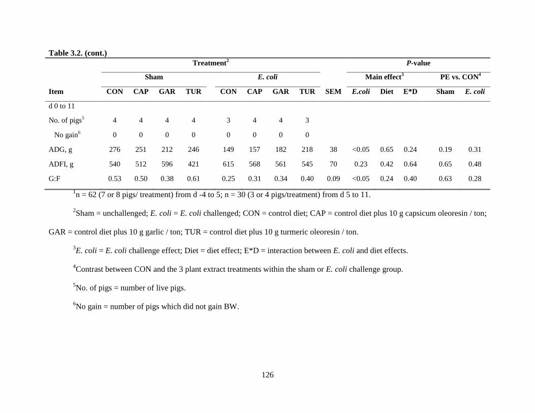
126
Table 3.2. (cont.)
Treatment2 P-value
Sham E. coli Main effect3 PE vs. CON
4
Item CON CAP GAR TUR CON CAP GAR TUR SEM E.coli Diet E*D Sham E. coli
d 0 to 11
No. of pigs5 4 4 4 4 3 4 4 3
No gain6 0 0 0 0 0 0 0 0
ADG, g 276 251 212 246 149 157 182 218 38 <0.05 0.65 0.24 0.19 0.31
ADFI, g 540 512 596 421 615 568 561 545 70 0.23 0.42 0.64 0.65 0.48
G:F 0.53 0.50 0.38 0.61 0.25 0.31 0.34 0.40 0.09 <0.05 0.24 0.40 0.63 0.28
1n = 62 (7 or 8 pigs/ treatment) from d -4 to 5; n = 30 (3 or 4 pigs/treatment) from d 5 to 11.
2Sham = unchallenged; E. coli = E. coli challenged; CON = control diet; CAP = control diet plus 10 g capsicum oleoresin / ton;
GAR = control diet plus 10 g garlic / ton; TUR = control diet plus 10 g turmeric oleoresin / ton.
3E. coli = E. coli challenge effect; Diet = diet effect; E*D = interaction between E. coli and diet effects.
4Contrast between CON and the 3 plant extract treatments within the sham or E. coli challenge group.
5No. of pigs = number of live pigs.
6No gain = number of pigs which did not gain BW.

127
Table 3.3. Effect of plant extracts on diarrhea score and frequency of diarrhea of pigs experimentally infected with a
pathogenic E. coli1
Treatment2 P-value
Sham E. coli Main effect3 PE vs. CON
4
Item CON CAP GAR TUR CON CAP GAR TUR SEM E.coli Diet E*D Sham E. coli
d 0 to 25 1.96 1.17 1.21 1.29 1.93 1.67 1.35 1.36 0.36 0.32 <0.05 0.70 <0.05 0.11
d 3 to 55 2.06 1.35 1.46 1.67 3.43 2.50 2.13 2.0 0.31 <0.05 <0.05 0.22 <0.05 <0.05
d 6 to 85 1.44 1.18 1.17 1.21 2.86 2.58 2.83 2.90 0.52 <0.05 0.92 0.96 0.49 0.91
d 9 to 115 1.09 1.04 1.08 1.00 3.51
a 2.13
b 1.21
c 1.15
c 0.26 <0.05 <0.05 <0.05 0.77 <0.05
Pig days6 64 64 64 64 53 64 64 53
Frequency7 20 4 7 9 40 26 17 16 <0.05 <0.05 <0.05 <0.05 <0.05
a, b, c within a row, means without a common superscript differ (P < 0.05) within the E. coli challenge group.
1n = 62 (7 or 8 pigs/ treatment) on d 0, 3, and 5; n = 30 (3 or 4 pigs/treatment) on d 8 and 11.
2Sham = unchallenged; E. coli = E. coli challenged; CON = control diet; CAP = control diet plus 10 g capsicum oleoresin / ton;
GAR = control diet plus 10 g garlic / ton; TUR = control diet plus 10 g turmeric oleoresin / ton.
3E. coli = E. coli challenge effect; Diet = diet effect; E*D = interaction between E. coli and diet effects.
4Contrast between CON and the 3 plant extract treatments within the sham or E. coli challenge group.
5Diarrhea score = 1, normal feces, 2, moist feces, 3, mild diarrhea, 4, severe diarrhea, 5, watery diarrhea.
6Pig days = number of pig x the number of days of diarrhea scoring.
7Frequency = number of pig days with diarrhea score ≥ 3. Statistical analysis was conducted by Chi-square test.

128
Table 3.4. Effect of plant extracts on culture score of feces from pigs experimentally infected with a pathogenic E. coli1
Treatment2 P-value
Sham E. coli Main effect3 PE vs. CON
4
Item CON CAP GAR TUR CON CAP GAR TUR SEM E.coli Diet E*D Sham E. coli
d 3
Coliform 5.3 5.5 6.0 6.6 6.5 7.0 6.4 6.1 0.43 <0.05 0.64 0.073 0.092 0.99
β-hemolytic 0 0 0 0 5.6 5.5 5.0 5.4 0.55 <0.05 0.94 0.94 1.00 0.60
β-hemo., ratio 0 0 0 0 0.86 0.79 0.77 0.89 0.07 <0.05 0.80 0.80 1.00 0.56
d 5
Coliform 5.8 6.5 5.6 6.1 8.0 8.0 7.9 8.0 0.69 <0.05 0.88 0.93 0.66 0.96
β-hemolytic 0 0 0 0 7.5 6.8 7.0 7.9 0.48 <0.05 0.62 0.62 1.00 0.57
β-hemo., ratio 0 0 0 0 0.94 0.84 0.89 0.99 0.06 <0.05 0.65 0.65 1.00 0.63
d 8
Coliform 5.8 7.3 6.0 5.8 6.8 7.8 7.8 7.3 0.60 <0.05 0.14 0.61 0.33 0.17
β-hemolytic 0.06 0.09 0 0 6.0 6.8 3.8 5.3 1.23 <0.05 0.48 0.54 0.98 0.50
β-hemo., ratio 0.05 0.07 0 0 0.85 0.87 0.50 0.70 0.15 <0.05 0.43 0.46 0.99 0.27
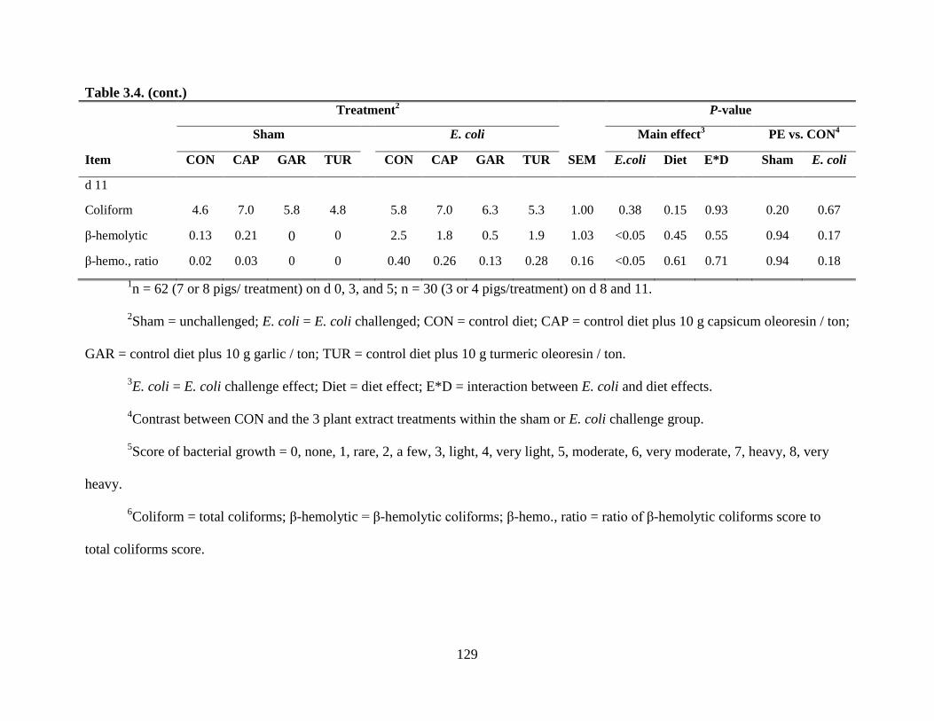
129
Table 3.4. (cont.)
Treatment2 P-value
Sham E. coli Main effect3 PE vs. CON
4
Item CON CAP GAR TUR CON CAP GAR TUR SEM E.coli Diet E*D Sham E. coli
d 11
Coliform 4.6 7.0 5.8 4.8 5.8 7.0 6.3 5.3 1.00 0.38 0.15 0.93 0.20 0.67
β-hemolytic 0.13 0.21 0 0 2.5 1.8 0.5 1.9 1.03 <0.05 0.45 0.55 0.94 0.17
β-hemo., ratio 0.02 0.03 0 0 0.40 0.26 0.13 0.28 0.16 <0.05 0.61 0.71 0.94 0.18
1n = 62 (7 or 8 pigs/ treatment) on d 0, 3, and 5; n = 30 (3 or 4 pigs/treatment) on d 8 and 11.
2Sham = unchallenged; E. coli = E. coli challenged; CON = control diet; CAP = control diet plus 10 g capsicum oleoresin / ton;
GAR = control diet plus 10 g garlic / ton; TUR = control diet plus 10 g turmeric oleoresin / ton.
3E. coli = E. coli challenge effect; Diet = diet effect; E*D = interaction between E. coli and diet effects.
4Contrast between CON and the 3 plant extract treatments within the sham or E. coli challenge group.
5Score of bacterial growth = 0, none, 1, rare, 2, a few, 3, light, 4, very light, 5, moderate, 6, very moderate, 7, heavy, 8, very
heavy.
6Coliform = total coliforms; β-hemolytic = β-hemolytic coliforms; β-hemo., ratio = ratio of β-hemolytic coliforms score to
total coliforms score.

130
Table 3.5. Effect of plant extracts on total and differential white blood cells of pigs experimentally infected with a pathogenic E.
coli1
Treatment2 P-value
Sham E. coli Main effect3 PE vs. CON
4
Item CON CAP GAR TUR CON CAP GAR TUR SEM E.coli Diet E*D Sham E. coli
d 5
WBC, 103/μL
5 15.2 14.5 17.0 13.8 21.1 14.5 14.6 18.3 1.69 0.091 0.19 <0.05 0.98 <0.05
Neu, 103/μL
5 10.22 7.96 10.37 8.40 10.16 7.66 8.59 9.15 1.32 0.70 0.28 0.79 0.36 0.27
Lym, 103/μL
5 4.15 5.63 5.49 4.54 8.25 6.17 5.35 8.36 0.97 <0.05 0.71 0.050 0.32 0.15
Mono, 103/μL
5 0.535 0.682 1.051 0.723 0.425 0.470 0.587 0.514 0.16 <0.05 0.14 0.66 0.10 0.58
Eos, 103/μL
5 0.098 0.099 0.069 0.079 0.086 0.105 0.050 0.188 0.047 0.52 0.45 0.49 0.76 0.61
Baso, 103/μL
5 0.031 0.062 0.032 0.002 0.028 0.054 0.029 0.060 0.032 0.55 0.62 0.56 0.97 0.53
Neu/Lym5 3.24 1.45 3.17 2.10 1.28 1.41 1.60 1.22 0.66 <0.05 0.36 0.44 0.17 0.86
d 11
WBC, 103/μL
5 16.7 15.8 19.0 14.4 32.2 21.6 23.0 24.3 3.44 <0.05 0.27 0.25 0.94 <0.05
Neu, 103/μL
5 7.96 8.28 8.49 6.71 17.01 10.26 14.00 10.19 1.87 <0.05 0.099 0.20 0.95 <0.05
Lym, 103/μL
5 8.57 6.66 9.86 6.99 13.48 9.43 7.63 12.30 2.43 0.062 0.45 0.20 0.74 0.10
Mono, 103/μL
5 0.264 0.890 0.549 0.473 1.473 1.536 0.934 1.778 0.299 <0.05 0.29 0.28 0.23 0.85
Eos, 103/μL
5 0.309 0.165 0.051 0.148 0.223 0.322 0.234 0.183 0.131 0.40 0.67 0.64 0.17 0.87
Baso, 103/μL
5 0 0.151 0 0.054 0.039 0.026 0.127 0.128 0.070 0.53 0.64 0.26 0.34 0.45
Neu/Lym5 1.13 1.37 0.91 0.97 1.44 1.11 2.29 1.04 0.42 0.17 0.49 0.17 0.91 0.94

131
Table 3.5. (cont.)
1n = 62 (7 or 8 pigs/ treatment) on d 5; n = 30 (3 or 4 pigs/treatment) on d 11.
2Sham = unchallenged; E. coli = E. coli challenged; CON = control diet; CAP = control diet plus 10 g capsicum oleoresin / ton;
GAR = control diet plus 10 g garlic / ton; TUR = control diet plus 10 g turmeric oleoresin / ton.
3E. coli = E. coli challenge effect; Diet = diet effect; E*D = interaction between E. coli and diet effects.
4Contrast between CON and the 3 plant extract treatments within the sham or E. coli challenge group.
5WBC = white blood cell; Neu = neutrophil; Lym = lymphocyte; Mono = monocyte; Eos = eosinophil; Baso = basophil;
Neu/Lym = neutrophils/lymphocytes.
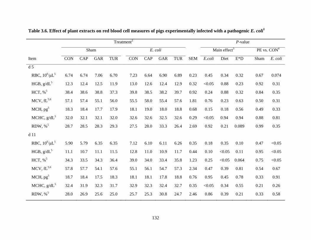
132
Table 3.6. Effect of plant extracts on red blood cell measures of pigs experimentally infected with a pathogenic E. coli1
Treatment2 P-value
Sham E. coli Main effect3 PE vs. CON
4
Item CON CAP GAR TUR CON CAP GAR TUR SEM E.coli Diet E*D Sham E. coli
d 5
RBC, 106/μL
5 6.74 6.74 7.06 6.70 7.23 6.64 6.90 6.89 0.23 0.45 0.34 0.32 0.67 0.074
HGB, g/dL5 12.3 12.4 12.5 11.9 13.0 12.6 12.4 12.9 0.32 <0.05 0.88 0.23 0.92 0.31
HCT, %5 38.4 38.6 38.8 37.3 39.8 38.5 38.2 39.7 0.92 0.24 0.88 0.32 0.84 0.35
MCV, fL5,6 57.1 57.4 55.1 56.0 55.5 58.0 55.4 57.6 1.81 0.76 0.23 0.63 0.50 0.31
MCH, pg5 18.3 18.4 17.7 17.9 18.1 19.0 18.0 18.8 0.68 0.15 0.18 0.56 0.49 0.33
MCHC, g/dL5 32.0 32.1 32.1 32.0 32.6 32.6 32.5 32.6 0.29 <0.05 0.94 0.94 0.88 0.81
RDW, %5 28.7 28.5 28.3 29.3 27.5 28.0 33.3 26.4 2.69 0.92 0.21 0.089 0.99 0.35
d 11
RBC, 106/μL
5 5.90 5.79 6.35 6.35 7.12 6.10 6.11 6.26 0.35 0.18 0.35 0.10 0.47 <0.05
HGB, g/dL5 11.1 10.7 11.1 11.5 12.8 11.0 10.9 11.7 0.44 0.10 <0.05 0.11 0.95 <0.05
HCT, %5 34.3 33.5 34.3 36.4 39.0 34.0 33.4 35.8 1.23 0.25 <0.05 0.064 0.75 <0.05
MCV, fL5,6 57.8 57.7 54.1 57.6 55.1 56.1 54.7 57.3 2.34 0.47 0.39 0.81 0.54 0.67
MCH, pg5 18.7 18.4 17.5 18.3 18.1 18.1 17.8 18.8 0.76 0.95 0.45 0.78 0.33 0.91
MCHC, g/dL5 32.4 31.9 32.3 31.7 32.9 32.3 32.4 32.7 0.35 <0.05 0.34 0.55 0.21 0.26
RDW, %5 28.0 26.9 25.6 25.0 25.7 25.3 30.8 24.7 2.46 0.86 0.39 0.21 0.33 0.58

133
Table 3.6. (cont.)
1n = 62 (7 or 8 pigs/ treatment) on d 5; n = 30 (3 or 4 pigs/treatment) on d 11.
2Sham = unchallenged; E. coli = E. coli challenged; CON = control diet; CAP = control diet plus 10 g capsicum oleoresin / ton;
GAR = control diet plus 10 g garlic / ton; TUR = control diet plus 10 g turmeric oleoresin / ton.
3E. coli = E. coli challenge effect; Diet = diet effect; E*D = interaction between E. coli and diet effects.
4Contrast between CON and the 3 plant extract treatments within the sham or E. coli challenge group.
5RBC = red blood cell; HGB = hemoglobin; HCT = packed cell volume; MCV = mean corpuscular volume; MCH = mean
corpuscular hemoglobin; MCHC = mean corpuscular hemoglobin concentration; RDW = red cell distribution width.
6fL= femtolitre (10
-15 L).

134
Table 3.7. Effects of plant extracts on cytokine and acute phase protein concentrations in the serum of pigs infected with a
pathogenic E. coli1
Treatment2 P-value
Sham E. coli Main effect3 PE vs. CON
4
Item CON CAP GAR TUR CON CAP GAR TUR SEM E.coli Diet E*D Sham E. coli
d 0
TNF-α, pg/mL5 52.31 61.66 56.83 44.63 48.15 60.10 63.25 63.37 8.66 0.48 0.48 0.45 0.79 0.15
IL-10, pg/mL 0.34 1.08 3.73 0.88 0.36 0.55 0.37 0.86 1.02 0.19 0.44 0.36 0.19 0.84
TGF-β, ng/mL5 23.12 21.99 21.24 22.11 25.17 20.46 20.38 23.99 1.18 0.63 <0.05 0.25 0.30 <0.01
CRP, μg/mL5 23.96 18.15 20.19 22.24 21.20 25.11 21.76 20.75 2.88 0.60 0.96 0.35 0.26 0.69
HP, μg/mL5 603.4 587.0 735.6 568.3 536.1 596.8 513.8 616.6 83.5 0.33 0.93 0.38 0.78 0.68
d 5
TNF-α, pg/mL5 58.97 68.27 56.59 58.06 79.55 61.82 72.2 63.08 5.76 <0.05 0.43 0.057 0.73 <0.05
IL-10, pg/mL 1.05 1.17 1.52 1.14 2.53 1.63 4.35 2.59 1.16 0.063 0.61 0.79 0.87 0.80
TGF-β, ng/mL5 22.64 24.11 21.71 21.78 23.40 21.22 21.66 21.03 1.33 0.41 0.52 0.50 0.94 0.15
CRP, μg/mL5 25.02 21.66 24.13 26.64 29.06 27.79 24.58 29.30 3.06 0.16 0.56 0.81 0.80 0.60
HP, μg/mL5 930.6 574.2 570.9 663.4 1399.5
a 823.1b 887.5
b 1269.6a 116.8 <0.05 <0.05 0.43 <0.05 <0.05

135
Table 3.7. (cont.)
Treatment2 P-value
Sham E. coli Main effect3 PE vs. CON
4
Item CON CAP GAR TUR CON CAP GAR TUR SEM E.coli Diet E*D Sham E. coli
d 11
TNF-α, pg/mL5 68.00 66.35 88.58 61.53 87.61 80.91 65.62 66.23 14.44 0.67 0.73 0.35 0.78 0.27
IL-10, pg/mL 0.52 0 0 0.16 0.01 0.13 0.32 0.76 0.38 0.57 0.70 0.39 0.20 0.32
TGF-β, ng/mL5 30.44 27.55 24.61 25.74 23.04 18.72 25.01 25.87 2.47 <0.05 0.38 0.070 0.066 0.85
CRP, μg/mL5 34.78 30.18 31.52 25.05 26.60 32.15 21.13 31.96 8.24 0.60 0.85 0.51 0.39 0.82
HP, μg/mL5 407.1 304.0 507.6 236.3 1271.6
a 937.8a 163.8
b 1363.0a 291.6 <0.05 0.091 <0.05 0.81 0.074
a, b within a row, means without a common superscript differ (P < 0.05) within the E. coli challenge group.
1n = 62 (7 or 8 pigs/ treatment) on d 0 and 5; n = 30 (3 or 4 pigs/treatment) on d 11.
2Sham = unchallenged; E. coli = E. coli challenged; CON = control diet; CAP = control diet plus 10 g capsicum oleoresin / ton;
GAR = control diet plus 10 g garlic / ton; TUR = control diet plus 10 g turmeric oleoresin / ton.
3E. coli = E. coli challenge effect; Diet = diet effect; E*D = interaction between E. coli and diet effects.
4Contrast between CON and the 3 plant extract treatments within the sham or E. coli challenge group.
5TNF-α = tumor necrosis factor-α, TGF-β = transforming growth factor-β, CRP = C-reactive protein, Hp = haptoglobin.

136
Table 3.8. Effects of plant extracts on histological characteristics of the small intestine of pigs infected with a pathogenic E.
coli1
Treatment2 P-value
Sham E. coli Main effect3 PE vs. CON
4
Item CON CAP GAR TUR CON CAP GAR TUR SEM E.coli Diet E*D Sham E. coli
d 5
Jejunum
Villi height, μm 315 334 320 259 259 271 317 320 21.6 <0.05 0.28 0.42 0.55 0.093
Crypt depth, μm 219 205 215 221 235 234 212 221 13.7 0.13 0.59 0.30 0.63 0.26
VH:CDH5 1.51 1.73 1.66 1.56 1.15 1.18 1.60 1.48 0.16 <0.05 0.20 0.27 0.39 0.10
Ileum
Villi height, μm 334 385 319 333 263 305 316 326 25.6 <0.05 0.18 0.15 0.63 <0.05
Crypt depth, μm 183 214 193 213 202 206 196 205 14.0 0.87 0.47 0.74 0.16 0.99
VH:CDH5 1.92 1.93 1.79 1.64 1.39 1.55 1.70 1.69 0.16 <0.05 0.91 0.26 0.46 0.17
Colon
Crypt depth, μm 335 333 389 361 338 318 315 332 19.6 <0.05 0.54 0.26 0.25 0.48
d 11
Jejunum
Villi height, μm 467 485 428 463 465 457 402 429 32.3 0.29 0.23 0.95 0.80 0.33
Crypt depth, μm 203 185 206 213 222 226 208 242 14.0 <0.05 0.34 0.48 0.93 0.85
VH:CDH5 2.43 2.75 2.13 2.25 2.19 2.09 2.05 1.83 0.21 <0.05 0.16 0.47 0.80 0.40

137
Table 3.8. (cont.)
Treatment2 P-value
Sham E. coli Main effect3 PE vs. CON
4
Item CON CAP GAR TUR CON CAP GAR TUR SEM E.coli Diet E*D Sham E. coli
Ileum
Villi height, μm 364 439 346 385 346 377 378 400 29.6 0.66 0.17 0.34 0.37 0.20
Crypt depth, μm 219 236 210 217 239 234 203 247 14.4 0.26 0.12 0.41 0.86 0.43
VH:CDH5 1.79 2.03 1.73 1.89 1.55 1.67 1.98 1.69 0.13 0.12 0.35 0.082 0.48 0.10
Colon
Crypt depth, μm 383 373 408 381 371 393 406 400 27.7 0.73 0.62 0.89 0.86 0.32
1n = 32 (4 pigs/ treatment) on d 5; n = 30 (3 or 4 pigs/treatment) on d 11.
2Sham = unchallenged; E. coli = E. coli challenged; CON = control diet; CAP = control diet plus 10 g capsicum oleoresin / ton;
GAR = control diet plus 10 g garlic / ton; TUR = control diet plus 10 g turmeric oleoresin / ton.
3E. coli = E. coli challenge effect; Diet = diet effect; E*D = interaction between E. coli and diet effects.
4Contrast between CON and the 3 plant extract treatments within the sham or E. coli challenge group.
5VH:CDH = villi height:crypt depth.

138
CHAPTER 4
PLANT EXTRACTS IMPROVE IMMUNE RESPONSES AND GROWTH EFFICIENCY
OF WEANED PIGS EXPERIMENTALLY INFECTED WITH PORCINE
REPRODUCTIVE AND RESPIRATORY SYNDROME VIRUS
ABSTRACT
A study was conducted to evaluate the effects of 3 different plant extracts (PE) on growth
performance and immune responses of weaned pigs experimentally infected with porcine
reproductive and respiratory syndrome virus (PRRSV). Weaned pigs (n = 64, 7.8 ± 0.3 kg BW)
were used in a 2 × 4 factorial arrangement with a randomized complete block design. The first
factor was with or without PRRSV challenge (intranasal; 105 50% tissue culture infective dose).
The second factor was 4 diets: a nursery basal diet (CON), 10 mg capsicum oleoresin (CAP),
garlic (GAR), or turmeric oleoresin (TUR)/kg diet. Pigs were housed in disease containment
chambers for 28 d: 14 d before and after the inoculation (d 0). Rectal temperature (RT) was
measured every 3 or 4 d post-inoculation (PI). The ADG, ADFI, and G:F were measured on d -
14 to 0, 0 to 7, and 7 to 14. Blood was collected on d 0, 7, and 14 to measure the total and
differential white blood cells (WBC), and serum was collected to measure viral load (VL) by
qPCR, PRRSV antibody titer (AT), tumor necrosis factor-alpha (TNF-α), IL-10, C-reactive
protein (CRP), and haptoglobin (Hp) by ELISA. The PRRSV infection decreased (P < 0.01)
ADG and ADFI from d 0 to 7, 7 to 14, and 0 to 14, and G:F from d 7 to 14 and 0 to 14, and
increased (P < 0.05) RT on d 7, 9, and 11, VL on d 7 and 14, AT on d 14, and TNF-α, IL-10,

139
CRP, and HP on d 7 and 14, compared with the unchallenged group. In addition, the PRRSV
infection reduced (P < 0.01) WBC and lymphocytes (LYM) on d 7, monocytes (MONO) on d 7
and d 14, and the ratio of neutrophils to LYM (NEU/LYM) on d 14, but increased NEU/LYM on
d 7, WBC and LYM on d 14. In the PRRSV challenged group, CAP reduced (P < 0.05) RT on d
4 and VL, TNF-α, and CRP on d 7, and increased IL-10 and Hp on d 14; GAR reduced (P < 0.05)
RT on d 4, and increased (P < 0.05) ADG from d 0 to 7 and Hp on d 14; TUR increased (P <
0.05) ADG from d 7 to 14, G:F from d 7 to 14 and from d 0 to 14, AT, and decreased VL on d 7
and 14 and TNF-α on d 7. The 3 PE tested did not influence the populations of peripheral
immune cells. In the unchallenged group, all piglets were PRRSV negative during the overall
period PI. The CAP increased (P < 0.05) ADFI from d 0 to 7 and overall period PI, and LYM on
7 compared with the CON. In conclusion, the 3 PE tested showed different effects on growth
efficiency and immune responses, and TUR might strengthen the immune responses and
efficiency of pigs infected with PRRSV.
KEY WORDS: growth, immune responses, plant extracts, PRRSV, weaned pigs
INTRODUCTION
Porcine reproductive and respiratory syndrome (PRRS), caused by a positive single-
stranded RNA virus (PRRSV), is one of most economically important diseases in the swine
industry (Wardley et al., 1996; Neumann et al., 2005). The general clinical signs of PRRS in
weaned pigs are fever, lethargy, respiratory depression, and decreased performance (Van Reeth
et al., 1999; Hall, 2005). The PRRSV has a complex interaction with the immune system because
the primary targets of PRRSV are immune cells, such as alveolar macrophages, monocytes,

140
anddendritic cells (Choi and Chae, 2002; Labarque et al., 2003). The PRRSV infection can
impair immune responses (Lopez Fuertes et al., 1999; Mulupuri et al., 2007), which may
increase the risk of secondary infection of pigs by other pathogenic bacteria or virus.
Plant extracts (PE) are secondary plant metabolites, which have been proposed as
effective feed additives in swine production due to their in vitro antimicrobial (Sokmen et al.,
2004), anti-inflammatory (Sosa et al., 2005), antioxidant (Dundar et al., 2008), or antiviral
(Sökmen et al., 2004) effects, and potential beneficial effects on pig performance and health in
vivo (Manzanilla et al., 2004; Allan and Bilkei, 2005). Our previous in vitro study indicated that
7 different PE had potential anti-inflammatory effects, as all PE tested inhibited the secretion of
tumor necrosis factor-alpha (TNF-α) from LPS-stimulated porcine alveolar macrophages (Liu et
al., 2011c).Another of our experiments showed capsicum oleoresin, garlic, and turmeric
oleoresin reduced diarrhea and enhanced immune responses of E. coli-infected pigs (Liu et al.,
2011a, b). However, it is not clear whether these 3 PE have beneficial effects on the immune
system of pigs with viral infections. Therefore, the objective of this study was to investigate
whether the 3 PE mentioned above alter growth performance, disease resistance, and immune
responses of nursery pigs experimentally infected with PRRSV.
MATERIALS AND METHODS
The protocol for this experiment was reviewed and approved by the Institutional Animal
Care and Use Committee of the University of Illinois at Urbana-Champaign. The experiment was
conducted in the disease containment chambers of the Edward R. Madigan Laboratory building
at the University of Illinois at Urbana-Champaign.

141
Animals, Housing, Experimental Design, and Diet
A total of 64 weaned piglets (7.8 ± 0.3 kg BW, 21 d old; PIC C-22 female × PIC line 337
male) with same number of gilts and barrows were selected from the Swine Research Center of
the University of Illinois at Urbana-Champaign. Prior to weaning, blood samples of all piglets
destined for this experiment were tested to verify they were free of PRRSV. After weaning, all
pigs were transferred to the disease containment chambers and randomly assigned to treatment in
a randomized complete block design with weight within sex as the blocks and pig as the
experimental unit. Pigs were housed in the individual pens for 28 days (14 d before and 14 d
after the PRRSV challenge (d 0)). There were a total of 64 individual pens, 4 in each of 16
chambers. There were 2 suites of 8 chambers, and each suite was used for either PRRSV
challenged or unchallenged pigs. The piglets had ad libitum access to feed and water.
The treatments were in a 2×4 factorial arrangement (with or without PRRSV challenge; 4
dietary treatments). There were 8 replicates per treatment. In the PRRSV challenge group, all 32
pigs were inoculated intranasally with 2 mL of high-virulence strain of PRRSV (Purdue isolate
P-129) containing 105 50% tissue culture infective dose. In the unchallenged group, pigs were
inoculated with 3 mL PBS as the sham control. The 4 dietary treatments were the complex
nursery basal diet (CON), and the addition of 10 mg capsicum oleoresin (CAP), 10 mg/kg garlic
(GAR), or 10 mg/kg turmeric oleoresin (TUR)/kg diet to the CON, respectively. All 3 plant
extracts were obtained from Pancosma S. A. (Geneva, Switzerland). Capsicum and turmeric are
extracted oleoresins, which were standardized to 6% capsaicin and dihydrocapsaicin, and 98%
curcuminoides, respectively. Garlic is a botanical extract from garlic, standardized to 40% propyl
thiosulfonates. The CON was formulated to meet or exceed NRC (1998) estimates of
requirements of weaned pigs (Table 4.1). Spray-dried plasma, antibiotics, or zinc oxide were not

142
included in the diets, and the experimental diets were provided to pigs throughout the
experimental period. After PRRSV inoculation, one pig from the infected GAR treatment was
culled on d 5 post-inoculation (PI) due to difficult breathing, and other two pigs from infected
CON and TUR treatments were euthanized on d 7 and 8 PI due to severe diarrhea and weight
loss.
Sample Collection
Blood samples were collected in duplicate (Ethylenediaminetetraacetic acid (EDTA)
blood and serum) from the jugular vein of each pig before the PRRSV challenge and d 7 and 14
PI. Whole blood samples were used to measure total and differential white blood cell counts.
Sera were used for determination of viral load, antibody titer, cytokines, and acute phase proteins
(APP).
Pigs and feeders were weighed on the day of weaning (d -14), the inoculation day (d 0),
and d 7 and 14. Growth performance (ADG, ADFI, and G:F) was measured for each interval
from d -14 to 0, 0 to 7, 7 to 14, and 0 to 14.
At the end of experiment (d 14 PI), all pigs were anesthetized by intramuscular injection
of a 1 mL combination of telazol, ketamine, and xylazine (2:1:1) per 23 kg BW. The final
mixture contained 100 mg telazol, 50 mg ketamin, and 50 mg xylazine in 1 mL (Fort Dodge
Animal Health, For Dodge, IA). After anesthesia, pigs were euthanized by intracardiac injection
with 78 mg sodium pentobarbital (Sleepaway, Henry Schein, Inc., Indianapolis, IN) per 1 kg of
BW, and lung tissues were collected and fixed in 10% neutral buffered formalin for further
histological analysis.

143
Clinical Signs and Histological Examination
Clinical observations were recorded daily PI. Rectal temperature (RT) for each pig was
measured before PRRSV inoculation and on d 4, 7, 9, 11, and 14 PI.
The fixed lung tissues were embedded in paraffin, sectioned at 5 μm, and stained with
hematoxylin and eosin. The slides were scanned by NanoZoomer Digital Pathology (NDP)
System (Hamamatsu Co., Bridgewater, NJ), and viewed in the associated NDP.view software.
Determination of Differential Blood Leukocytes, Viral Load, and PRRSV Antibody
Blood samples collected in EDTA were used to measure total and differential blood cell
counts by the Veterinary Clinical Pathology Laboratory at the University of Illinois at Urbana-
Champaign. Differential leukocyte proportions and concentrations were analyzed on a
multiparameter, automated hematology analyzer calibrated for porcine blood (CELL-DYN 3700,
Abbott Laboratories, Abbott Park, IL).
The serum viral load and PRRSV-specific antibody were analyzed by the Indiana
Animal Disease Diagnostic Laboratory at Purdue University. The viral load was measured by
PCR, and the results are presented as cycle threshold (Ct) values. A higher Ct value indicates a
lower viral load in the serum. The serum antibodies against PRRSV were measured by an ELISA
method according to the procedures described by the manufacturer (IDEXX, Westbrook, ME).
The results are shown as sample to positive ratio (S/P), calculated for each serum sample of the
infected pigs. A ratio of 0.4 or greater is considered positive.
Measurements of Serum Cytokines and Acute Phase Proteins
The serum TNF-α and IL-10 concentrations were measured by ELISA according to the
recommendations of the manufacturer (R&D Systems, Inc., Minneapolis, MN, USA). Briefly,
standard, control, and samples were added to the wells coated with monoclonal antibody specific

144
for each cytokine. After incubation for 2 h, the unbound substances were washed away, and an
enzyme-linked polyclonal antibody specific for the cytokine was added to the wells to sandwich
the cytokine immobilized during the first incubation. A further 2 h of incubation was followed by
a wash to remove any unbound antibody-enzyme reagents, and then a substrate solution was
added to the wells and color developed in proportion to the amount of the cytokine bound in the
initial step. The color development was stopped by adding the stop solution and the intensity of
the color was measured at 450 nm with the correction wavelength set at 530 nm. Concentrations
were calculated from a standard curve. All samples were analyzed in duplicate. The intra-assay
and inter-assay coefficients of variation provided by the manufacturer were lower than 4.4 and
7.1%, respectively.
The serum C-reactive protein (CRP) and haptoglobin (Hp) levels were measured by
commercially available kits according to the recommendation of manufacturer (GenWay Biotech,
Inc., San Diego, CA, USA). The detailed procedures were similar to those described above,
except for different incubation times. The intra-assay and inter-assay coefficients of variation
provided by the manufacturer were lower than 4.1 and 6.2%, respectively.
Statistical Analysis
Data were analyzed using the Proc Mixed procedure of SAS (SAS Inst. Inc, Cary, NC).
The statistical model included effects of PRRSV challenge, diet, and their interaction as fixed
effects. Block was a random effect. Specific contrasts were used to test comparisons between the
control and the PE treatments collectively within each challenge treatment. Differences among
the PE treatments within each challenge treatment were tested by pair-wise comparisons when
the overall diet effect was significant. Probability values of < 0.05 were considered to be
significant.

145
RESULTS
Growth Performance
Before the PRRSV challenge, dietary PE tended (P = 0.075) to decrease ADFI compared
to the CON (Table 4.2). The PRRSV infection reduced (P < 0.05) ADG, ADFI, and G:F during
the overall period PI (P < 0.01), but the supplementation of PE in the PRRSV infected group
increased (P < 0.05) G:F during the overall period PI. There was a significant (P < 0.05)
interaction between PRRSV infection and diet on ADFI for the overall period PI, as feeding CAP
and TUR increased, and GAR decreased overall ADFI in the sham group, but the opposite
pattern was found in the PRRSV-infected group. There were no differences among PE treatments.
Clinical Signs and Histopathology
After inoculation, the infected pigs showed the first signs of lethargy and anorexia on d 2
PI. Respiratory symptoms such as coughing and dyspnea were not detected. The PRRSV
infection significantly increased (P < 0.05) RT on d 7, 9, and 11 PI compared with the sham
group (Figure 4.1). An interaction between PRRSV infection and diet was observed (P = 0.036)
on d 4 PI, as the PRRSV-infected pigs fed the 3 PE diets had lower RT than those fed the CON,
but no difference between PE and CON was found in the sham pigs. All pigs in the sham group
were clinically healthy during the entire experimental period.
The PRRSV infected pigs had lung lesions, whereas the PRRSV-negative pigs remained
lesion-free (Figure 4.2). All PRRSV-infected pigs showed marked interstitial pneumonia
characterized by a severe septal infiltration with macrophages, lymphocytes, and type II
pneumocyte hypertrophy and hyperplasia. Also PRRSV-infected pigs showed accumulations of
necrotic debris and mixed inflammatory cells in alveolar spaces.

146
Viral Load and Antibody Titer in Serum
All pigs were verified as PRRSV-negative by qPCR before starting the experiment. All
pigs in the sham group were PRRSV-negative PI, whereas all pigs in the PRRSV-infected group
were PRRSV-positive on d 7 and 14 PI. In the PRRSV-infected group, feeding CAP and TUR
increased (P < 0.05) Ct values on d 7 PI, and TUR increased (P < 0.05) Ct value on d 14 PI
(Figure 4.3), indicating these PE decreased viral load compared with the CON. The Ct values of
serum virus on d 7 PI were lower than d 14 PI, which indicates serum viral load decreased during
this period.
No PRRSV-specific antibody was detected in the sham group. In the PRRSV-infected
group, the PRRSV-specific antibody was detected on d 7 PI, but the S/P ratios for all pigs were
lower than 0.4 (Figure 4.4), considered negative. The antibody titers of all infected pigs on d 14
PI were positive and greater than on d 7 PI. There was no PE effect on PRRSV antibody.
Complete Blood Counts
The PRRSV infection induced (P < 0.01) leukopenia on d 7 PI but increased the number
of white blood cells on d 14 PI (Table 4.3). In addition, the PRRSV infection decreased (P <
0.01) lymphocytes and increased (P < 0.01) the ratio of neutrophils to lymphocytes on d 7 PI, but,
the reverse pattern was detected on d 14 PI compared with d 7 PI. The supplementation of PE
tended to increase (P = 0.057) the number of lymphocytes on d 7 PI in the sham group. There
was no PE effect in the PRRSV-infected group.
The PRRSV infection decreased (P < 0.05) the number of red blood cells (RBC),
hemoglobin concentration (HGB), packed cell volume on d 7 and 14 PI, compared with the sham
group (Table 4.4). There were only sporadic effects of PE on RBC measurements in the sham
group, and none in the PRRSV-infected group.

147
Serum Cytokines and Acute Phase Proteins
A pro-inflammatory cytokine, TNF-α, and an anti-inflammatory cytokine, IL-10, were
detected in the serum of all pigs (Table 4.5). There was no effect of dietary PE supplementation
on serum TNF-α or IL-10 before the PRRSV infection. The PRRSV infection markedly
increased (P < 0.01) TNF-α and IL-10 on d 7 and 14 PI, but the PE treatments in the PRRSV-
infected pigs reduced serum TNF-α (P < 0.05) and increased (P < 0.05) serum IL-10 on d 7 PI as
compared to the CON. However, this pattern was not found on d 14 PI. There were differences
among the PE treatments on serum IL-10 concentration on d 7 PI, as PRRSV-infected pigs fed
CAP had higher IL-10 concentration than those fed GAR.
Two acute phase proteins, CRP and Hp were detected in the serum of all pigs (Table 4.5).
There was a significant (P < 0.05) diet effect on serum Hp on d 0 before PRRSV infection as
GAR had higher Hp concentration than TUR. The PRRSV infection increased (P < 0.01) serum
CRP and Hp levels on d 7 and 14 PI compared with the sham group, but the supplementation of
PE in PRRSV-infected pig diets reduced (P < 0.05) serum CRP and Hp compared with the CON
on d 14 PI. An interaction between PRRSV infection and diet was observed (P < 0.05) on d 14
PI, as the supplementation of PE decreased (P < 0.05) serum CRP of the PRRSV-infected pigs,
but not of uninfected pigs.
DISCUSSION
Porcine reproductive and respiratory syndrome is one of main causes of mortality of post-
weaning and growing pigs (NAHMS, 2008). The results reported here indicate that feeding PE to
nursery pigs enhanced the pigs’ immune responses to a PRRSV challenge and may help alleviate
negative impacts of infection, as indicated by reducing viral load, pro-inflammatory cytokine,

148
acute phase proteins, and increasing feed efficiency. These findings are in agreement with
previous data showing an improvement of immune responses in piglets fed different PE (Pu et al.,
2009; Liu et al., 2011a, b).
In this experiment, the PRRSV infection model was achieved successfully, as all of the
challenge pigs were PRRSV-positive, lethargic, and had increased rectal temperature and
reduced feed intake, whereas the unchallenged pigs were PRRSV-negative and clinically healthy.
Rectal temperatures of infected pigs were increased from d 4 PI and returned to normal at d 14 PI.
The infected pigs fed the CON diet had a higher average RT at d 4 PI than those fed the PE diets,
which indicates feeding PE may delay and shorten the appearance of pyrexia of infected pigs.
Lung lesions were detected in the PRRSV infected pigs at d 14 PI. All PRRSV-infected pigs
showed marked interstitial pneumonia and the accumulations of necrotic debris and mixed
inflammatory cells in alveolar spaces, consistent with the observation that the immune cells in
the lung are the main targets of PRRSV infection (Pol et al., 1991).
PRRSV infection resulted in a marked decrease in leukocyte populations at d 7 PI, but an
increase at d 14 PI, which indicates PRRSV causes the infected pigs to undergo a state of
immunosuppression for at least 1 week after infection. These results are consistent with those
reported earlier (Lohse et al., 2004; Shi et al., 2008; Che et al., 2011). The decline in peripheral
blood leukocyte populations may be explained by the apoptosis of immune cells at the early
stage of PRRSV infection. Apoptosis is often considered as an innate defense mechanism that
limits viral infection by elimination of infected cells. Therefore, many viruses inhibit apoptosis
of infected cells to prevent premature cell death and increase viral infection and rep lication
(Everett and McFadden, 1999). However, unlike other viruses, PRRSV induces the apoptosis of

149
monocytes/macrophages and other immune cells (Sur et al., 1998; Choi and Chae, 2002;
Labarque et al., 2003).
In the present experiment, PRRSV infection significantly increased the viral load in the
serum of infected pigs at d 7 PI, and the viral load decreased with time. Previous reports from
Labarque et al. (2000), Pu et al. (2009), and Che et al. (2011) have indicated that PRRSV viremia
peaks between 6 and 9 days post-infection. Feeding PE reduced viral load in PRRSV-infected
pigs at d 7 and 14 PI, although no differences in clinical signs were observed during these days
in the controlled conditions of this experiment. Pu et al. (2009) also reported that Hypericum
perforatum extract reduced the viremia of pigs infected with PRRSV. The reduction of viremia
indicates an anti-viral effect of PE.
Pro-inflammatory cytokines, such as TNF-α and IL-1β, primarily produced by monocytes
or macrophages, are expressed rapidly following infection (Van Reeth et al., 2002; Thacker,
2006). Many of these cytokines are important in the development of inflammation within the
respiratory tract. The TNF-α response to PRRSV in the present study was pronounced at d 7 and
d 14 PI, suggesting that this cytokine was probably involved in the early immune response. Choi
et al. (2001), Che et al. (2011), and Xiao et al. (2010) also found increased TNF-α mRNA
expression in the macrophages and lung, and increased serum TNF-α concentration during
PRRSV infection. The increased TNF-α in PRRSV infection may contribute to the macroscopic
lung lesions and high fever of pigs in the challenged group, because of the recruitment of more
immune cells to the site of infection (Che et al., 2011). The supplementation of PE in the diets
reduced serum TNF-α production of infected pigs, suggesting PE may protect against continuing
inflammation. This result is also consistent with an in vitro experiment from Liu et al. (2011b),
150
which found PE suppressed TNF-α secretion from LPS-treated porcine alveolar macrophages.
Therefore, both in vitro and in vivo experiments indicate anti-inflammatory effects of these PE.
The anti-inflammatory cytokine IL-10 is a potent immunosuppressive cytokine, which
can inhibit the activation of immune cells and the consequent production of proinflammatory
cytokines such as TNF-α. The result is suppression of the inflammatory response (Moore et al.,
2001). In the present experiment, PRRSV infection enhanced the production of IL-10, in
agreement with previous studies (Genini et al., 2008; Che et al., 2011). Suradhat et al. (2003) and
Charerntantanakul and Kasinrerk (2010) suggested that the up-regulation of IL-10 with PRRSV
infection may contribute to the poor innate and adaptive immune responses, because the
increased IL-10 at an early stage of PRRSV infection may enhance virus survival within the host
and delay the development of protective immunity. However, in this experiment, the serum IL-
10 production was not very high and both IL-10 and TNF-α levels were increased in PRRSV-
infected pigs. These results suggest that IL-10 might not play a critical role in the suppression of
immune responses at the early stage of PRRSV infection. The supplementation of PE enhanced
IL-10 production at d 7 PI which, together with the reduced TNF-α, indicates that PE might
alleviate the ongoing inflammation.
The production of TNF-α, IL-1β, and IL-6 triggers the synthesis of acute phase proteins,
such as C-reactive protein and haptoglobin by hepatocytes (Cohen, 2002; Gómez-Laguna et al.,
2010). Acute phase proteins induce a proinflammatory reaction and fever, but overexpression
can lead to an anti-inflammatory response (Petersen et al., 2004). Increased CRP and Hp were
detected in PRRSV infected pigs at d 7 and 14 PI in this experiment. The higher response of Hp
than CRP for PRRSV infection is consistent with the results of Gómez-Laguna et al. (2010), who
failed to find a difference in serum CRP concentrations between control and PRRSV-infected

151
pigs. Earlier studies also showed increased levels of serum Hp in PRRSV-infected pigs from d 5
to 21 PI, indicating its important role in mediating immune responses during PRRSV infection
(Díaz et al., 2005; Gnanandarajah et al., 2008). Acute phase proteins are mainly secreted by IL-
6-mediated hepatocytes; however, TNF-α has been shown to increase CRP production directly or
indirectly through the induction of or in synergy with IL-6 (Tilg et al., 1997; Petersen et al.,
2004). Thus, the reduced CRP production caused by feeding PE to PRRSV-infected pigs at d 14
PI may be partially explained by the reduced TNF-α. However, feeding PE enhanced the serum
Hp production of PRRSV-infected pigs, which may indicate the anti-inflammatory effect of Hp
if overexpressed.
In the present experiment, PRRSV infection significantly reduced growth rate and feed
efficiency, which has also been shown in some other studies (Doeschl-Wilson et al., 2009;
Escobar et al., 2004; Toepfer-Berg et al., 2004; Che et al., 2011). The proposed mechanisms
from Doeschl-Wilson et al. (2009) explained the relationship between PRRSV infection and
retarded growth of pigs: PRRSV infection leads to the loss of appetite and to competition for
resources between growth and immunity of pigs. PRRSV infection increased the production of
inflammatory mediators, such as pro-inflammatory cytokines and acute phase proteins. These
enhanced immune responses not only directly affect growth by systematic regulation, but also
compete for the resources need for growth. Feeding PE increased the feed efficiency of pigs
infected with PRRSV, which may be at least partially explained by the modulated immune
responses, as reducing TNF-α, CRP, and viral load.
In conclusion, the present results suggest that PE modulates the immune responses and
enhances the growth efficiency of weaned pigs infected with PRRSV in vivo. The decreases in
viral load, pro-inflammatory cytokine and C-reactive protein indicate that PE may help alleviate
152
the negative effects of PRRSV including inflammation. This result also confirms the anti-
inflammatory activity of PE found in vitro. The increased antibody titers, anti-inflammatory
cytokine, and haptoglobin also suggest that PE may enhance the immune responses of the host,
and thereby suppress ongoing inflammation and prevent secondary infections. Further studies are
needed to investigate the mechanisms behind these potential immunomodulatory effects of PE.
LITERATURE CITED
Allan, P. and G. Bilei. 2005. Oregano improves reproductive performance of sows.
Theriogenology. 63:716-721.
Baydar, H., O. Sağdiç, G. Özkan, and T. Karadoğan. 2004. Antibacterial activity and
composition of essential oils from Origanum, Thymbra and Satureja species with
commercial importance in Turkey. Food Control. 15:169-172.
Charerntantanakul, W. and W. Kasinrerk. 2010. Interleukin-10 antisence oligodeoxynucleotide
suppresses IL-10 expression and effects on proinfammatory cytokine responses to
porcine reproductive and respiratory syndrome virus. Viral Immunol. 23:425-435.
Che, T. M., R. W. Johnson, K. W. Kelley, W. G. Van Alstine, K. A. Dawson, C. A. Moran, and J.
E. Pettigrew. 2011. Mannan oligosaccharide improves immune responses and growth
efficiency of nursery pigs experimentally infected with porcine reproductive and
respiratory syndrome virus. J. Anim. Sci. In Press.
Choi, C. and C. Chae. 2002. Expression of tumor necrosis factor-α is associated with apoptosis
in lungs of pigs experimentally infected with porcine reproductive and respiratory
syndrome virus. Res. Vet. Sci. 72:45-49.
153
Choi, C., W. -S. Cho, B. Kim, and C. Chae. 2001. Expression of interferon-gamma and tumor
necrosis factor-alpha in pigs experimentally infected with porcine reproductive and
respiratory syndrome virus (PRRSV). J. Comp. Path. 127:106-113.
Cohen, J. 2002. The immunopathogenesis of sepsis. Nature. 420:885-891.
Díaz, I., L. Darwich, G. Pappaterra, J. Pujols, and E. Mateu. 2005. Immune responses of pigs
after experimental infection with a European strain of Porcine reproductive and
respiratory syndrome virus. J. Gen. Virol. 86:1943-1951.
Doeschl-Wilson, A. B., I. Kyriazakris, A. Vincent, M. F. Rothschild, E. Thacker, and L. Galina-
Pantoja. 2009. Clinical and pathological responses of pigs from two genetically diverse
commercial lines to porcine reproductive and respiratory syndrome virus infection. J.
Anim. Sci. 87:1638-1647.
Dundar, E., E. G. Olgun, S. Isiksoy, M. Kurkcuoglu, K. H. C. Baser, and C. Bal. 2008. The
effects of intra-rectal and intra-peritoneal application of Origanum onites L. essential oil
on 2,4,6-trinitrobenzenesulfolic acid-induced colitis in the rat. Exp. Toxical. Pathol.
59:399-408.
Escobar, J., W. G. Van Alstine, D. H. Baker, and R. W. Johnson. 2004. Decreased protein
accretion in pigs with viral and bacterial pneumonia is associated with increased
myostatin expression in muscle. J. Nutr. 134:3047-3053.
Everett, H. and G. McFadden. 1999. Apoptosis: an innate immune response to virus infection.
Trends Microbiol. 7:160-165.

154
Genini, S., P. L. Delputte, R. Malinverni, M. Cecere, A. Stella, H. J. Nauwynck, and E. Giuffra.
2008. Genome-wide transcriptional response of primary alveolar macrophages following
infection with porcine reproductive and respiratory syndrome virus. J. Gen. Virol.
89:2550-564.
Gnanandarajah, J. S., C. M. T. Dvorak, C. R. Johnson, and M. P. Murtaugh. 2008. Presence of
free haptoglobin alpha 1 S-subunit in acute porcine reproductive and respiratory
syndrome virus infection. J. Gen. Virol. 89:2746-2753.
Gómez-Laguna, J., F. J. Salguero, I. Barranco, F. J. Pallarés, I. M. Rodríguez-Gónez, A. Bernabé,
and L. Carrasco. 2010. Acute phase response in porcine reproductive and respiratory
syndrome virus infection. J. Comp. Path. 142:51-60.
Hall, W. 2005. Porcine reproductive and respiratory syndrome (PRRS) virus – a significant
disease of pigs. Aust. Vet. J. 83:260-261.
Labarque, G. G., H. J. Nauwynck, K. V. Reeth, and M. B. Pensaert. 2000. Effect of cellular
changes and onset of humoral immunity on the replication of porcine reproductive and
respiratory syndrome virus in the lungs of pigs. J. Gen. Virol. 81:1327-1334.
Labarque, G., S. Van Gucht, H. Nauwynck, K. Van Reeth, and M. Pensaert. 2003. Apoptosis in
the lungs of pigs infected with porcine reproductive and respiratory syndrome virus and
associations with the production of apoptogenic cytokines. Vet. Res. 34:249-260.
Liu, Y., M. Song, T. M. Che, J. A. Soares, D. Bravo, C. W. Maddox, and J. E. Pettigrew. 2011a.
Effects of plant extracts on diarrhea and intestinal morphology of newly weaned pigs
experimentally infected with a pathogenic E. coli. J. Anim. Sci. 86(E-Suppl. 2):96.

155
Liu, Y., M. Song, T. M. Che, J. A. Soares, D. Bravo, C. W. Maddox, and J. E. Pettigrew. 2011b.
Effects of plant extracts on peripheral blood immune cells and inflammatory mediators of
weaned pigs experimentally infected with a pathogenic E. coli. J. Anim. Sci. 89(E-Suppl.
1):248. (Abstr.)
Liu, Y., T. M. Che, D. Barvo, and J. E. Pettigew. 2011c. Effects of plant extracts on cytokine
production of porcine alveolar macrophages. J. Anim. Sci. 86(E-Suppl. 2):47.
Lohse, L., J. Nielsen, and L. Eriksen. 2004. Temporary CD8+ T-cell depletion in pigs does not
exacerbate infection with porcine reproductive and respiratory virus (PRRSV). Viral
Immunol. 17:594-603.
Manzanilla, E. G., J. F. Perez, M. Martin, C. Kramel, F. Baucells, and J. Gasa. 2004. Effect of
plant extracts and formic acid on the intestinal equilibrium of early-weaned pigs. J. Anim.
Sci. 82:3210-3218.
Moore, K. W., R. de Waal Malefyt, R. L. Coffman, and A. O’Garra. 2001. Interleukin-10 and the
interleukin-10 receptor. Annu. Rev. Immunol. 19:683-765.
Mulupuri, P., J. J. Zimmerman, J. Hermann, C. R. Johnson, J. P. Cano, W. Yu, S. A. Dee, and M.
P. Murtaugh. 2007. Antigen-specific B-cell responses to porcine reproductive and
respiratory syndrome virus infection. J. Virol. 82:358-370.
National Research Council (NRC). 1998. Pages 110-123 in Nutrient Requirements of Swine. 10th
rev. ed. Natl. Acd. Press, Washington, DC.
Neumann, E. J., J. B. Kliebenstein, C. D. Johnson, J. W. Mabry, E. J. Bush, A. H. Steitzinger, A.
L. Green, and J. J. Zimmerman. 2005. Assessment of the economic impact of porcine
reproductive and respiratory syndrome on swine production in the United States. J. Am.
Vet. Med. Assoc. 227:385-392.

156
Petersen, H. H., J. P. Nielsen, and P. M. H. Heegaard. 2004. Application of acute phase protein
measurements in veterinary clinical chemistry. Vet. Res. 35:163-187.
Pol, J. M. A, J. E. Vandijk, G. Wensvoort, and C. Terpstra. 1991. Pathological, ultrastructural,
and immunohistochemical changes caused by lelystad virus in experimentally induced
infections of mystery swine disease (Synonym – porcine epidemic abortion and
respiratory syndrome (PEARS)). Vet. Quart. 13:137-143.
Pu, X., J. Liang, R. Shang, X. Wang, Z. Wang, L. Hua, and L. Yu. 2009. Influence of Hypericum
perforatum extract on piglet infected with porcine respiratory and reproductive syndrome
virus. Agr. Sci. China. 8:730-739.
Shi, K., H. Li, X. Guo, X. Ge, H. Jia, S. Zheng, and H. Yang. 2008. Changes in peripheral blood
leukocyte subpopulations in piglets co-infected experimentally with porcine reproductive
and respiratory syndrome virus and porcine circovirus type 2. Vet. Microbiol. 129:367-
377.
Sökmen, M., J. Serkedjieva, D. Daferera, M. Gulluce, M. Polissiou, B, Tepe, H. A. Akpulat, F.
Sahin, and A. Sokmen. 2004. In vitro antioxidant, antimicrobial, and antiviral activities of
the essential oil and various extracts from herbal parts and callus cultures of Origanum
acutidens. J. Agric. Food Chem. 52:3309-3312.
Sosa, S., G. Altinier, M. Politi, A. Braca, I. Morelli, and R. D. Loggia. 2005. Extracts and
constituents of Lavandula multifida with topical anti-inflammatory activity.
Phytomedicine. 12:271-277.
Sur, J. -H., A. R. Doster, and F. A. Osorio. 1998. Apoptosis induced in vivo during acute
infection by porcine reproductive and respiratory syndrome virus. Vet. Pathol. 36:506-
514.

157
Suradhat, S., R. Thanawongnuwech, and Y. Poovorawan. 2003. Upregulation of IL-10 gene
expression in porcine peripheral blood mononuclear cells by porcine reproductive and
respiratory syndrome virus. J. Gen. Virol. 84:453-459.
Thacker, E. L. 2006. Lung inflammatory responses. Vet. Res. 37:469-486.
Tilg, H., C. A. Dinarello, and J. W. Mier. 2005. IL-6 and APPs: anti-inflammatory and
immunosuppressive mediators. Immunol. Today. 18:428-432.
Toepfer-Berg, T. L., J. Escobar, W. G. Van Alstine, D. H. Baker, J. Salak-Johnson, and R. W.
Johnson. 2004. Vitamin E supplementation does not mitigate the acute morbidity effects
of porcine reproductive and respiratory syndrome virus in nursery pigs. J. Anim. Sci.
82:1942-1951.
Van Reeth, K., G. Labarque, H. Nauwynck, and M. Pensaert. 1999. Differential production of
proinflammatory cytokines in the pig lung during different respiratory virus infections:
correlations with pathogenicity. Res. Vet. Sci. 67:47-52.
Van Reeth, K., S. Van Gucht, and M. Pensaert. 2002. In vivo studies on cytokine involvement
during acute viral respiratory disease of swine: troublesome but rewarding. Vet. Immunol.
Immunop. 87:161-168.
Xiao, S., D. Mo, Q. Wang, J. Jia, L. Qin, X. Yu, Y. Niu, X. Zhao, X. Liu, and Y. Chen. 2010.
Aberrant host immune response induced by highly virulent PRRSV identified by digital
gene expression tag profiling. BMC Genomics. 11:544-562.

158
TABLES AND FIGURES
Table 4.1. Ingredient composition of basal diet (as-fed basis)
Ingredient Amount, %
Corn, ground 41.54
Whey dried 15.00
Soybean meal, 47% 10.82
Fishmeal 10.00
Lactose 10.00
Soy protein concentrate 5.00
Poultry byproduct meal 4.27
Soybean oil 2.67
Mineral premix1 0.35
Vitamin premix2 0.20
L-Lysine∙HCl 0.05
DL-Met 0.05
L-Thr 0.03
L-Trp 0.02
Total 100.00
Calculated energy and nutrients
ME, kcal/kg 3480
CP, % 22.67
Fat, % 6.34
Ca, % 0.80
P, % 0.72
Available P, % 0.49
Lys, % 1.50
Lactose, % 21.00

159
Table 4.1. (cont.)
1Provided as milligrams per kilogram of diet: 3,000 of NaCl; 100 of Zn from zinc oxide;
90 of Fe from iron sulfate; 20 of Mn from manganese oxide; 8 of Cu from copper sulfate; 0.35 of
I from calcium iodide; 0.30 of Se from sodium selenite.
2Provided per kilogram of diet: 2,273 μg of retinyl acetate; 17 μg of cholecalciferol; 88
mg of DL-α-tocopheryl acetate; 4 mg of menadione from menadione sodium bisulfite complex;
33 mg of niacin; 24 mg of D-Ca-pantothenate; 9 mg of riboflavin; 35 μg of vitamin B12; 324 mg
of choline chloride.

160
Table 4.2. Effect of plant extract on growth performance of pigs experimentally infected with porcine reproductive and
respiratory syndrome virus1
Treatment2 P-value
Sham PRRSV SEM Main effect3 PE vs. CON
4
Item CON CAP GAR TUR CON CAP GAR TUR PRRSV Diet E*D Sham PRRSV
d -14, kg 7.79 7.90 7.85 7.85 7.82 7.80 7.82 7.82 0.35 0.85 1.00 0.99 0.74 0.97
d 0, kg 9.65 10.54 9.51 9.71 10.27 9.78 9.83 10.05 0.41 0.63 0.66 0.32 0.54 0.41
d 7, kg 12.79 13.89 12.33 12.55 11.96 12.72 12.14 12.06 0.63 <0.05 0.69 0.24 0.82 0.98
d 14, kg 17.2 19.2 16.7 17.4 14.3 14.2 14.5 15.3 0.87 <0.01 0.40 0.17 0.51 0.65
d -14 to 0
ADG, g 133 188 119 133 177 139 145 158 24.4 0.48 0.54 0.19 0.59 0.28
ADFI, g 414 461 416 369 515 490 433 387 44.3 0.17 0.10 0.73 0.99 0.13
G:F 0.37 0.42 0.25 0.38 0.36 0.29 0.33 0.41 0.07 0.81 0.36 0.35 0.74 0.84
d 0 to 7
ADG, g 448 479 402 405 239 278 328 288 49.0 <0.01 0.77 0.22 0.64 0.19
ADFI, g 745 943 714 807 600 604 729 625 67.2 <0.01 0.49 0.071 0.29 0.50
G:F 0.44 0.39 0.39 0.38 0.29 0.41 0.52 0.42 0.056 0.79 0.41 0.075 0.37 <0.05

161
Table 4.2. (cont.)
Treatment2 P-value
Sham PRRSV SEM Main effect3 PE vs. CON
4
Item CON CAP GAR TUR CON CAP GAR TUR PRRSV Diet E*D Sham PRRSV
d 7 to 14
ADG, g 636 761 625 693 333 359 339 469 49.9 <0.01 0.090 0.32 0.29 0.33
ADFI, g 1009 1246 1004 1100 765 682 757 682 77.1 <0.01 0.65 0.11 0.20 0.51
G:F 0.64 0.62 0.63 0.64 0.42 0.54 0.53 0.70 0.062 0.055 0.13 0.17 0.93 <0.05
d 0 to 14
ADG, g 542 620 513 549 286 318 334 378 42.7 <0.01 0.31 0.20 0.64 0.18
ADFI, g 877 1095 859 954 683 643 743 653 62.3 <0.01 0.49 <0.05 0.18 0.97
G:F 0.63 0.58 0.60 0.59 0.42 0.51 0.45 0.58 0.052 <0.01 0.45 0.14 0.48 0.065
1n = 60 (7 or 8 pigs/treatment).
2Sham = unchallenged; PRRSV = PRRSV challenged; CON = control diet; CAP = control diet plus 10 g capsicum oleoresin /
ton; GAR = control diet plus 10 g garlic / ton; TUR = control diet plus 10 g turmeric oleoresin / ton.
3PRRSV = PRRSV challenge effect; Diet = diet effect; E*D = interaction between PRRSV and diet effects.
4Contrast between CON and the 3 plant extract treatments within the sham or PRRSV challenge group.
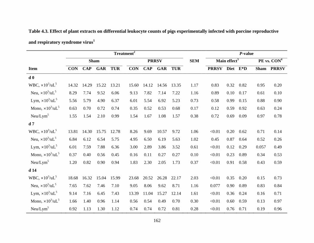
162
Table 4.3. Effect of plant extracts on differential leukocyte counts of pigs experimentally infected with porcine reproductive
and respiratory syndrome virus1
Treatment2 P-value
Sham PRRSV SEM Main effect3 PE vs. CON
4
Item CON CAP GAR TUR CON CAP GAR TUR PRRSV Diet E*D Sham PRRSV
d 0
WBC, ×103/uL
5 14.32 14.29 15.22 13.21 15.60 14.12 14.56 13.35 1.17 0.83 0.32 0.82 0.95 0.20
Neu, ×103/uL
5 8.29 7.74 9.52 6.06 9.13 7.82 7.14 7.22 1.16 0.89 0.10 0.17 0.61 0.10
Lym, ×103/uL
5 5.56 5.79 4.90 6.37 6.01 5.54 6.92 5.23 0.73 0.58 0.99 0.15 0.88 0.90
Mono, ×103/uL
5 0.63 0.70 0.72 0.74 0.35 0.52 0.53 0.68 0.17 0.12 0.59 0.92 0.63 0.24
Neu/Lym5 1.55 1.54 2.10 0.99 1.54 1.67 1.08 1.57 0.38 0.72 0.69 0.09 0.97 0.78
d 7
WBC, ×103/uL
5 13.81 14.30 15.75 12.78 8.26 9.69 10.57 9.72 1.06 <0.01 0.20 0.62 0.71 0.14
Neu, ×103/uL
5 6.84 6.12 6.54 5.75 4.95 6.50 6.19 5.63 1.02 0.45 0.87 0.64 0.52 0.26
Lym, ×103/uL
5 6.01 7.59 7.88 6.36 3.00 2.89 3.86 3.52 0.61 <0.01 0.12 0.29 0.057 0.49
Mono, ×103/uL
5 0.37 0.40 0.56 0.45 0.16 0.11 0.27 0.27 0.10 <0.01 0.23 0.89 0.34 0.53
Neu/Lym5 1.20 0.82 0.90 0.94 1.83 2.30 2.05 1.73 0.37 <0.01 0.91 0.58 0.43 0.59
d 14
WBC, ×103/uL
5 18.68 16.32 15.04 15.99 23.68 20.52 26.28 22.17 2.03 <0.01 0.35 0.20 0.15 0.73
Neu, ×103/uL
5 7.65 7.62 7.46 7.10 9.05 8.06 9.62 8.71 1.16 0.077 0.90 0.89 0.83 0.84
Lym, ×103/uL
5 9.14 7.16 6.45 7.43 13.39 11.04 15.27 12.14 1.61 <0.01 0.36 0.24 0.16 0.71
Mono, ×103/uL
5 1.66 1.40 0.96 1.14 0.56 0.54 0.49 0.70 0.30 <0.01 0.60 0.59 0.13 0.97
Neu/Lym5 0.92 1.13 1.30 1.12 0.74 0.74 0.72 0.81 0.28 <0.01 0.76 0.71 0.19 0.96

163
Table 4.3. (cont.)
1n = 60 (7 or 8 pigs/treatment).
2Sham = unchallenged; PRRSV = PRRSV challenged; CON = control diet; CAP = control diet plus 10 g capsicum oleoresin /
ton; GAR = control diet plus 10 g garlic / ton; TUR = control diet plus 10 g turmeric oleoresin / ton.
3PRRSV = PRRSV challenge effect; Diet = diet effect; E*D = interaction between PRRSV and diet effects.
4Contrast between CON and the 3 plant extract treatments within the sham or PRRSV challenge group.
5WBC = white blood cell; Neu = neutrophil; Lym = lymphocyte; Mono = monocyte; Neu/Lym = the ratio of neutrophils to
lymphocytes.

164
Table 4.4. Effect of plant extracts on red blood cell measures of pigs experimentally infected with porcine reproductive and
respiratory syndrome virus1
Treatment2 P-value
Sham PRRSV SEM Main effect3 PE vs. CON
4
Item CON CAP GAR TUR CON CAP GAR TUR PRRSV Diet E*D Sham PRRSV
d 0
RBC, ×106/uL
5 6.34 6.29 6.49 6.86 6.68 6.86 6.52 6.59 0.27 0.37 0.81 0.37 0.51 0.94
HGB, g/dL5 12.18 11.33 11.94 12.31 12.25 12.05 11.58 12.11 0.43 0.85 0.39 0.53 0.51 0.49
HCT, %5 36.45 34.44 36.36 37.34 37.65 36.44 36.60 37.00 1.33 0.39 0.47 0.77 0.79 0.53
MCV, fL5, 6
57.32 54.59 55.73 56.45 56.45 56.29 56.05 56.55 1.81 0.99 0.33 0.75 0.24 0.55
MCH, pg5 19.08 17.98 18.24 18.12 18.40 17.64 17.75 18.50 0.69 0.49 0.41 0.79 0.17 0.53
MCHC, g/dL5 33.34 32.97 32.86 33.01 32.55 33.01 31.74 32.73 0.42 0.045 0.24 0.40 0.38 0.89
RDW, %5 28.68 30.12 28.54 29.98 26.38 29.14 27.05 29.34 1.69 0.24 0.39 0.96 0.65 0.28
d 7
RBC, ×106/uL
5 5.89 6.48 6.32 6.39 6.00 5.82 5.97 5.83 0.20 <0.01 0.63 0.16 <0.05 0.53
HGB, g/dL5 11.16 11.88 11.23 11.18 10.43 10.25 10.34 10.55 0.40 <0.01 0.80 0.36 0.46 0.88
HCT, %5 33.37 35.40 33.78 33.46 32.68 31.71 32.04 32.75 1.25 <0.05 0.93 0.44 0.49 0.66
MCV, fL5, 6
56.63 54.60 53.60 52.71 54.68 54.44 53.55 56.14 1.98 0.74 0.50 0.22 0.063 0.98
MCH, pg5 18.98 18.32 17.80 17.63 17.45 17.63 17.30 18.13 0.66 0.060 0.46 0.093 <0.05 0.60
MCHC, g/dL5 33.49 33.57 33.23 33.45 32.00 32.36 32.36 32.28 0.30 <0.01 0.87 0.76 0.83 0.28
RDW, %5 27.79 27.47 27.97 29.54 25.30 27.09 26.66 26.20 1.91 <0.05 0.76 0.67 0.73 0.34
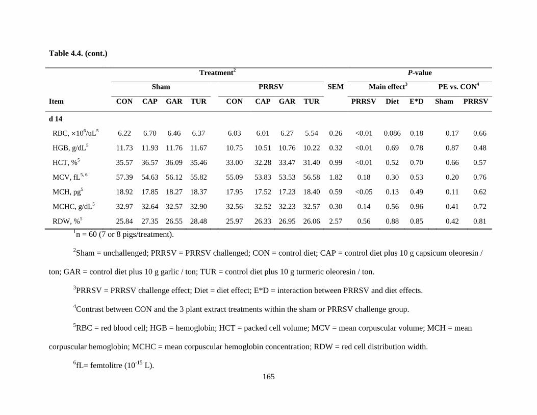
165
Table 4.4. (cont.)
Treatment2 P-value
Sham PRRSV SEM Main effect3 PE vs. CON
4
Item CON CAP GAR TUR CON CAP GAR TUR PRRSV Diet E*D Sham PRRSV
d 14
RBC, ×106/uL
5 6.22 6.70 6.46 6.37 6.03 6.01 6.27 5.54 0.26 <0.01 0.086 0.18 0.17 0.66
HGB, g/dL5 11.73 11.93 11.76 11.67 10.75 10.51 10.76 10.22 0.32 <0.01 0.69 0.78 0.87 0.48
HCT, %5 35.57 36.57 36.09 35.46 33.00 32.28 33.47 31.40 0.99 <0.01 0.52 0.70 0.66 0.57
MCV, fL5, 6
57.39 54.63 56.12 55.82 55.09 53.83 53.53 56.58 1.82 0.18 0.30 0.53 0.20 0.76
MCH, pg5 18.92 17.85 18.27 18.37 17.95 17.52 17.23 18.40 0.59 <0.05 0.13 0.49 0.11 0.62
MCHC, g/dL5 32.97 32.64 32.57 32.90 32.56 32.52 32.23 32.57 0.30 0.14 0.56 0.96 0.41 0.72
RDW, %5 25.84 27.35 26.55 28.48 25.97 26.33 26.95 26.06 2.57 0.56 0.88 0.85 0.42 0.81
1n = 60 (7 or 8 pigs/treatment).
2Sham = unchallenged; PRRSV = PRRSV challenged; CON = control diet; CAP = control diet plus 10 g capsicum oleoresin /
ton; GAR = control diet plus 10 g garlic / ton; TUR = control diet plus 10 g turmeric oleoresin / ton.
3PRRSV = PRRSV challenge effect; Diet = diet effect; E*D = interaction between PRRSV and diet effects.
4Contrast between CON and the 3 plant extract treatments within the sham or PRRSV challenge group.
5RBC = red blood cell; HGB = hemoglobin; HCT = packed cell volume; MCV = mean corpuscular volume; MCH = mean
corpuscular hemoglobin; MCHC = mean corpuscular hemoglobin concentration; RDW = red cell distribution width.
6fL= femtolitre (10
-15 L).

166
Table 4.5. Effects of plant extracts on cytokine and acute phase protein concentrations in the serum of pigs infected with
porcine reproductive and respiratory syndrome virus1
Treatment2 P-value
Sham PRRSV Main effect3 PE vs. CON
4
Item CON CAP GAR TUR CON CAP GAR TUR PRRSV Diet E*D Sham PRRSV
d 0
TNF-α, pg/mL5 67.8 52.7 54.6 55.9 60.5 55.4 60.2 52.4 6.75 0.88 0.33 0.72 0.086 0.51
IL-10, pg/mL 8.9 7.6 6.4 5.5 7.7 6.4 7.2 8.0 1.76 0.86 0.79 0.69 0.24 0.81
CRP, μg/mL5 22.9 16.8 18.4 24.8 24.5 26.0 27.4 22.3 5.34 0.26 0.97 0.63 0.64 0.91
HP, μg/mL5 257 194 280 154 221 376 367 167 62.8 0.11 <0.05 0.22 0.47 0.21
d 7
TNF-α, pg/mL5 56.5 47.7 44.3 54.2 179.5 146.2 159.6 139.4 10.95 <0.01 0.12 0.23 0.48 <0.05
IL-10, pg/mL 11.9 12.1 15.0 9.2 18.9
a 36.4
b 22.9
a 31.4
ab 4.62 <0.01 0.34 0.16 0.97 <0.05
CRP, μg/mL5 23.2 19.9 29.8 26.0 52.1 42.5 45.0 42.0 5.16 <0.01 0.49 0.45 0.69 0.14
HP, μg/mL5 302 204 436 388 1312 1572 1563 1322 173.0 <0.01 0.69 0.57 0.82 0.36
d 14
TNF-α, pg/mL5 42.2 41.3 33.4 46.8 201.0 172.0 207.8 177.8 15.27 <0.01 0.69 0.34 0.91 0.40
IL-10, pg/mL 4.5 4.1 4.0 6.5 15.2 15.4 16.5 17.2 2.15 <0.01 0.71 0.97 0.87 0.65
CRP, μg/mL5 16.8 18.4 27.6 23.4 41.0 27.9 32.8 34.4 3.63 <0.01 0.15 <0.05 0.087 <0.05
HP, μg/mL5 150 234 492 249 890 1503 1485 1035 152.9 <0.01 0.038 0.40 0.37 <0.05

167
Table 4.5. (cont.)
a, b within a row, means without a common superscript differ (P < 0.05) within the sham or PRRSV challenge group.
1n = 60 (7 or 8 pigs/treatment).
2Sham = unchallenged; PRRSV = PRRSV challenged; CON = control diet; CAP = control diet plus 10 g capsicum oleoresin /
ton; GAR = control diet plus 10 g garlic / ton; TUR = control diet plus 10 g turmeric oleoresin / ton.
3PRRSV = PRRSV challenge effect; Diet = diet effect; E*D = interaction between PRRSV and diet effects.
4Contrast between CON and the 3 plant extract treatments within the sham or PRRSV challenge group.
5TNF-α = tumor necrosis factor-alpha; CRP = C-reactive protein; Hp = haptoglobin.

168
Figure 4.1. Rectal temperature (RT) in pigs fed control (CON) or capsicum oleoresin (CAP),
garlic (GAR), or turmeric oleoresin (TUR) diets with or without porcine reproductive and
respiratory syndrome virus (PRRSV) infection. The RT of PRRSV-infected pigs at d 7 (P <
0.01), 9 (P < 0.01), and 11 (P < 0.05) postinfection (PI) was greater than that of uninfected pigs.
There was a PRRSV×diet interaction (P < 0.05) on RT at d 4 PI, as the PRRSV-infected pigs fed
the 3 PE diets had lower RT than those fed the CON, but no difference between PE and CON
was found in the sham pigs. Value were means ± pooled SEM, n = 7 or 8. Individual pig was an
experimental unit.
39.0
39.2
39.4
39.6
39.8
40.0
40.2
40.4
40.6
0 4 7 9 11 14
Rec
tal
tem
per
atu
re, °C
After PRRSV infection, d
CON
CAP
GAR
TUR
CON+PRRSV
CAP+PRRSV
GAR+PRRSV
TUR+PRRSV
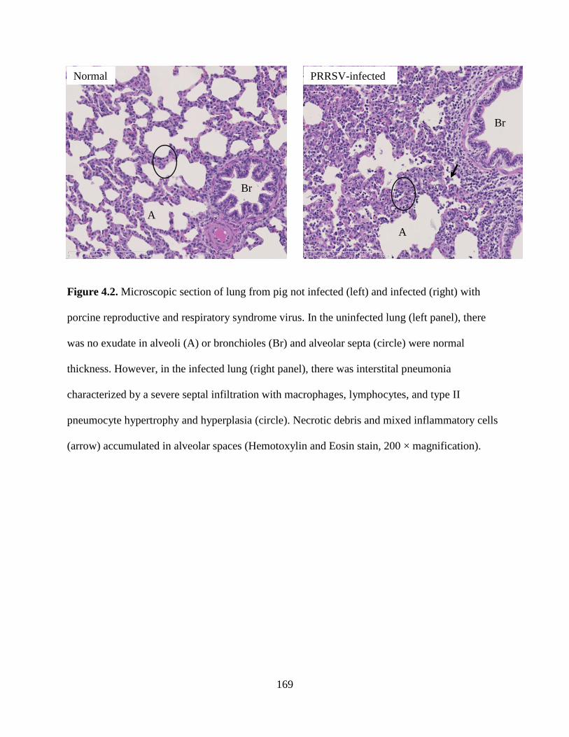
169
Figure 4.2. Microscopic section of lung from pig not infected (left) and infected (right) with
porcine reproductive and respiratory syndrome virus. In the uninfected lung (left panel), there
was no exudate in alveoli (A) or bronchioles (Br) and alveolar septa (circle) were normal
thickness. However, in the infected lung (right panel), there was interstital pneumonia
characterized by a severe septal infiltration with macrophages, lymphocytes, and type II
pneumocyte hypertrophy and hyperplasia (circle). Necrotic debris and mixed inflammatory cells
(arrow) accumulated in alveolar spaces (Hemotoxylin and Eosin stain, 200 × magnification).
Br
Br
A
A
Normal PRRSV-infected
A

170
Figure 4.3. Serum viral load in control- or plant extracts (PE)-fed pigs infected with
porcine reproductive and respiratory syndrome virus (PRRSV). ICON: pigs fed control diet in
the PRRSV-infected group; ICAP: pigs fed capsicum oleoresin diet in the PRRSV-infected
group; IGAR: pigs fed garlic diet in the PRRSV-infected group; ITUR: pigs fed turmeric
oleoresin diet in the PRRSV-infected group. Viral load is presented as cycle threshold (Ct)
values, where Ct value is inverse to the amount of virus. Capsicum oleoresin and turmeric
oleoresin increased Ct value at d 7 PI, and turmeric oleoresin increased Ct value at d 14 PI,
which indicates these 2 PE reduce the viral load in the serum of PRRSV-infected pigs. Values
were means ± pooled SEM, n = 7 or 8. The individual pig was the experimental unit. a, b
means
without a common superscript differ (P < 0.05) within the PRRSV challenge group.
a
a
b
ab
a
ab
b
b
0
5
10
15
20
25
30
7 14
Vir
al
loa
d i
n s
eru
m, C
t
After PRRSV infection, d
ICON
ICAP
IGAR
ITUR
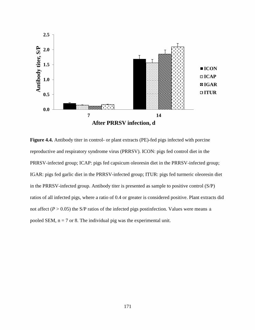
171
Figure 4.4. Antibody titer in control- or plant extracts (PE)-fed pigs infected with porcine
reproductive and respiratory syndrome virus (PRRSV). ICON: pigs fed control diet in the
PRRSV-infected group; ICAP: pigs fed capsicum oleoresin diet in the PRRSV-infected group;
IGAR: pigs fed garlic diet in the PRRSV-infected group; ITUR: pigs fed turmeric oleoresin diet
in the PRRSV-infected group. Antibody titer is presented as sample to positive control (S/P)
ratios of all infected pigs, where a ratio of 0.4 or greater is considered positive. Plant extracts did
not affect (P > 0.05) the S/P ratios of the infected pigs postinfection. Values were means ±
pooled SEM, n = 7 or 8. The individual pig was the experimental unit.
0.0
0.5
1.0
1.5
2.0
2.5
7 14
An
tib
od
y t
iter
, S
/P
After PRRSV infection, d
ICON
ICAP
IGAR
ITUR

172
CHAPTER 5
GENERAL SUMMARY, DISCUSSION, AND CONCLUSION
Many of the feed additives now available for use as “alternatives to antibiotics” in the pig
industry either alter microbial populations in the gastrointestinal tract or influence the immune
system of the pig. Plant extracts (PE) are extracted from parts of plant materials are among the
interesting additives, and have been largely employed for both human nutrition and the livestock
industry. Plant extracts are of potential interest due to their antiviral, antimicrobial, antioxidant,
anti-inflammatory, and other biological effects. This may lead to the ability to these PE, instead
of antibiotics, in diets to improve performance and health of animals. However, because of the
large variation in composition, the biological or practical effects of different batches of the same
PE may differ. Therefore, it is necessary to select pure principles for evaluating their possible
role as alternatives to antibiotics in livestock production. The important aim of these studies was
to evaluate the effects of several different PE on immune function and disease resistance of pigs.
This research contained 3 studies: (1) anti-inflammatory properties of 7 different PE on porcine
alveolar macrophage (PAM) in vitro; (2) the effects of dietary supplementation of 3 different PE
on growth performance, intestinal morphology, and immune responses of pigs infected with a
bacterial model; (3) the effects of dietary supplementation of the same 3 PE on growth
performance and immune responses of pigs infected with a viral model.
The first study tested the in vitro anti-inflammatory effects of 7 PE (anethol, capsicum
oleoresin, carvacrol, cinnamaldehyde, eugenol, garlic, and turmeric oleoresin) on PAM collected
from weaned pigs by bronchoalveolar lavage. Lipopolysaccharide (LPS) sharply stimulated the

173
secretion of 2 pro-inflammatory cytokines: TNF-α and IL-1β. The inclusion of each of the 7 PE
suppressed the secretion of TNF-α from LPS-treated PAM in a dose dependent manner. But, the
IL-1β response differed from that of TNF-α in the presence of LPS. The treatments with
carvacrol, cinnamaldehyde, eugenol, and garlic significantly suppressed the secretion of both
TNF-α and IL-1β from LPS-induced PAM, but anethol and capsicum oleoresin stimulated or did
not affect the secretion of IL-1β from LPS-induced PAM. These results indicate that all of PE
may have potent anti-inflammatory effects to varying degrees. Several PE increased the secretion
of TNF-α or IL-1β from PAM in the absence of LPS, which indicated that the inclusion of these
PE in the normal condition may be beneficial to the host, since the inflammatory mediators can
protect the host against infection. Plant extracts did not increase production of the 2 anti-
inflammatory cytokines, IL-10 and TGF-β, which suggests other mechanisms may be involved
in the reduction of pro-inflammatory cytokines from PAM by PE. In addition, the cell viability
test found different influences of 7 PE on PAM. The high level (200 μg/mL) of carvacrol and
garlic were cytotoxic to PAM, and very low levels of cinnamaldehyde (20 μg/mL) and turmeric
oleoresin (10 μg/mL) significantly inhibited cell viability of PAM. The cell viability results
indicate that the inhibitory effects of PE on the production of pro-inflammatory cytokines
resulted from mechanisms other than direct killing of cells. Otherwise, these results also suggest
different dose effects of these 7 PE and the importance of dose selection when we use PE in the
future.
Based on the results from the first study, capsicum oleoresin (CAP), garlic (GAR), and
turmeric oleoresin (TUR) were selected to perform the second and third experiments with
bacterial or viral challenge, because they showed different effects on the production of pro-
inflammatory cytokine IL-1β and cell viability of PAM. In the presence of LPS stimulation, CAP

174
increased the secretion of IL-1β and cell viability of PAM, GAR inhibited IL-1β production and
cell viability of PAM, TUR inhibited the cell viability of PAM but did not affect IL-1β
production.
In the second study, 10 mg CAP, GAR, or TUR was added per kg diet to investigate the
effects of supplementation of these PE on the immune responses, growth performance, and
intestinal morphology of weaned pigs infected with a pathogenic F-18 Eshcherichia coli (E. coli).
The supplementation of PE improved the gut health as indicated by increased villi height and the
ratio of villi height to crypt depth. In addition, feeding PE to E. coli challenged pigs reduced
diarrhea score, frequency of diarrhea, total white blood cell number, serum pro-inflammatory
cytokine TNF-α, and serum concentration of the acute phase protein haptoglobin, all of which
indicate that PE may alleviate the inflammation caused by E. coli infection. However, feeding
PE did not affect the proportions of β-hemolytic coliforms in feces of E. coli challenged pigs,
which indicates the reduction of diarrhea may be induced by mechanisms rather than an
antimicrobial effect of PE. The reduction of serum TNF-α, haptoglobin, and white blood cells
suggests feeding dietary PE may attenuate the over-expression of the systemic immunity and
inflammatory mediators of pigs infected with E. coli. On the other hand, these results also
indicate that the supplementation of low level of PE, 10 ppm, still has beneficial effects on pig
health probably by modulating the immune system of pigs. The 3 PE tested here showed
different effects on several measurements. Future studies are needed to investigate the
mechanisms through which different PE exert these effects.
The third study was conducted with a similar experimental design as the second study,
with same dietary treatments but a different challenge model, porcine reproductive and
respiratory syndrome virus (PRRSV). Similar to the post-weaning E. coli infection, PRRS is an

175
economically important disease in the swine industry. In this study, as expected, PRRSV
infection induced increased rectal temperature, serum viral load, serum specific antibody titer,
serum TNF-α, IL-10, C- oreactive protein, and haptoglobin, and reduced growth performance of
pigs. In addition, PRRSV infection reduced total white blood cells on d 7 post-inoculation (PI),
but increased total white blood cells on d 14 PI, which indicates the delayed immune responses
of pigs typical of PRRSV infection. In the PRRSV challenge group, the supplementation of PE
reduced viral load and serum TNF-α and C-reactive protein, which indicate that PE may alleviate
the negative effects of PRRSV, such as over-stimulation of immune responses and poor growth
rate. In addition, adding PE in pig diets increased growth efficiency, serum specific antibody titer,
and serum IL-10 and haptoglobin, which also suggest that PE may have the ability to enhance
the immune response of host, and thereafter may suppress ongoing inflammation and prevent the
secondary infection. The 3 PE tested here also showed some different effects on the
measurements, and TUR may be the most powerful candidate to strengthen the efficiency and
immune responses of pigs infected with PRRSV.
In overall conclusion, both in vitro and in vivo studies show evidence of potential anti-
inflammatory effects of PE, as PE reduced the diarrhea of pigs infected with E. coli and reduced
the serum viral load of pigs infected with PRRSV. Dietary PE can be supplemented as
alternatives to antibiotics, not to treat sick animals but to provide some prophylaxis to pigs. The
inclusion of PE in pig diets shows benefits in the face of both a bacterial challenge model and a
viral challenge model, which indicates the supplementation of PE may potentially improve pig
health and/or growth performance by modulating the immune system of pigs. Future research,
including measurement of gene expression in specific tissue or immune cells, should be
conducted to detect the mechanism through which different PE show different effects. Combined

176
infections of bacterial and viral pathogens should be investigated to detect the effect of PE on the
immune responses of pigs to combined diseases as pigs often encounter a complex of pathogens.
In addition, combinations of different PE may be also interesting in the future research since the
several PE tested in this project showed different influences in vitro and in vivo.

177
AUTHOR’S BIOGRAPHY
Yanhong Liu was born in Jiaozuo City, Henan Province, China. She earned her
Bachelor’s degree in Biological Sciences (2004) at China Agricultural University, Beijing, China.
After the completion of her Bachelor’s degree, she enrolled in the Ministry of Agriculture Feed
Industry Centre, also in China Agricultural University and received her Master’s degree under
the guidance of Dr. Defa Li, Spring 2007. During her Master program, she conducted several
different experiemnts, most of which were related to nutrition and immunology. The title of the
Master’s thesis was “Effects of particle size and physical form of diets on mast cell numbers,
histamine, and stem cell factor concentration in the small intestine of broiler chickens”.
After she accomplished her Master’s degree, she joined the swine nutrition group in
Department of Animal Sciences at the University of Illinois, Urbana-Champaign, IL, Spring
2008, in pursuit of Ph.D. degree under the guidance of Dr. James E. Pettigrew, mainly focusing
on the dietary effects on pig health. Her Ph.D. program is related to the plant extracts on immune
function and disease resistance in pigs with the goal of looking for some additives for nursery
pigs to improve their performance and disease resistance. During the Ph.D. program, she led
three research projects as co-investigator, made 5 research manuscripts, 9 abstracts with oral or
poster presentations, 1 research article, and 1 conference proceedings as a first author as well as
6 research manuscripts, 5 abstracts, and 2 conference proceedings as a co-author, received 2
graduate research paper award (3rd
in Ph.D. oral presentation competition and 2nd
in poster
competition).
Yanhong got married to Peng in June 2011, and will have their baby in Febrary 2012.
Upon the completion of her Ph.D. program, Yanhong will work as a postdoctoral research
associate in Dr. Pettigrew lab.