effects of electric field on histopathological study, electrical properties and enzymes function of...
TRANSCRIPT
Research Inventy: International Journal Of Engineering And Science
Vol.4, Issue 12 (December 2014), PP 25-37
Issn (e): 2278-4721, Issn (p):2319-6483, www.researchinventy.com
25
Effects of electric field on histopathological study, electrical
properties and enzymes function of liver of albino rats
*1Sahar E.Abo-Neima,
2Hussein A. Motaweh ,
3Marzoga F.Ragab
1Lecturer of Medical Biophysics, Department of physics, Faculty of Science, Damanhour University, Egypt.
2Professor Doctor, Department of physics, Faculty of Science, Damanhour University, Egypt.
3 Demonstrator, Department of physics, Faculty of Science, Omar El-Moktar University, Elgouba- Libya
* Corresponding Author: Dr Sahar Abo-Neima (Email: [email protected])
ABSTRACT - The present work was undertaken in order to investigate the effects of electric field (EF) of
strength 50Hz-3KV/m on the histopathology, dielectric properties and liver function tests in albino rats. Fifty
male albino rats were equally divided into three groups namely A, B, and C. Animals of group A used as control
group which didn't receive any treatment . Animals of group B was divided into two subgroups namely B1 and B2
which were discretely exposed to 50HZ, 3KV/m electric field for a period of 15 day (8 hours/day, 5day/week).
Group B2 animals were left to survive and housed at normal environmental conditions similar to control group A
for a period of 15 day post exposed. Animals of group C are divided into two subgroups namely C1 and C2 were
discretely exposed to the electric field for a period of 30 day (8 hours/day, 5day/week). Group C2 animals were
left to survive and housed at normal environmental conditions similar to control group A for a period of 15 day
post exposed. At the end of this period, blood and tissues samples were collected from all groups for
experimental investigations. The dielectric constant (έ), electrical conductivity (σ) was measured in frequency
range 42Hz-5MHz to investigate any changes in liver structure through studding histopathological examination.
Also, the liver function was studied through analysis of glutamic oxaloacetic transaminase (GOT), glutamic
pyruvie transaminase (GPT) and total protein (TP) after exposure to electric field this biochemical parameters
have been evaluated in the blood serum of rats. The obtained results show high significant changes in the value
of έ and σ of liver tissues for all groups exposed to EF as compared with control group. The levels of GOT and
GPT were increased up to four times their values during the period of exposure to EF. These variations were
recovered during two week after stopping exposure but they did not return to its original control values before
exposure. On microscopic level; liver histological observations in liver cells which revealed some alterations
including hepatic tissue with two portal tracts showing mild florous expansion and a dilated central vein, also
ghosts of hepatocytes denoting necrotic changes also shows hepatic tissue with dilated central veins engorged
with blood and splitting out to adjacent hepatocytes.
KEYWORDS - electric field, histopathology, liver enzymes, dielectric constant, conductivity.
I. INTRODUCTION
Electromagnetic fields (EMFs) exposure exists at home, workplaces as a result of all types of electrical
equipment and building wiring as well as a result of nearby power lines. It represents one of the invisible
environmental pollutant factors that affect animals and human health [1]. During the past decade considerable
evidence has been accumulated with regard to the biological effects, both in vivo and in vitro, of extremely low
frequency electric and magnetic fields, such as those originating from residentially proximate power lines,
household electrical wiring and diagnostic apparatus and therapy devices. Electric and magnetic fields
associated with production transmission, and use of electricity is ubiquitous in industrialized societies. Electric
fields exist whenever there is electric potential in a line. However, because reductions in field strength occur as
electric fields pass through walls and other objects, the potential for human exposure to electric fields in a home
environment is modest [2].
Several studies on animal cells have also shown that EMFs influence a large variety of cellular
functions [3]. The mechanisms (or some) of interaction with living cells involve, as reported, changes in the
intracellular levels of Ca2+
[4]. However, many of the proposed hypotheses assume that the cell membrane is
most likely the target for the primary impact of the field and that this interaction might affect the signal
transduction mechanisms at different levels [5].
It is likely that the disturbances lead to adaptive changes, which in turn result in altered lactate
dehydrogenase activity and accelerated transamination processes. EMFs penetrate human body and act on all

Effects of electric field on histopathological…
26
organs, altering the cell membrane potential and the distribution of ions and dipoles. These alterations may
influence biochemical processes in the cell, thus changing both biochemical parameters and enzyme activities of
serum [6].
Automation medical and research instruments which generate (EMFs) are widely diffused in recent
years, and the people are frequently exposed to it. Despite that the study of the effect of (EMFs) on living
organisms is a complex problem, but it is of more interest to give insight into the expected hazards and the
proper ways of its use and protection. The EMF penetrates the human body and act ions on all organs, altering
the cell membrane potential and the distribution of ions and dipoles [7].
The effects of the EMFs on living organisms are based on the molecular interaction between many
tissues and cellular systems, as well as on the level of cell organelles. The explanation of these mechanisms may
be the subject for further investigations in this area, and may constitute a basis for the effective and safe
application of EMFs in therapy [8, 9].
The mechanism of the interaction of EMFs with biological tissue associated with the changes in the
permeability of cell membranes as a result of the change in the concentration of ions in the extra- and
intracellular environment. The concentration changes are induced by the EMFs, which results from Lorentz
force causing the motion of charged particles in the magnetic field [10]. Under the effect of electromagnetic
field, the researchers observed the changes concerning both enzymatic complexes [11, 12, 13] and the
coagulation complexes [13].
EMFs were observed to influence enzyme action, signal transduction, protein synthesis and gene
expression. These activities play an important role in regulating cell growth and processes important to
promotion [14, 7]. Furthermore, alterations may influence biochemical processes in the cell, thus changing both
biochemical parameters and enzyme activities of the blood serum.
Recent electron microscopy studies on hepatocytes and liver tissue have shown that constant magnetic
fields exhibited structural changes in hepatocytes, primarily in the mitochondria and also split cell membrane
[15]. Moreover, constant and low frequency magnetic fields exert a preponderant controlling influence on the
thermoregulation, metabolism and hematology in rats [16].
The exposition of rats 1 hour/day for 10 consecutive days to a static magnetic field of 128 mT induced
an increase in hematocrit, hemoglobin, plasma fuel metabolites and tissue enzymes releases within the blood
[17].Several authors suggested that chemical and physical processes at the atomic level are the bases of
reactions between biomolecules in an EMF, since the field can magnetically affect the chemical bonds between
adjacent atoms with consequent production of free radicals [7,18].The magnetic field effects seem to be an ideal
means for investigating biological function. Significant area would be better understood if knowledge on
magnetic field effects on biological membrane is measured by physical parameters such as dielectric parameter
(έ) and conductivity (σ).The physical mechanism for the effects of weak EMFs ranging from microwave to radio
waves had been discussed [7].by the dielectric nature of all biological molecules especially those constituting
the biological membrane.
The aim of the present work is to study the effect of electric field on histopathology, electrical
properties and enzymes function of liver of albino rats the study pays attention to patients under investigation to
these tests to be protected against exposure to any source of electric field.
II. MATERIALS AND METHOD 2.1. EXPERIMENTAL ANIMALS
The experimental animals kept in the same conditions for 2 weeks for adaptation. In the present work
50 male albino rats were used, each of average weight 170±10gm .The animals were housed in the same
environmental conditions in plastic cages, and feed with constant balanced diet and tap water. Which were
equally divided into three groups namely A, B and C. Animals of group A are used as a control group and didn't
receive any treatment and housed at normal environmental conditions (the temperature inside the lab varied
between 22 ْ and 25 ْ C, lighting condition are natural light from large windows during the day and complete
darkness during the night). Animals of group B was divided into two subgroups namely B1 and B2 which were
discretely exposed to 50HZ, 3KV/m electric field for a period of 15 day (8 hours/day, 5day/week). Group B2
animals were left to survive and housed at normal environmental conditions similar to control group A1 for a
period of 15 day post exposed. Animals of group C are divided into two subgroups namely C1 and C2 were
discretely exposed to the electric field for a period of 30 day (8 hours/day, 5day/week). Group C2 animals were

Effects of electric field on histopathological…
27
left to survive and housed at normal environmental conditions similar to control group A1 for a period of 15 day
post exposed. At the end of this period, blood and tissues samples were collected from all groups for
experimental investigations.
2.2. ELECTRIC FIELD EXPOSURE FACILITY The exposure cage consisted of Perspex chamber, with an exposure volume of dimension 100x30x35
cm3 located between two parallel cupper plates, which extended vertically along two parallel sides of the
exposure cage as shown in figure (3-1). In order to prevent any animal shock from direct contacts with the
electrodes, the cupper plates were covered by a sheet of Polymethyl methacrylate. It is worthy to mention that,
the Perspex material has a negligible effect on the field homogeneity [19]. The two electrodes were connected
to a step up transformer with an output voltage of 3Kv when connected to the main supply. For more
precautions an electric timer was used to adjust the exposure times specially when mains fall. The electric field
inside the chamber was measured through the use of field meter and was found to be homogeneous and reads
3Kv/m.
2.3. BIOCHEMICAL ANALYSIS Liver tissues of the experimental animals were immediately removed after exposure to EF. Weighed
tissue samples were homogenized by a glass homogenizer after dilution by distilled water then the supernatant
fluid was separated by centrifugation at 3000 rpm for 15 minutes, and stored at –20ºC for biochemical analysis.
Liver glutamic oxaloacetic transaminases (GOT) and glutamic pyruvic transaminases (GPT) were determined
using the method adapted by Fischbach and Zawta [20]. Alkaline phosphatase was determined using the method
adapted by Bessey et al. [21], and total protein content was determined using the method adapted by Henery
[22]. Also blood serum was collected after blood centrifugation and stored at -20 ºC for biochemical analysis
[7].
2.4. THE DIELECTRIC AND CONDUCTIVITY MEASUREMENTS
The dielectric measurements were carried out for the liver samples in the frequency range
42Hz-5MHz using a loss Factor Meter type HIOKI 3532 LCR Hi TESTER; version 1.02, Japan as
shown in Fig.2, and cell types (PW 950/60) manufactured by Philips show Fig.3. Animals were
sacrificed then the liver was immediately excised and placed between a pair of 1cm diameter black
platinum circular electrodes for dielectric measurements, the sample between the electrodes was
maintained at constant pressure, and the distance between the electrodes was measured through the
use of a micrometer, while the liver sample was filling the whole volume between the electrodes.
During measurements, the sample between the electrodes was kept at a constant temperature of
24±0.10C the capacitance (C) of the tissue was measured at each frequency and the resistance (R) was

Effects of electric field on histopathological…
28
recorded each run was repeated three times .The relative permittivity έ of the sample was calculated
for each frequency using the relation:-
(1)
Where A is the area of electrode, d the distance between the two electrodes, εo is the permittivity of
free space and έ is the dielectric constant. The dielectric loss ε" is calculated from the relation:-
(2)
Where f is the applied frequency in Hertz, R and C are the resistance and capacitance of the sample at
resonance and δ is the loss angle. The electric conductivity σ is given by:
(3)
Where R is the resistance of the sample
Fig.2. Hioki 3532-50 LCR Hitester Bridge.
Fig.3. Cell used for measurement dielectric of biological liver tissues

Effects of electric field on histopathological…
29
2.5. HISTOLOGY ANALYSIS Specimens of liver tissues were taken from all groups and prepared for the histological and
histopathological sections following Banchroft and Stevens work, 2006 [23]. All of them were fixed in 10%
buffered formalin (10ml formalin in 30ml normal saline or sterilized distilled water).The tissues were
subsequently dehydrated in upgraded concentrations of alcohol (70% alcohol) cleansed in xylene. Several
sections of 3-6 micrometer thickness were cut, dried with blotting paper [24], using microtome then embedded
in paraffin and sections stained with Hematoxylin and Eosin (H&E) [25, 26]. The slides were then evaluated for
pathological changes under light microscope (100 x). Photographs were taken using Kodak digital 10.3 mega
pixels camera [27].
2.6. STATISTICAL EVALUATION All results are presented as mean ± standard error of the mean. Statistical significances of the
differences for all groups of samples were assessed using Student's t test. Differences were considered to be
statistically significant at p <0.05, high significant at p < 0.01 and very high significant at p < 0.001, not
significant at p > 0.01.
III. RESULTS AND DISCUSSION
EMF exposure exists at home, workplaces as a result of all types of electrical equipment and building
wiring as well as a result of nearby power lines. It represents one of the invisible environmental pollutant factors
that affect animals and human health [1]. How induced EMF can affect organ function and induced cellular
changes? Is a question, which has no definite answer? However, various mechanisms have been suggested.
EMF might amplify electric currents in tissues and cells or affect these currents through resonance with local
field focus [28].
In the present work, the low frequency EMF was chosen because it has been encountered in many work
places, medical practice and new technologies in use nowadays [29]. The rat liver function was studied through
analysis of glutamic oxaloacetic transaminase (GOT), glutamic pyruvie transaminase (GPT) and total protein
(TP) after exposure to electric field. The same biochemical parameters have been evaluated in the blood serum
of rats. The levels of GOT and GPT were increased to up to four timed their values during the period of
exposure to EF. Also, a recovery was carried out after 15 day from stopping the exposure to electric field. The
changes in liver enzymes and total protein from blood serum analysis are shown in Table.1.In a previous study
on the effect of static EMF on the liver, kidney and spleen tissues of rat showed that the liver tissue is more
affected by EMF than the other tissues.
Table .1
Average values of total protein TP, the glutamic oxaloacetic GOT and glutamic pyruvic GPT transaminases of
blood serum for all groups A ,B1,B2,C1 and C2 respectively. Values are the average of 10 experiments and
P<0.001 as compared to values for the control group.
TP
g/dL
GPT
U/L
GOT
U/L
Groups
6.4
66
43
A
7.3
84
259
B1
7.8
207
128
B2
8.7
184
86
C1
9.2
177
79
C2

Effects of electric field on histopathological…
30
The obtained data showed that EF produced alteration in biochemical parameters of the liver
transaminases GOT and GPT which have been widely utilized in mammalian toxicology as biomarkers of
specific organ dysfunction. In general the increase in transaminases activity is usually associated with
hepatocyte damage. These results are in agreement with the results recorded by Sihem [17]. The authors studied
the effects of sub-acute exposure to magnetic field on blood hematological and biochemical parameters in
female rats and found that the serum GPT activity remained unchanged in treated rats, while GOT activity was
increased, our present results agree with observations obtained by many authors [17, 30, 31].
The level of serum total proteins (TP) is significantly increased after exposure to EF. These results
were agreement with other findings reported by other works that in vivo exposure to a pulsed magnetic field at
1.5mT caused significant changes on plasma proteins in rats, difference in levels of plasma proteins were
observed between the control groups of the two studies. This observation supports the hypothesis that the state
of physiological equilibrium of a biological system is crucial to its response to a potentially effective EMF [2].
Valberg et al., [32], showed that the exposure to time varying magnetic field induces EF and this in turn may
cause large structural changes of the protein molecules imbedded in the cell membrane forming a new
membrane conformation. In this new conformation, the ions are able to pass through the membrane by binding
temporarily with the protein molecule, thus “hopping” through the membrane. Watanabe et al., 1997 [33]
showed that activities of GOT and GPT in the plasma, as indicator of hepatotoxicity, may alter the cell
membrane potential and distribution of ions and dipoles. Kula et al, 1999 [34] reported that the physicochemical
action of an EMF consists of electron, ion, dipolar, macrostructural and electrolytic polarization. Other factors
may also play a role, such as molecular excitation, biochemical activation, generation of radicals, weakening of
chemical bond and hydration change may alter relaxation protein fractions of serum.
The dielectric relaxation spectroscopy study showed a dielectric dispersion in the frequency region
from 42 KHz to 5MHz for both control and exposed groups to 50Hz–3KV/m electric field. In this frequency
range (β-dispersion) the relaxation mechanism is due to the counter ion molecules and proteins at the cellular
membrane. Fig.4. shows the variation of the dielectric constant έ with frequency of liver tissue for rats exposed
to EF.
Fig.4. revealed the variation of Permittivity with frequency within the range 42Hz, 5MHz of the liver tissue
suspension after exposure to 50Hz-3Kv/m electric field and recovery values after two weeks.

Effects of electric field on histopathological…
31
Fig.5. revealed the variation of dielectric loss with frequency within the range 42Hz –5MHz of the liver tissue
suspension after exposure to 50Hz-3Kv/m electric field and recovery values after two weeks.
Fig.6. revealed the variation of dielectric loss with frequency within the range 42Hz –5MHz of the liver tissue
suspension after exposure to 50Hz-3Kv/m electric field and recovery values after two weeks.
There was a pronounced decrease in conductivity of liver tissue suspension with frequency for all
groups as compared with control group as shown in Fig. 6. The decrease in conductivity due to field exposure
and the recovery groups not returned to the control value during the recovery period this is an indicator that
there is no improvement in the liver state. The relative high control value of hepatocytes membrane permittivity
έ and conductivity σ may be attributed to the high value of the membrane capacitance and conductance due to
normal activity of GOT and GPT and normal values of cell membrane potential and distribution of ions and
dipoles. So, the low values of the membrane permittivity, dielectric loss and conductivity after exposure to
electric field are due to the lipid peroxidation, which causes destruction of cell membrane [15, 36]. During

Effects of electric field on histopathological…
32
recovery, for group B2 the changes in σ, έ and ε" approximately return to its control value and the conductivity
was approximately returned to the control value except for group C2 it attained a higher value than the control
value.
Fig.7. indicates photomicrographs of liver sections for control rat A1. (a) Microscope Examination ME reveals
hexagonal hepatocytes separated by sinusoidal spaces.(b) ME reveals a central vein as well as a portal tract are
seen, cords of hepatocytes separated by sinusoidal spaces.(c) ME reveals a dilated central vein engorged with
blood is seen also liver tissue with dilated a portal tract to the left (H&E×100).
Effects of electric field on histopathological…
33
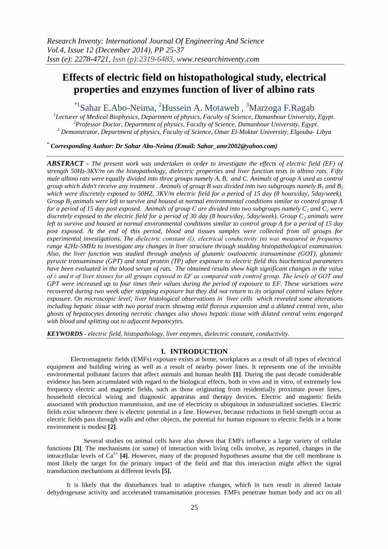
Fig.8. indicates photomicrographs of liver sections for group B1 (a) ME reveals a widely expanded portal tract
by fibrous tissue, with dilated congested portal vein and a dilated proliferating bile duct (H&E×40) (b) ME
reveals an area of bile duct proliferations as well as fibrous tissue bands (H&E×100) (c) ME reveals a portal
tract expanded by fibrous tissue and shows dilated congested portal vein and bile duct proliferations (H&E×100)
(d) ME reveals a rudely expanded portal tract with abundant proliferating bile ducts and a dilated congested
portal vein (H&E×100).
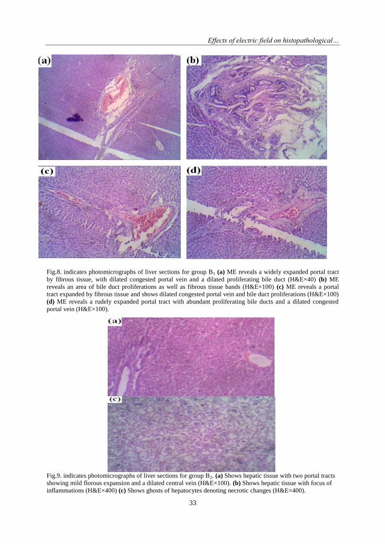
Fig.9. indicates photomicrographs of liver sections for group B2. (a) Shows hepatic tissue with two portal tracts
showing mild florous expansion and a dilated central vein (H&E×100). (b) Shows hepatic tissue with focus of
inflammations (H&E×400) (c) Shows ghosts of hepatocytes denoting necrotic changes (H&E×400).

Effects of electric field on histopathological…
34
Fig.10. indicates photomicrographs of liver sections for group C1. (a) Shows hepatic tissue with dilated central
vein and engorged sinusoids (H&E×400). (b) Shows hepatic tissue with dilated central veins engorged with
blood and splitting out to adjacent hepatocytes (H&E×400).
Fig.11. indicates photomicrographs of liver sections for group C2 (a) ME reveals a portal trace infiltrated by
moderated amount of lymphocytes with moderate spilling out to adjacent hepatocytes, moderate spotty necrosis
is noted (H&E×100) (b) ME reveals a portal tract with fibrous tissue expansion and is moderately infiltrated by
lymphocytes with mild to moderate spilling into adjacent hepatocytes, a dilated congested portal vein is seen
(H&E×100) (c) ME reveals a portal tract which is expanded by fibrous tissue and shows bile duct proliferations

Effects of electric field on histopathological…
35
and is moderately to heavily infiltrated by lymphocytes with moderato spilling into adjacent hepatocytes, a
dilated congested portal vein is seen (H&E×100) (d) ME reveals a portal tract heavily infiltrated with
lymphocytes with moderate spilling into adjacent hepatocytes , moderate to heavy spotty necrosis is seen
(H&E×100).
Liver histopathology for control animals A, the hepatic lobule appeared consisted of numerous lobules
bounded together with connective tissue. The portal areas appeared at the peripheries of hepatic lobules, each
containing a branch of the portal vein, a branch of the hepatic artery and branch of bile ductile embedded in
connective tissue. The hepatocytes had polygonal outlines with relatively large, rounded vesicular and central
nuclei. The blood sinusoids were seen alternate with the liver cell strands and were lined with Kupffer and
endothelial cells (Fig.7).
Liver histopathology for animals of group (B1) showing a portal-portal bridging septa containing
hyperplastic bile ductules embedded in fibrous tissues connecting between two portal areas which shows
numerous dilated, thin and elongated biliary proliferated ductile were observed at the peripheral areas of the
hepatic lobules which contained different sized surviving hepatocytes and fibroblasts also reveals a widely
expanded portal tract by fibrous tissue, with dilated congested portal vein and a dilated proliferating bile duct ,
shows an area of bile duct proliferations as well as fibrous tissue bands (Fig.8).
Liver histopathology for animals of group (B2) showing well developed fibrous septa containing
proliferated ductular structure surrounded by infiltrated inflammatory cells, also shows hepatic tissue with two
portal tracts showing mild florous expansion and a dilated central vein, shows hepatic tissue with focus of
inflammations shows also ghosts of hepatocytes denoting necrotic changes (Fig.9).
Liver histopathology for animals of group (C1) showing partial disappearance of proliferated biliary
epithelial cells and ductules and a conspicuous increase of the regenerating and surviving hepatocytes, shows
hepatic tissue with dilated central vein and engorged sinusoids and hepatic tissue with dilated central veins
engorged with blood and splitting out to adjacent hepatocytes (Fig.10).
Liver histopathology for animals of group (C2) reveals a portal trace infiltrated by moderated amount
of lymphocytes with moderate spilling out to adjacent hepatocytes, moderate spotty necrosis is noted also
appear portal tract with fibrous tissue expansion and is moderately infiltrated by lymphocytes with mild to
moderate spilling into adjacent hepatocytes, a dilated congested portal vein is seen, a portal tract which is
expanded by fibrous tissue and shows bile duct proliferations and is moderately to heavily infiltrated by
lymphocytes with moderato spilling into adjacent hepatocytes, a dilated congested portal vein is seen and also a
portal tract heavily infiltrated with lymphocytes with moderate spilling into adjacent hepatocytes , moderate to
heavy spotty necrosis is seen.
In the present study the most conspicuous histological change of rat’s livers after exposure to electric
field was the proliferation of bile duct epithelium - like cells as well as the distinct capacity of these cells to
differentiate into hepatocytes and/or biliary epithelial cells. These proliferated bile ductular cells were not
usually seen in normal liver but were observed in all the examined groups after exposure to EF to repopulate the
destroyed hepatic cells. Thus, the proliferating ductular cells might engage in hepatocyte regeneration [37, 38].
In this respect, numerous histological studies on the liver showed hyperplastic reactions with proliferation of
bile duct epithelium - like cells in the periportal areas of diseased livers [39, 40]. Moreover, other investigators
recorded that the prolonged exposure to EMF increased ductular proliferation in the liver [38, 39].
The present study also revealed that the ductular proliferated cells were often seen in the periportal
areas after hepatic injury in close association with proliferated fibroblasts. Also, the degree of activation of
ductular proliferated reaction was positively correlated with the degree of inflammatory activity and liver
damage. It was previously reported that areas of more severe injury were more closely associated with ductular
reaction [41, 42]. So, other workers recorded that the proliferating ductular cells were observed at the portal
areas after hepatic injuries in association with areas of necrosis, inflammation, malignant transformation [43].
However, other investigators reported that there was a negative correlation between the cell proliferation activity
of the bile ducts and the number of fibroblast like cells which were seen to be increased in number with time
after bile duct legation and liver injury [44].

Effects of electric field on histopathological…
36
In this respect, It was mentioned that ductular cells underwent hepatocytic differentiation and might be
considered as an early form of regenerating hepatocytes [45, 46]. Thus, the ductular reactions were observed
around the wound areas and the ductules extended to the injured areas to repopulate the injured hepatocytes
[47]. In the present study extended biliary ductules were observed surrounded by survived hepatocytes after
thirty days following the end of exposure to electric field.
IV. CONCLUSION
In conclusion, the results demonstrated that
1- This study suggests that, in humans under investigation, the activities of liver enzymes GOT and GPT may
increase and the conductivity may decrease by exposure to electric field generated during magnetic
resonance imaging or nuclear magnetic resonance procedures.
2- The decrease in conductivity due to field exposure and the recovery groups not returned to the control
value during the recovery period this is an indicator that there is no improvement in the liver state.
3- The prolonged exposure to electric field enhanced and increased ductular proliferation in the liver. The
hepatic regeneration was related to ductular proliferated cells. The histological investigation of the nature
of these cells would help to understand the mechanism of hepatocytes regeneration. Moreover, biliary
epithelial cells and ductular proliferated structures could be utilized as an early histological mark to
describe the diseased liver and might be considered as a prognostic indication for assessing the grading of
the severity of the hepatic injury and might open a way to cell based therapy for liver diseases.
REFERENCES
[1] Mervat.S and Zaghloul, Histological study on the effects of electromagnetic field on the liver of albino rats, Egypt.J. Histol, 32(1), 2009, 165-172.
[2] Magdiy El-Ashry, Mahmoud A Ibrahim and Esmail A Ali, The Influence of 50 Hz Magnetic Field on Liver enzymes , Suez
Canal Univ, Med J , 11(1), 2008, 59 -64. [3] Lagroye, I., and J.L. Poncy, Influence of 50-Hz magnetic fields and ionizing radiation on c-jun and c-fos oncoproteins,
Bioelectromagnetics,19,1998,112–116.
[4] Lyle DB. Fuchs TA, Casamento JP, Davis CC and Swicord ML, Intracellular calcium signalingbyJurkatT-lymphocytes exposed to a 60 Hz magnetic field, Bioelectromagnetics,18, 1997, 439-445.
[5] Bersani F, Marinelli F, Ognibcne A, Mntteucci A, Ceuchi S, Santi S, Squarzoni S and Maraldi NM, Intrnmcmbrane protein
distribution in cell cultures is affected by 50 Hz pulsed magnetic fields, Bioelctromagnetics;18, 1997, 463-469. [6] Duda D, Grzesik J and Pawlicki K, Changes in liver and kidney concentration of copper, manganese, cobalt and iron in rats
exposed to static and low frequency (50 T-lz) magnetic fields, J Trace Elcm Elektrolytes Health Dis,15,1991, 181-188.
[7] Samira M. Sallam, Azza M. Awad. Effect of static magnetic field on the electrical properties and enzyme function of rat liver. Romanian J. Biophys, 18(4),2008, 337–347.
[8] Ciejka E., Gorąca A.: Effect of a magnetic field of the parameters applied in magnetotherapy on selected biochemical blood
parameters, Balneolog Pol ,49, 2007,234-242. [9] Teresa Bachanek, Monika Sapula, Katarzyna Jarmolinska, and Ewa Wolanska. Effects of electromagnetic field on tissues of the
oral cavity of rats .A preliminary study, Bull Vet Inst Pulawy 54, 2010, 683-685.
[10] Liboff A.R, Electric field on cyclotron resonance, Bioelectromagnetics,18, 1997, 85-87. [11] Lailal-Kobierska A. Cieślar G. Sieroń A, Effect of slow alternating magnetic fields on intrasecretory and enzymatic function of
the spleen in rats, Acta Bio-Optica Informat Med,4 1998, , 139-151.
[12] Sieroń A,Application of magnetic fields in medicine.Medica Press, Bielsko-Biała, Poland, 2000. [13] Ciejka E., Gorąca A., Michalska M., Kostka B.: Effect of low frequency magnetic field on selected parameters of the clotting
system, Pol Merkuriusz Lek , 19,2005, 148-151.
[14] Murphy JC, Kaden DA, Warren J and Sivak A, Power frequency electric and magnetic fields -A review of genetic toxicology, Mutation Res, 296,1993, 221-240.
[15] Parafiniuk, M., E. Gorczynska, A. Gutsch, W. Parafiniuk, Effect of constant magnetic field on the liver of guinea pig: Electron
microscopic studies, Folia Histochem.Cytobiol, 30, 1992, 30, 119–124. [16] Abdelmelek , H., S. Chater, M. Sakly, Acute exposure to magnetic field depresses shivering thermogenesis in rat,
Biomedizinische Technik, Band 46, Ergänzungsband 2,2, 2001, ,164–166.
[17] Sihem, C., A. Hafedh, S. Mohsen, P.J. Mar, B.R. Khmais, Effects of sub-acute exposure to magnetic field on blood hematological and biochemical parameters in female rats,Turk. J. Hematol,23, 2006, 182–187.
[18] Simko, M., M.O. Mattsson, Extremely low frequency electromagnetic fields as effectors of cellular responses in vitro, possible
immune cell activation, J. Cell Biochem,93, 2004, 83–92. [19] Kaune W.T, A Prototype system for exposing small laboratory animals to 60 Hz vertical electric fields: electric measurements. In
biological effects of extremely low frequency electromagnetic fields, Proc. 18th annual Hanford life science symposium Richland
Washington, 1979,225. [20] Fischbach, F., B. Zawta, Age-dependent reference limits of several enzymes in plasma at different measurement temperatures,
Klin. Lab, 38,1992, 555–561.
[21] Bessey, O.A., O.H. Lowrery, M.J. Brock, A method for rapid determination of alkaline phosphate with five cubic millimeters of serum, J. Biol. Chem, 164, 1946, 321–329.

Effects of electric field on histopathological…
37
[22] Henery, R.J., Clinical chemistry, Harper and Row Publishers, New York, 1964.
[23] Bancroft J.D. and Stevens,Theory and Practice of Histological Techniques. Queens Medical Center, Notingham, University
Hospital NHS. 2006; Fourth Edition.
[24] Usunobun U, Josiah JS, Nwangwu S, Uhunmwagho SE, Omage K, Maduagwu EN, British J. pharm. & Toxico, 2(3),2011, 138-142.
[25] Chauhan, R.S,Veterinary laboratory diagnosis,1sted, International book distributing Co., Lucknow. , 2004, 269-285.
[26] Nawras Kadhum Mahdee AL-Nakeeb. The pathogenesis of experimental infection by Staphylococcus aureus in rabbits ,Kufa Journal For Veterinary Medical Sciences 2 (2), 2011, 127-140.
[27] Maisa "Mohammad Amin" Al-Qudah. Effect of aging on heart and ileum histology of male albino rats, Arch. Appl. Sci. Res, 4
(3),2012, 1345-1352. [28] Sagan,L.A., Epidemiological and laboratory studies of power frequency electric and magnetic fields.
J.Am.Med.Assoc,268,1992,625-629.
[29] Juutilainen, J, Effects of low frequency magnetic fields on embryonic development and pregnancy, Scand, J work Environ. Health,17,1991,147-158.
[30] Hudyma, A.A., The comparative effect of magnetic and laser irradiation of the liver and blood on the bile-seeretory function in
rats, Fiziol. Zh, 45(6), 1999, 31–36. [31] Ibrahim, M, M. EL-Ashry, and E. ALI, The influence of 50 Hz magnetic field on liver function, Romanian J. Biophys, 18(2),
2008, 113–122.
[32] Valberg Alberg, B.A., R. Kavat, C.N. Raffuty, Can low level 50/60 Hz electric and magnetic fields cause biological effects, Rad. Res, 2, 21, 1997.
[33] Watanabe, Y., M. Nakagwa , Y. Miyakoshi, Enhancement of lipid peroxidation in the liver of mice exposed to magnetic fields,
Industrial Health,35, 1997, 285–290. [34] Kula, B., A. Sobczak, R. Grabowska-Bochenek, D. Piskorska, Effect of electromagnetic field on serum biochemical parameters
in steel workers, J. Occup. Health, 41, 1999, 177–180.
[35] Parafiniuk, M., E. GorczynskaKA, A. GUTSCH, W. PARAFINIUK, Effect of constant magnetic field on the liver of guinea pig: Electron microscopic studies, Folia Histochem. Cytobiol, 30,1992, 119–124.
[36] Omar, H., S.M. Sallam, I.H. Ibrahim, M. Rizk, Dielectric properties of red blood cells pretreated by different types of vitamins,
Egypt. J. Biomed. Eng, 6, 2005, 97–104. [37] Roskams TA, Theise ND, Balabaud C, Bhagat G, Bhathal PS, Bioulac Sage P, Brunt EM, Crawford JM, Crosby HA, Desmet V,
Finegold MJ, Geller SA, Gouw AS, Hytiroglou P, Knisely AS, Kojiro M, Lefkowitch JH, Nakanuma Y, Olynyk JK, Park YN,
Portmann B, Saxena R, Scheuer PJ, Strain AJ, Thung SN, Wanless IR and West AB, Nomenclature of the finer branches of the biliary tree: Canals, ductules and ductular reactions in human livers. Hepatology Jun, 39(6), 2004, 1739-1745.
[38] Jensen CH, Jauho EI, Santoni Rugiu E, Holmskov U, Teisner B, Tygstrup N and Bisgaard HC. (2004): Transitamplifying
ductular (oval) cells and their hepatocytic progeny are characterized by a novel and distinctive expression of deltalike protein/preadipocyte factor 1/fetal antigen 1, Am. J. Pathol. Apr, 164(4), 2004, 1347-1359.
[39] Lopez P, Tunon MJ, Gonzalez P, Diez N, Bravo AM and Gonzalez Gallego J, Ductular proliferation and hepatic secretory
function in experimental fascioliasis. Exp. Parasitol. Aug; 77(1), 1993, 36-42.
[40] Yoshioka K, Mori A, Taniguchi K and Mutoh K, Cell proliferation activity of proliferating bile duct after bile duct ligation in
rats. Vet. Pathol. May, 42(3), 2005, 382-385. [41] Roskams T, Liver stem cells and their implication in hepatocellular and cholangiocarcinoma. Oncogene Jun 26;25(27),
2006,3818-3822.
[42] De Lima VM, Oliveira CP, Alves VA, Chammas MC, Oliveira EP, Stefano JT, de Mello ES, Cerri GG, Carrilho FJ and Caldwell SH, A rodent model of NASH with cirrhosis, oval cell proliferation and hepatocellular carcinoma, J. Hepatol. Dec,49(6),
2008,1055-1061.
[43] Wisse E and Knook DL. (1979): The investigation of sinusoidal cells: A new approach to the study of liver function. Prog. Liver Dis,6, 1979, 153-171.
[44] Knittel T, Kobold D, Piscaglia F, Saile B, Neubauer K, Mehde M, Timpl R and Ramadori G, Localization of liver
myofibroblasts and hepatic stellate cells in normal and diseased rat livers: Distinct roles of (myo-)fibroblast subpopulations in hepatic tissue repair, Histochem, Cell Biol. Nov;112(5), 1999, 387-401.
[45] Factor VM, Radaeva SA and Thorgeirsson SS, Origin and fate of oval cells in dipin-induced hepatocarcinogenesis in the mouse,
Am. J. Pathol. Aug; 145(2),1994, 409-422. [46] Sirica AE, Gainey TW and Mumaw VR, Ductular hepatocytes. Evidence for a bile ductular cell origin in furantreated rats.
Am.J.Pathol. Aug ,145(2), 1994, 375-383.
[47] Nagaya M, Kubota S, Suzuki N, Akashi K and Mitaka T, Thermoreversible gelation polymer induces the emergence of hepatic stem cells in the partially injured rat liverm, Hepatology May;43(5), 2006, 1053-1062.