effects of divided attention on temporal processing in...
TRANSCRIPT

Neuropsychology1999, Vol. 13, No. 1,10-21
Copyright 1999 by the American Psychological Association, Inc.0894-4105/99/$3.00
Effects of Divided Attention on Temporal Processing in PatientsWith Lesions of the Cerebellum or Frontal Lobe
Laurence CasiniCentre National de la Recherche Scientifique
Richard B. IvryUniversity of California, Berkeley
Prefrontal cortex and cerebellum have both been implicated in temporal processing tasksalthough the exact contribution of each system remains unclear. To investigate this issue,control participants and patients with either prefrontal or cerebellar lesions were tested ontemporal and nontemporal perceptual tasks under 2 levels of attentional load. Each trialinvolved a comparison between a standard tone and a subsequent comparison tone that variedin frequency, duration, or both. When participants had to make concurrent judgments on bothdimensions, patients with frontal lobe lesions were significantly impaired on both taskswhereas the variability of cerebellar patients increased in the duration task only. Thisdissociation suggests that deficits on temporal processing tasks observed in frontal patients canbe related to the attention demands of such tasks; cerebellar patients have a more specificproblem related to timing.
The brain is continually required to process temporalinformation. This can be seen in all of our everydayactivities: coordinating the gestures of a complex action,anticipating the duration of a signal light, or preparing theevening meal—all of these entail a system that is able toanticipate events in advance. Extensive research over thepast decade has sought to elucidate fundamental questionsconcerning how time is represented in the brain. Perfor-mance on time perception tasks entails multiple-componentoperations (Gibbon, Church, & Meek, 1984; Ivry & Hazel-tine, 1995; Treisman, 1963). In addition to the ability torepresent temporal information, such tasks require percep-tual, attentional, and memory processes. There has beensubstantial interest in the neuropsychological literature onthe neural systems involved in the perception and productionof relatively short intervals (reviewed in Gibbon, Malapani,Dale, & Gallistel, 1997; Ivry, 1996). Much of this work hasfocused on the cerebellum, frontal lobe, and basal ganglia.Performance on temporal processing tasks is disruptedfollowing lesions to any one of these three structures (forreviews, see Ivry, 1996; Meek, 1996). What remains to bedetermined is the functional role for these structures, as wellas the interactions between them in the course of temporal
Laurence Casini, Centre de Recherche en Neurosciences Cogni-tives, Centre National de la Recherche Scientifique (CNRS-CRNC), Marseille, France; Richard B. Ivry, Department of Psychol-ogy, University of California, Berkeley.
This work was supported by National Institutes of Health GrantsNS30256 and NS17778 and a postdoctoral fellowship from theDirection des Recherches et Techniques of France. We are gratefulto Robert Knight, Robert Rafal, Donatella Scabini, and NaomiShimizu for their assistance in recruiting, evaluating, and testingthe patients and to Eliot Hazeltine and Jennifer Mangels for theircomments and criticisms.
Correspondence concerning this article should be addressed toLaurence Casini, CNRS-CRNC, 31 chemin Joseph Aiguier, 13402Marseille Cedex 20, France. Electronic mail may be sent tocasini@ lnf.cnrs-mrs.fr.
processing. In this article, we focus on two of these regions;the prefrontal cortex and cerebellum.
The importance of the frontal lobes in temporal process-ing has been demonstrated in both animal and humanstudies. It has been suggested that many of the problemsexperienced by patients with frontal lesions reflect a prob-lem in the temporal organization of mental and motoractivities (Fuster, 1981; Nichelli, Clark, Hollnagel, & Graf-man, 1995; Stuss & Benson, 1984). A loss of temporalcoherence would obviously be a major impediment to theplanning and execution of goal-oriented behavior. Forexample, frontal patients have difficulty in reconstructingthe time sequence of a series of events or in makingjudgments concerning the temporal order of a series ofconsecutive stimuli (Mangels, 1997; Milner, Corsi, & Leo-nard, 1991; Shimamura, Janowsky, & Squire, 1990). Thesetests assess patients' memories for the temporal relationshipbetween items; that is, for judgments of relative time. Theydo not explicitly test memory for the duration of temporalintervals, a task that might require the representation ofabsolute time.
Evoked potential studies in humans have also implicatedfrontal regions in temporal processing tasks. Elbert, Ulrich,Rockstroh, and Lutzenberger (1991) observed slow corticalpotentials over the frontotemporal region when people wererequired to reproduce a target duration. Similarly, theevoked responses were linked to frontal regions during aduration discrimination task (Bruder et al., 1992). Althoughthese studies based their anatomical conclusions on therelative amplitude of slow cortical potentials, Casini andMacar (1996a, 1996b) used topographical analyses to local-ize the underlying generators in a more reliable manner. Thelevel of activation over dorsolateral prefrontal regions wasfound to be predictive of performance on a time reproduc-tion task: Activity level was inversely related to the accuracyof the produced interval.
It has also been proposed that the cerebellum plays acentral role in the representation of temporal information.
10
This
doc
umen
t is c
opyr
ight
ed b
y th
e A
mer
ican
Psy
chol
ogic
al A
ssoc
iatio
n or
one
of i
ts a
llied
pub
lishe
rs.
This
arti
cle
is in
tend
ed so
lely
for t
he p
erso
nal u
se o
f the
indi
vidu
al u
ser a
nd is
not
to b
e di
ssem
inat
ed b
road
ly.

FRONTAL LOBES AND CEREBELLUM IN TIME PERCEPTION 11
Support for this hypothesis comes from both empirical studyand theoretical analysis of this structure. Braitenberg (1967)hypothesized that the cerebellar cortex implemented aninterval-based timing system through a series of delay linesformed by parallel fiber activity. Although further anatomi-cal and physiological analyses questioned the idea of this"hardware" form of timing (Fahle & Braitenberg, 1984),other theorists have suggested that a "software" spectrum oftiming elements might emerge through the relatively slowsynaptic interactions that take place in the cerebellar cortex(Buonomano & Mauk, 1994; Fiala, Grossberg, & Bullock,1996). Classical conditioning studies of the eyeblink re-sponse have focused on the cerebellum (e.g., Daum et al.,1993b; Thompson, 1990; Woodruff-Pak, Papka, & Ivry,1996) and stressed the importance of temporal representa-tions (Ivry, 1993). Of relevance here is the fact that thecerebellar cortex is essential for the precise timing thatmakes this learned response adaptive (e.g., Ferret, Ruiz, &Mauk, 1993).
Ivry and his colleagues have looked for more directevidence of the role of the cerebellum in timing. Patientswith cerebellar lesions show increased variability on arepetitive tapping task (Ivry & Keele, 1989), with the deficitattributed to poor timing control rather than motor executionfor those patients with lesions in the more lateral regions ofthe neocerebellum (Franz, Ivry, & Helmuth, 1996; Ivry,Keele, & Diener, 1988). Moreover, cerebellar lesions wereassociated with poor acuity on perceptual tasks that requireprecise timing, including duration discrimination (Ivry &Keele, 1989) and velocity discrimination (Grill, Hallett,Marcus, & McShane, 1994; Ivry & Diener, 1991; Nawrot &Rizzo, 1995). Given that these patients do not show percep-tual deficits on nontemporal tasks such as loudness orposition discrimination, the cerebellar contribution appearsto be specific to those tasks that require a precise representa-tion of the fine timing between sensory and motor events.
Neuroimaging studies with positron emission tomography(PET) provide further evidence of prefrontal and cerebellarinvolvement in temporal processing tasks (as well as basalganglia). Increases in regional cerebral blood flow wereobserved in both areas when participants judged the durationof a visual stimulus that ranged in duration from 410 ms to910 ms, compared with a control condition in which thestimuli were passively observed (Maquet et al., 1996). Theseresults are in agreement with the findings of Jueptner andcolleagues (Jueptner, Flerlch, Weiller, Mueller, & Diener,1996; Jueptner et al., 1995). Compared with a passivestimulus-only condition, activation was greater in bothvermal and hemispheric loci when participants were re-quired to judge the duration of intervals marked by auditorysignals (Jueptner et al., 1995) and when participants had tojudge the velocity of a moving peg on their right hand(Jueptner, Flerlch, Weiller, Meuller, & Diener, 1996). In bothstudies, increased activity was also observed in dorsolateralprefrontal cortex.
To date, comparisons between the cerebellum and frontalcortex on temporal processing tasks have generally beenindirect. The rat model of internal timing has focused on thestriatal-frontal-hippocampal pathway, and the cerebellum
has not been included in the various lesion and pharmacologi-cal manipulations (Gibbon et al., 1997). Within the humanneuropsychological literature, there have been substantialdifferences in methodology between studies assessing cer-ebellar and frontal contributions on timing tasks (Ivry &Keele, 1989; Von Steinbuchel, Wittman, & Poeppel, 1996).Moreover, between the human and animal literatures, thedependent variables have been quite different. The ratstudies have focused on how lesions or pharmacologicalagents alter perceived duration, or bias, whereas the humancerebellar studies have focused on changes in the consis-tency of an internal timing system.
Mangels, Ivry, and Shimizu (1998) recently reported adirect comparison of patients with either cerebellar orprefrontal lesions on a series of perceptual tasks. In their firstexperiment, the performance of frontal and cerebellar pa-tients was compared on two duration discrimination tasks,one with intervals centered around 400 ms and the other withintervals centered around 4 s. The goal was to test theprediction that the cerebellar timing system was limited torelatively short intervals, those relevant for motor control,whereas the contribution of prefrontal cortex would becomemanifest at longer intervals. The results, however, onlyprovided support for the latter prediction. Compared withhealthy control participants, the cerebellar group was im-paired on both duration discrimination tasks, suggesting thatthis area was essential for the accurate representation oftemporal information across both interval ranges. In con-trast, the frontal group was only impaired on the 4-s versionof the task, consistent with the hypothesis that this region isessential for sustaining the information over a longer periodof time. Further support for this hypothesis was obtained in asecond experiment in which participants were required tocompare the frequency of two stimuli with either a 1-s or 4-sinterval separating the standard and comparison tones. Onlythe frontal patients showed a decrease in performance whenthe interstimulus interval was extended. These results areconsistent with the idea that the cerebellum is essential forproviding an accurate representation of temporal informa-tion whereas the contribution of prefrontal cortex is bestcharacterized in terms of a central role in working memory.
The Mangels et al. (1998) study provides an initial steptoward dissociating the specialized roles of the cerebellumand the prefrontal cortex in temporal processing tasks.Although their results indicated that the cerebellum plays acritical role in representing temporal information, the integ-rity of the prefrontal cortex was also found to be important,especially when either temporal or nontemporal judgmentswere required for stimuli extending over intervals of severalseconds. In the present study, we varied the attentional loadduring temporal and nontemporal perceptual tasks given thatsuch manipulations have been shown to influence bothacuity and subjective duration in time perception experi-ments (e.g., Macar, Grondin, & Casini, 1994; Zakay, 1989;reviewed in Brown, 1997). Patients with either cerebellar orprefrontal lesions were required to perform duration andfrequency discrimination tasks, either in isolation or intandem. We hypothesized that patients with prefrontallesions would show the greatest impairment in the dual-task
This
doc
umen
t is c
opyr
ight
ed b
y th
e A
mer
ican
Psy
chol
ogic
al A
ssoc
iatio
n or
one
of i
ts a
llied
pub
lishe
rs.
This
arti
cle
is in
tend
ed so
lely
for t
he p
erso
nal u
se o
f the
indi
vidu
al u
ser a
nd is
not
to b
e di
ssem
inat
ed b
road
ly.

12 CASINI AND IVRY
conditions, reflecting the role of prefrontal cortex in theallocation of attentional resources. In contrast, patients withcerebellar lesions were expected to exhibit similar atten-tional costs as control participants on the frequency discrimi-nation task. Demonstrating that the prefrontal patients aredisproportionately sensitive to the attentional requirementsin these tasks would provide further evidence that thecerebellum and prefrontal cortex make dissociable contribu-tions to temporal processing tasks.
Method
Participants
Three groups of participants were chosen: patients with frontallobe lesions, patients with cerebellar lesions, and healthy controls(Table 1). The patients were recruited with the assistance ofmembers of the Neurology Department at the Veterans Administra-tion Medical Center in Martinez, CA. The patients were initiallyidentified through a review of computerized tomography (CT) andmagnetic resonance imaging (MRI) records indicating a lesioninvolving either lateral prefrontal cortex or the hemispheric regionsof the cerebellum. Their medical records were then reviewed.Exclusion criteria included any past psychiatric disorders orsignificant medical problems related to other neurological events.All patients meeting these criteria were given a neurological andneuropsychological assessment.
Control group. Ten healthy, elderly people (8 men and 2women) served as a control group. They were recruited from thepatient and volunteer population at the Veterans AdministrationMedical Center, Martinez, CA. They were matched to the patientswith respect to age (M = 65.2, SD — 5.2) and education level(M= 14.2, SD= 1.3).
Patients with frontal lobe lesions. Five patients with unilaterallesions of frontal lobe were recruited. The patients had had a singlecerebral infarct in the dorsolateral prefrontal region (Figure 1). Thepatients averaged 69 years of age and 13.4 years of education. Theaverage lesion volume, estimated from quantitative analyses fromCT scans, was 61.3 cm3. The lesion was in the left hemisphere in 4of the patients. Three of the patients with left hemisphere damagepresented some evidence of aphasia in terms of dysfluency andword finding problems. These problems were relatively mild for 2of the patients and severe for the remaining patient (J.C.). Theaphasic problems, however, did not interfere with the patients'abilities to report their perceptual judgments in the currentexperiment.
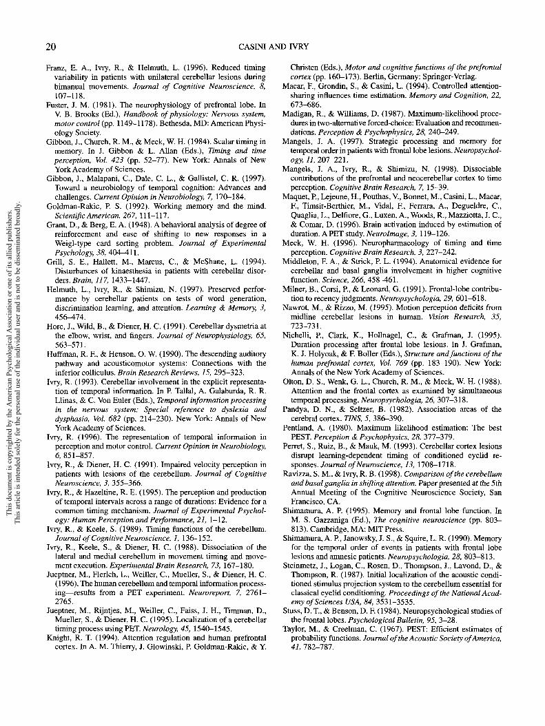
Patients with cerebellar damage. Eight patients with lesions ofthe cerebellum were recruited. Their mean age was 61.3 years andthey had an average of 12.1 years of education. Seven patients hadunilateral lesions due to either stroke or tumor (Figure 2). Estimatesof lesion volume were not available. The lesion was restricted tothe left cerebellar hemisphere for 3 patients and to the righthemisphere in the other 4 patients. The lesions appeared to extendinto the dentate nucleus for 4 of the patients with unilateral lesions(J.D., E.G., R.M., T.R.). The remaining patient had extensivecerebellar atrophy. The atrophy could be seen in an MRI at alllevels of the cerebellum with relative sparing of the anterior lobe.This pattern is consistent with a diagnosis of sporadic cerebellaratrophy and argues against alcohol-based atrophy.
Motor dysfunction of cerebellar patients was estimated by aneurologist using a clinical evaluation testing posture, gait, eyemovements, and volitional movements. A 5-point scale rangingfrom 0 (no evidence of cerebellar dysfunction) to 4 (severecerebellar dysfunction) was used. Overall clinical rating of motorsigns ranged from 0 to 2.5 (moderate). The relatively modest
Table 1Patient Information and Individual Performance Scores on the Neuropsychological Assessment Tests
Participant
FrontalO.A.R.M.J.C.A.L.E.B.
CerebellarM.B.J.D.T.K.B.C.J.L.R.M.T.R.B.H.
Control"
Sex
MMMFF
MMMMMMMM
8M,2F
Side
LLLLR
LLLRRRRBilat
Lesion
Vol.(cm3)
17.510.3
102.651.217.3
WAIS-R
Clin.
0.52.00.02.51.02.51.5
Age
6464716779
345276606954717465
Education(years)
1412161312
12123
161016161214
Info
128
1310
989
127
11169
13
Voc
127
119
1110
159
13191313
DigitSpan
1011
511
1015
119
11111513
DigitSym
1110107
15
965
1076
107
13
F,A, S
29247
2148
412219332625243444
WCST
Cat
16644
662626645
Pers.errors(%)
25.813.817.028.125.0
10.814.526.69.0
53.111.220.228.917
Note. Vol. (frontal patients only) = volume of lesion estimated from computerized tomography scans; Clin. (cerebellar patients only) =overall clinical rating of motor signs, ranging from 0 (no impairment) to 4 (severe impairment); WAIS-R = Wechsler Adult IntelligenceScale—Revised (Wechsler, 1987); Info = Information subtest; Voc = Vocabulary subtest; Digit Sym = Digit Symbol subtest; F, A, S =total number of words produced in 3 min on the F, A, S Letter Verbal Fluency Test (Benton & Hamsher, 1978); WCST = Wisconsin CardSorting Test (Grant & Berg, 1948); Cat = categories attained; Pers. errors = perseverative errors; M = male; F = female; L = unifocallesion on the left hemisphere; R = unifocal lesion on the right hemisphere; Bilat = lesion on both hemispheres."The data shown here for control participants are averages.
This
doc
umen
t is c
opyr
ight
ed b
y th
e A
mer
ican
Psy
chol
ogic
al A
ssoc
iatio
n or
one
of i
ts a
llied
pub
lishe
rs.
This
arti
cle
is in
tend
ed so
lely
for t
he p
erso
nal u
se o
f the
indi
vidu
al u
ser a
nd is
not
to b
e di
ssem
inat
ed b
road
ly.

FRONTAL LOBES AND CEREBELLUM IN TIME PERCEPTION 13
O
1
o
G 'O
.2 a.S.1
^C S•
C/3 tC Oo -o
.2? o<o
o CDLLJ
This
doc
umen
t is c
opyr
ight
ed b
y th
e A
mer
ican
Psy
chol
ogic
al A
ssoc
iatio
n or
one
of i
ts a
llied
pub
lishe
rs.
This
arti
cle
is in
tend
ed so
lely
for t
he p
erso
nal u
se o
f the
indi
vidu
al u
ser a
nd is
not
to b
e di
ssem
inat
ed b
road
ly.

14 CASINI AND IVRY
MB
TK
EC
RM
TR
Figure 2. Lesion reconstructions for the 7 patients with focal cerebellar lesions. Each row shows aseries of seven slices, going inferior to superior from left to right.
degree of motor dysfunction is likely due to the fact that the clinicalevaluation and testing occurred after an extended recovery periodof at least 1 year (and over 5 years for most of the patients).
The neuropsychological assessment showed comparable perfor-mance between the patients and control participants on all but twotests. The patients performed worse than the controls on the DigitSymbol subtest of the Wechsler Adult Intelligence Scale—Revised(WAIS-R; Wechsler, 1987), F(2, 19) = 6.76, p < .01, and theVerbal Fluency Test (FAS; Benton & Hamsher, 1978), F(2, 19) =5.82, p < .01. The only test revealing a difference between the
patient groups was the Digit Symbol test, f ( l l ) = 2.3, p < .05, withthe cerebellar group performing more poorly than the frontal group.There is considerable debate at present as to whether cerebellarlesions produce impairments on tasks designed to assess generaland specific aspects of cognitive function (Akshoomoff, Cour-chesne, Press, & Iragui, 1992; Daum et al., 1993a; Helmuth, Ivry,& Shimizu, 1997), and this issue is outside the focus of this article.However, it is noteworthy that the cerebellar patients wereimpaired on the two tasks in which speeded performance wasrequired in addition to accuracy.
This
doc
umen
t is c
opyr
ight
ed b
y th
e A
mer
ican
Psy
chol
ogic
al A
ssoc
iatio
n or
one
of i
ts a
llied
pub
lishe
rs.
This
arti
cle
is in
tend
ed so
lely
for t
he p
erso
nal u
se o
f the
indi
vidu
al u
ser a
nd is
not
to b
e di
ssem
inat
ed b
road
ly.
FRONTAL LOBES AND CEREBELLUM IN TIME PERCEPTION 15
Given that the focus of this article is on attentional factors, it isimportant to note that none of the patients had any signs ofattentional disorders as assessed by standard clinical procedures.There was no evidence of neglect or extinction and none of thepatients had obvious impairments of concentration or vigilance.
Procedure
Each participant was tested individually and completed threeblocks of trials. Two of these were single-task (ST) blocks in whichthe participants judged either the duration or the frequency of theauditory stimuli. In the third block, the dual-task (DT) condition,the participants were required to judge both the duration and thefrequency of the stimuli. All of the stimuli were generated by a PCcomputer and played over the internal speaker of the computer.
All of the tasks used a psychophysical procedure, ParameterEstimation By Sequential Testing (PEST), developed by Taylor andCreelman (1967) and extended by Pentland (1980). Two stimuli arepresented on each trial, a standard and a comparison. Theprocedure is designed to estimate the difference threshold requiredfor participants to accurately judge the comparison on approxi-mately 90% of the trials (corresponding to 1.5 SD units on the logitdistribution). The procedure is generic in that it can be used withany stimulus dimension. In the current study, we either manipulatedthe duration, the frequency, or both dimensions of the comparisonstimulus. The PEST procedure is adaptive in that it continually usesthe information obtained in previous trials in its estimate of thethreshold. Specifically, the test stimulus on each trial is the currentestimate of either the lower or upper difference threshold. Initially,this estimate is set to a single value for all of the participants.However, on the basis of individual performance, the differencebetween the standard and comparison will either become smaller orlarger. In effect, the PEST procedure creates a situation in which thesubjective experience for all participants is approximately equal.The adaptive procedure selects values so that the participant iscorrect on about 90% of the trials, with the values individuallyadjusted on the basis of each person's acuity.
Threshold estimates were bidirectional: for the duration discrimi-nation task, independent measures were made for both the shortand long thresholds, and for the frequency discrimination task,independent measures were made for both the lower and higherthresholds. The final estimates were based on 30 trials of eachthreshold, or a total of 60 trials per block. Computer simulationshave demonstrated that the PEST procedure is both efficient andstable (Madigan & Williams, 1987; Pentland, 1980). With 30 trialsper threshold, the procedure is likely to converge on the correctestimate over a wide range of starting values.
Duration perception task. The standard stimulus was a 600-Hztone, presented for a fixed duration of 400 ms. After a 1-sinterstimulus interval, the comparison was presented. The fre-quency of the comparison stimulus was fixed at 600 Hz, but thestimulus varied in duration. Participants were instructed that thefirst tone was the standard and that they were required to judge theduration of the comparison tone. On each trial, participants judgedwhether the second tone was shorter or longer than the standard andgave their responses verbally (short or long). The experimenterentered the response on the keyboard and the computer thendetermined the test value for the next trial. To reduce thecomputational process, the logit distribution was divided into 61equal steps with 6 ms between each step (range of comparisonvalues: 220 ms-580 ms).
Frequency perception task. The general procedure was identi-cal to that used in the duration task except that the second tone
varied in frequency rather than duration. The standard tone wasagain a 600-Hz tone, presented for 400 ms. The duration ofcomparison tone was also fixed at 400 ms, but now its frequencywas varied. Participants judged the frequency and gave theirresponse verbally (up or down). Each of the 61 steps were separatedby 1 Hz (range of comparison values: 570 Hz-630 Hz).
Dual task. In the dual-task block, both the duration andfrequency of the comparison stimulus were varied. On each trial,participants first heard the standard stimulus (600 Hz, 400 ms),followed after a 1-s interstimulus interval by the comparisonstimulus. Separate PEST procedures were used to make indepen-dent estimates for the two comparison values; pilot testing indi-cated that there were no consistent biases for one response to belinked to another (e.g., participants to be more likely to say shorterwhen they heard a high frequency tone). Participants had to givetwo responses on each trial, one indicating the duration of thecomparison tone (shorter or longer) and a second indicating thefrequency of the comparison tone (up or down). Participants werefree to make the two judgments in whichever order they preferred.The range of comparison values was the same as in the single taskconditions.
Order of tasks. The three blocks were completed in a single1-hr session. The single tasks were always performed first, with theorder of the duration and frequency tasks counterbalanced acrossparticipants. The dual task was always performed last. This notonly made it easier for the participants to understand the require-ments in this condition, but it also increased the likelihood that theparticipants would not attend to both dimensions in the single taskconditions. Although this ordering introduces a confound whencomparing single- and dual-task performance, our primary interestinvolves a single-dual comparison between the three groups ofparticipants.
Results
The dependent variables in this experiment were themeasures of perceptual acuity and bias provided by thePEST procedure. Acuity was operationalized as the differ-ence between the upper and lower difference thresholdestimates divided by three. This measure corresponds to 1standard deviation unit, measured in ms for the durationdiscrimination task and in Hz for the frequency discrimina-tion task. Larger standard deviations indicate that a greaterdifference was required between the standard and compari-son values in order to meet the criterion level of perfor-mance. The point of subjective equality (PSE) was taken asthe measure of bias and corresponded to the midpointbetween the two thresholds. This corresponds to the value atwhich participants were equally likely to respond shorterand up or longer and down. In the duration discriminationtask, a PSE greater than the standard of 400 ms indicates thatthe comparison duration was underestimated. More timemust elapse for the comparison to be judged equal induration to 400 ms. In the frequency discrimination task, aPSE greater than the standard of 600 Hz indicates that thecomparison frequency was underestimated. The frequencyof the comparison must be higher to be judged equal to thestandard.
Analyses of variance (ANOVAs) were done on the twoindices. One variable was group (controls, cerebellars, and
This
doc
umen
t is c
opyr
ight
ed b
y th
e A
mer
ican
Psy
chol
ogic
al A
ssoc
iatio
n or
one
of i
ts a
llied
pub
lishe
rs.
This
arti
cle
is in
tend
ed so
lely
for t
he p
erso
nal u
se o
f the
indi
vidu
al u
ser a
nd is
not
to b
e di
ssem
inat
ed b
road
ly.

16 CASINI AND IVRY
frontals) and the other variable was condition (single or dualtask). Because the units are not comparable on the two tasks,separate analyses were conducted on the duration andfrequency data.
Duration Perception Task
Standard deviation. Figure 3 A shows the mean standarddeviation scores for each group of participants in the ST andDT conditions. In both conditions, the values are higher forthe cerebellar and frontal groups in comparison to thecontrol participants, F(2, 20) = 5.l6,p< .01. For the targetcriterion, the patients required a larger difference in durationbetween the standard and comparison tone. There was nodifference between the two patient groups. The comparisonbetween the ST and DT conditions shows that performancewas poorer in the dual-task condition, F(l, 20) = 19.90, p <.0001. The percent increase in the difference threshold was29%, 41%, and 24% for the controls, cerebellars, and
Table 2Individual Difference Thresholds on Duration andFrequency Tasks in the Single- and Dual-Task Conditions
Patient
Duration (ms)
ST DT
Frequency (Hz)
ST DT
FrontalO.A.R.M.J.C.A.L.E.B.
CerebellarM.B.J.D.T.K.B.C.J.L.R.M.T.R.B.H.
2244386432
3644581838463634
5250607032
38667624448280116
79101114
2161851415411
1617151819
4131291611414
Note. ST = single-task condition; DT = dual-task condition.
90 T
60-
30-
B
Figure 3. Difference threshold estimated as 1 SD of the psycho-metric function on the duration (A) and frequency (B) tasks in thesingle- and dual-task conditions (ST and DT, respectively). Errorbars reflect 95% confidence intervals around each mean. CONT =controls; CERE = cerebellar lesions; FRONT = frontal lesions.
frontals, respectively. Although the increase was greatest forthe cerebellar patients, the Group X Condition interactionwas not significant, F(2, 20) = 2.10.
Table 2 presents the difference thresholds for each of thepatients individually. As can be seen, there is considerableoverlap between the scores for the cerebellar and frontalpatients. Three additional points are noteworthy. First,although the sample size was small, there was no cleardifference between the patients with focal left-sided cerebel-lar lesions (M.B., J.D., T.K.) and those with right-sidedcerebellar lesions (E.G., J.L., R.M., T.R.). Second, thedifference threshold increased most sharply for the patientwith bilateral cerebellar atrophy (B.H.). Atrophy patientstended to perform more poorly than the patients withunilateral lesions in a previous study of time perception(Ivry & Keele, 1989). Third, the only patient who failed toshow any increase in the DT condition was the 1 patient withright-hemisphere prefrontal damage (E.B.).
PSE. The mean PSE values are presented in Table 3.There was no significant difference between the three groupson this measure, F(2, 20) < 1. All of the means were longerthan the target duration of 400 ms, indicating that theparticipants consistently showed a bias to underestimate theduration of the comparison stimulus. Although the meanPSEs were larger in the dual-task condition, this effect wasonly marginally significant, F(l, 20) = 2.83, p < .10. TheGroup X Condition interaction was not significant, F(2,20) < 1.
Frequency Perception Task
Standard deviation. The mean standard deviation scoreson the frequency task are shown in Figure 3B. In bothconditions, the patients performed more poorly than thecontrol participants, F(2, 20) = 6.49, p < .01. The maineffect of condition, F(l, 20) = 17.56, p < .001, and theinteraction, F(2,20) = 13.47,p< .0001, were significant. Inthe single-task condition, both groups of patients showed
This
doc
umen
t is c
opyr
ight
ed b
y th
e A
mer
ican
Psy
chol
ogic
al A
ssoc
iatio
n or
one
of i
ts a
llied
pub
lishe
rs.
This
arti
cle
is in
tend
ed so
lely
for t
he p
erso
nal u
se o
f the
indi
vidu
al u
ser a
nd is
not
to b
e di
ssem
inat
ed b
road
ly.

FRONTAL LOBES AND CEREBELLUM IN TIME PERCEPTION 17
Table 3Means and Standard Errors for Point of SubjectiveEquality (PSE) Obtained in Single- and Dual-TaskConditions on Each Task for the Three Groups
Control Cerebellar Frontal
Task M SE M SE M SE
Duration (ms)STDT
Frequency (Hz)STDT
423.7441.1
599.9598.9
9.513.6
0.81.6
419.9441.6
600.1596.1
15.215.4
2.62.7
440.8448.0
604.7598.1
4.99.6
2.02.0
Note. ST = single-task condition; DT = dual-task condition.
poorer acuity than the controls, and the two groups did notdiffer from one another, F(l, 11) < 1. On the dual-taskblock, both groups of patients were also impaired relative tothe controls, but here the frontal patients performed signifi-cantly worse than the cerebellar patients, F(l, 11) = 9.84,/7< .01 .
Of central interest is the fact that only the frontal lobepatients showed a significant decrease in performancebetween the single- and dual-task conditions on the fre-quency task, f(8) = -4.67, p < .005. The dual-taskperformance of the controls, t ( l S ) = 0.87, and the cerebel-lars, f(14) = 0.93, on the frequency task was comparable tothat found under single-task conditions. A post hoc analysisrestricted to the two patient groups revealed a significantGroup X Condition interaction, F(l, 11) = 16.20, p < .005.Whereas all groups showed an increase in the durationdifference threshold under dual-task conditions, only thefrontals showed a concomitant increase in the frequencydifference threshold. The percentage increase in the differ-ence threshold for the controls, cerebellars, and frontals was9%, 0%, and 39%, respectively.
The difference thresholds for each patient on the fre-quency task are shown in Table 2. The effect of the DTcondition on the two groups is quite striking. All 5 of thefrontal patients showed an increase in the DT condition, andthe increase ranged from 5 Hz to 9 Hz. In contrast, thedifference threshold was larger in the DT condition for only4 of the 8 cerebellar patients, and here the increase was nevergreater than 3.33 Hz. In terms of the difference between theST and DT conditions, there was no overlap between the twogroups of patients.
PSE. The PSE values on the frequency task are shownin Table 3. The effect of condition was significant, F(1,20) =10.39, p < .005. All groups judged the comparison tone ashigher in frequency when they were in the dual-taskcondition. There was no significant difference betweengroups on this measure, F(2,20) < 1, nor was the interactionsignificant, F(2, 20) < 1.
Discussion
Previous research has shown that lesions to the frontallobes or cerebellum can impair performance on time-perception tasks. However, it has been difficult to identify
functional dissociations because of differences in methodol-ogy, dependent variables, and the lack of direct comparisons.In this experiment, we compared performance on a temporaland nontemporal task as a function of attentional load.Different patterns of interference were observed for the twopatient groups, providing an important step in understandinghow the cerebellum and frontal lobe may make differentialcontributions to tasks that require temporal processing.
The single-task results did not reveal any differencesbetween the patient groups. The cerebellar group was morevariable on the duration discrimination task in comparison tothe control participants, thus replicating previous results(Ivry & Keele, 1989; Mangels et al., 1998). However, unlikethe results of Mangels et al., the frontal patients were alsoimpaired on this task in the present study. Both of the patientgroups were also impaired on the frequency perception taskunder single-task conditions.
The poor performance on the frequency discriminationtask was unexpected. This task was intended to serve as anauditory control task. The patients may be more variablethan controls on any psychophysical task, reflecting general-ized problems in performing these relatively demandingtasks. For example, such tasks require that the participantsconcentrate for sustained periods of time because a block oftrials lasts for approximately 8 min. This may be moredifficult following brain damage.
On the other hand, it is possible that these deficits reflectspecific problems in making auditory discriminations. Ana-tomical studies in the monkey have shown that secondaryauditory-association areas innervate Area 46 of prefrontalcortex (Pandya & Seltzer, 1982). Similarly, the cerebellumreceives auditory inputs. In the rat, auditory regions of thecortex project to the parafloccular lobule of the cerebellumthrough both the mossy and the climbing fiber pathways(Azizi, Burne, & Woodward, 1985), and subcortical projec-tions to the cerebellar vermis have also been shown byelectrical stimulation of the inferior colliculus (Huffman &Henson, 1990). Moreover, in eyeblink conditioning studies,an auditory tone is frequently used as the conditioningstimulus and numerous studies have demonstrated that thissignal is projected through mossy fibers (Steinmetz et al.,1987; reviewed in Thompson, 1990). It is possible that thefrequency-perception problems occur as a result of damageto representations of auditory signals in either prefrontal orcerebellar regions. However, the functions of tuning to puretones of cerebellar neurons in the auditory projection regionof the vermis in the cat are quite broad (Aitkin & Boyd,1975). Individual neurons respond at a relatively constantrate over a range of several octaves, making it unlikely thatthey could support the fine discrimination capability re-quired on the frequency task. We are unaware of similarstudies of prefrontal neurons. Future studies will be requiredto examine these different hypotheses. It is, of course,possible that the frontal and cerebellar groups performpoorly on this task for different reasons.
More relevant to the focus of this experiment, thedual-task condition revealed an important difference inperformance between the two patient groups. When theattentional load was increased by requiring simultaneous
This
doc
umen
t is c
opyr
ight
ed b
y th
e A
mer
ican
Psy
chol
ogic
al A
ssoc
iatio
n or
one
of i
ts a
llied
pub
lishe
rs.
This
arti
cle
is in
tend
ed so
lely
for t
he p
erso
nal u
se o
f the
indi
vidu
al u
ser a
nd is
not
to b
e di
ssem
inat
ed b
road
ly.

18 CASINI AND IVRY
judgments of duration and frequency, all three groupsbecame more variable on the duration discrimination task.This result is consistent with numerous results obtained withhealthy participants indicating that temporal judgments canbe influenced by attentional manipulations (Casini & Macar,1997; Macar et al., 1994; Zakay, 1989). In contrast, only thepatients with prefrontal lesions showed a dual-task cost onthe frequency discrimination task. The difference thresholdincreased for all 5 patients in this group under the dual-taskcondition. The Group X Condition interaction was signifi-cant when the comparison was made between all threegroups as well as when the analysis was restricted to the twopatient groups. The latter analysis is especially importantgiven the fact that both the cerebellar and frontal patientswere more variable than the controls in the single-taskconditions.
Although we had predicted that the frontal patients wouldshow the greatest decrement in performance in the dual-taskcondition, we had anticipated that all of the groups wouldshow some cost during the dual-task block on both tasks.The failure to find any change in performance on thefrequency perception task for the control and cerebellargroup likely reflects a lack of sensitivity in this task. Theeffect of attention load was also found to be greater on atemporal task compared to a nontemporal task in a studydone by Macar et al. (1994). Nonetheless, this does notcompromise the primary finding showing a dissociationbetween the cerebellar and frontal groups in terms of theeffects of dividing attention between the two tasks.
This dissociation allows us to develop hypotheses concern-ing how the cerebellum and prefrontal cortex contribute tothese tasks. It is possible that the prefrontal lesions producedseparate disturbances in separable processing systems, onerelated to time perception and another related to frequencyperception. However, a more parsimonious interpretation isthat the frontal lesions disrupt attentional processes requiredin these tasks. Whenever the attentional demands of a taskare increased, patients with prefrontal lesions are chal-lenged, regardless of whether the task requires temporal ornontemporal processing. The cerebellar group, on the otherhand, did not show a generalized attentional problem.
Courchesne and colleagues (Courchesne et al., 1994;Akshoomoff, Courchesne, & Townsend, 1997) have pro-posed that the cerebellum plays a critical role in shiftingattention. The basic idea here is that, analogous to its role inmotor coordination, the cerebellum is essential for mentalcoordination by orienting perceptual systems to task-relevant stimuli. The primary evidence in support of thishypothesis has come from a divided attention task in whichparticipants must alternate between attending to one of twodimensions (Akshoomoff & Courchesne, 1992; Courchesneet al., 1994). However, Ravizza and Ivry (1998) found thatthe attentional problem appears to be related to the fact thatthis task requires rapid successive responses. When theattentional requirements are held constant but the motordemands are reduced, patients with focal cerebellar lesionsshow a significant improvement in performance. The currentresults provide further evidence that the deficits observed on
perceptual tasks in patients with cerebellar lesions are notrelated to an attentional problem.
We interpret the cerebellum deficit on the durationdiscrimination task as further evidence of the role of thisstructure in representing temporal information. The cerebel-lum has been shown to be essential for controlling theprecise timing in both motor control (e.g., Hore, Wild, &Diener, 1991) and sensorimotor learning (Ferret et al.,1993). Together with the perceptual problems shown bycerebellar patients on temporal tasks (Grill et al., 1994; Ivry& Diener, 1991; Ivry & Keele, 1989; Nawrot & Rizzo,1995), the timing hypothesis provides a theoretical umbrellafor these disparate results (see Ivry, 1996). Recent theoreti-cal conjectures have centered on the idea that timing withinthe cerebellum consists of a set of distributed functionalunits tuned to different temporal intervals (Buonomano &Mauk, 1994; Fiala et al., 1996; Ivry, 1996). For example,Buonomano and Mauk propose that the coding of durationcould emerge from relatively slow synaptic interactionsoccurring in the cerebellar cortex. Different temporal inter-vals would be coded through negative feedback loopsinvolving the interaction of granule and Golgi cells onPurkinje cells. Models such as these assume that temporalinformation is transformed into a spatial code (see also, Fialaet al., 1996). Lesions would be expected to add noise to thesystem, which would be reflected as increased variability onthe duration discrimination tasks in the current study.
However, the cerebellar timing system is only one func-tional component required for these tasks. Successful perfor-mance also depends on other operations such as thoseinvolved in attention, memory, and decision processes.There are obviously a number of ways in which anattentional process would be required for successful perfor-mance on these difficult discrimination tasks. In the single-task condition, the participant would want to focus on thetask-relevant dimension. The prefrontal cortex has beenhypothesized to assist in such filtering operations, perhapsby attenuating information from irrelevant information chan-nels (Knight, 1994). Different anatomical and neuroimagingstudies have revealed projections from the neocerebellarcortex to the prefrontal cortex (Middleton & Strick, 1994).Thus, the increased variability in frontal patients on theduration perception task could be viewed as a manifestationof a failure to fully attend to the temporal informationprovided by the cerebellum. The increased variability ob-served in the prefrontal patients on both tasks in thedual-task condition would be consistent with a putative rolefor this neural region in coordinating processing acrossdifferent processing systems (Corbetta, Miezin, Dobmeyer,Shulman, & Petersen, 1991).
The relationship between working memory and attentionremains unclear (see Shimamura, 1995). In the presentstudy, we have emphasized the attentional role of lateralprefrontal cortex. However, a functional account of thisregion could also be stated in terms of decision or memoryprocesses. Our psychophysical procedure requires a compari-son between two successive stimuli, separated by a 1-sinterval. A representation of the standard stimulus wouldhave to be maintained across the interstimulus interval as
This
doc
umen
t is c
opyr
ight
ed b
y th
e A
mer
ican
Psy
chol
ogic
al A
ssoc
iatio
n or
one
of i
ts a
llied
pub
lishe
rs.
This
arti
cle
is in
tend
ed so
lely
for t
he p
erso
nal u
se o
f the
indi
vidu
al u
ser a
nd is
not
to b
e di
ssem
inat
ed b
road
ly.

FRONTAL LOBES AND CEREBELLUM IN TIME PERCEPTION 19
well as during the presentation of the comparison, at leastuntil a decision is reached. Such an operation would fit withthe working memory operations associated with lateralprefrontal cortex (Goldman-Rakic, 1992). As noted earlier,Mangels et al. (1998) found that the performance of patientswith prefrontal lesions was especially sensitive to the delaybetween the offset of the standard and the offset of thecomparison. These patients, many of whom were in thecurrent study, showed an increase in variability as thisinterval was lengthened, an effect observed on both durationand frequency discrimination tasks. At present, we canconclude that the contribution of prefrontal cortex to perfor-mance on these tasks becomes more pronounced as the taskbecomes more difficult, either through the dual-task manipu-lation or by extending the temporal extent of the stimulusevents.
Over the past decade, an extensive literature has emergedexploring the neurological correlates of the hypothesizedcomponent operations involved in temporal processing tasks(for recent reviews see Gibbon et al., 1997; Ivry, 1996;Meek, 1996). In the scalar timing theory (Church, 1984), thebasic component of timing is a pacemaker that producesoutputs, called pulses, at a given rate. These pulses are keptin a counter gated by a switch. A comparator processcontrasts the value accumulated in the counter with valuesthat have been stored in reference memory on previoustrials. Responses are determined on the basis of thiscomparison.
Research on the basal ganglia has implicated this subcor-tical structure as a key component of an internal pacemaker(Meek, 1996; Gibbon et al., 1997). Executive and memoryprocesses have been associated with cortical processes,including a putative role of frontal cortex in an attentionalsystem required to monitor the output of the timing system(Olton, Wenk, Church, & Meek, 1988). Although the currentdata are in accord with this latter hypothesis, we havepostulated a central role for the cerebellum in the clockprocess, the outputs of the cerebellum being viewed asrepresentations of particular intervals rather than pulses asimplied by pacemaker models. It will be important in futurestudies to make direct comparisons between patients withcerebellar and basal ganglia dysfunction, seeking dissocia-tions as have been observed in the current experiment. Inthis manner, a functional analysis can be established of theneural systems involved in temporal processing.
References
Aitkin, L., & Boyd, J. (1975). Responses of single units incerebellar vermis of the cat to monaural and binaural stimuli.Journal of Neurophysiology, 38, 418^tl9.
Akshoomoff, N. A., & Courchesne, E. (1992). A new role for thecerebellum in cognitive operations. Behavioral Neuroscience,106, 731-738.
Akshoomoff, N. A., Courchesne, E., Press, G. A., & Iragui, V.(1992). Contribution of the cerebellum to neuropsychologicalfunctioning: Evidence from a case of cerebellar degenerativedisorder. Neuropsychologia, 30, 315-328.
Akshoomoff, N. A., Courchesne, E., & Townsend, J. (1997).Attention coordination and anticipatory control. InternationalReview ofNeurobiology, 41, 575-598.
Azizi, S. A., Burne, R. A., & Woodward, D. J. (1985). The auditorycorticopontocerebellar projection in the rat: Inputs to the parafloc-culus and midvermis. An anatomical and physiological study.Experimental Brain Research, 59, 36-49.
Benton, A. L., & Hamsher, K. (1978). The Multilingual AphasiaExamination. Iowa City: University of Iowa Press.
Braitenberg, V. (1967). Is the cerebellar cortex a biological clock inthe millisecond range? Progress in Brain Research, 25, 334-346.
Brown, S. W. (1997). Attentional resources in timing: Interferenceeffects in concurrent temporal and nontemporal working memorytasks. Perception and Psychophysics, 59, 1118-1140.
Bruder, G. E., Towey, J., Friedman, D., Erhan, H., Jasiukaitis, P., &Tenke, C. (1992). Lateral asymmetries of event-related potentialslow waves. Journal ofPsychophysiology, 6, 97-110.
Buonomano, D., & Mauk, M. (1994). Neural network model of thecerebellum: Temporal discrimination and the timing of motorresponses. Neural Computation, 6, 38-55.
Casini, L., & Macar, F. (1996a). Can the level of brain activityprovide an index of the temporal performance? NeuroscienceLetters, 219, 71-74.
Casini, L., & Macar, F. (1996b). Prefrontal slow potentials intemporal compared to nontemporal tasks. Journal ofPsychophysi-ology, 10, 252-264.
Casini, L., & Macar, F. (1997). Effects of attention manipulation onperceived duration and intensity in the visual modality. Memoryand Cognition, 25, 812-818.
Church, R. M. (1984). Properties of the internal clock. In J. Gibbon& L. Allan (Eds.), Timing and time perception, Vol. 423 (pp.566-582). New York: Annals of New York Academy of Sci-ences.
Corbetta, M., Miezin, F. M., Dobmeyer, S., Shulman, G. L., &Petersen, S. E. (1991). Selective and divided attention duringvisual discriminations of shape, color and speed: Functionalanatomy by positron emission tomography. Journal of Neurosci-ence, 11, 2383-2402.
Courchesne, E., Townsend, J., Akshoomoff, N. A., Saitoh, O.,Yeung-Courchesne, R., Lincoln, A., James, H., Haas, R. H.,Schriebman, L., & Lau, L. (1994). Impairment in shiftingattention in autistic and cerebellar patients. Behavioral Neurosci-ence, 108, 848-865.
Daum, I., Ackermann, H., Schugens, M. M., Reimold, C., Dich-gans, J., & Birbaumer, N. (1993a). The cerebellum and cognitivefunctions in humans. Behavioral Neuroscience, 107, 411-419.
Daum, I., Schugens, M. M., Ackermann, H., Lutzenberger, W.,Dichgans, J., & Birbaumer, N. (1993b). Classical conditioningafter cerebellar lesions in humans. Behavioral Neuroscience,107, 748-756.
Elbert, T., Ulrich, R., Rockstroh, B., & Lutzenberger, W. (1991).The processing of temporal intervals reflected by CNV-like brainpotentials. Psychophysiology, 28, 648-655.
Fahle, M., & Braitenberg, V. (1984). Some quantitative aspects ofcerebellar anatomy as a guide to speculation on cerebellarfunctions. In J. Bloedel, J. Dichgans, & W. Precht (Eds.),Cerebellar functions (pp. 186-200). Berlin, Germany: Springer-Verlag.
Fiala, J. C., Grossberg, S., & Bullock, D. (1996). Metabotropicglutamate receptor activation in cerebellar Purkinje cells assubstrate for adaptive timing of the classically conditionedeye-blink response. Journal of Neuroscience, 16, 3760-3774.
This
doc
umen
t is c
opyr
ight
ed b
y th
e A
mer
ican
Psy
chol
ogic
al A
ssoc
iatio
n or
one
of i
ts a
llied
pub
lishe
rs.
This
arti
cle
is in
tend
ed so
lely
for t
he p
erso
nal u
se o
f the
indi
vidu
al u
ser a
nd is
not
to b
e di
ssem
inat
ed b
road
ly.
20 CASINI AND IVRY
Franz, E. A., Ivry, R., & Helmuth, L. (1996). Reduced timingvariability in patients with unilateral cerebellar lesions duringbimanual movements. Journal of Cognitive Neuroscience, 8,107-118.
Fuster, J. M. (1981). The neurophysiology of prefrontal lobe. InV. B. Brooks (Ed.), Handbook of physiology: Nervous system,motor control (pp. 1149-1178). Bethesda, MD: American Physi-ology Society.
Gibbon, J., Church, R. M., & Meek, W. H. (1984). Scalar timing inmemory. In J. Gibbon & L. Allan (Eds.), Timing and timeperception, Vol. 423 (pp. 52-77). New York: Annals of NewYork Academy of Sciences.
Gibbon, J., Malapani, C., Dale, C. L., & Gallistel, C. R. (1997).Toward a neurobiology of temporal cognition: Advances andchallenges. Current Opinion in Neurobiology, 7, 170-184.
Goldman-Rakic, P. S. (1992). Working memory and the mind.Scientific American, 267, 111-117.
Grant, D., & Berg, E. A. (1948). Abehavioral analysis of degree ofreinforcement and ease of shifting to new responses in aWeigl-type card sorting problem. Journal of ExperimentalPsychology, 38, 404-411.
Grill, S. E., Hallett, M., Marcus, C., & McShane, L. (1994).Disturbances of kinaesthesia in patients with cerebellar disor-ders. Brain, 117, 1433-1447.
Helmuth, L., Ivry, R., & Shimizu, N. (1997). Preserved perfor-mance by cerebellar patients on tests of word generation,discrimination learning, and attention. Learning & Memory, 3,456-474.
Hore, J., Wild, B., & Diener, H. C. (1991). Cerebellar dysmetria atthe elbow, wrist, and fingers. Journal of Neurophysiology, 65,563-571.
Huffman, R. F., & Henson, O. W. (1990). The descending auditorypathway and acousticomotor systems: Connections with theinferior colliculus. Brain Research Reviews, 15, 295-323.
Ivry, R. (1993). Cerebellar involvement in the explicit representa-tion of temporal information. In P. Tallal, A. Galaburda, R. R.Llinas, & C. Von Euler (Eds.), Temporal information processingin the nervous system: Special reference to dyslexia anddysphasia, Vol. 682 (pp. 214-230). New York: Annals of NewYork Academy of Sciences.
Ivry, R. (1996). The representation of temporal information inperception and motor control. Current Opinion in Neurobiology,6, 851-857.
Ivry, R., & Diener, H. C. (1991). Impaired velocity perception inpatients with lesions of the cerebellum. Journal of CognitiveNeuroscience, 3, 355-366.
Ivry, R., & Hazeltine, R. E. (1995). The perception and productionof temporal intervals across a range of durations: Evidence for acommon timing mechanism. Journal of Experimental Psychol-ogy: Human Perception and Performance, 21, 1-12.
Ivry, R., & Keele, S. (1989). Timing functions of the cerebellum.Journal of Cognitive Neuroscience, 1, 136-152.
Ivry, R., Keele, S., & Diener, H. C. (1988). Dissociation of thelateral and medial cerebellum in movement timing and move-ment execution. Experimental Brain Research, 73, 167-180.
Jueptner, M., Flerlch, L., Weiller, C., Mueller, S., & Diener, H. C.(1996). The human cerebellum and temporal information process-ing—results from a PET experiment. Neuroreport, 7, 2761-2765.
Jueptner, M., Rijntjes, M., Weiller, C., Faiss, J. H., Timman, D.,Mueller, S., & Diener, H. C. (1995). Localization of a cerebellartiming process using PET. Neurology, 45, 1540-1545.
Knight, R. T. (1994). Attention regulation and human prefrontalcortex. In A. M. Thierry, J. Glowinski, P. Goldman-Rakic, & Y.
Christen (Eds.), Motor and cognitive functions of the prefrontalcortex (pp. 160-173). Berlin, Germany: Springer-Verlag.
Macar, F., Grondin, S., & Casini, L. (1994). Controlled attention-sharing influences time estimation. Memory and Cognition, 22,673-686.
Madigan, R., & Williams, D. (1987). Maximum-likelihood proce-dures in two-alternative forced-choice: Evaluation and recommen-dations. Perception & Psychophysics, 28, 240-249.
Mangels, J. A. (1997). Strategic processing and memory fortemporal order in patients with frontal lobe lesions. Neuropsychol-ogy, 11, 207-221.
Mangels, J. A., Ivry, R., & Shimizu, N. (1998). Dissociablecontributions of the prefrontal and neocerebellar cortex to timeperception. Cognitive Brain Research, 7, 15-39.
Maquet, P., Lejeune, H., Pouthas, V., Bonnet, M., Casini, L., Macar,F., Timsit-Berthier, M., Vidal, F., Ferrara, A., Degueldre, C.,Quaglia, L., Delfiore, G., Luxen, A., Woods, R., Mazziotta, J. C.,& Comar, D. (1996). Brain activation induced by estimation ofduration. A PET study. Neurolmage, 3, 119-126.
Meek, W. H. (1996). Neuropharmacology of timing and timeperception. Cognitive Brain Research, 3, 227-242.
Middleton, F. A., & Strick, P. L. (1994). Anatomical evidence forcerebellar and basal ganglia involvement in higher cognitivefunction. Science, 266, 458-461.
Milner, B., Corsi, P., & Leonard, G. (1991). Frontal-lobe contribu-tion to recency judgments. Neuropsychologia, 29, 601-618.
Nawrot, M., & Rizzo, M. (1995). Motion perception deficits frommidline cerebellar lesions in human. Vision Research, 35,723-731.
Nichelli, P., Clark, K., Hollnagel, C., & Grafman, J. (1995).Duration processing after frontal lobe lesions. In J. Grafman,K. J. Holyoak, & F. Boiler (Eds.), Structure and functions of thehuman prefrontal cortex, Vol. 769 (pp. 183-190). New York:Annals of the New York Academy of Sciences.
Olton, D. S., Wenk, G. L., Church, R. M., & Meek, W. H. (1988).Attention and the frontal cortex as examined by simultaneoustemporal processing. Neuropsychologia, 26, 307-318.
Pandya, D. N., & Seltzer, B. (1982). Association areas of thecerebral cortex. TINS, 5, 386-390.
Pentland, A. (1980). Maximum likelihood estimation: The bestPEST. Perception & Psychophysics, 28, 377-379.
Perret, S., Ruiz, B., & Mauk, M. (1993). Cerebellar cortex lesionsdisrupt learning-dependent timing of conditioned eyelid re-sponses. Journal of Neuroscience, 13, 1708-1718.
Ravizza, S. M., & Ivry, R. B. (1998). Comparison of the cerebellumand basal ganglia in shifting attention. Paper presented at the 5thAnnual Meeting of the Cognitive Neuroscience Society, SanFrancisco, CA.
Shimamura, A. P. (1995). Memory and frontal lobe function. InM. S. Gazzaniga (Ed.), The cognitive neuroscience (pp. 803-813). Cambridge, MA: MIT Press.
Shimamura, A. P., Janowsky, J. S., & Squire, L. R. (1990). Memoryfor the temporal order of events in patients with frontal lobelesions and amnesic patients. Neuropsychologia, 28, 803-813.
Steinmetz, J., Logan, C., Rosen, D., Thompson, J., Lavond, D., &Thompson, R. (1987). Initial localization of the acoustic condi-tioned stimulus projection system to the cerebellum essential forclassical eyelid conditioning. Proceedings of the National Acad-emy of Sciences USA, 84, 3531-3535.
Stuss, D. T, & Benson, D. F. (1984). Neuropsychological studies ofthe frontal lobes. Psychological Bulletin, 95, 3-28.
Taylor, M., & Creelman, C. (1967). PEST: Efficient estimates ofprobability functions. Journal of the Acoustic Society of America,41, 782-787.
This
doc
umen
t is c
opyr
ight
ed b
y th
e A
mer
ican
Psy
chol
ogic
al A
ssoc
iatio
n or
one
of i
ts a
llied
pub
lishe
rs.
This
arti
cle
is in
tend
ed so
lely
for t
he p
erso
nal u
se o
f the
indi
vidu
al u
ser a
nd is
not
to b
e di
ssem
inat
ed b
road
ly.

FRONTAL LOBES AND CEREBELLUM IN TIME PERCEPTION 21
Thompson, R. (1990). Neural mechanisms of classical condition-ing in mammals. Philosophical Transactions of the RoyalSociety (Section B), 329, 161-170.
Treisman, M. (1963). Temporal discrimination and the indifferenceinterval: Implications for a model of the "internal clock."Psychological Monographs, 77, 1-31.
Von Steinbuchel, N., Wittman, M., & Poeppel, E. (1996). Timing inperceptual and motor tasks after disturbances of the brain. InM. A. Pastor & J. Artieda (Eds.), Time, internal clocks andmovement, Vol. 115 (pp. 281-304). Amsterdam: Elsevier Sci-ence.
Wechsler, D. (1987). Wechsler Adult Intelligence Scale—Revised.San Antonio, TX: The Psychological Corporation.
Woodruff-Pak, D. S., Papka, M., & Ivry, R. (1996). Cerebellarinvolvement in eyeblink classical conditioning in humans.Neuropsychology, 10, 443-458.
Zakay, D. (1989). Subjective and attentional resource allocation:An integrated model of time estimation. In I. Levin & D. Zakay(Eds.), Time and human cognition, Vol. 59 (pp. 365-397).Amsterdam: Elsevier Science.
Received December 1, 1997Revision received June 19, 1998
Accepted June 22, 1998
New Editors Appointed, 2000-2005
The Publications and Communications Board of the American Psychological Associa-tion announces the appointment of three new editors for 6-year terms beginning in 2000.
As of January 1,1999, manuscripts should be directed as follows:
• For Experimental and Clinical Psychopharmacology, submit manuscripts toWarren K. Bickel, PhD, Department of Psychiatry, University of Vermont, 38Fletcher Place, Burlington, VT 05401-1419.
• For the Journal of Counseling Psychology, submit manuscripts to Jo-Ida C.Hansen, PhD, Department of Psychology, University of Minnesota, 75 EastRiver Road, Minneapolis, MN 55455-0344.
• For the Journal of Experimental Psychology: Human Perception and Perfor-mance, submit manuscripts to David A. Rosenbaum, PhD, Department of Psy-chology, Pennsylvania State University, 642 Moore Building, University Park,PA 16802-3104.
Manuscript submission patterns make the precise date of completion of the 1999 volumesuncertain. Current editors, Charles R. Schuster, PhD; Clara E. Hill, PhD; and Thomas H.Carr, PhD, respectively, will receive and consider manuscripts through December 31,1998. Should 1999 volumes be completed before that date, manuscripts will be redirectedto the new editors for consideration in 2000 volumes.
This
doc
umen
t is c
opyr
ight
ed b
y th
e A
mer
ican
Psy
chol
ogic
al A
ssoc
iatio
n or
one
of i
ts a
llied
pub
lishe
rs.
This
arti
cle
is in
tend
ed so
lely
for t
he p
erso
nal u
se o
f the
indi
vidu
al u
ser a
nd is
not
to b
e di
ssem
inat
ed b
road
ly.