effects of different molecular weight ... - synvisc one 9.pdfeffects of different molecular weight...
TRANSCRIPT

ARTHRITIS & RHEUMATISM Vol. 50, No. I, January 2004, pp 314-326
DOIIO.J002/art.IJ421
© 2004, American College of Rheumatology
Effects of Different Molecular Weight Elastoviscous Hyaluronan Solutions on Articular Nociceptive Afferents
Ana Gomis,1 Matthias Pawlak,2 Endre A. Balazs,3 Robert F. Schmidt,4 a·nd Carlos Belmonte1
Objective. To compare 3 different hyaluronan (HA) preparations used as therapeutic agents for osteoarthritis pain in humans in order to establish the degree to which a single application affects the sensitivity of nociceptors in both the normal and the acutely inflamed rat joint.
Methods. In anesthetized rats, single-unit recordings were performed from the medial articular nerve of the right knee joint under normal conditions and during an acute experimental arthritis. Fifty fine afferent units (conduction velocities 0.8-15.3 meters/second) responded to passive movements of the knee joint. They were exposed to a torque meter-controlled, standardized stimulus protocol consisting of innocuous and noxious inward and outward rotations of the joint. This stimulus protocol of SO seconds' duration was repeated every 5 minutes for 2-3 hours. Three commercially available HA preparations and a buffer solution, the solvent of these preparations, were injected intraarticularly after discharges resulting from 6 stimulus protocols were averaged and used as controls.
Results. Both in normal and in inflamed joints, the injection of Hyalgan did not reduce nerve impulse frequency of the evoked discharges. The injections of
Supported by grants from Genzyme Biosurgery, Cambridge, MA, and the Matrix Biology Institute, Ridgefield, NJ, and by grant MCYT.BFI2002-03788 from the Ministerio de Ciencia y Tecnologfa, Spain, and grant 01/1162 from the FIS, Spain. br. Schmidt's work was supported by an Alexander von Humboldt-}. C. Mutis Fellowship.
1 Ana Gomis, PhD, Carlos Belmonte, MD: Instituto de Neurociencias de Alicante, Universidad Miguel Hernandez-CSIC, San Juan de Alicante, Spain; 2Matthias Pawlak, PhD: Physiologisches Institut, Universitiit Wiirzburg, Wiirzburg, Germany; 3Endre A. Balazs, MD: Matrix Biology Institute, Ridgefield, New Jersey; 4Robert F. Schmidt, MD: Instituto de Neurociencias de Alicante, Universidad Miguel Hermindez-CSIC, San Juan de Alicante, Spain, and Physiologisches Institut, Universitiit Wiirzburg, Wilrzburg, Germany.
Address correspondence and reprint requests to Ana Gomis, PhD, Universidad Miguel Hernandez-CSIC, Instituto de Neurociencias de Alicante, 03550 Sant Joan d'Aiacant, Alicante, Spain. E-mail: [email protected].
Submitted for publication January 24, 2003; accepted in revised form September 4, 2003.
314
Orthovisc had no effect in normal joints, but produced a transient frequency reduction of the evoked discharge in inflamed joints. Synvisc significantly reduced (by an average of 50%) the impulse discharge in both normal and inflamed joints 50 minutes after injection, and this level of impulse discharge continued until the end of the recording period (120-130 minutes after injection). The buffer, which had elastoviscous properties substantially different from those of Hyalgan, Orthovisc, and Synvisc, had no such effect.
Conclusion. We conclude that the elastoviscous properties of HA solutions are determining factors in reducing pain-eliciting nerve activity both in normal and in inflamed rat joints.
Elastoviscous hyaluronan (HA) solutions and HA derivatives (hylans) have been used in veterinary medicine since the early 1970s for treatment of arthritis pain (for review, see ref. 1). This therapeutic approach was based on the finding that long-lasting analgesia could be achieved in the joints of arthritic horses by replacing the pathologic synovial fluid, which has a low elastoviscosity, with a highly purified HA solution of normal elastoviscosity but significantly greater concentration than that of the healthy joint fluid. As a result of this discovery, highly purified HA and hylan solutions became available worldwide for the treatment of arthritis pain in humans and animals (for review, see refs. 2 and 3). This new therapeutic paradigm was called viscosupplementation.
HA products of variable elastoviscosity have been used in viscosupplementation. The differences in rheologic properties ( elastoviscosity) among these therapeutic products are primarily due to variations in their average molecular weight. Clinical studies in human osteoarthritis showed that in order to achieve clinically significant short-term and long-term pain relief, and depending on the specific product used, the viscosupplementation process had to be repeated 2-10 times at

HA SOLUTIONS IN THE REDUCTION OF PAIN-ELICIT!Na' NERVE ACTIVITY 315
weekly intervals. It was also observed that the greater the elastoviscous properties of the preparation, the fewer repeat injections were needed for long-lasting pain relief (4). The availability of HA preparations of different elastoviscosity, combined with the use of different treatment schedules, obscured somewhat the efficacy of this therapy and slowed considerably the acceptance of this new treatment for arthritis pain in both human and veterinary medicine.
The dependence of the analgesic effects of HA solutions on their elastoviscosity had also been shown in early experimental studies in arthritic joints of horses and dogs (5). The fast pain-relieving effect of intraarticular application of highly elastoviscous HA solutions in these experiments was later confirmed using behavioral models in horses, sheep, and rodents (6-9). These studies showed that pain induced by intraarticular injections of bradykinin, prostaglandin E2 (PGE2), substance P, histamine, or acetic acid was alleviated by injections of high molecular weight HA solutions.
Suppression of the behavioral signs of pain was more pronounced and longer lasting when high molecular weight, high-concentration HA solutions were used. The intraarticular production of PGE2 and bradykinin after injection of monosodium urate monohydrate crystals was also suppressed by prior injection of high molecular weight HA into the joint (10).
With regard to the mode of action ofviscosupplementation, it was envisaged that the exchange of pathologic fluid of low elastoviscosity and low HA concentration with a pure HA solution of high elastoviscosity and high concentration causes an immediate decrease in the concentration of pain-stimulating and inflammatory agents in the joint, and that the injection of a high concentration (10-20 times higher than normal) of higher molecular weight HA into an arthritic joint results in a nearly normal intraarticular fluid. It was proposed that this sudden increase in HA concentration and elastoviscosity provides protection and attenuates forces transmitted to the nociceptive nerve endings where pain signals originate. The injected HA flowed out of the joint in 5-7 days, but the analgesic effect persisted longer. Thus, it was suggested that treatment also restores the homeostasis of the synthesis of high molecular weight, high-concentration HA in the joint (4).
The protective effect of elastoviscous HA solutions on nerve endings was demonstrated in a series of animal experiments in which movement-induced nerve impulse activity in single fine fibers of normal and inflamed knee joints of the cat was reduced by intraar-
ticular injection of highly elastoviscous hylan solutions, but not by solutions of the same concentration with no elastoviscous properties (11,12). The elastoviscosity of the hylan was eliminated not by a decrease in concentration, but by cleaving the long, linear polymer molecules into very short molecular segments without changing the chemical properties of the molecule. These observations support the hypothesis that highly elastoviscous HA or hylan solutions in the joint reduce the tension transmitted by joint movements to the transducing elements in the membranes of articular nociceptors located in the synovial tissue of the joint (12).
The recent introduction of the rat knee joint model to record from single fine articular afferents when using knee joint movements of defined torque (13,14) has made it possible to test the above hypothesis regarding the mode of action of HA and hylans on joint nociceptors in a much more rigorous and systematic manner than before. In particular, it has now become feasible to compare HA and hylan preparations of various compositions for any similarities or differences they may have in their analgesic effects when tested on either normal or inflamed joints. This study is the first to compare 3 different HA preparations used as therapeutic agents for osteoarthritis pain in humans in order to establish the degree to which one single application affects the sensitivity of nociceptors in both the normal and the acutely inflamed rat joint.
MATERIALS AND METHODS
Test materials. We tested 3 HA products available in the countries of the European Union and in the US. These included Synvisc (Genzyme Biosurgery, Cambridge, MA), Hyalgan (Fidia, Padua, Italy), and Orthovisc (Anika, Woburn, MA). All 3 preparations, derived from chicken combs, are so-called noninflammatory fractions of HA, which means that they are free of inflammatory agents, sterile, and have a very low pyrogen content ( <0.05 IU/mg). Hyalgan and Orthovisc are made of HAs with somewhat similar molecular weight distributions. The average molecular weight of Orthovisc, calculated from intrinsic viscosity and light scattering measurements, is higher than that of Hyalgan (1.3 million versus 0.7 million) (see Table 1). Because of the presence of a greater number of molecules with molecular weight >1 million (see Figure 1), and because of the greater concentration of HA in the product (1.5% ), Orthovisc has a considerably greater shear viscosity at a low shear rate (0.002 seconds-1
) and a greater dynamic elastic modulus (G') at a wide range of frequencies (0.5-2.5 Hz) than Hyalgan. Table 1 and Figure 1 clearly demonstrate the differences between the two products.
Synvisc is a somewhat different product because it contains 80% (per volume) hylan A and 20% (per volume) hylan B. Hylan A is a water-soluble, derivative of HA with an average molecular weight of 6 million. Figure 1 shows that

316 GOMIS ET AL
Table 1. Concentrations, average molecular weights, and rheologic properties of the 3 hyaluronan preparations used for intraarticular injections in the rat knee joint cavity•
Average molecular Shear viscosity at Dynamic elastic modulus
Hyaluronan or hylan weight calculated from 0.002 seconds- 1 (G'), Pa
concentration, % intrinsic viscosity, X 106 Pa ·second At 0.5 Hz At 2.5 Hz
Synvisc O.St 6t 1,400 100 120 Orthovisc 1.5 1.3 160 40 90 Hyalgan 1 0.7 0.6 0.08 0.6
• The dynamic modulus and shear viscosity were measured at a broad range of frequencies (Hz) and shear rates (seconds- 1). Data at only 2 selected frequencies (0.5 Hz and 2.5 Hz) and at the lowest measurable shear rate (0.002 seconds- 1
) are shown. See Materials and Methods for details. t Hylan A and hylan B. :j: Hylan A component only.
hylan A contains molecules predominantly with a mass >2 million. In contrast, Hyalgan has no molecules with a mass >2 million, and -15% of molecules in Orthovisc have that mass. The concentration of hylan A in Synvisc is 1%, but it is diluted with hylan B (20% of the total volume), resulting in a final HA concentration of 0.8% in Synvisc. Hylan B is a crosslinked hylan A. The crosslinks are his-ethyl sulfonyl bridges that produce a highly hydrated (95.5% water), water-insoluble but hyaluronidase-digestible, viscoelastic gel {15). The gel particles in Synvisc are 0.3-1 mm (average) in size. They are very easily deformable and contribute only 10% to the elastoviscous properties of Synvisc. Therefore, the much greater shear viscosity (at 0.002 seconds-1
) and dynamic elasticity (G') at a wide range of frequencies (0.5-2.5 Hz) of Synvisc must be attributed to hylan A, not to hylan B. The great difference between the 3 products is in their rheologic properties. Since the concentration of HA (or hylan) is similar in all the products (0.8-1.5%), the major difference that explains the variation in rheologic properties is the molecular weight distribution (Table 1 and Figure 1).
All 3 products contain physiologic concentrations of NaCl adjusted to a mean ± SD pH of 7.2 ± 0.3 with a weak phosphate buffer. This buffered solution was used as a control.
0.12
~ 0.1
iii hylanA z 0.08 w c
0.00 ...J
~ 0.0.
i= 0. 0 0.02
1x10⁷ 1x1o' 1x10 5
MOLECULAR WEIGHT Figure 1. Polydispersity of the two hyaluronan (HA) preparations (Orthovisc and Hyalgan) and of the hylan A component of Synvisc. The optical density is a measure of the amount of HA and hylan molecules with a given molecular weight.
Characterization of test materials. Agarose gel electrophoresis, according to the methods of Lee and Cowman (16), was performed to demonstrate the polydispersity of all 3 HA products. Four standard sodium HA preparations, representing 4 different average molecular weights (6.3 X 10⁶
, 4.2 x 10⁶,
5.6 X 10⁵, and 2.5 X 10⁵
) were included with every electrophoretic run to establish a linear relationship between electrophoretic mobility and the log of the weight average molecular weight, ranging from 0.2 to 6 X 10⁶
. The average molecular weight of the products was determined at 25oC by measuring limiting viscosity numbers using a Cannon-Ubbelohde semimicro dilution viscometer (Cannon Instrument Company, State College, PA). The HA was dissolved in 0.15 N NaCI at a concentration ranging between 0.01 mglml and 0.1 mglml. The average molecular weight was calculated from the limiting viscosity number ['IJ] by the equation ['IJ] = K'Mw •, where K' = 0.397, Mw = weight average molecular weight, and a = 0.601 {17).
The shear viscosity and the dynamic elastic modulus (G') were determined using a Couette-type viscometer manufactured by Bohlin Instruments (East Brunswick, NJ). The shear viscosity in Pa · second was determined at selected intervals between the shear rates of 0.002 seconds- 1 and 100 seconds- 1 (at 25°C). The dynamic elastic modulus in Pa was determined at selected intervals between the frequencies of 0.01 Hz and 10Hz (at 25°C).
Application of test materials. All test materials (50-75 J.Ll) were applied with an injection needle into the joint cavity from the lateral side. This was followed by a few passive knee joint movements to facilitate distribution in the cavity.
General procedures. The study was performed on 50 adult male Wistar rats weighing 300-350 grams. All of them were treated in accordance with institutional animal care guidelines. Anesthesia was induced by intraperitoneal injection of thiopentone (100-120 mglkg body weight). The trachea was cannulated. A catheter was placed into the left femoral vein to repeatedly supply small additional doses of thiopentone in order to maintain a deep level of anesthesia throughout the experiment. Arterial blood pressure was continuously monitored with a pressure transducer via a catheter in the left femoral artery. The absence of any changes in blood pressure in response to noxious stimuli confirmed the depth of anesthesia. The rats were paralyzed by intravenous administration of gallamine triethiodide (5 mg/100 grn of body weight; Sigma, St. Louis, MO) and artificially ventilated. Expiratory C02 (MM200

HA SOLUTIONS IN THE REDUCTION OF PAIN-ELICITING NERVE ACTIVITY 317
C02 meter; Heyer-Artema, Sundbyberg, Sweden) and body temperature were registered and kept at physiologic levels.
All recordings were done on the right hind limb. After the dissection of the knee joint area and the thigh, a fine catheter was inserted in a retrograde manner into the right saphenous artery distally from the branching of the small artery supplying the medial aspect of the knee joint for close intraarterial bolus applications of KCl into the joint area (see below).
Stabilization of the right femur was accomplished by fixation with a special grip, and a pool was formed using skin flaps and filled with warm paraffin oil. In this pool, the whole medial part of the leg was exposed, including the knee joint and the patellar ligament. A shoe-like holder for the hind paw, including the ankle joint, of the rat was connected to a lever via a metal rod equipped with a strain gauge and a torque meter (Hottinger Baldwin Messtechnik, Darmstadt, Germany) to measure the applied torque when performing inward and outward rotations of the calf against the thigh (14).
Identification and recording of knee joint afferent units. The recording techniques were analogous to those used in the knee joint of the cat (11,12). Briefly, the saphenous nerve was cut both in the inguinal region and distally from the joint, leaving only the medial articular nerve intact. Fine filaments were dissected at the proximal end of the nerve using sharpened watchmaker forceps. To visualize the filaments, a black Perspex platform was used. After putting the filaments over a silver wire electrode for extracellular recording, the receptive fields of the afferent units were explored by recording the nerve impulse responses to mechanical stimuli evoked by probing the tissue over the knee joint and its surroundings with a hand-held glass rod.
The afferent terminal of the nerve fiber so identified was electrically stimulated (5-15V for 0.5 msec) within its receptive field to calculate the conduction velocity from the latency of the evoked impulse and the distance between the stimulating and recording electrodes. The conduction velocities in this investigation ranged from 0.8 meters/second to 15.3 meters/second (mean ::':: SEM 3.8 ::':: 0.6 meters/second) for those 29 units for which they could be unequivocally determined and for the additional 17 units for which the conduction velocity measurements gave slightly varying values depending on the location of the stimulating electrode in the receptive field. For these units, the values of the fastest conduction velocities obtained were included in the mean. In another 4 units of this study, the receptive fields were inaccessible to the stimulating electrode.
In order to ensure that the units under observation were being reached by the blood supply to the medial aspects of the knee joint, their responses to a close intraarterial bolus injection of KCI (0.2 ml, 320 mM, via the catheter in the right saphenous artery) were routinely observed at the beginning of the recording period, and only units that responded with a short-latency, brief high-frequency burst of discharges were included in this study (18). The injection of KCl was repeated at the end of the experiment to confirm that the unit under observation remained excitable.
The sensitivity to knee joint movements of all articular afferent units identified in this way was further explored using the protocol shown in Figure 2D. The maneuver started from the middle (the resting) position of the joint and consisted of
a passive, manually performed 10-second outward rotation (OR) within the working range of the joint (10-20 mNm innocuous OR) followed by a 10-second OR which exceeded the normal working range of the joint (40-60 mN-m noxious OR [NOR]). Thereafter, the joint was returned to the resting position for 10 seconds, and the same 2 steps were performed using inward rotations (IRs; innocuous IR and noxious IR [NIR]). Such a complete movement cycle (innocuous OR, NOR, followed by innocuous IR, NIR) with a total duration of 50 seconds was repeated every 5 minutes. In Figure 2, the responses of an afferent nerve fiber to this mechanical protocol are plotted as instantaneous frequency (Figure 2A), as frequency histograms in impulses/second (Figure 2B), and as original nerve impulse recordings (Figure 2C).
Inflammation of the knee joint. An experimental inflammation of the right knee joint was induced by intraarticular injection of kaolin, a mechanical irritant (0.05 ml, 4%; Sigma) and, 10 minutes later, of carrageenan, a chemical irritant (0.05 ml, 2%; Sigma) (19). After each injection, the joint was flexed and extended for 10 minutes to distribute the irritants throughout the joint cavity. Recordings from units innervating inflamed joints were started 4-5 hours after initiation of the inflammation.
Activity measurement. The discharges recorded during these movements were analyzed by counting separately the number of impulses evoked by each of the 4 movements (innocuous OR, NOR, innocuous IR, NIR). These data are represented in Figures 4-6 in the following ways: 1) To evaluate the effects of the various HAs on the responses evoked by innocuous ORs and IRs, the numbers of impulses obtained for these movements in the course of each movement cycle were added. 2) The same was done with the number of impulses obtained during the noxious movements of each movement cycle (NORs and NIRs). The data obtained in this way for both types of movement are displayed in Figure 4 (open circles for the innocuous movements and solid triangles for the noxious movements [see below]). 3) Finally, in the graphs shown in Figures 5 and 6, the total number of impulses obtained during the complete movement cycle (i.e., innocuous OR + NOR + innocuous IR + NIR) was measured and plotted.
The control baselines shown in Figures 4-6 were obtained by averaging the respective numbers of discharges resulting from the 6 complete movement cycles immediately preceding the injection of the test substances and expressing them as 100%. The subsequent numbers of impulses from these units obtained according to procedures 1-3 above were again averaged and expressed as percentages of the control movement discharges. All plots and graphs displayed in Figures 4-7 were averaged from at least 6 individual experiments (see figure legends for details).
Statistical analysis. Data are reported as the mean ::':: SEM. Mean frequencies before and at different times after the injection of the test substances (15, 60, and 100 minutes) were compared statistically using Student's paired t-test and the Wilcoxon signed rank test. Comparisons between the effects of buffer and test solutions were performed using one-way analysis of variance (ANOVA). P values Jess than 0.05 were considered significant.
318 GOMIS ET AL
A :::lJ: 0 '-" 60 i G>:.,
i : oN 80~ lij g 40
~ 2
cG> ·~ ; co :::l 20 . :;-..;;)!:,~ -cr ;r._ ~£ 0
. "· . -...:~ _.,_
B ~j Ill
li> 30 Q) Ill 20
~ "3 0. 10
L E L 0 -c
D 10 s
:~L NOR
E OR z E
NIR IR L
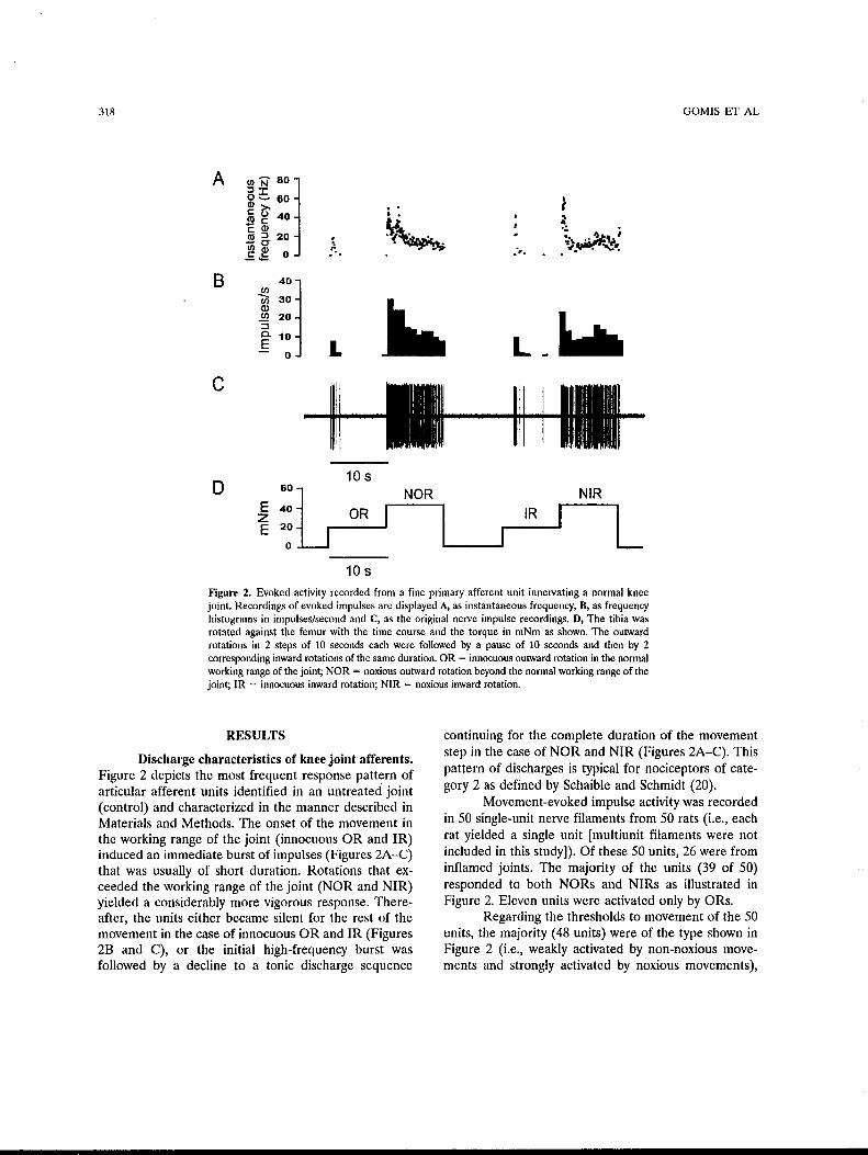
10 s Figure 2. Evoked activity recorded from a fine primary afferent unit innervating a normal knee joint. Recordings of evoked impulses are displayed A, as instantaneous frequency, B, as frequency histograms in impulses/second and C, as the original nerve impulse recordings. D, The tibia was rotated against the femur with the time course and the torque in mNm as shown. The outward rotations in 2 steps of 10 seconds each were followed by a pause of 10 seconds and then by 2 corresponding inward rotations of the same duration. OR = innocuous outward rotation in the normal working range of the joint; NOR = noxious outward rotation beyond the normal working range of the joint; IR = innocuous inward rotation; NIR = noxious inward rotation.
RESULTS
Discharge characteristics of knee joint afferents. Figure 2 depicts the most frequent response pattern of articular afferent units identified in an untreated joint (control) and characterized in the manner described in Materials and Methods. The onset of the movement in the working range of the joint (innocuous OR and IR) induced an immediate burst of impulses (Figures 2A-C) that was usually of short duration. Rotations that exceeded the working range of the joint (NOR and NIR) yielded a considerably more vigorous response. Thereafter, the units either became silent for the rest of the movement in the case of innocuous OR and IR (Figures 2B and C), or the initial high-frequency burst was followed by a decline to a tonic discharge sequence
continuing for the complete duration of the movement step in the case of NOR and NIR (Figures 2A-C). This pattern of discharges is typical for nociceptors of category 2 as defined by Schaible and Schmidt (20).
Movement-evoked impulse activity was recorded in 50 single-unit nerve filaments from 50 rats (i.e., each rat yielded a single unit [multiunit filaments were not included in this study]). Of these 50 units, 26 were from inflamed joints. The majority of the units (39 of 50) responded to both NORs and NIRs as illustrated in Figure 2. Eleven units were activated only by ORs.
Regarding the thresholds to movement of the 50 units, the majority (48 units) were of the type shown in Figure 2 (i.e., weakly activated by non-noxious movements and strongly activated by noxious movements),

HA SOLUTIONS IN THE REDUCfiON OF PAIN-ELICITING NERVE ACfiVITY 319
while 2 units responded only to noxious movements (nociceptors of category 3 [21 ]). Spontaneous activity was very rarely encountered, and this phenomenon will not be considered further in this report.
To test the efficacy of 3 HA and hylan preparations on the discharge characteristics of fine articular afferents in normal or inflamed joints, a series of movement sequences of the type displayed in Figure 2D was recorded as described in Materials and Methods. Thereafter, the test substance was injected into the articular cavity, and the movement sequences were continued at the same rate.
Effects of Synvisc on the movement-evoked discharges of tine articular primary afferents. An example of the effect of an injection of Synvisc into the inflamed joint is illustrated in the specimen records displayed in Figure 3. Figure 3A shows the control responses to the movement protocol (Figure 3D), whereas Figures 3B and C illustrate the movement-evoked responses 10 minutes and 80 minutes, respectively, after injection. As early as 10 minutes after the Synvisc application, the total number of impulses per discharge signaling noxious joint movement (NOR and NIR) to the central nervous system was reduced by 26% of the control value. Eighty minutes after the Synvisc injection, the total number of impulses in the discharge was reduced by 40% compared with the same sequence of movements under control conditions.
The unit in Figure 3 had a rather high threshold to movement and did not produce more than 2 impulses to innocuous OR or to innocuous IR. In other units with somewhat lower thresholds to innocuous movements, non-noxious rotations did produce greater numbers of impulses (Figure 2). The average responses before and after Synvisc application to movements in the working range of the joint are illustrated by open circles for the units from normal and inflamed joints in Figures 4A and B, respectively. For the same units, the effects of Synvisc on the responses to movement into the noxious range (NOR and NIR) are displayed by solid triangles in Figures 4A and B. Figure 4A shows the average responses of 10 units from normal joints, and Figure 4B shows the average responses of 8 units from inflamed joints. Impulse activity decayed slowly after Synvisc injection, particularly for the discharges evoked by movements in the working range of the joint. Both in normal and in inflamed joints, mean impulse activity evoked by non-noxious movements was significantly different from control responses (P < 0.05 by paired t-test) at 60 minutes and 100 minutes after injection. The decrease in the impulse activity evoked by noxious
A gJ ~ 1oo 0 ~ 80
8
D
~ (j' 60
~ ~ 40 ctl ::l 20 u;g .E.:= 0
o~ 80 ~ ~ 100 l ~ (j' 60
~ ai 40 .l!l fr 20 ~ ~ 0 _,._
E z E
100l 80 60
40 20
0
60
Control
~ ........... •• >-•
10 min after injection
.. .~ .... -..
80 min after injection
10 s
NOR NIR
10 s
Figure 3. Effect of Synvisc on the evoked activity in a fine primary afferent unit (conduction velocity 1.45 meters/second) innervating an inflamed joint. A, Instantaneous frequency and original nerve impulse recordings before intraarticular injection of 50 JLI Synvisc. B, Same parameters 10 minutes after intraarticular injection of 50 JLI Synvisc. C, Same parameters 80 minutes after intraarticular injection of 50 JLI Synvisc. D, Movement protocol us.ed in all experiments of the study (see Figure 20). See Figure 2 for definitions.
movements became significant 100 minutes after injection (P < 0.05).
The comparison of the movement-evoked responses to non-noxious and noxious stimulation has to take into account the borderline sensitivity of these category 2-type nociceptive afferents to movements in the working range of the joint. The numbers of impulses averaged for outward and inward movements in the working range (innocuous OR and IR) are several

320
A 11i0
c
~ 100
E
0
Normal knee joints
Synvisc
l .. ~ ..................................................... .
I ::::::::..-1 ~ 0 ~ ~ Iii) ~ 100 1~ 1~ 11i0 1~
min
0 ~ ~ Iii) Iii) 100 1~ 1~
min
{) io 4o 6o 8o 100 12o 14o 100 1Bo
nin
B 200
~ 100
~ 100
~ 0
D
F ooo
J: ~ 200
8 100
0
GOMIS ET AL
Inflamed knee joints
Synvi&c
0 ~ ~ Iii) ~ 100 1~ 1~ 11i0
min
0
0
Hyalgan
~ ~ Iii) Iii) 100 1~ 1~
nin
Qthovlsc
~ ~ Iii) Iii) 100 1~ 1~
nin Figure 4. Effects of the 3 elastoviscous solutions on the movement-evoked impulse activity of fine primary afferents innervating normal and inflamed knee joints. The movement protocol is the same as that illustrated in Figures 20 and 30. A and B, Effect of Synvisc injection. C and D, Effect of Hyalgan injection. E and F, Effect of Orthovisc injection. Open circles represent the average of the counted impulses for the non-noxious movement in both directions (OR and IR) over the periods of time shown, expressed as the mean and SEM percentage of the average control responses (first 6 complete movement cycles). Solid triangles represent the same analysis for the noxious movement in both directions (NOR and NIR). Dotted lines indicate control baselines. Arrows indicate times when test substances were injected into knee joints. See Figure 2 for definitions.
orders of magnitude smaller than those for outward and inward movements outside the working range (NOR and NIR). This is clearly seen in the specimen recordings obtained from normal and inflamed joints (Figures 2 and 3). Therefore, in Figures 5 and 6, we plotted the total afferent inflow evoked by a complete movement cycle, thus Jumping together the numbers of impulses evoked by the full displacement of the joint (values for innocuous OR, NOR, innocuous IR, and NIR of each movement protocol) from normal and inflamed joints (Figures 5 and 6, respectively).
The effects of Synvisc in normal and inflamed
joints are displayed in Figures SA and 6A, respectively. Synvisc consistently reduced the number of impulses evoked by movement soon after the injections, with no sign of recovery during the more than 2 hours of the recording period. During that time, in both the normal and inflamed joints, the evoked activity was significantly reduced to ~70% of the control frequency (P < 0.05 at 60 minutes and 100 minutes after the injection, by paired t-test and Wilcoxon signed rank test).
Effects of Hyalgan on the movement-evoked discharges of fine articular primary afferents. The sequence of events just described was repeated in a

HA SOLUTIONS IN THE REDUCTION OF PAIN-ELICITING NERVE AcTIVITY 321
Normal knee joints
A Q) 120 (/) c 100 0 c. (/) Q) 80 ,_
0 60 ,_ -c 8 40
~ 0 20
c Q) 140 (/) c
120 0 c. (/)
100 ~
e 80 ...... c 0 60 (.)
~ 0 40
' Synvisc
····-n~q., ................................................. 'I\!" i, ··--h,t ' 1ft~{': , fT If -. -
0
0
40
.:~,T <I "tt I· ...... "- 1-J t·~ " - J . '
80
min 120 160
Orthovisc
40 80
min 120 160
B (J) 170 (/) c
150 0 c. (/)
130 Q) ,_
e 110 -c 0 90 (.)
~ Q 70
0 120
80
40
0
120
80
40
0 CD II) c:: 120 0 0. II)
80 ~ :g 40 c: 8 0 '#.
Hyalgan
~Tt TfwJ _ _ _ _ . . .... T T .. J.fJl~"J::tl -1 j ..... a~ JiHt!}f~ ................................. .
0 40 80
min
control 15mln 60mln 100mln
120 160
Synvisc
Hyalgan
Orthovisc
Figure 5. Effects of the 3 elastoviscous solutions on the movement-evoked impulse activity of fine primary afferents innervating normal knee joints. The movement protocol is the same as that illustrated in Figures 2D and 3D. Graphs in A-C display the a.veraged total number (OR + NOR + IR + NIR) of evoked impulses of 6-10 primary afferent units over the period of time shown, expressed as the mean :!: SEM percentage of the average control responses. Dotted lines indicate control baselines. Arrows indicate times when test substances"were injected into knee joints. A, Effect of Synvisc injection (n = 10). B, Effect of Hyalgan injection (n = 6). C, Effect of Orthovisc injection (n = 8). D, Evoked impulse activity under control conditions and at different times after the injection of the test substances is expressed as the mean and SEM percentage of the control response, which was taken as 100%. Each column represents the average of 3 consecutive recordings taken before the designated times. * = P < 0.05 versus control. See Figure 2 for definitions.
series of 12 experiments in which Hyalgan was injected into 6 normal and 6 inflamed knee joints. The results are displayed in Figures 4C and D, 5B, and 6B in the same way as described for the experiments using Synvisc. In the normal joint, Hyalgan injection led to a slight increase in the number of impulses evoked by
innocuous ORs and IRs in all units recorded in this series (Figure 4C, open circles). This gradual increase was also present-although less pronounced-in the units from inflamed joints (Figure 4D, open circles). The number of impulses evoked by noxious movements (NORs and NIRs) (Figures 4C and D, solid

322 GOMIS ET AL
Inflamed knee joint
A (]) 120 Ill c: 0 0. Ill 80 (]) .... e ..... 40 c: 8 ~ 0 0
c (]) 140 Ill c: 0 0. 100 Ill (]) .... e 60 -c: 8 ~ 0 20
Synvisc .,v~·-·········· ................................... .
\~·'!-~,··;··.·
0 40
0 40
; : " \ l ~ - ; ; : ; ;
. "·1 . : :
80
min 120
Orthovisc
80
min 120
160
160
B (]) 180 r/) c: 0 0. 140 r/) (]) .... e 100 -c: 8
::,e 0 60
0 120
Hyalgan
I . .
~A 0 40 80
min 120 160
Synvisc
Hyalgan
Orthovisc
conlrol 15 min 60 min 100 min
Figure 6. Effects of the 3 different elastoviscous solutions on the movement-evoked impulse activity of fine primary afferents innervating inflamed knee joints. The movement protocol is the same as that illustrated in Figures 2D and 3D. Graphs in A-C display the averaged total number (OR + NOR + IR + NIR) of evoked impulses of 6-8 primary afferent units over the period of time shown, expressed as the mean ± SEM percentage of the average control responses. Dotted lines indicate control baselines. Arrows indicate times when test substances were injected into knee joints. A, Effect of Synvisc injection (n = 8). B, Effect of Hyalgan injection (n = 6). C, Effect of Orthovisc injection (n = 6). D, Evoked impulse activity under control conditions and at different times after the injection of the test substances is expressed as the mean and SEM percentage of the control response, which was taken as 100%. Each column represents the average of 3 consecutive recordings taken before the designated times. * = P < 0.05 versus control. See Figure 2 for definitions.
triangles) remained unaltered during the observation periods.
The absence of any attenuating effect of Hyalgan application was confirmed when the total afferent outflow in response to innocuous OR, innocuous IR, NOR, and NIR for the units from the 6 normal joints and 6 inflamed joints (Figures 5B and 6B, respectively) were combined. In this combined picture, the increase in activity observed in the normal joint 100 minutes after the application ( -120% of the control discharge) was not significant.
Effects of Orthovisc on the movement-evoked discharges of fine articular primary afferents. Orthovisc was injected into 8 normal and 6 inflamed joints when we recorded the movement-evoked activity from single primary afferent units as in the two series of experiments described above. The results obtained in response to non-noxious and noxious movements for units from normal and inflamed joints are displayed in Figures 4E and F, respectively, in the same way as shown in Figures 4A-D. No significant changes in impulse activity evoked by non-noxious (innocuous OR and IR) and noxious

HA SOLUTIONS IN THE REDUCTION OF PAIN-ELICITING NERVE ACTIVITY 323
Q) 140 Buffer C/) c::
~rnij __ 0 120 0. C/) Q) L..
0 100
L.. ...... c:: 80 0 ()
~ 0 60
0 30 60 90 120 150 180
min Figure 7. Movement-evoked activity in 6 primary afferent units innervating inflamed knee joints before and after intraarticular injection (arrow) of the buffer solution. Stimulus protocol, data recording, evaluation, and graphic display are the same as those in Figures SA-C and 6A-C.
(NOR and NIR) movements were observed in normal joints following injection of Orthovisc. In contrast, in inflamed joints, a small and brief, but nevertheless significant, reduction of the discharges evoked by noxious movements was observed 15 minutes after injection (P < 0.05 by paired t-test) (see also Figure 6C).
The total afferent outflows in response to the complete movement protocol are plotted in Figure 5C for the units from normal joints and in Figure 6C for the units from inflamed joints. The total number of impulses evoked by the complete movement cycle in normal joints showed a small decline 1 hour after the injection, but this was not significant. In 6 inflamed joints, however, the number of impulses per movement sequence shortly after the Orthovisc application dropped significantly to -70% of the control values and thereafter returned slowly to the control level (Figure 6C).
The gtaphs displayed in Figures 5D and 6D summarize the major results of this study 15, 60, and 100 minutes after the injection of the 3 substances. Significant reductions in the number of impulses in the evoked discharges were obtained for Orthovisc only at 15 minutes in inflamed joints and for Synvisc at 60 and 100/110 minutes in normal as well as inflamed joints.
Ineffectiveness of the buffer solution. Figure 7 shows the absence of any effect of intraarticular injection of the buffer solution into 6 inflamed knee joints on the mean firing discharge of 6 afferent units evoked by mechanical stimulation of the type shown in Figures 2 and 3. Conduction velocity of the fibers ranged from
1.19 meters/second to 3.33 meters/second (average 2.13 meters/second). During the 2 hours of the recording period following the intraarticular application of the buffer, the average number of impulses recorded per movement sequence increased somewhat, an observation similar to those seen in Figures 4C and 5B. However, the increase in the impulse discharge did not become significant. The effect of buffer solution on normal joints was not tested. When comparisons were made between average values of impulse activity after injection of the buffer and after injection of the different test substances at 15 minutes, 60 minutes, and 100 minutes (by one-way ANOV A), results were similar to those seen when mean values of impulse activity before injection were used as control (by paired t-test).
DISCUSSION
Regarding the neurophysiologic aspects of our study, the present series of experiments compared under standardized conditions the effects of various HA and hylan preparations of different concentration and molecular size on the evoked discharges of fine articular primary afferent units from normal and inflamed knee joints of the rat. Only the most elastoviscous HA derivative, Synvisc, reduced the impulse activity in sensory fibers evoked by movements within and outside the working range of both normal and inflamed joints. In inflamed joints, but not in normal joints, a more modest and brief effect was obtained with Orthovisc. Finally, Hyalgan, the HA solution with the lowest elastoviscosity, was ineffective in both normal and inflamed joints. The intensity of acute pain experienced during noxious stimulation of peripheral tissues appears to be roughly proportional to the firing frequency of nociceptor fibers (22). Accordingly, only Synvisc would have a durable analgesic effect on experimentally induced joint pain via its attenuating action on joint nociceptor activity.
Transduction and encoding of innocuous and noxious forces into nerve impulses by sensory nerve afferents innervating the soft tissues of the joint take place in the peripheral, mechanosensory nerve endings (19-21). These are equipped with stretch-activated channels, and the general model of the mechanosensory transduction apparatus is envisaged as an ion channel that detects deflections of an external structure, such as the extracellular matrix, relative to an internal structure, such as the cytoskeleton. Deflection resulting from a deformation of the tissue where the nociceptor ending is located changes tension in all elements of the system, and the transduction channels respond by increasing

324
their opening probability, permitting the entry of a large number of cations (23).
Mechanosensory endings in the capsule and synovial tissue of the joints are embedded in the intercellular matrix of the coHagen fiber network with HA and proteoglycans. It has been proposed that HA is a critical element in a "pericellular molecular cage" that transmits tensile and compression forces to the mechanosensory apparatus (24,25). Thus, we speculate that the reduction of nociceptor activity caused by intraarticular injection of highly elastoviscous HA or hylan solutions is due to a reduced transmission of the gating force to the stretchactivated channels in nociceptor nerve terminals. In favor of this hypothesis is the reduction of the opening probability of stretch-activated channels of Xenopus oocytes activated by external pressure when they are immersed in elastoviscous HA solutions, both in the intact cells and in isolated membrane patches (26).
The present experiments confirm the attenuation of impulse activity of nociceptors by elastoviscous HA and hylan solutions (11,13) and illustrate the role played by molecular size and concentration of HA and hylan molecules in obtaining this effect. The average molecular weight and the concentration determine the elastic and viscous properties of solutions of polymers like HA and its derivative, hylan (27-29). HA in human synovial fluid has an average molecular weight of 4-5 million and a concentration of 2.5-4 mg/ml (27). Under slow deformation frequencies, it behaves as a viscous and pseudoplastic fluid, while at high rates of deformation, it acts as a coiled molecular shock absorber, storing the energy transmitted as elastic deformation and thus behaving like an elastic body (30).
In the present experiments, both in normal and inflamed joints, Synvisc, a solution containing hylan A with an elastoviscosity comparable with that of healthy synovial fluid, was effective in reducing the stimulating effect of moderate and strong forces on fine medial articular nerve afferents with low and high thresholds to mechanical stimuli. The elastoviscous behavior reported for Synvisc (15) predicts that this substance may absorb an important part of the mechanical energy of the stimulus. This would reduce the transmission of force to the mechanotransduction apparatus, thus decreasing the response of knee joint sensory fibers to mechanical forces and the stimulus force applied to the knee sensory fiber, as was observed in the present experiments.
An interesting observation was that the reduction of nerve activity caused by Synvisc was more pronounced for movements performed within the working range of the joint. It is possible that when strong forces are
GOMJS ET AL
transmitted to the mechanosensory channels, the filtering effect of viscoelastic molecules becomes proportionally less important. Solutions with lower elastoviscosity (Hyalgan and Orthovisc) are much less effective in converting mechanical energy into elastic deformation. Consequently, their attenuating effect on movementevoked afferent activity in our experiments was much less pronounced or even unnoticeable both in normal and in inflamed joints.
The attenuating effect of Synvisc in the normal and in the inflamed joints started 30 minutes after injection and lasted throughout the entire experiment. Orthovisc, when injected into the inflamed joint, caused a slight but significant attenuation of the movementevoked discharges that lasted only -1 hour. The shortlived reduction of the impulse discharges by Orthovisc in the inflamed joint is intriguing. Perhaps the injected solution containing 15 mg/ml of HA transiently raised the concentration of HA, which is low in an acutely inflamed joint exudate (estimated to be 1-2 mg/ml). Later on, when passive joint movements mix and dilute the injected HA with the joint exudate, elastoviscosity falls again and the analgesic effect disappears. Also, we cannot completely rule out the possibility that a part of the analgesic effect of HA is caused by its direct interaction with specific cell surface receptors, as suggested by Gotoh and coworkers (8). In our experiments, as well as in behavioral studies (7), the various HA preparations tested had identical chemical composition and polyanionic properties. Thus, both sets of data favor the hypothesis that a mechanical filtering action, such as elastoviscosity, is the principal cause of the differences in the effect on nerve discharges of the substances being investigated.
Regarding the clinical significance of our findings, in healthy joints, movements within and outside the normal range of motion elicited nerve discharges that were significantly attenuated by Synvisc, the most elastoviscous solution with the lowest concentration of HA (0.8% ). At the other extreme, Hyalgan, with the lowest elastoviscosity and with a somewhat higher concentration of HA (1% ), was ineffective. One can assume that in the healthy joint, release of inflammatory mediators is negligible, even after repeated motions. Therefore, the effect must be mainly on the mechanotransduction apparatus.
This interpretation is further supported by the data obtained in the inflamed knee joints. There, chemical agents released by the irritation secondary to the intraarticular injections of kaolin and carrageenan sensitize joint nociceptors and enhance their responsiveness

HA SOLUTIONS IN THE REDUCfiON OF PAIN-ELICITING NERVE ACfiVITY 325
(20). This effect appears to be slightly more pronounced in the inflamed knee joint of the cat than in that of the rat. In these conditions, Synvisc reduced movementevoked activity in the same proportion as in healthy knee joints. The possibility that intraarticular HA exerts an antiinflammatory action has been suggested repeatedly and is supported by experimental evidence obtained both in vitro and in animal models (refs. 7 and 31-33, and for review, see ref. 24). If this is the case, the contribution of such an antiinflammatory effect to the reduction of impulse activity in the present experimental model was small at best. Thus, filtering of mechanical forces appears to be the main mechanism by which intraarticular HA or hylan solutions attenuate impulse activity in healthy and in acutely inflamed joints.
One must be careful, however, in assuming that the pain relief observed in human osteoarthritic knee joints after injection of HA preparations of various elastoviscosities is directly comparable with the results reported from the present study. The joint pathology developed in the acute rat arthritis model is different from the pathophysiology of any painful human arthritis. Moreover, no systematic, well-controlled clinical study has been made analyzing the pain-relieving effect of viscosupplementation during the first 24 hours. Finally, only 1 clinical study has compared the pain-relieving effects of low- and high-elastoviscous hylan and HA preparations, namely, Synvisc and Artzal (average molecular weight 0.75 million; Seikagaku Kogyo, Tokyo, Japan) (34). In that study, after the first injection of 1 ml of either preparation, significant pain relief (pain at rest, weight-bearing pain, overall pain) was observed after 1 week. No data were collected during the first week, and the pain relief effects of both preparations after 1 week were not significantly different. After 2 more injections of each preparation 1 week apart, significantly greater pain relief was observed with Synvisc than with Artzal at 8 weeks and 12 weeks after the beginning of the treatment.
Despite the limitations of extrapolating animal results to clinical situations, our findings support the importance of elastoviscosity in determining the analgesic properties of viscosupplementation materials. It is well established that this elastoviscosity depends on the average molecular weight, the polydispersity, and the concentration of the HA or hylan molecules in the various products available as therapeutic agents for arthritis pain. The data also indicate that further increasing the elastic and viscous properties of the HAs or hylans used for intraarticular injection may result in
products with more effective analgesic properties on joint pain.
ACKNOWLEDGMENTS
Synvisc and its buffer solution were gifts of Biomatrix, Inc. The authors wish to express their thanks to Eva Quintero, Ana Miralles, and Maria Oppmann for technical assistance.
REFERENCES
1. Balazs EA, Denlinger JL. Sodium hyaluronate and joint function. Equine Vet Sci 1985;5:217-28.
2. Adams ME. Viscosupplementation as articular therapy. Perspective. In: Laurent T, editor. The chemistry, biology and medical applications of hyaluronan and its derivatives. London: Portland Press; 1996. p. 243-53.
3. Weiss C. Hyaluronan and hylan in the treatment of osteoarthritis. In: Kennedy JF, Phillips GO, Williams PA, Hascall VC, editors. Hyaluronan. Cambridge: Woodhead; 2002. p. 467-82.
4. Balazs EA, Denlinger JL. Viscosupplementation: a new concept in the treatment of osteoarthritis. J Rheumatol 1993;20:3-9.
5. Rydell N, Balazs EA. Effect of intraarticular injection of hyaluronic acid on the clinical symptoms of osteoarthritis and on granulation tissue formation. Clin Orthop 1971;80:25-32.
6. Gingerich DA, Auer JA, Fackelman GE. Effect of exogenous hyaluronic acid on joint function in experimentally induced equine osteoarthritis: dosage titration studies. Res Vet Sci 1981;30:192-7.
7. Golob S, Miyazaki K, Onaya J, Sakamoto T, Tokuyasu K, Namik 0. [Experimental knee pain model in rats and analgesic effect of sodium hyaluronate (SPH)) Nippon Yakurigaku Zasshi 1988;92: 17-27. Japanese.
8. Gotoh S, Onaya JI, Abe M, Miyazaki K, Hamai A, Horie K, et al. Effects of the molecular weight of hyaluronic acid and its action mechanisms on experimental joint pain in rats. Ann Rheum Dis 1993;52:817-22.
9. Ghosh P, Read R, Armstrong S, Wilson D, Marshall R, McNair P. The effects of intraarticular administration of hyaluronan in a model of early osteoarthritis in sheep. I. Gait analysis and radiological and morphological studies. Semin Arthritis Rheum 1993; 22:18-30.
10. Aihara S, Murakami N, Ishii R, Kariya K, Azuma Y, Hamada K, et al. [Effects of sodium hyaluronate on the nociceptive response of rats with experimentally induced arthritis] Nippon Yakurigaku Zasshi 1992;100:359-65. Japanese.
11. Pozo MA, Balazs EA, Belmonte C. Reduction of sensory response to passive movements of inflamed knee joints by hylan, a hyaluronan derivative. Exp Brain Res 1997;116:3-9.
12. Belmonte C, Pozo MA, Balazs EA. ModulatioA by hyaluronan and its derivatives (hylans) of sensory nerve activity signalling articular pain. In: Laurent TC, editor. The chemistry, biology and medical applications of hyaluronan and its derivatives. London: Portland Press; 1998. p. 205-17.
13. Pawlak M, Gomis A, Just S, Heppelmann B, Belmonte C, Schmidt RF. Mechanoprotective actions of elastoviscous hylans on articular pain receptors. In: Kennedy JF, Phillips GO, Williams PA, Hascall VC, editors. Hyaluronan. Cambridge: Woodhead; 2002. p. 341-52.
14. Just S, Pawlak M, Heppelmann B. Responses of fine primary afferent nerve fibres innervating the rat knee joint to defined torque. J Neurosci Methods 2000;103:157-62.
15. Balazs EA, Leshchiner EA. Hyaluronan, its crosslinked derivative-hylan-and their medical applications. In: Inagaki H, Phillips GO, editors. Cellulosics utilization: research and rewards in cellulosics. New York: Elsevier Applied Science; 1989. p. 233-41.

326
16. Lee HG, Cowman MK. An agarose gel electrophoretic method for analysis of hyaluronan molecular weight distribution. Anal Biochem 1994;219:278-87,
17. Bothner H, Waaler T, Wik 0. Limiting viscosity number and weight average molecular weight of hyaluronate samples produced by heat degradation. lnt J Bioi Macromol1988;10:287-91.
18. Kanaka R, Schaible H-G, Schmidt RF. Activation of fine articular afferent units by bradykinin. Brain Res 1985;327:81-90.
19. Schaible H-G, Schmidt RF. Effects of an experimental arthritis on the sensory properties of fine articular afferent units. J Neurophysiol 1985;54:1109-22.
20. Schaible H-G, Schmidt RF. Time course of mechanosensitivity changes in articular afferents during a developing experimental arthritis. J Neurophysiol 1988;60:2180-95.
21. Schaible H-G, Schmidt RF. Responses of fine medial articular nerve afferents to passive movements of knee joint. J Neurophysiol 1983;49:1118-26.
22. Torebjiirk HE, Schmelz M, Handwerker HO. Functional properties of human cutaneous nociceptors and their role in pain and hyperalgesia. In: Belmonte C, Cerver6 F, editors. Neurobiology of nociceptors. Oxford: Oxford University Press; 1996. p. 349-69.
23. Belmonte C. Transduction and encoding of noxious stimuli. In: Schmidt RF, Willis WD, editors. Encyclopedic reference of pain. Heidelberg: Springer Verlag. In press. (online at http://encref. springer.de ).
24. Balazs EA. The viscoelastic intercellular matrix and control of cell function by hyaluronan. In: Laurent TC, editor. The chemistry, biology and medical applications of hyaluronan and its derivatives. London: Portland Press; 1998. p. 185-204.
25. Balazs EA. Analgesic effect of elastoviscous hyaluronan solutions and the treatment of arthritic pain. Cells Tissues Organs 2003;174: 711-24.
26. De Ia Pei\a E, Sala S, Rovira JC, Schmidt RF, Belmonte C.
GOMIS ET AL
Elastoviscous substances with analgesic effects on joint pain reduce stretch-activated ion channel activity 'in vitro.' Pain 2002; 99:501-8.
27. Balazs EA, Gibbs DA. The rheological properties and biological function of hyaluronic acid. In: Balazs EA, editor. Chemistry and molecular biology of the intercellular matrix. London and New York: Academy Press; 1970. p. 1241-53.
28. Gibbs D, Merrill E, Smith K, Balazs EA. Rheology of hyaluronic acid. Biopolymers 1968;6:777-91.
29. Milas M, Rinaudo M, Roure I, AI-Assaf S, Phillips GO, Williams P A. Rheological behavior of hyaluronan, Heal on and hylan in aqueous solutions. In: Kennedy JF, Phillips GO, Williams ·PA, Hascall VC, editors. Hyaluronan. Cambridge (UK): Woodhead; 2002. p. 181-94.
30. Balazs EA. The physical properties of synovial fluid and the special role of hyaluronic acid. In: Helfet A, editor. Disorders of the knee. Philadelphia: JB Lippincott; 1982. p. 61-74.
31. Balazs EA, Darzynkiewicz Z. The effect of hyaluronic acid on fibroblasts, mononuclear phagocytes and lymphocytes. In: Kulonen E, Pikkarainen J, editors. Biology of the fibroblast. London: Academic Press; 1973. p. 237-52.
32. Frean SP, Lees P. Effects of polysulfated glycosaminoglycans and hyaluronan on prostaglandin E2 production by cultured equine synoviocytes. Am J Vet Res 2000;61:499-505.
33. Akatsuka M, Yamamoto Y, Tobetto K, Yasui T, Ando T. In vitro effects of hyaluronan on prostaglandin E2 induction by interleukin-1 in rabbit articular chondrocytes. Agents Actions 1993;38: 122-5.
34. Wobig M, Bach G, Beks P, Dickhut A, Runzheimer J, Schwieger G, et al. The role of elastoviscosity in the efficacy of viscosupplementation for osteoarthritis of the knee: a comparison of hylan G-F 20 and a lower-molecular-weight hyaluronan. Clin Ther 1999;21:1549-62.