effects of da-5513 on alcohol metabolism and alcoholic ...effects of da-5513 on serum alcohol and...
TRANSCRIPT
49
Lab Anim Res 2018: 34(2), 49-57
https://doi.org/10.5625/lar.2018.34.2.49
ISSN 1738-6055 (Print)
ISSN 2233-7660 (Online)
Effects of DA-5513 on alcohol metabolism andalcoholic fatty liver in rats
Jae Young Yu1,#, Hanh Thuy Nguyen2,3,#, Chul Soon Yong2, Hyoung Geun Park1, Joon Ho Jun1, Jong Oh Kim2,*1Department of Formulation Development, Dong-A Pharmaceutical Co. Ltd., Yongin, Korea
2College of Pharmacy, Yeungnam University, Gyeongsan, Korea3National Institute of Pharmaceutical Technology, Hanoi University of Pharmacy, Hanoi, Vietnam
Hangover is characterized by a number of unpleasant physical and mental symptoms that occur afterheavy alcohol drinking. In addition, consistently excessive alcohol intake is considered as a major reasoncauses liver disease. The present study investigated the in vivo effects of DA-5513 (Morning care® KangHwang) on biological parameters relevant to hangover relief and alcoholic fatty liver. Blood alcohol andacetaldehyde concentrations were determined in rats administered a single dose of alcohol and treatedwith DA-5513 or commercially available hangover relief beverages (Yeomyung® and Ukon®). The effectsof DA-5513 on alcoholic fatty liver were also determined in rats fed alcohol-containing Lieber-DeCarlidiets for 4 weeks. Serum liver function markers (aspartate and alanine aminotransferase activities) andserum/liver lipid levels were assessed. Blood alcohol and acetaldehyde concentrations were lower in thegroups treated with DA-5513 or Yeomyung®, as compared with control rats. However, Ukon® did notproduce any significant effects on these parameters. Treatment with DA-5513 significantly reduced serumaspartate and alanine aminotransferase activities and markedly reduced serum cholesterol andtriglyceride levels, as compared with control rats. Histological observations using Oil Red O stainingfound that DA-5513 delayed the development of alcoholic fatty liver by reversing hepatic fataccumulation. These findings suggest that DA-5513 could have a beneficial effect on alcohol-inducedhangovers and has the potential to ameliorate alcoholic fatty liver.
Keywords: Morning care®, hangover, acetaldehyde, alcohol-induced fatty liver, hepatic triglyceride
Received 9 March 2018; Revised version received 19 April 2018; Accepted 25 April 2018
Alcoholic liver disease has been demonstrated to be a
major cause of morbidity and mortality worldwide in
individuals with consistently excessive alcohol intake
[1]. Hangover is characterized by a number of unpleasant
physical and mental symptoms that occur after heavy
alcohol drinking [2,3]. Alcohol is initially oxidized to
acetaldehyde by the alcohol dehydrogenase (ADH)
enzyme; this is subsequently converted to acetate by
aldehyde dehydrogenase (ALDH) in the liver [2,3].
Acetaldehyde is much more toxic than ethanol and this
metabolite may cause the physical symptoms of hangover
such as fatigue, headache, increased sensitivity to light
and sound, redness of the eyes, muscle aches, and thirst
[3]. Furthermore, long-term consumption of alcohol in
large quantities may cause chronic liver diseases and
hepatic steatosis (alcoholic fatty liver), which is defined
as excess lipid accumulation in the cytoplasm of hepatocytes;
this is regarded as a significant risk factor for hepatic
fibrosis and cirrhosis [4]. Thus, reduction of alcohol-
induced hepatic fat accumulation may block or delay the
progression of steatosis to advanced stages of alcoholic
liver disease.
Multiple mechanisms contribute to the pathogenesis of
alcoholic hepatic steatosis, including increased de novo
#These authors contributed equally to this work.
*Corresponding author: Jong Oh Kim, 214-1, Dae-dong, College of Pharmacy, Yeungnam University, Gyeongsan, 712-749, KoreaTel: +82-53-810-2813; Fax: +82-53-810-4654; E-mail: [email protected]
This is an Open Access article distributed under the terms of the Creative Commons Attribution Non-Commercial License (http://creativecommons.org/licenses/by-nc/3.0) which permits unrestricted non-commercial use, distribution, and reproduction in any medium, provided the original work is properly cited.
50 Jae Young Yu et al.
Lab Anim Res | June, 2018 | Vol. 34, No. 2
hepatic lipogenesis, impaired mitochondrial fatty acid β-
oxidation, and reduced export of very low-density lipoprotein
[5]. However, the complex mechanisms involved in
alcoholic fatty liver formation have been debated in the
literature [6]. Accumulating evidence has suggested that
adipose tissue dysfunction might impact on hepatic lipid
metabolism [7,8]. A direct link between adipose triglyceride
(TG) loss and hepatic TG gain was revealed using
deuterium-labeled TG in alcohol-fed mice [9]. Dysregulation
of lipid homeostasis was also evidenced in clinical
studies, which identified a lower fat mass during the
development of alcoholic fatty liver [10]. Adipose tissue
plays a crucial role as a major metabolic buffering system
in lipid homeostasis [11,12], and modification of alcohol-
induced dysfunction in this tissue might therefore provide
an important target for the development of functional
foods to prevent alcoholic fatty liver [13]. Many treatments
have been reported to prevent and/or reduce the severity
of hangover and alcoholic fatty liver symptoms, including
innumerable folk remedies and recommendations.
Yeomyung® (YM, Glami, Gwangwon, Korea) is the
most commonly sold hangover recovery drink in Korea,
and Ukon® (UK, Enagic, Okinawa, Japan) is a turmeric
extract-based beverage that is marketed as an anti-
hangover drink and has enjoyed huge success in Japan.
In addition, various products have been released on the
basis that they decrease the accumulation of fat in the
liver, prevent liver damage, and alleviate alcohol-related
hangovers. However, the mechanism of action of these
products has not been revealed, and scientific research
into this area is therefore required. DA-5513 (Morning
care® Kang Hwang) was developed by the Dong-A
Pharm. Co. (Yongin, Korea) and has been approved by
the Korea Food and Drug Administration as an over-the-
counter treatment for hangover. The proprietary liquid
DA-5513 formulation was developed using seven herbal
extracts, including GMT-ALC-5L (fermented rice embryo
and bean extract), Curcuma longa L., Trapa japonica
Flerov., Silybum marianum L., Paullinia cupana Mart.,
Pueraria thunbergiana Benth., and honey. These natural
products have been used in traditional oriental medicines
to prevent alcohol-induced hangover and protect the
liver against diverse hepatotoxins such as ethanol, carbon
tetrachloride, antitubercular agents, and thioacetamide
[14-20].
The present study was therefore designed to investigate
the effect of DA-5513 on hangover relief, which would
be associated with the rapid elimination of alcohol and
acetaldehyde. Furthermore, a chronic ethanol-treated rat
model was developed to evaluate the ability of this
product to protect from alcoholic fatty liver.
Materials and Methods
Materials
DA-5513 was provided by the Dong-A Pharm. Co.
(Yongin, Korea), while Yeomyung® (YM, Glami,
Gwangwon, Korea) and Ukon® (UK, Enagic, Okinawa,
Japan) were purchased from the market. All other
chemicals used were of analytical grade.
Animals
Seven-week-old male Wistar rats were purchased
from Chung-Ang Lab Animal Inc. (Seoul, Korea) and
acclimatized to the laboratory setting (22.0±2.0oC, 12-h
light/dark cycles, 55%±5% humidity) with free access to
water and food (Samyang Co, Incheon, Korea) for a
week before the experiment. The experimental protocol
was approved by the Institutional Animal Care and Use
Committee of Dong-A Pharm. Co. (Yongin, Korea), and
all experimental procedures were conducted in compliance
with this company’s guidelines for the care and use of
laboratory animals.
Alcohol-induced hangover model
The rats (200-250 g) were randomly divided into four
groups (n=8 per group). Each group had a single oral
administration of the following test treatments: ethanol
(control group), and ethanol with either DA-5513, YM,
or UK. Then, after 0.5 h, 10 mL/kg of body weight was
administered. At 0.5, 1, 2, 4, and 6 h post-ethanol
administration, 0.2 mL blood was collected from each
rat and left at room temperature for 30 min before
centrifugation at 3,000 rpm for 15 min. The levels of
alcohol and acetaldehyde in the supernatant were
measured using kits for the detection of ethanol (Roche
Co., Darmstadt, Germany) and acetaldehyde (R-Biopharm,
Darmstadt, Germany). During the metabolism of ethanol
to acetaldehyde and acetate, nicotinamide adenine
dinucleotide (NAD+) is converted to NADH. Thus, the
concentration of NADH was determined by measuring
absorbance at 340 nm.
Alcoholic fatty liver model
Rats (220-230 g) were randomly divided into four
groups based on their body weight (n=10 per group):

Effects of DA-5513 on alcohol metabolism and alcoholic fatty liver 51
Lab Anim Res | June, 2018 | Vol. 34, No. 2
untreated control (CON), ethanol-treated control (ED),
and ethanol-treated with UK or DA-5513 groups. The
rats were fed a standard Lieber-DeCarli ethanol diet
(36% ethanol-derived calories) for 4 weeks [21,22]; pair-
fed control rats were administered dextran-maltose to
match the alcohol-derived calories in the ethanol diet.
DA-5513 or UK was introduced into the alcohol diet by
gradually mixing it with distilled water and feeding it at
the same time each day throughout the experiment. The
rats were sacrificed using ether anesthesia; blood samples
were centrifuged (1,500 g, 4oC, 10 min) to separate the
serum and stored at −80oC until analysis. The livers were
quickly removed and preserved in phosphate-buffered
formalin for histological examination. The rest of the
liver was frozen at −80oC prior to analysis of hepatic TG
levels.
Serum levels of alanine aminotransferase (ALT),
aspartate aminotransferase (AST), total cholesterol (T-
CHO), and TG were monitored by standard clinical
chemistry assays on an Automated Chemistry Analyzer
(Prestige 24I; Tokyo Boeki Medical System, Tokyo,
Japan). Total liver lipids were extracted from homogenates
prepared from 100 mg rat liver using chloroform:methanol
(2:1, v/v) [23]. The TG levels within total lipid samples
were determined enzymatically using a commercially
available enzymatic kit (Sigma Chem. Co.) according to
the manufacturer’s protocol.
Histological study
As proposed by Levene et al., Oil Red O can be used
to identify lipids and quantify hepatic steatosis. Several
protocols have been developed, including paraffin-
embedded sections and cryosections (or frozen sections)
[24,25]. In this study, frozen liver tissues were cut into
4-mm sections and affixed to microscope slides. Sections
were stained with Oil Red O solution buffer. Histo-
pathologic examinations of the liver sections were
conducted by a pathologist and were peer-reviewed. The
slides were dehydrated, dealcoholized, mounted utilizing
Canada balsam, and assessed for inflammation and tissue
damage utilizing an Olympus microscope (Olympus,
Tokyo, Japan) [26,27].
Statistical analysis
Data are given as the mean and standard deviation, and
inter-group differences were analyzed using one-way
ANOVA by Duncan’s method. Differences were considered
significant at a P-value <0.05, and very significant at a
P-value <0.01.
Results
Effects of DA-5513 on serum alcohol and acetaldehyde
concentrations
In order to determine the effects of DA-5513 on
hangovers, blood alcohol levels were investigated and
presented in Table 1. DA-5513, YM, or UK was
administered orally 0.5 h before 25% ethanol consumption,
and blood was collected 0.5, 1, 2, 4, and 6 h after alcohol
was administered. Administration of UK resulted in a
lower blood alcohol concentration than that observed in
the control group; however, this difference was not
statistically significant (P>0.05). On the other hand, DA-
5513 and YM produced a similar significant reduction of
the blood alcohol concentration at each of the time-
points examined, as compared with the control group
(Table 1).
The blood acetaldehyde concentration, which was
highest in the CON group, peaked 0.5 h after ethanol
consumption (Table 2). Rats treated with UK showed
lower blood acetaldehyde levels than control rats, but
these differences were not statistically significant. Both
DA-5513 and YM significantly reduced the acetaldehyde
levels in the blood at every time-point examined, as
Table 1. Blood alcohol concentrations (mg/mL) in rats
GroupTime (h)
0.5 1 2 4 6
Control 53.68±3.65 49.22±2.78 54.63±1.65 46.89±2.89 41.22±3.22
UK 49.66±2.16 50.55±3.22 49.33±4.12 43.22±5.13 38.55±5.32
YM 45.63±1.99* 43.56±2.35* 42.55±1.48* 33.99±2.99* 32.56±3.11*
DA-5513 43.66±3.55* 43.56±3.66* 42.31±2.33* 37.88±3.22* 31.47±2.98*
Control, ethanol-treated control; UK, treated with ethanol and UK; YM, treated with ethanol and YM; DA-5513, treated with ethanoland DA-5513.The data represent mean±standard deviation (n=8).*P<0.01 vs Control
52 Jae Young Yu et al.
Lab Anim Res | June, 2018 | Vol. 34, No. 2
compared with the control group. However, this effect
was more marked for DA-5513 than for YM throughout
the study. Taken together, these findings indicated that
DA-5512 provided more effective reduction of alcohol
and acetaldehyde levels than other products, suggesting
that it has the potential to act as a hangover relief
beverage.
Effects of DA-5513 on serum AST and ALT
All rats were fed the standard Lieber-DeCarli ethanol
diet for 4 weeks and hepatotoxicity was evaluated by
clinical chemistry. As seen in Figure 1A and 1B, the ED
group showed a marked increase in the levels of serum
AST and ALT (by approximately 1.3- and 1.4-fold,
respectively), as compared to the untreated control group
Table 2. Blood acetaldehyde concentrations (µg/mL) in rats
GroupTime (h)
0.5 1 2 4 6
Control 85.65±5.66 68.44±1.99 54.65±2.22 53.22±3.64 39.55±1.22
UK 80.66±7.19 60.55±6.88 48.66±6.59 47.55±5.23 35.66±4.66
YM 64.88±8.22* 55.89±5.69* 46.55±4.55* 43.55±4.26* 32.66±3.94*
DA-5513 62.45±6.55* 49.88±6.99* 36.85±6.25* 34.56±3.84* 29.68±3.74*
Control, ethanol-treated control; UK, treated with ethanol and UK; YM, treated with ethanol and YM; DA-5513, treated with ethanoland DA-5513.The data represent mean±standard deviation (n=8).*P<0.01 vs Control
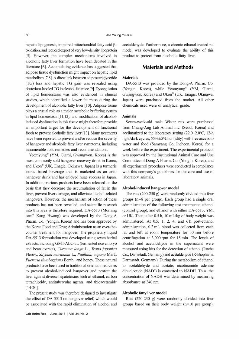
Figure 1. Effects of DA-5513 on serum (A) AST activity and (B) ALT activity in chronic ethanol-treated rats. CON, control diet; ED,alcohol diet; ED+UK, alcohol diet with UK; ED+DA-5513, alcohol diet with DA-5513. The data represent mean±SD (n=10);**P<0.01 and *P<0.05.
Figure 2. Effects of DA-5513 on serum (A) TG and (B) T-CHO levels in chronic ethanol-treated rats. CON, control diet; ED, alcoholdiet; ED+UK, alcohol diet with UK; ED+DA-5513, alcohol diet with DA-5513. The data represent mean±SD (n=10); **P<0.01 and*P<0.05.
Effects of DA-5513 on alcohol metabolism and alcoholic fatty liver 53
Lab Anim Res | June, 2018 | Vol. 34, No. 2
(P<0.01). The administration of UK product after
ethanol diet could reduce the ALT level (P<0.05) but not
AST level in comparison with ED group. In contrast, the
blood samples of the animals treated with DA-5513
revealed significant hepatoprotective activity, as evidenced
by an amelioration of this increase in serum AST and
ALT levels (P<0.01 and P<0.05, respectively).
Effects of DA-5513 on serum and liver lipid profiles
The serum T-CHO and TG levels (Figure 2A and 2B)
increased significantly in the ED group (by approximately
1.2- and 1.8-fold, respectively) (P<0.01). Animals that
received DA-5513 showed a significantly lower level of
both serum TG and T-CHO than that of ED group
(P<0.01 and P<0.05, respectively) while the difference
between UK and ED group was not significant.
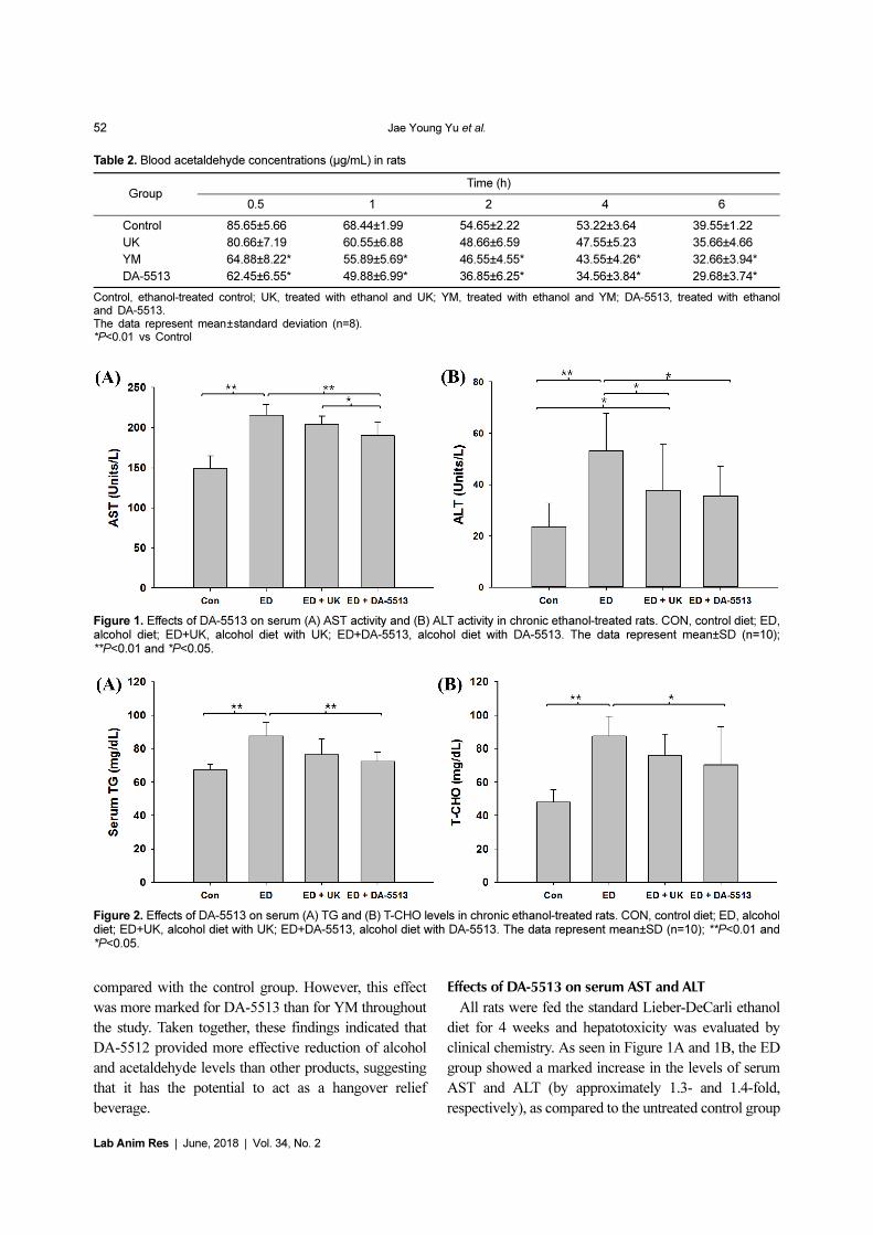
In addition, qualitative hepatic TG measurement, which
further confirmed the histological results, demonstrated
that alcohol feeding with Lieber-DeCarly diet greatly
increased the hepatic TG level in mice by 1.8-fold, in
comparison with control group (Figure 3). This elevation
was significant decreased by concomitant administration
of UK product (P<0.05) or DA-5513 (P<0.01).
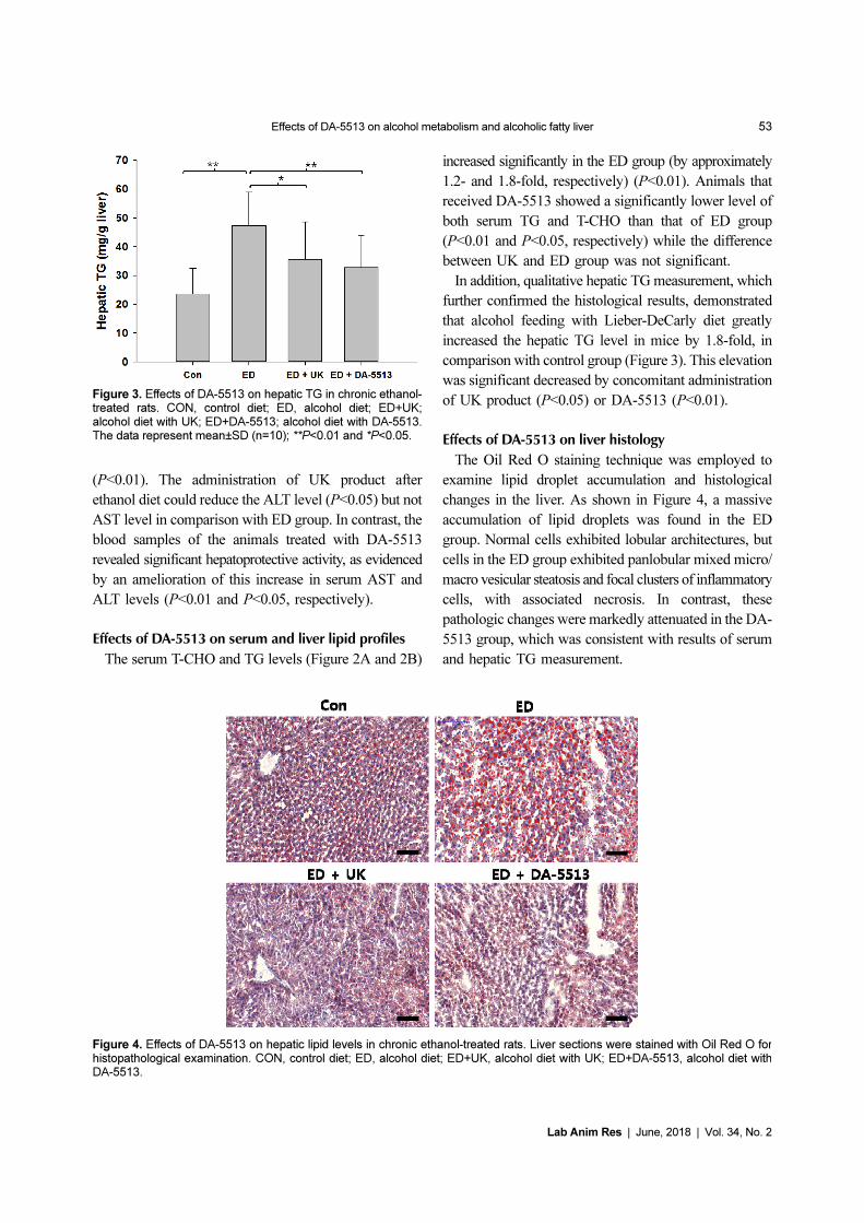
Effects of DA-5513 on liver histology
The Oil Red O staining technique was employed to
examine lipid droplet accumulation and histological
changes in the liver. As shown in Figure 4, a massive
accumulation of lipid droplets was found in the ED
group. Normal cells exhibited lobular architectures, but
cells in the ED group exhibited panlobular mixed micro/
macro vesicular steatosis and focal clusters of inflammatory
cells, with associated necrosis. In contrast, these
pathologic changes were markedly attenuated in the DA-
5513 group, which was consistent with results of serum
and hepatic TG measurement.
Figure 3. Effects of DA-5513 on hepatic TG in chronic ethanol-treated rats. CON, control diet; ED, alcohol diet; ED+UK;alcohol diet with UK; ED+DA-5513; alcohol diet with DA-5513.The data represent mean±SD (n=10); **P<0.01 and *P<0.05.
Figure 4. Effects of DA-5513 on hepatic lipid levels in chronic ethanol-treated rats. Liver sections were stained with Oil Red O forhistopathological examination. CON, control diet; ED, alcohol diet; ED+UK, alcohol diet with UK; ED+DA-5513, alcohol diet withDA-5513.
54 Jae Young Yu et al.
Lab Anim Res | June, 2018 | Vol. 34, No. 2
Discussion
Heavy alcohol drinking can result in several alcohol-
induced hangover symptoms, which are attributed to the
physiological effects of alcohol and its metabolites. It is
well established that accumulation of acetaldehyde, an
intermediate alcohol metabolite, plays a pivotal role in
the development of hangover [2,3]. In order to determine
the effects of DA-5513 on hangovers, we measured rat
blood alcohol and acetaldehyde levels at different time-
points after the administration of alcohol. Two commercially
available products, YM and UK, were used as positive
control treatments. The administration of DA-5513 and
YM was associated with lower blood alcohol and
acetaldehyde levels over the time-course of the experiment.
However, the reduction in the acetaldehyde level
observed in rats treated with DA-5513 was 20% and
27% greater than that observed in rats treated with YM
and UK, respectively. These findings indicated that DA-
5512 produced more effective reduction of the acetaldehyde
level than YM, demonstrating that it had the potential to
act as a hangover relief beverage.
The liver is the largest internal organ in the human
body and it has many different roles. One of its most
important functions is to filter harmful substances from
the blood. The liver commonly repairs itself by rebuilding
new liver cells when the old ones are damaged. However,
chronic alcohol ingestion can lead to the development of
liver diseases such as fatty liver, alcoholic hepatitis, and
cirrhosis [4]. This liver damage occurs through several
interrelated pathways. The oxidative reactions involved
in alcohol metabolism generate hydrogen, which
converts NAD to NADH, increasing the redox potential
(NADH/NAD) of the liver [28]. This increase in redox
potential inhibits fatty acid oxidation and gluconeogenesis,
promoting fat accumulation in the liver. In addition,
chronic alcoholism induces the microsomal ethanol-
oxidizing system to break down alcohol, mainly in the
endoplasmic reticulum [29]. This pathway, where
cytochrome P450 2E1 is the main enzyme, can account
for 20% of alcohol metabolism. This enzyme is
upregulated by chronic alcohol use, and generates free
radicals and harmful reactive oxygen species via the
oxidation of nicotinamide adenine dinucleotide phosphate
(NADPH) to NADP [28]. This oxidative stress promotes
hepatocyte necrosis and apoptosis, and lipid peroxidation,
which causes inflammation and fibrosis. Inflammation is
also exacerbated by acetaldehyde, which can bind
covalently to cellular proteins, forming antigenic adducts
[30,31].
Apart from the inconvenient symptoms of hangover,
long-term consumption of alcohol in large quantities is
the leading cause of liver disease and hepatic steatosis.
Alcoholic fatty liver disease results from the deposition
of fat and the accumulation of TG in liver cells. The
potential pathophysiologic mechanisms involved in fatty
liver include a reduction in mitochondrial fatty acid β-
oxidation, increased endogenous fatty acid synthesis or
enhanced delivery of fatty acids to the liver, and deficient
incorporation or export of TG as very low-density
lipoproteins [32,33]. The Lieber-DeCarli liquid diet
model is used to induce alcoholic fatty liver disease in
animals, where it causes liver injury, steatosis, and
oxidative stress. This model has been used to investigate
the relationship between alcohol and therapeutic agents
[22,34]. Previous data have indicated that feeding mice
with a standard Lieber-DeCarli formula for 2 weeks was
sufficient to induce significant steatosis [22]. The degree
of steatosis, determined by the hepatic TG concentration,
revealed that the liver samples met the criteria for a
clinical diagnosis of steatosis [35]. In our study, mice
treated with the Lieber-DeCarli diet for 4 weeks showed
steatosis, as confirmed by a significant increase in serum
ALT and AST activities, T-CHO, and TG levels. Our
data showed that dietary DA-5513 markedly attenuated
the hepatic steatosis observed in this model, as indicated
by Oil Red O staining, hepatic TG quantification, and
serum measures of AST, ALT, T-CHO, and TG.
Mono-and poly-herbal preparations have been used in
traditional medical systems for the treatment of liver
disease since long before recorded history; some of these
products appear to have positive effects on this
potentially reversible disease. Both basic and clinical
studies have suggested that herbal medicines and their
constituents such as Gynostemma pentaphyllum (Thunb.)
Makino, Panax notoginseng (Burkill) F.H.Chen (saponins),
Crataegus pinnatifida Bunge (penta-oligogalacturonide),
Dioscorea opposita Thunb. (dioscin), Punica granatum
L. (gallic acid), glycyrrhizin, silymarin, Prunus armeniaca
L (kernels), and baicalin may have modest benefits in the
treatment of fatty liver disease [36]. Therefore, DA-
5513, which is composed of several herbal extracts, was
investigated as a hangover remedy that may reduce the
blood alcohol concentration, as well as preventing
alcoholic fatty liver. GMT-ALC-5L (fermented rice
embryo and bean extract) promotes the health of both
Effects of DA-5513 on alcohol metabolism and alcoholic fatty liver 55
Lab Anim Res | June, 2018 | Vol. 34, No. 2
habitual alcohol drinkers and non-habitual alcohol
drinkers. This preparation has been demonstrated to
reduce blood alcohol levels and promote liver function
recovery by modulating alcohol-metabolizing enzymes
[37,38]. Turmeric (Curcuma longa) has been used in
traditional medicines as a household remedy for various
diseases, including biliary disorders, anorexia, cough,
diabetic wounds, rheumatism, sinusitis, and hepatic
disorders. In vitro and in vivo animal studies have
provided evidence for the hepatoprotective effects of
turmeric against a variety of hepatotoxic substances,
including carbon tetrachloride, galactosamine, pentobarbitol,
1-chloro-2,4-dinitrobenzene, 4-hydroxynonenal, and
acetaminophen (paracetamol) [39]. These hepatoprotective
effects may stem from the potent antioxidant effects of
turmeric. Dietary supplementation of turmeric in rats
(1% turmeric by weight for 10 weeks) was found to
significantly protect against iron-induced lipid peroxide
formation [40]. Curcumin is also helpful in the relief of
hangover. It exhibited an inhibitory effect on alcohol
intoxication in humans, as evidenced by a reduced blood
acetaldehyde concentration and reduced discomfort [41,
42]. Curcumin significantly reduces plasma low-density
and very low-density lipoprotein levels, reduces T-CHO
levels in the liver, and increases the α-tocopherol level
in rat plasma, suggesting an in vivo interaction between
curcumin and α-tocopherol that may increase the
bioavailability of vitamin E and decrease T-CHO levels
[43]. Silybum marianum (milk thistle) has been used for
centuries as an herbal medicine for the treatment of liver
disease. Silymarin’s hepatoprotective effects involve
several mechanisms including an antioxidant effect,
inhibition of lipid peroxidation, enhancement of liver
detoxification via inhibition of phase I detoxification,
and an increased hepatocyte protein synthesis, thereby
promoting hepatic tissue regeneration [16]. Animal
studies have also demonstrated that silybin could reduce
the conversion of hepatic stellate cells into myofibroblasts,
thus slowing or even reversing fibrosis [16]. Clinical
studies of patients with chronic alcoholic liver disease in
Austria and Hungary demonstrated that silymarin
administration resulted in a normalization of serum liver
enzyme and total bilirubin levels in patients with
alcoholic liver disease, in addition to improved liver
tissue histology [44]. In patients with cirrhosis, long-
term (41 months) administration of silymarin at 420 mg
per day resulted in a significant increase in survival, as
compared to the placebo group [45]. Although most
studies of water chestnuts (Trapa japonica Flerov.) have
focused on their nutritional and ecological value, they
have also been reported to have antioxidant, anti-cancer,
and anti-diabetic effects; these were associated with a
reduction in blood glucose level, and inhibition of α-
amylase and α-glucosidase [15]. Trapa japonica is also
used as an ethno-medicine for the treatment of gastric
ulcer, diarrhea, alcohol hangover, and dysentery [46].
Trapa japonica Flerov. was reported to significantly
inhibit the production of reactive oxygen species, thus
protecting the liver from tert-butyl hydroperoxide (t-
BHP)-induced damage by stabilizing antioxidant systems
and regulating the mitochondrial membranes within liver
cells [47]. In vivo models have indicated that Trapa
japonica Flerov. significantly attenuated t-BHP-induced
increases in serum glutamate oxaloacetate transaminase
and glutamate pyruvate transaminase levels, and in hepatic
malondialdehyde levels [48]. Guarana has also been
used as a tonic for hangovers, neuralgia and menstrual
headaches, leucorrhea, diarrhea, and fevers; this compound
has been shown to prevent DNA damage in carbon
tetrachloride-treated rats [18,20]. Kudzu (Pueraria
thunbergiana) is employed in traditional Chinese medicine
and its major isoflavone constituent, puerarin, has
antioxidant activity and a variety of biological actions in
cardiovascular disease, gynecological disease, osteoporosis,
cognition, and diabetic nephropathy [49]. Studies of
Pueraria flos showed that it increased the acetaldehyde
removal rate in both rats and humans after alcohol
consumption, and reduced hangover symptoms [50].
The kudzu vine is potentially highly beneficial in the
treatment of liver damage, as it scavenges reactive free
radicals and boosts the endogenous antioxidant system.
Kudzu vine extract significantly reduced the cytotoxicity
and production of reactive oxygen species induced by t-
BHP in vitro and lowered the plasma levels of ALT and
AST in a rat model of carbon tetrachloride-induced
hepatotoxicity [15]. Another ingredient that may help to
counteract the effects of heavy alcohol drinking is honey.
Honey contains fructose, a sugar that promotes alcohol
metabolism [51]. Furthermore, honey has considerable
anti-inflammatory, antioxidant, and antitumor activities,
and plays a key role in normalizing kidney function and
protecting the liver from a range of toxic agents [52].
Consistent with these findings, the combination of these
herbal ingredients in DA-5513 significantly ameliorated
hepatic steatosis, as evidenced by its effects on hepatic
TG, serum ALT and AST activities, and serum T-CHO

56 Jae Young Yu et al.
Lab Anim Res | June, 2018 | Vol. 34, No. 2
and TG levels.
In summary, these findings indicated that DA-5513
produced beneficial effects on alcohol metabolite levels
and alcoholic fatty liver in rats. Further studies are
required to investigate the antioxidant activity of this
preparation, and its effects on lipid mechanism, in rats
administered alcohol.
Acknowledgments
This research was supported by the Yeungnam
University research grants in 2017.
Conflict of interests There is a conflict of interest
regarding the publication of this manuscript. JY Yu, HG
Park, and JH Jun are employees of the Dong-A
Pharmaceutical Co. Ltd. The other authors have no
conflicts of interest to declare.
References
1. Rehm J, Mathers C, Popova S, Thavorncharoensap M,Teerawattananon Y, Patra J. Global burden of disease and injuryand economic cost attributable to alcohol use and alcohol-usedisorders. Lancet 2009; 373(9682): 2223-2233.
2. Wiese J, McPherson S, Odden MC, Shlipak MG. Effect ofOpuntia ficus indica on symptoms of the alcohol hangover. ArchIntern Med 2004; 164(12): 1334-1340.
3. Swift R, Davidson D. Alcohol hangover: mechanisms andmediators. Alcohol Health Res World 1998; 22(1): 54-60.
4. Lieber CS. Alcoholic fatty liver: its pathogenesis and mechanismof progression to inflammation and fibrosis. Alcohol 2004; 34(1):9-19.
5. Altamirano J, Bataller R. Alcoholic liver disease: pathogenesisand new targets for therapy. Nat Rev Gastroenterol Hepatol 2011;8(9): 491-501.
6. Purohit V, Gao B, Song BJ. Molecular mechanisms of alcoholicfatty liver. Alcohol Clin Exp Res 2009; 33(2): 191-205.
7. Lafontan M, Girard J. Impact of visceral adipose tissue on livermetabolism: Part I: Heterogeneity of adipose tissue and functionalproperties of visceral adipose tissue. Diabetes Metab 2008; 34(4):317-327.
8. Wree A, Kahraman A, Gerken G, Canbay A. Obesity affects theliver-the link between adipocytes and hepatocytes. Digestion2011; 83(1-2): 124-133.
9. Zhong W, Zhao Y, Tang Y, Wei X, Shi X, Sun W, Sun X, Yin X,Sun X , Kim S, McClain CJ, Zhang X, Zhou Z. Chronic alcoholexposure stimulates adipose tissue lipolysis in mice: role ofreverse triglyceride transport in the pathogenesis of alcoholicsteatosis. Am J Pathol 2012; 180(3): 998-1007.
10. Addolorato G, Capristo E, Greco AV, Stefanini GF, Gasbarrini G.Influence of chronic alcohol abuse on body weight and energymetabolism: is excess ethanol consumption a risk factor forobesity or malnutrition? J Intern Med 1998; 244(5): 387-395.
11. Li Y, Wong K, Giles A, Jiang J, Lee JW, Adams AC,Kharitonenkov A, Yang Q, Gao B, Guarente L , Zang M. HepaticSIRT1 attenuates hepatic steatosis and controls energy balance inmice by inducing fibroblast growth factor 21. Gastroenterology2014; 146(2): 539-549. e7.
12. Suter PM, Schutz Y, Jequier E. The effect of ethanol on fat storage
in healthy subjects. N Engl J Med 1992; 326(15): 983-987.13. Neuschwander-Tetri BA. Hepatic lipotoxicity and the
pathogenesis of nonalcoholic steatohepatitis: the central role ofnontriglyceride fatty acid metabolites. Hepatology 2010; 52(2):774-788.
14. Pulido-Moran M, Moreno-Fernandez J, Ramirez-Tortosa C,Ramirez-Tortosa M. Curcumin and health. Molecules 2016;21(3): 264.
15. You Y, Duan X, Wei X, Su X, Zhao M, Sun J, Ruenroengklin N,Jiang Y. Identification of major phenolic compounds of Chinesewater chestnut and their antioxidant activity. Molecules 2007;12(4): 842-852.
16. Vargas-Mendoza N, Madrigal-Santillán E, Morales-González Á,Esquivel-Soto J, Esquivel-Chirino C, García-Luna y González-Rubio M, Gayosso-de-Lucio JA, Morales-González JA.Hepatoprotective effect of silymarin. World J Hepatol 2014; 6(3):144-149.
17. Chang BY, Lee DS, Lee JK, Kim YC, Cho HK, Kim SY.Protective activity of kudzu (Pueraria thunbergiana) vine onchemically-induced hepatotoxicity: in vitro and in vivo studies.BMC Complement Altern Med 2016; 16: 39.
18. Mattei R, Dias RF, Espínola EB, Carlini EA, Barros SB. Guarana(Paullinia cupana): toxic behavioral effects in laboratory animalsand antioxidants activity in vitro. J Ethnopharmacol 1998; 60(2):111-116.
19. Cheng N, Du B, Wang Y, Gao H, Cao W, Zheng J, Feng F.Antioxidant properties of jujube honey and its protective effectsagainst chronic alcohol-induced liver damage in mice. Food Funct2014; 5(5): 900-908.
20. Kober H, Tatsch E, Torbitz VD, Cargnin LP, Sangoi MB, BochiGV, da Silva AR, Barbisan F, Ribeiro EE, da Cruz IB, MorescoRN. Genoprotective and hepatoprotective effects of Guarana(Paullinia cupana Mart. var. sorbilis) on CCl4-induced liverdamage in rats. Drug Chem Toxicol 2016; 39(1): 48-52.
21. Kim CI, Leo MA, Lowe N, Lieber CS. Differential effects ofretinoids and chronic ethanol consumption on membranes in rats.J Nutr 1988; 118(9): 1097-1103.
22. Yin HQ, Lee BH. Temporal changes in the hepatic fatty liver inmice receiving standard Lieber-DeCarli diet. ToxicologicalResearch 2008; 24(2): 113-117.
23. Bligh EG, Dyer WJ. A rapid method of total lipid extraction andpurification. Canadian journal of biochemistry and physiology1959; 37(8): 911-917.
24. Levene AP, Kudo H, Thursz MR, Anstee QM, Goldin RD. Is oilred-O staining and digital image analysis the gold standard forquantifying steatosis in the liver? Hepatology 2010; 51(5): 1859.
25. Kucherenko MM, Marrone AK, Rishko VM, Yatsenko AS,Klepzig A, Shcherbata HR. Paraffin-embedded and frozensections of drosophila adult muscles. J Vis Exp 2010; (46): 2438.
26. Fakhoury-Sayegh N, Trak-Smayra V, Khazzaka A, Esseily F,Obeid O, Lahoud-Zouein M, Younes H. Characteristics ofnonalcoholic fatty liver disease induced in wistar rats followingfour different diets. Nutr Res Pract 2015; 9(4): 350-357.
27. Yogalakshmi B, Sreeja S, Geetha R, Radika MK, Anuradha CV.Grape Seed Proanthocyanidin Rescues Rats from Steatosis: AComparative and Combination Study with Metformin. J Lipids2013; 2013: 153897.
28. Lieber CS. Alcohol: its metabolism and interaction with nutrients.Annual review of nutrition 2000; 20(1): 395-430.
29. Lu Y, Cederbaum AI. CYP2E1 and oxidative liver injury byalcohol. Free Radic Biol Med 2008; 44(5): 723-738.
30. Guengerich FP, Beaune PH, Umbenhauer DR, Churchill PF, BorkRW, Dannan GA, Knodell RG, Lloyd RS, Martin MV.Cytochrome P-450 enzymes involved in genetic polymorphism ofdrug oxidation in humans. Biochem Soc Trans 1987; 15(4): 576-578.
31. Stewart S, Jones D, Day CP. Alcoholic liver disease: new insightsinto mechanisms and preventative strategies. Trends Mol Med2001; 7(9): 408-413.
Effects of DA-5513 on alcohol metabolism and alcoholic fatty liver 57
Lab Anim Res | June, 2018 | Vol. 34, No. 2
32. Ceni E, Mello T, Galli A. Pathogenesis of alcoholic liver disease:role of oxidative metabolism. World J Gastroenterol 2014; 20(47):17756-17772.
33. Lakshman MR. Some novel insights into the pathogenesis ofalcoholic steatosis. Alcohol 2004; 34(1): 45-48.
34. Lieber CS, DeCarli LM, Sorrell MF. Experimental methods ofethanol administration. Hepatology 1989; 10(4): 501-510.
35. Adams LA, Lymp JF, St Sauver J, Sanderson SO, Lindor KD,Feldstein A, Angulo P. The natural history of nonalcoholic fattyliver disease: a population-based cohort study. Gastroenterology2005; 129(1): 113-121.
36. Hong M, Li S, Tan HY, Wang N, Tsao SW, Feng Y. Current statusof herbal medicines in chronic liver disease therapy: the biologicaleffects, molecular targets and future prospects. Int J Mol Sci 2015;16(12): 28705-28745.
37. Lee HS, Song J, Kim TM, Joo SS, Park D, Jeon JH, Shin S, ParkHK, Lee WK, Ly SY, Kim MR, Lee DI, Kim YB. Effects of apreparation of combined glutathione-enriched yeast and riceembryo/soybean extracts on ethanol hangover. J Med Food 2009;12(6): 1359-1367.
38. Takahashi H, Greenway H, Matsumura H, Tsutsumi N, NakazonoM. Rice alcohol dehydrogenase 1 promotes survival and has amajor impact on carbohydrate metabolism in the embryo andendosperm when seeds are germinated in partially oxygenatedwater. Ann Bot 2014; 113(5): 851-859.
39. Kiso Y, Suzuki Y, Watanabe N, Oshima Y, Hikino H.Antihepatotoxic principles of Curcuma longa rhizomes. Plantamedica 1983; 49(11): 185-187.
40. Reddy AC, Lokesh BR. Effect of dietary turmeric (Curcumalonga) on iron-induced lipid peroxidation in the rat liver. FoodChem Toxicol 1994; 32(3): 279-283.
41. Sasaki H, Sunagawa Y, Takahashi K, Imaizumi A, Fukuda H,Hashimoto T, Wada H, Katanasaka Y, Kakeya H, Fujita M,Hasegawa K, Morimoto T. Innovative preparation of curcumin forimproved oral bioavailability. Biol Pharm Bull 2011; 34(5): 660-665.
42. Hamano T, Nishi M, Itoh T, Ebihara S, Watanabe Y. The effect ofbeverage containing curcuma longa L. extract on the alcoholmetabolism of healthy volunteers. Oyo Yakuri (Pharmacometrics)2007; 72(1-2): 31-38.
43. Kamal-Eldin A, Frank J, Razdan A, Tengblad S, Basu S, VessbyB. Effects of dietary phenolic compounds on tocopherol,cholesterol, and fatty acids in rats. Lipids 2000; 35(4): 427-435.
44. Fehér J, Deák G, Müzes G, Láng I, Niederland V, Nékám K,Kárteszi M. Liver-protective action of silymarin therapy inchronic alcoholic liver diseases. Orv Hetil 1989; 130(51): 2723-2727.
45. Ferenci P, Dragosics B, Dittrich H, Frank H, Benda L, Lochs H,Meryn S, Base W, Schneider B. Randomized controlled trial ofsilymarin treatment in patients with cirrhosis of the liver. J Hepatol1989; 9(1): 105-113.
46. Kim YS, Hwang JW, Jang JH, Son S, Seo IB, Jeong JH, Kim EH,Moon SH, Jeon BT, Park PJ. Trapa japonica pericarp extractreduces LPS-induced inflammation in macrophages and acutelung injury in mice. Molecules 2016; 21(3): 392.
47. Kim YS, Hwang JW, Han YK, Kwon HJ, Hong H, Kim EH,Moon SH, Jeon BT, Park PJ. Antioxidant activity and protectiveeffects of Trapa japonica pericarp extracts against tert-butylhydroperoxide-induced oxidative damage in Chang cells.Food Chem Toxicol 2014; 64: 49-56.
48. Kim YS, Kim EK, Hwang JW, Seo IB, Jang JH, Son S, Jeong JH,Moon SH, Jeon BT, Park PJ. Characterization of the antioxidantfraction of Trapa japonica pericarp and its hepatic protectiveeffects in vitro and in vivo. Food Funct 2016; 7(3): 1689-1699.
49. Chen G, Li L. Nutrient consumption and production of isoflavonesin bioreactor cultures of Pueraria Iobata (Willd). J Environ Biol2007; 28(2): 321-326.
50. Yamazaki T, Hosono T, Matsushita Y, Kawashima K, Someya M,Nakajima Y, Narui K, Hibi Y, Ishizaki M, Kinjo J, Nohara T.Pharmacological studies on Puerariae Flos. IV: Effects of Puerariathomsonii dried flower extracts on blood ethanol and acetaldehydelevels in humans. Int J Clin Pharmacol Res 2002; 22(1): 23-28.
51. Shi P, Chen B, Chen C, Xu J, Shen Z, Miao X, Yao H. Honeyreduces blood alcohol concentration but not affects the level ofserum MDA and GSH-Px activity in intoxicated male micemodels. BMC Complement Altern Med 2015; 15: 225.
52. Lowenstein LM, Simone R, Boulter P, Nathan P. Effect of fructoseon alcohol concentrations in the blood in man. JAMA 1970;213(11): 1899-1901.