effects of computerized cognitive training on neuroimaging outcomes … · research article open...
TRANSCRIPT

RESEARCH ARTICLE Open Access
Effects of computerized cognitive trainingon neuroimaging outcomes in older adults:a systematic reviewLisanne F. ten Brinke, Jennifer C. Davis, Cindy K. Barha and Teresa Liu-Ambrose*
Abstract
Background: Worldwide, the population is aging and the number of individuals diagnosed with dementia is risingrapidly. Currently, there are no effective pharmaceutical cures. Hence, identifying lifestyle approaches that may prevent,delay, or treat cognitive impairment and dementia in older adults is becoming increasingly important. ComputerizedCognitive Training (CCT) is a promising strategy to combat cognitive decline. Yet, the underlying mechanisms of the effectof CCT on cognition remain poorly understood. Hence, the primary objective of this systematic review was to examinepeer-reviewed literature ascertaining the effect of CCT on both structural and functional neuroimaging measures amongolder adults to gain insight into the underlying mechanisms by which CCT may benefit cognitive function.
Methods: In accordance with PRISMA guidelines, we used the following databases: MEDLINE, EMBASE, and CINAHL. Twoindependent reviewers abstracted data using pre-defined terms. These included: main study characteristics such as thetype of training (i.e., single- versus multi-domain), participant demographics (age ≥ 50 years; no psychiatric conditions),and the inclusion of neuroimaging outcomes. The Physiotherapy Evidence Database (PEDro) scale was used to assessquality of all studies included in this systematic review.
Results: Nine studies were included in this systematic review, with four studies including multiple MRI sequences. Resultsof this systematic review are mixed: CCT was found to increase and decrease both brain structure and function in olderadults. In addition, depending on region of interest, both increases and decreases in structure and function were associatedwith behavioural performance.
Conclusions: Of all studies included in this systematic review, results from the highest quality studies, which were tworandomized controlled trials, demonstrated that multi-domain CCT could lead to increases in hippocampal functionalconnectivity. Further high quality studies that include an active control, a sample size calculation, and an appropriatetraining dosage, are needed to confirm these findings and their relation to cognition.
Keywords: Computerized cognitive training, Neuroimaging, Brain structure, Brain function, Older adults
BackgroundWith our ageing population, the incidence of dementiais rising rapidly. Currently, over 47 million people world-wide are diagnosed with dementia and this number isexpected to triple by 2050 [1]. In 2010 it was estimatedthat the worldwide cost of dementia was 604 billion USdollars [1]. Thus it is imperative to find strategies thatpromote cognitive healthy aging to minimize the projected
societal, health, and economic burden by reducing ordelaying the potential progression to mild cognitive impair-ment or dementia.Currently, there is no pharmaceutical cure for dementia.
As such, identifying lifestyle approaches that may prevent,delay, or even treat cognitive impairment and dementia inolder adults is becoming increasingly important [2]. Evenwhen an effective pharmacological therapy is available,lifestyle approaches (i.e., exercise, nutrition, and cognitivetraining) can be used in conjunction as lifestyle interven-tions result in multidimensional benefits [3]. In recentyears, there is growing interest in complex mental activity
* Correspondence: [email protected], Mobility, and Cognitive Neuroscience Laboratory, Department ofPhysical Therapy, Djavad Mowafaghian Centre for Brain Health, University ofBritish Columbia, 2215 Wesbrook Mall, Vancouver, BC V6T 1Z3, Canada
© The Author(s). 2017 Open Access This article is distributed under the terms of the Creative Commons Attribution 4.0International License (http://creativecommons.org/licenses/by/4.0/), which permits unrestricted use, distribution, andreproduction in any medium, provided you give appropriate credit to the original author(s) and the source, provide a link tothe Creative Commons license, and indicate if changes were made. The Creative Commons Public Domain Dedication waiver(http://creativecommons.org/publicdomain/zero/1.0/) applies to the data made available in this article, unless otherwise stated.
ten Brinke et al. BMC Geriatrics (2017) 17:139 DOI 10.1186/s12877-017-0529-x
as a strategy to promote healthy cognitive aging. Complexmental activity comprises all activities that are cognitivelychallenging for an individual [4], such as memory andexecutive functioning training, or dance. A meta-analysisof human cohort studies provides robust evidence thatcomplex patterns of mental activity in early, mid-life, andlate-life stages is associated with a significant reduction indementia incidence [5]. Furthermore, they found an asso-ciation between increased levels of complex mental activ-ity in late life and lower dementia rates, independent ofother predictors. Finally, it showed a dose-response rela-tionship between the amount of complex mental activitiesin late life and dementia risk [5].Computerized cognitive training (CCT) is one example
of complex mental activity that could be used to promotehealthy cognitive aging. CCT is defined as cognitive train-ing on an individual electronic device (e.g., computer,laptop, tablet/iPad) that requires a physical response suchas a button press, and excludes training that primarily re-quires an individual to perform two tasks simultaneously,in order to compare performance with single-task con-ditions (i.e., dual-task training). Notably, CCT is an ap-proach that could be used by those who are limited intheir ability to physically participate in other strategies,such as exercise. A meta-analyses shows that CCTimproved overall cognitive performance in older adults[6]. Specifically it showed improvements in verbal andnon-verbal memory, working memory, processing speed,and visuospatial skills [6]. Recent randomized controlledtrials (RCT’s) of CCT in older adults showed that bothtwo and three months of training resulted in improvedglobal cognition compared with an active control group[7, 8]. Additionally, an RCT showed that CCT resultedin improvements in memory and processing speedwhich were still visible twelve months post-training[7], and shows that CCT is able to maintain its bene-fits. Playing a real-time strategy video game for 23.5 himproved performance in executive functions, indicat-ing transfer of training after participating in complexmental activities [9]. Thus, current evidence suggeststhat CCT is a promising strategy for promoting healthycognitive aging.Cognitive training is based on the notion that the brain,
even with age, can change for the better, if given the ap-propriate environmental stimuli, thoughts, and emotions[10]. This capacity of the brain is called “neuroplasticity”.In the same way that physical training improves physicalabilities, cognitive training (or brain training) may induceneuroplastic changes in the brain, resulting in improvedcognitive abilities. One of the fundamental principles ofneuroplasticity is the concept of synaptic plasticity – thenotion that individual connections within the brain areconstantly being removed or recreated, largely dependentupon how they are used [11]. Cognitive training aims to
harness this principle of neuroplasticity by using guidedpractice on a set of tasks related to memory, attention, orother cognitive processes.To gain more insight in what potential neuroplastic
changes CCT may induce; incorporating different neuro-imaging techniques in studies could be a good approachto help demonstrate these changes in the brain. Forexample, synaptic plasticity as a result of stimulationby CCT could potentially be captured by functionalconnectivity, measured with resting-state functionalmagnetic resonance imaging (rsfMRI), by strengthen-ing connections within and between networks [12]. Todate, it is not well established how CCT impacts re-gional brain volume, functional activity, and functionalor structural connectivity in older adults. Althoughwork has been done among younger adults illustratingchanges in functional activity in the middle frontalgyrus and superior and inferior parietal cortices afterworking memory training [13], these findings don’tnecessarily translate to an older adult population.Therefore, gaps remain in understanding the under-lying mechanisms of training-induced neuroplasticityin older adults. Addressing this knowledge void, thissystematic review aims to ascertain the mechanisms bywhich CCT exerts an impact on brain structure andfunction by using different neuroimaging techniquessuch as volumetric magnetic resonance imaging (MRI),task-based functional MRI (fMRI), rsfMRI, and diffu-sion tensor imaging (DTI). Through understanding theunderlying neural mechanisms of CCT, our goal is toprovide knowledge on how to design improved and tar-geted interventions that help combat or prevent cogni-tive decline throughout life.
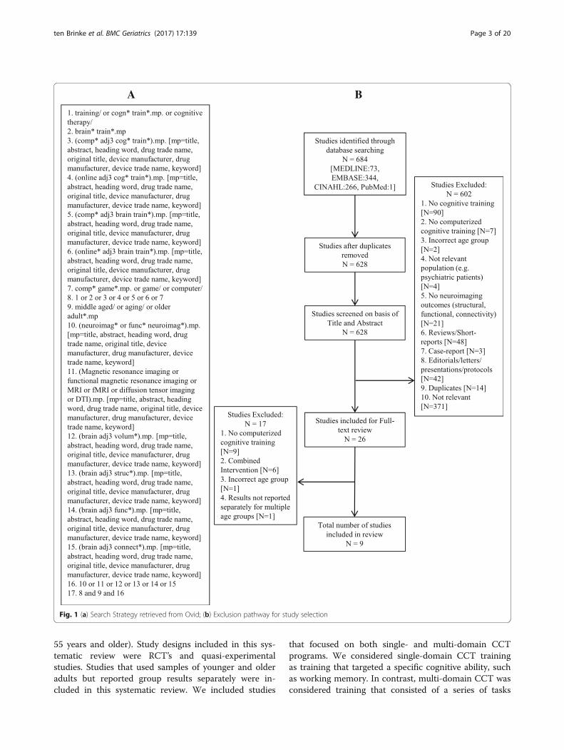
MethodsSearch strategyIn accordance with the Preferred Reporting Items forSystematic reviews and Meta-Analyses (PRISMA) state-ment [14], we conducted a comprehensive search ofMEDLINE, EMBASE, and CINAHL databases to identifyall the studies that investigated neuroimaging outcomesresulting from CCT interventions. We limited oursearch to adults aged 55 years and older with and with-out cognitive impairment, who have not been diagnosedwith dementia. We did not limit the search based onpublication date, as CCT is a relative novel researchtopic. The final search (see Fig. 1a for search strategy)was done on July 7 (2016) and included a check forrecent publications in PubMed.
Study selectionWe selected studies that had a CCT intervention withneuroimaging outcomes (e.g. volumetric structural MRI,functional MRI, DTI) in an older adult population (age
ten Brinke et al. BMC Geriatrics (2017) 17:139 Page 2 of 20
55 years and older). Study designs included in this sys-tematic review were RCT’s and quasi-experimentalstudies. Studies that used samples of younger and olderadults but reported group results separately were in-cluded in this systematic review. We included studies
that focused on both single- and multi-domain CCTprograms. We considered single-domain CCT trainingas training that targeted a specific cognitive ability, suchas working memory. In contrast, multi-domain CCT wasconsidered training that consisted of a series of tasks
A B
Fig. 1 (a) Search Strategy retrieved from Ovid; (b) Exclusion pathway for study selection
ten Brinke et al. BMC Geriatrics (2017) 17:139 Page 3 of 20

that targeted multiple cognitive abilities (e.g., executivefunctions and memory). We excluded studies that didnot focus on CCT or studies that used CCT in combin-ation with other types of intervention (e.g., non-CCT,exercise), reviews and short reports. A full list of exclu-sion criteria and the exclusion pathway is displayed inFig. 1b. Critical review of titles and abstracts resulted in26 articles for full-text review.
Data extraction and quality assessmentWe developed a list of data extraction items. This list in-cluded reference, study sample, study design, MRI magnet,neuroimaging outcomes, cognitive function measured,training program/task, cognitive domain trained, descrip-tion of training, training frequency and duration, totalhours of training, supervised/home-based training, andcontrol group. Two authors [LTB and CKB] independentlyextracted the data from the included studies. Discrepancieswere discussed and solved by two authors [JCD and TLA].The Physiotherapy Evidence Database (PEDro) scale
[15] was used to assess the quality of the included stud-ies. We [LTB and TLA] added three additional items tothe PEDro scale to ensure a proper assessment of inter-vention studies using neuroimaging outcomes. Thesethree items included were: 1) cognition measured to as-sist the interpretation of neuroimaging results; 2) samplesize calculation; and 3) compliance reported (yes/no). Toanswer the items in the quality assessment, we used a ‘+’for items that were present and a ‘-‘ for items that wereabsent. The quality assessment was performed independ-ently by two authors [LTB and CKB]. Discrepancies werediscussed and reviewed by two authors [JCD and TLA].Consensus between two authors [LTB and CKB] wasachieved after discussion (K=0.98). Because item one ofthe PEDro scale is related to external validity, it is notincluded in the overall PEDro score. Therefore, the max-imum quality assessment score calculated by the PEDrowas 10 points (each ‘+’ indicates one point), and will bereported in the results. Studies with a PEDro score of 6/10 or higher were considered studies of moderate tohigh quality. The additional item list had a maximumscore of three points and trends from this list will bedescriptively discussed in the results.
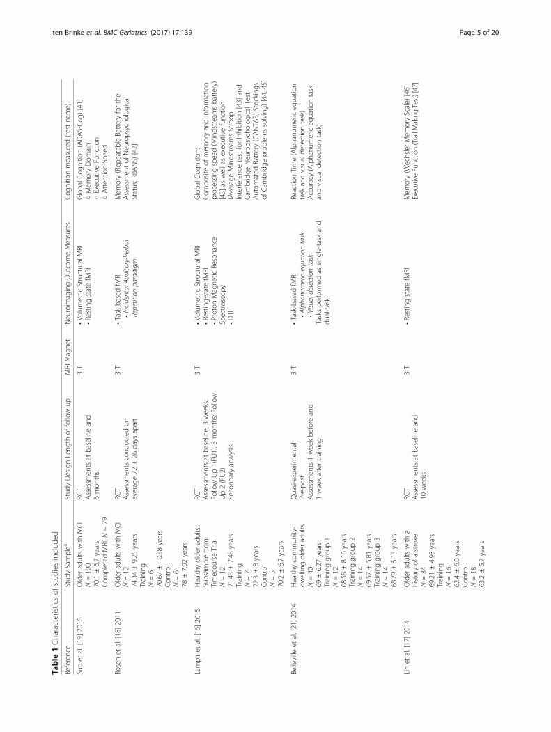
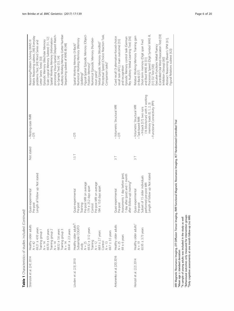
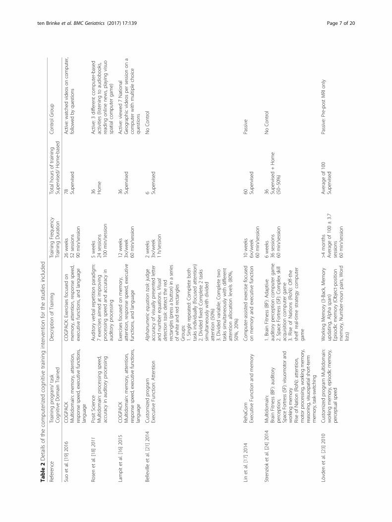
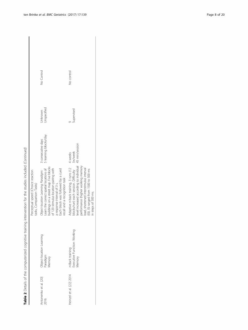
ResultsOverview of studies includedOf the 684 articles identified, nine were included in thissystematic review (Table 1). These nine papers includedfour RCT’s [16–19] and five quasi-experimental studies[20–24]; all nine studies had a different study duration.Details of the interventions included are provided inTable 2. The results are categorized into four categories:1) Volumetric structural imaging (n = 4) [16, 19, 20, 22];2) Task-based fMRI (n = 3) [18, 21, 22]; 3) Connectivity
(n = 7) [16, 17, 19, 20, 22–24]; and 4) Correlationbetween imaging outcomes and cognitive function out-comes (n = 8) [16–22, 24], (Table 3). Results are re-ported in order of study quality, starting with the highestquality.
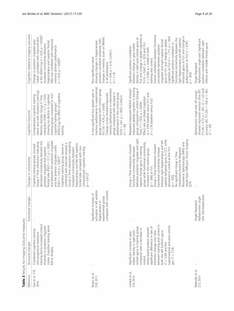
Structural imaging (n = 4)Four studies [16, 19, 20, 22] reported volumetric and cor-tical thickness outcomes (Table 3). A randomized con-trolled study (full factorial design) multi-domain cognitivetraining study using Cogpack [19], older adults with mildcognitive impairment (MCI) trained for a total of 78 hover a period of 6 months under supervision. Combinedcognitive training with resistance training resulted in in-creased cortical thickness in the posterior cingulate cortex.However, in the same study they found that cognitivetraining alone led to a decrease in the posterior cingulatecortex thickness. However, there was no difference in de-crease in thickness compared with the control group.In addition, a twelve-week supervised multi-domain
CCT study [16] using the same program (CogPack)showed that 36 h of training resulted in an increase ingrey matter density in the right post-central gyrus com-pared with a decrease in the active control group. Add-itionally, the training resulted in a difference in rate ofthickness change over time in both the left fusiformgyrus and the supramarginal and post-central gyri.In contrast, in an object-location learning paradigm
study [20] participants performed training on threeconsecutive days where they had to learn the correctspatial location of buildings on a street map. On eachtraining day, the training was followed by a cued recalland recognition task. Hippocampal volumes was mea-sured pre- and post-training. The authors found thatthe object-location learning paradigm did not lead tochanges in hippocampal volume.In another quasi-experimental study [22], participants
performed an adaptive working memory training (n-Back) for twelve 45-min sessions over 4 weeks. Difficultylevel of the training was based on individual perform-ance and increases over time. Results showed that thetraining did not result in changes in grey matter volumein the working memory network.In summary, one RCT [19] found cortical thinning as
a result of cognitive training alone. In contrast, anotherRCT [16] found an increase in grey matter density fol-lowing training. Finally, one study [22] found that cogni-tive training did not result in changes in grey matter,and one study [20] found that cognitive training did notlead to changes in hippocampal volume.
Task-based fMRI (n = 3)Three [18, 21, 22] of the eight included studies examinedthe effect of a CCT intervention on brain function as
ten Brinke et al. BMC Geriatrics (2017) 17:139 Page 4 of 20
Table
1Characteristicsof
stud
iesinclud
ed
Reference
Stud
ySamplea
Stud
yDesignLeng
thof
follow-up
MRI
Magne
tNeuroim
agingOutcomeMeasures
Cog
nitio
nmeasured(testname)
Suoet
al.[19]2016
Older
adultswith
MCI
N=100
70.1±6.7years
Com
pleted
MRI:N
=79
RCT
Assessm
entsat
baselineand
6mon
ths
3T
•Vo
lumetric
StructuralMRI
•Resting-statefM
RIGlobalC
ognitio
n(ADAS-Cog
)[41]
○Mem
oryDom
ain
○ExecutiveFunctio
n○Atten
tion-Speed
Rosenet
al.[18]2011
Older
adultswith
MCI
N=12
74.34±9.25
years
Training
N=6
70.67±10.58years
Con
trol
N=6
78±7.92
years
RCT
Assessm
entscond
uctedon
average72
±26
days
apart
3T
•Task-based
fMRI
•IncidentalAu
ditory-Verbal
Repetitionparadigm
Mem
ory(Rep
eatableBatteryforthe
Assessm
entof
Neuropsycho
logical
Status:RBA
NS)
[42]
Lampitet
al.[16]2015
Health
yolde
radults:
Subsam
plefro
mTimecou
rseTrial
N=12
71.43±7.48
years
Training
N=7
72.3±8years
Con
trol
N=5
70.2±6.7years
RCT
Assessm
entsat
baseline,3weeks:
Follow
Up1(FU
1),3
mon
ths:Follow
Up2(FU2)
Second
aryanalysis
3T
•Vo
lumetric
StructuralMRI
•Resting-statefM
RI•Proton
Magne
ticResonance
Spectroscopy
•DTI
GlobalC
ognitio
n:Com
posite
ofmem
oryandinform
ation
processing
speed(M
indstreamsbattery)
[43]
aswellasexecutivefunctio
n(Average
MindstreamsStroop
Interfe
rencetestforInhibitio
n[43]
and
Cam
bridge
Neuropsycho
logicalTest
Autom
ated
Battery(CANTA
B)Stocking
sof
Cam
bridge
prob
lemssolving)
[44,45]
Belleville
etal.[21]2014
Health
ycommun
ity-
dwellingolde
radults
N=40
69±6.27
years
Training
grou
p1
N=12
68.58±8.16
years
Training
grou
p2
N=14
69.57±5.81
years
Training
grou
p3
N=14
68.79±5.13
years
Quasi-experim
ental
Pre-po
stAssessm
ents1weekbe
fore
and
1weekaftertraining
3T
•Task-based
fMRI
•Alph
anum
ericequationtask
•Visualdetectiontask
Taskspe
rform
edas
sing
le-taskand
dual-task
Reactio
nTime(Alphanu
mericeq
uatio
ntask
andvisualde
tectiontask)
Accuracy(Alphanu
mericeq
uatio
ntask
andvisualde
tectiontask)
Linet
al.[17]2014
Older
adultswith
ahistoryof
astroke
N=34
69.21±4.93
years
Training
N=16
62.4±6.0years
Con
trol
N=18
63.2±5.7years
RCT
Assessm
entsat
baselineand
10weeks
3T
•RestingstatefM
RIMem
ory(W
echslerMem
oryScale)
[46]
ExecutiveFunction(TrailMakingTest)[47]
ten Brinke et al. BMC Geriatrics (2017) 17:139 Page 5 of 20
Table
1Characteristicsof
stud
iesinclud
ed(Con
tinued)
Strenzioket
al.[24]2014
Health
yolde
radults
N=42
69.21±4.93
years
Training
grou
p1
N=14
69.70±6.9years
Training
grou
p2
N=14
68.52±5.6years
Training
grou
p3
N=14
69.41±2.3years
Quasi-experim
ental
Pre-po
stLeng
thof
follow
up:N
otstated
Not
stated
•Resting-statefM
RI•DTI
Reason
ing/Prob
lem
Solving(W
AISIII
Matrix
Reason
ingsubtest,[48]
Everyday
prob
lemsTest,[49]WordSeriesand
Letter
SeriesTests)[50]
Episod
icMem
ory(W
echslerMem
ory
ScaleLogicalM
emorySubtest)[51,52]
SpatialW
orking
Mem
ory(Inform
ation-
processing
Visuo-SpatialD
elayed
Match-
to-Sam
pleTest)[53,54]
Aud
itoryWorking
Mem
ory(LetterN
umber
Sequ
encing
subtesto
fWAISIII)
[48]
Lövden
etal.[23]2010
Health
yolde
radultsb:
Subsam
pleCOGITO
stud
yN=25
69.32±3.12
years
Training
N=12
68.9±2.7years
Con
trol
N=13
69.7±3.5years
Quasi-experim
ental
Pre-po
stTraining
:Pre-po
stMRI
onaverage
179±25.2days
apart
Con
trol:
Pre-po
stMRI
onaverage
184±15.0days
apart
1.5T
•DTI
SpatialW
orking
Mem
ory(3-Back)c
Num
ericalWorking
Mem
ory(M
emory
Upd
ating)
c
Figu
ral-SpatialEpisodicMem
ory(Object-
Positio
nMem
ory)c
Num
ericalEpisod
icMem
ory(Num
ber-
noun
pairs)c
VerbalEpisod
icMem
ory(W
ordlist)c
Percep
tualSpeed(Cho
iceReactio
nTask,
Com
parison
tasks)c
Anton
enko
etal.[20]2016
Health
yolde
radults
N=25
69±6years
Quasi-experim
ental
Pre-po
stAssessm
ents1daybe
fore
(pre),
1dayafter(post)and1mon
thafter(fo
llow-up)
training
d
3T
•Vo
lumetric
StructuralMRI
•DTI
Cuedrecall(3-alternative-forced
-cho
ice
recalltask
(AFC
);mainou
tcom
e)[55]
andrecogn
ition
Episod
icMem
orycontroltask(German
ReyAud
itory
VerbalLearning
Test)[56]
Heinzelet
al.[22]2014
Health
yolde
radultsc
N=19
65.95±3.73
years
Quasi-experim
ental
Pre-po
stSubset
of15
olde
rindividu
als
perfo
rmed
pre-po
stMRI
Leng
thof
follow
up:N
otstated
3T
•Vo
lumetric
StructuralMRI
•Task-based
fMRI
•N-back[57]:tworuns
(16blocks/run
)with
4working
mem
oryloads(0,1,2,3)
•Functio
nalC
onne
ctivity
(PPI)
RelativeWorking
Mem
oryTraining
gain
(n-Back)[57]
Short-term
mem
ory(Digitspan
Fwd
andBw
dWAISIII)[51]
Processing
Speed(DigitSymbo
lWAISIII,
[51]
D2Test[58])
ExecutiveFunctions:VerbalFluency
(Contro
lledOralW
ordAssociationTest)[59]
Inhibitio
n(Stroo
p)[60]
AbstractReason
ing(Raven
’sSPM
[61],
Figu
ralR
elations
subtest[62])
MRI
Mag
netic
Resona
nceIm
aging,
DTIDiffusionTensor
Imag
ing,
fMRI
functio
nalM
agne
ticRe
sona
nceIm
aging,
RCTRa
ndom
ized
Con
trolledTrial
a Meanag
e±stan
dard
deviation
bAsampleof
youn
gad
ults
was
includ
edin
thestud
yas
well
c Beh
avioural
outcom
eson
lymeasuredforinterven
tiongrou
psdOnlycogn
itive
assessmen
tsat
onemon
thfollow-up(noMRI)
ten Brinke et al. BMC Geriatrics (2017) 17:139 Page 6 of 20
Table
2Detailsof
thecompu
terized
cogn
itive
training
interven
tionforthestud
iesinclud
ed
Reference
Training
prog
ram/task
Cog
nitiveDom
ainTraine
dDescriptio
nof
Training
Training
Freq
uency
Training
Duration
Totalh
oursof
training
Supe
rvised
/Hom
e-based
Con
trol
Group
Suoet
al.[19]2016
COGPA
CK
Multid
omain:mem
ory,attention,
respon
sespeed,executivefunctions,
lang
uage
COGPA
CK:Exercisesfocusedon
mem
ory,attention,respon
sespeed,
executivefunctio
ns,and
lang
uage
26weeks
52sessions
90min/session
78 Supe
rvised
Active:watched
videos
oncompu
ter,
followed
byqu
estio
ns
Rosenet
al.[18]2011
PositScience
Multid
omain:processing
speed,
accuracy
inauditory
processing
Aud
itory
verbalrepe
titionparadigm
:7exercisesaimed
atim
proving
processing
speedandaccuracy
inauditory
processing
5weeks
24sessions
100min/session
36 Hom
eActive:3different
compu
ter-based
activities
(listen
ingto
audiob
ooks,
readingon
linene
ws,playingvisuo
spatialcom
putergame)
Lampitet
al.[16]2015
COGPA
CK
Multid
omain:mem
ory,attention,
respon
sespeed,executivefunctions,
lang
uage
Exercisesfocusedon
mem
ory,
attention,respon
sespeed,
executive
functio
ns,and
lang
uage
12weeks
3×/w
eek
60min/session
36 Supe
rvised
Active:view
ed7National
Geo
graphicvide
ospe
rsessionon
acompu
terwith
multip
lechoice
questio
ns
Belleville
etal.[21]2014
Customized
prog
ram
ExecutiveFunctio
n:Atten
tion
Alph
anum
ericequationtask:jud
geaccuracy
ofvisuallypresen
tedletter
andnu
mbe
req
uatio
ns.Visu
aldetectiontask:d
etectthered
rectangles
(pressabu
tton
)inaseries
ofwhite
andredrectangles
Group
s:1.Sing
lerepe
ated
:Com
pletebo
thtasksindividu
ally(fo
cusedattention)
2.Divided
fixed
:Com
plete2tasks
simultane
ouslywith
divide
dattention(50%
)3.Divided
variable:Com
pletetw
otaskssimultane
ouslywith
different
attentionallocatio
nlevels(80%
,50%,20%)
2weeks
3×/w
eek
1h/session
6 Supe
rvised
NoCon
trol
Linet
al.[17]2014
RehaCom
ExecutiveFunctio
nandmem
ory
Com
puter-assisted
exercise
focused
onmem
oryandexecutivefunctio
n10
weeks
6×/w
eek
60min/session
60 Supe
rvised
Passive
Strenzioket
al.[24]2014
Multid
omain:
BrainFitness(BF):aud
itory
percep
tion;
SpaceFortress(SF):visu
omotor
and
working
mem
ory
Rise
ofNation(RoN
):attention,
motor
processin
g,working
mem
ory,
reason
ing,visuospatialsho
rt-term
mem
ory,task-switching
1.BrainFitness(BF):A
daptive
auditory
percep
tioncompu
tergame
2.SpaceFortress
(SF):C
omplex
skill
acqu
isition
compu
tergame
3.Rise
ofNations
(RoN
):Off-the
shelfreal-tim
estrategy
computer
game
6weeks
36sessions
60min/session
36 Supe
rvised
+Hom
e(50–50%)
NoCon
trol
Lövden
etal.[23]2010
Customized
prog
ram
Multid
omain:
working
mem
ory,episo
dicmem
ory,
perceptualspeed
Working
Mem
ory(3-Back,Mem
ory
updatin
g,Alpha
span)
Episod
icmem
ory(Object-po
sitio
nmem
ory,Num
ber-no
unpairs,W
ord
lists)
>4mon
ths
Average
of100±3.7
sessions
60min/session
Average
of100
Supe
rvised
Passive:Pre-po
stMRI
only
ten Brinke et al. BMC Geriatrics (2017) 17:139 Page 7 of 20
Table
2Detailsof
thecompu
terized
cogn
itive
training
interven
tionforthestud
iesinclud
ed(Con
tinued)
Percep
tualspeed(Cho
icereactio
ntasks,Com
parison
Tasks)
Anton
enko
etal.[20]
2016
Object-locatio
nLearning
Paradigm
Mem
ory
Object-locationLearning
Paradigm
:Learnthecorrectspatiallocations
ofbu
ildings
onastreet
map.Fiveblocks
of120stimulus-lo
catio
npairing
with
arespon
seintervalof
3s.
Each
blockwas
followed
byacued
recallandarecogn
ition
task
3consecutivedays
5learning
blocks/day
Unkno
wn
Unspe
cified
NoCon
trol
Heinzelet
al.[22]2014
n-Back
training
ExecutiveFunctio
n:Working
Mem
ory
Adaptiven-back
training
,3runs
(12
blocks/run
)each
session.Difficulty
levelincreased
accordingto
individu
alperfo
rmance
(higherw
orking
mem
ory
load,sho
rtened
interstim
ulus
interval
(ISI).ISIrangedfro
m1500
to500ms
instepsof
500ms.
4weeks
3×/w
eek
45min/session
9 Supe
rvised
Nocontrol
ten Brinke et al. BMC Geriatrics (2017) 17:139 Page 8 of 20
Table
3Results
forIm
agingOutcomemeasures
Reference
Structuralchange
sFunctio
nalchang
esChang
esin
conn
ectivity
Cog
nitio
nOutcome
Cog
nitio
nrelatedto
imagingou
tcom
e
Suoet
al.[19]
2016
Com
bine
dcogn
itive
training
andprog
ressiveresistance
training
ledto
increasedcortical
thicknessin
posteriorcing
ulate
cortex.C
ognitivetraining
alon
eledto
atroph
y.
-Cog
nitivetraining
grou
psshow
edGroup
XTimeinteractionindicatin
gde
creasedconn
ectivity
betw
eenthe
posteriorcing
ulateandsupe
rior
frontallobe
(F(67)
=31.7,p
<0.001)
andbe
tweenthepo
steriorcing
ulate
andtheanterio
rcing
ulatecortex
(F(67)
=13.9,p
<0.001)a .
Cog
nitivetraining
grou
p(alone
orcombine
dwith
exercise)sho
wed
aGroup
XTimeinteractionindicatin
gincreasedconn
ectivity
betw
een
hipp
ocam
pusandtheleftsupe
rior
frontallobe
comparedwith
non-
compu
terized
cogn
itive
training
(p=0.012)a
Com
puterized
cogn
itive
training
(alone
andwith
resistance
training
):Mem
orydo
main:Group
XTime
interaction(F(90)
=5.7,p<0.02)
show
ingno
declinein
cogn
itive
training
grou
pcomparedto
non-
cogn
itive
training
grou
psa
ADAS-Cog
:Noeffect
ofcogn
itive
training
Chang
ein
posteriorcing
ulategrey
mattercorrelated
with
improvem
ent
intheADAS-Cog
(r=0.25,p
=0.030)a .
Increasedconn
ectivity
betw
een
hipp
ocam
pusandsupe
riorfro
ntal
lobe
was
correlated
with
improved
mem
orydo
mainpe
rform
ance
(r=0.33,p
=0.005)a
Rosenet
al.
[18]
2011
-Sign
ificant
increase
ofactivationin
leftanterio
rhipp
ocam
pusin
expe
rimen
talg
roup
comparedwith
controls.
-Ano
n-sign
ificant
butgreatergain
inmem
orype
rform
ance
inexpe
rimen
tal
grou
pcomparedwith
controlg
roup
(F(1,10)
=4.76,p
=0.054).
Chang
escores
show
edim
proved
mem
orype
rform
ance
ininterven
tion
grou
pcomparedwith
decrease
inpe
rform
ance
inthecontrolg
roup
(t(10)
=2.61,p
<.0027,Coh
en’s
d=1.38)
Non
-significanttren
dshow
ingchange
sin
hipp
ocam
pal
activationcorrelated
positivelywith
change
sin
mem
oryscoreon
RBANS
inallp
articipants
(r=0.49,p
=0.10,C
ohen
’sd=1.14)
Lampitet
al.
[16]
2015
Sign
ificant
increase
ingrey
matterde
nsity
inrig
htpo
st-
centralg
yrus
intraining
grou
pcomparedwith
ade
crease
incontrol.
Vertex-based
analysisshow
edsign
ificant
differencein
rate
ofthicknesschange
over
time
betw
eentraining
andcontrolin
both
theleftfusiform
gyrus
(T>3.39)andthe
supram
arginaland
post-cen
tral
gyri(T
>2.24).
-Group
xTimeinteractionshow
edfunctio
nalcon
nectivity
decrease
betw
eenpo
steriorcing
ulateandrig
htsupe
riorfro
ntalgyrusin
training
grou
pwhilefunctio
nalcon
nectivity
increasedin
thecontrolg
roup
(p=.006)at
FU1.
Group
xTimeinteractionshow
edfunctio
nalcon
nectivity
increase
betw
eenrig
hthipp
ocam
pusandleft
supe
riortempo
ralg
yrus
inCCT,while
decreasedin
controlatfirstFU
1(p
=.029).
Nosign
ificant
Group
xTime
interactions
foun
dforMagne
ticResonanceSpectroscopy
(MRS)a
ndwho
lebrainDiffusionTensor
Imaging
(DTI)
Repe
ated
-measuredANOVA
show
edim
proved
glob
alcogn
ition
intraining
grou
pcomparedto
control(Group
XTime,F=7.833,p=0.003).
Effect
size
onGlobalC
ognitio
n(d
=0.94
baselineversus
FU1and
d=2.18
baselineversus
FU2)
Sign
ificant
positivecorrelation
betw
eenchange
ingrey
matter
density
inrig
htpo
st-cen
tralgyrusat
FU2andchange
inglob
alcogn
ition
atFU
1(r=0.647,p=.023)andFU
2(r=0.584,p=0.046)
inbo
thtraining
andcontrol.
Inversecorrelationbe
tweenfunctio
nal
conn
ectivity
betw
eenpo
sterior
cing
ulateandrig
htsupe
riorfro
ntal
gyrusat
FU1andchange
inglob
alcogn
ition
atFU
2(r=−.771,p
=.003).
Sign
ificant
positivecorrelationin
functio
nalcon
nectivity
betw
eenthe
right
hipp
ocam
pusandleftsupe
rior
tempo
ralg
yrus
atFU
1andchange
inglob
alcogn
ition
atFU
2(r=0.591,
p=.043).
Belleville
etal.
[21]
2014
-Sing
leRepe
ated
:Alph
anum
ericsin
gle
task:D
ecreased
post-
-Alph
anum
ericsin
gletask:A
llgrou
psshow
edim
proved
reactio
ntim
e(RT;
F(1,34)=9.75,p
<.001,η
2=.22)
and
accuracy
(AC;F(1,34)
=14.8,p
=.001,
η2=.30)
Sing
leRepe
ated
:Alph
anum
ericsin
gletask:Significant
positivecorrelationbe
tweenrig
htinferio
randmiddlefro
ntalgyrus
ten Brinke et al. BMC Geriatrics (2017) 17:139 Page 9 of 20
Table
3Results
forIm
agingOutcomemeasures(Con
tinued)
training
activationin
inferiorand
right
middle
frontalgyrus
(t=5.91),leftmiddle
frontalgyrus(t=4.57)
andleftthalam
us(t=5.37).
Visualdetectionsin
gle
task:nochange
Dualtask:no
change
Divided
Fixed
Alphanum
ericsin
gletask:
nochange
Visualdetectionsin
gle
task:D
ecreased
post-
training
activationin
right
cerebe
llum
(t=4.73)andrig
htmid
dleoccipitalg
yrus
(t=4.68)w
hen
perfo
rmingthevisual
detection
task.
Dualtask(50/50):Sm
all
increase
inpo
st-training
activationin
right
and
leftmiddlefro
ntalgyrus
(areas
11,47;t=4.41
andt=4.52
respectively).
Divided
Variable
Alph
anum
ericsin
gletask:
nochange
Visualdetectionsin
gle
task:n
ochange
Dualtask:
Sign
ificant
increased
activationin
right
middlefro
ntalgyrus
(area10;for
20%
attentionallocatio
nt=5.35
and50%
attentionallocatio
nt=4.78).Noredu
ced
post-trainingactivation
in80%
attention
allocatio
n.
Visualdetectionsin
gletask:N
ochange
Dualtask(costscore)b:
Sing
lerepe
ated
:Noim
provem
entsin
dualtasking
Divided
Fixed:
Redu
ceddu
al-task
cost(F(1,34)
=6.97,p
<.001,η
2=.45)
Divided
Variable:Redu
ceddu
al-task
costandwereableto
mod
ifyattentional
priority(F(2,33)=5.17,p
<.001,η
2=.34)
activationandreactio
ntim
e(r=.56,
p<.05).
Divided
Variable:Sign
ificant
negative
correlation(posttraining
)be
tween
activationof
right
supe
riorandmiddle
frontalgyrus(Brodm
annarea
10)and
attentionalcost(r=−.55,p<.05)
ten Brinke et al. BMC Geriatrics (2017) 17:139 Page 10 of 20
Table
3Results
forIm
agingOutcomemeasures(Con
tinued)
Linet
al.[17]
2014
--
Training
grou
p:Sign
ificant
increased
functio
nalcon
nectivity
in(all
p’s<0.005):
1.Lefthipp
ocam
pus-rig
htinferio
rfro
ntalgyrus
2.Lefthipp
ocam
pus-rig
htmiddle
frontalgyrus
3.Righ
thipp
ocam
pus-leftmiddle
frontalgyrus
4.Righ
thipp
ocam
pus-leftinferio
rfro
ntalgyrus
5.Righ
thipp
ocam
pus-leftsupe
rior
frontalgyrus
6.Righ
thipp
ocam
pus-leftparietallob
eCon
trol
grou
p:Sign
ificantlyde
creasedfunctio
nal
conn
ectivity
(allp’s<0.005):
1.Lefthipp
ocam
pus-rig
htmiddle
occipitalg
yrus
2.Righ
thipp
ocam
pus-rig
htpo
sterior
lobe
orcerebe
llum
3.Righ
thipp
ocam
pus-leftsupe
rior
tempo
ralg
yrus
Training
grou
p:1.Sign
ificant
improved
scores
on5/7
subtestsfro
mWechslerMem
oryScale,
namely:Men
talcon
trol
(p=0.003),
Logicalm
emory(p
<0.001),D
igits
forw
ardandbackward(p
=0.014),
Visualreprod
uctio
n(p
=0.008),and
Associatedlearning
(p<0.001).
2.Im
proved
Mem
oryqu
otient
(p=0.005)
3.Im
proved
perfo
rmance
onTrail
MakingTest-A
(p<0.001)
Con
trol
grou
p:no
sign
ificant
change
sbe
tweenbaselineand10-w
eekscores
Training
grou
p:sign
ificant
positive
correlations
betw
een(allp’s<0.001):
1.Mem
oryqu
otient
andfunctio
nal
conn
ectivity
oflefthipp
ocam
pus-rig
htfro
ntallobe
(r=0.64)
2.Mem
oryqu
otient
andfunctio
nal
conn
ectivity
ofrig
hthipp
ocam
pus-left
frontallobe
(r=0.85)
3.Mem
oryqu
otient
andfunctio
nal
conn
ectivity
ofrig
hthipp
ocam
pus-left
parietallob
e(r=0.79)
4.TrailM
akingTest-A
scoreandfunc
tionalcon
nectivity
ofleft
hipp
ocam
pus-rig
htfro
ntallobe
(r=0.94)
5.TrailM
akingTest-A
andfunctio
nal
conn
ectivity
ofrig
hthipp
ocam
pus-left
frontallobe
(r=0.68)
Con
trol
grou
p:no
sign
ificant
correlations
betw
eencogn
ition
and
functio
nalcon
nectivity
c
Strenzioket
al.
[24]
2014
--
VentralN
etwork:
Axialdiffu
sivity
(AD)in
therig
htoccipito-tem
poralw
hite
matter
sign
ificantlyincreasedafterBF
compared
with
ade
crease
afterSF
andRO
N(p
<0.05)
DorsalN
etwork:
Functio
nalcon
nectivity
betw
eenrig
htsupe
riorparietalcortex(SPC
)andleft
posteriorinferio
rtempo
rallob
e(ITL)
decreasedin
SFandincreasedin
RON
(p=0.02).
Functio
nalcon
nectivity
betw
eenrig
htSPCandleftanterio
rITLde
creasedin
BFandshow
edan
increase
inRO
N(p
=0.03)
Univariate
ANOVA
show
edmain
effectsof
training
grou
p:Reason
ingon
Everyday
Prob
lemsTest:
Maineffect
oftraining
grou
p(F(2,39)
=5.34,p
<0.01,p
artial
η2=0.215).
BFandSF
show
edim
proved
perfo
rmance
aftertraining
andRO
Nshow
edno
effect.
SpatialW
orking
Mem
ory:
Maineffect
oftraining
grou
p(F(2,39)
=5.03,p
<0.001,partial
η2=0.205).
SFim
proved
perfo
rmance
after
training
,RONde
creasedpe
rform
ance,
andBF
show
edno
effect.
Matrix
Reason
ing:
Maineffect
oftraining
grou
p(F(2,39)
=3.40,p
<0.044,partial
η2=0.148).
Largestgainsseen
inBF
andasm
aller
gain
inRO
N.The
SFgrou
pshow
eda
decrease
inreason
ingaftertraining
Cog
nitio
nandWhite
MatterIntegrity
Positivecorrelationbe
tweenchange
inthalam
icADandchange
inworking
mem
orype
rform
ance
inall
participants(r=0.44,p
<0.005).
Neg
ativecorrelationbe
tweenchange
sin
occipito-tem
poralA
Dandeveryday
prob
lem
solving(r=−0.32,p
<.05)
andspatialw
orking
mem
oryaccuracy
(r=−0.35,p
<.05).
Neg
ativecorrelationbe
tweenchange
sin
occipito-tem
poral-p
arietalA
Dand
spatialw
orking
mem
oryaccuracy
(r=−0.40,p
<0.05).
Cog
nitio
n&Functio
nalC
onne
ctivity
Positivecorrelationbe
tweenchange
sin
SPC-posterio
rITLconn
ectivity
and
change
sin
everyday
prob
lem
solving
time(r=−0.57,p
<.001).
Lövden
etal.
[23]
2010
--
MeanDiffusivity
(MD)
Group
XTimeinteractionfoun
dfor
segm
ent1(gen
u)of
corpus
callosum,
show
ingade
crease
inMD
Unkno
wn:analysiscombine
dyoun
ger
andolde
rsubsets
Unkno
wn:analysiscombine
dyoun
ger
andolde
rsubsets
ten Brinke et al. BMC Geriatrics (2017) 17:139 Page 11 of 20

Table
3Results
forIm
agingOutcomemeasures(Con
tinued)
(t(11)=
2.39,p
=.036).Nochange
sin
controlg
roup
Fractio
nalA
nisotrop
y(FA)
Group
XTimeinteractionfoun
dfor
segm
ent1of
corpus
callosum,
show
ingan
increase
inFA
(t(11)=
3.12,p
=.010)
Anton
enko
etal.[20]2016
Hippo
campalvolum
e:no
differencepreto
posttraining
(p=0.505)
MeanDiffusivity
(MD):Asign
ificant
decrease
infornixMDwas
foun
dat
post-trainingcomparedwith
pre-
training
(p=0.036).
Nodifferencein
hipp
ocam
palM
Dfro
mpre-
topo
st-training(p
=0.669).
Fractio
nalA
nisotrop
y(FA):Ano
n-sign
ificant
increase
infornixFA
was
foun
dbe
tweenpre-
andpo
st-training
(p=0.114)
%Co
rrectduringtraining
:Task
perfo
rmance
sign
ificantlyim
proved
inacurviline
arconvex
manne
rover
the3training
days
learning
-Highe
rincrease
infornixFA
from
pre
topo
stassessmen
twas
sign
ificantly
relatedto
better
averagerecall
perfo
rmance
ontheob
ject-lo
catio
ntask
durin
gtraining
,at1-daypo
stand
follow-up(r=0.431,p=0.031)
-Chang
ein
fornixFA
didno
tcorrelate
with
episod
icmem
orype
rform
ance
onthecontroltask(Rey
Aud
itory
VerbalLearning
Test;p
=0.214)
-Chang
ein
fornixMDdidno
tcorrelatewith
recallpe
rform
ance
p=0.728
-Chang
ein
hipp
ocam
palM
Dor
volumedidno
tcorrelatewith
recall
perfo
rmance
(p=0.688andp=0.758,
respectively)
Heinzelet
al.
[22]
2014
Nosign
ificant
change
ingrey
mattervolumeof
working
mem
oryne
tworkpo
sttraining
(t(14)=
0.83,p
=.421)
Nosign
ificant
2(tim
e)×3(working
mem
ory
load)interaction
(F=.24,p=.714,p
artial
η2=.024).
Sign
ificant
maineffect
oftim
e(F=12.68,
p=.003,p
artial
η2=.475)driven
byBO
LDde
crease
in1-
back
(t=.99,p=.029).
A2(tim
e)×3(load)repe
ated
measures
ANOVA
show
edno
change
sin
conn
ectivity
inworking
mem
ory
netw
ork(F(2,28)
=1.08,p
=.355,
partialη
2=.071)
n-Back:p
airedt-testsshow
edim
proved
perfo
rmance
on1-Back
(t(18)
=3.37,p
=.003),2-ack
(t(18)
=7.47,p
<.001),and3-Back
(t(18)
=4.86,p
<.001)d.
Repe
ated
-measuresMANOVA
(factor
time)
show
edim
provem
entsin
neurop
sycholog
icalmeasuresafter
training
.Postho
cpairedt-tests
show
edim
provem
entsin
DigitSpan
Fwd(t(18)
=2.97,p
=0.008),D
2test
(t(18)
=6.48,p
<0.001),D
igitSymbo
l(t(18)
=2.76,p
=0.013),Stroo
pInterfe
rence(t(18)
=3.28,p
=0.004),
andFigu
ralR
elations
(t(18)
=4.73,
p<0.001).N
oim
provem
entsafter
training
werefoun
din
DigitSpan
Bwd,
VerbalFluency,andRaven’s
SPM.d
Non
-significanttren
dbe
tweenBO
LDactivationat
baselineandrelative
improvem
entin
DigitSpan
Fwd
(r=.43,p=.067)
a Thisstud
ywas
afullfactoriald
esign
bTh
isdu
al-taskcost
represen
tstheprop
ortio
nallossof
performan
cein
thedu
al-taskcond
ition
asafunctio
nof
performan
cein
thesing
le-taskcond
ition
.Alarger
scorerepresen
tsalarger
dual-taskcost
c Not
specified
whe
ther
correlations
wereba
sedon
chan
gescores
orscores
atweek10
dRe
sults
repo
rted
forallo
lder
participan
ts(N
=19
)
ten Brinke et al. BMC Geriatrics (2017) 17:139 Page 12 of 20
measured via task-based fMRI (Table 3). An RCT [18]showed that 2200 min of cognitive training over a periodof 5 weeks resulted in a significant increase in leftanterior hippocampus activity compared with an activecontrol group. The cognitive training consisted of sevengames aimed to improve auditory processing speed andaccuracy. Task difficulty was adjusted throughout thetraining based on individual performance. The activecontrol group performed computer-based activities suchas reading online newspapers and playing computergames targeting visuospatial abilities.A two-week quasi-experimental study looked at fo-
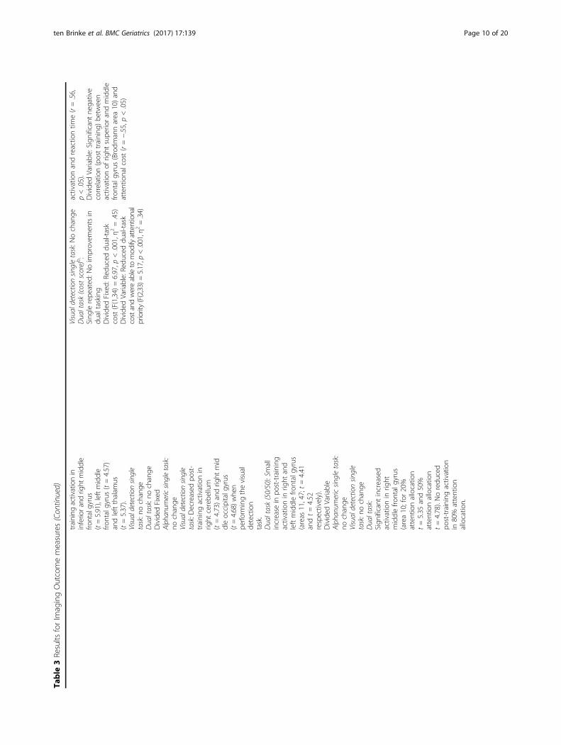
cused and divided (fixed and variable) attention training[21]. In the focused attention training, two tasks (i.e.,alphanumeric task and a visual detection task) were per-formed back to back but separate so participants focusedon one task at a time. In the divided attention training,participants performed two tasks at the same time withan equal amount of attention (fixed) or under differentattention allocations (variable). Results showed thattraining a single alphanumeric task for 6 h over twoweeks decreased activation in the inferior and right mid-dle frontal gyrus, in the left middle frontal gyrus and inthe left thalamus. No differences in functional brain acti-vation were found after performing the single visualdetection task or the in the dual task condition. Partici-pants who were assigned to training where they per-formed both the alphanumeric task and the visualdetection task at the same time (i.e., dual task) did notshow differences in performance during the alpha-numeric task in the scanner. However, participantsshowed decreased functional brain activation at post-training compared with pre-training in the cerebellumand right middle occipital gyrus during the single visualdetection task. Additionally, participants showed a slightincrease in activation in both the right and left middlefrontal gyrus. Finally, participants who were assigned tothe training group where they had to perform dual tasksunder different attention allocation levels (i.e., 80%, 50%,or 20%), showed increased activation in the right middlefrontal gyrus (area 10) for 20% and 50% attention alloca-tion when performing the dual task. No significantchanges in functional brain activation were found duringthe 80% attention allocation task, neither during thealphanumeric single task, nor during the visual detectionsingle task performance.In an adaptive n-back training program [22], partici-
pants performed 12 sessions of approximately 45 mineach over 4 weeks. The difficulty level of the trainingwas based on individual performance and was increasedacross training sessions by increasing working memoryload and decreasing the interstimulus interval. Results ofthis study showed a non-significant time (2) by workingmemory load (3) interaction, with a significant main
effect of time. This main effect of time demonstrates areduction in working memory network functional brainactivity measured by the Blood Oxygen Level Dependent(BOLD) signal after 12 training sessions. Only decreasesin the 1-back (and not 2-back or 3-back) condition weresignificant, which indicates this main effect of time isdriven by the BOLD signal during the 1-back condition.In summary, an RCT [18] showed that 2200 min of
CCT resulted in increased in left anterior hippocampusactivity compared with an active control group. Onequasi-experimental study [21] showed that depending onthe task and region of interest, all training conditions re-sulted in both increased and decreased activity. Finally, asecond quasi-experimental study [22] found that 12 ses-sions of n-back training resulted in a significant decreasein working memory activity; however decrease in activitywas driven by performance on the 1-back condition.
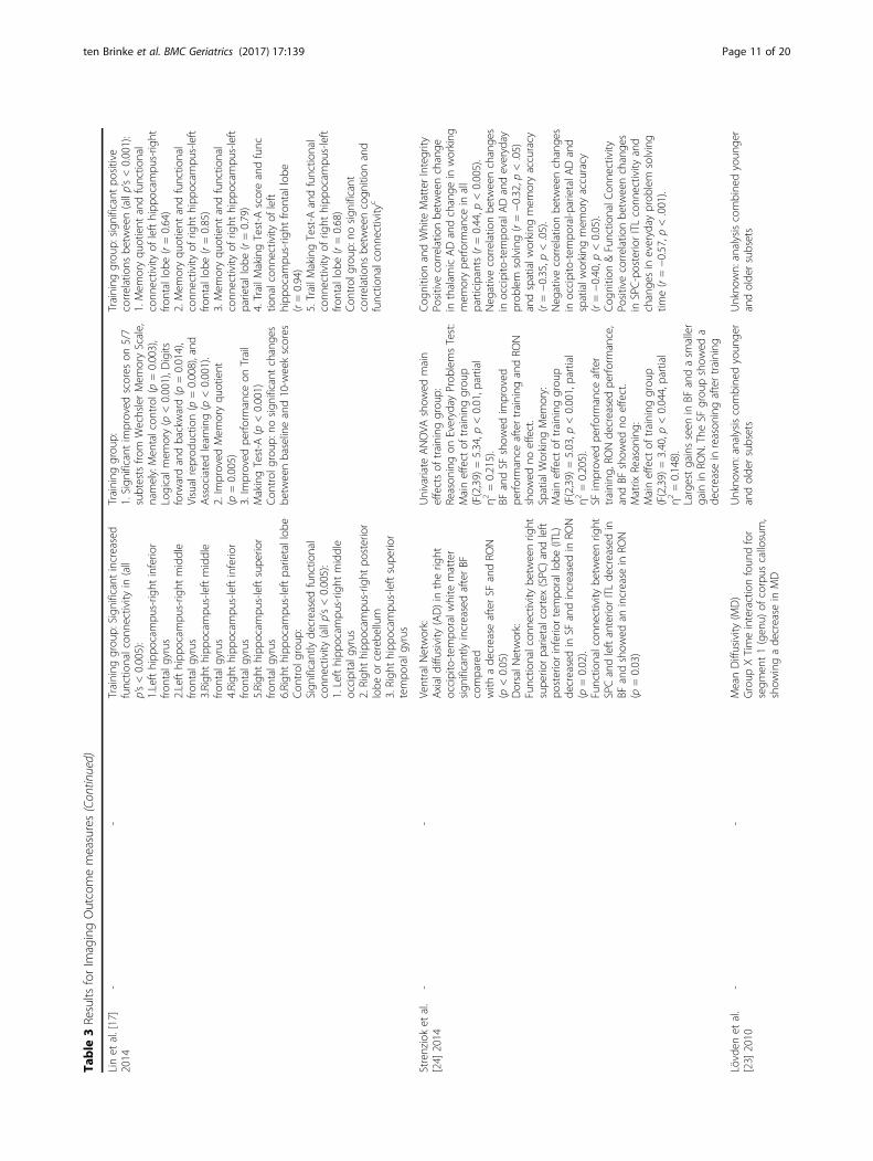
ConnectivityResting-state fMRI (n = 5)Five studies [16, 17, 19, 22, 24] looked at changes in func-tional connectivity after CCT (Table 3). An RCT [19] ex-amined the effect of progressive resistance training (PRT),computerized multi-domain cognitive training (CCT), ora combined intervention on brain structure and functionin older adults with mild cognitive impairment (MCI).The study duration was 26 weeks, with a total of 78 h oftraining. In the cognitive training groups (i.e., PRT + CCT,and CCT + Sham), the posterior cingulate cortex showedsignificant decreases in resting-state functional connectiv-ity with both the superior frontal lobe and the anteriorcingulate cortex. In addition, increases in resting-statefunctional connectivity between the hippocampus and theleft superior frontal lobe were found compared withgroups without CCT.A second RCT of 12 weeks of multimodal CCT [16]
showed that 36 h of cognitive training resulted in de-creases in resting-state functional connectivity betweenthe posterior cingulate and the right superior frontalgyrus, while the control group showed significant in-creases in resting-state functional connectivity. In con-trast, CCT resulted in increased resting-state functionalconnectivity between the right hippocampus and the leftsuperior temporal gyrus compared with a decrease inconnectivity in the control group.Another RCT [17] looked at the effects of a 10-week
computer assisted training focused on executive func-tioning and memory in older adults with a history ofstroke. The authors found that training, compared witha passive control group, significantly increased resting-state functional connectivity in multiple areas. The lefthippocampus showed significantly increased connectivitywith the right inferior frontal gyrus and the right middlefrontal gyrus. Additionally, the right hippocampus showed
ten Brinke et al. BMC Geriatrics (2017) 17:139 Page 13 of 20

increased resting-state functional connectivity with the leftmiddle frontal gyrus, the left inferior frontal gyrus, the leftsuperior frontal gyrus and the left parietal lobe. In con-trast, the control group showed significant decreases inresting-state functional connectivity over the 10 weeks(see Table 3 for connectivity decreases).A quasi experiment investigating the effect of three
different computer programs [24] found an increasedresting-state functional connectivity in the dorsal net-work between the right superior parietal cortex (SPC)and left posterior inferior temporal lobe (ITL) in Rise OfNation (RON) compared with a decrease in Space Fort-ress (SF). Finally, Brain Fitness (BF) resulted in signifi-cantly decreased resting-state functional connectivitybetween the right SPC and the left anterior ITL com-pared with an increase in RON.Finally, a quasi-experimental study [22] looking at the
effects of an adaptive n-back training program in olderadults found that the 5-week training did not result inchanges in task-based functional connectivity in theworking memory network.
Structural connectivity (n = 4)Four studies [16, 20, 23, 24] examined changes in struc-tural connectivity, using DTI, after CCT (Table 3).Whole brain diffusion tensor imaging (DTI) of an RCTof 12 weeks of multimodal CCT [16] showed that 36 hof cognitive training did not result in changes in struc-tural connectivity after training.A quasi-experiment in healthy older adults looked at
the effect of three different training protocols on brainstructure [24]. The participants trained for 36 h over aperiod of 6 weeks; half of the training was supervised,and the other half was performed at their own homes.One training group performed BF, an auditory percep-tion game; the second training group performed SF, acomplex skill acquisition game focused on visuomotorand working memory skills; and the third group per-formed RON, an off-the shelf real-time strategy gamefocused on for example attention, motor processing,working memory and reasoning. The authors foundchanges in the ventral and dorsal network. Axial diffu-sivity (AD) was increased in the right occipito-temporalwhite matter in the BF group, compared with a decreasein SF and RON.Another quasi-experimental study [23] of approxi-
mately 100 h of multi-domain cognitive training in bothyoung and healthy older adults performed DiffusionTensor Imaging (DTI) to look at the effects of trainingon structural connectivity in the brain. Result showed asignificant decrease in MD in the genu of the corpuscallosum compared with a passive control group whoshowed no changes in MD. They also found a significant
increase of fractional anisotropy (FA) in the genu of thecorpus callosum compared with the control group.Diffusion Tensor Imaging results from a third quasi-
experimental study [20] that involved 3 consecutive daysof training an object-location learning paradigm, showedthat the 3-day training resulted in a significant decreasein mean diffusivity (MD) in the fornix at post-trainingcompared with pre-training. No changes in MD werefound in the hippocampus as a result of the training. Inaddition, the results showed an increase in FA in the for-nix, however this increase was not significant.In summary, the seven [16, 17, 19, 20, 22–24] above
mentioned rsfMRI and DTI studies showed both in-creases and decreases in functional and structural con-nectivity after CCT. The variety in study protocol (i.e.,training type, duration) and the regions of interestchosen for neuroimaging analysis makes the comparisonbetween studies difficult.
Correlation between imaging outcomes and cognitivefunction outcomes (n = 8)Eight studies [16–22, 24] assessed the association betweencognitive performance and neuroimaging findings (Table 3).An RCT in older adults with a history of stroke [19]found that increases in posterior cingulate grey matterwere associated with improvements in global cognition.Additionally, a cognitive training by time interactionshowed that the increased connectivity between thehippocampus and the left superior frontal lobe was relatedto increased memory domain performance. However, thisinteraction takes into account all training groups thathad a cognitive training component (i.e., also cognitivetraining combined with resistance training). The inclu-sion of the combination group might have influencedthis interaction.In contrast, an RCT looking at CCT in older
adults with MCI [18] found no significant correla-tions between neuroimaging and cognitive results.However, the authors found a non-significant trendsuggesting that, in both groups, increases in hippocampalactivity might be related to improved memory scores onthe RBANS.An RCT of multimodal CCT [16] found that increased
grey matter density in the right posterior central gyrus wasassociated with improved global cognition at 3 weeks and3 months. This association was found in both the trainingand control group. In addition, it was found that a decreasein resting-state functional connectivity between the poster-ior cingulate and the superior frontal gyrus after 3 weeksof training was related to an increased change in globalcognition after 3 months of training. Increased resting-state functional connectivity between the right hippo-campus and the left superior temporal gyrus measures
ten Brinke et al. BMC Geriatrics (2017) 17:139 Page 14 of 20
after three weeks of training was associated with in-creases in global cognition after 3 months of training.A quasi-experimental study [21] found that in partici-
pants performing the alphanumeric task in the single taskcondition (i.e., focus on one task at the time), there was asignificant positive correlation between both the right in-ferior and the middle frontal gyrus activation and reactiontime. Thus shorter reaction time (i.e., better performance)was associated with a decrease in brain activation. In thedivided variable condition (i.e., dual task with different at-tention allocation levels), there was a negative correlationbetween activation of the right superior and middle frontalgyrus and attentional cost post training. This correlationindicates that a better training performance (i.e., lowerattentional cost during dual task performance) was associ-ated with higher levels of brain activation.An RCT in older adults with a history of stroke [17]
revealed that in the multimodal cognitive training group,resting-state functional connectivity between the lefthippocampus and both the right frontal lobe and rightfrontal lobe, was associated with improved performancein memory executive function respectively. Additionally,increases in resting-state functional connectivity betweenthe right hippocampus and the left frontal lobe and theleft parietal lobe were associated with increases of memoryand executive functioning. No significant associationsbetween functional connectivity and behavioural perform-ance were found in the control group.A quasi-experimental study looking at the effect of three
different types of cognitive training on brain structure andfunction [24] found that in the BF training group an in-crease in thalamic AD was associated with an increase inworking memory performance. By comparing BF and SF,the authors found that an increase in occipito-temporalAD was associated with a decrease in everyday problemsolving time. Additionally, they found an associationbetween the increase in both the occipito-temporal ADand occipito-temporal-parietal AD and accuracy of spatialworking memory tasks, indicating that a greater AD wasassociated with a smaller increase in accuracy on thememory task. Finally, looking at the contrast between SFand RON, functional connectivity decreases between thesuperior parietal cortex (SPC) and the posterior inferiortemporal lobe (ITL) were related to better performanceon every day problem solving tasks (i.e., decrease in timefor task completion).In another quasi-experimental study [20], participants
training for 3 consecutive days on an object-locationlearning paradigm. The authors found that the previousmentioned increase in fornix FA on the post-test com-pared with pre-test was significantly associated with betterrecall performance. Thus, a higher increase in fornix FAover the course of the training resulted in a better recallperformance on the object-location learning paradigm
task. Changes in fornix MD, hippocampal MD, and hippo-campal volume were not associated with recall perform-ance. Performance on the episodic memory control taskwas not associated with changes in fornix FA.The last quasi-experimental study [22] looked at
changes in short term memory (digit span) and found anon-significant trend between task-based functional acti-vation at baseline and improvement in digit span, whichindicates that an increased activation might lead to in-creased short term memory performance.In summary, eight [16–22, 24] of the nine studies
[16–24] included demonstrated an association betweenchanges in neuroimaging measures (volumetric orconnectivity) and changes in behavioural outcomes. De-pending on the region of interest (i.e., both volumetricand connectivity), both increases and decreases in ac-tivity resulted in improved cognitive performance. Onestudy [18] found no significant association between neu-roimaging and behavioural measures. One study [23] didnot report the association between neuroimaging and cog-nition in older adults specifically.
Quality assessment of the included studiesThe quality of studies included in this systematic reviewvaried substantially (Table 4). On average, the nine in-cluded studies met 7 of the 11 PEDro criteria. Two stud-ies of the highest quality [18, 19] meeting 9 of the 10PEDro criteria; however, five [17, 20, 22–24] studiesfailed to meet five or more study quality criteria. Item 11(i.e., included point measures and variability measures)was met for all nine studies. Item 8 (key outcome mea-sured for 85% of subjects) and nine (outcome data ana-lyzed by intention to treat) were met by seven of thenine studies [18–24]. Item 6, (i.e., blinding of all who ad-ministered the training) commonly received a negativeresponse (i.e., one of the studies [19] blinded trainingadministers). Frequent issues were failure to meet or re-port: 1) allocation concealment (n = 4) [20, 22–24]; 2)blinding of all subjects (n = 6) [17, 20–24]; 3) blinding ofall who administered the training (n = 8) [16–18, 20–24]; 4)blinding of assessors who measured at least one keyoutcome (n = 5) [20–24]; and 5) between-group statis-tical comparisons for at least one key outcome (n = 4)[17, 20–22]. Item 9 (participants with available outcomemeasures received the treatment or control conditionallocated) received 78% overall rater agreement betweenthe authors [LTB and CKB], where the remaining ques-tions received a 100% overall rater agreement between theauthors [LTB and CKB].Of the three additional items, selected by the authors
[LTB and TLA], item 12 (inclusion of cognitive out-comes to assist neuroimaging interpretation) was ad-dressed by all nine studies [16–24]. Items 13 (sample
ten Brinke et al. BMC Geriatrics (2017) 17:139 Page 15 of 20
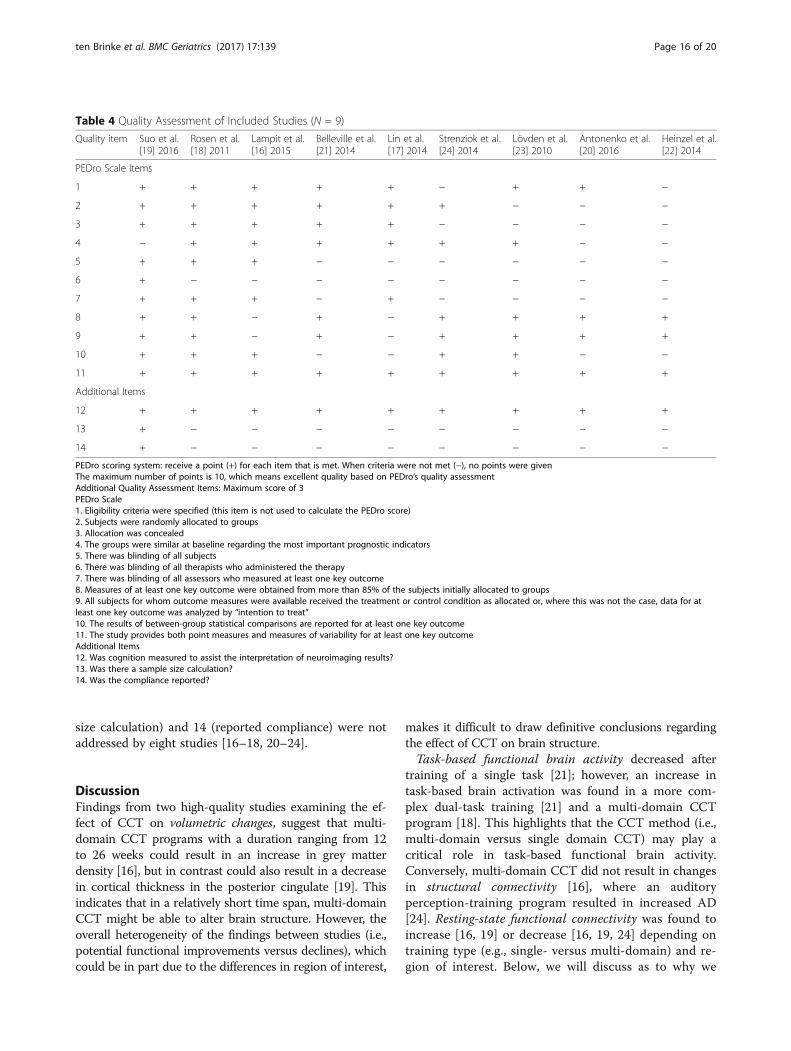
size calculation) and 14 (reported compliance) were notaddressed by eight studies [16–18, 20–24].
DiscussionFindings from two high-quality studies examining the ef-fect of CCT on volumetric changes, suggest that multi-domain CCT programs with a duration ranging from 12to 26 weeks could result in an increase in grey matterdensity [16], but in contrast could also result in a decreasein cortical thickness in the posterior cingulate [19]. Thisindicates that in a relatively short time span, multi-domainCCT might be able to alter brain structure. However, theoverall heterogeneity of the findings between studies (i.e.,potential functional improvements versus declines), whichcould be in part due to the differences in region of interest,
makes it difficult to draw definitive conclusions regardingthe effect of CCT on brain structure.Task-based functional brain activity decreased after
training of a single task [21]; however, an increase intask-based brain activation was found in a more com-plex dual-task training [21] and a multi-domain CCTprogram [18]. This highlights that the CCT method (i.e.,multi-domain versus single domain CCT) may play acritical role in task-based functional brain activity.Conversely, multi-domain CCT did not result in changesin structural connectivity [16], where an auditoryperception-training program resulted in increased AD[24]. Resting-state functional connectivity was found toincrease [16, 19] or decrease [16, 19, 24] depending ontraining type (e.g., single- versus multi-domain) and re-gion of interest. Below, we will discuss as to why we
Table 4 Quality Assessment of Included Studies (N = 9)
Quality item Suo et al.[19] 2016
Rosen et al.[18] 2011
Lampit et al.[16] 2015
Belleville et al.[21] 2014
Lin et al.[17] 2014
Strenziok et al.[24] 2014
Lövden et al.[23] 2010
Antonenko et al.[20] 2016
Heinzel et al.[22] 2014
PEDro Scale Items
1 + + + + + − + + −
2 + + + + + + − − −
3 + + + + + − − − −
4 − + + + + + + − −
5 + + + − − − − − −
6 + − − − − − − − −
7 + + + − + − − − −
8 + + − + − + + + +
9 + + − + − + + + +
10 + + + − − + + − −
11 + + + + + + + + +
Additional Items
12 + + + + + + + + +
13 + − − − − − − − −
14 + − − − − − − − −
PEDro scoring system: receive a point (+) for each item that is met. When criteria were not met (−), no points were givenThe maximum number of points is 10, which means excellent quality based on PEDro’s quality assessmentAdditional Quality Assessment Items: Maximum score of 3PEDro Scale1. Eligibility criteria were specified (this item is not used to calculate the PEDro score)2. Subjects were randomly allocated to groups3. Allocation was concealed4. The groups were similar at baseline regarding the most important prognostic indicators5. There was blinding of all subjects6. There was blinding of all therapists who administered the therapy7. There was blinding of all assessors who measured at least one key outcome8. Measures of at least one key outcome were obtained from more than 85% of the subjects initially allocated to groups9. All subjects for whom outcome measures were available received the treatment or control condition as allocated or, where this was not the case, data for atleast one key outcome was analyzed by “intention to treat”10. The results of between-group statistical comparisons are reported for at least one key outcome11. The study provides both point measures and measures of variability for at least one key outcomeAdditional Items12. Was cognition measured to assist the interpretation of neuroimaging results?13. Was there a sample size calculation?14. Was the compliance reported?
ten Brinke et al. BMC Geriatrics (2017) 17:139 Page 16 of 20
might see a discrepancy between single- and multi-domain CCT effects, and why this discrepancy mightaffect both structural and resting-state functional con-nectivity differently.
Task-based functional activityFunctional activation patterns in the brain change withaging as a result of neurophysiological changes. Comparedwith younger adults, functional activation patterns be-come less coordinated and localized in older adults, whichresult in loss of cognitive performance [25]. In the currentreview, three studies looked at functional activity in thebrain while performing a task in the scanner. Activitylevels in the brain while performing a task were both in-creased and decreased, depending on the type of trainingand region of interest. All three studies focused on differ-ent brain regions, which makes comparison difficult.However, results suggest that engaging in a more diverseor complex training (e.g., multi-domain CCT or dual-tasktraining) might lead to an increased functional activation[18, 21] compared with training of a single task [21, 22].In contrast, a short report focusing on transfer of trainingshowed results that five weeks of training (i.e., letter mem-ory and updating tasks) resulted in increases in task-related functional activity in the striatum compared with apassive control group [26]. Though, besides the focus ondifferent brain regions, the vast differences in study de-sign, such as the training duration, the presence or ab-sence of a control group, and the small number of studiesask for prudence for making assumptions.
Structural connectivity and type of trainingDTI is an imaging technique used to determine thewhite matter microstructure of the brain by looking athow water molecules diffuse within the brain (i.e., thedirection and amount of diffusion) [27, 28]. DTI is oftenquantified by measures such as FA and MD; which pro-vide information about the direction of diffusivity andmolecular diffusion rate, respectively. Decreases in FAand increases in MD might indicate lower levels of mye-lin or the presence of axonal injury, as water moleculesare able to diffuse more freely (i.e., isotropic) [29, 30].However, rather than looking at one specific DTI scalar(e.g., FA, MD), scalars need to be combined with otherneuroimaging measures (e.g., T2, PD, FLAIR) to give amore detailed and accurate picture of for example whitematter abnormalities that might occur within the brain[30]. Studies have linked loss of white matter integrity,as measured with DTI, to be associated with age-relatedcognitive decline in otherwise healthy older adults [31].In addition, a meta-analysis focusing on DTI in MCIand Alzheimer’s Disease found increased MD in bothMCI and Alzheimer’s Disease, as well as decreased FA in
Alzheimers’ Disease compared with controls. More se-vere levels of Alzheimer’s Disease (i.e., lower scores onthe Mini-Mental State Examination) were associatedwith reductions in FA [32].Few studies looked at the effect of CCT on structural
connectivity using DTI. One study of moderate-to-highquality (PEDro score of 7/10) found no changes in struc-tural connectivity after 12 weeks of multi-domain CCT,which could be due to the small sample size [16]. Thesefindings are in contrast with a quasi-experimental study[22] that found that an average of 100 h of training overfour months resulted in decreased MD and increased FAin the genu of the corpus callosum. These findings sug-gest that multi-domain CCT is able to alter white mattermicrostructure in the brain in older adults. This findingcould be promising as disruptions in white matterorganization are often paired with cognitive decline [33].However, a limitation of this quasi-experimental study isthe lack of an active control group. Thus, we need morehigh quality studies to replicate these findings and toexamine how multi-domain CCT might be able to alterwhite matter microstructure.Increases in AD in the right occipito-temporal white
matter were found in a study examining the effect of anadaptive auditory perception computer game (i.e., single-domain). This increased AD was correlated with a lowerscore in everyday problem solving and spatial workingmemory accuracy [24]. However, due to the absence ofan included control group, this study used contrastsbetween the three training groups to look at improve-ments between groups. Therefore, results will morelikely provide information about the effect of the train-ing groups in relation to each other (i.e., which interven-tion shows the best results), than give informationwhether the intervention actually works.
Functional connectivity and type of trainingResting-state fMRI is used to map networks in the brain,such as the well-established Default Mode Network(DMN) and the Central Executive Network (CEN). Thesenetworks are activated in both the presence [34] or the ab-sence of a (cognitive) task [35, 36]. In patients with MCIor Alzheimer’s Disease, these functional networks in thebrain are found to be disrupted [37, 38]. In addition, wecan measure functional networks in the brain while per-forming a task with task-based fMRI.Two studies [16, 19] showed that a multi-domain CCT
intervention resulted in increased resting-state functionalconnectivity of the hippocampus. One high quality study(i.e., PEDro score of 9/10) found that a 26-week multi-domain CCT program alone (versus combination of CCTwith resistance training) resulted in increased resting-statefunctional connectivity between the hippocampus and the
ten Brinke et al. BMC Geriatrics (2017) 17:139 Page 17 of 20
left superior frontal lobe [19]. Additionally, a study withthe same CCT program (i.e., COGPACK) found thatmulti-domain CCT resulted in increased resting-statefunctional connectivity between the right hippocampusand the left superior temporal gyrus after only three weeksof training [16]. These improvements in resting-statefunctional connectivity were significantly correlated withimproved memory performance [19] and changes in glo-bal cognition at follow-up [16], respectively.In accordance, an RCT of multi-domain CCT in older
adults with a history of a stroke [17] found that CCT in-creased resting-state functional connectivity between thehippocampus and both the inferior frontal gyrus and themiddle frontal gyrus. These increases in resting-sate func-tional connectivity were associated with significant positivechanges in memory quotient and processing speed (TrailMaking Test-A). Literature shows that resting-state func-tional connectivity between the hippocampus and the su-perior frontal lobe is reduced in MCI [37, 38]. Therefore,the current findings might indicate that multi-domain CCTcould lead to improved cognitive performance throughstrengthening hippocampal functional networks and pre-venting memory loss that might be manifested by loss inhippocampal functional connectivity. However, the bio-logical underpinnings of this change in connectivity are stillunclear. Current histological findings suggest training in-duced neuroplasticity could be a result of dendritic branch-ing, synaptogenesis or other factors such as angiogenesis[39]. Besides more human studies, we need to combineknowledge acquired from both human and animal (histo-logical analyses), to help understand how multi-domainCCTcould result in these functional changes in the brain.Immediate comparison between the results of a single-
versus multi-domain program can be made within onequasi-experimental study [24]. Participants in this studywere randomly assigned to one of three included cognitivetraining programs. Participants who were randomized inBrain Fitness, a training program considered more single-domain in nature, showed decreased resting-state func-tional connectivity between the superior parietal cortex andthe inferior temporal lobe. In contrast, participants whowere assigned to Rise of Nation, a more multi-domaintraining, showed increased resting-state functional connect-ivity between the superior parietal cortex and the inferiortemporal lobe. This contrast could be due to the nature ofthe training (i.e., single-domain versus multi-domain), asanother quasi-experimental study [22] of single-domainCCT showed no changes in task-based functional connect-ivity following training.A recent study [40] comparing non-computerized
single-domain and multi-domain training found thatmulti-domain cognitive training mainly resulted in in-creased memory proficiency, while single-domain train-ing primarily – but not only - enhances visuospatial
and attentional benefits. Results of the current system-atic review are in accordance with these findings, as themulti-domain CCT shows improvements in resting-state functional connectivity of hippocampus-frontallobe and hippocampus-temporal lobe, which was asso-ciated with improvements in memory. Single-domainCCT did not result in similar findings. Gains in cogni-tion resulting from multi-domain were more prone tosustain compared to gains acquired in single-domaincognitive training. Thus, multi-domain cognitive train-ing might result in more widespread gains in cognitivefunctions, which maintain visible over a longer periodof time compared to single-domain cognitive training.
Quality assessmentThe quality of studies was heterogeneous. Commonlymissed criteria, were those that focused on blinding of par-ticipants, blinding of individuals who delivered the CCT,and blinding of the assessors. These issues could result intobias (i.e., either positively or negatively) during training andfollow-up measurements due to expectations of both studyexaminers (treatment delivery or assessors) and partici-pants. However, five [20–24] of the nine included studieswere quasi-experimental and therefore the key characteris-tic of the more superior RCT, randomization into either anexperimental or a control group, was lacking in thesestudies. The absence of a proper control group in thesefive quasi-experimental designs affects the interpret-ation of the results of the study; instead of whether atreatment works, quasi-experimental studies provideinformation on whether an intervention is more effect-ive than a standard or alternative treatment.Finally, of the three additionally included quality assess-
ment criteria (i.e., item 12–14) two criteria (i.e., sample sizecalculation, compliance reported) were only met by onestudy [19]. The absence of sample size calculations and re-ported compliance in the remaining studies [16–18, 20–24],could result in a lack of power, which increases the chancesof false negatives (i.e., type-II errors). This could mean thatpotential effects of CCT on neuroimaging parameterssimply could not be detected due to a small samplesize, and not because they were not present.
LimitationsThe studies included in this systematic review variedvastly in study design and CCT delivery, which resultedin a great deal of heterogeneity mainly in outcomes offunctional and structural connectivity. Only four of thenine included studies were RCT’s [16–19]. However, thetype of control group used varied; some studies includedactive controls, whether other control groups were of apassive nature (i.e., usual care). The inclusion of a con-trol group, with a preference for the so-called activecontrol groups, is recommendable in future studies. In
ten Brinke et al. BMC Geriatrics (2017) 17:139 Page 18 of 20

addition, the heterogeneity of the findings in thissystematic review might also be due to the large vari-ability in type of training (single- versus multi-domain)and the dosage and duration of training (i.e., daysversus months). Thus, the heterogeneous nature of thestudy designs in this review makes it difficult to drawconclusions. To better understand the relevant mecha-nisms of CCT, neuroimaging outcomes need to be accom-panied with behavioural data. Furthermore, there arelimited investigations regarding the transfer effects ofCCT and the pattern of neuroplasticity associated withtransfer. A high-quality study design, which includesfor example an active control group, a literature-basedtraining duration and dosage, and a sample size calcula-tion, would help increase the consistency and compar-ability of findings, which in turn would help increasethe ability to draw appropriate conclusions.
ConclusionsThis systematic review is an essential first step towardsunderstanding the complex volumetric and functionalchanges, as well as changes in structural and functionalconnectivity that underlie CCT in older adults. However,the highly heterogeneous nature of the results in thissystematic review, potentially due to the large variabilityin study design, indicates that more high-quality studiesare needed to confirm and expand upon these findings.In addition, these studies do not provide informationregarding the physiological and cellular mechanisms caus-ing these structural changes. More histological studies areneeded to gain insight whether these CCT induced changesmight be a result of for example neurogenesis or synapticplasticity. Future studies should focus on multi-domainCCT, since this type of training has the potential to inducemore widespread and long-lasting effects on cognition.
AbbreviationsAD: Axial diffusivity; BF: Brain fitness; BOLD: Blood oxygen level dependent;CCT: Computerized cognitive training; CEN: Central executive network;DMN: Default mode network; DTI: Diffusion tensor imaging; FA: Fractionalanisotropy; fMRI: functional magnetic resonance imaging; ITL: Inferior temporallobe; MCI: Mild cognitive impairment; MD: Mean diffusivity; MRI: Magneticresonance imaging; PEDro: Physiotherapy evidence database; PRT: Progressiveresistance training; RCT: Randomized controlled trial; RON: Rise of Nation;rsfMRI: resting-state functional magnetic resonance imaging; SF: Spacefortress; SPL: Superior parietal cortex
FundingThis work was supported by funding from the Jack Brown and FamilyAlzheimer Research Foundation Society.
Availability of data and materialsAll data supporting the conclusions of this article are included within the article.
Authors’ contributionsLTB wrote the first draft of the manuscript. JCD and CKB provided helpedwith data extraction and drafting of tables. TLA and JCD conceived the studyconcept and design. TLA, JCD, and CKB wrote portions of the manuscriptand critically reviewed the manuscript. All authors (TLA, JCD, CKB and LTB)have read and approved the manuscript.
Authors’ informationLTB is a Mitacs Accelerate Doctoral Trainee. CKB is a Michael Smith Foundationfor Health Research Postdoctoral Fellow. TLA is a Canada Research Chair in PhysicalActivity, Mobility and Cognitive Neuroscience.
Ethics approval and consent to participateNot applicable.
Consent for publicationNot applicable.
Competing interestsThe authors declare that they have no competing interests.
Publisher’s NoteSpringer Nature remains neutral with regard to jurisdictional claims in publishedmaps and institutional affiliations.
Received: 11 October 2016 Accepted: 30 June 2017
References1. World Health Organization: Governments commit to advancements in
dementia research and care. http://www.who.int/mediacentre/news/releases/2015/action-on-dementia/en/. Accessed 1 Aug 2016.
2. Polidori MC, Nelles G, Pientka L. Prevention of dementia: focus on lifestyle.Int J Alzheimers Dis. 2010;2010:1–9.
3. Ngandu T, Lehtisalo J, Solomon A, Levalahti E, Ahtiluoto S, Antikainen R,Backman L, Hanninen T, Jula A, Laatikainen T, et al. A 2 year multidomainintervention of diet, exercise, cognitive training, and vascular risk monitoringversus control to prevent cognitive decline in at-risk elderly people (FINGER): arandomised controlled trial. Lancet. 2015;385(9984):2255–63.
4. Valenzuela M, Sachdev PS. Harnessing brain and cognitive reserve for theprevention of dementia. Indian J Psychiatry. 2009;51(Suppl 1):S16–21.
5. Valenzuela MJ, Sachdev P. Brain reserve and dementia: a systematic review.Psychol Med. 2006;36(4):441–54.
6. Lampit A, Hallock H, Valenzuela M. Computerized cognitive training incognitively healthy older adults: a systematic review and meta-analysis ofeffect modifiers. PLoS Med. 2014;11(11):e1001756.
7. Lampit A, Hallock H, Moss R, Kwok S, Rosser M, Lukjanenko M, et al. Thetimecourse of global cognitive gains from supervised computer-assistedcognitive training: a randomised, active-controlled trial in elderly withmultiple dementia risk factors. J Prev Alzheimers Dis. 2014;1(1):33–9.
8. Smith GE, Housen P, Yaffe K, Ruff R, Kennison RF, Mahncke HW, Zelinski EM.A cognitive training program based on principles of brain plasticity: resultsfrom the Improvement in Memory with Plasticity-based Adaptive CognitiveTraining (IMPACT) study. J Am Geriatr Soc. 2009;57(4):594–603.
9. Basak C, Boot WR, Voss MW, Kramer AF. Can training in a real-time strategyvideo game attenuate cognitive decline in older adults? Psychol Aging.2008;23(4):765–77.
10. Bruel-Jungerman E, Davis S, Laroche S. Brain plasticity mechanisms andmemory: a party of four. Neuroscientist. 2007;13(5):492–505.
11. Citri A, Malenka RC. Synaptic plasticity: multiple forms, functions, andmechanisms. Neuropsychopharmacology. 2008;33(1):18–41.
12. Guerra-Carrillo B, Mackey AP, Bunge SA. Resting-state fMRI: a window intohuman brain plasticity. Neuroscientist. 2014;20(5):522–33.
13. Olesen PJ, Westerberg H, Klingberg T. Increased prefrontal and parietalactivity after training of working memory. Nat Neurosci. 2004;7(1):75–9.
14. Moher D, Liberati A, Tetzlaff J, Altman DG, Group P. Preferred reportingitems for systematic reviews and meta-analyses: the PRISMA statement. AnnIntern Med. 2009;151(4):264–9. W264
15. Maher CG, Sherrington C, Herbert RD, Moseley AM, Elkins M. Reliability ofthe PEDro scale for rating quality of randomized controlled trials. Phys Ther.2003;83(8):713–21.
16. Lampit A, Hallock H, Suo C, Naismith SL, Valenzuela M. Cognitive training-induced short-term functional and long-term structural plastic change isrelated to gains in global cognition in healthy older adults: a pilot study.Front Aging Neurosci. 2015;7:14.
17. Lin ZC, Tao J, Gao YL, Yin DZ, Chen AZ, Chen LD. Analysis of centralmechanism of cognitive training on cognitive impairment after stroke:
ten Brinke et al. BMC Geriatrics (2017) 17:139 Page 19 of 20
Resting-state functional magnetic resonance imaging study. J Int Med Res.2014;42(3):659–68.
18. Rosen AC, Sugiura L, Kramer JH, Whitfield-Gabrieli S, Gabrieli JD. Cognitivetraining changes hippocampal function in mild cognitive impairment: apilot study. J Alzheimers Dis. 2011;26(Suppl 3):349–57.
19. Suo C, Singh MF, Gates N, Wen W, Sachdev P, Brodaty H, Saigal N, Wilson GC,Meiklejohn J, Singh N, et al. Therapeutically relevant structural and functionalmechanisms triggered by physical and cognitive exercise. Mol Psychiatry. 2016;
20. Antonenko D, Kulzow N, Cesarz ME, Schindler K, Grittner U, Floel A.Hippocampal pathway plasticity is associated with the ability to form novelmemories in older adults. Front Aging Neurosci. 2016;8:61.
21. Belleville S, Mellah S, de Boysson C, Demonet JF, Bier B. The pattern and lociof training-induced brain changes in healthy older adults are predicted bythe nature of the intervention. PLoS One. 2014;9(8):e102710.
22. Heinzel S, Lorenz RC, Brockhaus WR, Wustenberg T, Kathmann N, Heinz A,Rapp MA. Working memory load-dependent brain response predictsbehavioral training gains in older adults. J Neurosci. 2014;34(4):1224–33.
23. Lovden M, Bodammer NC, Kuhn S, Kaufmann J, Schutze H, Tempelmann C,Heinze HJ, Duzel E, Schmiedek F, Lindenberger U. Experience-dependentplasticity of white-matter microstructure extends into old age.Neuropsychologia. 2010;48(13):3878–83.
24. Strenziok M, Parasuraman R, Clarke E, Cisler DS, Thompson JC, GreenwoodPM. Neurocognitive enhancement in older adults: comparison of threecognitive training tasks to test a hypothesis of training transfer in brainconnectivity. NeuroImage. 2014;85(Pt 3):1027–39.
25. Bishop NA, Lu T, Yankner BA. Neural mechanisms of ageing and cognitivedecline. Nature. 2010;464(7288):529–35.
26. Dahlin E, Neely AS, Larsson A, Backman L, Nyberg L. Transfer of learning afterupdating training mediated by the striatum. Science. 2008;320(5882):1510–2.
27. Mori S. Introduction to diffusion tensor imaging. 1st ed. Amsterdam: ElsevierScience; 2007.
28. Mori S, Zhang J. Principles of diffusion tensor imaging and its applicationsto basic neuroscience research. Neuron. 2006;51(5):527–39.
29. Soares JM, Marques P, Alves V, Sousa N. A hitchhiker’s guide to diffusiontensor imaging. Front Neurosci. 2013;7:31.
30. Alexander AL, Lee JE, Lazar M, Field AS. Diffusion tensor imaging of thebrain. Neurotherapeutics. 2007;4(3):316–29.
31. Kerchner GA, Racine CA, Hale S, Wilheim R, Laluz V, Miller BL, Kramer JH.Cognitive processing speed in older adults: relationship with white matterintegrity. PLoS One. 2012;7(11):e50425.
32. Sexton CE, Kalu UG, Filippini N, Mackay CE, Ebmeier KP. A meta-analysis ofdiffusion tensor imaging in mild cognitive impairment and Alzheimer’sdisease. Neurobiol Aging. 2011;32(12):2322 e2325–18.
33. Bartzokis G. Alzheimer's disease as homeostatic responses to age-relatedmyelin breakdown. Neurobiol Aging. 2011;32(8):1341–71.
34. Seeley WW, Menon V, Schatzberg AF, Keller J, Glover GH, Kenna H, Reiss AL,Greicius MD. Dissociable intrinsic connectivity networks for salienceprocessing and executive control. J Neurosci. 2007;27(9):2349–56.
35. Raichle ME, MacLeod AM, Snyder AZ, Powers WJ, Gusnard DA, ShulmanGL. A default mode of brain function. Proc Natl Acad Sci U S A. 2001;98(2):676–82.
36. Greicius MD, Krasnow B, Reiss AL, Menon V. Functional connectivity in theresting brain: a network analysis of the default mode hypothesis. Proc NatlAcad Sci U S A. 2003;100(1):253–8.
37. Bai F, Watson DR, Yu H, Shi Y, Yuan Y, Zhang Z. Abnormal resting-statefunctional connectivity of posterior cingulate cortex in amnestic type mildcognitive impairment. Brain Res. 2009;1302:167–74.
38. Wang Z, Liang P, Jia X, Qi Z, Yu L, Yang Y, Zhou W, Lu J, Li K.Baseline and longitudinal patterns of hippocampal connectivity in mildcognitive impairment: evidence from resting state fMRI. J Neurol Sci.2011;309(1–2):79–85.
39. Zatorre RJ, Fields RD, Johansen-Berg H. Plasticity in gray and white:neuroimaging changes in brain structure during learning. Nat Neurosci.2012;15(4):528–36.
40. Cheng Y, Wu W, Feng W, Wang J, Chen Y, Shen Y, Li Q, Zhang X, Li C.The effects of multi-domain versus single-domain cognitive training innon-demented older people: a randomized controlled trial. BMC Med.2012;10:30.
41. Graham DP, Cully JA, Snow AL, Massman P, Doody R. The Alzheimer’sDisease Assessment Scale-Cognitive subscale: normative data for older adultcontrols. Alzheimer Dis Assoc Disord. 2004;18(4):236–40.
42. Randolph C, Tierney MC, Mohr E, Chase TN. The repeatable battery for theassessment of neuropsychological status (RBANS): preliminary clinicalvalidity. J Clin Exp Neuropsychol. 1998;20(3):310–9.
43. Dwolatzky T, Whitehead V, Doniger GM, Simon ES, Schweiger A, Jaffe D,Chertkow H. Validity of a novel computerized cognitive battery for mildcognitive impairment. BMC Geriatr. 2003;3:4.
44. Sahakian BJ, Owen AM. Computerized assessment in neuropsychiatry usingCANTAB: discussion paper. J R Soc Med. 1992;85(7):399–402.
45. Robbins TW, James M, Owen AM, Sahakian BJ, McInnes L, Rabbitt P.Cambridge neuropsychological test automated battery (CANTAB): a factoranalytic study of a large sample of normal elderly volunteers. Dementia.1994;5(5):266–81.
46. Wechsler D. A standardized memory scale for clinical use. Aust J Psychol.1945;19:87–95.
47. Hachinski V, Iadecola C, Petersen RC, Breteler MM, Nyenhuis DL, Black SE,Powers WJ, DeCarli C, Merino JG, Kalaria RN, et al. National Institute ofNeurological Disorders and Stroke-Canadian Stroke Network vascularcognitive impairment harmonization standards. Stroke. 2006;37(9):2220–41.
48. Wechsler D. Wechsler Adult Intelligence Scale. 3rd ed. San Antonio:Psychological Corporation; 1997.
49. Willis SLM, M. Manual for the Everyday Problems Test. University Park:Pennsylvania State University; 1993.
50. Schaie KW. Manual for the Schaie-Thurstone adult mental abilities test(STAMAT). Palo Alto: Consulting Psychological Press; 1985.
51. Wechsler D. Wechsler memory scale–revised: manual. San Antonio:Psychology Corporation; 1987.
52. Wechsler D. Wechsler memory scale—revised. San Antonio: PsychologicalCorporation; 2009.
53. Greenwood PM, Lambert C, Sunderland T, Parasuraman R. Effects ofapolipoprotein E genotype on spatial attention, working memory, and theirinteraction in healthy, middle-aged adults: results From the National Instituteof Mental Health’s BIOCARD study. Neuropsychology. 2005;19(2):199–211.
54. Parasuraman R, Greenwood PM, Kumar R, Fossella J. Beyond heritability:neurotransmitter genes differentially modulate visuospatial attention andworking memory. Psychol Sci. 2005;16(3):200–7.
55. Floel A, Suttorp W, Kohl O, Kurten J, Lohmann H, Breitenstein C, Knecht S.Non-invasive brain stimulation improves object-location learning in theelderly. Neurobiol Aging. 2012;33(8):1682–9.
56. Helmstadter CL, M. & Lux S. Verbaler Lern- und Merkfähigkeitstest (VLMT),Manual. Göttingen: Belz-Test; 2001.
57. Cohen JD, Perlstein WM, Braver TS, Nystrom LE, Noll DC, Jonides J, Smith EE.Temporal dynamics of brain activation during a working memory task.Nature. 1997;386(6625):604–8.
58. Brickenkamp R. Test d2. The d2 test of attention. 9th ed. Goettingen:Hogrefe; 2002.
59. Benton AH, K. Multilingual aphasia examination. Iowa City: AJA Associates; 1989.60. Stroop JR. Studies of Interference in serial verbal reactions. J Exp Psychol.
1935;18:643–62.61. Raven J, Summer B, Birchfield M, Brosier G, Burciaga L, Bykrit B. Manual for
raven’s progressive matrices and vocabulary scales. Research supplementno. 3: a compendium of North American normative and validity studies.Oxford: Oxford Psychologists Press; 1990.
62. Horn W: Leistungspruefsystem LPS, vol. Hogrefe, 2 edn. Goettingen:Germany; 1983.
• We accept pre-submission inquiries
• Our selector tool helps you to find the most relevant journal
• We provide round the clock customer support
• Convenient online submission
• Thorough peer review
• Inclusion in PubMed and all major indexing services
• Maximum visibility for your research
Submit your manuscript atwww.biomedcentral.com/submit
Submit your next manuscript to BioMed Central and we will help you at every step:
ten Brinke et al. BMC Geriatrics (2017) 17:139 Page 20 of 20