effects of cognitive training on attention and neural ... · traumatic brain injury ... with other...
TRANSCRIPT

Effects of Cognitive Training on Attention and Neural Processing Following Pediatric
Traumatic Brain Injury (TBI)
Principal Investigator: Shari Wade, PhD
Co Investigators: Peter Chiu, PhD; Weihong Yuan, PhD.
Final Report

2
Table of Contents
I. Introduction………………………………………………………………………...… 3
II. Executive Summary…………………………………………………………………. 3
III. Information/Qualifications – Principal and all co-investigators………………….. 7
IV. A review of the literature related to the project topic…………………………….. 8
V. Historical perspectives on the topic of this report…………………..…………….. 18
VI. A brief review of the current status of the topic in Ohio, the surrounding states, and
nationally…………………..………………………………………………………… 18
VII. Future trends, both regionally and nationally…………………………….……… 19
VIII. Financial issues and considerations……………………………………….………. 19
IX. Education and training issues and considerations……………………………….. 19
X. Legislative and regulatory issues and considerations………………………......... 19
XI. Data and information issues and considerations…………………………………. 20
XII. An analysis of the researcher findings……………………………………………. 22
XIII. Conclusions…………………………………………………………………………. 34
XIV. Recommendations………………………………………………………………….. 34
XV. References…………………………………………………………………………… 35

3
I. Introduction
Traumatic brain injury (TBI) is the leading cause of acquired disability in children affecting
hundreds of young Ohioans each year and resulting in significant deficits in cognition, behavior,
and social development (Janusz et al., 2002; Langlois et al., 2006; Max et al., 1998). Changes in
attention and executive function skills (EF) are particularly common post-TBI and may
contribute to academic and social difficulties. However, few evidence-based programs exist to
remediate attention difficulties following TBI. In this study, we piloted the Attention
Intervention and Management Program (AIM) designed to improve attention and reduce EF
deficits. Additionally, we examined whether AIM resulted in brain-based changes in the neural
substrates of attention, working memory, and EF skills using fMRI in 15 adolescents who
sustained a moderate to severe TBI and were experiencing persistent problems with attention and
EF. We also used diffusion tensor imaging (DTI) to characterize increases in white matter
integrity associated with AIM and their relationship to EF performance. Our overarching goals
were to provide preliminary evidence regarding the efficacy of AIM and to establish the
relationship between improvements in attention and EF and the integrity of neural substrate
underlying the functional and structural network in the brain before and after receiving AIM.
II. Executive summary
Background
Traumatic brain injury (TBI) is the leading cause of disability in children (Langlois et al.,
2006). Each day approximately 11 Ohioans under 22 years of age suffer TBIs (Ohio Brain Injury
Association Website). Impairments in attention are among the most frequently reported
symptoms by parents and teachers following pediatric TBI (Max et al., 2005). Further, these
cognitive disabilities are responsible for a wide range of academic and adjustment issues (Fay et

4
al., 1994; Loken et al., 1995). Broadly defined, attention encompasses all of the mental
processes, operations, and systems requisite for acquiring and applying information. It interacts
with other cognitive functions including perception, memory/learning; organization, and
reasoning. In fact, attention is core to the integration of these systems (Sohlberg & Mateer,
2001). A number of different attentional subcomponents with interconnected neural systems
have been identified and shown to be differentially disrupted following trauma (Posner &
Rothbart, 2007; Sohlberg & Mateer, 2001) including: maintenance or sustained attention,
attentional selectivity, attentional capacity, and the ability to effectively shift attention. Given the
prevalence of attention difficulties and Secondary Attention Deficit Hyperactive Disorder
(ADHD) following TBI, it is imperative to identify treatments to effectively address attention
impairments (Levin et al., 2007). The Attention Improvement Management program sought to
remediate attention and EF difficulties in youth with TBI.
Participants
Participants included children with a history of TBI and healthy comparison
children recruited from Cincinnati Children’s Hospital Medical Center (CCHMC). Seven
participants from the previous pilot were included in the current study. Children aged 9-
18 years with complicated mild to severe TBI (GCS [Glasgow Coma Scale; Teasdale &
Jennett, 1974] score of ≤ 12 or a GCS of 13-15 accompanied by abnormalities on
imaging) were recruited from the CCHMC Trauma Registry, ongoing study participants,
and hospital clinicians that identified potentially eligible participants. In order to
minimize potential adherence problems, participants aged 18 were required to reside in
the home for the duration of the study. Additional eligibility for the TBI group included:
attentional impairments on the Vanderbilt ADHD Diagnostic Parent Rating Scale,

5
Attention Subscale (Wolraich et al., 2003; endorsed at least 4 out of 9 attention items
with a frequency score of 2 or 3), at least 1 year post injury to ensure relatively complete
neural recovery, and convenient access to a computer. A comparison cohort of children
aged 9-18 years and matched with the TBI group on age and sex were recruited through
CCHMC allowing us to control for practice effects and determine whether changes
associated with AIM are reliable. Exclusionary criteria for the healthy comparison group
included a diagnosis of ADHD or attentional impairments on the Vanderbilt ADHD
Diagnostic Parent Rating Scale, Attention Subscale (endorsed at least 4 out of 9 attention
items with a frequency score of 2 or 3). Exclusionary criteria for both controls and
children with TBI included a diagnosis of cognitive disability or inability to operate a
computer. Participants were consented in accordance with the Institutional Review
Board.
A total of 22 children with TBI and 11 healthy comparison children were
originally enrolled in the pilot study. Nine participants with TBI dropped out before
study completion (41%) and one participant with TBI was excluded due to non-adherence
with the number of required home practice sessions. There were no significant
differences in age, sex, ethnicity, or GCS between completers and non-completers (all p >
.05).
Participants who completed the study ranged in age from 9 and 15 years (M =
13.3, SD = 2.2). There were no significant differences between the TBI and healthy
comparison groups in age, sex, ethnicity, or handedness (all p > .05; see Table 1). Of the
12 participants with TBI who completed the study, five had complicated mild injuries,
two had moderate injuries, and five had severe brain injuries. Participants with TBI were

6
an average of 5 years post-injury (M = 5.3, SD = 2.8, range 1.1 – 9.1). See Table 2 for
characteristics of the participants with TBI.
Table 1. Participant Characteristics
TBI
n = 12
Healthy
Comparison
n = 11
p
Age at pre-test (M [SD]) 13.22 (2.5) 13.37 (2.1) .882
Sex (% male) 42 38 .267
Ethnicity (% non-white) 17 15 .401
Handedness (% right) 75 77 .360
Note. TBI = traumatic brain injury
Table 2. Demographic and Injury Characteristics of Each Participant with TBI
Age Sex Ethnicity Mechanism of
Injury
GCS Time Since
Injury
(years)
14.84 M AA Pedestrian 13 3.52
15.58 F C Pedestrian 3 4.11
14.34 F C Recreational 9 3.67
15.59 M C Recreational 14 5.84
13.44 F C Fall 3 8.36
13.69 F C Recreational 3 7.77
13.71 F C MVA 15 7.71
9.25 F C MVA 7 3.50
9.17 F C Recreational 11 1.50
9.50 M C Fall 15 1.08
15.02 M C MVA 3 9.10
14.44 M AA Fall 15 8.02
Note. AA = African American; C = Caucasian; GCS = Glasgow Coma Scale; F =
female; M = male; MVA = motor vehicle accident; Pedestrian = pedestrian collision
with motor vehicle; TBI = traumatic brain injury
Methodology
This project provided data regarding the efficacy of a research-based intervention
to remediate attention and EF deficits following pediatric TBI. It also shed light on
whether cognitive remediation contributes to brain-based changes in attention, working
memory, and EF, thus providing support for the theory of neural remodeling or
reallocation. To our knowledge, this was among the first investigations of a cognitive
remediation intervention for pediatric TBI that uses functional imaging to understand

7
treatment effects. The overarching objectives of the project were to provide preliminary
evidence regarding the efficacy of AIM and to establish the relationship between
improvements in attention and EF and the integrity of neural substrate underlying the
functional and structural network in the brain before and after receiving AIM. The
primary outcomes of the study were the measures of attention, behavior, and executive
function skills that were completed pre-and post-intervention. Results indicated
improvements in parent-reported executive function behaviors, limited aspects of
attention, and patient-identified goals. Improvements in neuropsychological performance
may have been limited by an absence of initial deficits on many of the measures. Changes
in structural connectivity and the neural substrates of executive attention correspond to
improvements in parent-reported EF, suggesting that AIM results in both neural and
behavioral changes.
Conclusions
This project provides novel information about the efficacy of a computerized
attention drill program for adolescents with TBI and attention impairments. Findings
from this project should provide crucial evidence for subsequent large-scale
investigations of the neural underpinnings of cognitive remediation interventions for
children and adolescents with TBI.
III. Information/qualifications – principal and all co-investigators
Principal Investigator – Shari Wade, PhD – Dr. Wade is Professor of Pediatrics at
CCHMC and Director of the Rehabilitation Research and Training Center for Pediatric
TBI Interventions. Dr. Wade is a leader in the field of research on recovery from

8
pediatric TBI with particular emphasis on developing evidence-based interventions to
improve long-term child and family functioning.
Co –Principal Investigator – Weihong Yuan, PhD – Dr. Yuan is trained in biomedical
engineering and received his Ph.D. (2000) from Rutgers, The State University of New
Jersey. In 2005, Dr. Yuan joined faculty of Imaging Research Center and Pediatric
Neuroimaging Research Consortium at CCHMC. His research focuses on the application
of functional MRI and diffusion tensor imaging in pediatric populations including
hydrocephalus, epilepsy, traumatic brain injury, supratentorial tumors, and spina bifida.
Co- Investigator – Peter Chiu, PhD – Dr. Chiu is Director of the Cognitive
Neuroscience Laboratory at the University of Cincinnati and Associate Professor in the
Department of Psychology at University of Cincinnati. For the past decade, he has
studied how the brain works in children and adolescents in domains of attention,
language, memory, decision making and other areas of cognition using behavioral and
brain imaging methods. He has collaborated with the Cincinnati Children’s Hospital
Medical Center (CCHMC) TBI team led by Dr. Shari Wade for the past 5 years on
various brain imaging studies of TBI.
Co – Investigator –Nicolay Walz, PhD – Dr. Walz is a Clinical Neuropsychologist and
Associate Associate Professor of Behavioral Medicine at CCHMC. Dr. Walz has
collaborated with Dr. Wade for the past decade on studies examining predictors of
recovery following childhood TBI and interventions to improve outcomes.
Co – Investigator – McKay Moore Sohlberg, PhD – Dr. Sohlberg is Professor of Speech
Pathology at the University of Oregon. She is an internationally recognized leader in the
field of TBI rehabilitation. Dr. Sohlberg's research focuses on the development and

9
evaluation of therapy programs and tools to increase community reintegration for people
with brain injury. In addition, Dr. Sohlberg developed the AIM intervention.
IV. A review of the literature related to the project topic
Traumatic brain injury (TBI) is the leading cause of disability in children
(Langlois et al., 2006). Each day approximately 11 Ohioans under 22 years of age suffer
TBIs (Ohio Brain Injury Association Website). Impairments in attention are among the
most frequently reported symptoms by parents and teachers following pediatric TBI
(Max et al., 2005). Further, these cognitive disabilities are responsible for a wide range of
academic and adjustment issues (Fay et al., 1994; Loken et al., 1995). Broadly defined,
attention encompasses all of the mental processes, operations, and systems requisite for
acquiring and applying information. It interacts with other cognitive functions including
perception, memory/learning; organization, and reasoning. In fact, attention is core to the
integration of these systems (Sohlberg & Mateer, 2001). A number of different
attentional subcomponents with interconnected neural systems have been identified and
shown to be differentially disrupted following trauma (Posner & Rothbart, 2007;
Sohlberg & Mateer, 2001) including: maintenance or sustained attention, attentional
selectivity, attentional capacity, and the ability to effectively shift attention. Given the
prevalence of attention difficulties and Secondary Attention Deficit Hyperactive Disorder
(ADHD) following TBI, it is imperative to identify treatments to effectively address
attention impairments (Levin et al., 2007).
Background and Rationale for the AIM Intervention
Sohlberg and Mateer (2001) described a clinical model of attention that was
derived by examining cognitive theories of attention in concert with clinical observations

10
from the assessment and rehabilitation of individuals with traumatic brain injuries. These
authors divided attention into five components: focused attention, sustained attention,
selective attention, alternating attention, and divided attention. However, based on
increasing evidence regarding the functional importance of executive control and
working memory, these constructs have been incorporated into their revised model
(Sohlberg & Mateer, 2010). This clinical model of attentional processes forms the
foundation of the AIM intervention. Further rationale and description is provided below.
Although a number of different paradigms have been employed to ameliorate
attention deficits in children, metacognitive strategy training and direct attention training
are supported by the most extensive evidence base. Thus the proposed study will evaluate
a manualized intervention integrating these two approaches for the treatment of attention
deficits in children with TBI. Metacognitive strategy training refers to teaching self-
monitoring, self-management, and goal-setting strategies to improve attention, behavior,
and academic performance. There is considerable empirical support for the efficacy of
training in reflecting on one’s thoughts and actions and using that information to regulate
one’s learning and behavior (Kennedy et al., 2008). When paired with attention training,
metacognitive strategy instruction typically emphasizes facilitating efficient allocation of
cognitive resources. It often includes provision of feedback, goal setting and self-
awareness enhancement. Recent reviews (Reid et al., 2005; Mooney et al., 2005) provide
support for the utility of metacognitive strategy training in improving target behaviors in
children with ADHD and social and emotional behavior disorders respectively. However,
data regarding their efficacy for TBI are lacking.

11
Unlike strategy training, direct attention training aims to improve the underlying
attention deficit by targeting specific attention skills such as sustained attention, working
memory, and shifting from one task to another (Butler et al., 2008; Sohlberg et al., 2003).
The premise is that attentional abilities can be improved by providing structured
opportunities for exercising particular aspect of attention. Treatments typically involve
children engaging in a series of repetitive drills or exercises that are designed to provide
opportunities for practice on tasks with increasingly greater attentional demands.
Direct attention training, like metacognitive strategy training has been evaluated
with positive findings in a number of pediatric groups with attention deficits, including
child survivors of cancers affecting the central nervous system (Butler et al., 2008),
children with ADHD (Penkman, 2004), fetal alcohol syndrome (Vernescu, 2008) and
developmental learning disabilities (Stevens et al., 2008). Recent studies have
investigated the efficacy of attention training and metacognitive strategy instruction to
treat pediatric patients with attention deficits due to Acquired Brain Injury (ABI).
Galbiati and colleagues (2009) completed a study on 65 patients (with 25 as non-treated
controls) with TBI, ages 6-18 years, utilizing computerized direct attention training
intervention (i.e., Rehacom and Attention and Concentration) along with their clinician-
delivered cognitive rehabilitation intervention. The computerized, direct attention-
training program was similar to the proposed AIM activities with tasks designed to
address vigilance, attention and concentration, response and response behavior (in the
Rehacom Intervention) and selective and sustained attention, attention span, divided
attention, shifting, and resistance to distraction (in the Attention and Concentration
intervention). After intervention, treated students showed significant improvements on

12
the Continuous Performance Task (CPT) Overall Index as well as reductions in
impulsiveness on the task and omission errors when compared to controls. Additionally,
students that received the intervention demonstrated significant improvement on
measures of adaptive behavior including daily living skills, social skills, and
communication (as reported by parents) both at post-testing as well as in one year follow-
up compared to controls. Similar findings were reported by Butler and colleagues when
treating children undergoing cancer treatments (Butler et al., 2008). van’t Hooft and
colleagues (2007) also examined the impact of direct attention training and strategy
instruction on children (ages 9-17) with acquired brain injury. Treated students showed
significant improvement in sustained and selective attention as well as verbal working
memory at post-test as well as 6-months post treatment when compared to control
students.
All three of the existing studies evaluating the efficacy of direct attention training
and strategy instruction in children with ABI (Butler, 2008; van’t Hooft, et al., 2007;
Galbiati et. al, 2009) provide support for integrating both attention and
behavioral/metacognitive training in working with students with TBI or ABI. However,
in each of these studies, the metacognitive strategy development was delivered by a
clinician. While this may be an ideal way to deliver interventions, issues related to
availability of well-trained interventionists as well as access by geographic local remain a
critical stumbling-block to provision of services to all that need it in a timely manner
(Galbiati et. al, 2009; Kesler, Lacayo, & Jo, 2011).
Considerable evidence supports the potential utility of both Attention Process
Training (APT) and strategy training to address the attention and executive functioning

13
difficulties arising from pediatric TBI. Given the pervasive nature of secondary attention
difficulties, an intervention package integrating both process and strategy training is
likely to result in greater improvements than an intervention including only one of these
approaches. Direct attention training builds on the emerging literature on experience-
dependent neural plasticity (i.e., repeated practice allows new pathways to be established
in the brain). However, it has been criticized as decontextualized, thereby resulting in a
lack of generalization to new settings and situations. In response to these concerns, the
Attention Intervention and Management program (AIM) integrates attention training with
the use of metacognitive strategies to ensure that the child can apply these skills across
settings and situations.
The proposed AIM intervention is a fully computer-delivered intervention that
provides direct attention and metacognitive strategy development. Clinicians are led
through a computer-based assessment procedure to assist in the selection of attention
training tasks and metacognitive strategies as well as the levels of prompting for each
individual client. Additionally, the AIM is set up to facilitate home practice with its
capacity to capture and send performance data remotely. The emphasis on home practice
addresses the gulf between the efficacy research which emphasizes intensive, daily
sessions and clinical delivery constraints.
Rationale for fMRI Study to Examine Neural Remodeling Associated with AIM
Repeated activation and stimulation of attentional systems are hypothesized to
facilitate changes in cognitive capacity through underlying changes in neural circuitry
(Posner & Rothbart, 2007). Knowledge of the mechanisms believed to underlie neural
plasticity or experience-dependent recovery such as the modification of synaptic

14
connectivity is mounting (Posner & Rothbart, 2005), and provides a brain-based
explanation for improvements seen following attention training. Biological mechanisms
have been identified that could account for the changes observed following attention
training. We know that the brain is a dynamic organ capable of reorganization following
neurological impairment. When we provide attention training combined with the
metacognitive training, our goal is to positively influence that neural reorganization and
encourage connections that will boost attention and executive functions. The research
evaluating neuroplasticity while nascent is growing with the advent of different
technologies that allow us to measure activity in different neural networks. Several
studies have particular relevance to identifying possible mechanisms responsible for
benefits with direct attention training. Recently, Kessler and colleagues (2011) evaluated
the effects of a computerized cognitive program with attention and executive function
drills on children with cancer-related brain injury. Their results suggested that the
exercises were effective for improving executive attention with corresponding
neurobiologic changes documented with fMRI. Similarly, Kim and colleagues (2009)
used fMRI to explore possible neural remodeling following attention training in adult
patients with traumatic brain injury. Ten patients completed attention training three times
per week for 4 consecutive weeks using tasks designed to train sustained and divided
attention in both visual and auditory modes. Participants then received follow-up fMRI
studies using a visuospatial attention task and were compared to healthy individuals.
Following the cognitive training, the patients with TBI demonstrated improved
performance on attention tasks accompanied by changes in attention network activation
including a decrease in frontal lobe activity and an increase in the anterior cingulated

15
cortex activity. The authors suggested that the results demonstrate the plasticity of the
neural networks, and the ability for attention training to induce redistribution of the
visuospatial attention network in patients with TBI. The AIM program incorporates
attention and executive function drills similar to these two studies and has the added
metacognitive training component. These studies provide the foundation for the second
aim of our study-to examine neural correlates of improvements in attention and related
abilities.
Previous Research on Neural Changes following Pediatric TBI
We previously shed light on the utility of using DTI and fMRI in characterizing
underlying neural changes following TBI in young children (Karunanayaka et al., 2007;
Kramer et al., 2008; Kurowski et al., 2009; Walz et al., 2008; Yuan et al., 2007) and
adolescents (Tlustos et al., 2011). In the initial study, families of 23 children consented to
participate and imaging data were successfully obtained on > 90% of participants. DTI
images were acquired on a 3.0 Tesla Siemens Trio Magnetic Resonance Imaging (MRI)
scanner using 12-direction diffusion-weighted spin-echo-planar imaging (EPI) scan.
Results revealed a significant decrease of fractional anisotropy (FA) values in several
white matter regions correlating strongly with injury severity in the children with TBI
(Yuan et al., 2007). These findings provided initial evidence that that DTI is a sensitive
and predictive index of white matter injuries following TBI in young children. Given that
only two of the TBI group had severe injuries (i.e, Glasgow Coma Scale (GCS) scores <
8), they also support the notion that mild/moderate TBI in young children may result in
persistent white matter alteration.

16
Children also completed an fMRI task assessing sustained attention (Continuous
Performance Task: CPT; Kramer et al., 2008). In this task, single digits (“0”, “9” etc.)
appeared one at a time centrally on the screen at the rate of one per second and
participants had to press a button if a number was repeated on two consecutive trials.
Examination of group differences revealed several areas of significantly greater
activation in the TBI group relative to the orthopedic injuries (OI) group, including
bilateral frontal cortex, right fusiform gyrus, bilateral occipital areas (BA 18,19), bilateral
parietal cortex, and posterior cingulate. Thus, we found over-activation of the relevant
attention network in the parietal and frontal regions in children with TBI relative to
controls. These findings contrast with those obtained in studies of Attention Deficit
Hyperactivity Disorder where under-activation of the attention network has been
documented. These results provided the first evidence that children’s brains function
differently following TBI and that these differences in activation can be reflected in task
performance, thereby providing a critical first step in understanding the neural
underpinnings of the cognitive and behavioral deficits commonly observed following TBI
in children.
In a subsequent study, supported by a grant from the Ohio Department of Public
Safety, we examined the neural substrates of executive function skills following TBI in
adolescents. Two of the fMRI tasks assessed aspects of attention relevant to the AIM
intervention and the current proposal. As described in a recently published paper (Tlustos
et al., 2011), we used fMRI to examine one aspect of attention, interference control, in
11 adolescents, aged 12–16 years, (mean age, 15.7 years) with TBI who were at least 1
year post-injury and 11 age-matched typically developing control participants (TC)

17
(mean age, 15.2 years). Participants completed a Counting Stroop task with 2 main
conditions: (1) a neutral condition requiring the counting of animal words and (2) an
interference condition in which mismatched number words were counted. Both TBI and
TC adolescents activated similar networks of brain regions relevant to interference
control, but the TBI group showed higher levels of activation relative to the TC group in
multiple brain areas within this network, including predominantly right frontal and
parietal regions. Findings of greater activation of the relevant neural network in the TBI
group are consistent with recent fMRI findings using other interference control
paradigms with individuals with a history of TBI.
As part of the same Ohio Department of Public Safety/Emergency Medical
Services- (ODPS/EMS) funded study, we investigated the neural basis of emotionally-
mediated response inhibition in adolescents with TBI compared to typically-developing
adolescents using fMRI. While undergoing fMRI, 10 participants with TBI and 9 healthy
controls saw faces with varying emotional expressions and were instructed to “go” (press
a button) on pictures displaying happy, sad, or fearful, and “no-go” (withhold pressing)
on angry pictures. Groups showed similar performance on the Emotion Go/No-Go task
during “Go” trials, with a trend towards higher accuracy among the healthy controls on
“No-Go” trials. There were no group differences on the Interference score. A group
comparison revealed that participants in the control group demonstrated greater
inhibition-related activation than participants in the TBI group in superior medial frontal
lobe, right precentral gyrus, medial precuneus, and left postcentral gyrus and inferior
parietal lobe. The study provides preliminary evidence that adolescents with TBI show
differential patterns of neural activation than their typically-developing counterparts

18
during an emotionally-mediated inhibition task, particularly within regions known to be
related to inhibition, perspective-taking, and self-monitoring. Together these studies
provide foundation for the current study.
V. Historical perspectives
In the United States, it is estimated that approximately 4 million TBIs occur
annually. In children aged 0-19 years, there are approximately 600,000 emergency
department visits, 40,000 hospitalizations, and 3,000 deaths annually in the United States
related to TBI (Faul, Xu, Wald, & Coronado, 2010). Children aged 0-4 years have the
highest rate of TBI (1,256 per 100,000) and teens aged 15-19 years also have an
increased incidence (757 per 100,000) (Faul et al., 2010). It is estimated that more than
50% of TBIs in children are due to falls, approximately 25% are due to being struck
by/against an object (e.g., colliding with a moving or stationary object), and around 7%
are due to motor vehicle accidents. TBI in children is also associated with a large
economic and societal cost. Hospital charges associated with pediatric TBI are over
$2.56 billion annually in the United States (Shi et al., 2009). Further, it is estimated that
pediatric TBIs results in $60 billion direct and indirect medical costs in the United States
(Finkelstein, Corso, & Miller, 2006).
VI. A brief review of the current status of the topic in Ohio, the surrounding states, and
nationally
Each day approximately 11 Ohioans under 22 years of age suffer TBIs (Ohio
Brain Injury Association Website). There has been a spike in recent media attention
highlighting the short-and long-term effects of concussion. These reports may lead to
heighten public awareness regarding the health and behavioral sequelae associated with

19
TBIs in general. Investigators in Ohio are recognized both nationally and internationally
as the source of some of the most important findings regarding TBI outcomes and
intervention in the past two decades (Yeates et al., 2004; Taylor et al., 2004; DePompei
ref; Wade et al 2006; 2014). The University of Pittsburgh is also conducting important
research on management and recovery following mild TBI or concussion (references to
Collins and Lovell papers). Nationally, both the Institute of Medicine and The American
Congress of Rehabilitation Medicine issued recent reports regarding the efficacy of
cognitive remediation for attention problems following TBI in adults. However, data
regarding attention training/cognitive remediation following TBI in children is still
limited (Sohlberg, Harn, MacPherson, & Wade, 2014).
VII. Future trends, both regionally and nationally
There is a need to develop effective treatment protocols for children and
adolescents with TBI and attention deficits. Currently, there is a paucity of strong
evidence-based interventions available for management of attention problems in children
with TBI. Future research will continue to examine the efficacy of various treatment
options that may alleviate attention deficits in children and adolescents with TBI, the
utility of combined interventions (attention training and medication), and the optimal
timing and intensity of treatments. Developing effective treatment of attention problems
may lead to improved functioning after injury.
VIII. Financial issues and considerations
Not applicable for this research
IX. Education and training issues and considerations
Not applicable for this research
20
X. Legislative and regulatory issues and considerations
Not applicable for this research
XI. Data and information issues and considerations
The following standardized measures were administered pre- and post-treatment
to assess changes in attention, working memory, and executive function skills. Each of
these measures has substantial evidence documenting the reliability and validity for this
population and are regularly used in these types of studies.
The Behavior Rating Inventory of Executive Function (BRIEF; Gioia et al., 2000). The
BRIEF is a well-validated self- and parent/teacher- measure of daily behaviors associated
with executive functioning that are often affected following TBI. The Global Executive
Composite (GEC) of the parent- and adolescent self-report versions of the BRIEF served
as a summary measure of problems with behavior regulation and metacognition. The
BRIEF also assesses executive function abilities across eight clinical scales (Inhibition,
Shift, Emotional Control, Plan/Organize, Organization of Materials, Monitor) thereby
providing information regarding patterns of improvement.
The Test of Everyday Attention-for Children (TEA-Ch; Manly, Anderson, Nimmo-Smith,
Turner, Watson, & Robertson, 2001). Subtests from the TEA-Ch were administered to
assess aspects of working memory and attention. Specifically, the Code Transmission
task provided a measure of working memory and sustained attention, the Walk/Don't
Walk task provided a measure of inhibition, the Sky Search task provided a measure of
selective/focused attention and the Score! task provided a measure of sustained attention.

21
Delis–Kaplan Executive Function System (D-KEFS; Delis, Kaplan, & Kramer, 2001).
Specific subtests from the D-KEFS were administered to assess executive functions such
as flexibility of thinking, inhibition, problem solving, planning, impulse control, concept
formation, abstract thinking, and creativity in both verbal and spatial modalities. The
Trail Making (flexibility of thinking on a motor task); Color-word Interference (verbal
inhibition); and Tower (planning and reasoning, impulsivity) subtests were administered
to assess improvements on laboratory measures of inhibition and EF.
AIM Program Gathered Data. Because the program is computer delivered, it gathers a
range of data related to the frequency of practice across a week, the types of attention and
working memory tasks utilized (sustained, selective, working memory, suppression, or
alternating attention tasks), types of strategies (see Table 2) and task accuracy. During
the weekly clinic visit, the clinician also recorded reasons for modifying tasks or
strategies (e.g., criteria met, too difficult; participant appeared bored, limited progress; set
off somatic symptoms) and hypothesized reasons for lack of compliance or engagement
(e.g., seemed bored, lacked self-confidence, technology issues, family stressors or
competing activities).
Goal Attainment Scale (GAS; Malec, 1999). AIM uses an automated process to structure
Goal Attainment Scaling (GAS), a criterion-referenced measure of a person’s goal
achievement using a collaborative interview process involving the clinician, participant
and parent. GAS quantifies summary outcomes across participants receiving the same
intervention, but who have different individual goals (Ottenbacher & Cusick, 1990;
Trombly, Radomski, Trexel, & Burnet-Smith, 2002). For direct cognitive interventions,
GAS provides an ecological measure of generalization to activities that are meaningful to
22
participants and their families. Consistent with previous research, goal attainment was
rated on a 5-point scale (–2 to +2). The midpoint of 0 was established as the predicted
expected level of performance, with –1 and +1 indicating somewhat less than and
somewhat greater than expected performance, respectively.
XII. An analysis of the researcher findings
Feasibility & Dose
As noted earlier, nine of 22 participants with TBI (41%) dropped out of the study
before completion. Of the participants who dropped out, four completed no intervention
sessions, three completed a single intervention session, and two dropped out after two
and four sessions, respectively. One additional participant completed the minimum
number of in-clinic intervention sessions but did not complete the minimum number of
home practice sessions and was, therefore, excluded. Reasons cited for discontinuation
included health or family factors (n = 1); too time-consuming (n = 2); dissatisfaction with
the program or clinician (n = 3); or the family was lost to follow-up (n = 3). Treatment
dosage for participants who completed the study varied. The number of in-clinic 60-90
minute sessions ranged from 10 to 13 (more clinic sessions were added for participants
who had not completed at least two home practice sessions), and the number of self-
initiated 20 to 40 minute home practice sessions ranged from 11-44 (M = 22.2, SD = 9.5).
Neuropsychological Test Performance
Pre- and post-test means and standard deviations for each group are reported in
Table 3, along with results of t-tests for pre-test group differences and the mixed model
Group x Time interaction and their respective effect sizes. The TBI group showed

23
significantly poorer performance at pre-test relative to the healthy comparison group on
the TEA-Ch Code Transmission subtest and the D-KEFS TMT Combined Sequencing
score, both with large effect sizes. Group differences at pre-test for the TEA-Ch
Walk/Don’t Walk subtest and the D-KEFS CWIT Inhibition/Switching and TT Total
Achievement did not reach statistical significance in our small sample; however, effect
sizes were medium and large, respectively, for the former two measures.

Table 3. Results of Mixed Model Analysis
Measure Pre-Test
Effect of Group at Pre-
Test Post-Test
Group x Time
Interaction
Neuropsychological Tests
TBI Comparison t p effect
size
TBI Comparison F p effect
size
TEA-Ch Code
Transmission
6.83 (3.9) 10.45 (2.3) 2.70 .013 1.02 8.50 (3.9) 9.00 (3.2) 6.85 .016 0.88
TEA-Ch Walk/Don’t
Walk*
5.54 (3.8) 7.00 (2.4) 1.08 .292 0.40 8.18 (4.2) 8.00 (3.9) 1.07 .314 0.45
D-KEFS TMT Combined
Sequencing
8.42 (5.4) 12.36 (2.1) 2.25 .035 0.93 9.33 (4.5) 11.64 (3.2) 1.18 .289 0.39
D-KEFS CWIT
Inhibition/Switching
8.67 (3.6) 11.00 (1.9) 1.92 .069 0.73 9.50 (4.3) 10.82 (1.7) 1.42 .246 0.32
D-KEFS TT Total
Achievement
9.58 (2.5) 10.82 (1.7) 1.35 .191 0.52 10.45 (2.6) 12.36 (1.9) 0.34 .564 -0.26
Parent BRIEF
BRI 64.50 (9.2) 45.27 (6.7) -5.68 <.001 -1.74 55.92 (8.8) 47.91 (8.0) 13.98 .001 -1.02
MI 71.92 (9.3) 47.55 (7.9) -6.75 <.001 -1.82 65.17 (8.4) 49.45 (8.9) 21.46 <.001 -0.65
GEC 70.25 (7.9) 46.55 (7.6) -7.32 <.001 -1.87 62.67 (7.8) 48.36 (8.8) 17.92 <.001 -0.74
Child BRIEF
BRI 62.33 (10.9) 44.56 (12.7) -3.19 .006 -1.23 59.00 (10.1) 45.56 (15.9) 0.81 .382 -0.30
MI 64.44 (7.6) 44.67 (12.4) -4.07 <.001 -1.40 61.00 (9.6) 46.00 (15.0) 2.19 .158 -0.35
GEC 64.78 (9.2) 44.22 (13.3) -3.82 .002 -1.38 60.44 (9.1) 45.44 (16.0) 1.88 .190 -0.37
Note. Values are means and standard deviations; scores are scaled scores for neuropsychological tests and T scores for the BRIEF
*TEA-Ch Walk/Don’t Walk score is missing for one child with TBI due to motor impairment; BRI = Behavioral Regulation Index; BRIEF =
Behavior Rating Inventory of Executive Function; D-KEFS = Delis–Kaplan Executive Function System; CWIT = Color-Word Interference Test;
GEC = Global Executive Composite; MI = Metacognitive Index; TBI = traumatic brain injury; TEA-Ch = Test of Everyday Attention for
Children; TMT = Trail Making Test; TT = Tower Test
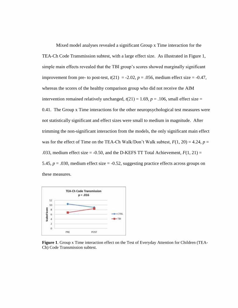
Mixed model analyses revealed a significant Group x Time interaction for the
TEA-Ch Code Transmission subtest, with a large effect size. As illustrated in Figure 1,
simple main effects revealed that the TBI group’s scores showed marginally significant
improvement from pre- to post-test, t(21) = -2.02, p = .056, medium effect size = -0.47,
whereas the scores of the healthy comparison group who did not receive the AIM
intervention remained relatively unchanged, t(21) = 1.69, p = .106, small effect size =
0.41. The Group x Time interactions for the other neuropsychological test measures were
not statistically significant and effect sizes were small to medium in magnitude. After
trimming the non-significant interaction from the models, the only significant main effect
was for the effect of Time on the TEA-Ch Walk/Don’t Walk subtest, F(1, 20) = 4.24, p =
.033, medium effect size = -0.50, and the D-KEFS TT Total Achievement, F(1, 21) =
5.45, p = .030, medium effect size = -0.52, suggesting practice effects across groups on
these measures.
Figure 1. Group x Time interaction effect on the Test of Everyday Attention for Children (TEA-
Ch) Code Transmission subtest.
26
Parent- and Child-Report Measures
Pre- and post-test means and standard deviations for each group are also reported
in Table 3, along with results of t-tests for pre-test group differences and the mixed model
Group x Time interaction and their respective effect sizes. The TBI group showed
significantly more parent- and child-reported behaviors associated with executive
dysfunction at pre-test relative to the healthy comparison group on all three examined
BRIEF scores, all with large effect sizes.
Mixed model analyses revealed significant Group x Time interactions for all three
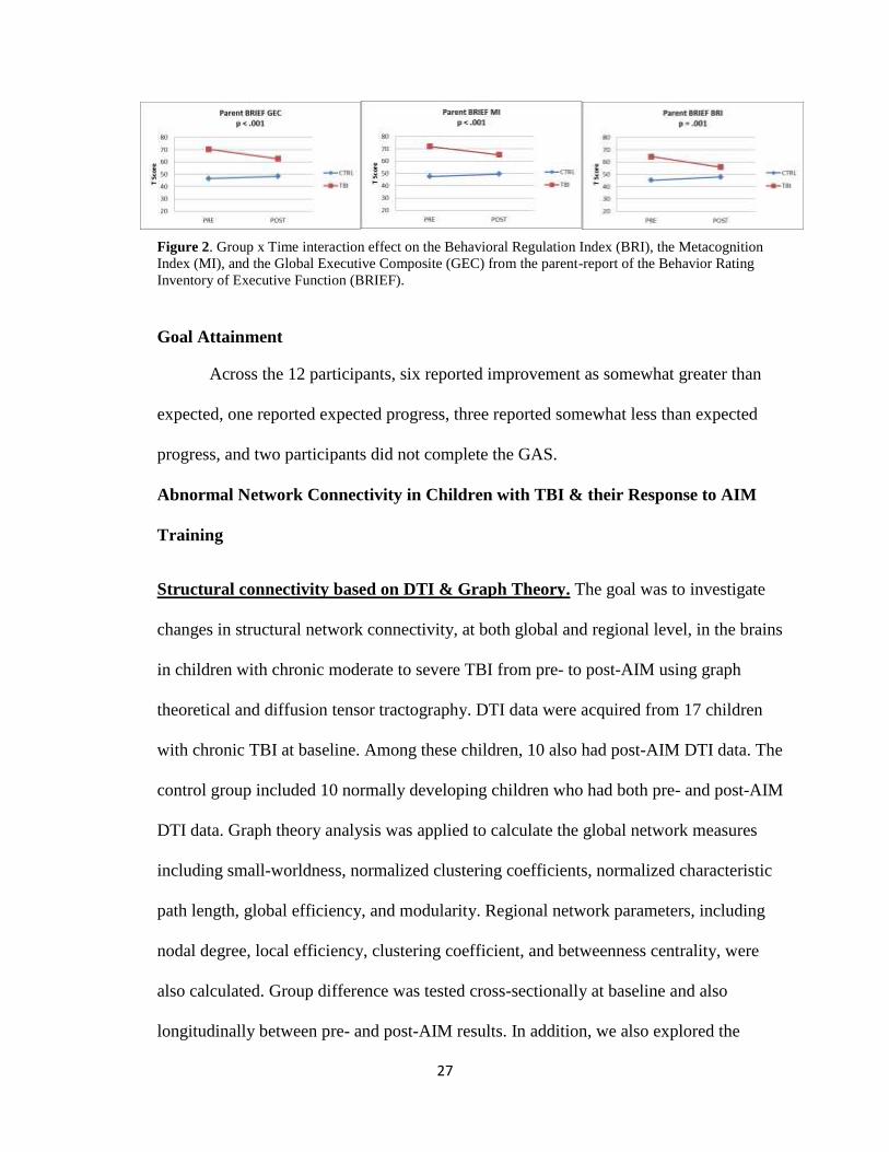
parent-reported BRIEF scores, with medium to large effect sizes. As illustrated in Figure
2, simple main effects revealed that the TBI group’s scores improved significantly from
pre- to post-test, (BRI: t(21) = 4.14, p < .001, large effect size = 0.78; MI: t(21) = 5.22, p
< .001, medium effect size = 0.51; GEC: t(21) = 4.94, p < .001, medium effect size =
0.60), whereas the scores of the healthy comparison group who did not receive the AIM
intervention remained relatively unchanged, (BRI: t(21) = -1.22, p = .237, small effect
size = -0.24; MI: t(21) = -1.41, p = .172, trivial effect size = -0.14; GEC: t(21) = -1.13, p
= .270, trivial effect size = -0.14). The Group x Time interactions for all three child-
reported BRIEF scores were not statistically significant and effect sizes were small in
magnitude. After trimming the non-significant interactions from the child-report BRIEF
models, there were significant main effect of Group on each measure (BRI: F(1, 16) =
9.37, p = .008, large effect size = -1.13; MI: F(1, 16) = 17.13, p < .001, large effect size =
-1.42; GEC: F(1, 16) = 13.21, p = .002, large effect size = -1.30), indicating poorer self-
reported EF in the TBI group across time.
27
Figure 2. Group x Time interaction effect on the Behavioral Regulation Index (BRI), the Metacognition
Index (MI), and the Global Executive Composite (GEC) from the parent-report of the Behavior Rating
Inventory of Executive Function (BRIEF).
Goal Attainment
Across the 12 participants, six reported improvement as somewhat greater than
expected, one reported expected progress, three reported somewhat less than expected
progress, and two participants did not complete the GAS.
Abnormal Network Connectivity in Children with TBI & their Response to AIM
Training
Structural connectivity based on DTI & Graph Theory. The goal was to investigate
changes in structural network connectivity, at both global and regional level, in the brains
in children with chronic moderate to severe TBI from pre- to post-AIM using graph
theoretical and diffusion tensor tractography. DTI data were acquired from 17 children
with chronic TBI at baseline. Among these children, 10 also had post-AIM DTI data. The
control group included 10 normally developing children who had both pre- and post-AIM
DTI data. Graph theory analysis was applied to calculate the global network measures
including small-worldness, normalized clustering coefficients, normalized characteristic
path length, global efficiency, and modularity. Regional network parameters, including
nodal degree, local efficiency, clustering coefficient, and betweenness centrality, were
also calculated. Group difference was tested cross-sectionally at baseline and also
longitudinally between pre- and post-AIM results. In addition, we also explored the

28
correlation between the changes in network connectivity and the changes in behavioral
outcomes (Walk-. Code- , pMI, pBRI,GEC) in response to the AIM training.
Figure 3. Comparison of Global network measures. Figure 4. Comparison of local network measures
Global network Measures: At baseline, children with chronic TBI were found to have
significantly higher small-worldness than the controls (two-tailed t-test, p=0.023, Figure
1). No significant group difference was found at baseline in other global network
measures. Longitudinally, significant decrease in the small-worldness was found in
children with chronic TBI after AIM training (n=10, two-tailed paired t-test, p=0.001).
No significant longitudinal change was found in the small-worldness in the control group.
Most importantly, changes in connectivity between the initial and follow-up scans
differed significantly between the groups (∆σ = -0.056±0.037 in TBI vs. ∆σ = 0.017 ±
0.044 in control, t=4.07, df = 19, p = 0.0007). Regional network Measures: A series of
brain regions showed significant group differences at baseline in various regional
network measures. However, among these regions, only the angular gyrus in TBI group
showed significant pre- vs. post-AIM change in nodal clustering coefficient and local
efficiency (both p<0.05, see Figure 4 for clustering coefficient result).
Association between changes in structural connectivity and changes in behavioral
outcomes: Significant correlations were found between longitudinal changes in

29
behavioral outcomes and changes in structural connectivity at both global and regional
level. Specifically, we found that the change in parent-reported metacognitive functioning
increased with mean local efficiency (p=0.008, Fig 3A) and the change in parent-reported
behavioral regulation was inversely correlated with the change in network modularity
(p=0.046, Fig 3B). It was also found that changes in parent-reported behavior regulation
decreased with the change in nodal clustering coefficient in angular gyrus at trend level
(p=0.07, Fig 3C).
Figure 5. Correlation between behavioral outcome with global (A, B) and local (C) structural connectivity.
Functional connectivity based on resting state fMRI & Graph Theory: We also
employed functional connectivity analysis based on resting state fMRI (rs-fMRI) and
graph theory to investigate changes in functional connectivity in response to AIM
training. The network connectivity parameters and statistical analyses used in the
functional connectivity analysis were similar to those used in the analysis of structural
connectivity. Among the 17 children with TBI recruited for imaging at baseline, 2 were
excluded for compliance reasons. Eight of the remaining 15 children with TBI had both
pre- and post-AIM rs-fMRI data. Among the 11 normal children recruited for imaging, 4
were excluded for motion artifact. Therefore, only 7 controls with both pre- and post-
AIM refMRI data were available in the final analysis.

30
Figure 6. Comparison of global functional connectivity
Global network Measures: At baseline, children with chronic TBI were found to have
significantly lower small-worldness than the controls (two-tailed t-test, p=0.022, Figure
4). Longitudinally, small-wordness increased at trend level in children with chronic TBI
after AIM training (n=8, two-tailed paired t-test, p=0.09). No significant longitudinal
change was found in the small-worldness in the control group. The two groups were
found to have a trend level difference in their response to the AIM training (∆σ =
0.17±0.25 in TBI vs. ∆σ = -0.055±0.27 in control, t=1.77, df = 14, p = 0.099).
Regional network Measures: A series of brain regions showed significant group
differences at baseline in various regional network measures (p<0.05). Among these
regions, middle occipital gyrus in TBI pts showed significant decrease in local efficiency
(p<0.03) and nodal clustering coefficient (p<0.03) after the AIM. Superior frontal gyrus
in TBI pts showed trend level decrease (p=0.08) in nodal clustering coefficient after
AIM.
Figure 7. Correlation between behavioral outcome with global (A, B) and local (C) functional connectivity.

31
Association between changes in functional connectivity and changes in behavioral
outcomes: Significant correlations were found between longitudinal changes in
behavioral outcomes and changes in functional connectivity at both global and regional
level. Specifically, we found that the change in WALK subtest increased with both
network modularity (p=0.011, Fig 5A) and normalized clustering coefficient (p=0.031,
Fig 5B). It was also found the change in WALK subtest decreased with the change in
nodal clustering coefficient in superior frontal gyrus at trend level (p=0.071, Fig 5C).
Overall Summary for graph analysis of network connectivity: Our data showed both
structural and functional connectivity based on graph theory were sensitive to detect
abnormalities of brain network associated with TBI at both global and regional level.
Using both approaches, we found significant changes in network measures in response to
the AIM training. In addition, we found that the changes in some of the network
measures were correlated, either significantly or at trend level, with the changes observed
in the behavioral outcomes. These results strongly suggest that the network connectivity
approach will provide a new avenue for potential diagnosis and prognosis for children
with TBI as well as a monitoring tool to quantify response to AIM training.
Counting Stroop as an Attentional Probe: Behavioral and Functional Neuroimaging
Results: One of the goals of the current project is to recruit participants with TBI who
show signs of attentional impairments. As a result, there are multiple challenges for
securing usable fMRI data from these participants, including heightened susceptibility to
movement artifacts in image acquisition, non-compliance to task directions, and
suboptimal task performance. Of the 12 participants with TBI who completed the AIM
training, only 6 contributed usable fMRI and behavioral data for both fMRI sessions

32
whereas 8 from the control group did so. Total performance for groups improved from
time1 to time2 for participants with TBI (from 79% at time1 to 87% at time2) as well as
for control participants (from 87% at time1 to 90% at time2), but neither the group effect
nor the interaction effect was significant. Performance was not different between groups
at time1 or time2.
Figure 8. The dominant pattern of the preliminary findings on training effects for participants
with TBI shows areas with higher levels of inhibition-related activation at time1 than at time2 (p
= 0.05 uncorrected, cluster size = 25; slice location z = -20 to +56). Such areas overlap with
network areas typically activated in the Counting Stroop task, including right medial temporal
lobe, right lingual gyrus, right middle frontal gyrus, left post-central gyrus, medial frontal gyrus,
and left middle frontal gyrus.

33
Figure 9. Preliminary findings on time effects for control participants. The only area that shows
higher levels of activation at time1 than at time2 is anterior cingulate (slice z=+20, region shown
in red). All other areas show lower level of attention at time1 than at time2. Convention as in
Figure 8.
Our preliminary findings suggest that there is a differential effect of time for the two
groups. While participants with TBI showed mostly a drop in levels of inhibition-related
activation across a number of areas, mostly in the frontal lobes, from pre-training to post-
training, control participants showed mostly an increase in levels of inhibition-related
activation across a number of areas, including medial and superior temporal cortices,
subgenual anterior cingulate, left insular cortex, and superior frontal areas, when
performing Counting Stroop the first time versus a second time. The findings with the
participants with TBI are consistent with the idea that there is an improvement in task
performance accompanied by a drop in related brain activation level following AIMS
training.

34
XIII. Conclusions
The results of this study are consistent with previous intervention studies
suggesting that broad based training that includes both domain-general and domain
specific approaches holds promise as an intervention to remediate attention, WM and EF
deficits in the pediatric acquired brain injury population. Effects were greatest on parent
reported executive dysfunction which improved significantly from pre- to post-treatment;
whereas effects on neuropsychological performance were more limited. Improvements in
parent-reported executive dysfunction corresponded to changes in both structural and
functional connectivity on neuroimaging suggesting that changes in parent-reported
behavior were reflected in corresponding neural changes. Structural and functional
connectivity analyses appear to provide a viable approach for quantifying neural changes
associated with attention training in children.
XIV. Recommendations
Despite its apparent utility, relatively high levels of attrition suggest that attention
should be given to making the intervention more engaging and doable for participants. As
delivered, the intervention required 10 or more weekly face to face visits as well as 2 or
more independent practice sessions. Although intensity is considered to be an important
treatment component, many participants had difficulty committing to this level of
practice. Future research is needed to identify both the optimal timing and intensity of
interventions such as AIM as well as strategies for making it more user-friendly.
35
XV. References
Butler RW, Fairclough DL, Katz ER, et al. A multicenter, randomized clinical trial of a
cognitive remediation program for childhood survivors of a pediatric malignancy.
Journal of Consulting & Clinical Psychology. 2008; 76(3): 367-378.
Collins, M.W., Lovell, M.R., Iverson, G.L., Cantu, R.C., Maroon, J.C., Field, M.
Cumulative effects of concussion in high school athletes. Neurosurgery.2002; 51:
1175-1181.
Delis DC, Kaplan E, Kramer JH. 2001. The Delis-Kaplan Executive Function System.
San Antonio: The Psychological Corporation.
DePompei, R. & Williams, J. Working with families after TBI- A family centered
approach. Topics in Language Disorders. 1994; 15: 68-81.
Faul, M., Xu, L., Wald, M. M., & Coronado, V. G. (2010). Traumatic brain injury in the
United States: emergency department visits, hospitalizations and deaths 2002-2006
Retrieved from http://www.cdc.gov/traumaticbraininjury/pdf/blue_book.pdf
Fay GC, Jaffe KM, Polissar NL, Liao S, Rivara JB, Martin KM. Outcome of pediatric
traumatic brain injury at 3 years: a cohort study. Archives of Physical Medicine and
Rehabilitation. 1994; 75: 733-741.
Finkelstein, E., Corso, P., & Miller, T. R. (2006). The Incidence and Economic Burden of
Injuries in the United States The Incidence and Economic Burden of Injuries in the
United States. New York, NY: Oxford University Press

36
Friston KJ, Holmes AP, Price CJ, Buchel C, Worsley KJ. Multisubject fMRI studies and
conjunction analyses. Neuroimage. 1999; 10(4): 385-396.
Galbiati S, Recla M, Pastore V, Liscio M, Bardoni A, Castelli E, Strazzer S. Attention
remediation following traumatic brain injury in childhood and adolescence.
Neuropsychology. 2009; 23: 40-49.
Gioia GA, Isquith PK, Guy SC. Behavior rating of executive function. Child
Neuropsychology. 2000; 6: 235-238.
Janusz JA, Kirkwood MW, Yeates KO, Taylor HG. Social problem-solving skills in
children with traumatic brain injury: Long-term outcomes and prediction of social
competence. Child Neuropsychology. 2002; 8: 179-194.
Karunanayaka P, Holland S, Yuan W, et al. Abnormalities in language circuitry in
children with traumatic brain injury, an fMRI study. Neurorehabilitation and
Neural Repair. 2007; 22: 355-369.
Kennedy MRT, Coelho C, Turkstra L, Ylvisaker M, Sohlberg MM, Yorkston K, Chiou
H-H, Kan P-F. Intervention for executive functions after traumatic brain injury: A
systematic review, meta-analysis and clinical recommendations.
Neuropsychological Rehabilitation. 2008; 18(3): 257-299.
Kesler SR, Lacayo NJ, Jo B. A pilot study of an online cognitive rehabilitation program
for executive function skills in children with cancer-related brain injury. Brain
Injury. 2011; 25(1): 101-112.

37
Kim Y-H, Yoo W-K, Ko M-H, Park C-H, Kim ST, Na DL. Plasticity of the attentional
network after brain injury and cognitive rehabilitation. Neurorehabilitation and
Neural Repair. 2009; 23(5): 468-477.
Kramer ME, Chiu CY, Walz NC, et al. Long-term neural processing of attention
following early childhood traumatic brain injury: fMRI and neurobehavioral
outcomes. Journal of the International Neuropsychological Society. 2008; 14(3):
424-435.
Kurowski B, Wade S, Cecil K, Walz N, Weihong Y, Rajagopal R, Holland S. Correlation
of Diffusion Tensor Imaging with Executive Function Measures after Early
Childhood Traumatic Brain Injury. Journal of Pediatric Rehabilitation Medicine.
2009; 2 (4): 273-283.
Langlois JA, Rutland-Brown W, Thomas KE. 2006. Traumatic brain injury in the
United States: emergency department visits, hospitalizations, and deaths. Atlanta:
Centers for Disease Control and Prevention.
Levin H, Hanten G, Max J, et al. Symptoms of attention-deficit/hyperactivity disorder
following traumatic brain injury in children. Journal of Developmental and
Behavioral Pediatrics. 2007; 28(2): 108-118.
Loken WJ, Thornton AE, Otto RL, Long CJ. Sustained attention after severe closed head
injury. Neuropsychology. 1995; 9: 592-598.
Lovell, M.R., Iverson, G.L., Collins, G.L., Podell, K., Johnston, K.M., Pardini, D.,
Pardini, J., Norwig, J., & Maroon, J. Measurement of symptoms following sports-

38
related concussion: reliability and normative data for the post-concussion scale.
2006; 13(3): 166-174.
Malec J. Goal attainment scaling in rehabilitation. Neuropsychological Rehabilitation.
1999; 9(3/4): 253-275.
Manly T, Robertson I H, Anderson V, Nimmo-Smith I. 1999. TEA-Ch: The Test of
Everyday Attention for Children Manual. Bury St. Edmunds, UK: Thames Valley
Test Company Limited.
Max JE, Koele SL, Smith WL, Sato Y, Lindgren SD, Robin DA, Arndt S. Psychiatric
disorders in children and adolescents after severe traumatic brain injury: A
controlled study. Journal of the American Academy of Child and Adolescent
Psychiatry. 1998; 37: 832-840.
Max JE, Schachar RJ, Levin HS, Ewing-Cobbs L, Chapman SB, Maureen Dennis M,
Ann Saunders A, Landis J. Predictors of attention-deficit/hyperactivity disorder
within 6 months after pediatric traumatic brain injury. Journal of the American
Academy of Child & Adolescent Psychiatry. 2005; 44(10): 1032-1040.
Mooney P, Ryan JB, Uhing BM, Reid R, Epstein MH. A review of self-management
interventions targeting academic outcomes for students with emotional and
behavioral disorders. Journal of Behavioral Education. 2005; 14(3): 203-221.
Ohio Brain Injury Association. Retrieved March 8, 2011 from http://www.biaoh.org/om.
Ottenbacher, K. J., & Cusick, A. Goal attainment scaling as a method of clinical service

39
evaluation. American Journal of Occupational Therapy. 1990; 44, 519–525.
Penkman L. Remediation of attention deficits in children: a focus on childhood cancer,
traumatic brain injury and attention deficit disorder. Pediatric Rehabilitation.
2004; 7(2): 111-123.
Posner MI. Rothbart MK. Influencing brain networks: implications for education. Trends
in Cognitive Sciences. 2005; 9(3): 99-103.
Posner MI. Rothbart MK. Research on attention networks as a model for the integration
of psychological science. Annual Review of Psychology. 2007; 58: 1-23.
Reid R, Trout A L, Schartz M. Self-regulation interventions for children with attention
deficit/hyperactivity disorder. Exceptional Children. 2005; 71(4): 361-377.
Schmithorst V, Holland, SK. Dardzinski BJ. 2000. CCHIPS: Cincinnati Children's
Hospital Imaging Processing Software .http://www.irc.chmcc.org/chips.htm.
Shi, J., Xiang, H., Wheeler, K., Smith, G. A., Stallones, L., Groner, J., & Wang, Z. Costs,
mortality likelihood and outcomes of hospitalized US children with traumatic brain
injuries. Brain Injury. 2009; 23(7): 602-611.
Smith SM, Jenkinson M, Johansen-Berg H, et al. Tract-based spatial statistics: voxelwise
analysis of multi-subject diffusion data. Neuroimage. 2006; 31(4): 1487-1505.
Sohlberg, MM, Harn, B., MacPherson, H., Wade, S.L. A pilot study evaluation attention
and strategy training following pediatric traumatic brain injury. Clinical Practice
in Pediatric Psychology. 2014; 2 (3): 263-280.

40
Sohlberg MM, Avery J, Kennedy M, et al. Practice guidelines for direct attention
training. Journal of Medical Speech Language Pathology. 2003; 11(3): xix-xxxix.
Sohlberg MM, Mateer C. 2001. Cognitive Rehabilitation: An Integrated
Neuropsychological Approach. New York: Guilford Publications.
Sohlberg MM, Mateer CA. 2010. Attention Process Training III: A direct attention
training program for persons with acquired brain injury. Lash & Associates
Publishing/Training Inc.
Stevens C, Fanning J, Coch D, Sanders L, Neville H. Neural mechanisms of selective
auditory attention are enhanced by computerized training: Electrophysiological
evidence from language-impaired and typically developing children. Brain
Research. 2008; 1205: 55-69.
Taylor, H. G. (2004). Research on outcomes of pediatric traumatic brain injury: current
advances and future directions. Dev Neuropsychol, 25(1-2), 199-225
Teasdale, G., Jennett, B. Assessment of coma and impaired consciousness: a practical
scale. The Lancet. 1974; 2:81-84
Tlustos SJ, Chiu C-YP, Walz NC, Holland SK, Bernard L, Wade SL. Neural correlates of
interference control in adolescents with traumatic brain injury: functional magnetic
resonance imaging study of the Counting Stroop Task. Journal of the International
Neuropsychological Society. 2011;17:181-189.
Trombly, C. A., Radomski, M. V., Trexel, C., Burnett-Smith, S. E. Occupational therapy
and achievement of self-identified goals by adults with acquired brain injury:

41
Phase II. The American Journal of Occupational Therapy. 2002; 56, 489–498.
van't Hooft I, Andersson K, Sejersen T, Bartfai B, von Wendt L. Attention and memory
training in children with acquired brain injuries. Acta Paediatrica. 2003; 92(8):
935-940.
van't Hooft I, Andersson K, Bergman B, Sejersen T, von Wendt L, Bartfai A. Sustained
favorable effects of cognitive training in children with acquired brain injuries.
Neurorehabilitation. 2007; 22(2): 109-116.
Vernescu R. 2008. Sustained attention training in children with fetal alcohol spectrum
disorder. Unpublished doctoral dissertation, Department of Psychology, Memorial
University, St. John’s Newfoundland.
Wade, S.L., Carey, J., Wolfe, C.R. An online family intervention to reduce parental
distress following pediatric brain injury. Journal of Consulting and Clinical
Psychology. 2006; 74(3): 445-454.
Wade SL, Karver CL, Taylor HG, et al. Counselor-assisted problem solving improves
caregiver efficacy following adolescent brain injury. Rehabilitation Psychology.
2014; 59(1):1
Walz NC, Cecil K, Wade SL, Michaud L. Late proton MR spectroscopy following
traumatic brain injury during early childhood: relationship with neurobehavioral
outcomes. Journal of Neurotrauma. 2008; 25: 94-103.
Wolraich ML, Lambert W, Doffing MA, Bickman L, Simmons T, Worley K.

42
Psychometric properties of the Vanderbilt ADHD Diagnostic Parent Rating Scale
in a referred population. J Pediatr Psychol. 2003; 28: 559– 568.
Worsley KJ, Liao CH, Aston J, Petre V, Duncan GH, Morales F, Evans AC. A general
statistical analysis for fMRI data. Neuroimage. 2002; 15(1): 1-15.
Yeates, K. O., Swift, E., Taylor, H. G., Wade, S. L., Drotar, D., Stancin, T., & Minich, N.
(2004). Short- and long-term social outcomes following pediatric traumatic brain
injury. J Int Neuropsychol Soc, 10(3), 412-426.
Yuan W, Holland SK, Schmithorst VJ, et al. Diffusion tensor MR imaging reveals
persistent white matter alteration after traumatic brain injury experienced during
early childhood. American Journal of Neuroradiology. 2007; 28: 1919-1925.