effect of thermal processing on mealworm allergenicity · pdf fileeffect of thermal processing...
TRANSCRIPT

Mol. Nutr. Food Res. 2015, 59, 1855–1864 1855DOI 10.1002/mnfr.201500138
RESEARCH ARTICLE
Effect of thermal processing on mealworm allergenicity
Henrike Broekman1, Andre Knulst1, Stans den Hartog Jager1, Francesca Monteleone2,Marco Gaspari2, Govardus de Jong3, Geert Houben1,3 and Kitty Verhoeckx1,3
1 Department of Dermatology/Allergology, University Medical Centre Utrecht (UMCU), Utrecht, The Netherlands2 Dipartimento di Medicina Sperimentale e Clinica, Universita “Magna Græcia” di Catanzaro, Catanzaro, Italy3 TNO, Zeist, the Netherlands
Received: February 20, 2015Revised: May 28, 2015
Accepted: June 3, 2015
Scope: The growing world population requires the exploration of new sustainable proteinsources to ensure food security. Insects such as mealworm are promising candidates. Forsafety reasons, a risk assessment, including allergy risks, is needed. Since allergenicity can beinfluenced by thermal processing, it is highly important to take this into account.Methods and results: Fresh mealworm was heat processed and extracted by a sequentialextraction method using in succession Tris, urea, and a combined SDS/DTT buffer. Extractswere tested using immunoblot, basophil activation test and skin prick test in 15 shrimp allergicpatients, previously indicated as population at risk for mealworm allergy. Immunoblots showeda difference in IgE binding between processed and unprocessed mealworm extracts. However,this was due to change in solubility. Some allergens were soluble in urea buffer, but becamemore soluble in Tris buffer and vice versa. IgE binding was seen for all extracts in blot andbasophil activation test. The results from 13 skin prick tests showed a skin reaction similarbetween processed and unprocessed mealworm.Conclusion: Thermal processing did not lower allergenicity but clearly changed solubilityof mealworm allergens. A sequential extraction method allowed for assessment of a broaderprotein panel.
Keywords:
Food allergy / Mealworm / Sequential extraction method / Solubility thermal pro-cessing
1 Introduction
A huge shortage of protein sources for human food con-sumption is expected in the near future due to the growingworld population [1]. Sustainable protein sources are beingexplored to solve the coming food insecurity problem. Thelarvae of the yellow mealworm beetle (Tenebrio molitor) is agood candidate and is already for sale in Great Britain, theUS, and in the major supermarkets in the Netherlands andBelgium [2, 3]. However, a thorough safety assessment, andin particular an allergenicity risk assessment, is yet to be per-formed [4]. Allergenicity is not only a theoretical threat, since0.1–5.7% of the pediatric and 0.1–3.2% of the adult Europeanpopulation has a food allergy [5]. Moreover, previously we [6]found that IgE from patients sensitized to shrimp and house
Correspondence: Henrike BroekmanE-mail: [email protected]
Abbreviations: BAT, basophil activation test; PBST, PBS-containing 1% Tween 20; SPT, skin prick test; TPSM, total peptide-spectrum matches
dust mite (Der p 10; closely related species), binds to meal-worm proteins. The relevant proteins were identified as thepan allergens tropomyosin and arginine kinase, which aremajor allergens in shellfish (e.g. shrimp and lobster). Aller-genicity can be influenced by factors such as matrix [7] andprocessing—for instance, by changing protein structure andthus IgE-binding epitopes [8]. This was previously reportedfor other foods such as peanut, tree nuts, and apple [9–12].Thermal processing by dry roasting enhanced allergenicityof peanut [12], while for tree nuts the allergenic propertieschanged in such a way that most pollen allergic patients re-acting to tree nuts had no clinical reaction after eating the heatprocessed food [12, 13]. Thus processing may have an impacton the risk of getting an allergic reaction for mealworm. Sincemealworm is closely related to shellfish one might expect thatprocessing may alter the allergenicity of mealworm proteinsin a comparable manner to shellfish.
For instance, Nakamura et al. [14] reported that thermalprocessing resulted in an enhanced IgE-binding capacity ofscallop tropomyosin in dot blot and competitive ELISA usingserum from scallop allergic patients. This enhanced capac-ity was suggested to be a result of glycation between free
C© 2015 WILEY-VCH Verlag GmbH & Co. KGaA, Weinheim www.mnf-journal.com

1856 H. Broekman et al. Mol. Nutr. Food Res. 2015, 59, 1855–1864
amino acids and aldehyde or ketone groups of sugars dur-ing heating. The same group found an opposite result afterMaillard reaction with squid tropomyosin [15]. Samson et al.[16] found no significant difference between boiled and rawshrimp extract using immunoblot with serum from shrimpallergic patients. However, interindividual differences in pro-tein recognition were observed. Carnes et al. [17] reported thatboiled extracts of shrimp and lobster had higher IgE-bindingcapacity in ELISA and recorded greater skin reactivity in skinprick test (SPT). A similar finding was observed by Liu et al.[18] when testing shrimp tropomyosin. Taken together, theresults in the above-mentioned papers are to some extent con-tradicting. This could be due to solubility issues. Therefore,more attention should be paid to the preparation of extractsto ensure the presence of a representative set of proteins forallergenicity assessment.
Most studies reported the effect of processing using im-munoblot and ELISA. Unfortunately, these methods lack in-formation on the functionality of IgE binding, which canbe measured using SPT and basophil activation test (BAT).These tests are therefore preferred over immunoblot andELISA in allergenicity assessment. However, they cannotreplace food challenges—the “gold” standard. Immunoblot,BAT, and SPT were used in this study to test the effect ofprocessing on mealworm allergenicity. Shrimp allergic pa-tients were tested due to the lack of a sufficient number ofmealworm allergic patients. To ensure that most relevant pro-teins were covered, a sequential protein extraction methodwas used and the presence of allergens was confirmed usingnanoLC–MS.
2 Materials and methods
2.1 Patient selection and screening
Three sera from patients diagnosed with shrimp allergy atthe University Medical Centre Utrecht, the Netherlands, wereused to test the effect of processing on protein solubility us-ing immunoblot. For allergenicity testing, 15 adult patientsdiagnosed with shrimp allergy, based on suggestive historyand sensitization were included. All patients reacted positiveto mealworm protein in SPT and serology. All patients gaveinformed consent before answering the questions and forthe performance of SPT and blood collection. The study wasapproved by the local ethics committee.
2.2 Thermal processing of mealworm
Raw and freeze dried Yellow mealworms in final larval stagewere kindly provided by Dutch insect farm Kreca (Ermelo,The Netherlands). Raw mealworms (50 g) were heat pro-cessed by various methods: Blanching for 1 min at 100�C,boiling in 300 mL water for 10 min at 100�C, baking for3.5 min at 1000 Watt on an induction cooker (Prima Donna
tsi-199k), or frying for 30 s at 180�C in peanut oil. All pro-cessed and unprocessed mealworms were stored at −20�Cuntil further use.
2.3 Mealworm extract preparation
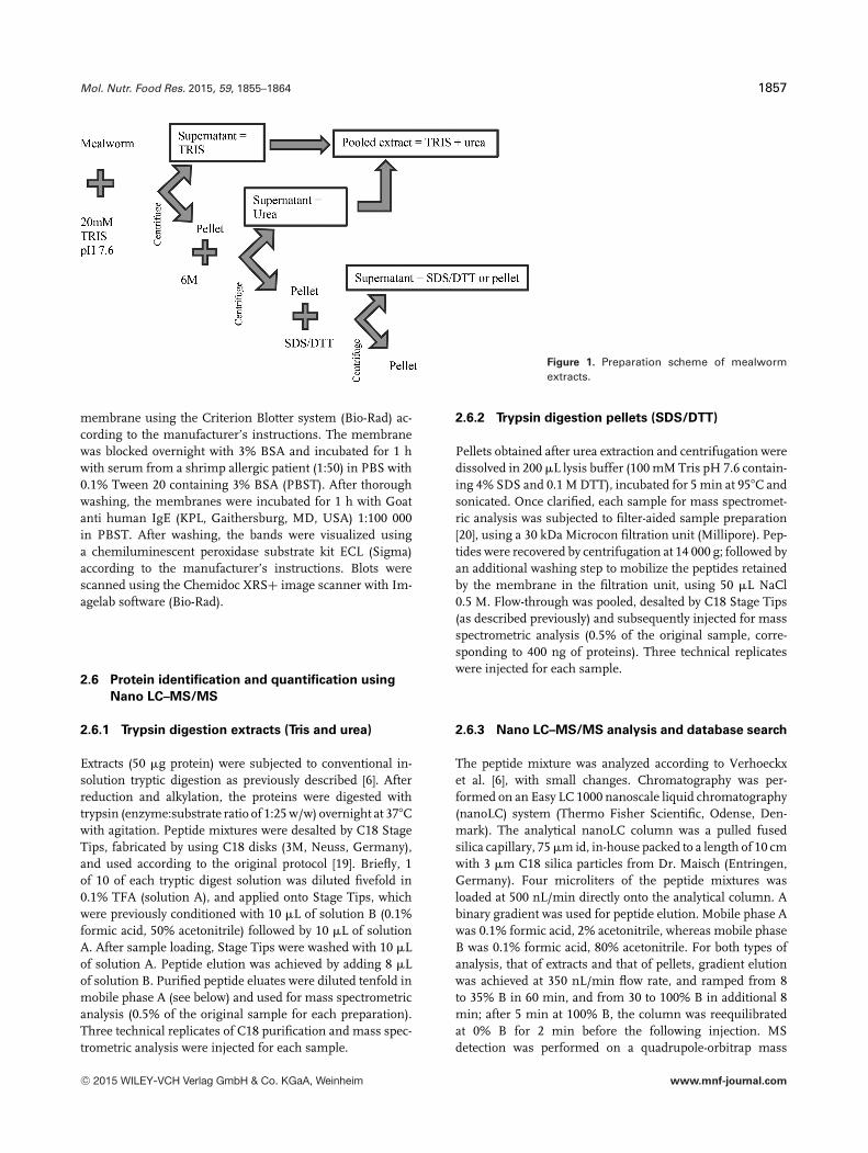
Five grams of raw, freeze dried, and processed mealwormswere extracted using a sequential protein extraction method(see Fig. 1). First, the mealworms were mixed with 25 mLice cold Tris buffer (20 mM Tris buffer pH 7.6 containing1 mM phenylthiocarbamide (Sigma Aldrich) and Halt Pro-tease Inhibitor Cocktail (Thermo Scientific)). The amount ofmealworm was corrected for weight gain or weight loss dueto processing. Subsequently, the mealworms were disrupted,using an ultraturrax (3 × 10 s) under continuous cooling. Af-ter centrifugation (30 min, 15 000 × g at 4�C), the supernatantwas recovered.
The insoluble residue was washed once with 5 mL Trisbuffer (as described above). The 30 and 5 mL supernatantswere combined. Twenty-five milliliter was used for samplecleanup and concentration using TCA precipitation. Second,the remaining pellet was extracted overnight at 4�C with 30mL urea buffer (6 M urea in 20 mM Tris buffer pH 7.6 con-taining 1 mM phenylthiocarbamide and Halt Protease In-hibitor Cocktail). The sample was subsequently centrifugedand the supernatant was collected. The pellet was washedonce more with 5 mL urea buffer, centrifuged, and the su-pernatant was combined with the 30 mL urea supernatant.Twenty-five milliliters of the extract was TCA precipitated.Tris and urea extracts were combined (1:1) for the BAT. Fi-nally, the insoluble residue was almost completely dissolvedat room temperature in 20 mL SDS/DTT buffer (20 mM TrispH 7.6, 2% SDS, and 1% DTT) and the supernatant wascollected after centrifugation. All TCA precipitated sampleswere redissolved in 6 M urea buffer and stored at −20�C be-fore further use. Protein concentration was determined usingthe Bradford method (Bio-Rad, Hercules, CA, USA).
2.4 SDS-PAGE gel of processed mealworm extracts
For SDS-PAGE, the Criterion system with a 10–20% ReadyGel Tris-HCl gel (Bio-Rad) was used according to the man-ufacturer’s instructions. All mealworm extracts (10 �g persample) were loaded on the gel under reducing conditions(Laemmli buffer). After protein separation, the proteins werevisualized using Coomassie-staining (Instant Blue, Expedi-ton, UK).
2.5 Immunoblot with serum of shrimp allergic
patients
All mealworm extracts were applied on the SDS-PAGE asdescribed above and transferred to a polyvinyldifluoride
C© 2015 WILEY-VCH Verlag GmbH & Co. KGaA, Weinheim www.mnf-journal.com
Mol. Nutr. Food Res. 2015, 59, 1855–1864 1857
Figure 1. Preparation scheme of mealwormextracts.
membrane using the Criterion Blotter system (Bio-Rad) ac-cording to the manufacturer’s instructions. The membranewas blocked overnight with 3% BSA and incubated for 1 hwith serum from a shrimp allergic patient (1:50) in PBS with0.1% Tween 20 containing 3% BSA (PBST). After thoroughwashing, the membranes were incubated for 1 h with Goatanti human IgE (KPL, Gaithersburg, MD, USA) 1:100 000in PBST. After washing, the bands were visualized usinga chemiluminescent peroxidase substrate kit ECL (Sigma)according to the manufacturer’s instructions. Blots werescanned using the Chemidoc XRS+ image scanner with Im-agelab software (Bio-Rad).
2.6 Protein identification and quantification using
Nano LC–MS/MS
2.6.1 Trypsin digestion extracts (Tris and urea)
Extracts (50 �g protein) were subjected to conventional in-solution tryptic digestion as previously described [6]. Afterreduction and alkylation, the proteins were digested withtrypsin (enzyme:substrate ratio of 1:25 w/w) overnight at 37�Cwith agitation. Peptide mixtures were desalted by C18 StageTips, fabricated by using C18 disks (3M, Neuss, Germany),and used according to the original protocol [19]. Briefly, 1of 10 of each tryptic digest solution was diluted fivefold in0.1% TFA (solution A), and applied onto Stage Tips, whichwere previously conditioned with 10 �L of solution B (0.1%formic acid, 50% acetonitrile) followed by 10 �L of solutionA. After sample loading, Stage Tips were washed with 10 �Lof solution A. Peptide elution was achieved by adding 8 �Lof solution B. Purified peptide eluates were diluted tenfold inmobile phase A (see below) and used for mass spectrometricanalysis (0.5% of the original sample for each preparation).Three technical replicates of C18 purification and mass spec-trometric analysis were injected for each sample.
2.6.2 Trypsin digestion pellets (SDS/DTT)
Pellets obtained after urea extraction and centrifugation weredissolved in 200 �L lysis buffer (100 mM Tris pH 7.6 contain-ing 4% SDS and 0.1 M DTT), incubated for 5 min at 95�C andsonicated. Once clarified, each sample for mass spectromet-ric analysis was subjected to filter-aided sample preparation[20], using a 30 kDa Microcon filtration unit (Millipore). Pep-tides were recovered by centrifugation at 14 000 g; followed byan additional washing step to mobilize the peptides retainedby the membrane in the filtration unit, using 50 �L NaCl0.5 M. Flow-through was pooled, desalted by C18 Stage Tips(as described previously) and subsequently injected for massspectrometric analysis (0.5% of the original sample, corre-sponding to 400 ng of proteins). Three technical replicateswere injected for each sample.
2.6.3 Nano LC–MS/MS analysis and database search
The peptide mixture was analyzed according to Verhoeckxet al. [6], with small changes. Chromatography was per-formed on an Easy LC 1000 nanoscale liquid chromatography(nanoLC) system (Thermo Fisher Scientific, Odense, Den-mark). The analytical nanoLC column was a pulled fusedsilica capillary, 75 �m id, in-house packed to a length of 10 cmwith 3 �m C18 silica particles from Dr. Maisch (Entringen,Germany). Four microliters of the peptide mixtures wasloaded at 500 nL/min directly onto the analytical column. Abinary gradient was used for peptide elution. Mobile phase Awas 0.1% formic acid, 2% acetonitrile, whereas mobile phaseB was 0.1% formic acid, 80% acetonitrile. For both types ofanalysis, that of extracts and that of pellets, gradient elutionwas achieved at 350 nL/min flow rate, and ramped from 8to 35% B in 60 min, and from 30 to 100% B in additional 8min; after 5 min at 100% B, the column was reequilibratedat 0% B for 2 min before the following injection. MSdetection was performed on a quadrupole-orbitrap mass
C© 2015 WILEY-VCH Verlag GmbH & Co. KGaA, Weinheim www.mnf-journal.com

1858 H. Broekman et al. Mol. Nutr. Food Res. 2015, 59, 1855–1864
spectrometer Q-Exactive (Thermo Fisher Scientific, Bremen,Germany) operating in positive-ion mode, with nanoelec-trospray (nESI) potential at 1800 V applied on the columnfront end via a tee piece. Data-dependent acquisitionwas performed by using a top-12 method with resolution(FWHM), AGC target, and maximum injection time (ms) forfull MS and MS/MS of, respectively, 70 000/17 500, 106/105,50/60. Mass window for precursor ion isolation was 1.6 m/z,whereas normalized collision energy was 25. Ion thresholdfor triggering MS/MS events was 2 × 10⁴. Dynamic exclusionwas 30 s. Data was processed using Proteome Discoverer1.3 (Thermo Fisher Scientific), using Sequest as searchengine, and the Swiss Prot database accessed on February2013 as sequence database (3 123 840 sequences for Metazoataxonomy). The following search parameters were used: MStolerance 15 ppm; MS/MS tolerance 0.02 Da; fixed modifica-tions carbamidomethyl cysteine; enzyme trypsin; maximummissed cleavages 1; taxonomy Metazoa. Search results werefiltered by q values using Percolator integrated in ProteomeDiscoverer, to achieve a peptide-level FDR of less than 1%.
2.6.4 Relative protein quantification
A label-free approach was adopted for relative quantifica-tion of allergens, using five unique peptides for argininekinase and three unique peptides for tropomyosin. Peakareas for each peptide were calculated using extracted ionchromatograms (XICs) via the Xcalibur software (ThermoFisher Scientific). Peak areas for each peptide were subse-quently normalized using the total peptide-spectrum matches(TPSM) of the corresponding LC–MS/MS analysis. The Trisfreeze dried sample, one with the highest TPSM, was cho-sen to confirm linearity between injected amount and TPSM.Triplicate measurements of peak area were averaged for eachpeptide and expressed as relative value compared to the aver-age area of the same peptide in the Tris unprocessed sample.Relative quantification at the protein level was achieved for allproteins by taking the median value of all associated peptides.
2.6.5 BAT using shrimp allergic patient serum
BAT was performed as described by Meulenbroek et al.[21] with minor modifications. Cells were incubated witha dilution series (1:107–1:102) of processed and unpro-cessed mealworm extracts (combined TRIS and urea ex-tracts (5 mg/mL) and SDS/DTT extracts (no concentrationdetermined)). Shrimp extract (ALK), 2 mg/mL, and shrimptropomyosin Pen a 1 (Indoor Biotechnologies), 1 mg/mL,were used as positive controls. CD63, CD123, and CD203c ex-pression was analyzed by flow cytometry using FACSCantoII and FACSDiva software (BD Bioscience, USA). The re-sults were expressed as a percentage of CD63+ basophils.Basophils of two patients did not respond in repetition to anyof the extracts, nor to the positive control. Basophils of a third
patient showed spontaneous release of CD63 on the negativecontrol. These three patients were therefore excluded.
2.6.6 SPT with processed mealworm extracts
SPT solutions of the processed and unprocessed mealworms(0.4 mg/mL) were kindly provided by ALK (ALK-Abello,Spain). These solutions were prepared in PBS, which hasmore or less the same extraction characteristics as the Trisbuffer mentioned above. The solutions were applied on theflexor aspect of the forearm using 1 mm tip lancets (ALK).Histamine dihydrochloride 10 mg/mL and glycerol diluentwere used as positive and negative controls, respectively. SPTreactivity was recorded after 15 min and measured as theratio of the mean of the wheal elicited by the tested extractand histamine control. When the ratio was 0.5 or greater,the reaction was regarded as positive. No statistical tests wereperformed due to the limited size of the group.
3 Results
3.1 Heat processing changes solubility
It can be concluded from Fig. 2 that protein profiles signif-icantly changed after heat processing in all tested extracts(Tris, urea, and SDS/DTT). Bands of proteins from the Trisextracts with MW < 25 kDa and at ±50 kDa were moreintense in all heat-processed extracts compared to the unpro-cessed extracts (raw and freeze dried).
In case of the urea extract, protein bands with a MW of ±40kDa were more pronounced in unprocessed extracts, whereasbands near 45 and 50 kDa were more pronounced in all heat-processed extracts. In the SDS/DTT extract the same band at±45 kDa diminishes after heating, while a band appears at±37 kDa. In addition, high molecular weight proteins (70–200 kDa) were detected in the SDS/DTT buffer extracts afterheating. These changes in protein profiles were the result ofchanges in solubility, as shown by the LC–MS analysis.
LC–MS analysis of processed and unprocessed Tris, urea,and SDS/DTT extracts identified a wide range of proteins inmealworm. Putative mealworm allergens (e.g. tropomyosin,arginine kinase, myosin light chain, and triosephosphate iso-merase), identified in Tris and urea extract were previouslyreported by us [6]. However, in this study we also identi-fied putative allergens in the SDS/DTT extract (Table 1). Themost dominant protein in the SDS/DTT extract was argininekinase after heat processing.
The concentrations of these putative mealworm allergenswere different in the tested extracts. It can be concluded fromFig. 3 that processing causes a shift in solubility from Tris tourea and vice versa. For instance, arginine kinase, which wasabundant in raw and freeze dried mealworm Tris extracts, wasalmost undetectable in heat processed mealworm Tris extract.However, it became detectable in urea extracts after heat pro-cessing. For tropomyosin the opposite effect was found. The
C© 2015 WILEY-VCH Verlag GmbH & Co. KGaA, Weinheim www.mnf-journal.com

Mol. Nutr. Food Res. 2015, 59, 1855–1864 1859
Figure 2. Coomassie-stained SDS-PAGE gels ofextracts from processed mealworms. Tris, urea,and SDS-DTT extracts were prepared from raw,freeze dried, blanched, boiled, baked, and friedmealworm.
solubility of tropomyosin in Tris buffer improved after heatprocessing. The same phenomenon was seen for other aller-gens such as myosin light chain, which behaved in a similarmanner to tropomyosin on processing. Triosephosphate iso-merase, another allergen, behaved similar to arginine kinase.However, quantification was difficult, due to detection lim-its (data not shown). In conclusion, solubility of mealwormallergens changed after heat processing.
3.2 Heat processing does not obviously change
IgE-binding capacity
The above-mentioned change in allergen solubility was con-firmed by the immunoblot of the three shrimp allergic pa-tients (Fig. 4). The immunoblots showed IgE binding toproteins in all tested mealworm extracts, Tris, urea, andSDS/DTT. In the lane of raw and freeze dried Tris extracta protein band at ±40 kDa can be seen, which was previouslyidentified as arginine kinase [6]. After processing, this bandbecomes more pronounced at a slightly higher MW. Compar-ing these results with the LC–MS analysis (Fig. 3), this bandis most likely tropomyosin (±37 kDa). In the lane of raw andfreeze dried urea extract, a ±37 kDa band (previously identi-fied after in-gel digestion, as tropomyosin [6]) was detected.The intensity of this band diminished after processing. Thedecline of tropomyosin band intensity in the urea extract is inaccordance with the LC–MS data (Fig. 3). Estimation of theoverall effect of processing on IgE-binding capacity is difficultdue to this shift in solubility. For this reason, a pool of Trisand urea extract was used for IgE cross-linking functionalitytesting.
3.3 Heat processing does not change IgE
cross-linking functionality
From the 12 useful BATs, 11 showed activation after incu-bation with both the mixed Tris /urea extracts as well as the
SDS/DTT extracts, indicated by an elevation in the percent-age of CD63+ cells (Fig. 5). Basophils of one patient reactedsolely to proteins in the SDS/DTT extract.
Overall, activity of basophils to processed or unprocessedmealworm proteins was not clearly different. However, ba-sophils from three patients were somewhat more stronglyactivated by the processed Tris /urea mealworm extracts,than by the unprocessed mealworm extracts. All 15 patientsshowed a positive skin reaction to shrimp, house dust mite,and mealworm extract. In addition to unprocessed meal-worm, all showed a positive skin reaction to blanched, boiled,baked, and fried extracts (Table 2).
However, some interindividual differences were seen inskin reactivity. Two patients had an increased skin reaction toprocessed extracts. The wheal size increased from 2+ to 3+from unprocessed to processed. Skin reaction of one patientdecreased by blanching and of one patient by frying. Over-all, SPT reactions were comparable between all processedextracts in 13 of 15 patients.
4 Discussion
From the results obtained in this study it can be concludedthat heat processing influences protein solubility. Some pro-teins became less soluble in Tris buffer due to heat-induceddenaturation but these proteins could still be solubilized ina chaotropic reagent such as urea (arginine kinase). Otherproteins that under natural conditions were insoluble in Trisbuffer became more soluble after heating (tropomyosin). Fur-thermore, processing did not lower IgE-binding capacity andIgE cross-linking functionality of mealworm allergens (e.g.tropomyosin, arginine kinase). A representative panel of pro-teins was assessed due to the use of a sequential extractionmethod. To the best of our knowledge, this is the first studyto assess the effect of thermal processing on mealworm al-lergenicity. Furthermore, the sequential protein extractionmethod used in this study has, as far as we know, never beenused to assess the effect of processing on allergenicity.
C© 2015 WILEY-VCH Verlag GmbH & Co. KGaA, Weinheim www.mnf-journal.com

1860 H. Broekman et al. Mol. Nutr. Food Res. 2015, 59, 1855–1864
Table 1. Proteins identified in SDS/DTT extract using LC–MS/MS
Protein (source) Accession Score Sequence Peptides PSM Masscoverage (%) identified (kDa)
UnprocessedMyosin heavy chain-like (Triobolium
castaneum)D6WVJ3 401 33 85 158 262.1
Actin-87E (Drosophila melanogaster) P10981 188 42 20 79 41.8Actin-2 (Diphyllobothrium dendriticum) P53456 146 15 18 54 41.7Actin, cytoskeletal 1 (Lytechinus pictus) P53465 128 19 10 60 41.8Fibronectin_type3 (T. castaneum) D6W7B4 112 7 50 53 989.6Actinin (Apis mellifera) H9K1K1 88 10 26 37 101.8Hemocyanin (T. molitor) Q9Y1W5 67 30 20 31 90.6Ca-transporting ATPase sarcoplasmic/
endoplasmic reticulum type (Drosophilapseudoobscura)
Q292Q0 65 17 17 26 109.1
ATPase_P-type_Ca-transporter(T. castaneum)
D6WZH8 63 11 17 26 109.8
Hexamerin 2 (T. molitor) Q95PI7 59 18 11 25 84.5Larval cuticle protein A2B (T. molitor) P80682 52 43 6 15 12.3Cuticle protein (T. molitor) Q9TXE4 46 27 3 15 23.2ATP synthase subunit alpha (T. castaneum) D6WSI9 42 23 12 19 59.5Tubulin beta-1 chain (Manduca sexta) O17449 40 26 11 19 50.2Troponin T-like (T. castaneum) D6W953 39 14 5 16 45.7
ProcessedMyosin heavy chain like (T. castaneum) D6WVJ3 418 34 89 155 262.1Actin-87E (D. melanogaster) P10981 218 47 23 275 41.8Actin, cytaloplasmic (Xenopusl aevis) O93400 211 48 22 175 41.7Kinase tranferase (T. castaneum) D6W7B4 147 13 97 183 989.6Alpha-actin like (T. castaneum) D2A2×1 122 45 46 147 106.7Actinin (T. castaneum) B3P8U6 86 29 27 66 106.7Hemocyanin (T. molitor) Q9Y1W5 82 36 29 106 90.6Ca-transporting ATPase sarcoplasmic/
endoplasmic reticulum type
(D. pseudoobscura)
Q292Q0 76 21 24 91 109.1
Arginine kinase (Xylosandrus crassiusculus) D5L6P4 75 48 15 93 27.0Alpha tubulin (D. melanogaster) K7WKV5 64 43 16 60 46.1Tubulin beta-1 chain (M. sexta) O17449 57 35 17 77 50.2Filamin-B like (T. castaneum) D6W7G0 56 12 26 65 267.3ATP synthase subunit alpha (T. castaneum) D6WSI9 54 35 20 62 59.5Prophenoloxidase (T. molitor) O97047 51 36 25 63 79.1Hexamerin 2 (T. molitor) Q95PI7 47 27 18 58 84.5
PSM, peptide-spectrum matches, value that represents the number of MS/MS spectra that matched peptide sequences assigned to thatparticular protein. Score, the sum of individual Sequest scores of all the identified peptides which were assigned to the protein itself. Thescore is the probability that the observed match is not a random event.Top 15 proteins identified by LC–MS/MS in the SDS/DTT fraction in the unprocessed and processed extracts. Arranged on highest meanscore of three measurements. Sequence coverage, peptides identified, and PSM are given as a mean of three measurements. Identificationwas based on homology with metazoan proteins in the Swiss Prot database. Proteins are noted in bold when assigned as allergen by theIUIS allergen nomenclature subcommittee.
Heat processing strongly changed the solubility charac-teristics of mealworm proteins. Change in allergen solubility,might be caused by changes in 3D structure of the proteins af-ter heat treatment. Some proteins will lose, irreversibly, theirfunctional properties and solubility and form aggregates dueto denaturation, while others may have increased solubility.Tropomyosin is a muscle protein, which forms a complexwith the insoluble actin and troponin and is heat stable as aresult of its coiled coil helical construction [22]. The improvedsolubility is most probably due to breakage of interactions
with these other difficult to solubilize proteins. However, noevidence could be found in literature to corroborate this. An-other possibility is the formation of soluble aggregates, whichwas also demonstrated for the Japanese cedar pollen allergenCry j 1 by Aoki et al. [23]. Moreover, Usui et al. [24] showedthat heat processing of purified tropomyosin from shrimp didnot induce the formation of insoluble aggregates. However,difference in solubility in PBS buffer between heated and un-heated tropomyosin was not in agreement with our results.This might be due to fact that heat processing of tropomyosin
C© 2015 WILEY-VCH Verlag GmbH & Co. KGaA, Weinheim www.mnf-journal.com

Mol. Nutr. Food Res. 2015, 59, 1855–1864 1861
Figure 3. LC–MS analysis of tropomyosin and arginine kinase in processed mealworm extracts (raw, freeze dried, blanched, boiled, baked,and fried, respectively). The results are presented as mean of three LC–MS analysis and calculated as ratio relative to the amount in theraw Tris extract.
was not tested in its natural environment (complex with actin)and thus breakage of interactions with other proteins cannotbe demonstrated.
In contrast, arginine kinase is a globular protein, whichtends to unfold during heating, exposing hydrophobic aminoacids, which are normally inside the protein. The exposed hy-drophobic amino acids from different molecules will interactin such a way, that formation of larger protein aggregateswill occur [25]. Cross-linking of arginine kinase may alsobe caused by polyphenol oxidase-mediated cross-linking. Inmost cases these aggregates become insoluble. Further aggre-gation of globular proteins during heating is favored throughthe formation of disulphide bridges. To solubilize these ag-gregates a more stringent buffer is needed, which confirms
our finding that arginine kinase was not detected in the Trisbuffer after heating.
Another possibility for the LC–MS detection oftropomyosin in Tris buffer after heat processing is the im-proved digestibility after heating. This effect was also seen ina study from Takagi et al. who showed that thermal treatmentmarkedly increased the digestibility of ovalbumin [26]. Thisis because ovalbumin is a globular protein that unfolds dur-ing heating, which exposes amino acid sequences that can behydrolyzed by trypsin. However, for mealworm tropomyosinthis improved digestibility after heating was not confirmedby the immunoblot. Furthermore, enhancement of trypsindigestion is not expected for tropomyosin because of its he-lical structure that upon heating will not suddenly expose
Figure 4. Immunoblot of processedmealworms with serum from one ofthe three shrimp allergic patients. Themealworms were extracted with a Tris,urea, and SDS-DTT buffer, respectively.
C© 2015 WILEY-VCH Verlag GmbH & Co. KGaA, Weinheim www.mnf-journal.com

1862 H. Broekman et al. Mol. Nutr. Food Res. 2015, 59, 1855–1864
Figure 5. BAT with extracts (left: pooled Tris and urea extract, right: SDS/DTT extract) from processed mealworms (freeze dried, fresh,blanched, cooked, baked, and fried, respectively). Maximum percentage of CD63+ basophils were calculated with respect to IgE positivecontrol. Each line represents one patient.
Table 2. SPT results, expressed as a ratio of histamine control,using extracts from the different processed mealworms(raw, blanched, boiled, baked, and fried) in 15 shrimpallergic patients
No. Sex Age (yr) Raw Blanched Boiled Baked Fried
1 F 46 3+ 3+ 3+ 3+ 2+2 F 23 2+ 3+ 3+ 3+ 3+3 M 69 2+ 2+ 3+ 2+ 2+4 M 45 2+ 2+ 2+ 3+ 2+5 F 27 3+ 2+ 3+ 3+ 3+6 M 19 2+ 3+ 2+ 2+ 2+7 F 60 2+ 2+ 2+ 2+ 2+8 M 30 2+ 3+ 3+ 3+ 2+9 M 27 2+ 2+ 2+ 2+ 2+10 F 47 2+ 2+ 2+ 2+ 2+11 F 52 0 0 1+ 1+ 1+12 M 26 2+ 2+ 2+ 2+ 2+13 M 34 2+ 2+ 3+ 2+ 3+14 F 23 2+ 2+ 2+ 2+ 2+15 M 46 2+ 2+ 3+ 3+ 3+
M, male; F, female.Mean SPT as a ratio of histamine control (3+).
different amino acid sequences. Therefore, it is more likelythat extractability and thus solubility is the main reason forthe difference between the unprocessed and heat processedsamples instead of improved digestibility.
When testing only protein extracts prepared in Tris buffer,which is the routine procedure, one could wrongly concludethat IgE-binding capacity to tropomyosin would be elevateddue to heat processing [17]. However, the correct explana-
tion is that possibly important allergens are overlooked whenusing just one buffer type. This was demonstrated by ourimmunoblot data and might also be the case in some studies[16–18] where induction of IgE binding after heat processingwas observed. In these studies only PBS extracts, which isa nondenaturing extraction buffer similar to the Tris buffer,were used. Solubility issues are often encountered when pro-teins are processed. In most cases this phenomenon is notrecognized since the composition of the protein extracts isnot identified [27].
IgE binding on the immunoblot was detected in all testedextracts, indicating that in mealworm there are more aller-gens present than the ones identified in Tris buffer. Themost dominant IgE-binding proteins in the Tris extract and inthe urea extract were identified as tropomyosin and argininekinase, respectively, which confirms our previous findings[6].
In the SDS/DTT extract, arginine kinase (±40 kDa band)was also identified and in addition IgE binding to proteinswith a higher molecular weight was detected. The high MWproteins that were identified in the SDS/DTT extracts by LC–MS/MS were myosin heavy chain, paramyosin, and hemo-cyanin. It is not clear if these are the same IgE-binding pro-teins as detected in the immunoblot. However, myosin heavychain and paramyosin were recently identified as shrimp al-lergens [28–30]. Moreover, paramyosin and hemocyanin areincluded in the IUIS database as arthropod allergens. Sincemealworm and shrimp are closely related it can be envisionedthat paramyosin and myosin heavy chain could be potentialmealworm allergens. Another option is that the high molec-ular weight proteins are the result of arginine kinase cross-linking. According to LC–MS identification arginine kinase
C© 2015 WILEY-VCH Verlag GmbH & Co. KGaA, Weinheim www.mnf-journal.com

Mol. Nutr. Food Res. 2015, 59, 1855–1864 1863
was also detected in the SDS/DTT extracts especially afterheat processing.
Processing did not change IgE functionality in BAT andSPT. The advantage of BAT over SPT is that BAT allows test-ing of extracts prepared with stringent buffers such as ureaand SDS/DTT, while due to patient safety SPT only allowsprotein extracts prepared according to clinical guidelines insterile PBS buffers [31]. The results from the BAT indicate,besides some interindividual variability in basophil activa-tion, no significant effect due to heat processing. Processingshowed only induced basophil activation in three patients.
In SPT, 13 of 15 patients, did not react differently to theprocessed extracts. Only two patients (other than those inBAT) showed a trend of increased skin reaction. This mightbe caused by the increased solubility of some allergens inPBS after heat processing, which might also be the case inthe study of Nowak-Wegrzyn et al. [32]. The authors reportedthat boiled shrimp extract induced larger skin response com-pared to raw shrimp extract in some shrimp allergic patients.Together these results strengthen the need for different ex-traction buffers to assess the allergenicity of a broad repre-sentative protein panel.
The strength of this study was the combined use of clinical,ex vivo and in vitro tests in combination with a sequential ex-traction method and LC–MS analysis. This allowed inclusionof a broader panel of mealworm proteins in the allergenicityassessment, than usually studied.
In conclusion, heat processing did not lower the aller-genicity of mealworm proteins, but clearly changed thesolubility of these proteins. A sequential extraction methodallowed for inclusion of a broader protein panel in theallergenicity assessment of mealworm.
The authors are members of the COST Action ImpARASFA1402. Supported by grants from TNO and the Dutch food andconsumer product safety authority.
The authors have declared no conflict of interest.
5 References
[1] Tilman, D., Balzer, C., Hill, J., Befort, B. L., Global food de-mand and the sustainable intensification of agriculture. Proc.Natl. Acad. Sci. USA 2011, 108, 20260–20264.
[2] FAO, FAO Forestry Paper: Edible Insects: Future Prospectsfor Food and Feed Security, FAO, Rome 2013.
[3] vanHuis, A., Potential of insects as food and feed in assuringfood security. Annu. Rev. Entomol. 2013, 58, 563–583.
[4] FAO/WHO. Foods Derived from Modern Biotechnology,FAO/WHO, Rome 2009.
[5] Nwaru, B. I., Hickstein, L., Panesar, S. S., Roberts, G. et al.,Prevalence of common food allergies in Europe: a systematicreview and meta-analysis. Allergy 2014, 69, 992–1007.
[6] Verhoeckx, K. C., van Broekhoven, S., den Hartog-Jager, C.F., Gaspari, M. et al., House dust mite (Der p 10) and crus-
tacean allergic patients may react to food containing Yellowmealworm proteins. Food Chem. Toxicol. 2014, 65, 364–373.
[7] Schulten, V., Lauer, I., Scheurer, S., Thalhammer, T. et al., Afood matrix reduces digestion and absorption of food aller-gens in vivo. Mol. Nutr. Food Res. 2011, 55, 1484–1491.
[8] Davis, P. J., Williams, S. C., Protein modification by thermalprocessing. Allergy 1998, 53, 102–105.
[9] Bohle, B., Zwolfer, B., Heratizadeh, A., Jahn-Schmid, B. et al.,Cooking birch pollen-related food: divergent consequencesfor IgE- and T cell-mediated reactivity in vitro and in vivo. J.Allergy Clin. Immunol. 2006, 118, 242–249.
[10] Blanc, F., Vissers, Y. M., Adel-Patient, K., Rigby, N. M. et al.,Boiling peanut Ara h 1 results in the formation of aggregateswith reduced allergenicity. Mol Nutr Food Res. 2011, 55(12),1887–1894.
[11] Hansen, K. S., Ballmer-Weber, B. K., Luttkopf, D., Skov, P.S. et al., Roasted hazelnuts–allergenic activity evaluatedby double-blind, placebo-controlled food challenge. Allergy2003, 58, 132–138.
[12] Beyer, K., Morrow, E., Li, X. M., Bardina, L. et al., Effectsof cooking methods on peanut allergenicity. The Journal ofallergy and clinical immunology 2001, 107(6), 1077–1081.
[13] Worm, M., Hompes, S., Fiedler, E. M., Illner, A. K. et al., Im-pact of native, heat-processed and encapsulated hazelnutson the allergic response in hazelnut-allergic patients. Clin.Exp. Allergy 2009, 39, 159–166.
[14] Nakamura, A., Watanabe, K., Ojima, T., Ahn, D. H. et al., Effectof maillard reaction on allergenicity of scallop tropomyosin.J. Agric. Food Chem. 2005, 53, 7559–7564.
[15] Nakamura, A., Sasaki, F., Watanabe, K., Ojima, T.et al., Changes in allergenicity and digestibility of squidtropomyosin during the Maillard reaction with ribose.J. Agric. Food Chem. 2006, 54, 9529–9534.
[16] Samson, K. T., Chen, F. H., Miura, K., Odajima, Y. et al., IgEbinding to raw and boiled shrimp proteins in atopic andnonatopic patients with adverse reactions to shrimp. Int.Arch. Allergy Immunol. 2004, 133, 225–232.
[17] Carnes, J., Ferrer, A., Huertas, A. J., Andreu, C. et al., Theuse of raw or boiled crustacean extracts for the diagnosis ofseafood allergic individuals. Ann. Allergy Asthma Immunol.2007, 98, 349–354.
[18] Liu, G. M., Cheng, H., Nesbit, J. B., Su, W. J. et al., Effectsof boiling on the IgE-binding properties of tropomyosin ofshrimp (Litopenaeus vannamei). J. Food Sci. 2010, 75, T1–T5.
[19] Rappsilber, J., Mann, M., Ishihama, Y. Protocol for micro-purification, enrichment, pre-fractionation and storage ofpeptides for proteomics using StageTips. Nat. Protoc. 2007,2, 1896–1906.
[20] Wisniewski, J. R., Ostasiewicz, P., Mann, M., High recoveryFASP applied to the proteomic analysis of microdissectedformalin fixed paraffin embedded cancer tissues retrievesknown colon cancer markers. J. Proteome Res. 2011, 10,3040–3049.
[21] Meulenbroek, L. A., de Jong, R. J., den Hartog Jager, C. F.,Monsuur, H. N. et al., IgG antibodies in food allergy influ-ence allergen-antibody complex formation and binding to
C© 2015 WILEY-VCH Verlag GmbH & Co. KGaA, Weinheim www.mnf-journal.com

1864 H. Broekman et al. Mol. Nutr. Food Res. 2015, 59, 1855–1864
B cells: a role for complement receptors. J. Immunol. 2013,191, 3526–3533.
[22] Hillberg, L., Zhao Rathje, L. S., Nyakern-Meazza, M., Helfand,B. et al., Tropomyosins are present in lamellipodia of motilecells. Eur. J. Cell Biol. 2006, 85(5), 399–409.
[23] Aoki, R., Saito, A., Usui, M., Azakami, H. et al., Reduction ofantigenicity of Cry j 1, a major allergen of Japanese cedarpollen, by thermal denaturation. J. Agric. Food Chem. 2009.57, 4995–4999.
[24] Usui, M., Harada, A., Ishimaru, T., Sakumichi, E. et al., Con-tribution of structural reversibility to the heat stability of thetropomyosin shrimp allergen. Biosci. Biotechnol. Biochem.2013, 77, 948–953.
[25] Yu, H. L., Ruan, W. W., Cao, M. J., Cai, Q. F. et al., Identifica-tion of physicochemical properties of Scylla paramamosainallergen, arginin kinase. J. Sci. Food Agric. 2013. 93, 245–253.
[26] Takagi, K., Teshima, R., Okunuki, H., Sawada, J. Comparativestudy of in vitro digestibility of food proteins and effect ofpreheating on the digestion. Biol. Pharm Bull. 2003, 26, 969–973.
[27] Kamath, S. D., Rahman, A. M., Voskamp, A., Komoda, T.et al., Effect of heat processing on antibody reactivity to
allergen variants and fragments of black tiger prawn: a com-prehensive allergenomic approach. Mol. Nutr. Food Res.2014, 58, 1144–1155.
[28] Khanaruksombat, S., Srisomsap, C., Chokchaichamnankit,D., Punyarit, P. et al., Identification of a novel allergen frommuscle and various organs in banana shrimp (Fennerope-naeus merguiensis). Ann. Allergy Asthma Immunol. 2014,113, 301–306.
[29] Suzuki, M., Shimizu, K., Kobayashi, Y., Ishizaki, S. et al.,Paramyosin from the disc abalone Haliotis discus discus.J. Food Biochem. 2014, 38(4), 444–451.
[30] Giuffrida, M. G., Villalta, D., Mistrello, G., Amato, S. et al.,Shrimp allergy beyond tropomyosin in Italy: clinical rele-vance of Arginine Kinase, sarcoplasmic calcium binding pro-tein and hemocyanin. Eur. Ann. Allergy Clin. Immunol. 2014,46, 172–177.
[31] Dreborg, S. F., Frew, A., Position paper: allergen standard-ization and skin tests. Allergy 1993, 48, 49–54.
[32] Nowak-Wegrzyn, A., Fiocchi, A., Rare, medium, or well done?The effect of heating and food matrix on food protein aller-genicity. Curr. Opin. Allergy Clin. Immunol. 2009, 9, 234–237.
C© 2015 WILEY-VCH Verlag GmbH & Co. KGaA, Weinheim www.mnf-journal.com