effect of super-eba as a root end filling on healing …...effect of super-eba as a root end filling...
TRANSCRIPT

0099-2399/95/2101-0013/$03.00/0 JOURNAL OF ENDODONTICS Copyright © 1995 by The American Association of Endodontists
Printed in U.S.A. VOL. 21, NO. 1, JANUARY 1995
Effect of Super-EBA as a Root End Filling on Healing after Replantation
T. R. Pitt Ford, BDS, PhD, J. O. Andreasen, S. O. Dorn, DDS, and S. P. Kariyawasam
The effect of Super-EBA cement as a root-end filling placed in teeth before replantation was examined in eight molar roots in monkeys. After extraction, root ends were resected, the canals contaminated with oral bacteria, root-end cavities prepared, and fillings of Super-EBA placed before replantation. After 8 wk, the jaws were removed and prepared for histological examination.
The tissue response to Super-EBA was very mild, with only a few inflammatory cells being observed at the root end of 3 of the 8 roots filled. Previous work showed a similarly mild response to Interme- diate Restorative Material and a very much more severe response to amalgam.
It is concluded that the tissue response to Super- EBA as a root-end filling is acceptable and consid- erabiy more favorable than that to amalgam.
Super-EBA cements were developed in the 1960's as less irritant substitutes for zinc phosphate cements in restorative dentistry (1). They differ from other zinc oxide-eugenol ce- ments in that the liquid contains 62.5% o-ethoxy benzoic acid, as well as eugenol. The compressive strength and solu- bility have been reported to be similar to zinc phosphate cement (1). In 1970, Hendra (1) advocated Super-EBA cement for root-end filling. Eight years later, Oynick and Oynick (2) reported that it had been used as a root-end filling in over 200 teeth, although detailed follow-up was not available.
In 1990, Dorn and Gartner (3) published the results of a retrospective clinical study of 65 teeth filled with Super-EBA and achieved a 95% success rate, considerably better than amalgam. Histological investigations appear to be absent apart from that of a single tooth by Oynick and Oynick (2).
The aim of this study was to investigate the tissue response to root end fillings of Super-EBA in an experimental model reported previously (4).
MATERIALS AND METHODS
The method has been reported in detail previously (4), but is briefly summarized. Mandibular first molars, which have
two roots, were used in monkeys. Under general anesthesia, four teeth were extracted using gentle luxation movements. The root apices were resected flat with a diamond wheel under a constant flow of saline, before a 2-mm-deep root end cavity was drilled with a round bur (diameter 0.8 mm); the cavity was then washed and dried. Infection was introduced into the root canal before the root end was Idled with Super-EBA cement (Staident, Staines, UK) mixed according to the man- ufacturer's instructions.
After completion of the root end Idling procedure, the molars were replanted and left unsplinted for 8 weeks. The monkeys were then killed, and the jaw section was removed. The tissue blocks were fixed in formalin before being demin- eralized in EDTA. After embedding, the tissue blocks were sectioned and stained.
Sections were assessed for inflammation at the root end adjacent to the Idling material. The severity of the inflam- mation was recorded as: none = no inflammatory cells, few cells = few inflammatory cells, moderate = inflammatory cells did not obscure the normal tissues, and severe = inflam- matory cells replaced normal tissues. The extent of inflam- mation was recorded as _<0.1, _<0.2, _<0.5, or >0.5 mm. The presence or absence of root resorption exposing the root end ffiling material was noted, and where present the greatest extent toward the crown was recorded as part of the length of the Idling (to the nearest 1/4). The presence or absence of ankylosis of the tooth in any part, noticeable dissolution of the root-end filling material, giant cells, a fibrous capsule, cementum deposition on the filling material, epithelium cov- eting the filling material, and bacteria in any part of the root canal were noted.
13
RESULTS
The severity and extent of inflammation around the root ends are reported in Table 1. Data for amalgam and Inter- mediate Restorative Material (IRM) (Caulk, Dentsply, Mil- ford, DE) have been reported previously (4). All the roots filled with Super-EBA cement had inflammation in the none/ few inflammatory-cell categories, in contrast to the other materials. However, the differences between IRM and Super- EBA were small and not significant (p > 0.05, ×2 test). Figure I shows a root end filled with Super-EBA, and a very small number of lymphocytes can be observed in fibrous tissue.

14 Pitt Ford et al. Joumal of Endodontics
TABLE 1. NO. of roots with inflammation, its severity and extent, and the mean extent of giant cell coverage for each material
Inflammation No. of
Material Roots No. of Severity No. of Extent (mm)
N* F M S 0 <0.1 <0.2 <0.5 >0.5
Amalgam 12 1 0 5 6 1 0 3 0 8 IRM 30 12 13 4 1 12 16 1 1 0 Super-EBA 8 5 3 0 0 5 1 2 0 0
* N, no inflammatory cells; F, few inflammatory cells; M, moderate inflammation; S, severe inflammation.
FUG 2. Different section of same root shown in Fig. 1 at higher magnification showing giant cells (arrow) on the surface of the root- end filling and some lymphocytes (L) in the fibrous tissue (hematoxylin and eosin stain; original magnification × 115).
Frog 1. Root end of a tooth filled with Super-EBA cement, which has dissolved during processing. At the true root end, there is fibrous tissue containing a few lymphocytes. Laterally, there is more inflam- mation and resorption affects <3/4 length of the filling (hematoxylin and eosin stain; odginal magnification x45).
Figure 2 shows a different section of the same root at higher magnification.
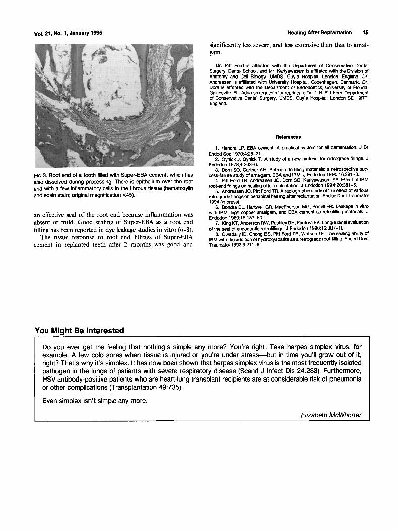
The presence of stainable bacteria in the root canals, the presence of ankylosis, active root resorption, and the extent of resorption of the root adjacent to the filling are recorded in Table 2. There was no observable difference between the materials, except possibly for the amount of resorption. No root end was covered by cementum, but one was covered by epithelium (Fig. 3).
DISCUSSION
The findings of this investigation with Super-EBA cement are broadly similar to those with IRM reported previously (4).
TABLE 2. NO. of teeth shown to contain bacteria, exhibiting ankylosis, demonstrating active root resorption, and the extent of resorption alongside the retrograde filling for each material
No. with No. with No. of Bacteria in No. Extent of Material Active Root Teeth Root Ankylosed Resorption
Canal Resorption
Amalgam 6 6 0 5 m IRM 15 9 8 13 0.33 Super-EBA 4 3 1 4 0.47
In view of the small number of roots involved, it would be unwise to conclude that the tissue response to Super-EBA was better than that to IRM. The results contrast with the severe response to amalgam (4). There is also agreement with radio- graphic studies of superior healing compared with amalgam (3, 5).
In common with IRM, more severe inflammation was noted at the sides of the roots where dentin had been resorbed. The resorption was more severe with Super-EBA cement. Although the resorption was without a doubt initiated by the procedure of extraction and replantation, it would seem pos- sible that some substance may have been leached out of the Super-EBA cement enhancing the resorption. However, where the material was exposed to the tissues at the root end, the tissue response was very good, implying that no further substances were being leached out.
Because the root canals of most of the teeth were shown to be still infected at 8 wk, Super-EBA cement seemed to provide
Vol. 21, No. 1, January 1995 Healing After Replantation 15
significantly less severe, and less extensive than that to amal- gam.
Dr. Pitt Ford is affiliated with the Department of Conservative Dental Surgery, Dental School, and Mr. Kariyawasam is affiliated with the Division of Anatomy and Cell Biology, UMDS, Guy's Hospital, London, England. Dr. Andreasan is affiliated with University Hospital, Copenhagen, Denmark. Dr. Dora is affiliated with the Department of Endodontics, University of Flodda, Gainesville, FL. Address requests for reprints to Dr. T. R. Pitt Ford, Department of Conservative Dental Surgery, UMDS, Guy's Hospital, London SE1 9RT, England.
FIG 3. Root end of a tooth filled with Super-EBA cement, which has also dissolved during processing. There is epithelium over the root end with a few inflammatory cells in the fibrous tissue (hematoxylin and eosin stain; original magnification x45).
an effective seal of the root end because inflammation was absent or mild. Good sealing of Super-EBA as a root end filling has been reported in dye leakage studies in vitro (6-8).
The tissue response to root end fillings of Super-EBA cement in replanted teeth after 2 months was good and
References
1. Handra LP. EBA cement. A practical system for all cementation. J Br Endod Soc 1970;4:28-31.
2. Oynick J, Oynick T. A study of a new material for retrograde fillings. J Endodon 1978;4:203-6.
3. Dom SO, Gartner AH. Retrograde tilling materials: a retrospective suc- cess-failure study of amalgam, EBA and IRM. J Endodon 1990;16:391-3.
4. Pitt Ford TR, Andreasen JO, Dom SO, Kariyawasam SP. Effect of IRM root-and fillings on healing after replantation. J Endodon 1994;20:381-5.
5. Andreasen JO, Pitt Ford TR. A radiographic study of the effect of various retrograde fillings on periapical healing after replantation. Endod Dent Traumatol 1994 (in press).
6. Bondra DL, Hartwell GR, MacPherson MG, Portell FR. Leakage in vitro with IRM, high copper amalgam, and EBA cement as retrefilling materials. J Endodon 1989;15:157-60.
7. King KT, Anderson RW, Pashley DH, Pantera EA. Longitudinal evaluation of the seal of endodontic retrotillings. J Endodon 1990;16:307-10.
8. Owadally ID, Chong BS, Pitt Ford TR, Watson TF. The sealing ability of IRM with the addition of hydroxyapatite as a retrograde root filling. Endod Dent Traumato11993;9:211-5.
You Might Be Interested
Do you ever get the feeling that nothing's simple any more? You're right. Take herpes simplex virus, for example. A few cold sores when tissue is injured or you're under stress--but in time you'll grow out of it, right? That's why it's simplex. It has now been shown that herpes simplex virus is the most frequently isolated pathogen in the lungs of patients with severe respiratory disease (Scand J Infect Dis 24:283). Furthermore, HSV antibody-positive patients who are heart-lung transplant recipients are at considerable risk of pneumonia or other complications (Transplantation 49:735).
Even simplex isn't simple any more.
Elizabeth McWhorter