effect of moringa oleifera stem extract on hydrogen
TRANSCRIPT

RESEARCH ARTICLE Open Access
Effect of Moringa oleifera stem extract onhydrogen peroxide-induced opacity ofcultured mouse lensLei Qi1, Yu Zhou2*, Weijie Li2, Mali Zheng1, Ruisheng Zhong1, Xin Jin2 and Yuan Lin1
Abstract
Background: Moringa oleifera, also known as horseradish tree or drumstick tree, has strong antioxidant properties.In the present study, we investigated the potential effect of Moringa oleifera stem extract (MOSE) on cataractformation induced by oxidative stress in cultured mouse lenses.
Methods: Mouse lenses cultured in vitro were pretreated with MOSE (0.5 and 1mg/mL) for 24 h. Then, 1 mMhydrogen peroxide was added, and mouse lenses were cultured for a further 24 h. The medium was then changedto normal culture medium. After 48 h, lens opacification, reactive oxygen species (ROS) generation, reducedglutathione (GSH) content, and activities of superoxide dismutase (SOD) and catalase (CAT) were measured in lenstissues. In addition, the protein expression of peroxisome proliferator-activated receptor alpha (PPARα), a nuclearreceptor with potential benefits to improve vision-threatening eye diseases, was assayed.
Results: MOSE (1 mg/mL) alleviated lens opacification, reduced ROS generation, increased GSH content, andelevated SOD and CAT activities in cultured lenses. Moreover, MOSE upregulated the expressions of SOD, CAT, andPPARα.Conclusions: This study showed that MOSE alleviates oxidative stress-induced cataract formation, and themechanism of the effect is mainly related to its improvement of the endogenous antioxidant system in the lens.
Keywords: Moringa oleifera, Lens, Organ culture, Oxidative stress, Antioxidant
BackgroundThe lens has a transparent, elastic avascular refractiveorganization and plays an important role in visual for-mation. Cataracts are characterized by gradual accumu-lation of cloudy deposits on the ocular lens and havebeen a leading cause of visual impairment and blindnessworldwide for centuries. Although modern cataract sur-gery is safe and effective, there are still many problems,such as high costs, loss of normal functions of postoper-ative eyes, and a high incidence of after-cataract. As theaging population increases, cataracts have become an in-creasingly serious issue [1, 2]. Thus, there is a great de-mand for safe, effective, and inexpensive agents toprevent or delay the onset of cataracts.
Cataracts are multifactorial eye diseases associatedwith several risk factors such as aging, diabetes, expos-ure to sunlight, and hypertension. However, oxidativestress caused by reactive oxygen species (ROS) has longbeen regarded as the major mechanism by which cellsare damaged and cataracts are formed [3–6]. Underphysiological conditions, lenses can compensate for amild degree of oxidant stress and remove oxidative dam-aged molecules by elevating endogenous antioxidantssuch as reduced glutathione (GSH) and activating anti-oxidant enzymes, such as superoxide dismutase (SOD)and catalase (CAT), which play important roles in pro-tecting the lens against oxidative stress. However, insome cases such as aging, ROS production is excessiveor the ability of the lens to scavenge ROS decreases, andoxidative stress injuries may occur and then cataractsare formed [3, 4].
© The Author(s). 2019 Open Access This article is distributed under the terms of the Creative Commons Attribution 4.0International License (http://creativecommons.org/licenses/by/4.0/), which permits unrestricted use, distribution, andreproduction in any medium, provided you give appropriate credit to the original author(s) and the source, provide a link tothe Creative Commons license, and indicate if changes were made. The Creative Commons Public Domain Dedication waiver(http://creativecommons.org/publicdomain/zero/1.0/) applies to the data made available in this article, unless otherwise stated.
* Correspondence: [email protected] of Basic Medical Science, School of Medicine, XiamenUniversity, Xiamen 361102, People’s Republic of ChinaFull list of author information is available at the end of the article
Qi et al. BMC Complementary and Alternative Medicine (2019) 19:144 https://doi.org/10.1186/s12906-019-2555-z

Because of the major role of oxidative stress in cata-ract formation, natural antioxidants with high activityand few side effects have attracted increasing attentionto delay the onset or progression of cataracts [7–10].Moringa oleifera, also known as horseradish tree ordrumstick tree, belongs to the Moringaceae family andhas been used in nutritious foods and traditional medi-cines for the treatment of various diseases such asrheumatism, inflammation, and diabetes in many Asiancountries [11]. Particularly, Moringa oleifera is one ofthe best known and most widely distributed species thatis rich in natural antioxidants [12, 13]. Moringa oleiferaleaf extracts was reported to inhibit the ROS formationinduced by H2O2 and enhanced the activities and mRNAexpressions of SOD and CAT in KB cells [14] and inHEK-293 Cells [15]. Recently the Moringa oleifera leafextract was reported to protect yeast cells against oxida-tive stress induced by cadmium and H2O2 through thereduction of intracellular ROS levels [16]. Regular intakeof Moringa oleifera leaves through diet decreased thelipid per oxidation and increase the SOD and CAT activ-ities in a diabetes-induced oxidative stress model in rats[17]. Moringa oleifera seed extract can inhibit the ROSformation induced by high fat diet in mice [18]. Moringaoleifera root extract attenuated beryllium-induced oxida-tive stress in rats [19]. All these significant antioxidantactivities of Moringa oleifera from both in vitro as wellas in vivo studies suggest that Moringa oliefera may in-hibit the cataract formation induced by oxidative stress.Although some studies have reported that the Moringaoleifera leaf extract has potential inhibitory effects onhigh sugar-induced cataract in goat lens in vitro [20] andselenite-induced cataract in rat pups [21], no study haveactually been conducted on the protective effects ofMoringa oleifera on oxidative stress induced cataract. Inaddition, compared with the traditional uses of theleaves, flowers, and seeds of Moringa oleifera, its stem isnot often consumed, and the stem may even be consid-ered as an agricultural by-product. However, Moringaoleifera stem is very abundant and inexpensive. Thus,any health benefit from it may reach a large part of thepopulation. Therefore, it is worthwhile to investigate thepotential effects of Moringa oleifera stem on delayingthe onset or progression of cataracts induced by oxida-tive stress. In addition, PPARs (including three isoforms:α, γ, and β/δ) are ligand-activated transcription factorsof the nuclear hormone receptor and play key roles inmaintaining glucose and lipid homeostasis by modulat-ing gene expression. Recent studies indicate that PPARshave potential benefits to improve or prevent variousvision-threatening eye diseases such as diabetic retinop-athy, glaucoma, and diabetic macular edema [22–24].Therefore, in this research, we also evaluate the effect ofMoringa oleifera stem on the expression of PPARs.
Lens organ cultures provide a simple and effectiveplatform to screen for candidate compounds that protectagainst cataract formation [25, 26]. Hydrogen peroxide(H2O2) is the main intracellular ROS in the aqueoushumor, which causes cataract development [27, 28], andis often used to induce cataract formation in vitro [8].Therefore, in the present study, we focused on the pro-tective effect of Moringa oleifera stem extract (MOSE)against cataract formation and explored its underlyingmechanism using a cataract formation model induced byH2O2 in lens organ culture. Luteolin is a flavonoidpresent in the leaves and stems of many plants and somereports indicate that luteolin exerts protective effects onselenite [29, 30] and STZ [31]-induced cataracts. Soluteolin was used as a reference for its established anti-oxidant property in our research.
MethodsAnimalsExperiments were performed using male BALB/c miceweighing 20–22 g (Certificate No. SCXK 2012–0002;Shanghai SLAC Laboratory Animal Co. Ltd., Shanghai,China). Mice were housed under a controlledtemperature (22 ± 1 °C) with a 12 h light/dark cycle, andallowed free access to food and water. All experimentalprotocols described in the present study were approvedby the Animal Care and Use Committee of Xiamen Uni-versity (LACUC: XMULAC20150077). All proceduresfor the animal study were conducted in accordance withARRIVE guidelines, and every effort was made to allevi-ate the suffering of the animals.
ReagentsMedium 199 (M-199), fetal bovine serum (FBS), and anantibiotic solution were purchased from Gibco (GrandIsland, NY, USA). 2,2-Diphenyl-1-1-picrylhydrazyl(DPPH), rutin, and luteolin were purchased from Sigma-Aldrich (St. Louis, MO, USA). 2′, 7′-Dichlorofluoresceindiacetate (DCFH-DA) and radioimmunoprecipitationassay (RIPA) lysis buffer were purchased from Solarbio(Beijing, China). Other materials used are specified indetail in the following sections.
Preparation of MOSEDry Moringa oleifera stems were purchased from Xia-men Jinzhu Ecological Agriculture Co. Ltd. (Xiamen, Fu-jian, China). Plant identification was done by Dr.Chenqin, an expert from the School of PharmaceuticalScience of the Xiamen University (Xiamen, China). Thevoucher specimen (No. 20140002) was kept at the KeyLaboratory of Chiral Drugs, Medical College, XiamenUniversity, Xiamen, China. The dry Moringa oleiferastems were then powdered and extracted with 70% etha-nol at 85 °C for 2 h. Then, the supernatant was filtered
Qi et al. BMC Complementary and Alternative Medicine (2019) 19:144 Page 2 of 9

using Whatman filter paper and vacuum evaporated toobtain the ethanol MOSE. The extracts were freeze driedinto powder form for storage. For experimental use, thefreeze-dried powder of MOSE was freshly diluted withM-199 and then filtered through a 0.22 μM microfiltra-tion membrane.
Determination of the total flavonoid content in MOSEThe total flavonoid content in MOSE was measuredusing a NaNO2-Al (NO3)3-NaOH colorimetric assay[32]. Briefly, 10 mL MOSE solution (0.1 mg/mL, dilutedwith 100% ethanol) was mixed with 1 mL of 5% NaNO2,and then 1mL of 10% Al(NO3)3 was added. After 5 min,5 mL of 1M NaOH was added to the mixture. The vol-ume was increased to 25mL with 60% ethanol, and themixture was allowed to rest for 15 min. Absorbance wasmeasured at 510 nm. All determinations were performedin triplicate. The total flavonoid content was expressedas mg of rutin equivalents per g of dried MOSE.
Assay of the DPPH radical scavenging capacityThe effects of MOSE and luteolin on DPPH scavengingwere measured according to a previously reportedmethod [33]. A DPPH radical solution was prepared bydissolving 2mg DPPH in 50mL of 70% EtOH. MOSE orluteolin was also dissolved in 70% EtOH at various con-centrations. The DPPH radical solution (225 μL) wasmixed with 75 μL of each sample in a 96-well micro-plate. An equal volume of EtOH was added to the con-trol well. Absorbance at 517 nm was measured after 30min of reaction at room temperature in the dark using amicroplate reader. Lower absorbance of the reactionmixture indicated a higher DPPH free radical scavengingactivity. The percentage of DPPH radical inhibition wascalculated as follows:DPPH radical inhibition (%) = 100% × [(A – B) / A].Where A is the absorbance value of the control reac-
tion (containing DPPH solution only) and B is the ab-sorbance value of the test reaction (containing theDPPH solution and sample). The antioxidant activity ofthe compound was expressed as IC50 and is defined asthe concentration (l g/mL) of compound that inhibitedthe formation of DPPH radicals by 50%.
Lens organ cultureThe lens organ culture was prepared according to previ-ous reports with some modifications [25, 34, 35]. First,mice were brought to the carbon dioxide (CO2) euthan-asia apparatus and then exposed to CO2 until completecessation of breathing was observed. The mice werethen be decapitated and the lenses were isolated throughthe posterior approach from the eyes and transferred to6-well plates containing 4 mLM-199 medium with 1%penicillin-streptomycin and 2% FBS per well. Then, the
lenses were incubated at 37 °C in a 5% CO2 incubator.After 24 h, each lens was observed under an anatomicalmicroscope (Leica S6D; Leica Microsystems, Wetzlar,Germany), and transparent lenses were selected for fur-ther experiments. The selected lenses were divided intothe following groups: normal control group (lenses cul-tured in normal medium without H2O2 exposure);MOSE-treated control group (lenses cultured in mediumwith 1 mg/mL MOSE without H2O2 exposure); vehiclecontrol group (lenses cultured in normal medium beforeH2O2 exposure); MOSE (0.5 mg/mL)-treated group(lenses cultured in medium with 0.5 mg/mL MOSE be-fore H2O2 exposure); MOSE (1 mg/mL)-treated group(lenses cultured in medium with 1 mg/mL MOSE beforeH2O2 exposure); luteolin (0.05 mg/mL)-treated group(lenses cultured in medium with 0.05 mg/mL luteolinbefore H2O2 exposure).The lenses were incubated in the medium for 24 h and
then treated with or without 1 mM H2O2 for 24 h,followed by incubation in fresh medium for another 48h. At the end of the experiments, each lens was exam-ined under the anatomical microscope for morphologicalchanges and then removed from the culture dish, care-fully blotted on wet filter paper, weighed, and then im-mediately frozen for subsequent analysis.
Measurement of lens opacificationThe opacity of lens in each group (n = 6 per group) wasexamined under the anatomical microscope equippedwith a charge-coupled device camera. The mean grayvalue of each lens was measured according to a previousreport [36] using ImageJ software (Wayne Rasband Na-tional Institutes of Health, USA). The results wereexpressed as the fold change of the average gray value ofthe lens from that in the normal control group.
Assessment of ROS generation in lensesDirect evidence of intracellular oxidation was observedin lens homogenates using the oxidant sensitive probeDCFH-DA, according to the method of other’s reports[37–40] with a slight modification. The DCFH-DA fluor-escent probe is oxidized by ROS to produce DCF that ishighly fluorescent at 530 nm. At the end of the experi-ments, the lenses (n = 6 per group) were homogenizedin a glass homogenizer with 0.9% saline at a ratio of 1:9.To measure ROS generation, the homogenates (100 μL)of each sample were mixed with 100 μL DCFH-DA(20 μM) in a 96-well microplate, and then incubated at37 °C in the dark for 30 min. The homogenates werecentrifuged at 3000×g for 15 min at 4 °C, and the fluor-escence of the supernatants was measured using a spec-trofluorometer (488 nm excitation and 520 nm emission;Varioskan, Thermo, USA). The result was calculated asthe fluorescence intensity per mg of protein and
Qi et al. BMC Complementary and Alternative Medicine (2019) 19:144 Page 3 of 9

expressed as the fold change of fluorescence intensityfrom the normal control group.
Assays of GSH content and activities of anti-oxidativeenzymes (SOD and CAT) in lensAfter experiments, the lenses were washed with cold0.9% saline, dried with filter paper, and weighed. Then,the lenses (n = 6 per group) were homogenized in a glasshomogenizer with 0.9% saline at a ratio of 1:9. The ho-mogenates were centrifuged at 3000×g for 15 min at4 °C, and the supernatants were collected for assays.GSH content and total SOD and CAT activities weremeasured using specific assay kits (Nanjing JianchengBioengineering Institute, Nanjing, China), according tothe manufacturer’s instructions. The protein content ofthe supernatant was determined using a BCA kit (Apply-gen Technologies Inc., Beijing, China). For all assays, theactivity was calculated as the fold change from thecontrol.
Western blot analysisAt the end of experiments, the lenses (n = 3 per group)were washed with PBS and lysed in RIPA buffer contain-ing protease inhibitors (Aidlab Biotechnologies, Beijing,China) for 30 min on ice. The lens lysates were centri-fuged at 12,000×g for 20 min at 4 °C, and the proteinswere quantified using the BCA kit. Protein samples(80 μg) were separated by 10% sodium dodecylsulfate-polyacrylamide gel electrophoresis and transferred topolyvinylidene fluoride membranes (Millipore, BillericaMA, USA). The membranes were blocked with 5% fat-free dry milk for 2 h and then incubated with a rabbitpolyclonal antibody against SOD (1:300, R&D Systems,Minneapolis, MN, USA), CAT (1:300, Abcam, Cam-bridge, MA, USA), peroxisome proliferator-activated re-ceptor alpha (PPARα) (1:500, Abcam, Cambridge, MA,USA), or GAPDH (1:1000, R&D Systems MN, USA) at4 °C overnight. Then, the membranes were incubatedwith horseradish peroxidase-conjugated anti-rabbit IgG
(1:1000, Cell Signaling Technology Inc., USA) for 2 h. Fi-nally, the protein bands were developed using enhancedchemiluminescence reagents (Millipore). Images wereobtained using a Kodak Image Station 4000R (EastmanKodak Co., Rochester, NY, USA) and analyzed usingKodak Image Software. The optical densities of specificimmunopositive bands were normalized to the GAPDHband in the same sample.
Statistical analysesEach experiment was performed at least three times.The results are expressed as means ± standard error ofthe mean (SEM). Statistical analyses were performed byone-way analysis of variance, followed by Tukey’s post-hoc test using Prism 5 software for Windows (GraphPadSoftware Inc., San Diego, CA, USA). Values of P < 0.05were considered as statistically significant.
ResultsTotal flavonoid content in MOSEThe total flavonoid content was measured by a NaNO2-Al (NO3)3-NaOH colorimetric assay. The flavonoid con-tent in MOSE was 169.7 ± 3.015 mg rutin equivalents/gMOSE dry weight, indicating that 1 g MOSE is equiva-lent to 169.7 mg rutin.
DPPH-scavenging capacityTo determine the effect of MOSE and luteolin onradical scavenging, we measured their effects on scav-enging DPPH radicals. Both MOSE and luteolin sig-nificantly reduced DPPH radicals in a dose-dependentmanner (P < 0.05 vs. control group, Fig. 1a). The IC50
of DPPH radical scavenging was 0.105 ± 0.0004 mg/mLfor MOSE (Fig. 1a) and 0.014 ± 0.0007 mg/mL forluteolin (Fig. 1b). Therefore, the free radical scaven-ging activity of 1 mg MOSE was approximatelyequivalent to that of 0.13 mg luteolin.
Fig. 1 DPPH radical scavenging assay of MOSE (a) and luteolin (b). The results are expressed as percentage inhibition of DPPH radical formation.Data are expressed as the mean ± SEM of three separate experiments
Qi et al. BMC Complementary and Alternative Medicine (2019) 19:144 Page 4 of 9
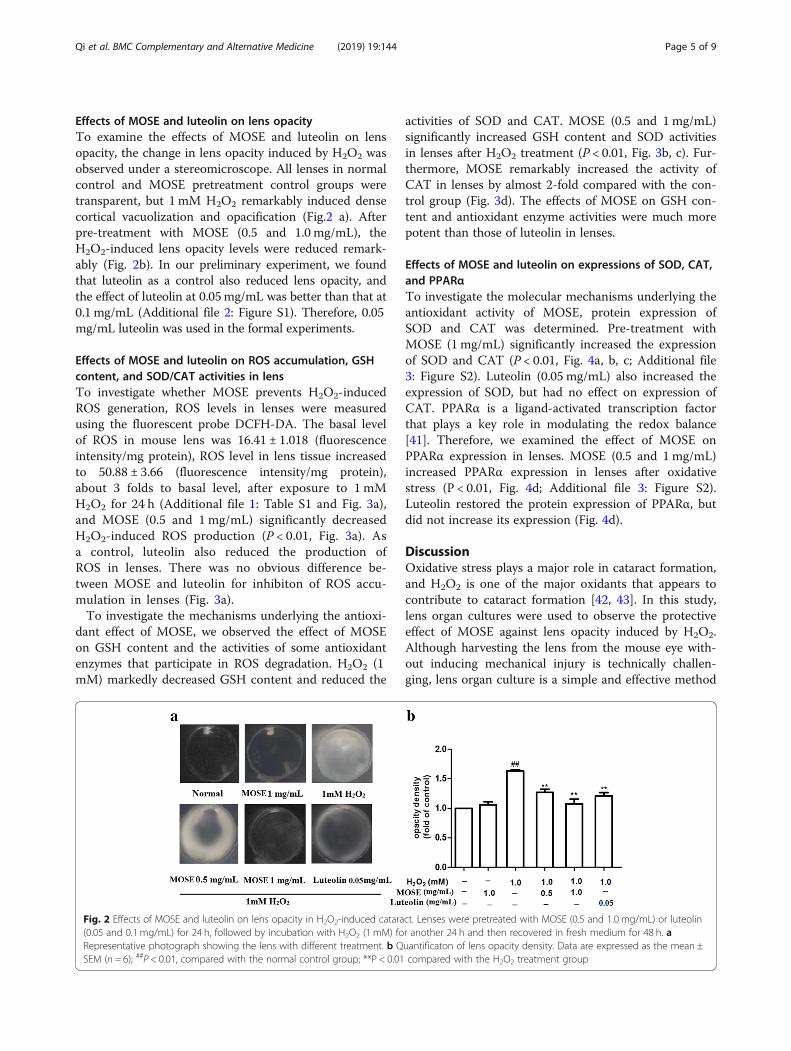
Effects of MOSE and luteolin on lens opacityTo examine the effects of MOSE and luteolin on lensopacity, the change in lens opacity induced by H2O2 wasobserved under a stereomicroscope. All lenses in normalcontrol and MOSE pretreatment control groups weretransparent, but 1 mM H2O2 remarkably induced densecortical vacuolization and opacification (Fig.2 a). Afterpre-treatment with MOSE (0.5 and 1.0 mg/mL), theH2O2-induced lens opacity levels were reduced remark-ably (Fig. 2b). In our preliminary experiment, we foundthat luteolin as a control also reduced lens opacity, andthe effect of luteolin at 0.05mg/mL was better than that at0.1 mg/mL (Additional file 2: Figure S1). Therefore, 0.05mg/mL luteolin was used in the formal experiments.
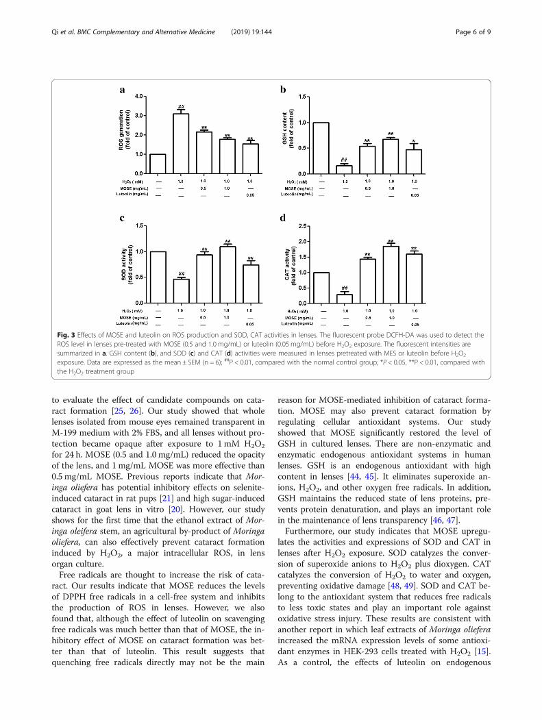
Effects of MOSE and luteolin on ROS accumulation, GSHcontent, and SOD/CAT activities in lensTo investigate whether MOSE prevents H2O2-inducedROS generation, ROS levels in lenses were measuredusing the fluorescent probe DCFH-DA. The basal levelof ROS in mouse lens was 16.41 ± 1.018 (fluorescenceintensity/mg protein), ROS level in lens tissue increasedto 50.88 ± 3.66 (fluorescence intensity/mg protein),about 3 folds to basal level, after exposure to 1 mMH2O2 for 24 h (Additional file 1: Table S1 and Fig. 3a),and MOSE (0.5 and 1 mg/mL) significantly decreasedH2O2-induced ROS production (P < 0.01, Fig. 3a). Asa control, luteolin also reduced the production ofROS in lenses. There was no obvious difference be-tween MOSE and luteolin for inhibiton of ROS accu-mulation in lenses (Fig. 3a).To investigate the mechanisms underlying the antioxi-
dant effect of MOSE, we observed the effect of MOSEon GSH content and the activities of some antioxidantenzymes that participate in ROS degradation. H2O2 (1mM) markedly decreased GSH content and reduced the
activities of SOD and CAT. MOSE (0.5 and 1mg/mL)significantly increased GSH content and SOD activitiesin lenses after H2O2 treatment (P < 0.01, Fig. 3b, c). Fur-thermore, MOSE remarkably increased the activity ofCAT in lenses by almost 2-fold compared with the con-trol group (Fig. 3d). The effects of MOSE on GSH con-tent and antioxidant enzyme activities were much morepotent than those of luteolin in lenses.
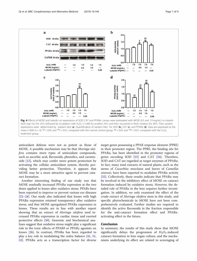
Effects of MOSE and luteolin on expressions of SOD, CAT,and PPARαTo investigate the molecular mechanisms underlying theantioxidant activity of MOSE, protein expression ofSOD and CAT was determined. Pre-treatment withMOSE (1 mg/mL) significantly increased the expressionof SOD and CAT (P < 0.01, Fig. 4a, b, c; Additional file3: Figure S2). Luteolin (0.05 mg/mL) also increased theexpression of SOD, but had no effect on expression ofCAT. PPARα is a ligand-activated transcription factorthat plays a key role in modulating the redox balance[41]. Therefore, we examined the effect of MOSE onPPARα expression in lenses. MOSE (0.5 and 1mg/mL)increased PPARα expression in lenses after oxidativestress (P < 0.01, Fig. 4d; Additional file 3: Figure S2).Luteolin restored the protein expression of PPARα, butdid not increase its expression (Fig. 4d).
DiscussionOxidative stress plays a major role in cataract formation,and H2O2 is one of the major oxidants that appears tocontribute to cataract formation [42, 43]. In this study,lens organ cultures were used to observe the protectiveeffect of MOSE against lens opacity induced by H2O2.Although harvesting the lens from the mouse eye with-out inducing mechanical injury is technically challen-ging, lens organ culture is a simple and effective method
Fig. 2 Effects of MOSE and luteolin on lens opacity in H2O2-induced cataract. Lenses were pretreated with MOSE (0.5 and 1.0 mg/mL) or luteolin(0.05 and 0.1 mg/mL) for 24 h, followed by incubation with H2O2 (1 mM) for another 24 h and then recovered in fresh medium for 48 h. aRepresentative photograph showing the lens with different treatment. b Quantificaton of lens opacity density. Data are expressed as the mean ±SEM (n = 6); ##P < 0.01, compared with the normal control group; **P < 0.01 compared with the H2O2 treatment group
Qi et al. BMC Complementary and Alternative Medicine (2019) 19:144 Page 5 of 9
to evaluate the effect of candidate compounds on cata-ract formation [25, 26]. Our study showed that wholelenses isolated from mouse eyes remained transparent inM-199 medium with 2% FBS, and all lenses without pro-tection became opaque after exposure to 1 mM H2O2
for 24 h. MOSE (0.5 and 1.0 mg/mL) reduced the opacityof the lens, and 1 mg/mL MOSE was more effective than0.5 mg/mL MOSE. Previous reports indicate that Mor-inga oliefera has potential inhibitory effects on selenite-induced cataract in rat pups [21] and high sugar-inducedcataract in goat lens in vitro [20]. However, our studyshows for the first time that the ethanol extract of Mor-inga oleifera stem, an agricultural by-product of Moringaoliefera, can also effectively prevent cataract formationinduced by H2O2, a major intracellular ROS, in lensorgan culture.Free radicals are thought to increase the risk of cata-
ract. Our results indicate that MOSE reduces the levelsof DPPH free radicals in a cell-free system and inhibitsthe production of ROS in lenses. However, we alsofound that, although the effect of luteolin on scavengingfree radicals was much better than that of MOSE, the in-hibitory effect of MOSE on cataract formation was bet-ter than that of luteolin. This result suggests thatquenching free radicals directly may not be the main
reason for MOSE-mediated inhibition of cataract forma-tion. MOSE may also prevent cataract formation byregulating cellular antioxidant systems. Our studyshowed that MOSE significantly restored the level ofGSH in cultured lenses. There are non-enzymatic andenzymatic endogenous antioxidant systems in humanlenses. GSH is an endogenous antioxidant with highcontent in lenses [44, 45]. It eliminates superoxide an-ions, H2O2, and other oxygen free radicals. In addition,GSH maintains the reduced state of lens proteins, pre-vents protein denaturation, and plays an important rolein the maintenance of lens transparency [46, 47].Furthermore, our study indicates that MOSE upregu-
lates the activities and expressions of SOD and CAT inlenses after H2O2 exposure. SOD catalyzes the conver-sion of superoxide anions to H2O2 plus dioxygen. CATcatalyzes the conversion of H2O2 to water and oxygen,preventing oxidative damage [48, 49]. SOD and CAT be-long to the antioxidant system that reduces free radicalsto less toxic states and play an important role againstoxidative stress injury. These results are consistent withanother report in which leaf extracts of Moringa olieferaincreased the mRNA expression levels of some antioxi-dant enzymes in HEK-293 cells treated with H2O2 [15].As a control, the effects of luteolin on endogenous
Fig. 3 Effects of MOSE and luteolin on ROS production and SOD, CAT activities in lenses. The fluorescent probe DCFH-DA was used to detect theROS level in lenses pre-treated with MOSE (0.5 and 1.0 mg/mL) or luteolin (0.05 mg/mL) before H2O2 exposure. The fluorescent intensities aresummarized in a. GSH content (b), and SOD (c) and CAT (d) activities were measured in lenses pretreated with MES or luteolin before H2O2
exposure. Data are expressed as the mean ± SEM (n = 6); ##P < 0.01, compared with the normal control group; *P < 0.05, **P < 0.01, compared withthe H2O2 treatment group
Qi et al. BMC Complementary and Alternative Medicine (2019) 19:144 Page 6 of 9
antioxidant defense were not as potent as those ofMOSE. A possible mechanism may be that Moringa olei-fera contains more types of antioxidant compounds,such as ascorbic acid, flavonoids, phenolics, and caroten-oids [12], which may confer more potent protection byactivating the cellular antioxidant system, thereby pro-viding better protection. Therefore, it appears thatMOSE may be a more attractive agent to prevent cata-ract formation.Another interesting finding of our study was that
MOSE markedly increased PPARα expression at the twodoses applied to lenses after oxidative stress. PPARs havebeen reported to improve or prevent various eye diseases[22–24]. Our study also indicated that lenses with highPPARα expression retained transparency after oxidativestress, and that MOSE upregulated PPARα expression inlenses. These results are in line with another reportshowing that an extract of Moringa oleifera seed in-creased PPARα expression in cardiac tissue and exertedprotective effects [50]. Genomic and biochemical ana-lyses suggest that oxidative stress might play a significantrole in the toxic effects of PPARδ or PPARγ agonists onlenses [26]. In contrast, PPARα has been regarded toplay a key role in modulating the redox balance [41, 51,52]. PPARα acts as a transcription factor for diverse
target genes possessing a PPAR response element (PPRE)in their promoter region. The PPRE, the binding site forPPARα, has been identified in the promoter regions ofgenes encoding SOD [53] and CAT [54]. Therefore,SOD and CAT are regarded as target enzymes of PPARα.In fact, many total extracts of natural plants, such as thestems of Cucurbita moschata and leaves of Camelliasinensis, have been reported to modulate PPARα activity[55]. Collectively, these results indicate that PPARα maybe involved in the inhibitory effect of MOSE on cataractformation induced by oxidative stress. However, the de-tailed role of PPARα in the lens requires further investi-gation. In addition, we only examined the effect of thecrude extract of Moringa oleifera stem in this study, andspecific phytochemicals in MOSE have not been com-prehensively evaluated. Further studies are required toidentify the active flavonoids in the fraction responsiblefor the anti-cataract formation effect and PPARα-activating effect in the future.
ConclusionIn summary, the results of this study show that MOSEsignificantly delays the progression of H2O2-inducedcataract formation in lens organ culture, and the mecha-nisms underlying its effect are related to scavenging of
Fig. 4 Effects of MOSE and luteolin on expressions of SOD, CAT and PPARα. Lenses were pretreated with MOSE (0.5 and 1.0 mg/mL) or luteolin(0.05 mg/ mL) for 24 h, followed by incubation with H2O2 (1 mM) for another 24 h, and then recovered in fresh medium for 48 h. Then, proteinexpressions were determined by western blot (a). Quantification of western blot for SOD (b), CAT (c), and PPARα (d). Data are expressed as themean ± SEM (n = 3); #P < 0.05 and ##P < 0.01, compared with the normal control group; *P < 0.05 and **P < 0.01, compared with the H2O2
treatment group
Qi et al. BMC Complementary and Alternative Medicine (2019) 19:144 Page 7 of 9
free radicals, increasing GSH content, and enhanced ac-tivities and expressions of SOD, CAT, and PPARα. Mor-inga oleifera stem, as a kind of natural antioxidant, hasextensive sources and no obvious adverse effects. There-fore, the inhibitory effect of MOSE on cataract forma-tion has great clinical interest. It can be used as apotential natural medicine in the prevention or treat-ment of cataract, especially induced by diabetes.
Additional files
Additional file 1: Table S1. Quantitative analysis of ROS inlens(fluorescence intensity/mg protein, means ± SEM, n = 6. (DOCX 19 kb)
Additional file 2: Figure S1. Effects of luteolin on lens opacity in H2O2-induced cataract. Lenses were pretreated with luteolin (0.01, 0.05 and 0.1mg/mL) for 24 h, followed by incubation with H2O2 (1 mM) for another24 h and then recovered in fresh medium for 48 h. Data are expressed asthe mean ± SEM (n = 6); ##P < 0.01, compared with the normal controlgroup; **P < 0.01 compared with the H2O2 treatment group. (TIF 584 kb)
Additional file 3: Figure S2. The original gel images of Fig. 4a. (TIF 155 kb)
AbbreviationsCAT: Catalase; DCFH-DA: 2′, 7′-Dichlorofluorescein diacetate; DPPH: 2,2-Diphenyl-1-1-picrylhydrazyl; FBS: Fetal bovine serum; GSH: Reducedglutathione; H2O2: Hydrogen peroxide; M-199: Medium 199; MOSE: Moringaoleifera stem extract; PPARα: Proliferator-activated receptor alpha;RIPA: Radioimmunoprecipitation assay; ROS: Reactive oxygen species;SOD: Superoxide dismutase
AcknowledgementsThe authors thank Wenliang Deng (Information & Network Services ofXiamen University) for his computer technical assistance.
Authors’ contributionsXJ and YL were responsible for the conception and design of this study. LQ,WJL and MLZ acquired the data. LQ analyzed and interpreted the data, anddrafted the manuscript. YZ revised the manuscript and did the final approvalof the manuscript. All authors read and approved the final version of themanuscript.
FundingThis work was supported by the Special Funds for the Science andTechnology Program of Traditional Chinese Medicine from Fujian ProvincialDepartment of Health (Grant No.wzhw201302), the Special Funds for theScience and Technology Program of Public Wellbeing from Xiamen Scienceand Technology Bureau (Grant No.3502Z20144030), the Medical InnovationProject of Fujian, China(Grant No.2015-CXB-43)and the Special Funds fromthe Xiamen Chuanming Biological Technology Co. Ltd. (GrantNo.XDHT2015241A).
Availability of data and materialsAll data generated or analyzed during this study are included in thispublished article. More details are available from the corresponding authoron reasonable request.
Ethics approval and consent to participateAll experimental protocols described in the present study were approved bythe Animal Care and Use Committee of Xiamen University (LACUC:XMULAC20150077). All procedures for the animal study were conducted inaccordance with ARRIVE guidelines, and every effort was made to alleviatethe suffering of the animals.
Consent for publicationNot applicable.
Competing interestsThe authors declare that they have no competing interests.
Author details1Department of Ophthalmology, Xiamen Hospital of Traditional ChineseMedicine, Xiamen 361005, People’s Republic of China. 2Department of BasicMedical Science, School of Medicine, Xiamen University, Xiamen 361102,People’s Republic of China.
Received: 8 August 2018 Accepted: 10 June 2019
References1. Lee CM, Afshari NA. The global state of cataract blindness. Curr Opin
Ophthalmol. 2017;28(1):98–103.2. Fukuoka H, Afshari NA. The impact of age-related cataract on measures of
frailty in an aging global population. Curr Opin Ophthalmol. 2017;28(1):93–7.3. Lofgren S. Solar ultraviolet radiation cataract. Exp Eye Res. 2016.4. Smith AJ, Ball SS, Manzar K, Bowater RP, Wormstone IM. Ku80 counters
oxidative stress-induced DNA damage and cataract formation in the humanLens. Invest Ophthalmol Vis Sci. 2015;56(13):7868–74.
5. Kruk J, Kubasik-Kladna K, Aboul-Enein HY. The role oxidative stress in thepathogenesis of eye diseases: current status and a dual role of physicalactivity. Mini Rev Med Chem. 2015;16(3):241–57.
6. Ji Y, Cai L, Zheng T, Ye H, Rong X, Rao J, Lu Y. The mechanism of UVBirradiation induced-apoptosis in cataract. Mol Cell Biochem. 2015;401(1–2):87–95.
7. Sunkireddy P, Jha SN, Kanwar JR, Yadav SC. Natural antioxidantbiomolecules promises future nanomedicine based therapy for cataract.Colloids Surf B Biointerfaces. 2013;112:554–62.
8. Dubey S, Saha S, Kaithwas G, Saraf SA. Effect of standardized fruit extract ofLuffa cylindrica on oxidative stress markers in hydrogen peroxide inducedcataract. Indian J Pharmacol. 2015;47(6):644–8.
9. Varma SD, Hegde KR. Kynurenine-induced photo oxidative damage to lensin vitro: protective effect of caffeine. Mol Cell Biochem. 2010;340(1–2):49–54.
10. Grover AK, Samson SE. Antioxidants and vision health: facts and fiction. MolCell Biochem. 2014;388(1–2):173–83.
11. Kou X, Li B, Olayanju JB, Drake JM, Chen N. Nutraceutical orpharmacological potential of Moringa oleifera lam. Nutrients. 2018;10(3).
12. Saini RK, Sivanesan I, Keum YS: Phytochemicals of Moringa oleifera: a reviewof their nutritional, therapeutic and industrial significance. 3 Biotech. 2016;6(2):203.
13. Stohs SJ, Hartman MJ. Review of the safety and efficacy of Moringa oleifera.Phytother Res. 2015;29(6):796–804.
14. Sreelatha S, Padma PR. Modulatory effects of Moringa oleifera extractsagainst hydrogen peroxide-induced cytotoxicity and oxidative damage.Hum Exp Toxicol. 2011;30(9):1359–68.
15. Vongsak B, Mangmool S, Gritsanapan W. Antioxidant activity and inductionof mRNA expressions of antioxidant enzymes in HEK-293 cells of Moringaoleifera leaf extract. Planta Med. 2015;81(12–13):1084–9.
16. Kerdsomboon K, Tatip S, Kosasih S, Auesukaree C. Soluble Moringa oleiferaleaf extract reduces intracellular cadmium accumulation and oxidative stressin Saccharomyces cerevisiae. J Biosci Bioeng. 2016;121(5):543–9.
17. Jaiswal D, Rai PK, Mehta S, Chatterji S, Shukla S, Rai DK, Sharma G, Sharma B,Khair S, Watal G. Role of Moringa oleifera in regulation of diabetes-inducedoxidative stress. Asian Pac J Trop Med. 2013;6(6):426–32.
18. Das N, Ganguli D, Dey S. Moringa oleifera lam. Seed extract prevents fat dietinduced oxidative stress in mice and protects liver cell-nuclei from hydroxylradical mediated damage. Indian J Exp Biol. 2015;53(12):794–802.
19. Agrawal ND, Nirala SK, Shukla S, Mathur R. Co-administration of adjuvantsalong with Moringa oleifera attenuates beryllium-induced oxidative stressand histopathological alterations in rats. Pharm Biol. 2015;53(10):1465–73.
20. Kurmi R, Ganeshpurkar A, Bansal D, Agnihotri A, Dubey N. Ethanol extract ofMoringa oliefera prevents in vitro glucose induced cataract on isolated goateye lens. Indian J Ophthalmol. 2014;62(2):154–7.
21. Sasikala V, Rooban BN, Priya SG, Sahasranamam V, Abraham A. Moringaoleifera prevents selenite-induced cataractogenesis in rat pups. J OculPharmacol Ther. 2010;26(5):441–7.
22. Moran E, Ding L, Wang Z, Cheng R, Chen Q, Moore R, Takahashi Y, Ma JX.Protective and antioxidant effects of PPARalpha in the ischemic retina.Invest Ophthalmol Vis Sci. 2014;55(7):4568–76.
Qi et al. BMC Complementary and Alternative Medicine (2019) 19:144 Page 8 of 9
23. Khatol P, Saraf S, Jain A. Peroxisome proliferated activated receptors (PPARs):opportunities and challenges for ocular therapy. Crit Rev Ther Drug CarrierSyst. 2018;35(1):65–97.
24. Chen Q, Qiu F, Zhou K, Matlock HG, Takahashi Y, Rajala RVS, Yang Y, MoranE, Ma JX. Pathogenic role of microRNA-21 in diabetic retinopathy throughdownregulation of PPARalpha. Diabetes. 2017;66(6):1671–82.
25. Qi HP, Wei SQ, Zhang LQ, Gao XC, Yu NN, Bi S, Cui H. Preventive effect ofdanshensu on selenite-induced cataractogenesis in cultured rat lens. ClinExp Ophthalmol. 2013;41(2):172–9.
26. Sampath S, McLean LA, Buono C, Moulin P, Wolf A, Chibout SD, Pognan F,Busch S, Shangari N, Cruz E, et al. The use of rat lens explant cultures tostudy the mechanism of drug-induced cataractogenesis. Toxicol Sci. 2012;126(1):128–39.
27. Basu S, Rajakaruna S, Dickinson BC, Chang CJ, Menko AS. Endogenoushydrogen peroxide production in the epithelium of the developingembryonic lens. Mol Vis. 2014;20:458–67.
28. Cornish KM, Williamson G, Sanderson J. Quercetin metabolism in the lens:role in inhibition of hydrogen peroxide induced cataract. Free Radic BiolMed. 2002;33(1):63–70.
29. Sreelakshmi V, Sasikala V, Abraham A. Luteolin supplementation preventsselenite-induced Cataractogenesis in Sprague Dawley rat pups. ChemBiodivers. 2015;12(12):1881–90.
30. Rooban BN, Sasikala V, Gayathri Devi V, Sahasranamam V, Abraham A.Prevention of selenite induced oxidative stress and cataractogenesis byluteolin isolated from Vitex negundo. Chem Biol Interact. 2012;196(1–2):30–8.
31. Chen Y, Sun XB, Lu HE, Wang F, Fan XH. Effect of luteoin in delayingcataract in STZ-induced diabetic rats. Arch Pharm Res. 2017;40(1):88–95.
32. Bao YF, Li JY, Zheng LF, Li HY. Antioxidant activities of cold-nature Tibetanherbs are signifcantly greater than hot-nature ones and are associated withtheir levels of total phenolic components. Chin J Nat Med. 2015;13(8):609–17.
33. Yamaguchi T, Takamura H, Matoba T, Terao J. HPLC method for evaluationof the free radical-scavenging activity of foods by using 1,1-diphenyl-2-picrylhydrazyl. Biosci Biotechnol Biochem. 1998;62(6):1201–4.
34. Devi VG, Rooban BN, Sasikala V, Sahasranamam V, Abraham A.Isorhamnetin-3-glucoside alleviates oxidative stress and opacification inselenite cataract in vitro. Toxicol in Vitro. 2010;24(6):1662–9.
35. Biju PG, Rooban BN, Lija Y, Devi VG, Sahasranamam V, Abraham A.Drevogenin D prevents selenite-induced oxidative stress and calpainactivation in cultured rat lens. Mol Vis. 2007;13:1121–9.
36. Lu Q, Yang T, Zhang M, Du L, Liu L, Zhang N, Guo H, Zhang F, Hu G, Yin X.Preventative effects of Ginkgo biloba extract (EGb761) on high glucose-cultured opacity of rat lens. Phytother Res. 2014;28(5):767–73.
37. Shukla R, Banerjee S, Tripathi YB. Antioxidant and Antiapoptotic effect of aqueousextract of Pueraria tuberosa (Roxb. Ex Willd.) DC. On streptozotocin-induceddiabetic nephropathy in rats. BMC Complement Altern Med. 2018;18(1):156.
38. Asha R, Gayathri Devi V, Abraham A. Lupeol, a pentacyclic triterpenoidisolated from Vernonia cinerea attenuate selenite induced cataract formationin Sprague Dawley rat pups. Chem Biol Interact. 2016;245:20–9.
39. Rooban BN, Sasikala V, Sahasranamam V, Abraham A. Amelioration ofselenite toxicity and cataractogenesis in cultured rat lenses by Vitexnegundo. Graefes Arch Clin Exp Ophthalmol. 2011;249(5):685–92.
40. Kim MS, Lee DY, Lee J, Kim HW, Sung SH, Han JS, Jeon WK. Terminaliachebula extract prevents scopolamine-induced amnesia via cholinergicmodulation and anti-oxidative effects in mice. BMC Complement AlternMed. 2018;18(1):136.
41. Aleshin S, Reiser G. Role of the peroxisome proliferator-activated receptors(PPAR)-alpha, beta/delta and gamma triad in regulation of reactive oxygenspecies signaling in brain. Biol Chem. 2013;394(12):1553–70.
42. Gao S, Qin T, Liu Z, Caceres MA, Ronchi CF, Chen CY, Yeum KJ, Taylor A,Blumberg JB, Liu Y, et al. Lutein and zeaxanthin supplementation reducesH2O2-induced oxidative damage in human lens epithelial cells. Mol Vis.2011;17:3180–90.
43. Smith AJ, Ball SS, Bowater RP, Wormstone IM. PARP-1 inhibition influencesthe oxidative stress response of the human lens. Redox Biol. 2016;8:354–62.
44. Harding JJ. Free and protein-bound glutathione in normal and cataractoushuman lenses. Biochem J. 1970;117(5):957–60.
45. Nye-Wood MG, Spraggins JM, Caprioli RM, Schey KL, Donaldson PJ, Grey AC.Spatial distributions of glutathione and its endogenous conjugates innormal bovine lens and a model of lens aging. Exp Eye Res. 2017;154:70–8.
46. Giblin FJ. Glutathione: a vital lens antioxidant. J Ocul Pharmacol Ther. 2000;16(2):121–35.
47. Sweeney MH, Truscott RJ. An impediment to glutathione diffusion in oldernormal human lenses: a possible precondition for nuclear cataract. Exp EyeRes. 1998;67(5):587–95.
48. Reczek CR, Chandel NS. ROS-dependent signal transduction. Curr Opin CellBiol. 2015;33:8–13.
49. Schieber M, Chandel NS. ROS function in redox signaling and oxidativestress. Curr Biol. 2014;24(10):R453–62.
50. Randriamboavonjy JI, Loirand G, Vaillant N, Lauzier B, Derbre S, Michalet S,Pacaud P, Tesse A. Cardiac protective effects of Moringa oleifera seeds inspontaneous hypertensive rats. Am J Hypertens. 2016;29(7):873–81.
51. Manea A, Manea SA, Todirita A, Albulescu IC, Raicu M, Sasson S, SimionescuM. High-glucose-increased expression and activation of NADPH oxidase inhuman vascular smooth muscle cells is mediated by 4-hydroxynonenal-activated PPARalpha and PPARbeta/delta. Cell Tissue Res. 2015;361(2):593–604.
52. Abdelmegeed MA, Moon KH, Hardwick JP, Gonzalez FJ, Song BJ. Role ofperoxisome proliferator-activated receptor-alpha in fasting-mediatedoxidative stress. Free Radic Biol Med. 2009;47(6):767–78.
53. Yoo HY, Chang MS, Rho HM. Induction of the rat cu/Zn superoxidedismutase gene through the peroxisome proliferator-responsive element byarachidonic acid. Gene. 1999;234(1):87–91.
54. Girnun GD, Domann FE, Moore SA, Robbins ME. Identification of afunctional peroxisome proliferator-activated receptor response element inthe rat catalase promoter. Mol Endocrinol. 2002;16(12):2793–801.
55. Rigano D, Sirignano C, Taglialatela-Scafati O. The potential of naturalproducts for targeting PPARalpha. Acta Pharm Sin B. 2017;7(4):427–38.
Publisher’s NoteSpringer Nature remains neutral with regard to jurisdictional claims inpublished maps and institutional affiliations.
Qi et al. BMC Complementary and Alternative Medicine (2019) 19:144 Page 9 of 9