effect of mild and moderate hypothermia on hypoxic injury in nearly pure neuronal culture
TRANSCRIPT

ORIGINAL ARTICLE
Effect of mild and moderate hypothermia on hypoxic injuryin nearly pure neuronal culture
Yu Hua • Kenjiro Hisano • Yuji Morimoto
Received: 1 December 2009 / Accepted: 5 July 2010 / Published online: 5 August 2010
� Japanese Society of Anesthesiologists 2010
Abstract
Purpose The effects of mild and moderate hypothermic
therapy on cerebral injury are still controversial. Our
hypothesis is that mild and moderate hypothermia should
have some effects on neurons themselves if they really
have protective effects. By using a nearly pure neuronal
culture, we evaluated the effects and mechanism of hypo-
thermia against hypoxic insult.
Methods A nearly pure neuronal culture from cortices of
18-day-old Wister rats was used. The neurons were
exposed to below 1% oxygen at 3 different temperatures
(30, 33 and 37�C). First, cell viability was measured by
assessing viable neurons with trypan blue. Second, to
evaluate the mechanism, the extracellular glutamate con-
centration was measured by high-performance liquid
chromatography after hypoxia; cell viability after exposure
to extrinsic glutamate was also evaluated. Next, mito-
chondrial membrane potential was estimated, by monitor-
ing aggregation of MitoCaptureTM, and the percentage of
apoptotic cells was evaluated by staining with Hoechst
33342 and propidium iodide.
Results After 24-h hypoxic insult, cell viability at 30 and
33�C was significantly higher than at 37�C. There was no
significant difference between extracellular concentrations
of glutamate after hypoxia or cell viability after glutamate
exposure among the 3 temperature groups. In moderate
hypothermia, the number of neurons with mitochondrial
injury and the percentage of apoptotic cells were signifi-
cantly reduced.
Conclusion Mild and moderate hypothermia inhibited
hypoxic neuronal cell death. The mechanism of this effect
may be related to protection of mitochondrial function,
presumably followed by inhibition of apoptosis, at least in
moderate hypothermia.
Keywords Hypothermia � Neurons � Hypoxia �Mitochondria � Glutamate
Introduction
Neuroprotective strategies against hypoxic, ischemic, and
traumatic brain injury have not been sufficiently estab-
lished [1, 2]. Among these, mild (33–36�C) and moderate
(28–32�C) hypothermic therapy have furnished fascinating
results for the central nervous system, including the hyp-
oxic brain, in many laboratory studies [3, 4]. The mecha-
nism of the protection by mild and moderate hypothermia
is not explained by inhibition of cerebral metabolism.
There have been many hypotheses, including inhibition of
the glutamate surge, intracellular signaling pathways, dis-
ruption of the blood–brain barrier, proliferation or activa-
tion of microglia, and the production of free radicals;
however, the mechanism remains unclarified [3].
In the clinical setting, 2 randomized controlled studies
from Europe and Australia demonstrated the efficacy of
mild hypothermic therapy for survivors of out-of-hospital
cardiac arrest [5, 6]. In addition, in 2 other randomized
controlled studies neurologic outcomes and mortality were
improved by induction of mild hypothermic therapy for
neonates with hypoxic–ischemic encephalopathy [7, 8]. On
the other hand, treatment with mild hypothermia was not
effective in improving outcome for patients with severe
brain injury [9]. Nor did mild intraoperative hypothermia
Y. Hua � K. Hisano � Y. Morimoto (&)
Department of Anesthesiology and Critical Care Medicine,
Hokkaido University Graduate School of Medicine, N15 W7,
Sapporo 0608638, Japan
e-mail: [email protected]
123
J Anesth (2010) 24:726–732
DOI 10.1007/s00540-010-0999-x

improve outcome after craniotomy among good-grade
patients with aneurismal subarachnoid hemorrhage [10].
Thus, the effectiveness of this therapy in the clinical setting
is still controversial.
These laboratory and clinical findings prompted us to
reappraise the effects of mild and moderate hypothermia.
We hypothesized that mild and moderate hypothermia
should have some effects on neurons themselves if they
really have a protective effect against hypoxic or ischemic
neuronal injury. In this study, we utilized a nearly pure
cortical neuronal culture and evaluated the effect of con-
comitant mild and moderate hypothermia during hypoxic
insult.
As a mechanism of ischemic/hypoxic cerebral injury,
the glutamate–calcium theory, called excitotoxic neuronal
cell death, is well known [11]. However, all clinical
pharmacological approaches targeting this cascade have
resulted in failure [11]. Meanwhile, the cell death-signaling
pathway related to mitochondrial injury leading to apop-
tosis has recently been demonstrated in the ischemic/hyp-
oxic brain [12]. Overloading of mitochondrial Ca2?
induces mitochondrial permeability transition, which leads
to cytochrome c release and caspase activation [11, 13].
Accordingly, we also examined the mechanism of the
effect of mild and moderate hypothermia with reference to
these two aspects. As far as we know, there have been no
studies evaluating the involvement of these 2 cascades
simultaneously using the same experimental model.
Materials and methods
Nearly pure neuronal culture
With institutional approval of animal care and use, a nearly
pure neuronal culture was prepared from the cortices of 18-
day-old Wister rat fetuses. The cortices were digested with
trypsin (Invitrogen, Carlsbad, CA, USA) and DNase I
(Sigma–Aldrich, St Louis, MO, USA), followed by
mechanical dissociation. Poly-L-lysine-coated plastic plates
(BD Bioscience, Franklin Lakes, NJ, USA) were seeded
with cortical cells at a density of 3000 cells/mm2. The cells
were grown in neurobasal medium (Invitrogen) with B27
minus AO (Invitrogen) and N2 supplements (Invitrogen)
and L-glutamine (Sigma–Aldrich). Serum-free neurobasal
medium was optimized for cell survival and neurite out-
growth of neurons and the almost complete absence of glial
cells without a reagent to inhibit them [14]. Cultures were
kept at 37�C in 5% CO2 in a humidified incubator. The
medium was changed every third day and the cells were
grown for 14 days. A pilot study showed that the propor-
tion of neurons among the cells was over 90% in our
culture system [15].
Hypoxic insult under mild and moderate hypothermia
After 14 days of culture, three dishes of the same sister
culture were randomly allocated to chambers in which the
temperature was maintained at 37, 33 (mild hypothermia),
or 30�C (moderate hypothermia) in a 100% humidified
atmosphere containing below 1% oxygen with the
remainder nitrogen. The temperature of each chamber was
accurately maintained using a thermostat (Digimulti D611;
Techno Seven Yokohama, Japan) and the concentration of
oxygen was continuously monitored with an oximeter
(JKO-25 II; Jiko, Tokyo, Japan). After 24 h of hypoxic
insult at each temperature, cell viability was measured.
In the preliminary study, we evaluated the changes in
the cell viability over time. Until 6 h after hypoxia, almost
no cells died. Cell viability finally decreased to below 50%
at 24 h after the start of hypoxia. Accordingly, we set the
exposure time at 24 h.
Cell viability
To evaluate cell viability, we used Shibuta’s method as
described previously [15]. Briefly, photomicrographs of 3
or 4 areas within the dish were taken shortly before
exposure to hypoxia. After 24 h, the cells were exposed to
0.4% trypan blue with phosphate-buffered saline (Invitro-
gen) to stain nonviable cells, and photomicrographs were
taken again in the same areas as before the exposure.
Approximately 1000 viable neurons per culture dish were
subjected to manual counting. A second observer unaware
of the arrangement of the photographs, the study design,
and the treatment procedure replicated all manual counts to
ensure count accuracy and minimal inter-observer vari-
ability. Survival was calculated by use of the formula:
100 9 (detected non-stained cells at the end of the exper-
iment/detected whole cells shortly before exposure).
Each dish of the same sister culture was assigned to one
of the three different temperatures. Cell viability in each
dish was measured after hypoxic exposure for 24 h. Mea-
surement was performed 16 times.
Glutamate–calcium cascade
Measurement of extracellular glutamate concentration
The extracelluar glutamate concentration was measured by
use of high-performance liquid chromatography (HPLC)
with an electrochemical detector (FLD-370; Eicom, Tokyo,
Japan) [16]. After 24 h hypoxia, the culture supernatants
were collected after the cells were sedimented by centri-
fugation and then stored at -80�C until assay within
2 weeks. The solution used to form fluorescent derivatives
consisted of o-phthalaldehyde containing mercaptoethanol.
J Anesth (2010) 24:726–732 727
123

10 ll of this solution was added to 30 ll of dialysate
sample diluted with 50% methanol and this mixture was
left to react for 150 s before HPLC analysis with fluori-
metric detection.
Each dish of the same sister culture was assigned to one
of the three different temperatures. The extracellular glu-
tamate concentration in each dish was measured after 24 h
of hypoxic exposure. Measurement was performed 11
times.
Cell viability after exposure to extrinsic glutamate
After 14 days of culture, fresh medium containing 250 lM
glutamate warmed at 37, 33, or 30�C was added to the 3
dishes from the same sister culture and each dish was
incubated for 30 min in a chamber set at the corresponding
temperature with 100% humidified room air. The medium
was then changed to the normal one and the dishes were
incubated for 23 h 30 min in the same atmosphere fol-
lowed by evaluation of the cell viability by the method
described above. The degree of glutamate exposure was
established as the cell viability was compatible with that
after 24 h of hypoxia at 37�C.
Each dish of the same sister culture was assigned to one
of the three different temperatures. Cell viability in each
dish was measured 24 h after glutamate exposure for
30 min. Measurement was performed 16 times.
Mitochondrial injury–apoptosis cascade
Quantitative evaluation of mitochondrial injury
Disruption of the mitochondrial transmembrane potential
was evaluated by using a MitoCaptureTM mitochondrial
apoptosis detection kit (Biovision, Mountainview, CA,
USA). In healthy cells, MitoCapture accumulates and
aggregates in the mitochondria, emitting bright red fluo-
rescence. When mitochondrial transmembrane potential
disappears (i.e., the mitochondria are injured), it remains in
the cytoplasm in its monomer form, fluorescing green.
After 6 h of hypoxia at 37, 33, or 30�C, cells from the same
sister culture were incubated with MitoCapture for 20 min
and analyzed under a nonconfocal fluorescence microscope
(Axiophoto; Zeiss, Jena, Germany). Because cell viability
did not decrease after 6 h of hypoxia in the preliminary
study, we estimated mitochondrial injury at that time.
Quantitative analysis counting approximately 1000 cells
was performed by a second observer unaware of the
arrangement of photographs, study design, and treatment
procedure. The percentage of the cells with mitochondrial
injury was calculated.
Each dish of the same sister culture was assigned to
one of the three different temperatures. The percentage
of cells with mitochondrial injury was evaluated
after 6 h of hypoxia. Measurement was performed 8
times.
Percentages of viable, apoptotic, and necrotic cells
The relative frequencies of apoptotic cells were examined
after 24 h of hypoxia at 37, 33, and 30�C by the methods of
Shimizu et al. as described before [15]. The cells were
stained for 30 min at 37�C with Hoechest 33342 (Sigma–
Aldrich) and propidium iodide (Sigma–Aldrich). They
were then analyzed under a nonconfocal fluorescence
microscope (Axiophoto; Zeiss). Quantitative analysis
counting about 1000 cells was performed by a second
observer unaware of the arrangement of photographs, study
design, and treatment procedure. Necrotic cells were
regarded as those with round red nuclei whereas apoptotic
cells had fragmented nuclei regardless of their color. The
percentages of viable, apoptotic, and necrotic cells were
calculated by use of the formula: 100 9 (viable, apoptotic,
or necrotic cells/whole cells).
Each dish of the same sister culture was assigned to one
of the three different temperatures. The percentages of
viable, apoptotic, and necrotic cells in each dish were
measured after 24 h of hypoxia. Measurement was per-
formed 14 times.
Statistics
Values are expressed as mean ± standard deviation. One-
factor ANOVA was performed among the temperature
groups in each experiment. When significant differences
were observed, the Tukey–Kramer test was performed as a
post-hoc test. Statistical significance was assumed when
p \ 0.05.
Results
Cell viability (Fig. 1)
Cell viability was 34.6 ± 4.7, 48.1 ± 11.4, and 47.5 ±
6.9% at 37, 33, and 30�C, respectively (p \ 0.01 in one-
factor ANOVA). Cell viability at 33 and 30�C was
significantly higher than that at 37�C.
Measurement of extracellular glutamate concentration
(Fig. 2)
Extracellular glutamate concentrations were 1.32 ± 0.72,
1.34 ± 0.87, and 1.22 ± 0.85 pmol/ll at 37, 33, and 30�C
respectively (p = 0.94 in one-factor ANOVA).
728 J Anesth (2010) 24:726–732
123
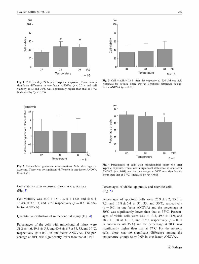
Cell viability after exposure to extrinsic glutamate
(Fig. 3)
Cell viability was 34.0 ± 15.1, 37.5 ± 17.0, and 41.0 ±
18.4% at 37, 33, and 30�C respectively (p = 0.51 in one-
factor ANOVA).
Quantitative evaluation of mitochondrial injury (Fig. 4)
Percentages of the cells with mitochondrial injury were
51.2 ± 4.6, 49.4 ± 5.5, and 40.6 ± 6.7 at 37, 33, and 30�C,
respectively (p \ 0.01 in one-factor ANOVA). The per-
centage at 30�C was significantly lower than that at 37�C.
Percentages of viable, apoptotic, and necrotic cells
(Fig. 5)
Percentages of apoptotic cells were 25.9 ± 8.2, 25.3 ±
7.2, and 17.8 ± 6.4 at 37, 33, and 30�C, respectively
(p = 0.01 in one-factor ANOVA) and the percentage at
30�C was significantly lower than that at 37�C. Percent-
ages of viable cells were 44.4 ± 13.3, 49.6 ± 11.9, and
58.2 ± 10.8 at 37, 33, and 30�C, respectively (p = 0.01
in one-factor ANOVA) and the percentage at 30�C was
significantly higher than that at 37�C. For the necrotic
cells, there was no significant difference among the
temperature groups (p = 0.09 in one-factor ANOVA).
Cel
l via
bilit
y
Temperaturen = 16
Fig. 1 Cell viability 24 h after hypoxic exposure. There was a
significant difference in one-factor ANOVA (p \ 0.01), and cell
viability at 33 and 30�C was significantly higher than that at 37�C
(indicated by *p \ 0.05)
Ext
race
llula
r gl
utam
ate
Con
cent
ratio
n
Temperature
(pmol/ml)
n = 11
Fig. 2 Extracellular glutamate concentrations 24 h after hypoxic
exposure. There was no significant difference in one-factor ANOVA
(p = 0.94)
Cel
l via
bilit
y
Temperature n = 16
Fig. 3 Cell viability 24 h after the exposure to 250 lM extrinsic
glutamate for 30 min. There was no significant difference in one-
factor ANOVA (p = 0.51)
Temperature
Per
cent
age
of c
ells
n = 8
Fig. 4 Percentages of cells with mitochondrial injury 6 h after
hypoxic exposure. There was a significant difference in one-factor
ANOVA (p \ 0.01) and the percentage at 30�C was significantly
lower than that at 37�C (indicated by *p \ 0.05)
J Anesth (2010) 24:726–732 729
123

Discussion
This study demonstrated that neuronal survival was sig-
nificantly increased by mild and moderate hypothermia
concomitant with 24 h hypoxia in nearly pure cortical
neuronal culture. To the best of our knowledge, there has
been only one previous report that examined the effect of
mild or moderate hypothermia on hypoxic neuronal insult
by using a nearly pure neuronal culture. That report by
Bossenmeyer-Pourie et al. evaluated the effect of con-
comitant hypothermia at 32�C on hypoxic insult by using a
nearly pure neuronal culture obtained from the rat embryo
forebrain [17]. In their study, cultures were returned to
normothermic and normoxic atmospheres for the next 96 h
after 6 h of hypoxia. Cell viability of the neurons under
normothermic hypoxia significantly decreased from 72 h
after the insult and hypothermia inhibited this decrease.
They also showed that both apoptosis and necrosis were
inhibited by hypothermia; however, the mechanism could
not be explored although they evaluated the time course of
DNA and protein synthesis by measuring the incorporation
of radiolabeled thymidine and L-leucine and also measured
the expression of Bcl-2 and HSP 70 [17]. In their study,
cell viability did not decrease after 6 h of hypoxia.
Therefore, the focus of that study was the effect of hypo-
thermia on reoxygenation injury. In our study, accordingly,
we tried to observe the effect of concomitant hypothermia
on ongoing hypoxic neuronal injury.
Our study demonstrated that mild and moderate hypo-
thermia did not reduce the extracellular concentration of
glutamate after hypoxia. Previous studies of global [18]
and focal [19] ischemia demonstrated that mild hypother-
mia inhibited the increase in glutamate release. In-vitro
studies using hippocampal slices [20] and mixed cortical
cell culture [21] also indicated that mild hypothermia
reduced the glutamate release after oxygen–glucose
deprivation. On the other hand, Asai et al. demonstrated, by
real-time measurement of glutamate, that intraischemic
elevation of glutamate did not differ between normother-
mia and mild hypothermia in severe global ischemia [22].
However, the glutamate level after ischemia markedly
decreased in hypothermia, suggesting the promotion of
post-ischemic reuptake of glutamate. In our study, the
change in the glutamate level after hypoxia was not eval-
uated, therefore, a significant difference might not be
noted.
Cell viability after exposure to extrinsic glutamate did
not significantly differ among the groups. Indeed, 15 min
of exposure to either 100 lM or 1 mM glutamate uni-
formly induced a marked increase in intracellular calcium,
with delayed recovery and massive neuronal death under
the conditions of both normothermia and moderate hypo-
thermia at 30�C in cultured hippocampal neurons [23].
Moreover, hypothermia at 34 and 28�C did not affect
changes in the cytosolic free calcium concentration
induced by NMDA in rat cortical brain slices [24]. These
studies indicate that mild and moderate hypothermia can-
not save neurons once glutamate is released during ische-
mia/hypoxia. Thus, inhibition of the glutamate–calcium
cascade might not have contributed much to the decrease in
hypoxic neuronal cell death due to mild and moderate
hypothermia in our experimental model.
On the other hand, in moderate hypothermia, the per-
centage of cells with mitochondrial injury was significantly
reduced and apoptosis due to hypoxic insult was signifi-
cantly inhibited. Accordingly, moderate hypothermia
might inhibit neuronal cell death induced by hypoxia by
suppression of the mitochondrial injury–apoptosis cascade
in our experimental model. However, the target of hypo-
thermia remains controversial. Yenari et al. [25] showed
that mild hypothermia significantly reduced the amount of
cytochrome c release 5 h after the onset of focal ischemia,
which indicated that the target of hypothermia was the
mitochondrion itself. In contrast, Zhao et al. [13] showed
biphasic cytochrome c release after global ischemia (5
and 48 h after ischemia). Caspase activity significantly
increased after the first phase of cytochrome c release. Mild
hypothermia did not block the first phase of cytochrome
c release, but significantly blocked caspase activity and the
second phase of cytochrome c release. This finding sug-
gested that the target of hypothermia was caspase activity
rather than mitochondria. In a study using gastric cancer
cells, mitochondrial injury detected by MitoCapture was
Per
cent
age
of c
ells
Temperature
Morphologic type of cells n = 14
Fig. 5 Percentages of viable, apoptotic, and necrotic cells 24 h after
hypoxic exposure. For apoptotic cells, there was a significant
difference in one-factor ANOVA (p = 0.01) and the percentage at
30�C was significantly lower than that at 37�C (indicated by
*p \ 0.05). For viable cells, there was a significant difference in
one-factor ANOVA (p = 0.01) and the percentage of viable cells at
30�C was significantly higher than that at 37�C (indicated by#p \ 0.05)
730 J Anesth (2010) 24:726–732
123

seen 2 h after the induction of apoptosis by ceramide, and
release of cytochrome c and activation of caspase-3 and
caspase-9 were observed 3 and 24 h after that [26]. This
suggested that mitochondrial changes detected by Mito-
Capture preceded the release of cytochrome c and the
activation of caspase. Our study showed that moderate
hypothermia inhibited the mitochondrial injury detected by
MitoCapture 6 h after the onset of hypoxia, at which time
almost no cells had died. Accordingly, in our experimental
model, moderate hypothermia might inhibit the mito-
chondrial injury–apoptosis cascade by acting on the mito-
chondrion itself.
With regard to the relationship between neuronal
apoptosis and hypothermia, Zhu et al. [27] evaluated the
effect of hypothermia at 30�C on brain injury and apoptotic
neuronal cell death in 7-day-old rats subjected to left
common carotid artery ligation and hypoxia for 1 h. Brain
infarct volumes and neuronal loss were significantly
reduced 72 h after ischemia/hypoxia. Cytochrome c release
and activation of caspase-3 and caspase-2 were signifi-
cantly diminished by hypothermia. The numbers of cyto-
chrome c-positive and TUNEL-positive cells (i.e.,
apoptotic cells) were also significantly reduced in the
hypothermia group. Xu et al. examined the effect of
hypothermia at 33�C on apoptosis induced by serum
deprivation in a nearly pure mouse cortical neuronal cul-
ture [28]. Hypothermia significantly reduced the number of
morphologically apoptotic neurons to less than half the
number seen in normothermic culture after 48 h. Shibano
et al. used serum-deprived PC 12 cells as the neuronal
apoptotic model and examined the direct effects of mild
and moderate hypothermia (29–35�C) [29]. After 96 h, the
number of apoptotic cells was over 90% and this propor-
tion decreased in a temperature-dependent fashion falling
to below 50% at 29�C.
To the best of our knowledge, this is the first preliminary
evaluation of the effects of mild and moderate hypothermia
on hypoxic neuronal injury from the aspect of the gluta-
mate–calcium and mitochondrial injury–apoptosis cas-
cades, simultaneously, using the same experimental model.
However, there are some limitations in our study. First, the
objective of this study was to evaluate the effect of con-
comitant hypothermia on ongoing hypoxic neuronal injury.
However, our results might indicate that hypothermia
merely delayed cell death and the intracellular cascade.
Accordingly, we may not be able to use the word ‘‘pro-
tection’’ in this study. It is unknown whether cell viability
decreased further after reoxygenation in our experimental
model, because cell injury was advanced compared with
that in Bossenmeyer-Pourie’s study. However, if intra-
hypoxic hypothermia inhibits the newly developed cell
injury after reoxygenation, we may be able to use the word
‘‘protection’’ for the first time. Second, it is not uncommon
for patients in critical conditions or during surgery to be
exposed to hypoxic conditions. In this study, accordingly,
we chose only hypoxia as a model of ischemic/hypoxic
insult. However, other types of insult, for example oxygen–
glucose deprivation, must be evaluated in the future. Third,
we could not clarify the mechanism of mild hypothermia in
this study. It was, however, demonstrated that concomitant
mild and moderate hypothermia could directly affect
ongoing hypoxic neuronal changes leading to cell death
and that, at least in moderate hypothermia, the mechanism
was related to suppression of the mitochondrial injury–
apoptosis cascade.
In conclusion, by using a nearly pure neuronal culture,
we showed that mild and moderate hypothermia could
inhibit ongoing neuronal injury by hypoxia, suggesting that
neurons themselves are a target for mild and moderate
hypothermic therapy. It is speculated that suppression of
the intracellular mitochondrial injury–apoptosis cascade
is the mechanism of the effect of, at least, moderate
hypothermia, whereas inhibition of the glutamate–calcium
cascade may not contribute to this inhibition in either mild
or moderate hypothermia.
Acknowledgments The authors thank Professor M. Yoshioka and
former Assistant Professor M. Matshumoto, Department of Neuro-
pharmacology in our university, for their teaching of HPLC mea-
surement. The authors also thank Ms Naoko Kimura for her technical
assistance.
References
1. Kawaguchi M, Furuya H, Patel PM. Neuroprotective effects of
anesthetic agents. J Anesth. 2005;19:150–6.
2. Grocott HP, Yoshitani K. Neuroprotection during cardiac sur-
gery. J Anesth. 2007;21:367–77.
3. Zhao H, Steinberg GK, Sapolsky RM. General versus specific
actions of mild-moderate hypothermia in attenuating cerebral
ischemic damage. J Cereb Blood Flow Metab. 2007;27:1879–94.
4. Miyazawa T, Tamura A, Fukui S, Hossmann KA. Effect of mild
hypothermia on focal cerebral ischemia. Review of experimental
studies. Neurol Res. 2003;25:457–64.
5. Bernard SA, Gray TW, Buist MD, Jones BM, Silvester W,
Gutteridge G, Smith K. Treatment of comatose survivors of out-
of-hospital cardiac arrest with induced hypothermia. N Engl J
Med. 2002;346:557–63.
6. Hypothermia after Cardiac Arrest Study G. Mild therapeutic
hypothermia to improve the neurologic outcome after cardiac
arrest. N Engl J Med. 2002;346:549–56.
7. Gluckman PD, Wyatt JS, Azzopardi D, Ballard R, Edwards AD,
Ferriero DM, Polin RA, Robertson CM, Thoresen M, Whitelaw
A, Gunn AJ. Selective head cooling with mild systemic hypo-
thermia after neonatal encephalopathy: multicentre randomised
trial. Lancet. 2005;365:663–70.
8. Shankaran S, Laptook AR, Ehrenkranz RA, Tyson JE, McDonald
SA, Donovan EF, Fanaroff AA, Poole WK, Wright LL, Higgins
RD, Finer NN, Carlo WA, Duara S, Oh W, Cotten CM, Ste-
venson DK, Stoll BJ, Lemons JA, Guillet R, Jobe AH, National
Institute of Child Health, Human Development Neonatal
J Anesth (2010) 24:726–732 731
123

Research. Whole-body hypothermia for neonates with hypoxic–
ischemic encephalopathy. N Engl J Med. 2005;353:1574–84.
9. Clifton GL, Miller ER, Choi SC, Levin HS, McCauley S, Smith
KR Jr, Muizelaar JP, Wagner FC Jr, Marion DW, Luerssen TG,
Chesnut RM, Schwartz M. Lack of effect of induction of
hypothermia after acute brain injury. N Engl J Med. 2001;344:
556–63.
10. Todd MM, Hindman BJ, Clarke WR, Torner JC. Intraoperative
Hypothermia for Aneurysm Surgery Trial. Mild intraoperative
hypothermia during surgery for intracranial aneurysm. N Engl J
Med. 2005;352:135–45.
11. Uchino H, Kuroda Y, Morota S, Hirabayashi G, Ishii N,
Shibasaki F, Ikeda Y, Hansson MJ, Elmer E. Probing the
molecular mechanisms of neuronal degeneration: importance of
mitochondrial dysfunction and calcineurin activation. J Anesth.
2008;22:253–62.
12. Chan PH. Mitochondria and neuronal death/survival signaling
pathways in cerebral ischemia. Neurochem Res. 2004;29:1943–9.
13. Zhao H, Yenari MA, Cheng D, Sapolsky RM, Steinberg GK.
Biphasic cytochrome c release after transient global ischemia and
its inhibition by hypothermia. J Cereb Blood Flow Metab.
2005;25:1119–29.
14. Xie C, Markesbery WR, Lovell MA. Survival of hippocampal
and cortical neurons in a mixture of MEM? and B27-supple-
mented neurobasal medium. Free Radic Biol Med. 2000;28:
665–72.
15. Hisano K, Watanabe M, Morimoto Y. Protective effects of free
radical scavenger edaravone against glutamate neurotoxicity in
nearly pure neuronal culture. J Anesth. 2009;23:363–9.
16. Matsumoto M, Togashi H, Kaku A, Kanno M, Tahara K,
Yoshioka M. Cortical GABAergic regulation of dopaminergic
responses to psychological stress in the rat in the rat dorsolateral
striatum. Synapse. 2005;56:117–21.
17. Bossenmeyer-pourie C, Koziel V, Daval J-L. Effects of hypo-
thermia on hypoxia-induced apoptosis in cultured neurons from
developing rat forebrain: comparison with preconditioning.
Pediatr Res. 2000;47:385–91.
18. Busto R, Globus MY-T, Dietrich WD, Martinez D, Valdes I,
Ginsberg MD. Effect of mild hypothermia on ischemia-induced
release of neurotransmitters and free fatty acids in rat brain.
Stroke. 1989;20:904–10.
19. Winfee CJ, Connolly ES, Fiore AJ, Solomon RA. Mild hypo-
thermia reduces penumbral glutamate levels in the rat focal
cerebral ischemia model. Neurosurgery. 1996;38:1216–22.
20. Berger R, Jensen A, Hossmann KA, Paschen W. Effect of mild
hypothermia during and after transient in vitro ischemia on
metabolic disturbances in hippocampal slices at different stages
of development. Dev Brain Res. 1998;105:67–77.
21. Bruno VM, Goldberg MP, Dugan LL, Giffard RG, Choi DW.
Neuroprotective effect of hypothermia in cortical cultures
exposed to oxygen–glucose deprivation or excitatory amino
acids. J Neurochem. 1994;63:1398–406.
22. Asai S, Zhao H, Kohno T, Takahashi Y, Nagata T, Ishikawa K.
Quantitative evaluation of extracellular glutamate concentration
in postischemic glutamate re-uptake, dependent on brain tem-
perature, in the rat following severe global brain ischemia. Brain
Res. 2000;864:60–8.
23. Arai H, Uto A, Ogawa Y, Sato K. Effect of low temperature on
glutamate-induced intracellular calcium accumulation and cell
death in cultured hippocampal neurons. Neurosci Lett. 1993;163:
132–4.
24. Bickler PE, Buck LT, Hansen BM. Effects of isoflurane and
hypothermia on glutamate receptor-mediated calcium influx in
brain slices. Anesthesiology. 1994;81:1461–9.
25. Yenari MA, Iwayama S, Cheng D, Sun GH, Fujimura M, Morita-
Fujimura Y, Chan PH, Steinberg GK. Mild hypothermia attenu-
ates cytochrome c release but does not alter Bcl-2 expression or
caspase activation after experimental stroke. J Cereb Blood Flow
Metab. 2002;22:29–38.
26. Belkhiri A, Dar AA, Zaika A, Kelley M, El-Rifai W. t-Darpp
promotes cancer cell survival by up-regulation of Bcl2 through
Akt-dependent mechanism. Cancer Res. 2008;68:395–403.
27. Zhu C, Wang X, Cheng X, Qiu L, Xu F, Simbruner G, Blomgren
K. Post-ischemic hypothermia-induced tissue protection and
diminished apoptosis after neonatal cerebral hypoxia–ischemia.
Brain Res. 2004;996:67–75.
28. Xu L, Yenari MA, Steinberg GK, Giffard RG. Mild hypothermia
reduces apoptosis of mouse neurons in vitro early in the cascade.
J Cereb Blood Flow Metab. 2002;22:21–8.
29. Shibano T, Morimoto Y, Kemmotsu O, Shikama H, Hisano K,
Hua Y. Effects of mild and moderate hypothermia on apoptosis in
neuronal PC12 cells. Br J Anaesth. 2002;89:301–5.
732 J Anesth (2010) 24:726–732
123