effect of membrane exposure on guided bone regeneration: a...
TRANSCRIPT
Clin Oral Impl Res 20181ndash11 wileyonlinelibrarycomjournalclr emsp|emsp1copy 2018 John Wiley amp Sons AS Published by John Wiley amp Sons Ltd
Accepted 22 November 2017
DOI 101111clr13121
R E V I E W A R T I C L E
Effect of membrane exposure on guided bone regeneration A systematic review and meta- analysis
Jeffrey Garcia1emsp|emspAustin Dodge1emsp|emspPaul Luepke1emsp|emspHom-Lay Wang2 emsp|emspYvonne Kapila3emsp|emspGuo-Hao Lin13
1Department of Surgical Sciences Marquette University School of Dentistry Milwaukee WI USA2Graduate Periodontics Department of Periodontics amp Oral Medicine University of Michigan School of Dentistry Ann Arbor MI USA3Department of Orofacial Sciences School of Dentistry University of California San Francisco San Francisco CA USA
CorrespondenceGuo-Hao Lin DDS MS Health Sciences Assistant Clinical Professor Department of Orofacial Sciences University of California San Francisco CA USAEmail ghlinumichedu guo-haolinucsfedu
AbstractAims This review aimed at investigating the effect of membrane exposure on guided bone regeneration (GBR) outcomes at peri- implant sites and edentulous ridgesMaterial and Methods Electronic and manual literature searches were conducted by two independent reviewers using four databases including MEDLINE EMBASE Web of Science and Cochrane Central Register of Controlled Trials for articles up to February 2017 Articles were included if they were human clinical trials or case series reporting outcomes of GBR procedures with and without membrane exposure A random- effects meta- analysis was conducted and the weighted mean difference (WMD) between the two groups and 95 confidence interval (CI) were reportedResults Overall eight articles were included in the quantitative analysis The WMD of thehorizontalbonegainatedentulousridgeswasminus7624(95CI=minus13752tominus1497p=01)betweensiteswithmembraneexposureandwithoutexposureInaddition theWMDof thedehiscence reductionatperi-implant siteswasminus2727(95CIofminus4587tominus868p=004)Bothanalysesshowedsignificantlyfavora-ble outcomes at the sites without membrane exposureConclusion Based on the findings of this study membrane exposure after GBR proce-dures has a significant detrimental influence on the outcome of bone augmentation Fortheedentulousridgesthesiteswithoutmembraneexposureachieved74morehorizontal bone gain than the sites with exposure For peri- implant dehiscence de-fects the sites without membrane exposure had 27 more defect reduction than the sites with exposure
K E Y W O R D S
alveolar ridge augmentation bone regeneration evidence-based dentistry meta-analysis review surgical wound dehiscence
1emsp |emspINTRODUCTION
Alveolar ridge dimensions provide the foundation for primary im-plant stability and long- term implant success Bone deficiencies present an immediate concern for clinicians and need to be ad-dressed early in the treatment planning process The etiology of ridge deficiencies can be either anatomic or pathologic in nature as
describedbyBuserMartinandBelser(2004)Inanattempttostan-dardize defect parameters several authors have developed classifi-cation systems (Allen Gainza Farthing amp Newbold 1985 Seibert 1983 Wang amp Al- Shammari 2002) Each author described three different types of ridge deficiencies based on the progression of the ridge resorption following edentulism horizontal vertical and a combination of the two dimensions When evaluating an edentulous
2emsp |emsp emspensp GARCIA et Al
site for future implant placement ideal treatment outcomes demand sufficient horizontal and vertical ridge dimensions To prevent fur-ther bone remodeling after implant placement Spray Black Morris and Ochi (2000) proposed a need for 18 mm of bone thickness at the buccal aspect of the implant and 05 mm at the palatal aspect to ensure long- term success In addition Tarnow Cho and Wallace (2000) also recommended a need for at least 15 mm of distance between an implant and the adjacent root and at least 3 mm of distance between two adjacent implants to accommodate ideal in-terproximal bone levels as well as for papilla support
Several techniques have been proposed and widely used to aug-ment deficient ridges Guided bone regeneration (GBR) is one of the most utilized techniques and it consists of using grafting materi-als in combination with a barrier either a nonresorbable membrane (Buser Bragger Lang amp Nyman 1990) or an absorbable membrane (Mellonig amp Nevins 1995) Other techniques include the use of a bone block graft (Misch 1997) or a ridge- split technique (Simion Baldoni amp Zaffe 1992) Additionally the use of distraction osteo-genesis to augment the edentulous ridge has also been proposed (Chiapasco Romeo amp Vogel 2001 Chin 1999) Recently the ldquosand-wichrdquo technique has been described to regenerate horizontal and vertical bone defects at peri- implant sites (Wang Misch amp Neiva 2004) All these techniques are effective however complicationscan occur during the healing phase of treatment creating undesired outcomes
Successful GBR procedures are dependent on four fundamental principles that must ensue during the surgery and throughout heal-ing These principles consist of primary closure angiogenesis space maintenance and stability of the wound the so- called PASS principle (WangampBoyapati2006)Complicationswithanyoftheseprinciplescan result in premature membrane exposure that potentially compro-mises the regenerative process To minimize the risk of complications clinicians should assess the amount of keratinized mucosa tissue bio-type vestibular depth flap flexibility bone defect type and size and type of membrane used (Chao Chang Fu Wang amp Chan 2015) Each one of these factors has been identified as a contributing factor in membrane exposure
As there is a need to understand the influence of membrane ex-posure on GBR outcomes the goal of this study was to compare the amount of bone gain after GBR procedures between sites with and without membrane exposure The primary outcome was the percent-age of horizontal bone gain at edentulous ridges The secondary out-come was the percentage of peri- implant bone dehiscence reduction at peri- implant sites
2emsp |emspMATERIAL AND METHODS
21emsp|emspFocused question
What is the effect of membrane exposure on bone augmentation out-comes after horizontal ridge augmentation at edentulous sites after GBR procedures or at peri- implant sites immediately after implant placement
22emsp|emspPICO question (problem intervention comparison outcome)
P Maxillary or mandibular partially edentulous healthy subjects who were to receive or had received dental implants to restore oral function
I GBR for horizontal ridge augmentation to augment bone width for future implant placement or to restore peri- implant dehiscence de-fects immediately after implant placement
C GBR outcomes between sites with and without membrane exposureO
bull Primary outcome percentage of horizontal bone gain at sites with and without membrane exposure at edentulous ridges
bull Secondary outcome percentage of peri-implant bone dehiscence reduction at sites with and without membrane exposure at peri-im-plant sites
23emsp|emspInformation sources
Electronic and manual literature searches were conducted by two inde-pendent reviewers (JG and AD) in four databases including MEDLINE EMBASE Web of Science and Cochrane Central Register of Controlled Trials for articles up to February 2017 Two reviewers independently extracted the data from studies (JG and AD) Publications that did not meet the inclusion criteria were excluded In case of disagreements consensus was reached by discussion with a third reviewer (GL)
24emsp|emspScreening process and data extraction
For the PubMed library combinations of controlled terms ([mh] rep-resented MeSH terms) and keywords ([tiab] represented titleabstract search and [all] represented full- text search) were used whenever possible As such the key terms used were as follows
(ldquoalveolar ridge augmentationrdquo[all] OR ldquoridge augmentationrdquo[all] OR ldquoguided bone regenerationrdquo[all])
AND(ldquodental implantsrdquo[mh] OR ldquoabsorbable implantsrdquo[mh] OR implant
[tiab] OR implants [tiab]) AND(ldquocomplicationrdquo[all] OR ldquocomplicationsrdquo[all] OR ldquoexposurerdquo[all])For the other databases the key terms used for the search in-
cluded GBR alveolar ridge augmentation dental implants complica-tion and exposure
The screening in such databases was limited to ldquoclinical studiesrdquo AND ldquohumansrdquo in all of the screening strategies In addition an elec-tronic screening of the grey literature at the New York Academy of Medicine Grey Literature Report (httpgreylitorg) and Google Scholar was conducted as recommended by high standards for systematic re-views that is Assessment of Multiple Systematic Reviews (AMSTAR) guidelines (Shea et al 2007)
Additionally a manual search of periodontics- related journals in-cluding Clinical Implant Dentistry and Related Research The International Journal of Oral amp Maxillofacial Implants Clinical Oral Implants Research
emspensp emsp | emsp3GARCIA et Al
Implant Dentistry European Journal of Oral Implantology Journal of Implantology International Journal of Oral and Maxillofacial Surgery Journal of Oral and Maxillofacial Surgery Journal of Dental Research Journal of Clinical Periodontology Journal of Periodontology and The International Journal of Periodontics amp Restorative Dentistry from January 2016 up toMarch 2017was performed to ensure a thor-ough screening process This systematic review was registered at the PROSPEROwebsite(registrationnumberCRD42017059598)
25emsp|emspInclusion criteria
Studies were included for the review if the following inclusion crite-ria were fulfilled clinical human prospective or retrospective study numberofstudysitesofge10GBRprocedureforridgeaugmentationsat partially edentulous ridges or peri- implant defects immediately after implant placement Studies with nonparticulate bone grafting materials (ie block grafts) no information regarding complications augmentation procedures other than GBR (distraction osteogenesis sinus lifts etc) vertical ridge augmentation and treatment of peri- implantitis were excluded from this study Article titles and abstracts were screened first for inclusion eligibility
26emsp|emspData extraction
Data recorded for each study included the study design number of participants bone grafting materials used membrane type se-lected initial bone width final bone width timing of complication onset and duration of follow- up after the augmentation surgery
27emsp|emspData analyses
The primary outcome was the percentage of horizontal bone gain at edentulous sites in groups with and without membrane exposure The secondary outcome was the percentage of dehiscence defect reduc-tion at peri- implant sites in groups with and without membrane expo-sure The pooled weighted mean difference (WMD) of the percentage of horizontal bone changes at edentulous sites and the percentage defect depth reduction at peri- implant sites was estimated using a computer program (RevMan version 50 The Nordic Cochrane Centre The Cochrane Collaboration Copenhagen 2008) The contribution of each article was weighed Random effects meta- analyses were applied to the selected studies to manage the inherent variability of results due to a great variety of study designs employed by different research groups Forest plots were generated to graphically represent the dif-ference in primary and secondary outcomes for all included studies using augmented sites as the analysis unit A pvalue=05wasusedasthe level of significance Heterogeneity was assessed with a chi- square test and I2 test Heterogeneity values range between 0 and 100 where lower values represent less heterogeneity In addition funnel plots were used to assess the presence of publication bias The data presented in this systematic review adhere to the PRISMA (Preferred Reporting Items for Systematic Review and Meta- Analyses) statement (Liberati et al 2009)
28emsp|emspRisk of bias assessment
The criteria used to assess the quality of the selected randomized con-trol trials (RCTs) were modified from the checklist of the Cochrane Center (Higgins amp Green 2011) For non- RCTs the Methodological Index for Non- Randomized Studies (MINORS) was used to rank the risk of bias of the included studies (Slim et al 2003) The degree of bias was categorized as follows low risk if all the criteria were met moderate risk when only one criterion was missing and high risk if two or more criteria were missing Two reviewers (JG and GL) assessed all the included articles independently
3emsp |emspRESULTS
31emsp|emspData extraction
A total of 837 records were identified through the electronic and man-ual search process after exclusion of duplicates (Figure 1) Of these 795werescreenedbasedonthetitleandorabstractcontentand42were selected for full- text assessment and inclusion in the qualitative evaluation Finally 34 studies (Antoun SitbonMartinez ampMissika2001vonArxampBuser2006vonArxampKurt1999Assenzaetal2001BahatampFontanessi2001BuserDulaHirtampSchenk1996Chiapasco Autelitano Rabbiosi amp Zaniboni 2013 Cordaro Amade amp Cordaro 2002 Cordaro Torsello Morcavallo amp di Torresanto 2011 FriedmannStrietzelMaretzkiPitaruampBernimoulin2002HerKangampFien2012KfirKfirampKaluski2007LeeKimYiampChoi2013Lindfors Tervonen Sandor amp Ylikontiola 2010 Lizio Corinaldesi amp Marchetti 2014 Lorenzoni Pertl Polansky Jakse ampWegscheider2002 Louis Gutta Said- Al- Naief amp Bartolucci 2008 Lyford Mills Knapp Scheyer amp Mellonig 2003 Mattout amp Mattout 2000Meloni et al 2017 Merli et al 2015 Miyamoto Funaki Yamauchi KodamaampTakahashi2012ParkBrooksOhampWang2009ParodiCarusiSantarelliampNanni1998ProussaefsampLozada20032006
F IGURE 1emspPRISMA flowchart demonstrates the inclusion studies selection process
4emsp |emsp emspensp GARCIA et Al
Proussaefs2003SethiampKaus2001Torresetal2010Ueharaetal2015 Urban Nagursky amp Lozada 2011 Urban Nagursky Lozada amp Nagy2013WessingEmmerichampBozkurt2016ZitzmannNaefampScharer 1997) were excluded from the quantitative analysis due to lack of data and eight studies (Annibali Bignozzi Sammartino La Monaca amp Cristalli 2012 Buser et al 1990 Chiapasco Abati Romeo amp Vogel 1999FuOhBenavidesRudekampWang2014GherQuinteroAssadMonacoampRichardson1994NowzariampSlots1995Parketal2008Tawil El- Ghoule amp Mawla 2001) that reported on the primary and secondary outcomes were meta- analyzed The kappa value between the two reviewers was 092 for titles and abstract evaluation and 088 for full- text evaluation The reasons for exclusion of specific studies are noted in Table S1 and the main features of the included studies are summarized in Table 1
32emsp|emspFeatures of the included studies
321emsp|emspStudy design and patient features
ThreeRCTs(Fuetal2014Gheretal1994Parketal2008)fourprospective case series (PCS Buser et al 1990 Chiapasco et al 1999 Nowzari amp Slots 1995 Tawil et al 2001) and one retrospective case series (RCS Annibali et al 2012) were included in this study The age range of the patients in the selected studies was 17ndash85 years All the studies indicated the participants were systemically healthy and only six studies (Annibali et al 2012 Chiapasco et al 1999 Fu et al 2014Gheretal1994NowzariampSlots1995Parketal2008)pro-vided more detailed exclusion criteria Annibali et al (2012) and Park et al (2008) excluded patients who smoked 10 or more cigarettes a day and patients with full- mouth plaque and bleeding scores of gt25 Chiapasco et al (1999) excluded heavy smokers alcohol abusers pa-tients with poor oral hygiene or active periodontal disease history of head and neck malignancies uncontrolled diabetes severe liver or renaldiseaseandimmunesystemdisordersFuetal(2014)excludedall patients who were smokers pregnant or had unstable periodontal diseasesorahistoryofdrugoralcoholabuseGheretal(1994)ex-cluded pregnant patients and Nowzari and Slots (1995) excluded pa-tientswhohadantibiotictherapywithin6monthsofthestudyperiodTwo studies (Annibali et al 2012 Chiapasco et al 1999) included patients who were light smokers (lt10 cigarettes per day)
The initial bone dimensions were provided by most of the studies exceptforthree(Fuetal2014Gheretal1994Parketal2008)The follow-up period after theGBR procedures ranging from 4 to14monthswas reportedbyall the includedstudiesAmong the in-cluded articles all studies reported no conflict of interest except one study (Park et al 2008) which reported a conflict of interest with two private corporations
322emsp|emspTypes of membranes used
All the included studies used barrier membranes for GBR procedures Only two studies (Buser et al 1990 Chiapasco et al 1999) that measured horizontal bone gain at edentulous ridges utilized expanded
polytetrafluoroethylene (e- PTFE) membranes Of the studies analyz-ing the reduction in bone dehiscences at peri- implant sites two stud-ies(Gheretal1994NowzariampSlots1995)exclusivelyusede-PTFEmembranes One study (Annibali et al 2012) used either titanium- reinforced e- PTFE or absorbable membranes however all membrane exposures occurred at sites with e- PTFE barriers The other three studies(Fuetal2014Parketal2008Tawiletal2001)onlyusedabsorbable membranes Tawil et al (2001) used an absorbable colla-gen membrane Park et al (2008) used either an acellular dermal ma-trix(ADM)oracollagenmembraneandFuetal(2014)introducedabovine pericardium membrane for GBR procedures
323emsp|emspTypes of bone grafting materials used
All included studies except for one (Nowzari amp Slots 1995) reported the grafting materials used for the GBR procedures For studies that measured horizontal bone gain at edentulous sites one (Chiapasco et al 1999) used autogenous bone grafts and the other (Buser et al 1990) did not use bone grafting material at all In terms of studies that analyzed dehiscence reduction at peri- implant sites one study (Tawil et al 2001) solely used autogenous cortical bone another study (Gher etal1994)useddemineralizedfreeze-driedboneallograft(DFDBA)twootherstudies(Fuetal2014Parketal2008)usedmineralizedallograft and the other one study (Annibali et al 2012) used a com-bination of autogenous bone chips and DFDBA or bovine xenograft
33emsp|emspIncidence of membrane exposure
For all studies the exposure of the membrane was clinically detected during the follow- up period The exposure resulted in surgical removal ofthemembraneinallbutfourstudies(Fuetal2014Gheretal1994Parketal2008Tawiletal2001)Gheretal (1994)attributed theexposure which occurred during the first 2 weeks postoperatively to inappropriate trimming of the membrane and used 012 chlorhexidine rinses for the duration of the healing time Two weeks postoperatively Tawil et al (2001) resutured the flaps to gain closure and did so without removing the membrane Park et al (2008) had five sites with collagen membraneexposureandtwositeswithADMexposureFuetal(2014)had three patients with partial cover screw exposure at 2 weeks yet the surgical site closed completely at the 1- month re- evaluation The remaining studies reported membrane exposures from 8 to 10 days to 36weekspostoperativelyOf these theadverseeventsof theexpo-sure were reported in two studies Buser et al (1990) reported unusual edema and Nowzari and Slots (1995) reported inflammation suppura-tion and pain The other two studies (Annibali et al 2012 Chiapasco et al 1999) did not comment on the events of the membrane exposure
34emsp|emspMeta- analysis of the primary outcome amp secondary outcome
As the size of the initial bone defect impacts the amount of defect reduction only studies reporting the percentage of bone defect re-duction were pooled for comparable comparisons
emspensp emsp | emsp5GARCIA et Al
TABLE 1emsp
Char
acte
ristic
s of
the
incl
uded
stu
dies
Def
ect h
eigh
t cha
nge
at p
eri- i
mpl
ant b
one
dehi
scen
ce si
tes a
fter
GBR
Stud
y na
me
Stud
y de
sign
N
umbe
r of
patie
nts
Expo
sure
Num
ber o
f si
tes
Gra
ft m
ater
ial
Mem
bran
e ty
peIn
itial
def
ect
heig
ht(m
m)
Fina
l def
ect
heig
ht (m
m)
Def
ect h
eigh
t re
duct
ion
(mm
)
Def
ect h
eigh
t fil
led
()
Com
plic
atio
n tim
ing
Follo
w- u
p tim
e (m
onth
s)
Ghe
r et a
l (1994)
RCT
N=36(aged
26ndash81)
No
16A
lloN
NA
NA
192
plusmn 2
18
NA
NA
6
Yes
27A
lloN
NA
NA
minus021plusmn293
NA
lt4months
Now
zari
and
Slot
s (1
995)
PCS
N=17(aged
17ndash65)
No
8N
AN
460plusmn128
056plusmn050
404plusmn145
8674plusmn1270
NA
9
Yes
9N
AN
486plusmn160
287
plusmn 1
88
199plusmn236
3665plusmn3714
2ndash36weeks
Taw
il et
al
(200
1)
PCS
N=17(aged
21ndash8
5)N
o15
Aut
oR
533
plusmn 2
13
040plusmn112
493plusmn222
922
2 plusmn
207
7N
A4ndash8
Yes
3A
uto
R5
00 plusmn
02
00 plusmn
03
00 plusmn
06000plusmn0
2 w
eeks
Park
et a
l (2
008)
RCT
N=18(aged
28ndash7
1)N
o11
Allo
RN
AN
AN
A83
00
plusmn 15
00
NA
6
Yes
7A
lloR
NA
NA
NA
5219plusmn2434
2ndash4weeks
Ann
ibal
i et
al
(201
2)
RCS
N=9(ageNA)
No
16A
uto+
Xen
oN
amp R
334plusmn253
NA
319
plusmn 2
37
958
7 plusmn
907
NA
6ndash9
Yes
5A
uto+
Xen
oN
530
plusmn 2
73
NA
500plusmn274
938
2 plusmn
108
31ndash
2 m
onth
s
Fu e
t al
(2014)
RCT
N=13(aged
31ndash64)
No
10A
lloR
NA
NA
NA
8827plusmn1451
NA
12
Yes
3A
lloR
NA
NA
NA
583
3 plusmn
381
92
wee
ks
Bone
wid
th c
hang
e at
ede
ntul
ous r
idge
s aft
er G
BR
Stud
y na
me
Stud
y de
sign
N o
f pa
tient
sEx
posu
reN
of s
ites
Gra
ft m
ater
ial
Mem
bran
e ty
peIn
itial
ridg
e w
idth
(mm
)Fi
nal r
idge
w
idth
(mm
)Fi
nal b
one
wid
th g
ain
(mm
)
Fina
l bon
e w
idth
gai
n (
)Co
mpl
icat
ion
timin
gFo
llow
- up
time
(mon
ths)
Buse
r et
al
(199
0)
PCS
N=10(aged
18ndash54)
No
7N
AN
35plusmn076
614plusmn117
264plusmn176
9162plusmn9019
NA
6ndash10
Yes
3N
AN
392
plusmn 1
01
600plusmn0
208
plusmn 1
01
6162plusmn4899
1 w
eek
to
3 m
onth
s
Chia
pasc
o et
al
(199
9)
PCS
N=15(aged
19ndash60)
No
13A
uto
N300plusmn061
604plusmn066
304plusmn088
10849plusmn4369
NA
11ndash14
Yes
2A
uto
N425plusmn035
475plusmn106
050
plusmn 0
71
111
1 plusmn
157
18ndash
10 d
ays
NA
not
ava
ilabl
eap
plic
able
GBR
gui
ded
bone
rege
nera
tion
RCT
ran
dom
ized
con
trol
led
tria
l PC
S p
rosp
ectiv
e ca
se s
erie
s R
CS r
etro
spec
tive
case
ser
ies
Aut
o a
utog
enou
s bo
ne g
raft
Allo
allo
graf
t Xe
no
xeno
graf
t Co
mbo
com
bina
tion
graf
ts N
non
reso
rbab
le m
embr
ane
R a
bsor
babl
e m
embr
ane
6emsp |emsp emspensp GARCIA et Al
Two studies (Buser et al 1990 Chiapasco et al 1999) reported outcomes on the percentage of horizontal bone gain after GBR at edentulous sites with and without membrane exposure Meta- analysis showedastatisticallysignificantdifference(WMD=minus7624witha95CIofminus13752tominus1497p=01Figure2)betweenthetwogroups favoring the group without membrane exposure A moderate level of heterogeneity was seen (pvalueforchi-squaretest=15andI2test=51)amongthepooledstudies
Fivestudies(Annibalietal2012Fuetal2014NowzariampSlots1995 Park et al 2008 Tawil et al 2001) reported outcomes on the percentage of bone dehiscence reduction after GBR at peri- implant sites Meta- analysis showed a statistically significant difference (WMD=minus2727with a 95CI of minus4587 to minus868p=004Figure 3) between the two groups favoring the group without mem-brane exposure A high level of heterogeneity was seen (p value for chi-square test=0001 and I2 test=83) among the pooled stud-ies Subgroup analysis based on the type of membranes used was also conducted For the nonresorbable membrane subgroup two studies (Annibali et al 2012 Nowzari amp Slots 1995) were pooled and meta- analysis showed no statistically significant difference (WMD=minus2456with a 95 CI of minus7155 to 2242 p=31)However this subgroup analysis also revealed a high level of heteroge-neity between the two studies (p value for chi- square test lt0007 and I2test=91)Fortheabsorbablemembranesubgroupthreestudies(Fuetal2014Parketal2008Tawiletal2001)wereincludedandtheserevealedastatisticallysignificantdifference(WMD=minus3183witha95CIofminus4095tominus2272p lt 0001) favoring the group without membrane exposure This subgroup analysis revealed a low level of heterogeneity among the pooled studies (p value for chi- square test=99andI2test=0)Funnelplotsfortheanalysisofedentulousridges and peri- implant sites were reported as Figures S1 and S2
35emsp|emspRisk of bias assessment
The results of the risk of bias assessment for included case series are summarized in Table 2 (RCT) and Table 3 (non- RCTs) One RCT (Gher etal1994)andonecaseseries (Annibalietal2012)wereconsid-ered to have a high risk of biasAnotherRCT (Fu etal 2014) andfour case series (Buser et al 1990 Chiapasco et al 1999 Nowzari amp Slots 1995 Tawil et al 2001) were considered to have a moderate
risk of bias One other RCT was considered to have a low risk of bias (Park et al 2008) The kappa value of the interexaminer agreement for risk of bias assessment was 100
4emsp |emspDISCUSSION
In 2001 Machtei (2001) investigated the effect of early membrane exposure on guided tissue and bone regeneration The results of the study showed a difference of six times greater bone gain if the healing period did not have early membrane exposure One of the main draw-backs of that investigation was that only two papers were included in the assessment Furthermore the results of the study were reported in millimeters instead of percentages of bone gain potentially intro-ducing issues with the validity of the comparisons Without identifying the dimensions of the initial defect size the amount of potential bone gain measured between the groups could yield incomparable data-sets For example one of the included studies (Annibali et al 2012) reported an average of 500 mm of bone gain for sites with membrane exposure which is greater than the 319 mm gained at sites without membrane exposure While this seems to suggest almost 2 mm of difference between the groups when analyzed by the percentage of defect reduction the two groups showed comparable percentages of defect reduction 9082 for sites without membrane exposure and 8750 for sites with exposure respectively In order to compare the outcomes more meaningfully and to avoid potentially biased analyses we used the percentage of horizontal bone gaindehiscence reduction instead of linear measurements as our study outcomes
The objective of using a barrier membrane is to prevent the in-growth of soft tissue while providing space for the graft and allowing oxygen and nutrients to enter the grafted site (Rakhmatia Ayukawa FuruhashiampKoyano2013)Priortoourstudynometa-analysishasbeen performed to compare the resulting bone gain of edentulous sites under exposed and nonexposed GBR membranes We found thatthesiteswithoutmembraneexposureachieved74morehor-izontal bone gain than those with exposure However it is worth mentioning that the two articles (Annibali et al 2012 Nowzari amp Slots 1995) exclusively used e- PTFE barriers instead of dense PTFE (d- PTFE) barriers The difference in bacterial resistance between e- PTFE and d- PTFE (d- PTFE being more resistant) could be significant
F IGURE 2emspForest plots representing the outcomes of the percentage of horizontal bone width gain after GBR at edentulous ridges between groupswithandwithoutmembraneexposureMeta-analysisshowedastatisticallysignificantdifference(WMD=minus7624witha95CIofminus13752tominus1497p=01)favoringthegroupwithoutmembraneexposure
emspensp emsp | emsp7GARCIA et Al
Studies have shown that intentionally exposed d- PTFE membranes for socket preservation procedures (Greenstein amp Carpentieri 2015) as well as GBR procedures (Hoffmann et al 2008 Waasdorp amp Feldman 2013) did not exhibit significantly compromised regenera-tion outcomes As the pore sizes of d- PTFE (less than 03 μm) barriers are much smaller than e- PTFE (05ndash30 μm) d- PTFE barriers provide a superior resistance to bacterial penetration A pore size of less than 03 μm (Bartee amp Carr 1995) is impervious to bacteria as the average size of bacteria is approximately 05ndash50 μm A larger pore size allows for easy bacterial contamination once a membrane is exposed to the oral cavity The presence of bacterial contamination compromises the integrity of the membrane and limits the regeneration process Once compromised the soft tissue begins to infiltrate the mem-brane resulting in a much more difficult removal of the membrane and less favorable outcomes (Rakhmatia et al 2013)
Regarding GBR at peri- implant dehiscence sites our study an-alyzed fivearticles (Annibalietal2012Fuetal2014NowzariampSlots 1995 Park et al 2008 Tawil et al 2001) that included 87 surgi-cal sites and found 27 more defect reduction at sites without mem-brane exposure compared to those with exposures This difference however is even more critical when considering absorbable (Fu et al 2014 Park etal 2008Tawil etal 2001) vs nonresorbablemem-branes As absorbable membranes are primarily metabolized through enzymatic degradation once they become exposed these membranes have a greater susceptibility to infection and a faster degradation rate Membrane exposures compromise space maintenance and cell exclu-sion properties leading to detrimental effects that are readily seen in subsequent outcomes It has been reported that bacterial invasion of the exposed absorbable membrane could occur as early as 3 weeks (Simion et al 1997) During the first week of exposure the outer surface of the barrier is colonized by bacteria and by week three to four the bacteria have invaded the entire thickness of the membrane Bacterial invasion results in membrane resorption and creation of ir-regular voids in the barrier which degrade the functional integrity of the barrier Even with the long- lasting cross- linked collagen barriers TalKozlovskyArtziNemcovskyandMoses(2008)foundthatboneregeneration outcomes were compromised once membranes were exposed
Interestingly one study (Park et al 2008) found a significant differ-ence in peri- implant dehiscence defect reduction between sites with and without barrier exposure in a collagen membrane group but not in an ADM group Four of six sites with ADM exposure were epithe-lialized after a 1- month period The authors attributed this favorable outcome to the polarized matrix nature of ADM which consisted of a basal lamina for epithelial cell migration and an underlying porous der-malmatrixforangiogeniccell ingrowth(CummingsKaldahlampAllen2005) However although the difference was not statistically signifi-cant it is worth noting that 25 less dehiscence defect reduction was still detected in the exposed ADM group than the nonexposed group
F IGURE 3emspForest plots representing the outcomes of the percentage of peri- implant bone dehiscence reduction after GBR between groups withandwithoutmembraneexposureMeta-analysisshowedastatisticallysignificantdifference(WMD=minus2727witha95CIofminus4587tominus868p=004)favoringthegroupwithoutmembraneexposure
TABLE 2emspRisk of bias assessment for the included RCT
Criteria Higgins and Green (2011)
Gher et al (1994)
Park et al (2008)
Fu et al (2014)
Sequence generation Yes Yes Yes
Randomization methods RCT RCT RCT
Allocation concealment method
Yes Yes
Examiner masked Yes No
All patients accounted for at end of study
Yes Yes Yes
Incomplete outcome data adequately addressed
Yes Yes Yes
Free of suggestion of selective outcome reporting
Yes Yes Yes
Estimated potential risk of bias
High Low Moderate
Not reported
8emsp |emsp emspensp GARCIA et Al
Our study findings are consistent with several previously published human studies reporting on membrane exposure which show early membrane removal and compromised bone regeneration (von Arx HardtampWallkamm1996ProussaefsampLozada2006) Ithasbeendemonstrated that the timelymembrane removalwithin4weeksofmembrane exposure could still potentially result in successful bone augmentation (Annibali etal 2012 Proussaefs amp Lozada 2006)Annibali et al (2012) treated patients with systemic antibiotic therapy (1gamoxicillinndashclavulanatetwiceadayfor6days)whenmembraneexposure occurred The membrane was subsequently removed after reentry surgery and the flap was closed The graft was allowed to heal for an additional period ranging from 3 to 8 months The study out-come showed a comparable amount of defect reduction between sites with and without membrane exposure Therefore proper manage-ment of membrane exposure could minimize the negative outcomes
Both peri- implant and edentulous sites exhibit a significant de-crease in regeneration outcomes if membranes become exposed Therefore it can be concluded that healing with primary intention is a crucial factor for GBR procedures Chao et al (2015) reported the soft tissue quality flap flexibility as well as the types of barrier mem-branes used have an important impact on wound opening Ideally an adequate amount of keratinized tissue a thick tissue biotype a deep vestibular depth and high flap flexibility with a use of absorbable
membranes might minimize the incidence of wound opening The au-thors also reported that the use of nonresorbable membranes poten-tially has higher risk of membrane exposure compared to absorbable ones due to a tendency to revert to their original shape after being molded However our search did not identify clinical data on d- PTFE or absorbable synthetic membranes thus the influence of exposure of these types of barriers after GBR procedures on clinical outcomes cannot be determined at this time
Other than membrane exposure another important factor to be considered is that of soft tissue dehiscence at surgical sites Two in-cludedstudies(Fuetal2014Parketal2008)hadacontrolgroupfor grafting materials alone without utilizing a barrier membrane Fu etal (2014) reported a detrimental effect ofwound dehiscence(minus246 of defect height reduction and minus1548 of defect widthreduction) on the surgical outcome However Park et al (2008) did not find a significant difference in defect height reduction and bone thickness gain between the sites with and without wound dehiscence The authors attributed this outcome to the use of the ldquosandwich bone augmentationrdquo technique Previous studies (Lorenzoni Pertl Keil ampWegscheider 1998 Lorenzoni et al 2002) have confirmed that an absence of soft tissue dehiscence caused significant gain in the quan-tity of bone augmentation Therefore it can be concluded that both wound dehiscence and membrane exposure are contributing factors
Criteria Slim et al (2003)
Buser et al (1990)
Nowzari and Slots (1995)
Chiapasco et al (1999)
Tawil et al (2001)
Annibali et al (2012)
A clearly stated aim
2 2 2 2 2
Inclusion of consecutive patients
2 2 2 2 2
Prospective collection of data
2 2 2 2 0
Endpoints appropriate to the aim of the study
2 2 2 2 2
Unbiased assessment of the study endpoint
2 2 2 2 2
Follow- up period appropriate to the aim of the study
2 2 2 2 2
Loss to follow- up less than 5
2 2 2 2 2
Prospective calculation of the study size
0 0 0 0 0
Estimated potential risk of bias
Moderate Moderate Moderate Moderate High
2 Adequate 1 Inadequate 0 Not reported
TABLE 3emspRisk of bias assessment for the included case series using the Methodological Index for Non- Randomized Studies (MINORS)
emspensp emsp | emsp9GARCIA et Al
to unfavorable grafting outcomes Clinically achieving tension- free primary closure and wound stability to minimize the chance of wound dehiscence and membrane exposure is important (Lim Lin Monje Chan amp Wang 2017)
There are several limitations in the current study First only eight papers were included in the current study Second most analyses were presented with high heterogeneity due to the different study designs follow- up periods materials used etc Third all the included studies were deemed to have moderate to high risk of bias This could be ex-plained by the difficulty of conducting prospective studies to analyze outcomes on membrane exposure as this type of adverse event is hard to predict Fourth patient- centered outcomes were not analyzed in the current review due to the limited data Fifth our study only identi-fied data on e- PTFE and collagen membranes Future studies investi-gating the impact of membrane exposure with other types of currently available barriers that is d- PTFE on clinical parameters are needed
5emsp |emspCONCLUSION
Based on the findings of the current study membrane exposure after GBR procedures has a significant detrimental influence on the amount of bone augmentation For GBR at edentulous sites sites without membraneexposure achieved74morehorizontal bonegain thansites with exposure For peri- implant dehiscence defects the sites without membrane exposure had 27 more defect reduction than sites with exposure However these outcomes are associated with collagen membranes and e- PTFE barriers Future studies investigating the impact of membrane exposure with other types of currently avail-able barriers on clinical parameters are needed
ORCID
Hom-Lay Wang httporcidorg0000-0003-4238-1799
Guo-Hao Lin httporcidorg0000-0003-1290-9994
REFERENCES OF ARTICLES INCLUDED IN QUALITATIVE ANALYSIS
Annibali S Bignozzi I Sammartino G La Monaca G amp Cristalli M P (2012) Horizontal and vertical ridge augmentation in localized alve-olar deficient sites A retrospective case series Implant Dentistry 21 175ndash185httpsdoiorg101097id0b013e31824ee3e9
Buser D Bragger U Lang N P amp Nyman S (1990) Regeneration and enlarge-ment of jaw bone using guided tissue regeneration Clinical Oral Implants Research 122ndash32httpsdoiorg101034j1600-05011990010104x
Chiapasco M Abati S Romeo E amp Vogel G (1999) Clinical out-come of autogenous bone blocks or guided bone regeneration with e- ptfe membranes for the reconstruction of narrow edentu-lous ridges Clinical Oral Implants Research 10 278ndash288 httpsdoiorg101034j1600-05011999100404x
Fu J H Oh T J Benavides E Rudek I ampWang H L (2014) Arandomized clinical trial evaluating the efficacy of the sandwich bone augmentation technique in increasing buccal bone thick-ness during implant placement surgery I Clinical and radiographic
parameters Clinical Oral Implants Research 25458ndash467httpsdoiorg101111clr12171
GherMEQuinteroGAssadDMonacoEampRichardsonAC(1994)Bone grafting and guided bone regeneration for immediate dental im-plants in humans Journal of Periodontology 65 881ndash891 httpsdoiorg101902jop1994659881
Nowzari H amp Slots J (1995) Microbiologic and clinical study of polytet-rafluoroethylene membranes for guided bone regeneration around implants The International Journal of Oral amp Maxillofacial Implants 10 67ndash73
ParkSHLeeKWOhTJMischCEShotwellJampWangHL(2008) Effect of absorbable membranes on sandwich bone aug-mentation Clinical Oral Implants Research 19 32ndash41 httpsdoiorg101111j1600-0501200701408x
Tawil G El-Ghoule G amp Mawla M (2001) Clinical evaluation of a bi-layered collagen membrane (bio- gide) supported by autografts in the treatment of bone defects around implants The International Journal of Oral amp Maxillofacial Implants 16857ndash863
REFERENCES
Allen E P Gainza C S Farthing G G amp Newbold D A (1985) Improved technique for localized ridge augmentation A report of 21 cases Journal of Periodontology 56 195ndash199 httpsdoiorg101902jop1985564195
Antoun H Sitbon J M Martinez H amp Missika P (2001) A pro-spective randomized study comparing two techniques of bone augmentation Onlay graft alone or associated with a mem-brane Clinical Oral Implants Research 12 632ndash639 httpsdoiorg101034j1600-05012001120612x
von Arx T amp Buser D (2006) Horizontal ridge augmentation usingautogenous block grafts and the guided bone regeneration tech-nique with collagen membranes A clinical study with 42 pa-tients Clinical Oral Implants Research 17 359ndash366 httpsdoiorg101111j1600-0501200501234x
vonArxTHardtNampWallkammB(1996)ThetimetechniqueAnewmethod for localized alveolar ridge augmentation prior to placement of dental implants The International Journal of Oral amp Maxillofacial Implants 11387ndash394
von Arx T amp Kurt B (1999) Implant placement and simulta-neous ridge augmentation using autogenous bone and a micro titanium mesh A prospective clinical study with 20 im-plants Clinical Oral Implants Research 10 24ndash33 httpsdoiorg101034j1600-05011999100104x
Assenza B Piattelli M Scarano A Lezzi G Petrone G amp Piattelli A (2001) Localized ridge augmentation using titanium micromesh Journal of Oral Implantology 27 287ndash292 httpsdoiorg1015631548-1336(2001)027lt0287LRAUTMgt23CO2
Bahat O amp Fontanessi R V (2001) Implant placement in three- dimensional grafts in the anterior jaw The International Journal of Periodontics amp Restorative Dentistry 21 357ndash365 httpsdoiorg1011607prd000423
BarteeBKampCarrJA(1995)Evaluationofahigh-densitypolytetrafluo-roethylene (n- PTFE) membrane as a barrier material to facilitate guided bone regeneration in the rat mandible Journal of Oral Implantology 21 88ndash95
BuserDDulaKHirtHPampSchenkRK (1996)Lateral ridgeaug-mentation using autografts and barrier membranes A clinical study with 40 partially edentulous patients Journal of Oral amp Maxillofacial Surgery 54 420ndash432 discussion 432-423 httpsdoiorg101016s0278-2391(96)90113-5
BuserDMartinWampBelserUC(2004)Optimizingestheticsforim-plant restorations in the anterior maxilla Anatomic and surgical con-siderations The International Journal of Oral amp Maxillofacial Implants 19(Suppl)43ndash61
10emsp |emsp emspensp GARCIA et Al
Chao Y C Chang P C Fu J H Wang H L amp Chan H L (2015) Surgical site assessment for soft tissue management in ridge augmentation pro-cedures The International Journal of Periodontics amp Restorative Dentistry 35e75ndashe83httpsdoiorg1011607prd2097
Chiapasco M Autelitano L Rabbiosi D amp Zaniboni M (2013) The role of pericranium grafts in the reduction of postoperative dehiscences and bone resorption after reconstruction of severely deficient edentulous ridges with autogenous onlay bone grafts Clinical Oral Implants Research 24679ndash687httpsdoiorg101111j1600-0501201202485x
Chiapasco M Romeo E amp Vogel G (2001) Vertical distraction osteogen-esis of edentulous ridges for improvement of oral implant positioning A clinical report of preliminary results The International Journal of Oral amp Maxillofacial Implants 1643ndash51
Chin M (1999) Distraction osteogenesis for dental implants Atlas of the Oral and Maxillofacial Surgery Clinics of North America 741ndash63
Cordaro L Amade D S amp Cordaro M (2002) Clinical results of alveolar ridge augmentation with mandibular block bone grafts in partially eden-tulous patients prior to implant placement Clinical Oral Implants Research 13103ndash111httpsdoiorg101034j1600-05012002130113x
Cordaro L Torsello F Morcavallo S amp di Torresanto V M (2011) Effect of bovine bone and collagen membranes on healing of mandibular bone blocks A prospective randomized controlled study Clinical Oral Implants Research 22 1145ndash1150 httpsdoiorg101111j1600-0501201002093x
Cummings LCKaldahlWBampAllenEP (2005)Histologicevalua-tion of autogenous connective tissue and acellular dermal matrix grafts in humans Journal of Periodontology 76 178ndash186httpsdoiorg101902jop2005762178
Friedmann A Strietzel F P Maretzki B Pitaru S amp Bernimoulin J P (2002) Histological assessment of augmented jaw bone uti-lizing a new collagen barrier membrane compared to a standard barrier membrane to protect a granular bone substitute mate-rial Clinical Oral Implants Research 13 587ndash594 httpsdoiorg101034j1600-05012002130603x
Greenstein G amp Carpentieri J R (2015) Utilization of d- ptfe barriers for post- extraction bone regeneration in preparation for dental implants Compendium of Continuing Education 36465ndash473
HerSKangTampFienMJ(2012)Titaniummeshasanalternativetoa membrane for ridge augmentation Journal of Oral amp Maxillofacial Surgery 70803ndash810httpsdoiorg101016jjoms201111017
Higgins J P amp Green S (2011) Cochrane Handbook for Systematic Reviews of Interventions Version 510 [updated March 2011] The Cochrane Collaboration 2011 Retrieved from wwwcochrane-handbookorg
Hoffmann O Bartee B K Beaumont C Kasaj A Deli G ampZafiropoulos G G (2008) Alveolar bone preservation in extraction sockets using non- resorbable dptfe membranes A retrospective non- randomized study Journal of Periodontology 791355ndash1369httpsdoiorg101902jop2008070502
KfirEKfirVampKaluskiE (2007) Immediateboneaugmentationafterinfected tooth extraction using titanium membranes Journal of Oral Implantology 33133ndash138httpsdoiorg1015631548-1336(2007)33[133IBAAIT]20CO2
Lee JY KimY KYiY J amp Choi J H (2013) Clinical evaluationof ridge augmentation using autogenous tooth bone graft ma-terial Case series study Journal of the Korean Association of Oral and Maxillofacial Surgeons 39 156ndash160 httpsdoiorg105125jkaoms2013394156
Liberati A Altman D G Tetzlaff J Mulrow C Gotzsche P C Ioannidis J P hellip Moher D (2009) The prisma statement for reporting systematic reviews and meta- analyses of studies that evaluate health care inter-ventions Explanation and elaboration Journal of Clinical Epidemiology 62e1ndashe34httpsdoiorg101016jjclinepi200906006
Lim G Lin G H Monje A Chan H L amp Wang H L (2017) Wound healing complications following guided bone regeneration for ridge augmentation A systematic review and meta- analysis The International
Journal of Oral amp Maxillofacial Implants [Epub ahead of print] httpsdoiorg1011607jomi5581
Lindfors LTTervonen EA SandorG K ampYlikontiola L P (2010)Guided bone regeneration using a titanium- reinforced eptfe mem-brane and particulate autogenous bone The effect of smoking and membrane exposure Oral Surgery Oral Medicine Oral Pathology Oral Radiology amp Endodontology 109 825ndash830 httpsdoiorg101016jtripleo200912035
Lizio G Corinaldesi G amp Marchetti C (2014) Alveolar ridge recon-struction with titanium mesh A three- dimensional evaluation of fac-tors affecting bone augmentation The International Journal of Oral amp Maxillofacial Implants 29 1354ndash1363 httpsdoiorg1011607jomi3417
LorenzoniMPertlCKeilCampWegscheiderWA (1998)Treatmentof peri- implant defects with guided bone regeneration A comparative clinical study with various membranes and bone grafts The International Journal of Oral amp Maxillofacial Implants 13639ndash646
Lorenzoni M Pertl C Polansky R A Jakse N amp Wegscheider W A (2002) Evaluation of implants placed with barrier mem-branes A retrospective follow- up study up to five years Clinical Oral Implants Research 13 274ndash280 httpsdoiorg101034 j1600-05012002130306x
Louis P J Gutta R Said-Al-Naief N amp Bartolucci A A (2008) Reconstruction of the maxilla and mandible with particulate bone graft and titanium mesh for implant placement Journal of Oral amp Maxillofacial Surgery 66235ndash245httpsdoiorg101016jjoms200708022
LyfordRHMillsMPKnappCIScheyerETampMellonigJT(2003)Clinical evaluation of freeze- dried block allografts for alveolar ridge augmentation A case series The International Journal of Periodontics amp Restorative Dentistry 23417ndash425httpsdoiorg1011607prd000543
Machtei E E (2001) The effect of membrane exposure on the out-come of regenerative procedures in humans A meta- analysis Journal of Periodontology 72 512ndash516 httpsdoiorg101902jop2001724512
Mattout P amp Mattout C (2000) Conditions for success in guided bone regeneration Retrospective study on 376 implant sitesJournal of Periodontology 71 1904ndash1909 httpsdoiorg101902jop200071121904
Mellonig J T amp Nevins M (1995) Guided bone regeneration of bone defects associated with implants An evidence- based outcome assess-ment The International Journal of Periodontics amp Restorative Dentistry 15168ndash185httpsdoiorg1011607prd000122
Meloni S M Jovanovic S A Urban I Canullo L Pisano M amp Tallarico M (2017) Horizontal ridge augmentation using gbr with a native colla-gen membrane and 11 ratio of particulated xenograft and autologous bone A 1- year prospective clinical study Clinical Implant Dentistry and Related Research 1938ndash45httpsdoiorg101111cid12429
Merli M Moscatelli M Mariotti G Pagliaro U Raffaelli E amp Nieri M (2015) Comparing membranes and bone substitutes in a one- stage procedure for horizontal bone augmentation A double- blind randomised controlled trial European Journal of Oral Implantology 8 271ndash281
Misch C M (1997) Comparison of intraoral donor sites for onlay graft-ing prior to implant placement The International Journal of Oral amp Maxillofacial Implants 12767ndash776
Miyamoto I Funaki K Yamauchi K Kodama T amp Takahashi T(2012) Alveolar ridge reconstruction with titanium mesh and au-togenous particulate bone graft Computed tomography- based evaluations of augmented bone quality and quantity Clinical Implant Dentistry and Related Research 14 304ndash311 httpsdoiorg101111j1708-8208200900257x
Park S H Brooks S L Oh T J amp Wang H L (2009) Effect of ridge morphology on guided bone regeneration outcome Conventional to-mographic study Journal of Periodontology 801231ndash1236httpsdoiorg101902jop2009090090
emspensp emsp | emsp11GARCIA et Al
Parodi R Carusi G Santarelli G amp Nanni F (1998) Implant placement in large edentulous ridges expanded by gbr using a bioresorbable col-lagen membrane The International Journal of Periodontics amp Restorative Dentistry 18266ndash275httpsdoiorg1011607prd000269
Proussaefs P amp Lozada J (2003) The use of resorbable collagen mem-brane in conjunction with autogenous bone graft and inorganic bovine mineral for buccallabial alveolar ridge augmentation A pilot study Journal of Prosthetic Dentistry 90530ndash538httpsdoiorg101016S0022391303005213
ProussaefsPampLozadaJ(2006)Useoftitaniummeshforstagedlocalizedalveolar ridge augmentation Clinical and histologic- histomorphometric evaluation Journal of Oral Implantology 32 237ndash247 httpsdoiorg1015631548-1336(2006)32[237UOTMFS]20CO2
Proussaefs P Lozada J KleinmanA RohrerM D ampMcMillan P J(2003) The use of titanium mesh in conjunction with autogenous bone graft and inorganic bovine bone mineral (Bio- Oss) for local-ized alveolar ridge augmentation A human study The International Journal of Periodontics amp Restorative Dentistry 23 185ndash195 httpsdoiorg1011607prd000515
RakhmatiaYDAyukawaYFuruhashiAampKoyanoK(2013)Currentbarrier membranes Titanium mesh and other membranes for guided bone regeneration in dental applications Journal of Prosthodontic Research 573ndash14httpsdoiorg101016jjpor201212001
Seibert J S (1983) Reconstruction of deformed partially edentulous ridges using full thickness onlay grafts Part I Technique and wound healing Compendium of Continuing Education 4437ndash453
SethiAampKausT (2001)Ridge augmentationusingmandibular blockbone grafts Preliminary results of an ongoing prospective study The International Journal of Oral amp Maxillofacial Implants 16 378ndash388
Shea B J Grimshaw J M Wells G A Boers M Andersson N Hamel C hellip Bouter L M (2007) Development of amstar A measurement tool to assess the methodological quality of systematic reviews BMC Medical Research Methodology 710httpsdoiorg1011861471-2288-7-10
Simion M Baldoni M amp Zaffe D (1992) Jawbone enlargement using imme-diate implant placement associated with a split- crest technique and guided tissue regeneration The International Journal of Periodontics amp Restorative Dentistry 12462ndash473httpsdoiorg1011607prd000036
Simion M Maglione M Iamoni F Scarano A Piattelli A amp Salvato A (1997) Bacterial penetration through resolut resorbable mem-brane in vitro An histological and scanning electron micro-scopic study Clinical Oral Implants Research 8 23ndash31 httpsdoiorg101111j1600-05011997tb00004x
Slim K Nini E Forestier D Kwiatkowski F Panis Y amp ChipponiJ (2003) Methodological index for non- randomized studies (mi-nors) Development and validation of a new instrument Australian and New Zealand Journal of Surgery 73 712ndash716 httpsdoiorg101046j1445-2197200302748x
Spray J R Black C G Morris H F amp Ochi S (2000) The influence of bone thickness on facial marginal bone response Stage 1 placement through stage 2 uncovering Annals of Periodontology 5 119ndash128 httpsdoiorg101902annals200051119
TalHKozlovskyAArtziZNemcovskyCEampMosesO(2008)Long-term bio- degradation of cross- linked and non- cross- linked collagen bar-riers in human guided bone regeneration Clinical Oral Implants Research 19295ndash302httpsdoiorg101111j1600-0501200701424x
Tarnow D P Cho S C amp Wallace S S (2000) The effect of inter- implant distance on the height of inter- implant bone crest Journal of Periodontology 71 546ndash549 httpsdoiorg101902jop2000714546
Torres J Tamimi F Alkhraisat M H Manchon A Linares R Prados-Frutos J C hellip Lopez Cabarcos E (2010) Platelet- rich plasma may prevent titanium- mesh exposure in alveolar ridge augmentation with anorganic bovine bone Journal of Clinical Periodontology 37943ndash951httpsdoiorg101111j1600-051X201001615x
Uehara SKuritaH ShimaneT SakaiHKamataTTeramotoYampYamada S (2015) Predictability of staged localized alveolar ridge aug-mentation using a micro titanium mesh Oral amp Maxillofacial Surgery 19 411ndash416httpsdoiorg101007s10006-015-0513-6
Urban I A Nagursky H amp Lozada J L (2011) Horizontal ridge aug-mentation with a resorbable membrane and particulated autogenous bone with or without anorganic bovine bone- derived mineral A pro-spective case series in 22 patients The International Journal of Oral amp Maxillofacial Implants 26404ndash414
Urban I A Nagursky H Lozada J L amp Nagy K (2013) Horizontalridge augmentation with a collagen membrane and a combination of particulated autogenous bone and anorganic bovine bone- derived mineral A prospective case series in 25 patients The International Journal of Periodontics amp Restorative Dentistry 33 299ndash307 httpsdoiorg1011607prd1407
Waasdorp J amp Feldman S (2013) Bone regeneration around immediate implants utilizing a dense polytetrafluoroethylene membrane without primary closure A report of 3 cases Journal of Oral Implantology 39 355ndash361httpsdoiorg101563AAID-JOI-D-10-00128
WangH L ampAl-Shammari K (2002)HVC ridge deficiency classifica-tion A therapeutically oriented classification The International Journal of Periodontics amp Restorative Dentistry 22 335ndash343 httpsdoiorg1011607prd000481
WangHLampBoyapatiL(2006)ldquoPASSrdquoprinciplesforpredictableboneregeneration Implant Dentistry 15 8ndash17 httpsdoiorg10109701id0000204762398260f
WangHLMischCampNeivaRF(2004)ldquoSandwichrdquoboneaugmenta-tion technique Rationale and report of pilot cases The International Journal of Periodontics amp Restorative Dentistry 24232ndash245
Wessing B EmmerichM amp BozkurtA (2016) Horizontal ridge aug-mentation with a novel resorbable collagen membrane A retrospec-tive analysis of 36 consecutive patients The International Journal of Periodontics amp Restorative Dentistry 36 179ndash187 httpsdoiorg1011607prd2065
Zitzmann N U Naef R amp Scharer P (1997) Resorbable versus nonre-sorbable membranes in combination with bio- oss for guided bone re-generation The International Journal of Oral amp Maxillofacial Implants 12 844ndash852
SUPPORTING INFORMATION
Additional Supporting Information may be found online in the sup-porting information tab for this article
How to cite this article Garcia J Dodge A Luepke P Wang H-LKapilaYLinG-HEffectofmembraneexposureonguided bone regeneration A systematic review and meta- analysis Clin Oral Impl Res 2018001ndash11 httpsdoiorg101111clr13121
本文献由ldquo学霸图书馆-文献云下载rdquo收集自网络仅供学习交流使用
学霸图书馆(wwwxuebalibcom)是一个ldquo整合众多图书馆数据库资源
提供一站式文献检索和下载服务rdquo的24 小时在线不限IP
图书馆
图书馆致力于便利促进学习与科研提供最强文献下载服务
图书馆导航
图书馆首页 文献云下载 图书馆入口 外文数据库大全 疑难文献辅助工具
- Effect of membrane exposure on guided bone regeneration A systematic review and meta-analysis
- 学霸图书馆
- link学霸图书馆
-
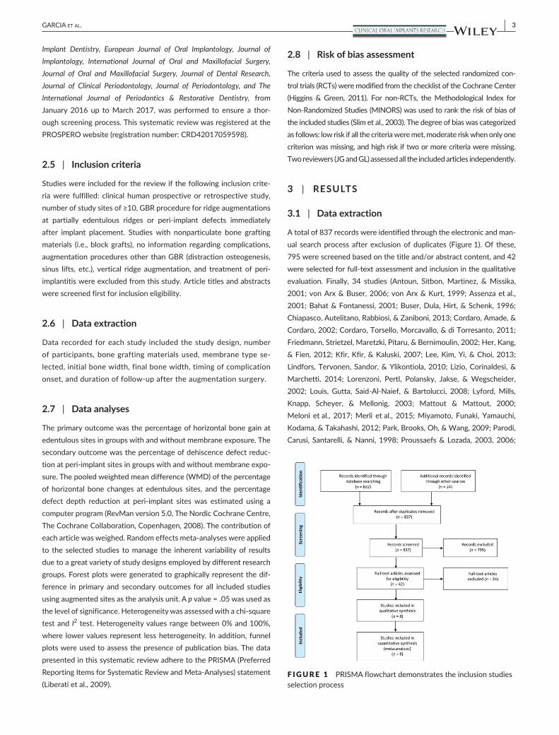