effect of low level laser therapy and calendula officinalis 3 … of low level laser therapy...
TRANSCRIPT

Volume 100 Number 2110 AJHM Summer 2007
Introduction
Skin fibroblasts are a vital component of the dermis; they not only produce and organize the
extracellular matrix of the dermis, but they also com-municate with each other and other cell types, play-ing a crucial role in regulating skin physiology.(1) Fibroblasts are a well established system for in vitro analysis of fibroblast growth, migration and collagen metabolism in wound healing.(2,3,4)
The laser is an electro-optical device capable of efficiently transmitting energy in the form of an in-tense beam of light. The radiant energy of the laser beam can be transformed into heat energy through its interaction with tissues. The interaction is depen-dent on the wavelength of the laser and the proper-ties of the tissue. Laser light is absorbed by water, and as most biologic tissues are composed mainly of water, the tissues are heated.(5) However, experi-ments following LLLT have shown that immediate increase of heat in the target tissue is negligible (+/- 1ºC). Low level lasers do not damage the hydrogen bonds since there is no heating effect. The effect is photochemical.(6) The basic tenet of LLLT is that it has a wavelength-dependent capability to alter cellular behavior in the absence of significant heat-
ing.(7)It is the coherence of laser which is the most sig-
nificant factor for skin penetration, thus allowing for a photochemical effect to occur.(8) When a laser beam is irradiated onto tissue, a local or topical primary response occurs at the cellular level; this is followed to some degree over a greater area by a systemic secondary response.
In the laboratory, Calendula extracts have been proven to be anti-edematous (9) antibacterial (10), antifungal (11) antiviral (12) and immuno-stimulat-ing.(13) Calendula officinalis is a remedy seldom used internally. Its major value lies in its local appli-cation to external wounds.(14) Topical application of Calendula officinalis markedly stimulates physi-ological regeneration and re-epithelization.(15) Dressing materials containing Calendula officinalis ointment applied to experimental animal wounds have also been found to enhance tissue repair.(16) Jouanny (17) advises its use externally as an anti-septic and cicatrizing agent. Phatak (18), Boericke (19) and Hering (20) state that Calendula officinalis promotes wound healing.
Interestingly enough, homœopathy has been ex-
Effect of Low Level Laser Therapy and Calendula officinalis 3 CH on Wound Healing in Human Skin Fibroblasts
Annelise Bresler (BA (WITS) M.Tech HOM), Denise Hawkins(M. Biomed), Radmila Razlog (M.Tech HOM) and Heidi Abrahamse (Prof).
Homeopathic Research
Abstract: Objective: This study aimed to determine the effectiveness of Low Level Laser Therapy (LLLT) and Calendula officinalis 3 CH or a combination of both treatment modalities on wounded human skin fibro-blasts (HSF) in vitro. Background data: LLLT is a form of phototherapy, involving the application of low level monochromatic and coherent light to injuries and lesions to stimulate healing. LLLT has been used to examine the influence of laser radiation on the healing process of wounds. Calendula preparations are mainly used for the treatment of wounds as it has anti-inflammatory, antibacterial and immuno-stimulating properties. Methods: Changes in normal and wounded fibroblast cell morphology were evaluated by light microscopy. Cellular responses to treatment were evaluated by assessing the cell viability and cytotoxicity. Results: The results demonstrate that Calendula officinalis 3 CH facilitates wound healing in the following two respects; it accelerated wound closure and increased cell viability. Laser irradiation improved the rate of wound closure in injured HSF cells in vitro while laser irradiation followed by Calendula officinalis 3 CH demonstrated a normalizing effect on wounded HSF cells.Keywords: wound healing, a comparison study of Calendula 3 CH and Laser phototherapy; Calendula, and wound healing; laser therapy, and wound healing.

AJHM Summer 2007 111Volume 100 Number 2
plained to work in a similar manner to LLLT, which is based on its biostimulatory effect. The Arndt-Schulz law is used by both modalities to elucidate the underlying mechanisms involved.(21,8) This law states that weak stimuli excite physiological activity while strong stimuli retard it.(8) It is evident from reviewing the literature that both modalities would argue that their treatment helps the body’s own in-nate capacity to heal.
Allied health professionals regularly care for a va-riety of wounds to the skin, namely abrasions, burns, surgical incisions, and, perhaps the most difficult to treat, ulcerations.(22) From acute wound manage-ment to augmentation of scar tissue remodelling, the clinician seeks to optimize wound care to pro-mote healing. A comprehensive treatment plan for wounds including complementary and alternative therapies may improve the rate of wound healing. This may result in reduced hospitalization thus limit-ing treatment and nursing time and reducing com-plications. The patient will be in a position to return to active life sooner and in doing so the economy at large will benefit.
Materials and MethodsCell Culture
Commercially available human skin fibroblast (HSF) cell lines (CRL1502 WS1) were obtained from the American Type Culture Collection (ATCC). Cells were grown in 25 ml of Eagles minimal essential
medium (EMEM) with Earle’s balanced salt solution (BSS) that was modified to contain 2 mM L-gluta-mine, 1.0 mM sodium pyruvate, 0.1 mM nonessential amino acids, 1% fungizone (BioWhittaker 17836-E) and 1% penicillin-streptomycin. In addition, it was supplemented with 10% fœtal bovine serum (Delta Bioproducts 14-501A1). The cultures were incubat-ed at 37ºC with 5% CO2 and 85% humidity. The cells were examined by inverse light microscopy and sub-cultured according to standard procedures.(23)
Experimental ProcedureThe HSF cells were subcultured from the 75 cm²
flask to 3.3 cm diameter culture plates. Approxi-mately 6 x 10⁵ cells in 3 ml culture medium were seeded in six sterile 3.3 cm diameter culture plates and incubated overnight to allow the cells to attach. The next day (Day 1) the plates were taken out of the incubator and 1 ml of culture medium was re-moved from each plate and discarded. Five of the six plates were wounded. To simulate a wound, me-chanical trauma was induced by means of a central scratch with a 1 ml sterile Pasteur pipette.(24,25,26) Each scratch was irregular and the wounds ranged from 1-2 mm in diameter. The plates were incubated at 37ºC for 30 min before each treatment regime.
Treatment was applied on two consecutive days (Day 1 and Day 2). Cells were visualised 24 hours (Day 2) and 48 hours (Day 3) after wound induc-tion with an inverted light microscope. Cellular re-
Plate Treatment No wound/ wound
Control/ Test
Key
1 No treatment No wound Control –W –C –I
2 No treatment Wound Control +W –C –I
3 Calendula officinalis 3 CH in 5% ethanol, 10 µl.
Wound Test + W +C –I
4 Irradiation with 632.8 nm Helium Neon (HeNe) laser, 5 J/cm² at calculated dosage.
Wound Test +W –C +I
5 Calendula officinalis 3 CH in 5% ethanol, 10 µl plus irradiation simultaneously.
Wound Test +W +C +I
6 Irradiation with 632.8 nm HeNe laser, 5 J/cm² at calculated dosage thereafter the plate was placed in the incubator for 4 min and subsequently the 10 µl of Calen-dula officinalis 3 CH in 5% ethanol was added.
Wound Test +W +I +C
Table 1: Experimental Procedure
Low Level Laser Therapy and Calendula

Volume 100 Number 2112 AJHM Summer 2007
sponses were measured on Day 3. Four of the plates received treatment while the remaining two served as controls. The experimental procedure is sum-marized in Table 1. The procedure was repeated six times (n=6).
Laser IrradiationLaser irradiation was carried out with a Helium
Neon (HeNe) laser, which has a wavelength of 632.8 nm, a power output of 18.8 mW and a power density of 2.07 mW/cm2. The dose delivered was 5 J/cm² (40 min 15 s) for a 3.3 cm spot size. Cell culture dishes were placed under the laser beam and irradiated with the culture dish lid off at room temperature in the dark on a dark surface.
Calendula officinalis 3 CHA preliminary study established that cells which
received 10 µl of Calendula officinalis 3 CH dispensed in 5% ethanol showed optimal signs of cell migra-tion with minimal cell death. A volume of 10 µl of Calendula officinalis 3 CH was added to the culture medium for specified plates on Day 1 and Day 2 of the experiment.
Cellular responsesCell morphology
Both wound contraction and re-epithelialization from the margins of the wound play an important role in wound closure.(27,28,29) Wound closure has been studied both in vivo and in vitro.(3,30,31) Two digital photos were taken of each wound on Day 2 and Day 3. Photos were evaluated for changes in migration (wound closure), haptotaxis (change in orientation of edge fibroblasts) and the number of fibroblasts present in the central scratch.(26)
ATP Cell Viability AssayThe CellTiter-Glo® (Promega G7571) lumines-
cent cell viability assay determines the number of viable cells in culture based on the quantitation of ATP present, which signals the presence of metaboli-cally active cells.(32) An equal volume of reconsti-tuted CellTiter-Glo® reagent was added to 50 µl of cell suspension (1x10⁵/100 µl). The contents were mixed for 3 minutes by hand to induce cell lysis. It was then incubated at room temperature for 10 minutes to stabilize the luminescent signal and the luminescence was recorded using a Junior EG&G Berthold Luminometer.
Trypan Blue Exclusion TestTrypan blue is a stain recommended for use in
estimating the proportion of viable cells in a popu-lation.(33,34) A vital dye such as trypan blue can enter a damaged cell membrane and stain the cyto-
plasm acting as an indicator of non-viable cells.(23) Live (viable) cells do not take up the dye and dead (non-viable) cells do. For the trypan exclusion test, 10 µl of the cell suspension was mixed with 10 µl of 0.4% Trypan blue (Sigma T8154). This solution was left at room temperature for 5 minutes and loaded into the hæmocytometer-counting chamber. The total number of cells per millilitre, the total num-ber of unstained (viable) and the total number of stained (non-viable) cells were counted using a light microscope. The viable cells were expressed as a percentage and calculated as the number of viable cells/total number X 100.
Lactate Dehydrogenase (LDH) Membrane Integrity Assay
The CytoTox 96® (Promega G1781) assay indi-rectly measures the lactate dehydrogenase activity present in the cytoplasm of intact cells. Cell quan-titation, therefore, can occur only if the cells are lysed to release the LDH present in the cell. The CytoTox 96® measures this stable cytosolic enzyme that is released upon cell lysis. Culture medium was removed from each plate on Day 3. 50 µl of culture medium was mixed with an equal volume of reconstituted substrate mix in a 96-well plate. The plate was covered with foil and incubated at room temperature for 30 minutes, protected from light. 50 µl of stop solution was added and the absorbance read at 490 nm.
Results
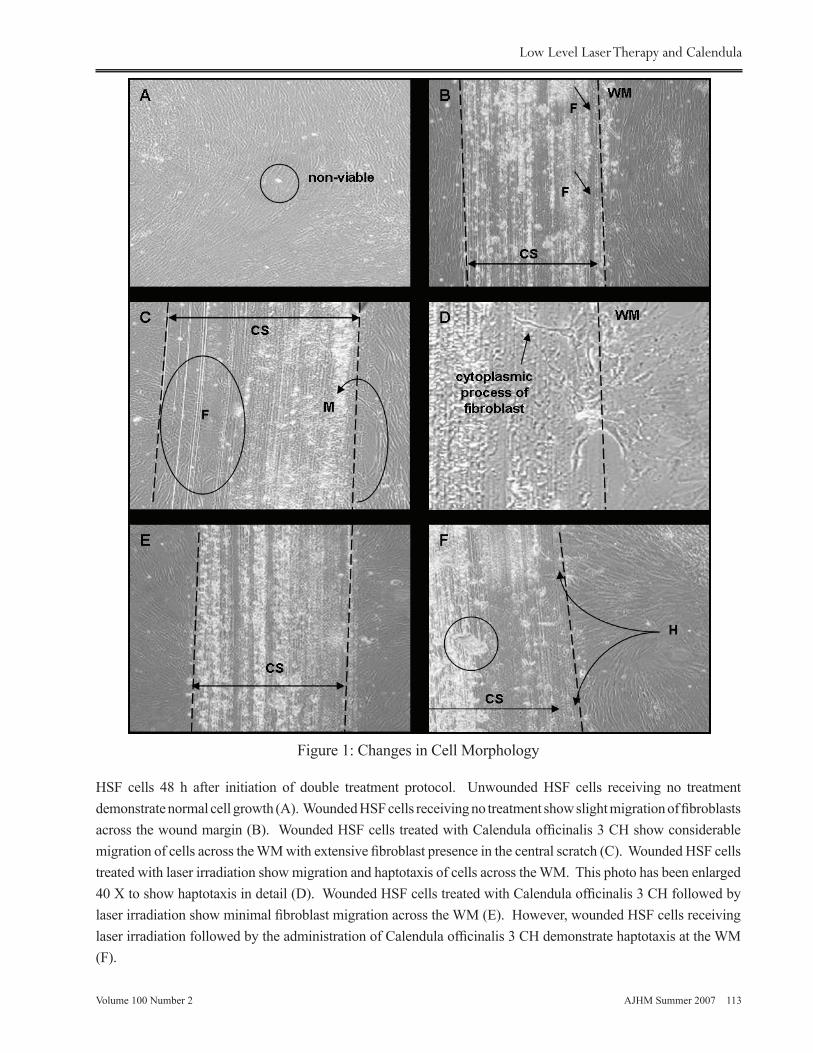
Changes in Cell MorphologyThe morphological changes observed after treat-
ment were recorded on Day 3 (Figure 1). The HSF cells that received no wound showed a normal state of biologic activity where normal growth was observed, highlighting the sustainability of the cul-tured environment to support continued existence and growth. Fibroblast activity was normal but also showed some non-viable cells (A). Wounded fibro-blasts that received no treatment showed fibroblast migration into the central scratch (CS) area. The morphological changes of the wounded untreated fibroblasts served as the control against which the individual treatments were compared (B).
Fibroblasts treated with Calendula officinalis 3 CH showed migration (M) of fibroblasts across the (wound margin) WM into the CS area. A significant amount of fibroblasts (F) can be seen in the CS (C). Although Figure 1 (D) has been taken under the 10 X magnification, it was enlarged to show haptotaxis (change in the orientation of the edge fibroblasts) in detail. Wounded HSF cells treated with laser ir-radiation show haptotaxis at the wound margin. The
Bresler, Hawkins, Razlog, Abrahamse
AJHM Summer 2007 113Volume 100 Number 2
Figure 1: Changes in Cell Morphology
HSF cells 48 h after initiation of double treatment protocol. Unwounded HSF cells receiving no treatment demonstrate normal cell growth (A). Wounded HSF cells receiving no treatment show slight migration of fibroblasts across the wound margin (B). Wounded HSF cells treated with Calendula officinalis 3 CH show considerable migration of cells across the WM with extensive fibroblast presence in the central scratch (C). Wounded HSF cells treated with laser irradiation show migration and haptotaxis of cells across the WM. This photo has been enlarged 40 X to show haptotaxis in detail (D). Wounded HSF cells treated with Calendula officinalis 3 CH followed by laser irradiation show minimal fibroblast migration across the WM (E). However, wounded HSF cells receiving laser irradiation followed by the administration of Calendula officinalis 3 CH demonstrate haptotaxis at the WM (F).
Low Level Laser Therapy and Calendula
Volume 100 Number 2114 AJHM Summer 2007
cytoplasmic processes of the fibroblasts can be seen to change their orientation at the WM as it invades the CS area (D).
Slight infiltration of fibroblasts into the CS area can be observed for cells treated with Calendula of-ficinalis 3 CH followed by laser irradiation on Day 3. Fibroblasts present in the CS were indicated with an arrow (E). Haptotaxis was visible for cells treated with laser irradiation followed by administration of Calendula officinalis 3 CH at the wound margin. Fragments of the plate, which came loose during the wound induction process, are encircled (F).
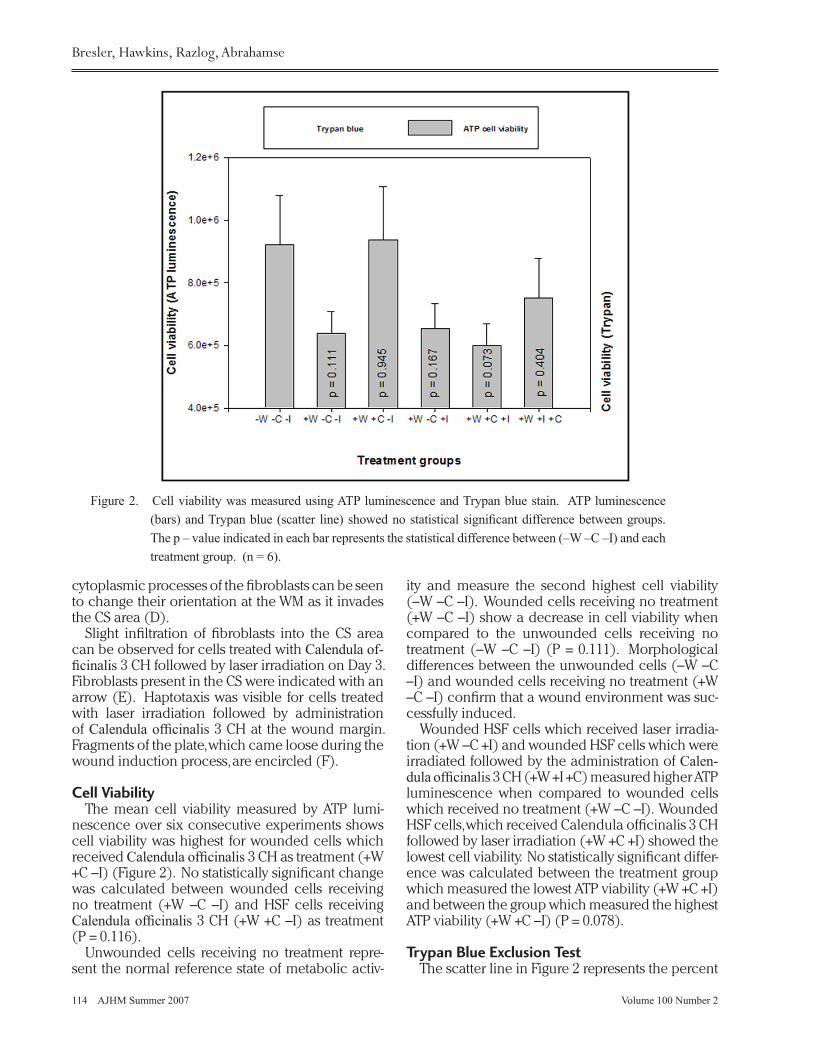
Cell ViabilityThe mean cell viability measured by ATP lumi-
nescence over six consecutive experiments shows cell viability was highest for wounded cells which received Calendula officinalis 3 CH as treatment (+W +C –I) (Figure 2). No statistically significant change was calculated between wounded cells receiving no treatment (+W –C –I) and HSF cells receiving Calendula officinalis 3 CH (+W +C –I) as treatment (P = 0.116).
Unwounded cells receiving no treatment repre-sent the normal reference state of metabolic activ-
ity and measure the second highest cell viability (–W –C –I). Wounded cells receiving no treatment (+W –C –I) show a decrease in cell viability when compared to the unwounded cells receiving no treatment (–W –C –I) (P = 0.111). Morphological differences between the unwounded cells (–W –C –I) and wounded cells receiving no treatment (+W –C –I) confirm that a wound environment was suc-cessfully induced.
Wounded HSF cells which received laser irradia-tion (+W –C +I) and wounded HSF cells which were irradiated followed by the administration of Calen-dula officinalis 3 CH (+W +I +C) measured higher ATP luminescence when compared to wounded cells which received no treatment (+W –C –I). Wounded HSF cells, which received Calendula officinalis 3 CH followed by laser irradiation (+W +C +I) showed the lowest cell viability. No statistically significant differ-ence was calculated between the treatment group which measured the lowest ATP viability (+W +C +I) and between the group which measured the highest ATP viability (+W +C –I) (P = 0.078).
Trypan Blue Exclusion Test The scatter line in Figure 2 represents the percent
Figure 2. Cell viability was measured using ATP luminescence and Trypan blue stain. ATP luminescence (bars) and Trypan blue (scatter line) showed no statistical significant difference between groups. The p – value indicated in each bar represents the statistical difference between (–W –C –I) and each treatment group. (n = 6).
Bresler, Hawkins, Razlog, Abrahamse

AJHM Summer 2007 115Volume 100 Number 2
age of viable cells for each procedure. Viability ranged between 90% and 96% and is read on the right Y-axis. HSF cells treated with Calendula officina-lis 3 CH (+W +C –I) and the unwounded cells receiv-ing no treatment (–W –C –I) calculated the highest percentage of viable cells, which corresponds with the results obtained for the ATP luminescence assay measuring cell viability.
This consistency between the two measuring tools for cell viability, namely ATP luminescence and Trypan blue was not established for the follow-ing two treatment groups: laser irradiation (+W –C +I) and irradiation followed by Calendula officinalis 3 CH (+W + I +C). Viability percentages were lower for these when compared to wounded cells receiv-ing no treatment (+W –C –I). This stands in contrast to the ATP cell viability measurements where both laser irradiated (+W –C +I) cells and irradiated cells followed by administration of Calendula officinalis 3 CH (+W +I +C) measured higher cell viability com-pared to wounded cells receiving no treatment (+W –C –I).
LDH Membrane Integrity Assay Lactate dehydrogenase (LDH) is released by
dying cells and therefore gives a quantitative mea-surement of the non-viable population.(35) LDH activity was lowest for both unwounded cells re-ceiving no treatment (–W –C –I) and wounded cells receiving irradiation followed by the administration of Calendula officinalis 3 CH (+W +I +C). Thus the unwounded cells and wounded cells receiving laser irradiation followed by the administration of Calendula officinalis 3 CH (+W +I +C) were the least cytotoxic (Figure 3).
The wounded HSF cells which received laser irradiation (+W –C +I) as treatment measured the highest LDH activity and were consequently iden-tified as the most cytotoxic treatment group. The wounded cells that received no treatment (+W –C –I) showed the second highest LDH activity, closely followed by the fibroblasts which received Calendula officinalis 3 CH (+W +C –I). Administra-tion of Calendula officinalis 3 CH followed by laser irradiation (+W +C +I) showed a cytotoxicity greater than wounded cells receiving laser irradiation fol-
Figure 3. Cytotoxicity was measured by means of a LDH membrane integrity assay. A statistical significant increase in cytotoxicity was measured for untreated HSF cells receiving no wound and those receiving a wound (P = 0.03). Furthermore, a statistically significant decrease in cytotoxicity was measured for wounded HSF cells receiving no treatment and wounded HSF cells treated with irradiation followed by the administration of Calendula officinalis 3 CH (P = 0.025). (n = 6, *P = less than or equal 0.05).
Low Level Laser Therapy and Calendula

Volume 100 Number 2116 AJHM Summer 2007
lowed by the administration of Calendula officinalis 3 CH (+W +I +C) indicating that laser irradiation may adversely affect the activity of Calendula.
A statistically significant increase in cytotoxicity was calculated between unwounded and wound-ed cells, which received no treatment (P = 0.03) showing that a wound environment was success-fully induced. Furthermore, a statistically significant decrease in cytotoxicity was measured between wounded cells receiving no treatment (+W –C –I) and wounded HSF cells receiving laser irradiation followed by the administration of Calendula officina-lis 3 CH (+W +I +C) (P = 0.025).
DiscussionThe stimulatory effects of LLLT at the cellular and
molecular levels have been shown in many studies (36), including in our own laboratory.(37) Laser light affects the mitochondrial respiratory chain by changing the electrical potential of cell membranes and, consequently, their selective permeability for so-dium, potassium and calcium ions, or by increasing the activity of certain enzymes such as cytochrome oxidase and adenosine triphosphate.(38) It also increases DNA synthesis,(39) collagen and pro-col-lagen production,(40,41) and may increase the cell proliferation (42) or alter locomotor characteristics of cells.(43) Although studies in both laboratory conditions and clinical settings have been numer-ous, the biochemical reactions induced by low level laser irradiation are still poorly understood.
Cordova et al.,(44) conducted a study with the aim to investigate the relationship between the beneficial properties of Calendula officinalis and its antioxidant properties. The butanolic fraction (BF) was studied because it is non-cytotoxic and is rich in a variety of bioactive metabolites including flavo-noids and terpenoids. The results obtained suggest the BF of Calendula officinalis possesses a significant free radical scavenging and antioxidant activity and that the proposed therapeutic efficacy of this plant could be due, in part, to these properties.
After analysis of the results there was an indica-tion that Calendula officinalis 3 CH can facilitate wound healing on wounded HSF cells in vitro in the following two respects - it accelerates wound closure and increases cell viability. Although no statistically significant difference was measured between the control group and the cells treated with Calendula officinalis 3 CH, the cell viability and cytotoxicity findings are encouraging. The lack of decrease in cell viability or increase in cytotoxicity despite wound induction and Calendula officinalis application may be indicative of the beneficial ef-fect that this treatment may have on wounded HSF cells in vitro. Furthermore, minor changes evident
on a cellular level may be more significant at a sys-temic level.
Laser treatment on its own showed increased ATP and a decrease in cytotoxicity when compared to wounded fibroblasts without treatment. Further-more, cell migration for wounded fibroblasts treated with laser irradiation showed an increase in migra-tion when compared to wounded cells without treatment. In conclusion, the cellular responses (mi-gration and cell viability) of wounded fibroblasts appeared to be stimulated by laser irradiation when the responses were compared to wounded cells receiving no treatment. Wounded cells which re-ceived no treatment (+W –C –I) showed a decrease in trypan blue, decrease in ATP cell viability and increase in LDH cytotoxicity when compared to unwounded untreated cells (–W –C –I) confirming that a central scratch successfully induced a wound environment.
Wounded HSF cells treated with Calendula offici-nalis 3 CH followed by laser irradiation proved to be the least favourable treatment. It seemed to lower cytotoxicity and early cell death. A suggested expla-nation would be that the laser irradiation might alter the chemical characteristics/bonds of Calendula of-ficinalis 3 CH dispensed in 5% ethanol consequently having a damaging effect on the cells and leading to lower cell viability.
Laser irradiation followed by the administration of Calendula officinalis 3 CH proved to be the bet-ter combination treatment procedure. It showed superior results with regard to cell morphology, cell viability and cytotoxicity when compared to the Ca-lendula officinalis 3 CH followed by irradiation treat-ment group. Laser irradiation followed by Calendula officinalis 3 CH demonstrated a normalising effect on wounded HSF cells. Results were comparable to unwounded cells thus designating this treatment procedure to be the most favourable.
ConclusionFrom this study cellular effects of LLLT and Ca-
lendula officinalis 3 CH on normal and wounded skin fibroblasts could be detected. Morphological changes indicate that a double treatment applica-tion of Calendula officinalis 3 CH increase wound closure on wounded HSF cells in vitro. Laser irradia-tion too showed morphological signs of increased wound closure for HSF cells after a double expo-sure. However, a synergistic relationship, based on morphology, was not established when combining the two treatments.
Although only a few statistically significant dif-ferences were obtained between the measurement tools and any of the treatment protocols, it is impor-tant and worth noting that small differences on the
Bresler, Hawkins, Razlog, Abrahamse

AJHM Summer 2007 117Volume 100 Number 2
cellular level might have a significant influence at the systemic level. Changes experienced at the cel-lular level might be amplified at the systemic level because of cell–cell interactions that may elicit a cascade of events.
References1. Cooper, M.L.Hansbrough, J.L., Spielvogel,
R.L., Bartel, R.L. and Naughton, G. “In-vivo optimization of a living dermal substitute employing cultured human fibroblasts on a biodegradable polyglycolic acid or polyglactin mesh”. Biomaterials 1991; 12: 243-248.
2. Gay, S., Martin, G.R., Müller, P.K., Timpl, R. and Kühn, K. “Simultaneous synthesis of types I and III collagen by fibroblasts in culture”. Proc. Natl. Acad. Sci 1976; 73(11): 4043-4040.
3. Laplante, A.F., Germain, L., Auger, F.A. and Moulin, V. “Mechanisms of wound reepithelialization: hints from a tissue-engineered reconstructed skin to long-standing questions”. FASEB J 2001; 15: 2377−2389.
4. El Ghalbzouri, A., Hensbergen, P., Gibbs, S. Kempenaar, J., Van der Schors, R. and Ponec, M. Fibroblasts facilitate re-epitheliazation in wounded skin equivalents”. Lab Invest 2004; 84: 102-112.
5. Arnoczky, S.P. and Aksan, A. “Thermal Modification of Connective Tissues: Basic Science Considerations and Clinical Implications”. J of the Am Academy of Orthopaedic Surg 2000; 8(5): 305-312.
6. UNL Environmental and Health Safety. (2003) Laser Classification and general safety control measures. http://ehs.unl.edu Downloaded 2005/03/28.
7. Basford, J.R. “Low Intensity Laser Therapy: Still Not an Established Clinical Tool”. Lasers in Surg and Med 1995; 16: 331-342.
8. Tunér, J. and Hode, L. Laser Therapy Clinical Practice and Scientific Background. Grängesberg: Prima Books 2002. pp. 8, 17, 27, 41, 49, 68, 70, 189, 320, 354, 358, 362.
9. Zitterl-Egsleer, K., Sosa, S., Jurenitsch, J., Schubert-Zsilavecz, M., Della Loggia, R., Tubaro, A., Bertoldi, M. and Franz, C. “Anti-oedematous activities of the main triterpenediol esters of marigold (Calendula officinalis L.)”. J of Ethnopharmy 1997; 57(2): 139-144.
10. Shah, J. Hompath Classic Premium Collection. (Version 8.0), 2004. form i.e. Computer program [Hompath], 8, New Hari Niwas, Dattatray Road, Santacruz(W), Mumbai-400 054, India.
11. De Klerk, M. The Anti-fungal properties of Calendula officinalis on Candida albicans, M:Tech Hom. Dissertation, Technikon
Witwatersrand, Johannesburg 1998. pp. iii. 12. Kalvatchev, Z., Walder, R. and Garzaro, D.
“Anti-HIV activity of extracts from Calendula officinalis flowers”. Biome Pharmacother 1997; 51(4): 176-180.
13. Amirghofran, Z., Azadbakht, M. and Karimi, M.H. “Evaluation of the immunomodulatory effects of five herbal plants”. J Ethnopharm 2000; 72(1-2): 167-172.
14. Kent, J.T. Lectures on Homoeopathic Materia Medica. 1907 original ed. New Dehli: B. Jain Publishers 2003. pp. 354.
15. Klouchek-Popova, E., Popov, A., Pavlova, N. and Krusteva, S. “Influence of the physiological regeneration and epithelialization using fractions isolated from Calendula officinalis”. Acta Physiol Pharmacol Bulg 1982: 8(4): 63-67.
16. Ansari, M. A., Jadon, N.S., Singh, S.P., Kumar, A. and Singh, H. “Effect of Calendula officinalis ointment, charmil and gelatin granules on wound healing in buffaloes: A histological study”. Indian Vet J 1997; 74(7): 594-597.
17. Jouanny, J. The essentials of Homeopathic Materia Medica. Boiron: Editions Boiron 1984. pp. 92.
18. Phatak, S.R. Materia Medica of Homoeopathic medicines. 1977 original ed. New Dehli: India Books and Periodical Publishers 2002 pp. 137-138.
19. Boericke, W. Pocket Manual of Homoeopathic Materia Medica and Repertory. 1927 original ed. New Dehli: B Jain Publishers (P) LTD 2002 pp. 156.
20. Hering, C. The Guiding Symptoms of our Materia Medica Vol III. 1997 reprint ed. New Dehli: B Jain Publishers (P) LTD 1997. pp. 261.
21. Callinan, P. Homoeopathy: How does it work? http://www.yourremedy.co.uk/how_it works.html. Downloaded 2006/02/02
22. Hopkins, J. T., McLoda, T.A., Seegmiller, J.G. and Baxter, G.D. “Low-Level Laser Therapy Facilitates Superficial Wound Healing in Humans: A Triple-Blind, Sham-Controlled Study”. Journal of Athletic Training 2004; 39(3): 223-229
23. Ausubel, R., Brent, R., Kingston, R.E., Moore, D.D., Seidan, J.G., Smith, J.A. and Struhl, K. Short protocols in molecular cloning. 4th ed. New York: Wiley and Sons Inc 1994 pp. 1-14.
24. Rigau, J., Sun, C., Trelles, M.A., and Berns, M (1995) Effects of the 633nm laser on the behaviour and morphology of primary fibroblasts in culture. In proceedings, Effects of low power light on biological systems, Barcelona, Spain, eds T.
25. Hawkins, D.H. and Abrahamse, H. “The role of laser fluence in cell viability, proliferation,
Low Level Laser Therapy and Calendula

Volume 100 Number 2118 AJHM Summer 2007
and membrane integrity of wounded human skin fibroblasts following helium-neon laser irradiation”. Lasers Surg Med 2006; 38(1): 74-83.
26. Cha, D., O’Brien, P., O’Toole, E.A., Woodley, D.T. and Hudson, L.G. “Enhanced modulation of keratinocyte motility by TGFα relative to EGF”. J Investig Dermatol 1996: 106: 590-597.
27. Clark, R.A. “Biology of dermal wound repair”. Dermatol Clin. 1993; 11: 647−666.
28. Grinnell, F. “Mini-Review on the Cellular Mechanisms of Disease”. Fibroblasts, Myofibroblasts, and Wound Contraction. J of Cell Biol 1994; 124(4): 401-404.
29. Martin, P. “Wound healing - aiming for perfect skin regeneration”. Science 1997; 276: 75-81.
30. Croft, C.B., and Tarin, D. “Ultrastructural studies of wound healing in mouse skin. Epithelial behaviour”. J Anat 1970; 106: 63−77.
31. Manios, A., Tzortzakjs, H. and Minogiannis N. “A differential model of wound epithelialization for full-thickness skin defects”. Ann MBC 1992; 9: 3−5.
32. Crounch, S.P.M. “The use of ATP bioluminescence as a measure of cell proliferation and cytoxicity”. Jour Immunol Meth 1993; 160: 81.
33. Phillips, H.J. and Terryberry, J.E. “Counting actively metabolizing tissue cultured cells”. Exp Cell. Res 1957; 13: 341-347.
34. Freshney, R.I. Culture of Animal Cells: A Manual of Basic Technique. 3rd ed. New York: Wiley-Liss. 1994 http://fscimage.fishersci.com/webimages_FSC/downloads/HyClone_ Protocol_3.pdf Downloaded 2006/02/20.
35. Doyle, A. and Griffiths, Bryan, J. Cell and Tissue Culture: Laboratory Procedures in Biotechnology. Chichester: John Wiley & Sons Ltd 1998. pp. 55, 141.
36. Pöntinen, P. J. Low Level Laser Therapy as a Medical Treatment modality. Tampere: Art Urpo Ltd 1992 pp.14-17, 134-135.
37. Abrahamse, H., Hawkins, D. and Houreld, N. “Effect of wavelength and fluence on morphology, cellular and genetic integrity of diabetic wounded human skin fibroblasts”. Spie International Optics and Photonics - Low Light Therapy 2006; 6140: 41-54.
38. Hrazdira, I. and Mornstein, V. Lékařská
biofyzika a přístrojová technika [Medical Biophysics and Instruments]. Neptun, Brno 2001.
39. Loevschal, H. and Arenholt-Bindlev, D. “Effects of low level diode laser (GaAIAs) irradiation on fibroblast of human mucosa in vitro.” Laser Surg Med 1994; 14: 347-354.
40. Balboni, G.C., Zonefrati, R., Brandi, M.L. and Repice, F. “Effects of HeNe/I.R. laser irradiation on two lines of normal human fibroblasts in vitro”. Arch Ital Anat Embriol 1986; 91:179-188.
41. Abergel, P., Lyons, R.F., Castel, J.C., Dwyer, R.M. and Uitto, J. “Biostimulation of wound healing by lasers: experimental approaches in animal models and in fibroblast cultures”. J Dermatol Surg Oncol 1987; 13: 127-133.
42. Web, C., Dyson, M. and Lewis, W. “Stimulatory effect of 660nm low level laser energy on hypertrophic scar-derived fibroblasts: possible mechanisms for increase cell counts”. Laser Surg Med 1998; 22: 294-301.
43. Noble, P.B., Shields, E.D., Blecher, P.D. and Bentley, K.C. “Locomotory characteristics of fibroblasts within a three-dimensional collagen lattice: modulation by helium/neon soft laser”. Laser Surg Med 1992; 12: 669-674.
44. Cordova, C.A., Siqueria, I.R., Netto, C.A., Yunes, R.A., Volpato, A.M., Cechinel Filho, V., Curi-Pedrosa, R. and Creczynski-Pasa, T.B. “Protective properties of butanolic extract of the Calendula officinalis L. (marigold) against lipid peroxidation of rat liver microsomes and action as free radical scavenger”. Redox Rep 2002; 7(2): 95-102.
All correspondence concerning this manuscript and request for reprints should be addressed to:
Prof. Heidi Abrahamse Laser Research GroupFaculty of HealthUniversity of JohannesburgP.O. Box 17011DoornfonteinSouth Africa2028TEL. +27 11 406 8145 FAX. +27 11 406 8202 EMAIL: [email protected]
Bresler, Hawkins, Razlog, Abrahamse