effect of laser therapy on gcf calprotectin level in …
TRANSCRIPT

EFFECT OF LASER THERAPY ON GCF
CALPROTECTIN LEVEL IN TYPE II DIABETIC
PATIENTS-A CLINICAL AND BIOCHEMICAL STUDY
Dissertation submitted to
THE TAMILNADU Dr. M.G.R. MEDICAL UNIVERSITY
In partial fulfillment for the Degree of
MASTER OF DENTAL SURGERY
BRANCH II
PERIODONTICS
APRIL 2016
brought to you by COREView metadata, citation and similar papers at core.ac.uk
provided by ePrints@TNMGRM (Tamil Nadu Dr. M.G.R. Medical University)

CERTIFICATE
This is to certify that Dr.K.R.KARTHIKEYAN, Post Graduate student in
the Department of Periodontics, J.K.K.Nattraja Dental College and Hospital,
Komarapalyam has done this dissertation titled “EFFECT OF LASER THERAPY
ON GCF CALPROTECTIN LEVEL IN TYPE II DIABETIC PATIE NTS – A
CLINICAL AND BIOCHEMICAL STUDY” under my direct guidance during
her post graduate study period 2013 - 2016.
This dissertation is submitted to THE TAMILNADU Dr. MGR
MEDICAL UNIVERSITY in partial fulfilment of the degree of MASTER OF
DENTAL SURGREY, BRANCH II – Periodontics.
DR.SUGUMARI ELAVARASU, DR.A.SIVAKUMAR,
Professor and Head, Principal,
J.K.K.N Dental college J.K.K.N Dental college
Komarapalayam Komarapalayam
Tamilnadu Tamilnadu

ACKNOWLEDGEMENT
I am greatly indebted to Dr.Sugumari Elavarasu, my Professor and Head of
the Department, Department of Periodontics, J.K.K.Nattraja Dental College and
Hospital, Komarapalayam, for her strenuous and dedicated efforts towards the post
graduate students and for her invaluable guidance, support and encouragement
throughout my post graduate study.
I would like to extend my heartfelt gratitude to Professor Dr.S.Sivakumar,
Principal, J.K.K.Nattraja Dental College and Hospital, for his kindness in allowing
me to utilize the facilities in the college.
I am highly obliged to my sir Dr.S.Thangakumaran, Reader for his valuable
suggestions, enthusiastic support and constant encouragement throughout my study.
I sincerely thank Dr. T.Arthiie, Reader for her valuable suggestions and
generous support. Her profound knowledge, patience and perseverance and his
incessant encouragement, guidance and support had benefited me and my
colleagues in every facet of our academic life.
I take this opportunity to express my humble gratitude to Dr. P.K. Sasi
Kumar, Reader, for his guidance and support throughout my study which had
benefited my academic life.
My heartful thanks to all my Postgraduate Colleagues, and all the Non-
Teaching Staff’s for their kind help during my postgraduate period.
I would like to thank all of my patients for their kind cooperation.
I also thank Dr. Saravanan, Genetech, Salem, for his support in lab
analysis.
Words are not enough to express my love and affection to my Father
Dr.K.Rajagunaseelan M.S Mother Mrs. R.Dhanalakshmi, for their mental and
physical support, without whom I would not have been able to reach this height.
Finally, without the grace of the ALMIGHTY this possibility would have
been just impossible.

CONTENTS
S. No. INDEX PAGE No.
1 INTRODUCTION 1
2 AIM AND OBJECTIVES 5
3 REVIEW OF LITERATURE 6
4 MATERIALS & METHODS 19
5 RESULTS 38
6 DISCUSSION 49
7 SUMMARY AND CONCLUSION 53
8 BIBLIOGRAPHY 54

ANNEXURE – 1 (TABLES)
TABLE No. TITLE
1 Comparison of clinical parameters in Group II subjects
2 Comparison of clinical parameters in Group II A subjects
3 Comparison of clinical parameters in Group II B subjects
4 Comparison of clinical parameters in group II A and B at 4th week post operative
5 Calprotectin levels
6 Inter Group comparison of Calprotectin levels
7 Intra Group comparison of Calprotectin levels
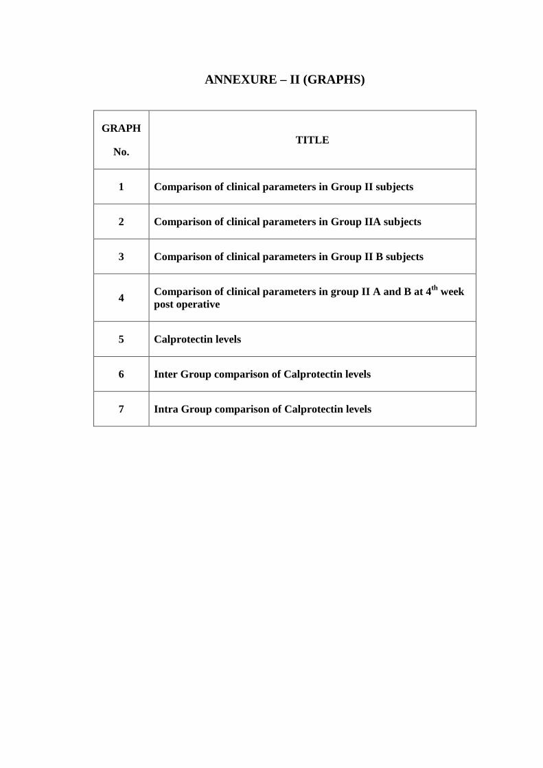
ANNEXURE – II (GRAPHS)
GRAPH
No. TITLE
1 Comparison of clinical parameters in Group II subjects
2 Comparison of clinical parameters in Group IIA subjects
3 Comparison of clinical parameters in Group II B subjects
4 Comparison of clinical parameters in group II A and B at 4th week post operative
5 Calprotectin levels
6 Inter Group comparison of Calprotectin levels
7 Intra Group comparison of Calprotectin levels

Introduction
1
Periodontitis is a disease that affects the tooth-supporting tissues and exhibits
a wide range of clinical, microbiological, and immunological manifestations. It is
associated with and probably caused by a multifaceted dynamic interaction among
specific infectious agents, host immune responses, hazardous environmental
exposure, and genetic propensity. Hormonal influences on the periodontium are
many, with widely varying clinical presentations and degrees of effect. The primary
hormonal factor is associated with diabetes mellitus.
Diabetes mellitus (DM) causes inflammatory complications, including
Retinopathy, Nephropathy, Neuropathy, Macro vascular disease, Altered wound
healing, Periodontal disease 1.
Diabetes mellitus is a chronic metabolic disorder caused by inherited or
acquired deficiency in production of insulin by the pancreas or by the
ineffectiveness of the insulin action. In diabetic patients, hyperglycemia could
indirectly exacerbate inflammatory tissue destruction through the body's scavenger
system against advanced glycation end-products and that hyperglycemia might
directly impair the biological functions of periodontal connective tissues through
cell–matrix interactions. It is suggested that an infection-mediated regulation cycle
of cytokine synthesis and secretion by chronic stimulus from lipopolysaccharide and
products of periodontopathogenic organisms may amplify the magnitude of the
advanced glycation end-product mediated cytokine response operative in DM. Dual
pathway of tissue destruction suggests that control of chronic periodontal infection
and gingival inflammation is essential for achieving long-term control of DM1,2.

Introduction
2
Calprotectin is a heterodimer of calcium-binding protein contained in the
cystosol of neutrophils, monocytes/macrophages and epithelial cells. This protein
belongs to the S100 protein family and forms approximately 5% of the total cellular
protein and 50% to 60% of cystosol protein in granulocytes. The biological role of
the S100 proteins are: regulation of the protein kinases, antimicrobial activity, and a
regulatory role in inflammatory reactions3.
Calprotectin exists normally in human plasma, saliva, and feces. Its level
elevates remarkable in various infections and tissue damaging disorder. In recently,
numerous studies have emerged documenting that increased levels of calprotectin
are present in patients with diabetes, cardiovascular diseases and various cancers.
Inflammed gingiva were found to contain vast numbers of calprotectin positive
macrophages around the vessels in central connective tissue and adjacent to the
epithelium. Calprotectin and its subunits have also been found in the gingival
crevicular fluid from diseased gingival sites of periodontal patients, and the
concentrations of calprotectin and its subunits were significantly higher in diseased
sites than in healthy sites.
The level of calprotectin in the gingival crevicular fluid of periodontitis
patients was positively correlated with clinical parameters and biochemical markers,
including probing depth, interleukin1β, prostaglandinE2, collagenase and aspartate
aminotransferase.
Calprotectin is released from leukocytes during inflammatory events in the
initial steps of inflammation and its extracellular function including antibacterial,
chemotactic for neutrophils and inhibition binding of pathogenic bacteria to

Introduction
3
epithelial cells, suggest that calprotectin may play a role in the immune response
mechanism of periodontal inflammation.
The calprotectin exerts a regulatory activity in inflammatory processes
through its effect on the survival or growth states of cells participating in the
inflammatory reaction. It is also possible that calprotectin, at a high concentration,
might have a deleterious effect on fibroblasts and influence the recovery of
inflammatory tissue. Therefore, the protein factor may be a new drug target to
control inflammatory reactions4.
The main goals of non surgical periodontal therapy (scaling and root
planning SRP) is to eliminate bacterial deposits and by removing the supragingival
and subgingival biofilms and to restore the biological compatibility of periodontally
diseased root surfaces for subsequent attachment of periodontal tissues to the treated
root surface. The mechanical therapy alone may fail to eliminate pathogenic bacteria
in the soft tissue and in areas that are inaccessible to periodontal instruments, such
as deep pockets, furcation areas, and root depressions5.
To overcome these limitations of mechanical therapy, several adjunctive
protocols have been developed. The use of lasers has been proposed for its
bactericidal and detoxification effects and for its capacity to reach sites that
mechanical instrumentation.
The laser technology was introduced in dentistry in the 80’s, their
performances as therapeutic methods constantly growing and being largely accepted.
Recently, the low frequency laser has been introduced. This type of laser is
characterized by a 400–900 nm low wavelength spectrum and biostimulation and
Introduction
4
biomodulation properties, due to the capacity of this wavelength spectrum to modify
tissue behavior, through its action on the calcium channels initiating an increase of
the cellular metabolism and proliferation rate6.
GCF calprotectin level decreases after the periodontal treatment7. The
present study was done to investigate the effect of laser as an adjunct to non-surgical
periodontal treatment in GCF calprotectin level of type II diabetic patients.
Hence the aim of this study was designed to estimate the levels of
Calprotectin level in GCF of subjects with chronic periodontitis with diabetic
mellitus, after 1st and 4th weeks of scaling and root planning and also scaling and
root planing with LASER in DM with chronic periodontitis subjects.

Aim and Objectives
5
The aim of the present study is:
1) To compare the effect of scaling root planing along with laser and scaling root
planing alone in type II diabetic patients.
2) Effect of laser therapy on GCF calprotectin level in type II diabetic patients.
Review of Literature
6
Diabetes mellitus and periodontitis:
Periodontal diseases are inflammatory in nature; they may alter glycemic
control in a similar manner to obesity. Diabetic patients with periodontal infection
have a greater risk of worsening glycemic control over time compared to diabetic
subjects without periodontitis.3 Periodontal intervention trials suggest a significant
potential metabolic benefit of periodontal therapy in people with diabetes. Several
studies of diabetic subjects with periodontitis have shown improvements in glycemic
control following scaling and root planning combined with adjunctive systemic
doxycycline therapy.8
Steven P E 20049, GCF samples were collected from 45 patients with Type
II DM and untreated chronic periodontitis. IL-1β levels were determined. He
concluded that poor glycemic control is associated with GCF IL-1β and also the
hyperglycaemia contributes to a heightened inflammatory response between poor
glycemic and periodontal destruction.
Campus G et al 200510, studied on total of 212 individuals participated in
that 71 Type II DM patients aged 61.0 ± 11.0 years and 141 non-diabetic controls in
good general health aged 59.1 ± 9.2 years. All subjects were given a clinical
periodontal examination for probing depth, attachment level, presence of calculus,
bleeding on probing, and assessment of plaque. Subgingival plaque samples were
obtained, and P. Gingivalis, P. Intermedia, and T. Forsythensis were identified using
multiplex polymerase chain reaction. Concluded patients with Type II DM
undoubtedly have susceptibility for more severe periodontal disease.
Review of Literature
7
Katz J et al 200511, gingival biopsies from eight patients with both type II
diabetes and chronic periodontitis and 14 healthy control subjects with chronic
periodontitis were immune histochemically stained for RAGE. It’s giving a
judgement that there was no change in the staining intensity for RAGE between both
groups, the increase in the mRNA for RAGE in the type II diabetes gingival
epithelium may indicate a possible involvement of this receptor in the periodontal
destruction in type II diabetes.
Calprotectin
Calprotectin is an important proinflammatory mediator with multiple
regulatory functions in periodontitis and diverse inflammatory diseases. It is
therefore reasonable to consider the calprotectin gene as an ideal candidate for
conferring genetic susceptibility to periodontitis.
Miyasaki et al 199812 identified calprotectin in adult dental calculus and
GCF. He indicated that calprotectin level in GCF from diseased pocket of patients
with periodontitis is significantly higher than the healthy gingival cervices.
Kido J et al 199913, in his study reviewed to investigate the correlations
between GCF calprotectin levels and clinical indicators (probing depth and bleeding
on probing, BOP), and the IL-1b or PGE2 levels in GCF. He derived that
calprotectin level in GCF correlates well with clinical biochemical markers of
periodontal disease and suggests that calprotectin may be useful for evaluating the
extent of periodontal inflammation.
Teuro N at al 200014, Association of GCF calprotectin level with GCF
volume, GI, level of biochemical markers including collagenase and AST in GCF
Review of Literature
8
was investigate to clarify the relationship between GCF calprotectin level and
periodontal inflammation. It can be inferred that GCF calprotectin level significant
correlates not only with clinical indicators but also with current biochemical marker
level.
Nisapakultorn K et al 200115, study was to assess the effect of epithelial
calprotectin expression on an invasive periodontal pathogen P. gingivalis, and to
provide evidence for the role of calprotectin in innate host defense in periodontal
disease. He observed that epithelial calprotectin expression may have a protective
role in periodontal diseases. Reduced P. gingivalis binding and invasion into
epithelial cells could reduce bacterial colonization and persistence during the
infections. And also increased calprotectin expression during periodontitis may
reflect complimentary innate mucosal host defense mechanisms in response to P.
gingivalis infection.
Nisapakultorn K et al 200116 studied the antimicrobial protein complex
epithelial resistance to invading bacteria; an epithelial cell line was stably
transfected to express the calprotectin complex. Data provide supporting evidence
that calprotectin expression in mucosal epithelial cells may play a protective role in
innate host defense. Calprotectin expression reduces epithelial invasion by
pathogens, including Listeria and Salmonella. Calprotectin mediates protection
against invasion in vitro by several novel mechanisms. Associated with expression,
bacterial binding is reduced. It remains unclear if the effect of calprotectin is
associated directly or indirectly with cell surface calprotectin. Cell surface α3
integrin is upregulated with calprotectin and is likely to reduce access to key
receptors. Calprotectin also interferes with internalization, perhaps by modifying

Review of Literature
9
calcium signaling and actin organization. These mechanisms may complement
antimicrobial activity of calprotectin within the cytoplasm. Based on in vitro
experiments, calprotectin is a multifunctional protein employing several modes of
action to contribute to innate epithelial immunity against infection.
Kido J et al 200317 studied on calprotectin which release by P-LPS was
induced via the CD14–TLR–NF-jB pathway and the cellular mechanism of
calprotectin release in human neutrophils. He concluded that calprotectin release is
induced by P-LPS via the CD14, TLR2, NF-κB signal pathway in human
neutrophils and may be dependent on microtubule and microfilament systems.
Que M L et al 200418 Fifteen healthy non-smoking subjects, aged 18–30,
were involved in this study GCF samples collected with Dura pore strips from 12
sites in each subject. Quantitative analyses of total proteins, MRP8/14, MRP14 and
MRP8 were performed by ELISA procedures. He observed that expression of
calprotectin in the early phase of experimental gingivitis is variable between
subjects, and two groups of subjects can be differentiated according to their response
patterns.
Suryono et al 200519 investigated the expression and production of
calprotectin from human monocytes by examining the effect of lipopolysaccharide
of P-LPS, TNF-α, and IL-1β by ELISA. Derived that P-LPS, TNF-α, and IL-1β
treated monocytes from human monocytes that this producton is associated with
activation of DNA C/EBPα binding complex.
Kaner D et al 200720 was analysed the calprotectin in GCF as a predictive
biomarker in periodontal therapy.
Review of Literature
10
Sun X et al 20113, studied the plasma concentrations of calprotectin and
were measured, using an enzyme immunoassay, in 139 patients with aggressive
periodontitis and in 88 periodontally healthy control subjects. He observed that
aggressive periodontitis is associated with elevated levels of plasma calprotectin and
that gene polymorphisms of S100A8 may influence the susceptibility and severity of
aggressive periodontitis.
Kaner et al 201121 found elevated levels of calprotectin in gingival
crevicular fluid and predicted disease activity in patients treated for generalized
aggressive periodontitis.
Sema B et al 201122 investigated gingival crevicular fluid (GCF)
calprotectin, osteocalcin and cross-linked N-terminal telopeptide (NTx) levels in
health along with different periodontal diseases. Results suggested that elevated
GCF calprotectin levels play a role as a reliable inflammatory marker in the
pathogenesis of periodontal disease. Fluctuating GCF levels of osteocalcin and NTx
might point out to the abnormal bone turnover in periodontitis. Data document for
the first time the role of NTx in the pathogenesis of different periodontal diseases.
Biomarkers
Biomarkers may be defined as a substance that is measured objectively and
evaluated as an indicator of normal biologic processes, pathogenic processes and
pharmacologic responses to a therapeutic intervention. Biomarkers are produced by
normal healthy individuals or by individuals affected by specific systemic diseases,
are tell-tale molecules that could be used to monitor health status, disease onset,
treatment response and outcome.

Review of Literature
11
Gingival crevicular fluid:
Gingival crevicular fluid is inflammatory exudate that seeps into gingival
crevices or periodontal pocket around teeth with inflamed gingival. The amount of
GCF produced at a given site significantly increase with the severity of gingival
inflammation23.
Several techniques have been employed for the collection of GCF and the
technique chosen will depend upon the objectives of the study as each technique has
advantages and disadvantages. The techniques can be divided into three basic
strategies, subject to various modifications in their application by different authors.
1) Gingival washing methods 2) Capillary tubing or micropipettes 3) Absorbent
filter paper strips24
Leo 196125 was one of the fiest to speculate on the mechanism of gingival
fluid production. The concentration of total protein in fluid from inflamed gingival
is very similar to that of serum while in normal crevices it is lower.
Pashely D H 197625 reviewed that in 1952; investigate the physiological
properties of the gingival pocket, observed increased fluid transduction and
emigration of leukocytes. Only through the efforts of Brill and co-workers, the
phenomenon of gingival fluid production was discovered and early attempts were
made to characterized the fluid,
Curtis et al 198826 reviewed that gingival crevicular fluid is regarded as a
promising medium for the detection of markers of periodontal disease activity,
because the fluid accumulation at the gingival margin, it will contain potential
markers derived not only from the host tissue and serum but also the subgingival
Review of Literature
12
microbial plaque and thus an extremely broad range of candidate molecule may be
investigated.
Uitto V J 200327 reviewed that gingival crevicular fluid is a complex
mixture of substance derived from serum, leukocytes, structural cells of the
periodontium and oral bacteria. These are indicators of periodontal disease and
healing after therapy.
Champagne et al 200328 explores the inflammatory mediator levels in
subjects with periodontal diseases. Their results suggest that some inflammatory
mediator levels increase with time in periodontitis patients both in active and stable
periodontal sites and that the risk for periodontitis is related to the overall systemic
inflammatory response of an individual.
Catherine M E 200328 reviewed that lipopolysaccharide triggers monocytes
to release inflammatory mediators (prostaglandin E2, thromboxane B, interleukins -1,
-6 and -8, tumor necrosis factor, and collagenase) that increase local destruction of the
connective tissues structural elements. Therefore, levels of monocytic inflammatory
mediators (including prostaglandin E2, interleukin-1, and tumor necrosis factor) in
GCF may well represent the ideal markers of disease activity at a site level.
Armitage G C 200424 reviewed the possible biomarkers in gingival
crevicular fluid for the progression of periodontitis. These components fall into 3
general categories.
1) Host derived enzyme and their inhibitors.
2) Inflammatory mediators and host response modifiers.
3) Tissue breakdown products.

Review of Literature
13
Over 65 GCF components have been examined as possible markers for the
progression of periodontitis.
1) Host derived enzyme:
Aspartate, aminotransferase, alkaline phosphatase, acid phosphatase, β-
glucuronidase, elastase, elastase inhibitors, cathepsins, trypsin like enzyme,
immunoglobulin-degrading enzyme, glycosidases, dipeptidyl peptidases, nonspecific
neutral proteinases, collegenases (MMP-1,3,8,13), gelatinases (MMP-2,9), TIMP-1,
stomyelysins, myeloperoxidases, lactate, arylsulfatase, creatinine kinase.
2) Inflammatory mediators and host response modifiers:
Cytokines, RANTES, prostaglandin E2, leukotriene B4, acute phase proteins,
auto antibodies, antibacterial antibodies, plasminogen activator, PA inhibitor-2,
substance P, vasoactive intestinal peptide, neurokinin A, neopterin, platelet-
activating factors, CD14, cystatins, calgranulin A.
3) Tissue breakdown products:
Glycosaminoglycans, hydroxyproline, fibronectin fragments, osteonectin,
osteocalcin, type I collagen peptide, osteopontin, laminin, calprotectin, haemoglobin
β-chain peptides, pyridinoline crosslinks (ICTP).
LASER
A laser is a device that produces coherent electromagnetic radiation. Laser
radiation is characterized by a low divergence of the radiation beam and, with few
exceptions, a well defined wavelength. The term laser is well known as the acronym
for light amplification by stimulated emission of radiation.

Review of Literature
14
Lasers are generally accepted and widely used as a tool for soft tissue
management. The major advantageous properties of lasers are relative ease of
ablation of tissues together with effective hemostasis and bacterial killing.
Gingivectomy, gingivoplasty and frenectomy are the most popular procedures
carried out using lasers.29
Walsh L J 199730 Despite more than 30 years of experience with low level
laser therapy (LLLT) or ‘biostimulation’ in dentistry, concerns remain as to its
effectiveness as a treatment modality. Reported LLLT can influence the behaviour
of many cell types, and that multiple effects can occur simultaneously.
Eberhard J 200331, compared the effectiveness of subgingival calculus
removal from periodontally involved root surfaces with an Er:YAG laser compared
to hand instrumentation in situ . Concluded that in vivo capability of the Er:YAG
laser to remove calculus from periodontally involved root surfaces, although the
effectiveness did not reach that achieved by hand instrumentation.
Qadri T et al 20056, studied in split-mouth, double-blind controlled clinical
trial the effects of irradiation with low-level lasers as an adjunctive treatment of
inflamed gingival tissue. They conclude that Additional treatment with low-level
lasers reduced periodontal gingival inflammation.
Charles M 200632 reported that clinical parameters of reductions in probing
depth or gains in clinical attachment level, that indicates when used as an adjunct to
SRP, mechanical, chemical, or laser curettage has little to no benefit beyond SRP
alone.
Review of Literature
15
Krause F 200733, To evaluate the removal of subgingival calculus and dental
hard tissues depending on the threshold level of a fluorescence feedback controlled
Er:YAG laser. Conclude that amount of residual calculus following laser irradiation
depends on the fluorescence threshold level for a feedback-controlled Er:YAG laser.
Isao I 200928 reviewed that animal study has reported that compared with
conventional scalpel surgery, laser surgery produces less pain with the oral soft
tissue incision.
Zand et al.200934 investigated the use of CO2 laser (1W of defocused
continuous mode) in 15 patients with recurrent aphthous ulcerations in comparison
to the placebo (recurrent aphthous ulcerations which were not treated). Both
ulcerations were covered with transparent gel without the use of anaesthetics. The
power of CO2 laser was 2-5mW after passing through gel which did not
significantly increase the temperature. The result was use of CO2 laser of low
intensity instantly reduces pain in patients with recurrent aphthous ulcerations
without any adverse effects
Carlos D et al 201035 studied the effect of low-power lasers in the
management of pathologies related to periodontal tissues. The clinical applications
of laser phototherapy (LPT) include periodontal inflammation modulation,
improvement in wound and bone healing processes, control of postoperative pain
and post treatment tooth hypersensitivity, and microbial reduction when associated
with an extrinsic photo sensitizer.
Gokce A 201136 evaluate the effect of low-level laser therapy (LLLT) as an
adjunct to non-surgical periodontal therapy of smoking and non-smoking patients

Review of Literature
16
with moderate to advanced chronic periodontitis. And he concluded that LLLT as an
adjunctive therapy to non-surgical periodontal treatment improves periodontal
healing.
Obradović R 201137 reported that Low level laser therapy was efficient in
gingival inflammation elimination and can be proposed as an adjuvant tool in basic
periodontal therapy of diabetic patients.
The American Academy of Periodontology (AAP) 201138 Erbium lasers
show the greatest potential for effective root debridement (SRP). There was the
potential for root surface damage during the process of in vivo calculus removal
since the Er:YAG is a hard tissue laser and the operator would not be able to
visualize what is being lased.
Radmila O 201239, to evaluate the effects of low-level laser therapy (LLLT)
by exfoliative cytology in patients with DM and gingival inflammation. And he
derived that LLLT as an adjunct in periodontal therapy reduces gingival
inflammation in patients with DM and periodontitis.
Catherine G 201240, studied was to compare the local biologic effects of
PDT, diode soft laser (DSL) therapy, and conventional deep scaling and root planing
(SRP) in residual pockets. And he concluded that, Levels of several cytokines and
acute-phase proteins significantly changed after treatment regardless of treatment
modality. There was no evidence for a specific DSL- or PDT-enhanced expression
of inflammatory mediators.
Obradović et al. 201241 treated patients with diabetes mellitus and
periodontal disease by use of LLLT (670 nm, 5 mW, 2 J/cm², 16 minutes for five

Review of Literature
17
days) together with conventional periodontal treatment. He observed that healing
was improved as well as collagenization and homogenization in gingival lamina
propria on the basis of histopathological findings.
Fabrizio S 20125, To investigate whether the adjunctive use of diode laser
provides additional benefits to scaling root planning alone in patients with chronic
periodontitis, a meta analysis was conducted according to the recommendations of
the Preferred Reporting Items for Systematic Reviews and Meta-analysis statement
and the Cochrane Collaboration and suggested that the use of diode laser as an
adjunctive therapy to conventional nonsurgical periodontal therapy did not provide
additional clinical benefit.
Soo L et al 201242 To compare a monotherapy of Er:YAG laser debridement
(ERL), wavelength 2940 nm, with mechanical scaling and root planing (SRP) for the
treatment of chronic periodontitis using clinical and patient-centred outcomes. He
observed that SRP resulted in a statistically significant greater short-term
improvement in clinical parameters and patient satisfaction compared with ERL.
Walter D 201343 evaluates the effect of a 980-nm diode laser as an adjunct
to scaling and root planing (SRP) treatment. He concluded that comparison of SRP
alone multiple adjunctive applications of a 980-nm diode laser with SRP showed PD
improvements only in moderate periodontal pockets (4 to 6 mm).
Jeffrey D 201344 found that Sites treated with the CO2 laser tended to show a
greater decrease in probing depths, greater amounts of recession, and greater gains
in clinical attachment levels, however the results were not statistically significantly
better than scaling and root planing alone.

Review of Literature
18
Mahnke et al. 199545.Determined that role of calprotectin in cellular
adhesion has been reported as the monoclonal antibody 27E10 inhibiting the
attachment of monocytes to collagen and fibronectin. On the other hand, these
extracellular matrix proteins induced the expression of calprotectin in parallel with
the release of inflammatory cytokines, tumor necrosis factor alpha (TNF-α) and
interleukin- 6 (IL-6) and production of superoxide anions. The relationship between
calprotectin expressions and higher capacity to release TNF α has also been shown
in human alveolar macrophages derived by bronchoalveolar lavage.
Andersen E et al 201046 measured the levels of MRP8/14 longitudinally
over 6 months in subjects with chronic periodontitis. And he confirmed clinical
improvements after scaling and root planing. There was a significant decrease of
MRP8/14 levels in diseased sites both at 3 and at 6 months after treatment.
Anderson E study based on Giannopoulou C 200647 study showed a
decrease of MRP8/14 after non-surgical periodontal therapy and significant changes
were noted even after 10 days when non-surgical therapy was supplemented with
systemic antibiotics. The improvements of biochemical parameters obtained in the
present trial also compared favourably with previous reports on the effect of
periodontal treatment on other gingival crevice fluid components.

Materials and Methods
19
A randomized controlled split mouth study was conducted to estimate the
level of CALPROTECTIN in GCF of periodontal health, disease and after
treatment. The protocol was reviewed and approved by institutional ethical board.
An informed consent was obtained from the patients and the study related procedure
was explained. A total of 60 subjects were recruited from the outpatient department
of periodontics, J.K.K.N Dental College and Hospital, Komarapalayam, Tamilnadu
based on the following criteria.
Inclusion criteria:
1) Age range 25- 65 years
2) Should at least 20 natural teeth
3) PD ≥4
4) Diabetic patients with chronic periodontitis
5) Systemic healthy subjects.
Exclusion criteria:
1) Treatment in the previous 6 months with anti-inflammatory drugs or antibiotics.
2) Haematological disorder.
3) Liver disorder.
4) Patients with bone disorder.
5) Pregnancy and lactating mother.
6) History of recent bacterial or viral infection.
7) Individual with habit of smoking and alcoholism.
Materials and Methods
20
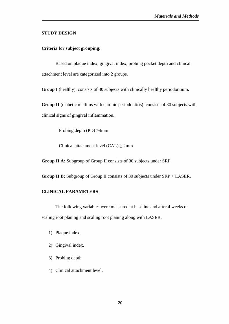
STUDY DESIGN
Criteria for subject grouping:
Based on plaque index, gingival index, probing pocket depth and clinical
attachment level are categorized into 2 groups.
Group I (healthy): consists of 30 subjects with clinically healthy periodontium.
Group II (diabetic mellitus with chronic periodontitis): consists of 30 subjects with
clinical signs of gingival inflammation.
Probing depth (PD) ≥4mm
Clinical attachment level (CAL) ≥ 2mm
Group II A: Subgroup of Group II consists of 30 subjects under SRP.
Group II B: Subgroup of Group II consists of 30 subjects under SRP + LASER.
CLINICAL PARAMETERS
The following variables were measured at baseline and after 4 weeks of
scaling root planing and scaling root planing along with LASER.
1) Plaque index.
2) Gingival index.
3) Probing depth.
4) Clinical attachment level.
Materials and Methods
21
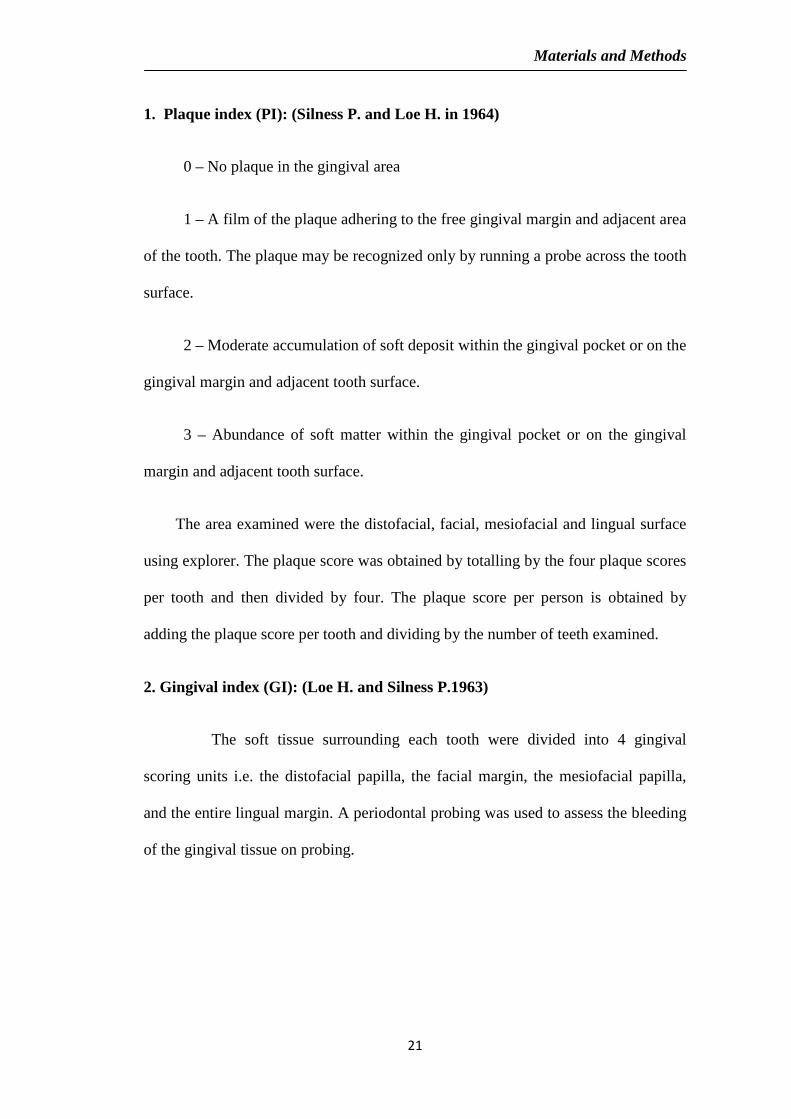
1. Plaque index (PI): (Silness P. and Loe H. in 1964)
0 – No plaque in the gingival area
1 – A film of the plaque adhering to the free gingival margin and adjacent area
of the tooth. The plaque may be recognized only by running a probe across the tooth
surface.
2 – Moderate accumulation of soft deposit within the gingival pocket or on the
gingival margin and adjacent tooth surface.
3 – Abundance of soft matter within the gingival pocket or on the gingival
margin and adjacent tooth surface.
The area examined were the distofacial, facial, mesiofacial and lingual surface
using explorer. The plaque score was obtained by totalling by the four plaque scores
per tooth and then divided by four. The plaque score per person is obtained by
adding the plaque score per tooth and dividing by the number of teeth examined.
2. Gingival index (GI): (Loe H. and Silness P.1963)
The soft tissue surrounding each tooth were divided into 4 gingival
scoring units i.e. the distofacial papilla, the facial margin, the mesiofacial papilla,
and the entire lingual margin. A periodontal probing was used to assess the bleeding
of the gingival tissue on probing.
Materials and Methods
22
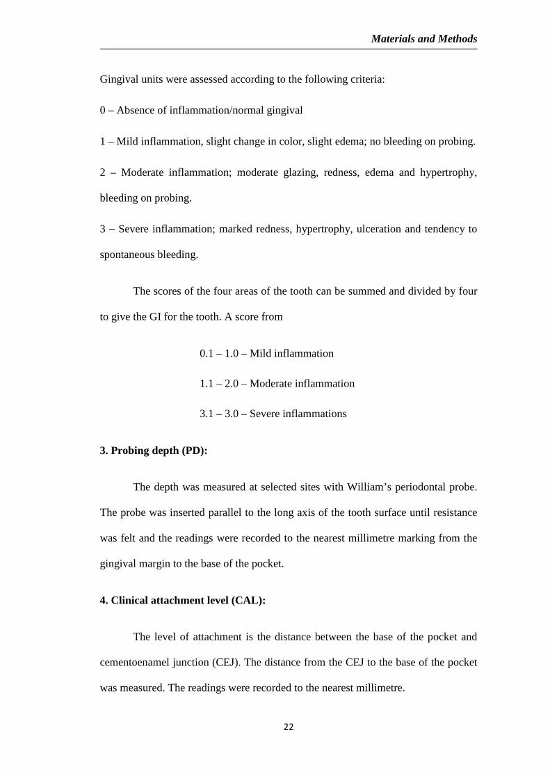
Gingival units were assessed according to the following criteria:
0 – Absence of inflammation/normal gingival
1 – Mild inflammation, slight change in color, slight edema; no bleeding on probing.
2 – Moderate inflammation; moderate glazing, redness, edema and hypertrophy,
bleeding on probing.
3 – Severe inflammation; marked redness, hypertrophy, ulceration and tendency to
spontaneous bleeding.
The scores of the four areas of the tooth can be summed and divided by four
to give the GI for the tooth. A score from
0.1 – 1.0 – Mild inflammation
1.1 – 2.0 – Moderate inflammation
3.1 – 3.0 – Severe inflammations
3. Probing depth (PD):
The depth was measured at selected sites with William’s periodontal probe.
The probe was inserted parallel to the long axis of the tooth surface until resistance
was felt and the readings were recorded to the nearest millimetre marking from the
gingival margin to the base of the pocket.
4. Clinical attachment level (CAL):
The level of attachment is the distance between the base of the pocket and
cementoenamel junction (CEJ). The distance from the CEJ to the base of the pocket
was measured. The readings were recorded to the nearest millimetre.
Materials and Methods
23
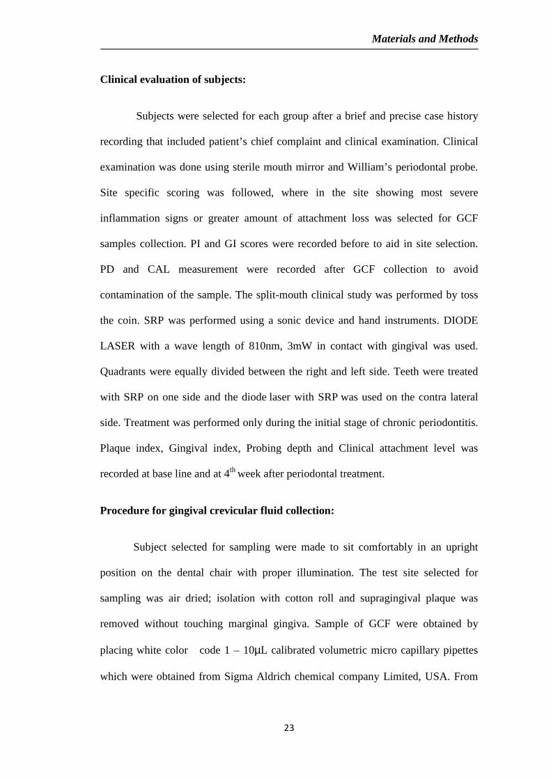
Clinical evaluation of subjects:
Subjects were selected for each group after a brief and precise case history
recording that included patient’s chief complaint and clinical examination. Clinical
examination was done using sterile mouth mirror and William’s periodontal probe.
Site specific scoring was followed, where in the site showing most severe
inflammation signs or greater amount of attachment loss was selected for GCF
samples collection. PI and GI scores were recorded before to aid in site selection.
PD and CAL measurement were recorded after GCF collection to avoid
contamination of the sample. The split-mouth clinical study was performed by toss
the coin. SRP was performed using a sonic device and hand instruments. DIODE
LASER with a wave length of 810nm, 3mW in contact with gingival was used.
Quadrants were equally divided between the right and left side. Teeth were treated
with SRP on one side and the diode laser with SRP was used on the contra lateral
side. Treatment was performed only during the initial stage of chronic periodontitis.
Plaque index, Gingival index, Probing depth and Clinical attachment level was
recorded at base line and at 4th week after periodontal treatment.
Procedure for gingival crevicular fluid collection:
Subject selected for sampling were made to sit comfortably in an upright
position on the dental chair with proper illumination. The test site selected for
sampling was air dried; isolation with cotton roll and supragingival plaque was
removed without touching marginal gingiva. Sample of GCF were obtained by
placing white color code 1 – 10µL calibrated volumetric micro capillary pipettes
which were obtained from Sigma Aldrich chemical company Limited, USA. From

Materials and Methods
24
each test sites, a standardized volume of 3µL was collected using the calibration on
the micropipette and placing the tip of the pipette extra crevicularly (unstimulated)
for 5 to 20 minutes. GCF contamination with saliva and blood were discarded.
Samples of GCF were collected at initial visit in Group I, Group II, Group
III, Group IV, Group V, Group VI subjects. Periodontal treatment (SRP and SRP +
LASER) was performed for diabetic with periodontitis patients at the same
appointment after GCF collection. After 1st and 4th week GCF was collected in
periodontally treated sites. The GCF collected was immediately transferred to plastic
vial and stored at -70°C till the time assay.
The samples were then assay for CALPROTECTIN levels by using Enzyme-
Linked Immunosorbent Assay kit.
METHOD OF ESTIMATION OF CALPROTECTIN:
The kit is a sandwich enzyme immunoassay for in vitro quantitative
measurement of calprotectin in human serum, plasma, tissue homogenates, cell
lysates and other biological fluids. The microtiter plate provided in this kit has been
precoated with a calprotectin. Standards or samples are then added to the appropriate
microtiter plate wells with a biotin-conjugated antibody specific to calprotectin.
Next, Avidin conjugated to Horseradish Peroxidase (HRP) is added to each
microplate well and incubated. After TMB substrate solution is added, only those
wells that contain calprotectin and enzyme conjugated avidin will exhibit a change
in color. The enzyme substrate reaction is terminated by the addition of sulphuric
acid solution and the colour change is measured spectrophotometrically at a
Materials and Methods
25
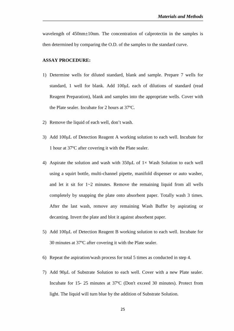
wavelength of 450nm±10nm. The concentration of calprotectin in the samples is
then determined by comparing the O.D. of the samples to the standard curve.
ASSAY PROCEDURE:
1) Determine wells for diluted standard, blank and sample. Prepare 7 wells for
standard, 1 well for blank. Add 100µL each of dilutions of standard (read
Reagent Preparation), blank and samples into the appropriate wells. Cover with
the Plate sealer. Incubate for 2 hours at 37ºC.
2) Remove the liquid of each well, don’t wash.
3) Add 100µL of Detection Reagent A working solution to each well. Incubate for
1 hour at 37ºC after covering it with the Plate sealer.
4) Aspirate the solution and wash with 350µL of 1× Wash Solution to each well
using a squirt bottle, multi-channel pipette, manifold dispenser or auto washer,
and let it sit for 1~2 minutes. Remove the remaining liquid from all wells
completely by snapping the plate onto absorbent paper. Totally wash 3 times.
After the last wash, remove any remaining Wash Buffer by aspirating or
decanting. Invert the plate and blot it against absorbent paper.
5) Add 100µL of Detection Reagent B working solution to each well. Incubate for
30 minutes at 37ºC after covering it with the Plate sealer.
6) Repeat the aspiration/wash process for total 5 times as conducted in step 4.
7) Add 90µL of Substrate Solution to each well. Cover with a new Plate sealer.
Incubate for 15- 25 minutes at 37ºC (Don't exceed 30 minutes). Protect from
light. The liquid will turn blue by the addition of Substrate Solution.

Materials and Methods
26
8) Add 50µL of Stop Solution to each well. The liquid will turn yellow by the
addition of Stop solution. Mix the liquid by tapping the side of the plate. If color
change does not appear uniform, gently tap the plate to ensure thorough mixing.
9) Remove any drop of water and fingerprint on the bottom of the plate and
confirm there is no bubble on the surface of the liquid. Then, run the microplate
reader and conduct measurement at 450nm immediately.

Materials and Methods
27
APPENDIX-1
PROFORMA
Op no: Date:
Name: Age/Sex:
Occupation: Address:
Chief complaint:
History of present illness:
Present medical history:
Past dental history:
Family history:
Materials and Methods
28
Pre operative clinical examination:
Plaque index: (Sillness P and Loe H 1964)
16 12 24
44 32 36
Score:
Interpretation:
Gingival index (Loe H & Sillness 1963)
16 12 24
44 32 36
Score:
Interpretation:
Scaling and root planing Scaling and root planing +
(Group II A) LASER (Group II B)
Materials and Methods
29
Group Group II A
Group II B
Periodontal pocket depth
Clinical attachment level
Investigation:
HB%: HBA1c:
Fasting glucose level:
Postprandial glucose level:
Diagnosis:
Post operative clinical examination:
Plaque index: (Sillness P and Loe H 1964)
16 12 24
44 32 36
Score:
Interpretation:

Materials and Methods
30
Gingival index (Loe H & sillness 1963 )
16 12 24
44 32 36
Score:
Interpretation:
Scaling and root planing Scaling and root planing +
(Group II A) LASER (Group II B)
Group Group II A Group II B
Periodontal pocket depth
Clinical attachment level
Materials and Methods
31
Consent form:
Department of Periodontics, JKK Nataraja Dental College,
Komarapalayam - 638183
Patient name
I have been explained about the nature and purpose of the study in which, I
have been asked to participate. I understand that I am free to withdraw my consent
and discontinue at any time without prejudice to me or effect on my treatment.
I have been given the opportunity to question about the material and study. I
have also given the consent for photographs to be taken at the beginning, during and
at the end of the study. I have fully agreed to participate in this study.
I hereby given the consent to be included in “EFFECT OF LASER
THERAPY ON GCF CALPROTECTIN LEVEL IN TYPE II DIABET IC
PATIENTS – A CLINICAL AND BIOCHEMICAL STUDY”
Station: Signature of the Patient
Date:
Signature of Professor/ HOD

Materials and Methods
32
APPENDIX - 2
ARMAMENTARIUM
1. Gloves
2. Mouth mask
3. Patient apron
4. Chair apron
5. Head cap
6. Sterile cotton rolls
7. Gauze
8. Kidney tray
9. Mouth mirror
10. Explorer
11. William’s periodontal probe
12. Tweezer
13. Ultrasonic scalers
14. Hu-Friedysupragingivalscalers
15. Hu-FriedyGracey Curettes
16. Microcapillary pipettes
17. Eppendorf tubes
Photographs
33
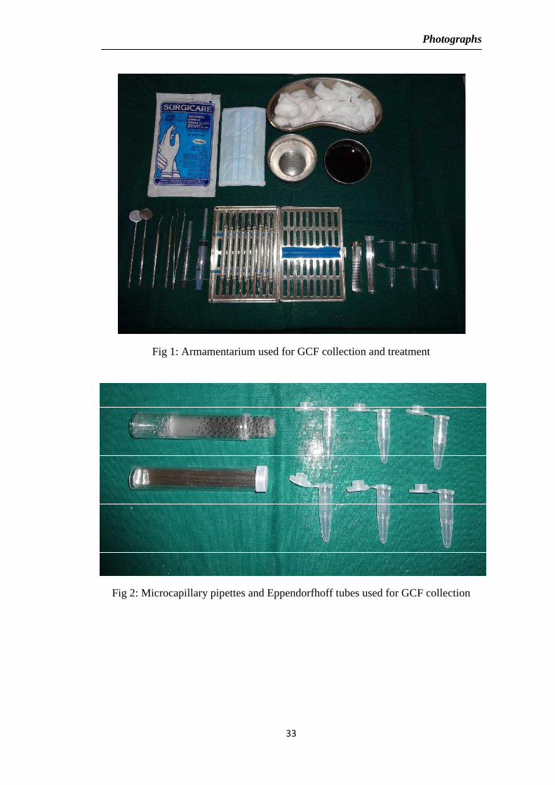
Fig 1: Armamentarium used for GCF collection and treatment
Fig 2: Microcapillary pipettes and Eppendorfhoff tubes used for GCF collection
Photographs
34
CLINICAL CASES
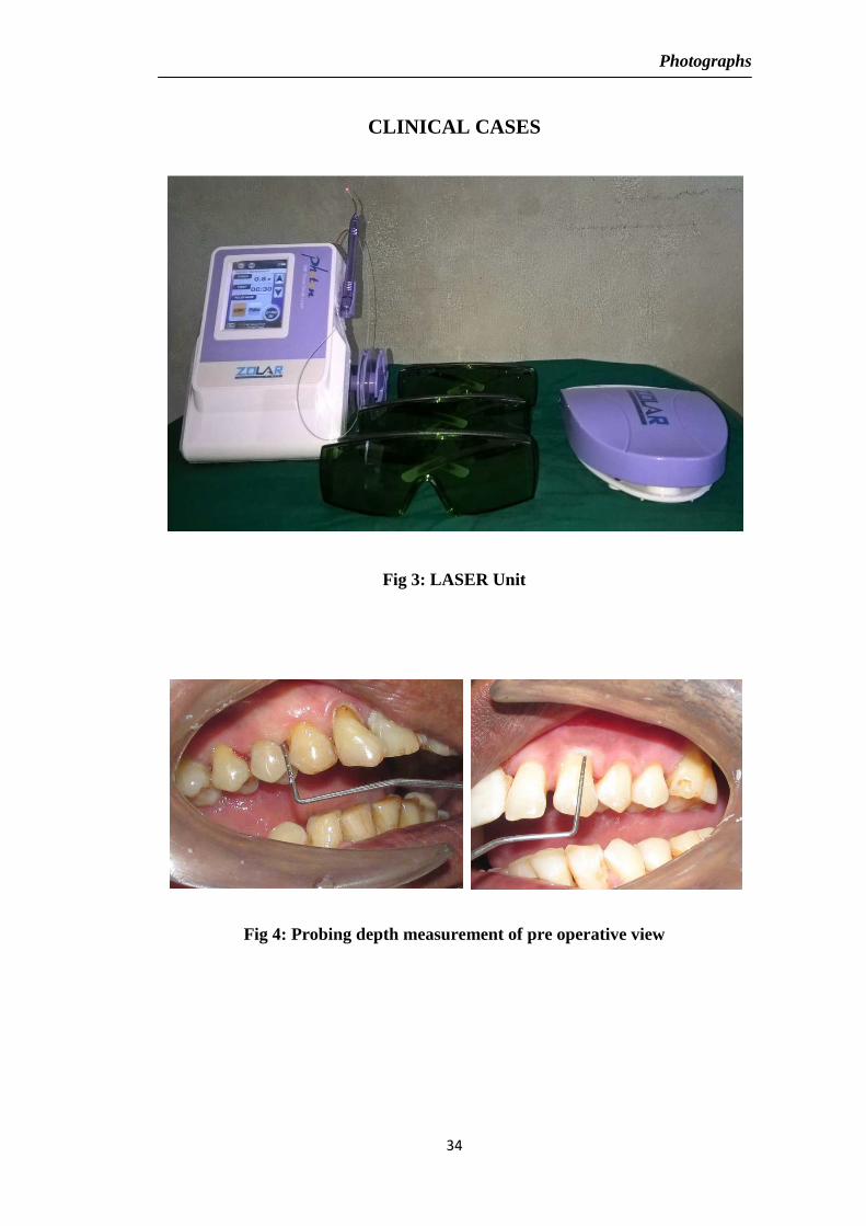
Fig 3: LASER Unit
Fig 4: Probing depth measurement of pre operative view

Photographs
35
Fig 5: GCF collection Fig 6: LASER Sulcular Debridement
Fig 7: probing depth measurement of post operative view

Photographs
36
LAB ANALYSIS
Fig 8: Calprotectin ELISA kit
Fig 9: ELISA PHOTOMETER Fig 10: ELISA ASSAY
Photographs
37
Results of ELISA
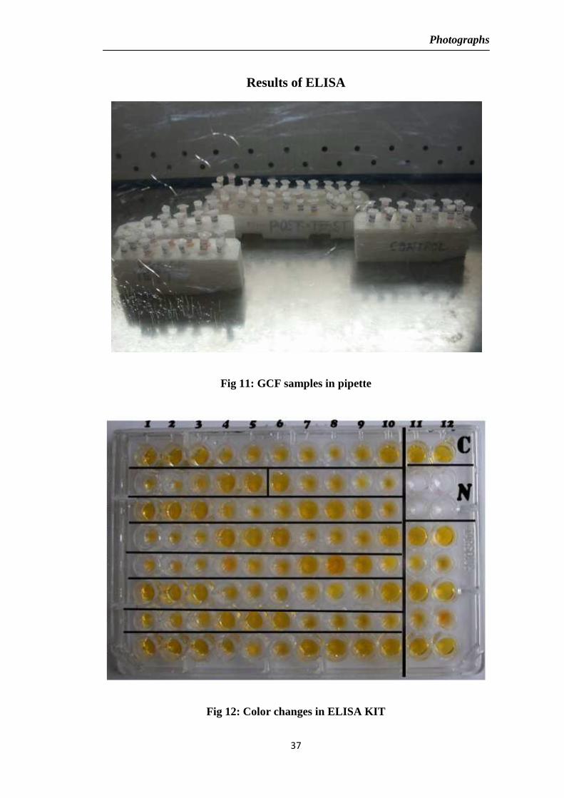
Fig 11: GCF samples in pipette
Fig 12: Color changes in ELISA KIT
Results
38
The present study was conducted to estimate the effect of laser therapy on
GCF calprotectin level in type II diabetic patients. Also to comparison was made
between the scaling root planing along with LASER to scaling root planing alone.
The randomized clinical study consist of 60 subjects divided into 2 groups i.e.,
healthy (Group I – 30 subjects), diabetic with chronic periodontitis (Group II – 30
subjects), aged between 25-65years from whom GCF was to collected to estimate the
calprotectin level in GCF using ELISA.
The result obtained were analysed statistically and comparisons were made
each group using paired student t – distribution test.
Comparison of clinical parameters:
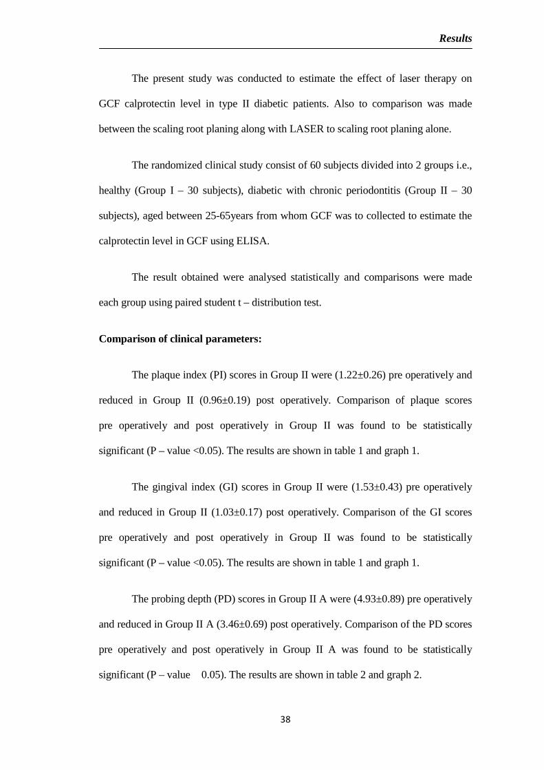
The plaque index (PI) scores in Group II were (1.22±0.26) pre operatively and
reduced in Group II (0.96±0.19) post operatively. Comparison of plaque scores
pre operatively and post operatively in Group II was found to be statistically
significant (P – value <0.05). The results are shown in table 1 and graph 1.
The gingival index (GI) scores in Group II were (1.53±0.43) pre operatively
and reduced in Group II (1.03±0.17) post operatively. Comparison of the GI scores
pre operatively and post operatively in Group II was found to be statistically
significant (P – value <0.05). The results are shown in table 1 and graph 1.
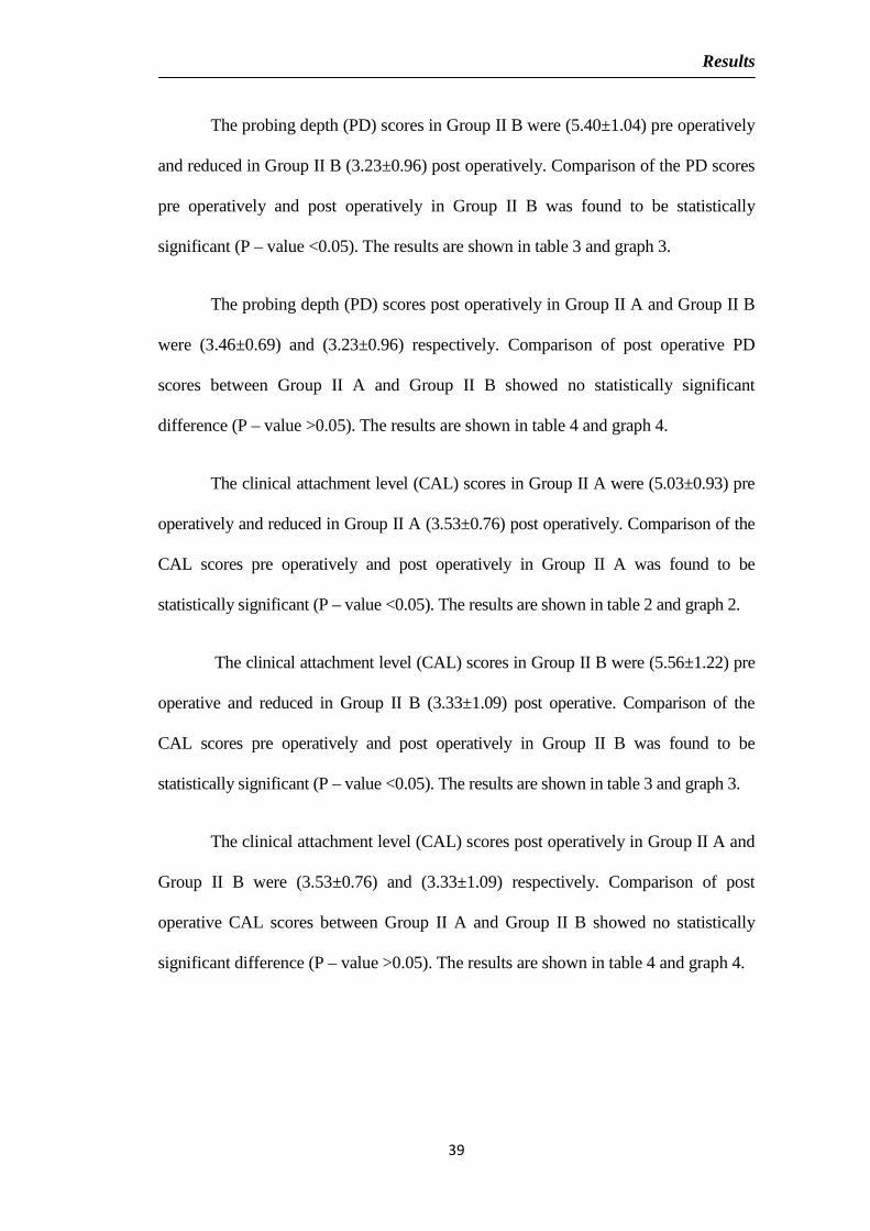
The probing depth (PD) scores in Group II A were (4.93±0.89) pre operatively
and reduced in Group II A (3.46±0.69) post operatively. Comparison of the PD scores
pre operatively and post operatively in Group II A was found to be statistically
significant (P – value �0.05). The results are shown in table 2 and graph 2.
Results
39
The probing depth (PD) scores in Group II B were (5.40±1.04) pre operatively
and reduced in Group II B (3.23±0.96) post operatively. Comparison of the PD scores
pre operatively and post operatively in Group II B was found to be statistically
significant (P – value <0.05). The results are shown in table 3 and graph 3.
The probing depth (PD) scores post operatively in Group II A and Group II B
were (3.46±0.69) and (3.23±0.96) respectively. Comparison of post operative PD
scores between Group II A and Group II B showed no statistically significant
difference (P – value >0.05). The results are shown in table 4 and graph 4.
The clinical attachment level (CAL) scores in Group II A were (5.03±0.93) pre
operatively and reduced in Group II A (3.53±0.76) post operatively. Comparison of the
CAL scores pre operatively and post operatively in Group II A was found to be
statistically significant (P – value <0.05). The results are shown in table 2 and graph 2.
The clinical attachment level (CAL) scores in Group II B were (5.56±1.22) pre
operative and reduced in Group II B (3.33±1.09) post operative. Comparison of the
CAL scores pre operatively and post operatively in Group II B was found to be
statistically significant (P – value <0.05). The results are shown in table 3 and graph 3.
The clinical attachment level (CAL) scores post operatively in Group II A and
Group II B were (3.53±0.76) and (3.33±1.09) respectively. Comparison of post
operative CAL scores between Group II A and Group II B showed no statistically
significant difference (P – value >0.05). The results are shown in table 4 and graph 4.

Results
40
Comparison of biochemical parameters:
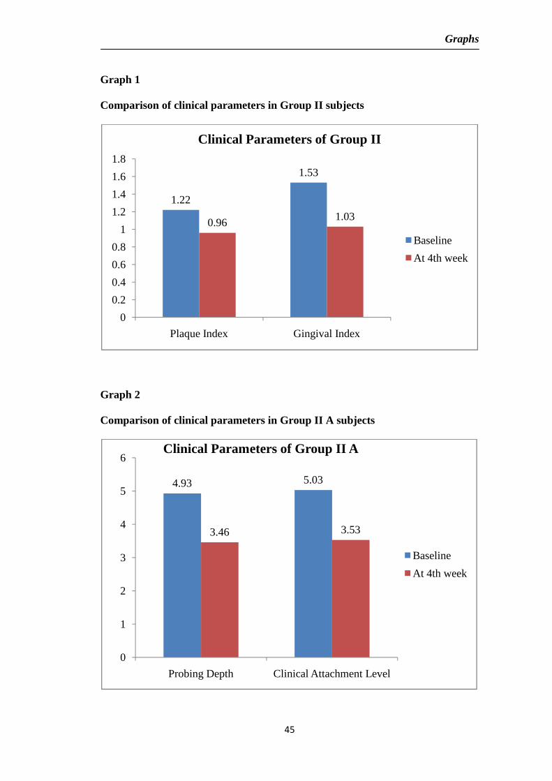
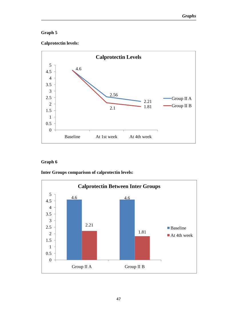
The GCF Calprotectin level in Group II A was (4.60±1.63) pre operatively
and reduced to (2.21±0.39) 4th week post operatively in Group II A. Comparison of
GCF Calprotectin level preoperatively and 4th week post operatively in Group II A
showed statistically significant difference (P – value of <0.05). The results are
shown in table 6 and graph 6.
The GCF Calprotectin level in Group II B was (4.60±1.63) pre operatively
and reduced to (1.81±0.24) 4th week post operatively in Group II B. Comparison of
GCF Calprotectin level preoperatively and 4th week post operatively in Group II B
showed statistically significant difference (P–value of <0.05). The results are shown
in table 6 and graph 6.
The GCF Calprotectin level 4th week post operatively was significantly
reduced in Group II B (1.81±0.24) compared to Group II A (2.21±0.39).
Comparison of GCF Calprotectin level 4th week post operatively between Group II
A and Group II B showed statistically significant difference (P – value <0.05). The
results are shown in table 7 and graph 7.
In summary, the results suggest that there is a statistically significant
difference between SRP and SRP + LASER Groups preoperatively and post
operatively in relation to Plaque index, Gingival index, Probing depth, Clinical
attachment level scores. The Calprotectin level was significantly reduced post
operatively in both SRP and SRP + LASER groups. There is statistically significant
difference among SRP and SRP + LASER groups in Calprotectin level.

Tables
41
Table 1
Comparison of clinical parameters in Group II subjects
Clinical parameters of Group II Base line At 4th week P - value
Plaque index 1.22±0.26 0.96±0.19 <0.05
Gingival index 1.53±0.43 1.03±0.17 <0.05
In Group II the P- Value between base line and 4th week post operative is
<0.05 (denotes statistically significant).
Table 2
Comparison of clinical parameters in Group II A subjects
In Group II A the P- Value between base line and 4th week post operative is
<0.05 (denotes statistically significant).
Clinical parameters of Group II A Base line At 4th week P - value
Probing depth 4.93±0.89 3.46±0.69 <0.05
Clinical attachment level 5.03±0.93 3.53±0.76 <0.05

Tables
42
Table 3
Comparison of clinical parameters in Group II B subjects
Clinical parameters of Group II B Base line At 4th week P value
Probing depth 5.40±1.04 3.23±0.96 <0.05
Clinical attachment level 5.56±1.22 3.33±1.09 <0.05
In Group II B the P- Value between base line and 4th week post operative is
<0.05 (denotes statistically significant).
Table 4
Comparison of clinical parameters in group II A and B at 4th week post
operative
Clinical parameter Group II A Group II B P Value
Probing depth 3.46±0.69 3.23±0.96 >0.05
Clinical attachment level 3.53±0.76 3.33±1.09 >0.05
P-Value between Group IIA and Group II B at 4th week post operative is
>0.05 (denotes statistically no significant).

Tables
43
Table 5
Calprotectin levels:
Groups Base line At 1st week At 4th week
II A 4.60±1.63 2.56±0.48 2.21±0.39
II B 4.60±1.63 2.10±0.32 1.81±0.24
Table 6
Inter Group comparison of Calprotectin levels:
Groups Base line At 4th week P value
II A 4.60±1.63 2.21±0.39 <0.05
II B 4.60±1.63 1.81±0.24 <0.05
Inter Group P-Value between Group IIA and Group II B in base line and at
4th week post operative is <0.05 (denotes statistically significant).

Tables
44
Table 7
Intra Group comparison of Calprotectin levels:
Periods II A II B P Value
At 1st week 2.56±0.48 2.10±0.32 <0.05
At 4th weeks 2.21±0.39 1.81±0.24 <0.05
Intra Group P-Value between Group IIA and Group II B in base line and at
4th week post operative is <0.05 (denotes statistically significant).
Graphs
45
Graph 1
Comparison of clinical parameters in Group II subjects
Graph 2
Comparison of clinical parameters in Group II A subjects
1.22
1.53
0.96 1.03
0
0.2
0.4
0.6
0.8
1
1.2
1.4
1.6
1.8
Plaque Index Gingival Index
Clinical Parameters of Group II
Baseline
At 4th week
4.93 5.03
3.46 3.53
0
1
2
3
4
5
6
Probing Depth Clinical Attachment Level
Clinical Parameters of Group II A
Baseline
At 4th week

Graphs
46
Graph 3
Comparison of clinical parameters in Group II B subjects
Graph 4
Comparison of clinical parameters in group II A and B at 4th week post
operative
5.4 5.56
3.23 3.33
0
1
2
3
4
5
6
Probing Depth Clinical Attachment Level
Clinical Parameters of Group II B
Baseline
At 4th week
3.46
3.53
3.23
3.33
3.053.1
3.153.2
3.253.3
3.353.4
3.453.5
3.553.6
Probing Depth Clinical Attachment Level
Clinical Parameters of Group II A & II B
Group II A
Group II B
Graphs
47
Graph 5
Calprotectin levels:
Graph 6
Inter Groups comparison of calprotectin levels:
4.6
2.56
2.21
2.1 1.81
0
0.5
1
1.5
2
2.5
3
3.5
4
4.5
5
Baseline At 1st week At 4th week
Calprotectin Levels
Group II A
Group II B
4.6 4.6
2.21
1.81
0
0.5
1
1.5
2
2.5
3
3.5
4
4.5
5
Group II A Group II B
Calprotectin Between Inter Groups
Baseline
At 4th week

Graphs
48
Graph 7
Intra Groups comparison of calprotectin levels:
2.56
2.212.11.81
0
0.5
1
1.5
2
2.5
3
At 1st week At 4th week
Comparison of Calprotectin between Intra Groups
II A
II B
Discussion
49
Periodontal diseases are a complex group of diseases characterized by
inflammation and the subsequent destruction of the tooth supporting tissue. The
destruction of the periodontal tissue is by bacterial infection and host immune
response, either directly or indirectly by various mediators that can activate
osteoclastic activity. As GCF permeates through the diseased soft tissue of the
periodontal pocket, it contains molecules from the periodontal disease process and it
is considered the most promising source of biochemical indicators like
calprotectin.13
According to Striz et al 200445, Calprotectin complex also known as L1
antigen, calgranulin A and B, macrophage migration inhibitory factor-related protein
8 and 14 (MRP8 and MRP14), S100A8/S100A9, and cystic fibrosis antigen, has
several functions in inflammatory reactions. It acts as a chemotactic factor and
regulates adhesion and migration of neutrophils or monocytes and is therefore
considered as a pro-inflammatory marker.
According to Kido J 199913 Higher levels of MRP 8 were detected in sites
with periodontitis and gingivitis than in healthy sites.
Our study comprised of 2 groups (healthy, diabetic with chronic
periodontitis) compared to that of Yukari K et al 20147 which included patients
with DM, chronic periodontitis, DM-P, and healthy individuals, but having no
provision to evaluate the effect of LASER on calprotectin levels in GCF which can
confirm the role of calprotectin in periodontal disease.

Discussion
50
In our study, plaque index and gingival index were examined pre operatively
and 4th week post operatively for all patients. The pre operative value of plaque
index and gingival index [(1.22±0.26) and (1.53±0.43) respectively] was
significantly reduced to [(0.96±0.19) and (1.03±0.17) respectively] postoperatively.
Probing depth and Clinical attachment level were measured in Group II A
pre operatively and 4th week post operatively. The 4th week post operative valve of
Probing depth and Clinical attachment level [(3.46±0.69) and (3.53±0.76)
respectively] was significantly reduced compared to pre operative valves
[(4.93±0.89) and (5.03±0.93) respectively] in Group II A.
Probing depth and Clinical attachment level was measured pre operatively
and 4th week post operatively in Group II B. The 4th week post operative valve of
Probing depth and Clinical attachment level [(3.23±0.96) and (3.33±1.09)
respectively] was significantly reduced compared to pre operative valves
[(5.40±1.04) and (5.56±1.22) respectively] in Group II B.
Among the groups, the probing depth and clinical attachment level values for
Group II B 4th week post operatively was significantly better. Dana V et al48
demonstrated that the application of low level laser therapy (LLLT), in addition to
standard procedures employed to treat periodontal disease, improved the outcome of
the treatment. And also had improved healing times, less pain, less bleeding, less
post surgery complications such as edema, inflammation, and infection compared
with the classic treatment only group.
Rozana D200549, demonstrated that local and general circulation of blood is
reinforced on laser. Therefore the wound healing is better and inflammation is
Discussion
51
reduced in Group II B 4th week post operative compared to Group II A 4th week post
operative group.
In our study, the mean concentration of Calprotectin in DM-P were reduced
from 4.60±1.63 µg/ml (Group II A) to 2.56±0.48 (Group II A 1st week post
operative), and 2.21±0.39 (Group II A 4th week post operative).
The value of Calprotectin in Group II B 4.60±1.63 µg/ml were reduced to
2.10±0.34 (Group II B 1st week post operative), and 1.81±0.24 (Group II B 4th week
post operative).
The study by Yukari K et al 20147 reported that the total amount of
calprotectin in GCF samples from periodontitis cases were significantly higher than
that from cases without periodontitis, but the calprotectin concentration in
periodontitis samples was slightly lower. This difference between calprotectin
amount and concentration was caused by the increase of GCF volume in
periodontitis. The calprotectin amount and concentration were not affected by DM,
and there was no significant correlation between GCF calprotectin level and HbA1c
value.
Suryono et al 200350 showed that MRP8/MRP14 existed in the epithelium
and connective tissue of a periodontitis patient’s gingival using the monoclonal
antibody 27E10, a macrophage marker that recognizes MRP8/MRP14 and suggested
that macrophage in the connective tissue and epithelium cells expressed these
proteins.

Discussion
52
Kido J 199913 demonstrated that the calprotectin level in GCF is positively
related to clinical indicators (probing depth and BOP) and levels of known
biochemical markers (IL-1b and PGE2) of periodontal disease, and it reflects the
degree of gingival inflammation in patients with periodontal disease. Although
Armitage 199610 showed that probing depth does not provide an accurate indication
of the current severity of periodontal inflammation, eventhough it has been widely
used to evaluate periodontal disease.
The main findings in our study was that the mean concentration of
calprotectin in GCF increased progressively from healthy to periodontitis which was
in accordance with that of study done by Kido J et al13 who reported increasing
calprotectin levels in GCF with progression of periodontal disease.
The correlation of Calprotectin concentration with four clinical parameters of
periodontal status was investigated. The Calprotectin level was positively correlated
with PI and GI. This indicated that the Calprotectin levels were associated with the
degree of periodontal tissue inflammation.
Moreover the Calprotectin concentration was positively correlated with the
PD and CAL. This indicated that the Calprotectin levels were positively correlated
with the degree of periodontal tissue destruction.

Summary and Conclusion
53
Some of the conclusions that can be drawn from this study are:
1) Calprotectin is detected in gingival crevicular fluid and its concentration is
found to be higher in patients with chronic periodontitis with diabetic
mellitus.
2) Treatment of periodontal diseases by scaling and root planing led to a
significant reduction in Calprotectin levels in GCF. There was a statistically
significant reduction more in LASER plus scaling and root planing Group
when compared to SRP alone.
3) Further, as the concentration of Calprotectin correlated positively with the
clinical parameters, it can be postulated that Calprotectin is actively involved
in the progression of periodontal disease and thus could be a marker of
periodontal destruction.
To conclude, as per our study, it can be postulated that greater the
periodontal destruction, there is substantial increase in Calprotectin concentration in
GCF.

Bibliography
54
1) Brian I. Mealey & Gloria I. Diabetes mellitus and Periodontal disease.
Periodontology 2000, Vol. 44, 2007, 127–153.
2) Hamdy Nassar, Alpdogan Kantarci & Thomas E. Van Dyke. Diabetic
periodontitis: a model for activated innate immunity and impaired resolution of
Inflammation. Periodontology 2000, Vol. 43, 2007, 233–244.
3) Sun X, Meng H, Shi D, Xu L, Zhang L, Chen Z, Feng X, Lu R. Analysis of
plasma calprotectin and polymorphisms of S100A8 in patients with aggressive
Periodontitis. J Periodont Res 2011; 46: 354–360.
4) Satoru Yui, Yuichi Nakatani, and Masaaki Mikami. Calprotectin
(S100A8/S100A9), an Inflammatory Protein Complex from Neutrophils with a
Broad Apoptosis-Inducing Activity. Pharmaceutical Society of Japan March
2003, Vol. 26, No. 6, 754.
5) Fabrizio Sgolastra & Marco Severino & Roberto Gatto & Annalisa
Monaco. Effectiveness of diode laser as adjunctive therapy to scaling root
planning in the treatment of chronic periodontitis:a meta-analysis. Lasers Med
Sci; 30 July 2012.
6) Qadri T, Miranda L, Tuner J, Gustafsson A. The short-term effects of low-
level lasers as adjunct therapy in the treatment of periodontal Inflammation.
J Clin Periodontol 2005; 32: 714–719.
Bibliography
55
7) Yukari Kajiura, Mika Bando, Yuji Inagaki, Toshihiko Nagata, and
Junichi Kido. Glycated Albumin and Calprotectin Levels in Gingival
Crevicular Fluid from Patients with Periodontitis and Type 2 Diabetes.
J Periodontol 2014; 85:1667-1675.
8) George W. Taylor, Brian A. Burt, Mark P. Becker, Robert J. Genco,
Marc Shlossman, William C. Knowler, and David J. Pettitt. Severe
Periodontitis and Risk for Poor Glycemic Control in Patients with Non-
Insulin-Dependent Diabetes Mellitus. Journal of Periodontology; 96; 67;1085-
1093.
9) Steven P. Engebretson, Judith Hey-Hadavi, Fernando J. Ehrhardt, Dan
Hsu. Gingival crevicular fluid level of interleukin-1β and Glycemic control in
patients with chronic periodontitis and type II diabetes. Journal of
periodontology; 2004, 75, 1203-1208.
10) Guglielmo Campus, Abeer Salem, Sergio Uzzau, Edoardo Baldoni, and
Giancarlo Tonolo. Diabetes and Periodontal Disease: A Case-Control Study.
J Periodontol 2005; 76: 418-425.
11) Katz J, Bhattacharyya I, Farkhondeh-Kish F, Perez FM, Caudle RM,
Heft MW. Expression of the receptor of advanced glycation end products in
gingival tissues of type II diabetes patients with chronic periodontal disease: a
study utilizing immunohistochemistry and RT-PCR. J Clin Periodontol 2005;
32: 40–44.

Bibliography
56
12) Kenneth T. Miyasaki, Alexandra Voganatsi, Thuc Huynh, Marvin
Marcus, and Steven Underwood. Calprotectin and Lactoferrin Levels in the
Gingival Crevicular Fluid of Children. Journal of Periodontology; 1998;
69:879-883.
13) Kido Jun-ichi, Nakamura T, Kido R, Ohishi K, Yamauchi N, Kataoka M,
Nagata T. Calprotectin in gingival crevicular fluid correlates with clinical and
biochemical markers of periodontal disease. J Clin Periodontol 1999; 26:
653–657.
14) Teruo Nakamura, Jun-ichi-Kido, Reiko Kido, Keijo Ohishi, Noriyoki
Yamachi, Masatoshi Kataoka. The association of calprotectin level in
Gingival crevicular fluid with Gingival index and the activities of collagenase
and aspartate aminotransferase in adult periodontitis patients. Journal of
Periodontol: 2000; 71;361-367.
15) Nisapakultorn K. Ross K.F and Herzberg M. C. Calprotectin Expression
In Vitro by Oral Epithelial Cells Confers Resistance to Infection by
Porphyromonas gingivalis. Infect Immun. 2001 Jul; 69(7):4242-7.
16) Kanokwan Nisapakultorn, Karen F. Ross, and Mark C. Herzberg.
Calprotectin Expression Inhibits Bacterial Binding to Mucosal Epithelial Cells.
Infect Immun. 2001 Jun; 69(6): 3692–3696.

Bibliography
57
17) Kido J, Kido R, Suryono, Kataoka M, Fagerhol MK, Nagata T.
Calprotectin release from human neutrophils is induced by Porphyromonas
gingivalis lipopolysaccharide via the CD-14–Toll-like receptor–nuclear factor
κB Pathway. J Periodont Res 2003; 38; 557–563.
18) Que ML, Andersen E, Mombelli A. Myeloid-related protein (MRP) 8/14
(calprotectin) and its subunits MRP8 and MRP14 in Plaque - induced early
gingival inflammation. J Clin Periodontol 2004; 31: 978–984.
19) Suryono, Jun-Ichi Kido, Noriko Hayashi, Masatoshi Kataoka. Calprotectin
in human monocytes: induction by Porphymonas gingivalis
lipopolysaccharide, Tumor necrosis factor-α and Interleukin-1β. J Periodontol
2005; 76; 437-442.
20) Kaner D. W. Hopfenmüller, J.P. Bernimoulin, B.M. Kleber, and A.
Friedmann. Calprotectin in GCF as a predictive biomarker in periodontal
therapy. Periodontal Research: Diagnosis–Neuroscience September 28, 2007.
21) Kaner D; Bernimoulin JP; Dietrich T; Kleber BM; Fri edmann A.
Calprotectin levels in gingival crevicular fluid predict disease activity in
patients treated for generalized aggressive periodontitis. J Periodontal Res.
2011 Aug; 46 (4):417-26.
22) Sema Becerika, Beral Afacana, Veli O zgen O zturka, Harika Atmacab
and Gulnur Emingila. Gingival crevicular fluid calprotectin, osteocalcin and
cross-linked N-terminal telopeptid levels in health and different periodontal
diseases. Disease Markers 31 (2011) 343–352.

Bibliography
58
23) Gareths, Griffiths. Formation, collection and significance of gingival crevice
Fluid. Periodontology 2000, Vol. 31, 2003, 32–42.
24) Gary C, Armitage. Analysis of gingival crevice fluid and risk of progression
of periodontitis. Periodontology 2000, vol.34, 2004, 109-119.
25) Pashley DH. A mechanistic analysis of gingival fluid production. J Periodont
Res 1976; 11: 121-134.
26) Curtis MA, Griffiths GS, Price SJ, Coulthurst SK, Johnson NW. The total
protein concentration of gingival crevicular fluid. Variation with sampling
time and gingival inflammation. J Clin Periodontol 1988; 15: 628-32.
27) Veli- Jukka Vitto. Gingival crevice fluid – an introduction. Periodontology
2000,Vol. 31, 2003, 9–11.
28) Catherine. M.E Champagne, William. Buchanan, Michael S. Reddy, John
S- Preisser. Potential for gingival crevice fluid measures as predictors of risk
for periodontal diseases. Periodontology 2000, Vol. 31, 2003, 167–180.
29) Isao Ishikawa, Akira Aoki, Aristeo A. Takasaki, Koji Mizutani, Katia M.
Sasaki & Yuichi Izumi. Application of lasers in periodontics: true innovation
or myth? Periodontology 2000, Vol. 50, 2009, 90–126.
30) Walsh L. J. The current status of low level laser therapy in dentistry. Part 1.
Soft tissue applications. Australian Dental Journal 1997; 42 :(4):247-54.

Bibliography
59
31) Eberhard J, Ehlers H, Falk W, Acil Y, Albers H-K, Jepsen S. Efficacy of
subgingival calculus removal with Er:YAG laser compared to mechanical
debridement: an in situ study. J Clin Periodontol 2003; 30: 511–518.
32) Charles M. Cobb. Lasers in Periodontics: A Review of the Literature.
J Periodontol 2006; 77: 545-564.
33) Krause F, Braun A, Brede O, Eberhard J, Frentzen M, Jepsen S.
Evaluation of selective calculus removal by a fluorescence feedback-controlled
Er:YAG laser in vitro.J Clin Periodontol 2007; 34: 66–71.
34) Vanja vucicevic Boras, Danica Vidovic Juras, Ana Andabak Rogulj,
Dragana Gabric Panduric, Zeljko Verzak and Vlaho Brailo. Applications
of Low Level Laser Therapy. A Textbook of Advanced Oral and Maxillofacial
Surgery; Chapter 12.
35) Carlos de Paula Eduardo & Patricia Moreira de Freitas & Marcella
Esteves-Oliveira & Ana Cecília Corrêa Aranha & Karen Müller Ramalho
& Alyne Simões & Marina Stella Bello-Silva & Jan Tunér. Laser
phototherapy in the treatment of periodontal disease. A review. Lasers Med Sci
17 july 2010.
36) Gokce Aykol, Ulku Baser, Ilay Maden, Zafer Kazak, Utku Onan, Sevda
Tanrikulu-Kucuk;Evin Ademoglu, Halim Issever, and Funda Yalcin. The
Effect of Low-Level Laser Therapy as an Adjunct to Non-Surgical Periodontal
Treatment. J Periodontol 2011;82:481-488.

Bibliography
60
37) Medicinski fakultet, Klinika za stomatologiju, Odeljenje za oralnu
medicinu I parodontologiju, Nis, Srbija. Low power laser efficacy in the
therapy of inflamed gingive in diabetics with parodontopathy. Vojnosanit
Pregl. 2011 Aug;68(8):684-9.
38) American Academy of Periodontology. Statement on the Efficacy of Lasers
in the Non-Surgical Treatment of Inflammatory Periodontal Disease.
J Periodontol • April 2011, Volume 82 • Number 4.
39) Radmila Obradović, Ph.D, Ljiljana Kesić, Ph.D, Dragan Mihailović, Ph.D,
Goran Jovanović, Ph.D, Slobodan Antić, Ph.D, and Zlata Brkić, Ph.D.
Low-Level Lasers as an Adjunct in Periodontal Therapy in Patients with
Diabetes Mellitus. Diabetes Technol Ther. 2012 Sep; 14(9): 799–803.
40) Catherine Giannopoulou, Isabelle Cappuyns, Jose Cancela, Norbert
Cionca, and Andrea Mombelli. Effect of Photodynamic Therapy, Diode
Laser, and Deep Scaling on Cytokine and Acute-Phase Protein Levels in
Gingival Crevicular Fluid of Residual Periodontal Pockets. J Periodontol
2012;83:1018-1027.
41) Mellitus Radmila Obradovic Ljiljana Kesic Dragan Mi hailovic Goran
Jovanovic Slobodan Antic and Zlata Brkic. Low-Level Lasers as an Adjunct
in Periodontal Therapy in Patients with Diabetes. Diabetes technology &
therapeutics volume 14, number 9, 2012; 799-803.

Bibliography
61
42) Soo L, Leichter JW, Windle J, Monteith B, Williams SM, Seymour GJ. A
comparison of Er:YAG laser and mechanical debridement for the non-surgical
treatment of chronic periodontitis: A randomized, prospective clinical study.
J Clin Periodontol 2012; 39: 537–545.
43) Walter Dukic, Ivona Bago, Andrej Aurer, and Marija Roguljic. Clinical
Effectiveness of Diode Laser Therapy as an Adjunct to Non-Surgical
Periodontal Treatment: A Randomized Clinical Study. J Periodontol 2013; 84:
1111-1117.
44) Jeffrey D. Pope, DDS, MS, Jeffrey A. Rossmann, DDS, MS, David
G. Kerns, DMD, MS, M.Miles Beach, DMD, MS, Daisha J. Cipher, PhD.
Use of the Carbon Dioxide Laser as an Adjunct to Scaling and Root Planing
for Clinical New Attachment: A Case Series. Clinical Advances in
Periodontics,vol-4,nov 2014, 209-215.
45) Striz Trebichavský. Calprotectin – a Pleiotropic Molecule in Acute and
Chronic Inflammation. Physiol. Res. 53: 245-253, 2004.
46) Andersen E, Dessaix IM, Perneger T, Mombelli A. Myeloid-related protein
(MRP8/14) expression in gingival crevice fluid in periodontal health and
disease and after treatment. J Periodont Res 2010; 45: 458–463.
47) Giannopoulou C, Andersen E. Brochut P, Plagnat D, and Mombelli A.
Enamel Matrix Derivative and Systemic Antibiotics as Adjuncts to Non-
Surgical Periodontal Treatment: Biologic Response. J Periodontol 2006; 77:
707-713.

Bibliography
62
48) Dana Vieru DDS, MS, phd, Martha Cortez DDS, Lewis Clayman MD,
DDS, and Anca Silvia Dumitriu DDS, MS, phd. Low level laser therapy in
the treatment of periodontal disease compared study diabetics, osteoporotic
patients, hepatic disease patients and healthy ones. Emergency dentistry,
London 2015, Oct 3, 1741-1749.
49) Rozana Dana vieru, Agafita Lefter and Sonia Herman. Effect of adjunct
LASER photo biomodulation in the classical treatment of periodontal disease.
Laser therapy; 2005, 14, 4, 161-169.
50) Suryono, Jun-ichi-kido, Noriko H. Effect of porphyromonas gingivalis
lipopolysaccharides, tumour necrosis factor-α and interleukin- 1β on
calprotectin release in human monocytes. J Periodontol 2003;74:1719-1724.