effect of infection kinetics on trophoblast cell …needle. the brain preparations were further...
TRANSCRIPT
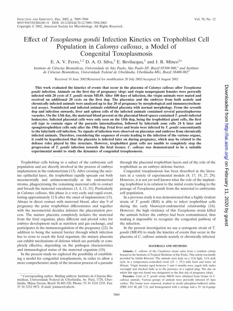
INFECTION AND IMMUNITY, Dec. 2002, p. 7089–7094 Vol. 70, No. 120019-9567/02/$04.00�0 DOI: 10.1128/IAI.70.12.7089–7094.2002Copyright © 2002, American Society for Microbiology. All Rights Reserved.
Effect of Toxoplasma gondii Infection Kinetics on Trophoblast CellPopulation in Calomys callosus, a Model of
Congenital ToxoplasmosisE. A. V. Ferro,1,2 D. A. O. Silva,2 E. Bevilacqua,1 and J. R. Mineo2*
Instituto de Ciencias Biomedicas, Universidade de Sao Paulo, Sao Paulo-SP, Brazil 05508-900,1 and Institutode Ciencias Biomedicas, Universidade Federal de Uberlandia, Uberlandia-MG, Brazil 38400-9022
Received 10 June 2002/Returned for modification 20 July 2002/Accepted 24 August 2002
This work evaluated the kinetics of events that occur in the placenta of Calomys callosus after Toxoplasmagondii infection. Animals on the first day of pregnancy (dop) and virgin nonpregnant females were perorallyinfected with 20 cysts of T. gondii strain ME49. After 100 days of infection, the virgin animals were mated andreceived an additional 20 cysts on the first dop. The placentas and the embryos from both acutely andchronically infected animals were analyzed up to day 20 of pregnancy by morphological and immunocytochem-ical assays. Noninfected and infected animals exhibited placenta with normal morphology. From the seventhdop and infection onwards, liver and spleen cells of the infected animals contained several parasitophorousvacuoles. On the 13th day, the maternal blood present at the placental blood spaces contained T. gondii-infectedleukocytes. Infected placental cells were only seen on the 15th dop, being the trophoblast giant cells, the firstcell type to contain signs of the parasite internalization, followed by labyrinth zone cells 24 h later andspongiotrophoblast cells only after the 19th dop. Fetal liver and brain were infected by T. gondii concomitantlyto the labyrinth cell infection. No signals of infection were observed on placentas and embryos from chronicallyinfected animals. Therefore, considering the sequence of events leading to the infection of the various organs,it could be hypothesized that the placenta is infected later on during pregnancy, which may be related to thedefense roles played by this structure. However, trophoblast giant cells are unable to completely stop theprogression of T. gondii infection towards the fetal tissues. C. callosus was demonstrated to be a suitableexperimental model to study the dynamics of congenital toxoplasmosis.
Trophoblast cells belong to a subset of the embryonic cellpopulation and are directly involved in the process of embryoimplantation in the endometrium (13). After crossing the uter-ine epithelial layer, the trophoblast rapidly spreads out bothmesometrially and antimesometrially at the endometrialstroma, phagocytosing the remaining maternal cells to contactand breach the maternal vasculature (3, 4, 13, 31). Particularlyin Calomys callosus, this phase is a very early and rapid event,lasting approximately 12 h after the onset of implantation (15).Always in direct contact with maternal blood, after day 9 ofpregnancy the polar trophoblast differentiates and togetherwith the mesometrial decidua initiates the placentation pro-cess. The mature placenta completely isolates the maternalfrom the fetal organism, plays different and pivotal roles forembryo development such as nutrition and gas exchange, andparticipates in the immunoregulation of the pregnancy (22). Inaddition to being the natural barrier through which infectionhas to cross to reach the fetal organism, the mature placentacan exhibit mechanisms of defense which are partially or com-pletely effective, depending on the pathogen characteristicsand immunological status of the maternal organism (10).
In the present study we explored the possibility of establish-ing a model for congenital toxoplasmosis, in order to allow amore comprehensive study of the invasion process of a parasite
through the placental trophoblast layers and of the role of thetrophoblast as an embryo defense barrier.
Congenital toxoplasmosis has been described in the litera-ture in a variety of experimental models (8, 17, 19, 27, 29).However, it is not yet well known what the role of the implant-ing trophoblast is in relation to the initial events leading to thepassage of Toxoplasma gondii from the maternal to embryoniccell population.
In a previous study it was demonstrated that a highly virulentstrain of T. gondii (RH) is able to infect trophoblast cellsduring the early blastocyst-endometrial relationship (16).However, the high virulence of this Toxoplasma strain killedthe animals before the embryo had been contaminated, thusmaking it impossible to recognize the congenital pathway ofthe infection.
In the present investigation we use a cystogenic strain of T.gondii (ME49) to study the kinetics of events that occur in theplacenta of C. callosus animals acutely or chronically infected.
MATERIALS AND METHODS
Animals. C. callosus of the Canabrava strain came from a resident colonyhoused at the Institute of Tropical Medicine of Sao Paulo. This colony was kindlyprovided by Judith Kloetzel. The animals were kept on a 12-h light, 12-h darkcycle in a temperature-controlled room (25 � 2°C) with food and water adlibitum. Virgin females aged between 3 and 4 months were caged with malesovernight and checked daily as to the presence of a vaginal plug. The day onwhich this sign was found was designated as the first day of pregnancy (dop).
Parasites. Cysts of T. gondii strain ME49 were obtained from brains of C.callosus animals. Various groups of animals were perorally infected 45 daysearlier. The brains were removed, washed in sterile phosphate-buffered saline(PBS; 0.01 M, pH 7.2), and homogenized with a syringe and a 25- by-8-gauge
* Corresponding author. Mailing address: Instituto de Ciencias Bio-medicas, Universidade Federal de Uberlandia, Av. Para, 1720, Uber-landia, Minas Gerais, Brazil 38.405-320. Phone: 55 34 3218 2195. Fax:55 34 3232 9871. E-mail: [email protected].
7089
on February 11, 2020 by guest
http://iai.asm.org/
Dow
nloaded from

needle. The brain preparations were further washed by centrifugation at 1,000 �g for 10 min in PBS.
Experimental infections. A total of 40 C. callosus animals were studied in thepresent investigation. These animals were divided into six groups of five animalsfor acute-phase infection and two groups of five animals for chronic-phaseinfection.
Acute-phase studies. Seronegative virgin females of C. callosus for T. gondii atthe first dop were perorally infected with 20 cysts. These animals were sacrificedfrom the 12th to 20th dop. Placentas and embryos, as well as blood samples, werecollected for morphological and serological analyses.
Chronic-phase studies. Virgin females were perorally infected with 20 cysts.After 100 days of infection, the group of seropositive animals was mated anddivided into two groups. The females of the first group were killed on the 20thdop. The females of the second group were reinfected at the first dop, with thesame procedure as carried out for the primary infection. These animals werekilled on the 20th dop and their placentas, embryos, and blood samples werecollected for morphological and serological analyses. Brains of some embryosfrom both groups of animals were removed and homogenized. The cell suspen-sions were obtained and inoculated by the intraperitoneal route into C. callosusanimals not yet infected with T. gondii. These animals were killed 30 days afterinoculation, and their livers and brains, as well as blood samples, were collectedfor morphological and serological analyses.
Morphological and immunocytochemical studies. For the purpose of conven-tional light microscopy, specimens were fixed by immersion with 10% parafor-maldehyde in 0.1 M phosphate buffer (pH 7.0), dehydrated, and embedded inparaffin or methacrylate resin. Sections were stained with toluidine blue orsubmitted to cytochemical reactions for carbohydrates (periodic acid-Schiff[PAS] stain) (20).
For ultrastructural studies, the material was fixed by immersion in 2.5% glu-taraldehyde plus 0.5 M sucrose in 0.1 M phosphate buffer (pH 7.0), dehydratedin ethanol, and embedded in Spurr resin. Ultrathin sections were stained withlead citrate and uranyl acetate, and they were then observed in a JEOL CX IIelectron microscope.
Immunolocalization of the parasites in paraffin sections placed on glass slideswas carried out in accordance with the following protocol: (i) the samples wereincubated for 10 min at room temperature with 5% acetic acid to block endog-enous alkaline phosphatase; (ii) in order to block nonspecific binding sites, thesamples were also treated with 2% normal goat serum diluted in PBS with 0.05M Tris, pH 7.4 (TBS) for 30 min at 37°C; (iii) the preparations were incubatedfor 12 h at 4°C with rabbit anti-T. gondii serum. Negative controls were carriedout by replacement of the primary antibodies with normal rabbit serum. (iv) Thepreparations were then rinsed in TBS and incubated with biotinylated goatanti-rabbit immunoglobulin G (Sigma Chemical Co., St. Louis, Mo.) for 30 minat 37°C. (v) The reaction signal was amplified by using the ABC system(Biomeda, Foster City, Calif.), developed with fast red-naphthol (Sigma Chem-ical Co.), and counterstained with Mayer’s hematoxylin.
ELISA and ELISA avidity. An enzyme-linked immunosorbent assay (ELISA)was carried out with some modifications (21). Briefly, polystyrene microtiterplates (Interlab, Sao Paulo, Brazil) were coated overnight at 4°C with solubleantigen from the RH strain of T. gondii at a protein concentration of 10 �g/ml insodium carbonate buffer (0.06 M, pH 9.6). The plates were washed three timeswith PBS plus 0.1% Tween 20 (PBST) and incubated with serum samples of C.callosus, in duplicate, diluted at 1:32 in PBST. After incubation for 45 min at37°C, the plates were incubated with an immunoenzymatic conjugate consistingof antibodies to gamma globulin from C. callosus linked to horseradish peroxi-dase type VI (Sigma Chemical Co.). After new washes, the reaction was devel-oped with a substrate solution consisting of hydrogen peroxide (0.04%) ando-phenylenediamine (0.5 mg/ml) diluted in citrate-phosphate buffer (0.1 M, pH5.0). After incubation for 15 min at room temperature, the reaction was stoppedwith 2 N H2SO4. The absorbances were measured at 492 nm by using a platereader system (Titertek Multiskan Plus; Flow Laboratories, Geneva, Switzer-land). The results were expressed by using the following ELISA index (EI)formula: EI � [(ABSx � ABSn)/(ABSp � ABSn)] � 100, where EI is the ELISAindex, ABSx is the sample mean absorbance, ABSn is the negative control meanabsorbance, and ABSp is the positive control mean absorbance (35).
Based on screening tests performed with negative and positive controls, thevalue of 15 was established as the cutoff for the ELISA index. Thus, samples thatpresented an EI above or equal to 15 were considered reactive samples.
The ELISA avidity assay was carried out as described above for the conven-tional ELISA, except for a modification in the serial washing step after incuba-tion with serum samples. This modification consisted of the addition of 6 M ureato the diluent (PBST) for 10 min at room temperature, in parallel with micro-
plates submitted to the conventional ELISA. The serum samples, as well aspositive and negative controls, were tested in quadruplicate at a dilution of 1:32.
The avidity index (AI) was the ratio between the absorbance (ABS) obtainedfor the microplates washed with urea (U�) and the microplates washed withouturea (U�). The AI values were expressed in percentages, as described elsewhere(9, 18), and calculated according to the following formula: AI � [ABS(U�)/ABS(U�)] � 100.
RESULTS
Morphological and immunocytochemical studies of theacute phase of T. gondii infection. Placentas of infected C.callosus animals showed a normal appearance, containing basaldecidua, spongiotrophoblast cells, giant trophoblastic cells,and a labyrinth zone (Fig. 1a).
On the 13th dop, leukocytes located in the maternal vascu-lature exhibited parasitophorous vacuoles with T. gondii intheir cytoplasm (Fig. 1b), although other organs such as theliver and spleen were infected earlier, at the 7th dop. Figuresresembling discharge of parasites in the maternal vascular bedcould also be observed (Fig. 1c). Exclusively on the 15th dop,steps of the invasion of the trophoblast giant cells by T. gondiicould be identified (Fig. 1d and e). No other trophoblast cellpopulation presented signals of infection in this particular pe-riod of the pregnancy (Fig. 2a).
Infection of labyrinth zone cells occurred on the 16th dop(Fig. 2b) concomitantly with infection of the brain and liver,and infected spongiotrophoblast cells were only observed closeto the end of pregnancy (19th dop) (Fig. 2c). In any of thetrophoblast cell populations, the parasites exhibited low reac-tivity to a PAS reaction (Fig. 2d).
Signals of infection of fetal tissue by T. gondii occurredconcomitantly with the infection of the labyrinth zone on the16th dop. At this time of the pregnancy, T. gondii infectioncould be found in fetal reticulocytes (Fig. 2e) and liver andbrain (Fig. 2f and g).
The above-mentioned findings were consistently observed inall animals from all groups in the acute-phase study.
Serological analysis of the acute phase of T. gondii infection.The results concerning the EI of the antibody showed ELISAindices higher than 15. Therefore, the animals were consideredpositive for T. gondii. Furthermore, the AIs were also very low(24.39%). These results are depicted in Fig. 3a, in which anacute phase of toxoplasmosis is characterized.
Serological, morphological, and immunocytochemical stud-ies of the chronic phase of T. gondii infection. (i) Serologicalanalysis before pregnancy. All animals that remained infectedwith T. gondii for 100 days or more showed EIs higher than 15and high AIs (80.34%), characterizing a chronic phase of tox-oplasmosis (Fig. 3b).
(ii) Immunocytochemical study. All females after 100 daysof infection had cysts in their brains. Placentas and embryosobtained from these females, however, did not show signals ofinfection upon immunocytochemical assay. In the same man-ner, placentas and embryos from females of the group thatreceived a second reinfection on day 1 of pregnancy, after 100days of the primary infection, were not infected. The above-mentioned findings were consistently observed in all animalsfrom both groups in the chronic-phase study.
Animals serologically negative for T. gondii, which receivedbrain homogenates of embryos obtained from females that
7090 FERRO ET AL. INFECT. IMMUN.
on February 11, 2020 by guest
http://iai.asm.org/
Dow
nloaded from
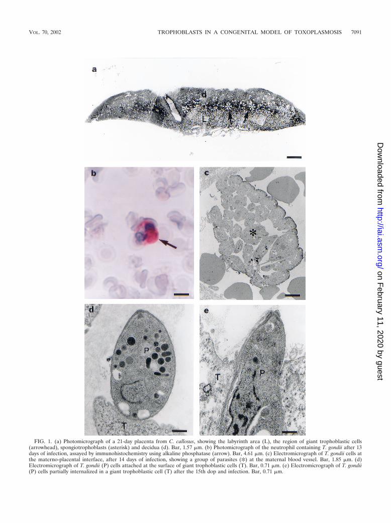
FIG. 1. (a) Photomicrograph of a 21-day placenta from C. callosus, showing the labyrinth area (L), the region of giant trophoblastic cells(arrowhead), spongiotrophoblasts (asterisk) and decidua (d). Bar, 1.57 �m. (b) Photomicrograph of the neutrophil containing T. gondii after 13days of infection, assayed by immunohistochemistry using alkaline phosphatase (arrow). Bar, 4.61 �m. (c) Electromicrograph of T. gondii cells atthe materno-placental interface, after 14 days of infection, showing a group of parasites (✻) at the maternal blood vessel. Bar, 1.85 �m. (d)Electromicrograph of T. gondii (P) cells attached at the surface of giant trophoblastic cells (T). Bar, 0.71 �m. (e) Electromicrograph of T. gondii(P) cells partially internalized in a giant trophoblastic cell (T) after the 15th dop and infection. Bar, 0.71 �m.
VOL. 70, 2002 TROPHOBLASTS IN A CONGENITAL MODEL OF TOXOPLASMOSIS 7091
on February 11, 2020 by guest
http://iai.asm.org/
Dow
nloaded from

FIG. 2. Photomicrographs showing the kinetics of infection of the trophoblastic cell populations from placentas of C. callosus and fetal tissues.(a) Rupture of a giant trophoblastic cell (T) by T. gondii (asterisk) after the 15th dop and infection. Bar, 11 �m. (b) Presence of T. gondii (arrow)in the region of labyrinth after the 16th dop. Bar, 11 �m. (c) Presence of T. gondii (arrow) in a spongiotrophoblast at the 19th dop. Bar, 11 �m.(d) T. gondii inside a giant trophoblastic cell at the 20th dop, showing that the parasite was unstained by PAS (arrow). Bar, 11 �m. (e)Electromicrograph from a fetal blood vessel in the labyrinth region at the 20th dop, showing the presence of T. gondii (asterisk) inside a reticulocyte(r). Bar, 1.30 �m. (f) Fetal nervous tissue containing T. gondii stained by alkaline phosphatase substrate in the immunohistochemistry assay (arrow)at the 16th dop. Bar, 11 �m. (g) Fetal liver containing T. gondii inside a hepatocyte stained by alkaline phosphatase substrate in the immuno-histochemistry assay (arrow). Bar, 48 �m.
7092 FERRO ET AL. INFECT. IMMUN.
on February 11, 2020 by guest
http://iai.asm.org/
Dow
nloaded from

received a second infection on the first dop also did not showsigns of infection in the liver. The serum analysis of theseanimals revealed EIs lower than 15, indicating that there wasnot congenital transmission to those embryos.
Serological analysis after pregnancy. As shown in Fig. 3b,the serum analysis presented EIs higher than 15 and high AIsat 120 days of infection and the 20th dop. Very similar resultswere also observed in the females reinoculated 100 days afterthe primary infection with 20 cysts of T. gondii and analyzed atthe 20th dop. Serum samples from these animals exhibited EIshigher than 15 and high AIs (Fig. 3b).
DISCUSSION
Experimental models of congenital toxoplasmosis have beendescribed in the literature since the early 1950s (2, 8, 17, 19, 23,26, 27). However, there is no information concerning the ki-netics of events leading to the understanding of how T. gondiitachyzoites reach the fetal tissues. In addition, no data describ-ing the role played by trophoblast cells interacting with the
parasites in the placenta from infected mothers have beenpublished.
In this study we present the results from experiments show-ing the serial events in different compartments of the placentasfrom C. callosus animals infected with the ME-49 strain of T.gondii. Our data demonstrated that the infection of the pla-centa starts with giant trophoblastic cells, at the 15th dop,followed by cells from the labyrinth at dop 16 and spongiotro-phoblasts at dop 19.
These findings are anatomically comprehensive and havealso been found in other congenital infection models (1, 5, 34).The progression of chlamydial infection in the mouse placentashows that the first cell population to be affected is the deciduaand neutrophils located at the limit of the maternal and fetaltissues. On the embryonic side, the first fetal invaded area isthe giant trophoblastic cell layer (5). In a similar manner, otherintracellular pathogens, such as Coxiella burnetti and Brucellaabortus, also preferentially first invade this cell population (1,34).
In rodents, at least two points seem to explain these findings.First, the boundary region between the fetal and maternalplacenta is characterized by the presence of a low number ofmacrophages and T cells, making this region more susceptibleto pathogen infections (25). Furthermore, the giant cells of thejunctional zone of the placenta form a mesh which receives thematernal blood directly from the uterine vessels (38). Theblood goes through the spaces of this mesh before reaching thelabyrinth zone for molecular exchange.
As already described by Fadul et al. (14), T. gondii is sys-temically spread through maternal leukocytes and, when thesecells are lysed at the maternal-fetal interface, the parasites mayinfect the giant trophoblastic cells. The labyrinth area is themain region of exchange of nutrients between maternal andfetal blood and, therefore, once infected by pathogens there isno difficulty for the pathogens to reach the fetal tissues. Thishypothesis is compatible with our experimental model, since aconcurrence between the infection of the labyrinth area andthe fetal tissues was observed. However, the infection of thelabyrinth area started only after the infection of the gianttrophoblastic cells. The spreading of the parasites toward fetaltissues occurred very quickly right after the infection of thelabyrinth zone. Probably cells from an erythrocytic lineage alsocontribute to carry the parasites to fetal tissues, as described byTanabe et al. (32).
Concerning the late infection of the spongiotrophoblast, onecould hypothesize that this phenomenon was due to the pres-ence of a wide layer of giant trophoblast cells protecting thosematernal blood cells. However, the rationale for a late infec-tion of the spongiotrophoblast remains unclear and deservesmore investigation, considering that these cells present endo-crine and stock functions (7, 24, 30). Therefore, consideringthe sequence of events leading to the infection of the variousorgans, it could be hypothesized that the placenta is infectedlater on during pregnancy, which may be related to the defenseroles played by this structure.
The serological evaluation of T. gondii infection in relationto its time of occurrence is very important to define the acutephase, considering that it is in this phase that the major prob-lems of congenital infection in humans and experimental mod-els occur.
FIG. 3. (a) Acute phase of infection by T. gondii in C. callosus;shown are means of EI and AI values for antibodies in C. callosus fromthe 15th to 20th dop and inoculation with T. gondii strain ME49. (b)Chronic phase of infection by T. gondii in C. callosus; shown are EIsand AIs for antibodies in C. callosus after 100 days of infection with T.gondii strain ME49 and before pregnancy (100/0); after 120 days ofinfection and the 20th dop (120/20); and after 120 days of infection andthe 20th dop with also reinfection with 20 cysts of T. gondii strainME49 (120/20�).
VOL. 70, 2002 TROPHOBLASTS IN A CONGENITAL MODEL OF TOXOPLASMOSIS 7093
on February 11, 2020 by guest
http://iai.asm.org/
Dow
nloaded from
Determination of the low avidity of immunoglobulin G an-tibodies to T. gondii is a very useful tool for evaluating acuteinfection, reflecting the degree of maturation of these mole-cules during the immune response (6). Our results clearlydemonstrate that congenital transmission only occurs when theantibodies present have low avidity, and these findings werecorrelated with the acute phase of infection, when the parasitesinside the giant trophoblasts were unstained by PAS, charac-terizing the presence of tachyzoites (12).
Among the experimental models, rats, unlike mice, are goodanimals for the study of congenital toxoplasmosis. Similarly asoccurs in humans, chronically infected pregnant rats protecttheir fetuses from congenital infection (23, 26, 33, 37), eventhough it was found that the occurrence of a low percentage ofcongenital transmission of T. gondii occurs during the chronicphase of maternal infection (11). Our results showed that allfemales in the chronic phase of infection, which were moni-tored by the presence of PAS-positive cerebral cysts and high-avidity antibodies to T. gondii, did not transmit the infection totheir fetuses.
Although the macaque model is considered the closestmodel to human congenital toxoplasmosis (28, 36), there aremany restrictions to working with primates and, therefore, thesearch for alternative experimental models to study congenitaltoxoplasmosis is very useful. Taken together, the results pre-sented herein demonstrate that C. callosus is an appropriateexperimental model to study the dynamics of congenital toxo-plasmosis that occurs in humans and in a wide range of mam-mals.
ACKNOWLEDGMENTS
This work was financially supported by Brazilian Research Agencies(FAPEMIG, FAPESP, CNPq, and CAPES).
We thank Nelly Patriarcha for revising the manuscript.
REFERENCES
1. Baumgartner, W., and S. Bachmann. 1992. Histological and immunocyto-chemical characterization of Coxiella burnetti-associated lesions in the mu-rine uterus and placenta. Infect. Immun. 60:5232–5241.
2. Bervely, J. K. A. 1959. Congenital transmission of toxoplasmosis throughsuccessive generations of mice. Nature 183:1348–1349.
3. Bevilacqua, E., and P. A. Abrahamsohn. 1989. Trophoblast invasion duringimplantation of the mouse embryo. Arch. Biol. Med. Exp. 22:107–118.
4. Billington, W. D. 1971. Biology of trophoblast. Adv. Reprod. Physiol. 5:27–66.
5. Buendía, A. J., J. Sanchez, M. C. Martínez, P. Camara, J. A. Navarro, A.Rodolakis, and J. Salinas. 1988. Kinetics of infection and effects on placentalcell population in murine model of Chlamydia psittaci-induced abortion.Infect. Immun. 66:2128–2134.
6. Camargo, M. E., S. M. Silva, P. G. Leser, and C. H. Granato. 1991. Avidezde anticorpos IgG específicos como marcadores de infeccao primaria recentepelo Toxoplasma gondii. Rev. Inst. Med. Trop. S. Paulo 33:213–218.
7. Campbell, W. J., S. Deb, S. C. Kwok, J. A. Joslin, and M. J. Soares. 1989.Differential expression of placental lactogen II and prolactin-like protein-Ain the rat chorioallantoic placenta. Endocrinology 125:1565–1574.
8. Cowen, D., and A. Wolf. 1950. Experimental congenital toxoplasmosis I. Thevagina as a portal of entry of Toxoplasma in the mouse. J. Exp. Med.92:393–402.
9. Cozon, G. J. N., J. Ferrandiz, H. Nebhi, M. Wallon, and F. Peyron. 1998.Estimation of the avidity of immunoglobulin G for routine diagnosis ofchronic Toxoplasma gondii infection in pregnant women. Eur. J. Clin. Mi-crobiol. Infect. Dis. 17:32–36.
10. Djurkovic-Djakovic, O. 1995. Toxoplasma infection and pathological out-come of pregnancy. Gynecol. Obstet. Investig. 40:36–41.
11. Dubey, J. P., S. K. Shen, O. C. H. Knok, and P. Thulliez. 1997. Toxoplas-
mosis in rats (Ratus norvegicus): congenital transmission to first and secondgeneration offspring and isolation of Toxoplasma gondii from seronegativerats. Parasitology 115:9–14.
12. Dubey, J. P., D. S. Lindsay, and C. A. Speer. 1998. Structures of Toxoplasmagondii tachyzoites, bradyzoites, and sporozoites and biology and develop-ment of tissue cyst. Clin. Microbiol. Rev. 11:267–299.
13. Enders, A. C., and S. Schlafke. 1969. Cytological aspects of trophoblast-uterine interaction in early implantation. Am. J. Anat. 125:1–30.
14. Fadul, C. E., J. Y. Channon, and L. H. Kasper. 1995. Survival of immuno-globulin G-opsonized Toxoplasma gondii in nonadherent human monocytes.Infect. Immun. 63:4290.
15. Ferro, E. A. V., and E. Bevilacqua. 1994. Trophoblastic invasion of theuterine epithelium in Calomys callosus (Rodentia, Cricetidae). J. Morphol.221:139–152.
16. Ferro, E. A. V., E. Bevilacqua, S. Favoreto, Jr., D. A. O. Silva, R. A. Mortara,and J. R. Mineo. 1999. Calomys callosus (Rodentia: Cricetidae) trophoblastcells as host cells to Toxoplasma gondii in early pregnancy. Parasitol. Res.85:647–654.
17. Graham, D. I., J. Hay, W. M. Hutchison, and J. C. Siim. 1984. Encephalitisin mice with congenital ocular toxoplasmosis. J. Pathol. 142:265–277.
18. Holliman, R. E., R. Raymond, N. Renton, and J. D. Johnson. 1994. Thediagnosis of toxoplasmosis using IgG avidity. Epidemiol. Infect. 112:399–408.
19. Hutchison, W. M., J. Hay, W. R. Lee, and J. C. Siim. 1982. A study ofcataract in murine congenital toxoplasmosis. Ann. Trop. Med. Parasitol.76:53–70.
20. McMannus, J. F. A. 1948. Histological and histochemical use of periodicacid. Stain Technol. 23:99–108.
21. Mineo, J. R., M. E. Camargo, and A. W. Ferreira. 1980. Enzyme-linkedimmunosorbent assay for antibodies to Toxoplasma gondii polysaccharides inhuman toxoplasmosis. Infect. Immun. 27:283–287.
22. Mossman, H. W. 1987. Vertebrate fetal membranes. Rutgers UniversityPress, New Brunswick, N.J.
23. Paulino, J. P., and R. W. A. Vitor. 1999. Experimental congenital toxosplas-mosis in Wistar and Holtzmann rats. Parasite 6:63–66.
24. Rasmusssen, C. A., K. E. Orwig, S. Vellucci, and M. J. Soares. 1997. Dualexpression of prolactin related protein in decidua and trophoblast tissuepregnancy in rats. Biol. Reprod. 56:647–654.
25. Redline, R. W., and C. Y. Lu. 1988. Specific defects in the anti-listerialimmune response in discrete regions of the murine uterus and placentaaccount for susceptibility to infection. J. Immunol. 140:3947–3955.
26. Remington, J. S., L. Jabobs, and L. Melton. 1961. Congenital transmission oftoxoplasmosis from mother animals with acute and chronic infection. J. In-fect. Dis. 108:163–173.
27. Roberts, C. W., and J. Alexander. 1992. Studies on a murine model ofcongenital toxoplasmosis: vertical disease transmission only occurs inBALB/c mice infected for the first time during pregnancy. Parasitology104:19–23.
28. Schoondermarkvandeven, E., W. Melchers, J. Galama, W. Camps, T. Eskes,and J. Meuwissen. 1993. Congenital toxoplasmosis: an experimental study inrhesus monkeys for transmission and prenatal diagnosis. Exp. Parasitol.77:200–211.
29. Sims, T. A., J. Hay, and I. C. Talbot. 1988. Host-parasite relationship in thebrains of mice with congenital toxoplasmosis. J. Pathol. 156:255–261.
30. Soares, M. J., B. M. Chapman, C. A. Rasmussen, G. Daí, T. Kamei, and K. E.Orwing. 1996. Differentiation of trophoblast endocrine cells. Placenta 17:277–289.
31. Tachi, S., C. Tachi, and H. R. Lindner. 1970. Ultrastructural features ofblastocyst attachment and trophoblastic invasion in the rat. J. Reprod. Fert.21:37–56.
32. Tanabe, K., T. Asi, I. Kimata, and S. Takada. 1979. Penetration of matu-rating red blood cells by Toxoplasma gondii. J. Gen. Microbiol. 113:433–437.
33. Thiermann, I. E. 1957. Transmission congenita del Toxoplasma gondii enratas com infection leve. Biologica (Santiago) 23:59–67.
34. Tobias, L., D. O. Cordes, and G. G. Schurig. 1993. Placental pathology of thepregnant mouse inoculated with Brucella abortus strain 2308. Vet. Pathol.30:119–129.
35. Turunen, H., K. A. Vuorio, and P. O. Leiniki. 1983. Determination of IgG,IgM and IgA antibody responses in human toxoplasmosis by enzyme-linkedimmunosorbent assay (ELISA). Scand. J. Dis. 15:307–311.
36. Wong, M. M., W. J. Kozek, S. L. Karr, Jr., M. A. Brayton, J. H. Theis, andA. G. Hendrickx. 1979. Experimental congenital infection of Toxoplasmagondii in Macaca arctoides. Asian J. Infect. Dis. 3:61–67.
37. Zenner, L., J. Estaquier, F. Darcy, P. Maes, A. Capron, and M. F. Cesbron-Delaw. 1999. Protective immunity in the rat model of congenital toxoplas-mosis and the potential of excreted-secreted antigens as vaccine compo-nents. Parasite Immunol. 21:261–272.
38. Zuckermann, F., and J. R. Head. 1986. Isolation and characterization oftrophoblast from murine placenta. Placenta 7:349–364.
Editor: W. A. Petri, Jr.
7094 FERRO ET AL. INFECT. IMMUN.
on February 11, 2020 by guest
http://iai.asm.org/
Dow
nloaded from