effect of grain size on mechanical, surface and biological properties of microwave sintered...
TRANSCRIPT
Materials Science and Engineering C 33 (2013) 2846–2854
Contents lists available at SciVerse ScienceDirect
Materials Science and Engineering C
j ourna l homepage: www.e lsev ie r .com/ locate /msec
Effect of grain size on mechanical, surface and biological properties of microwavesintered hydroxyapatite
Sudip Dasgupta, Solaiman Tarafder, Amit Bandyopadhyay, Susmita Bose ⁎W. M. Keck Biomedical Materials Research Laboratory, School of Mechanical and Materials Engineering, Washington State University, Pullman, WA 99164-2920, USA
⁎ Corresponding author. Tel.: +1 509 335 7461; fax:E-mail address: [email protected] (S. Bose).
0928-4931/$ – see front matter © 2013 Published by Elhttp://dx.doi.org/10.1016/j.msec.2013.03.004
a b s t r a c t
a r t i c l e i n f oArticle history:Received 23 June 2012Received in revised form 18 February 2013Accepted 1 March 2013Available online 14 March 2013
Keywords:NanostructureMicrowave sinteringGrain sizeHydroxyapatiteMechanical propertyVinculin and ALP expression
Hydroxyapatite (HA) compacts having average grain sizes of 168 ± 0.086 nm, 1.48 ± 0.627 μm and 5.01 ±1.02 μm are processed from synthesized HA powder by microwave sintering at varying sintering temperaturefor different times. Superior mechanical and biological properties are shown by nano-grain HA compacts ascompared to their micron grained counterparts. Compressive strength, indentation hardness, and indenta-tion fracture toughness are increased with the decrease in HA grain size. The highest surface energy and max-imum wettability are exhibited by nano-grain HA. HA compacts are assessed for cell–material interaction bySEM, MTT and immunochemistry assays using human osteoblast cell line for 1, 5 and 11 days. MTT assaysshowed higher number of living cells and faster proliferation on nano-grain HA surface. Osteoblast cells onnano-grain HA surface expressed significantly higher amount of vinculin and alkaline phosphatase (ALP) pro-tein markers for cell adhesion and differentiation respectively. This study shows the effect of grain size onphysical, mechanical and in vitro biological properties of microwave sintered HA compacts.
© 2013 Published by Elsevier B.V.
1. Introduction
Hydroxyapatite (HA, Ca10(PO4)6(OH)2), the structural prototypeof tooth and bone mineral, has been used as an important implantand scaffold material, and drug delivery agent, with significant clini-cal potential in medicine, dentistry and orthopedics [1–3]. From ex-tensive in vitro and in vivo studies, synthetic HA has been shown tolack local or systemic toxicity, to stimulate bone growth without im-mune response complications [4,5] and to spontaneously integrateinto the host hard tissue [1]. Biological and mechanical properties ofsynthetic HA are largely determined by its particle size, morphology,crystallinity, and composition, which depend on the synthesis precur-sors and processing.
Bone is a composite material consisting of nanoscale mineralparticles and a matrix of collagen fibers. Nanocrystalline HA (nanoHA) would be more interesting than micro-sized HA from a biolog-ical and medical viewpoint because of its similarity to minerals innatural bone. Compared to conventional microscale HA, whichlacks phase purity and homogeneity, nano HA offers the possibilityto enhance the rate of bone-bonding formation and to have excel-lent mechanical properties due to its high surface area to volumeratio, superior chemical homogeneity and microstructural uniformi-ty [6]. Furthermore, nano HA was shown to be able to inhibit thegrowth of certain kinds of cancer cells, such as the liver, throat and
+1 509 335 4662.
sevier B.V.
bone cancer cells, while having little side effect on normal cells [7].The rate of HA bonding to bone was demonstrated to be dependentnot on the composition but on the release of calcium and phosphateions from HA, determining the development of implant–bone inter-facial strength [8]. Consequently, sufficient dissolution of calciumand phosphate species is necessary to form bone-like apatite andbone bonding. The dissolution of nano HA has been proven to bevery different from that of microscale HA. The dissolution of nanoHA is dominated by its particle size [1]. The particle-size effect isexplained by the fact that small sized particles of HA may be degrad-able and stimulate bone ingrowth as they dissolve in the physiolog-ical environment [9]. Although nano HA has been extensivelystudied, most work has been focused on its synthesis procedures,structure analysis and applications [6,10]. Until now, to the best ofour knowledge, no detailed investigation has been carried out deal-ing with the effects of the synthesis parameters on the structure ofnano HA.
It has been reported that surface properties, such as surface area,charge, and topography, depend on the grain size of a material [11].In this respect, nanostructured materials possess higher surfacearea with increased portions of surface defects and grain-boundaries [12]. Meanwhile, hydroxyapatite (HA) has been consid-ered as a good candidate for designing hard tissue implants becauseof its excellent biological properties such as nontoxicity, lack of in-flammatory response and immunological reactions, and is able to in-timately bond to new bone [13,14]. Webster et al. have reported thatosteoblast adhesion and proliferation were significantly greater onnanostructured alumina and HA than on conventional formulations

2847S. Dasgupta et al. / Materials Science and Engineering C 33 (2013) 2846–2854
of the same ceramic after 3 and 5 days [15,16]. More importantly,synthesis of alkaline phosphatase and deposition of calciumcontaining mineral was significantly greater by osteoblasts culturedon nanostructured ceramics than conventional ceramics after 21 and28 days [11,17].
The efficiency of HA ceramics as orthopedic implant greatly de-pends on its grain size. Not only the osteoblast cells show grain sizedependent activities on HA compacts, but mechanical properties ofHA compacts also greatly vary with change in grain size in sinteredHA microstructure. Particularly, orthopedic implant exhibits re-markably different bioactivities and mechanical reliability at nano-scale. Thus it is interesting to investigate how one can monitor thebiological as well as mechanical properties of HA ceramics bychanging its grain size. Microwave sintering of ceramics to achieveimproved mechanical properties has widely been used by the sci-entific community [18]. Notable advantages of microwave sinteringare rapid volumetric heating rate, shorter processing time and costeffectiveness in terms of energy savings [18–20]. Heating uniformi-ty caused by volumetric heating, and thus, shorter sintering time ofmicrowave sintering leads to achieve finer grain. Controlled graingrowth and finer microstructure due to microwave sintering resultin high mechanical properties in sintered ceramics.
The objective of this study was to understand the influence ofgrain size on mechanical properties and bioactivity of sintered HA.In the present study, we processed HA compacts with differentgrain size using microwave sintering. The mechanical properties ofthese HA compacts are compared with those of HA compacts reportedpreviously [19]. Surface energy of nano and micron size HA compactsis also compared in this study. In vitro bone cell–material interactionof HA compacts was investigated for adhesion, proliferation and dif-ferentiation of human osteoblast cells using SEM, MTT assay and con-focal microscopy.
2. Materials and methods
2.1. Synthesis of HA nanopowders
HA nanopowder was synthesized using an emulsion synthesisrout [21]. Briefly, 5 M aqueous solution of Ca2+-ion was preparedby dissolving 0.01 mol (2.362 g) of Ca(NO3)2·4H2O in 2 ml distilledwater. 0.006 mol (0.686 g) of phosphoric acid (H3PO4) (85.7%) wasadded to the system to maintain Ca to P molar ratio 1.67. Organicphase of the emulsion was prepared by the addition of 10 vol.% sur-factant, poly(oxyethylene)12 nonylphenol ether (NP12), in cyclo-hexane with vigorous stirring. An aqueous to organic ratio (A/O) of1:15 was maintained for HA nanopowder synthesis. The pH of theemulsion was adjusted to 9 with dropwise addition of NH4OH to ini-tiate reaction between Ca(NO3)2·4H2O and H3PO4 to form HAnanocrystals. All reactions were aged for 24 h at room temperatureto grow non-agglomerated HA nanocrystals with high crystallinity.After aging, the emulsion was evaporated on the hot plate at150 °C followed by complete drying at 450 °C. Dry precursor powderwas calcined at 650 °C for 4 h to get carbon free crystalline HAnanopowder.
2.2. Consolidation of calcined HA powders
As synthesized HA nanopowders were processed following theprocedure as described in our previous work [19]. To investigatethe effect of calcination temperature on particle size, the as synthe-sized HA nanopowders were calcined at 800 °C for 4 h and 900 °Cfor 10 h. From here on, we will denote the as synthesized HAnanopowders after processing as HA (I), and as synthesized HAnanopowders calcined at 800 and 900 °C as HA (II) and HA (III), re-spectively. Particle size distribution was measured using dynamiclight scattering (DLS) technique (NICOMPTM 380, Santa Barbara,
CA). Disks with approximate dimensions of 12 mm in diameterand 2 mm in height were prepared by uniaxial pressing at50 MPa, followed by cold isostatic pressing at 345 MPa. Cylindricalcompacts with approximately 6 mm in diameter and 9 mm inheight were also prepared using the same technique for mechani-cal testing.
2.3. Sintering and characterization of HA
The disks and cylindrical HA were sintered in a 3 kW microwavefurnace [MW-L0316V, LongTech Co., Ltd, ChangSha, HuNan, P.R.China] at different temperature for different time interval as de-scribed in Table 1. The constituent phases of sintered HA (I), HA (II)and HA (III) compacts were analyzed using a Philips fully automatedX-ray diffractometer with CuKα radiation and a Ni filter. The diffrac-tometer was operated at 35 kV and 30 mA over the 2θ range of 20to 60° at a step size of 0.02° and a count time of 0.5 s per step. Micro-structure was characterized using a field-emission scanning electronmicroscope (FESEM) (FEI Inc., OR, USA). Sintered HA grain sizeswere determined from SEM images via a linear intercept method[22] using the equation G = (L / N)C, where G is the average grainsize (μm), L is the test line length (cm), N is the number of intersec-tions with grain boundaries along test line L; and C is the conversionfactor (μm/cm) of the picture on which the test lines were drawn asobtained from the scale bar.
The sintered HA pellets were characterized for microhardness andindentation fracture toughness. Fracture toughness was calculatedfrom the radial crack length that appeared after the indentation test.The equation for the calculation of fracture toughness is givenbelow [23].
Fracture toughness KICð Þ ¼ 0:016� E=Hð Þ1=2 � P=C3=2
where, E is Young's modulus of the sample, H is microhardness inGPa, P is the applied load and C is the half of the crack length. Com-pressive strength analysis was performed with cylindrical HAcompacts.
2.4. Contact angle measurement and surface energy calculation
Contact angles of different liquids on sintered HA disk surfacewere measured using the sessile drop method on a face contactangle set-up equipped with a microscope and a camera [ModelVCA Optima, AST products, Billerica, MA, USA]. Contact angleswere determined with liquids such as water, formamide, glycerol,and cell culture media. The liquid surface tension and their compo-nents were used in the following equation [24] to calculate the sur-face energy:
γL 1þ cosθð Þ ¼ 2 γSLWγL
LW� �1=2 þ 2 γS
þγL−
� �1=2 þ 2 γS−γL
þ� �1=2
where, θ is the contact angle of liquid L and solid S, γLW is the apolarcomponent of the surface energy, γ+ is the Lewis acid component(electron acceptor) and γ− is the Lewis base component (electrondonor).
2.5. In vitro cell–material interactions
In vitro cell–material interactions were studied for a maximum in-cubation period of 11 days using human fetal osteoblast (hFOB) cell(ATCC, VA, USA). Triplicate samples per group were evaluated for allexperiments. Each sample was sterilized by autoclaving at 121 °Cfor 20 min prior to the cell culture experiment. Following this, cellswere seeded onto the sample surfaces, placed in a 24-well plate.Cells were seeded at a density of 1 × 10⁴/well. Then 1 ml of McCoy's

0 200 400 600 800 1000 1200
Diameter (nm)
Inte
nsi
ty (
a.u
.)
HA (III)
HA (II)
HA (I)
Fig. 1. Particle size distribution of HA (I), HA (II) and HA (III) powders.
Table 1Mechanical properties of HA compacts with variation in sintering temperature and grain size.
HA compacts Grain size (μm) Compressive strength(MPa)
Microhardness(GPa)
Indentation fracturetoughness (MPa m1/2)
HA (I): sintered at 1000 °C for 20 min 0.168 ± 0.086 a 395 ± 42a 8.4 ± 0.4a 1.9 ± 0.2a
HA (I): sintered at 1100 °C for 20 min 0.52 ± 0.092a 328 ± 58a 7.3 ± 0.3a 1.5 ± 0.3a
HA (I): sintered at 1150 °C for 20 min 1.16 ± 0.17a 278 ± 35a 6.3 ± 0.5a 1.2 ± 0.2a
HA (II): sintered at 1150 °C for 30 min 1.48 ± 0.627 165 ± 25 5.4 ± 0.58 1.1 ± 0.09HA (III): sintered at 1150 °C for 45 min 5.01 ± 1.02 88 ± 29 3.8 ± 0.42 0.8 ± 0.07
a From reference [19].
2848 S. Dasgupta et al. / Materials Science and Engineering C 33 (2013) 2846–2854
5A medium (with L-glutamine, without phenol red and sodium bicar-bonate) supplemented with 5% fetal calf serum (FCS), and 5% bovinecalf serum (BCS) was added to each well. Cells were maintained at37 °C under an atmosphere of 5% CO2 and 95% air. The culturemedia was changed every other day.
2.5.1. Cell morphologyFor SEM observation, cell-cultured disk samples were placed in
0.1 M phosphate buffered saline (PBS) and rinsed quickly. Sampleswere subsequently fixed with 2% paraformaldehyde and 2% glutaral-dehyde in 0.1 M cacodylate buffer overnight at 4 °C. Following threerinses in 0.1 M cacodylate buffer, each sample was post-fixed in 2%osmium tetroxide (OsO4) for 2 h at room temperature. The fixedsamples were then again rinsed three times in 0.1 M cacodylateand dehydrated in an ethanol series (30%, 50%, 70%, 95% and 100%three times). Samples were then dried using acetone andhexamethyldisilazane (HMDS). Dried samples were mounted onaluminum stubs, gold sputter coated (Technis Hummer, San Jose,CA), and observed under SEM.
2.5.2. Cell proliferation using MTT assayThe MTT assay (Sigma, St. Louis, MO) was performed to assess cell
proliferation on sintered HA disks. The MTT solution of 5 mg/ml wasprepared by dissolving MTT in PBS, and filter sterilized. The MTT wasdiluted (50 μl into 450 μl) in serum free, phenol red-free Dulbecco'sminimum essential medium (DMEM). 500 μl diluted MTT solutionwas then added to each sample in 24-well plates. After 2 h incuba-tion, 500 μl of solubilization solution made up of 10% Triton X-100,0.1 N HCl and isopropanol were added to dissolve the formazan crys-tals. 100 μl of the resulting supernatant was transferred into a 96-wellplate, and read by a plate reader at 570 nm. Data are presented asmean ± standard deviation.
2.6. Immunochemistry and confocal microscopy
After culturing the cells on HA disks for a pre-specified number ofdays, the cells were fixed in 4% paraformaldehyde in 0.1 M phosphatebuffer that were kept for 24 h at 4 °C for future use. Those sampleswere permeabilized by 0.5% Triton X-100 in 0.1 M phosphate-bufferedsaline (PBS) for 10 min and blockedwith TBST/BSA (Tris-buffered salinewith 1% bovine serum albumin, 250 mM NaCl, pH 8.3) for 1 h at roomtemperature. Primary antibody against alkaline phosphatase (ALP)(Sigma-Aldrich, St. Louis, MO) or vinculin (Sigma-Aldrich, St. Louis,MO) was added at a 1:100 dilution and incubated at room temperatureovernight. Vinculin and ALP were used to study osteoblast cell at-tachment and differentiation, respectively, onto HA compacts. Thefollowing day, samples were rinsed with TBST/BSA three times for10 min each. The secondary antibody, Oregon green goatanti-mouse (GAM) (Molecular Probes, Eugene, OR), was added at1:100 dilution and incubated at room temperature for 1 h. After2 × 5 min washing with TBST/BSA followed by 5 min washing withPBS, samples were then mounted on coverslips with Vectashield
MountingMedium (Vector Labs, Burlingame, CA) with propidium io-dide (PI) and observed in confocal scanning laser microscopy(BioRad 1024 RMC, Hercules, CA, USA). Specific absorption ofvinculin or ALP was identified by the expression of green fluores-cence and nuclei counterstained with the chemical dye PI presentin the mounting medium were expressed as red fluorescence. Forgreen fluorescence excitation Ar ion (488 nm, 30 mW) laser wasused at 50% of maximum output at full power and for red fluores-cence excitation He–Ne (543 nm, 1.2 mW) laser was used at full out-put and full power. A combination of a band pass filter transmitting505–570 nm and a long pass filter at 560 nm was applied to obtainfluorescence images. Since the test samples were opaque, the confo-cal pinholes were opened up to 560 lm to pass more fluorescencelight to improve fluorescence image quality. Presence of higheramount of green fluorescence represents higher amount of vinculinexpression by these cells.
2.7. Statistical analysis
Data for grain size, compressive strength, microhardness, indenta-tion fracture toughness, contact angles, surface energy and MTT assayis presented as mean ± standard deviation. Statistical analysis wasperformed on MTT assay results using student's t-test, and Pvalue b 0.05 was considered significant.
3. Results
3.1. Particle size analysis of calcined HA particles
Fig. 1 shows the particle size distribution of HA (I), HA (II), and HA(III) measured by DLS technique. The particle size of as synthesizedHA (I) nanopowders varied between 30 and 60 nm. After calcination
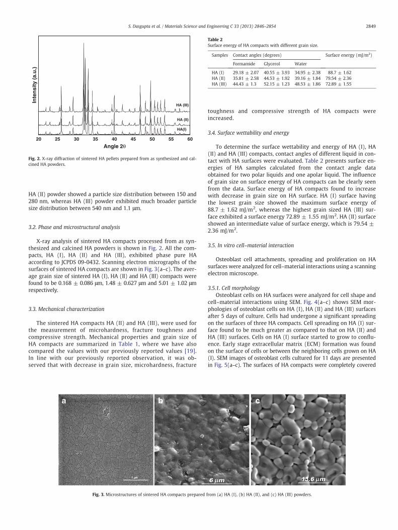
Table 2Surface energy of HA compacts with different grain size.
Samples Contact angles (degrees) Surface energy (mJ/m2)
Formamide Glycerol Water
HA (I) 29.18 ± 2.07 40.55 ± 3.93 34.95 ± 2.38 88.7 ± 1.62HA (II) 35.81 ± 2.58 44.53 ± 1.92 39.16 ± 1.84 79.54 ± 2.36HA (III) 44.43 ± 1.3 52.15 ± 1.23 48.53 ± 1.86 72.89 ± 1.55
20 25 30 35 40 45 50 55 60
Angle 2θ
Inte
nsi
ty (
a.u
.)
HA(I)
HA (II)
HA (III)
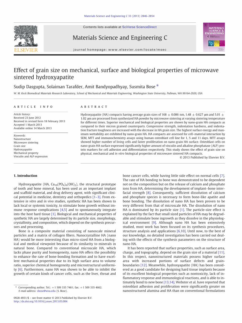
Fig. 2. X-ray diffraction of sintered HA pellets prepared from as synthesized and cal-cined HA powders.
2849S. Dasgupta et al. / Materials Science and Engineering C 33 (2013) 2846–2854
HA (II) powder showed a particle size distribution between 150 and280 nm, whereas HA (III) powder exhibited much broader particlesize distribution between 540 nm and 1.1 μm.
3.2. Phase and microstructural analysis
X-ray analysis of sintered HA compacts processed from as syn-thesized and calcined HA powders is shown in Fig. 2. All the com-pacts, HA (I), HA (II) and HA (III), exhibited phase pure HAaccording to JCPDS 09-0432. Scanning electron micrographs of thesurfaces of sintered HA compacts are shown in Fig. 3(a–c). The aver-age grain size of sintered HA (I), HA (II) and HA (III) compacts werefound to be 0.168 ± 0.086 μm, 1.48 ± 0.627 μm and 5.01 ± 1.02 μmrespectively.
3.3. Mechanical characterization
The sintered HA compacts HA (II) and HA (III), were used forthe measurement of microhardness, fracture toughness andcompressive strength. Mechanical properties and grain size ofHA compacts are summarized in Table 1, where we have alsocompared the values with our previously reported values [19].In line with our previously reported observation, it was ob-served that with decrease in grain size, microhardness, fracture
Fig. 3. Microstructures of sintered HA compacts prepared
toughness and compressive strength of HA compacts wereincreased.
3.4. Surface wettability and energy
To determine the surface wettability and energy of HA (I), HA(II) and HA (III) compacts, contact angles of different liquid in con-tact with HA surfaces were evaluated. Table 2 presents surface en-ergies of HA samples calculated from the contact angle dataobtained for two polar liquids and one apolar liquid. The influenceof grain size on surface energy of HA compacts can be clearly seenfrom the data. Surface energy of HA compacts found to increasewith decrease in grain size on HA surface. HA (I) surface havingthe lowest grain size showed the maximum surface energy of88.7 ± 1.62 mJ/m2, whereas the highest grain sized HA (III) sur-face exhibited a surface energy 72.89 ± 1.55 mJ/m2. HA (II) surfaceshowed an intermediate value of surface energy, which is 79.54 ±2.36 mJ/m2.
3.5. In vitro cell–material interaction
Osteoblast cell attachments, spreading and proliferation on HAsurfaces were analyzed for cell–material interactions using a scanningelectron microscope.
3.5.1. Cell morphologyOsteoblast cells on HA surfaces were analyzed for cell shape and
cell–material interactions using SEM. Fig. 4(a–c) shows SEM mor-phologies of osteoblast cells on HA (I), HA (II) and HA (III) surfacesafter 5 days of culture. Cells had undergone a significant spreadingon the surfaces of three HA compacts. Cell spreading on HA (I) sur-face found to be much greater as compared to that on HA (II) andHA (III) surfaces. Cells on HA (I) surface started to grow to conflu-ence. Early stage extracellular matrix (ECM) formation was foundon the surface of cells or between the neighboring cells grown on HA(I). SEM images of osteoblast cells cultured for 11 days are presentedin Fig. 5(a–c). The surfaces of HA compacts were completely covered
from (a) HA (I), (b) HA (II), and (c) HA (III) powders.

Fig. 4. SEM micrographs of osteoblast cells after 5 days of culture on (a) HA (I), (b) HA (II), and (c) HA (III) compacts.
2850 S. Dasgupta et al. / Materials Science and Engineering C 33 (2013) 2846–2854
by a dense and confluent cellular multilayer forming a three-dimensional fibril network. The presence of ECM could be detected onthe surface of cells.
3.5.2. MTT assayMTT assay was used to determine osteoblast cell proliferation on
HA compacts. Fig. 6 shows a comparison of cell densities on threeHA compacts over the course of the experiment. Cell proliferationwas evident over the duration of the experiment, on all the HA com-pacts. Cell densities were significantly higher on HA (I) compact ascompared to HA (II) and HA (III) at all time points. Again cell prolifer-ation on HA (II) compact found to be greater compared to that on HA(III) compact.
3.6. Immunocytochemistry and confocal microscopy
3.6.1. Vinculin expressionThe formation of focal adhesion plaques is a prerequisite process for
the development of the signaling transduction in cell attachment, and isone of the important indicators for cell activity on the substrates.Vinculin aids in the assemblage of focal contacts by cross-linking andrecruiting other proteins to form adhesive plaques [25]. Vinculin alsoacts as an adhesion molecule between the cells and substratum. It ismostly located at points of focal adhesion plaque, so the existence ofvinculin represents the formation of focal adhesion plaque. As cells at-tach to one another and to the substratum, adhesive proteins interactwith and form bonds to adhesion receptors within the cellular mem-brane. Antibody bound to vinculin expressed green fluorescence andnuclei stained with propidium iodide (PI) in the mounting mediumexpressed red fluorescence. The formation of vinculin-positive focal ad-hesion plaques increased gradually with cell culture time on all three
Fig. 5. SEM micrographs of osteoblast cells after 11 days of cu
HAcompacts as shown in Figs. 7–9. At 1 day after seeding,most fluores-cence staining of vinculin occurred on HA (I) substrate (Fig. 7a),followed by HA (II) (Fig. 7b) and HA (III) (Fig. 7c). The formation offocal adhesion plaque on HA (I) substrates was earlier than that onHA (II) and HA (III) substrates. Vinculin staining on HA (I) was alsomore than that on HA (II) and HA (III) at 5 days (Fig. 8) and 11 days(Fig. 9). Compared with HA (III) substrate, the vinculin staining on HA(II) substrate was more intense at all day points of cell cultureexperiment.
3.6.2. Alkaline phosphatase expressionImmunocytochemistry of osteoblast cells was used to determine
whether the cells express an osteoblastic phenotype on samples ornot. A major characteristic of osteoblasts is the expression of alkalinephosphatase (ALP). Antibody bound to ALP expressed green fluores-cence and nuclei stained with propidium iodide (PI) in the mountingmedium expressed red fluorescence. Fig. 10(a–c) shows the confocalmicrographs of ALP expression in one day cultured osteoblast cells onHA (I), HA (II) and HA (III) substrates. No positive immune stainingfor ALP was detected on any substrate after day 1. After day 5, osteo-blast cells cultured on HA substrates displayed positive signal for ALP,but with different patterns and levels as depicted in Fig. 11. After5 days of culture, cells on HA (I) (Fig. 11a) showed stronger greenfluorescence for ALP followed by HA (II) (Fig. 11b) and HA (III)(Fig. 11c). With the increase in culture time, ALP activity increasedsignificantly on all the HA compacts. After 11 days of cell culture,ALP expression on HA (I) (Fig. 12a) was more intense compared tothat observed on HA (II) (Fig. 12b) and HA (III) (Fig. 12c) compacts.Again HA (II) (Fig. 12b) compact exhibited stronger green fluores-cence staining for ALP than HA (III) (Fig. 12c) compact after 11 daysof cell culture.
lture on (a) HA (I), (b) HA (II), and (c) HA (III) compacts.

Fig. 6. MTT assay of cells on HA (I), HA (II) and HA (III) compacts.
2851S. Dasgupta et al. / Materials Science and Engineering C 33 (2013) 2846–2854
4. Discussion
Final grain size in sintered ceramic compact depends onstarting particle size and surface area of powder, sintering tem-perature and time of green ceramic compact. All other factorsbeing constant, the higher the particle size in green compact, thehigher will be the grain size in sintered compact. HA powders cal-cined at higher temperature for longer amount of time possessedparticles of higher size compared to powders calcined at lowertemperature for shorter duration. Thus HA (III) green compact,which was composed of the largest sized HA particles among all
Fig. 8. Confocal micrographs of vinculin expression in osteoblast cell
Fig. 7. Confocal micrographs of vinculin expression in osteoblast cell
the HA compacts showed the largest grain size. The observed po-rosity in the HA (III) microstructure (Fig. 3) could probably bedue to larger particle size. Again, green HA (III) compact wassintered in microwave furnace for longer time compared to HA(II) compact, which resulted in higher grain growth in thesintered HA (III) microstructure. HA (I) green compacts werecomposed of particles of the highest surface area, which resultedin the highest driving force for sintering. That is why, HA (I)green compact could be sintered at lower temperature with noor minimum grain growth in sintered microstructure.
For HA compacts with identical densities, mechanical propertiessuch as compressive strength, hardness and fracture toughness, areenhanced with decrease in grain size in sintered microstructure.With decrease in grain size, the inherent flaw size in sintered micro-structure is reduced which leads to the enhancement of compres-sive strength. Again as the number of grain boundaries per unitvolume is increased with decrease in grain size, finer grain sizedcompacts offer more resistance to crack propagation and dislocationmotion resulting in higher hardness and fracture toughness. ThusHA (I) nanocompacts having the lowest grain size showed the max-imum compressive strength, hardness and fracture toughness thanHA (II) and HA (III) compacts. Porous materials usually have alower mechanical strength than their dense counterparts [18]. Thelowest mechanical properties for HA (III) (Table 1) might haveresulted from a combined effect of larger grain size and the intrinsicporosity.
Our result shows surface energy of HA compact increased withdecrease in grain size. This increased surface energy of HA compacts
s cultured on (a) HA (I), (b) HA (II), and (c) HA (III) after day 5.
s cultured on (a) HA (I), (b) HA (II), and (c) HA (III) after day 1.

Fig. 9. Confocal micrographs of vinculin expression in osteoblast cells cultured on (a) HA (I), (b) HA (II), and (c) HA (III) after day 11. Green fluorescence indicating antibody boundto ALP, red fluorescence indicating antibody bound to DNA (nucleus).
2852 S. Dasgupta et al. / Materials Science and Engineering C 33 (2013) 2846–2854
with smaller grain size is due to increased wettability. Webster et al.[26] showed that surface area increases with decrease in grain size.Thus, increased surface energy of the HA compacts with decreasinggrain size is caused by increased surface area. Surfaces with high en-ergy facilitate increased adsorption of cell adhesive protein [27,28],which ultimately promote better osteoblast interactions. The mostcritical stage of osteoblast cell–material interaction is adhesion andspreading which ultimately governs cell's capacity to proliferateand differentiate. The MTT result shows that, at all day points ofcell culture, the number of cells on HA (I) compact was significantlyhigher than those on HA (II) and HA (III). This result suggests thatnanograined HA compact enhanced cellular attachment. This is fur-ther supported by SEM micrographs of osteoblast cells after 5 and11 days of culture (Fig. 4). Surface energy directly influences two im-portant phenomena for an efficient cell–biomaterial interaction,namely protein adsorption and cell attachment. After immersion inbiological fluids, all implant materials are coated by a protein layer.The presence of this pre-adsorbed protein layer is essential in medi-ating cell response to the material. For HA ceramics, protein adsorp-tion is governed by electrostatic interaction between HA surface andprotein [29]. With decrease in grain size of HA compact the numberof sites for electrostatic interaction between HA and protein in-creased. Thus protein adsorption and subsequent highest cell attach-ment are observed on nanograined HA (I) compact, followed by HA(II) and HA (III) compact. This fact is further endorsed by the resultsfrom vinculin protein expression on cell cultured HA (I), HA (II) andHA (III) surfaces. HA (I) compact showed the strongest vinculin
Fig. 10. Confocal micrographs of ALP expression in osteoblast cells
expression all through the cell culture experiment, signifies thatHA (I) surface generated most number of focal points for cell adhe-sion. Decreased grain size on HA surface creates more number offocal points for cell adhesion, this is further confirmed by thehigher amount of vinculin expression on HA (II) compact comparedto that on HA (III) compact. Again after 5 days of culture, SEM mi-crographs show that cell spreading on nanograined HA (I) compactwas the highest followed by HA (II) and HA (III) compacts, whichwas due to higher wettability of lower grained HA compacts. Thusthe surface of HA (I) compacts being conducive for both cell attach-ment and spreading encouraged cell proliferation most efficientlycompared to HA (II) and HA (III) compacts. Higher degree of cellproliferation on HA (II) surface as compared to HA (III) surfacewas due to higher amount of cell attachment and spreading onformer.
As osteoblast cells proliferate and then differentiate, it ex-presses a number of osteoblastic phenotypic markers. ALP is amajor characteristic marker of osteoblasts. ALP is regarded as anearly marker for osteoblast differentiation, and it is generally ac-cepted that as the specific activity of ALP in a population of bonecells increases there is a corresponding shift to a more differentiat-ed state. Though after 1 day no green fluorescence from ALP wasobserved from any of the HA compacts, after 5 days abundantamount of ALP protein expression was found on HA (I) surface.ALP expression after 5 and 11 days is far more intense on HA (I)compact as compared to HA (II) and HA (III) compacts. Higherlevels of ALP expression on HA (I) could be related to faster
cultured on (a) HA (I), (b) HA (II), and (c) HA (III) after day 1.

Fig. 12. Confocal micrographs of ALP expression in osteoblast cells cultured on (a) HA (I), (b) HA (II), and (c) HA (III) after day 11. Green fluorescence indicating antibody bound toALP, red fluorescence indicating antibody bound to DNA (nucleus).
Fig. 11. Confocal micrographs of ALP expression in osteoblast cells cultured on (a) HA (I), (b) HA (II), and (c) HA (III) after day 5.
2853S. Dasgupta et al. / Materials Science and Engineering C 33 (2013) 2846–2854
differentiation of osteoblast cells on HA (I) surface. Thus,nanograined HA (I) compacts facilitated rapid differentiation andstrong adhesion of osteoblast cells. Again HA (II) compact showedstronger ALP expression than HA (III) compact after 5 and11 days. These facts suggest that differentiation of osteoblast cellsbecome faster as the grain size of HA compact decreased. In gener-al, osteoblast cells attach, adhere, spread, proliferate and then dif-ferentiate faster as the grain size of HA compact decreased. Ourstudy shows that surface energy as well as mechanical and biolog-ical properties can be enhanced by decreasing the grain size of cal-cium phosphate ceramics.
5. Conclusions
With the variation in microwave sintering temperature andsintering cycle, HA compacts with grain size varying between 168 ±0.086 nm and 5.01 ± 1.02 μm are fabricated. Surface energy is in-creased with decrease in grain size in the sintered HA compacts. HAcompact with grain size 168 ± 86 nmexhibited the highest surface en-ergy of 97.30 ± 4.28 mJ/m2, while HA compacts with an average grainsize of 1.48 ± 0.627 and 5.1 ± 1.07 μm exhibited surface energy of81.88 ± 3.21 mJ/m2 and 71.92 ± 1.82 mJ/m2 respectively. Cell adhe-sion, proliferation and differentiation are tested with vinculin, MTTassay and alkaline phosphatase, respectively. Higher number of focalcontacts, higher cell density and faster differentiation are shown bynano-grain HA compared to micron grained counterparts. In thisstudy, we have shown that grain size has significant effect on
mechanical, physical and in vitro biological properties of sintered HAcompacts processed by microwave sintering.
Acknowledgment
The authors like to thank the financial support from the NationalInstitute of Health (NIH) R01 EB007351 and the National ScienceFoundation (NSF) under the Presidential Career Award for Scientistsand Engineers (PECASE) CTS # 0134476 for this work.
References
[1] M. Roy, G.A. Fielding, H. Beyenal, A. Bandyopadhyay, S. Bose, ACS Appl. Mater.Interfaces 4 (2012) 1341–1349.
[2] S. Bose, M. Roy, A. Bandyopadhyay, Trends Biotechnol. 30 (2012) 546–554.[3] S. Bose, S. Tarafder, Acta Biomater. 8 (2012) 1401–1421.[4] M. Jarcho, Dent. Clin. North Am. 36 (1992) 19–26.[5] M. Tirrell, E. Kokkoli, M. Biesalski, Surf. Sci. 500 (2002) 61–68.[6] S.A. Catledge, M.D. Fries, Y.K. Vohra, W.R. Lacefield, J.E. Lemons, S. Woodard, R.
Venugopalan, J. Nanosci. Nanotechnol. 2 (2002) 293–301.[7] J. Dumbleton, M.T. Manley, J. Bone Joint Surg. Am. 86A (2004) 2526–2540.[8] C.A. van Blitterswijk, J.J. Grote, W. Kuypers, W.T.H. Daems, K. de Groot, Biomate-
rials 7 (1986) 137–143.[9] M. Uota, H. Arakawa, N. Kitamura, T. Yoshimura, J. Tanaka, T. Kijima, Langmuir 21
(2005) 4724–4728.[10] C.J. Liao, F.H. Lin, K.S. Chen, J.S. Sun, Biomaterials 20 (1999) 1807–1813.[11] Y. Honga, H. Fana, B. Lia, B. Guob, M. Liuc, X. Zhang, Mater. Sci. Eng., R 70 (2010)
225–242.[12] K.J. Klabunde, J. Stark, O. Koper, C. Mohs, D. Park, S. Decker, Y. Jiang, I. Lagadic, D.
Zhang, J. Phys. Chem. 100 (1996) 12142–12153.[13] M. Vallet-Regi, J.M. Gonzalez-Calbet, Prog. Solid State Chem. 32 (2004) 1–31.[14] C.J. Damien, J.R. Parsons, J. Appl. Biomater. 2 (1991) 187–208.

2854 S. Dasgupta et al. / Materials Science and Engineering C 33 (2013) 2846–2854
[15] T.J. Webster, L.S. Schadler, R.W. Siegel, R. Bizios, Tissue Eng. 7 (2001) 291–301.[16] Michael Nelson, Ganesan Balasundaram, Thomas J. Webster, Int. J. Nanomedicine
1 (2006) 339–349.[17] T.J. Webster, C. Ergun, R.H. Doremus, R.W. Siegel, R. Bizios, Biomaterials 21 (2000)
1803–1810.[18] S. Tarafder, V.K. Balla, N.M. Davies, A. Bandyopadhyay, S. Bose, J. Tissue Eng.
Regen. Med. (2012), http://dx.doi.org/10.1002/term.555.[19] S. Bose, S. Dasgupta, S. Tarafder, A. Bandyopadhyay, Acta Biomater. 6 (2010)
3782–3790.[20] A. Chanda, S. Dasgupta, S. Bose, A. Bandyopadhyay, Mater. Sci. Eng., C 29 (2009)
1144–1149.[21] S. Bose, S.K. Saha, Chem. Mater. 15 (2003) 4464–4469.[22] J.E. Hilliard, Metal Prog. 85 (1964) 99–102.
[23] G.R. Anstis, P. Chantikul, B.R. Lawn, D.B. Marshall, J. Am. Ceram. Soc. 64 (1981)533–538.
[24] C.J. Oss, R.F. Giese Jr., R.J. Good, Langmuir 6 (1990) 1711–1713.[25] E. Zammir, B. Geiger, J. Cell Sci. 114 (2001) 3583–3590.[26] T.J. Webster, R.W. Siegel, R. Bizios, Biomaterials 20 (1999) 1221–1227.[27] C. Yao, V. Perla, J. McKenzie, E. Slamovich, T.J. Webster, J. Biomed. Nanotechnol. 1
(2005) 68–77.[28] A. Michiardi, C. Aparicio, B.D. Ratner, J.A. Planell, J. Gil, Biomaterials 28 (2007)
586–594.[29] S. Tarafder, S. Banerjee, A. Bandyopadhyay, S. Bose, Langmuir 26 (2010)
16625–16629.