effect of exogenous ammonium on photosynthetic o2 evolution and ultrastructural organization of...
TRANSCRIPT

1021-4437/04/5105- © 2004
MAIK “Nauka
/Interperiodica”0590
Russian Journal of Plant Physiology, Vol. 51, No. 5, 2004, pp. 590–596. Translated from Fiziologiya Rastenii, Vol. 51, No. 5, 2004, pp. 658–665.Original Russian Text Copyright © 2004 by Smolov, Ladygin, Semenova.
INTRODUCTION
Culturing of plant cells
in vitro
is accompanied bystructural changes in chloroplasts, which are associatedwith a reduction of grana in the thylakoid system [1]. Itis believed that these changes are due to the presence ofcarbohydrates in nutrient medium [2]. A decrease in theconcentration of sugars from 3 to 0.5% or their replace-ment by inorganic carbon source, such as air CO
2
,results in new formation of grana in the chloroplasts ofcultured cells. Grana organization of the lamellar chlo-roplast structure is known to be directly related to thepresence of pigment–protein complexes of PSII inchloroplasts [3].
Another component of the nutrient medium, ammo-nium, can also affect chlorophyll content in callus cells[4]. Moreover, the ratio of the photosynthetic rate to therate of dark respiration increased in an ammonium-con-taining medium [4], indicating a reorganization ofmetabolism in favor of the phototrophy. The increase inchlorophyll content and a greater proportion of pho-totrophic processes in cell metabolism could be relatedto an improvement of the photosynthetic apparatusinduced by exogenous ammonium.
Analysis of the composition of pigment–proteincomplexes of chloroplasts in soybean callus grown onammonium-containing or ammonium-free medium didnot reveal significant qualitative differences between
the treatments; however, there were distinct quantita-tive differences in the content of chlorophylls, caro-tenoids, and their complexes with proteins of LHC andPSI [5].
Therefore, it was understandable that the cell per-forms its “program” of the formation of the photosyn-thesizing structure irrespective of the presence ofammonium in nutrient medium, and ammonium couldbe only a factor that promoted execution of this “pro-grams.”
The goal of this work was to study the effect ofexogenous ammonium on the photosynthetic evolutionof O
2
and structural organization of chloroplasts andother organelles in the cultured soybean callus.
MATERIALS AND METHODS
We studied mixotrophic culture of soybean (
Glycinemax
L.) callus grown on two modified nutrient MSmedia, namely A and B media.
In the A medium, the concentrations of ammoniumand nitrate were equal to 7 and 26 mM, respectively.The B medium contained only nitrate at a concentrationof 32 mM. Such amounts of nitrogen componentsallowed us to equalize the two media in terms of nitro-gen content (about 460 mg of nitrogen per 1 l of themedium). In both media, organic source of nitrogen,casein hydrolyzate, was omitted. Other components ofthe nutrient medium were typical of the standard MSmedium [6].
Effect of Exogenous Ammonium on Photosynthetic O
2
Evolution and Ultrastructural Organization of Cells in Soybean Callus
A. P. Smolov*, V. G. Ladygin*, and G. A. Semenova**
*Institute of Basic Problems of Biology, Russian Academy of Sciences, Pushchino, Moscow oblast, 142290 Russia;e-mail: [email protected]
**Institute of Theoretical and Experimental Biophysics, Russian Academy of Sciences, Pushchino, Moscow oblast, 142290 Russia
Received February 10, 2004
Abstract
—The effect of exogenous ammonium on O
2
evolution and structural organization of cells in mix-otrophic callus of soybean (
Glycine max
L.). Chlorophyll content increased in the presence of ammonium innutrient medium. Under these conditions, the rate of photosynthetic O
2
evolution per unit biomass increased;however, the photosynthetic rate decreased when calculated per unit of chlorophyll content. The presence ofammonium in nutrient medium favored the formation of the protein-synthesizing machinery in cells, whichmanifested itself in an increase of the number of ribosomes, and directly enhanced protein synthesis, as followsfrom the expansion of chloroplast membrane systems and greater electron density of mitochondrial matrix andcytoplasm of mixotrophic cells. Possible sites of ammonium participation in the formation of the functionalstructure in plant cells are discussed.
Key words: Glycine max - callus - ammonium - O
2
evolution - organelles - ultrastructure
Abbreviations
: MS—Murashige and Skoog nutrient medium;PS—photosystem; LHC—light-harvesting complex; PEP—phos-phoenolpyruvate; Chl—chlorophyll.
RUSSIAN JOURNAL OF PLANT PHYSIOLOGY
Vol. 51
No. 5
2004
EFFECT OF EXOGENOUS AMMONIUM 591
The cultures were grown in 250-ml conical flaskscontaining 50 ml of the nutrient medium. Cultural con-ditions were 16 h light/8 h darkness, the illuminance of4–5 klx, a temperature of 27
°
C. The duration of cultur-ing was 35–40 days.
The rate of photosynthetic O
2
evolution by calluscells was measured by a manometric method [7]; theeffect of light intensity on the rate of photosynthetic O
2
evolution at the time of the maximum photosyntheticactivity of callus was measured potentiometericallywith a Clark-type electrode [8].
Chlorophyll content in callus tissues was measuredby the method of Arnon [9].
For electron microscopic examination, callus pieceswere fixed in 2.5% glutaraldehyde with postfixation in1% osmium tetroxide in phosphate buffer, pH 7.4. Fur-ther procedures including dehydration, polymerization,sectioning, and section contrasting were carried out bystandard methods [10].
The results are representative for one of three repli-cate experiments.
RESULTS
Photosynthetic Activity of Soybean Callus Cells
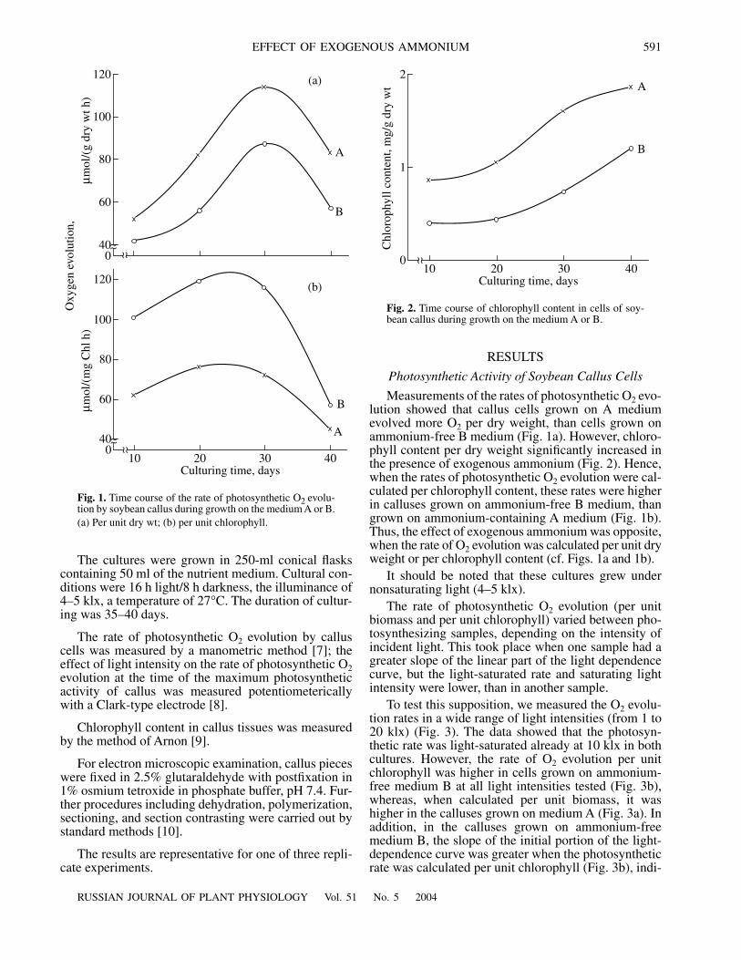
Measurements of the rates of photosynthetic O
2
evo-lution showed that callus cells grown on A mediumevolved more O
2
per dry weight, than cells grown onammonium-free B medium (Fig. 1a). However, chloro-phyll content per dry weight significantly increased inthe presence of exogenous ammonium (Fig. 2). Hence,when the rates of photosynthetic O
2
evolution were cal-culated per chlorophyll content, these rates were higherin calluses grown on ammonium-free B medium, thangrown on ammonium-containing A medium (Fig. 1b).Thus, the effect of exogenous ammonium was opposite,when the rate of O
2
evolution was calculated per unit dryweight or per chlorophyll content (cf. Figs. 1a and 1b).
It should be noted that these cultures grew undernonsaturating light (4–5 klx).
The rate of photosynthetic O
2
evolution (per unitbiomass and per unit chlorophyll) varied between pho-tosynthesizing samples, depending on the intensity ofincident light. This took place when one sample had agreater slope of the linear part of the light dependencecurve, but the light-saturated rate and saturating lightintensity were lower, than in another sample.
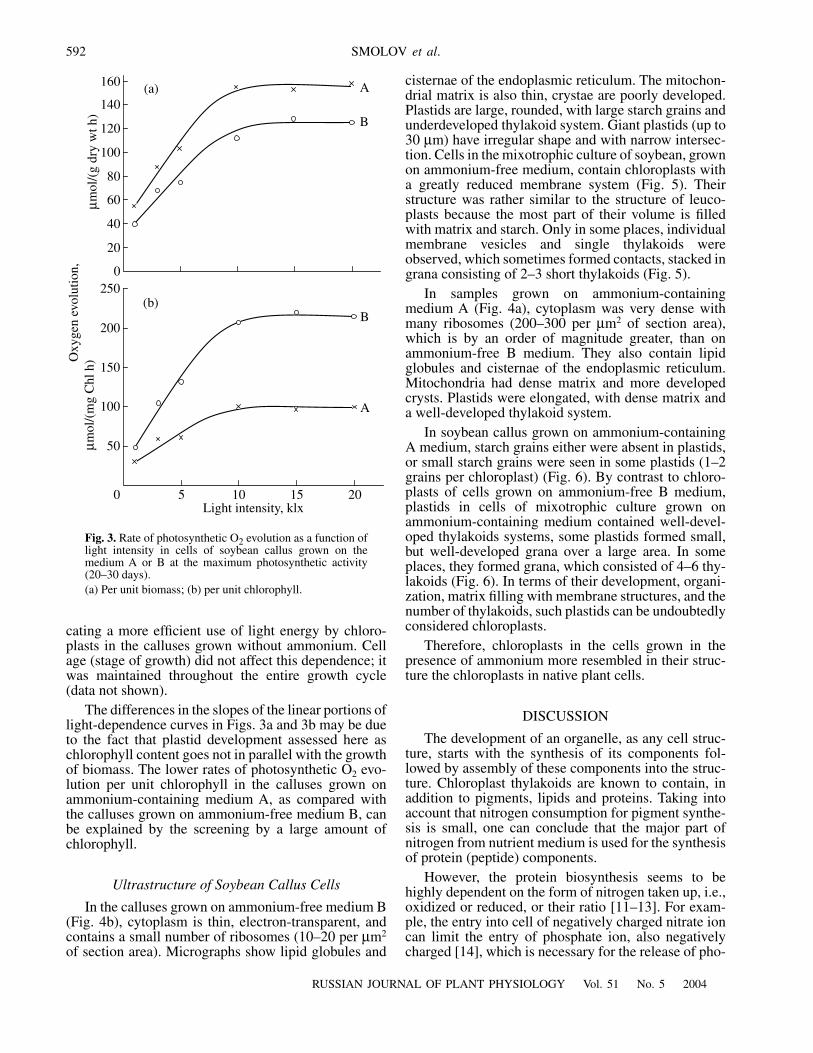
To test this supposition, we measured the O
2
evolu-tion rates in a wide range of light intensities (from 1 to20 klx) (Fig. 3). The data showed that the photosyn-thetic rate was light-saturated already at 10 klx in bothcultures. However, the rate of O
2
evolution per unitchlorophyll was higher in cells grown on ammonium-free medium B at all light intensities tested (Fig. 3b),whereas, when calculated per unit biomass, it washigher in the calluses grown on medium A (Fig. 3a). Inaddition, in the calluses grown on ammonium-freemedium B, the slope of the initial portion of the light-dependence curve was greater when the photosyntheticrate was calculated per unit chlorophyll (Fig. 3b), indi-
~~
~ ~040
60
80
100
120
Oxy
gen
evol
utio
n
,
µ
mol
/(g
dry
wt h
)
(a)
(b)
A
B
~~
~ ~
10 20 30 40
Culturing time, days
040
60
80
100
120
µ
mol
/(m
g C
hl h
)
A
B
Fig. 1.
Time course of the rate of photosynthetic O
2
evolu-tion by soybean callus during growth on the medium A or B.(a) Per unit dry wt; (b) per unit chlorophyll.
~ ~
10 20 30 40
Culturing time, days
0
1
2
Chl
orop
hyll
cont
ent,
mg/
g dr
y w
t
A
B
Fig. 2.
Time course of chlorophyll content in cells of soy-bean callus during growth on the medium A or B.
592
RUSSIAN JOURNAL OF PLANT PHYSIOLOGY
Vol. 51
No. 5
2004
SMOLOV
et al
.
cating a more efficient use of light energy by chloro-plasts in the calluses grown without ammonium. Cellage (stage of growth) did not affect this dependence; itwas maintained throughout the entire growth cycle(data not shown).
The differences in the slopes of the linear portions oflight-dependence curves in Figs. 3a and 3b may be dueto the fact that plastid development assessed here aschlorophyll content goes not in parallel with the growthof biomass. The lower rates of photosynthetic O
2
evo-lution per unit chlorophyll in the calluses grown onammonium-containing medium A, as compared withthe calluses grown on ammonium-free medium B, canbe explained by the screening by a large amount ofchlorophyll.
Ultrastructure of Soybean Callus Cells
In the calluses grown on ammonium-free medium B(Fig. 4b), cytoplasm is thin, electron-transparent, andcontains a small number of ribosomes (10–20 per
µ
m
2
of section area). Micrographs show lipid globules and
cisternae of the endoplasmic reticulum. The mitochon-drial matrix is also thin, crystae are poorly developed.Plastids are large, rounded, with large starch grains andunderdeveloped thylakoid system. Giant plastids (up to30
µ
m) have irregular shape and with narrow intersec-tion. Cells in the mixotrophic culture of soybean, grownon ammonium-free medium, contain chloroplasts witha greatly reduced membrane system (Fig. 5). Theirstructure was rather similar to the structure of leuco-plasts because the most part of their volume is filledwith matrix and starch. Only in some places, individualmembrane vesicles and single thylakoids wereobserved, which sometimes formed contacts, stacked ingrana consisting of 2–3 short thylakoids (Fig. 5).
In samples grown on ammonium-containingmedium A (Fig. 4a), cytoplasm was very dense withmany ribosomes (200–300 per
µ
m
2
of section area),which is by an order of magnitude greater, than onammonium-free B medium. They also contain lipidglobules and cisternae of the endoplasmic reticulum.Mitochondria had dense matrix and more developedcrysts. Plastids were elongated, with dense matrix anda well-developed thylakoid system.
In soybean callus grown on ammonium-containingA medium, starch grains either were absent in plastids,or small starch grains were seen in some plastids (1–2grains per chloroplast) (Fig. 6). By contrast to chloro-plasts of cells grown on ammonium-free B medium,plastids in cells of mixotrophic culture grown onammonium-containing medium contained well-devel-oped thylakoids systems, some plastids formed small,but well-developed grana over a large area. In someplaces, they formed grana, which consisted of 4–6 thy-lakoids (Fig. 6). In terms of their development, organi-zation, matrix filling with membrane structures, and thenumber of thylakoids, such plastids can be undoubtedlyconsidered chloroplasts.
Therefore, chloroplasts in the cells grown in thepresence of ammonium more resembled in their struc-ture the chloroplasts in native plant cells.
DISCUSSION
The development of an organelle, as any cell struc-ture, starts with the synthesis of its components fol-lowed by assembly of these components into the struc-ture. Chloroplast thylakoids are known to contain, inaddition to pigments, lipids and proteins. Taking intoaccount that nitrogen consumption for pigment synthe-sis is small, one can conclude that the major part ofnitrogen from nutrient medium is used for the synthesisof protein (peptide) components.
However, the protein biosynthesis seems to behighly dependent on the form of nitrogen taken up, i.e.,oxidized or reduced, or their ratio [11–13]. For exam-ple, the entry into cell of negatively charged nitrate ioncan limit the entry of phosphate ion, also negativelycharged [14], which is necessary for the release of pho-
0
20
40
60
80
100
120
140
160
Oxy
gen
evol
utio
n,
µ
mol
/(g
dry
wt h
)
A
B
(a)
(b)
50 10 15 20
Light intensity, klx
50
100
150
200
250
A
B
µ
mol
/(m
g C
hl h
)
Fig. 3.
Rate of photosynthetic O
2
evolution as a function oflight intensity in cells of soybean callus grown on themedium A or B at the maximum photosynthetic activity(20–30 days).(a) Per unit biomass; (b) per unit chlorophyll.

RUSSIAN JOURNAL OF PLANT PHYSIOLOGY
Vol. 51
No. 5
2004
EFFECT OF EXOGENOUS AMMONIUM 593
tosynthetic products from chloroplasts. It is likely thatthis was a cause of the accumulation of storage carbo-hydrates in chloroplasts of callus grown on pure nitratemedium (medium B), like in chloroplasts of pea andwheat leaf cells [14] (Fig. 5). The presence of positivelycharged ammonium ion in the nutrient medium and itsentry into the cell may relieve this limitation (Fig. 6),and newly synthesized photosynthetic carbohydratesare rapidly used in the biosynthesis of macromolecules.
However, the enhanced protein synthesis should bemaintained by accelerated production of amino acidsrequired for protein synthesis.
Ammonium was shown to stimulate the het-erotrophic (nonphotosynthetic) carbon dioxide fixa-tion, PEP-carboxylation in cultured cells [15]. Thisresults in the formation of a large amount of organicacids. Keto acids can enter in amination reactions withammonium absorbed from the medium, leading to theformation of amino acids.
(a)
(b)
0.5
µ
m
0.5
µ
m
C
R
ME
Mt
Mt
R
ER
Mt
ME
DNA
C
Mt
V
Fig. 4.
Ultrastructure of cytoplasm and mitochondria in the cells of soybean callus grown on medium (a) A or (b) B.C—cytoplasm, ER—endoplasmic reticulum, V—vacuole, Mt—mitochondria, ME—mitochondrial membrane, R—ribosome,DNA—mitochondrial DNA.

594
RUSSIAN JOURNAL OF PLANT PHYSIOLOGY
Vol. 51
No. 5
2004
SMOLOV
et al
.
However, this substrate role of ammonium was notconfirmed in experiments with Paul’s Scarlet Rose cells[12]. The authors noted that a twofold increase in theprotein content in the cells could not be secured by theaddition of 10% of ammonium nitrogen (on the back-ground of nitrate nitrogen). In addition, the stimulation
of PEP-carboxylase reaction in
Acer pseudoplatanus
cells was also observed in the presence of methylamine,a nonmetabolizable compound [15].
Therefore, in addition to a direct substrate role,ammonium ion can be a factor, which provides condi-
0.5
µ
m
0.5
µ
m
OG
CW CW
íh
SCE
VCE
MV
OG
íh
S
DNA
Fig. 5.
Ultrastructural organization of chloroplasts in cells of soybean callus grown on the ammonium-free B medium.CW—cell wall, CE—chloroplast envelope, S—starch, OG—osmiophilc globules, MV—membrane vesicles, DNA—DNA-con-taining region, Th—thylakoids. See Fig. 4 for other designations.

RUSSIAN JOURNAL OF PLANT PHYSIOLOGY
Vol. 51
No. 5
2004
EFFECT OF EXOGENOUS AMMONIUM 595
0.5
µ
m 0.5
µ
m
OG
CE
V
íh
CW
CE
CE
G
CE
CW
Gíh
OG
Fig. 6.
Ultrastructural organization of chloroplasts in cells of soybean callus grown on the ammonium-containing medium A.CW—cell wall, CE—chloroplast envelope, OG—osmiophilc globules, Th—thylakoids, G—grana.

596
RUSSIAN JOURNAL OF PLANT PHYSIOLOGY
Vol. 51
No. 5
2004
SMOLOV
et al
.
tions (for example, pH) for the enhancement of proteinsynthesis due to ribosome formation.
Further, the pigments synthesized in the presence ofammonium (Fig. 2), with proteins and lipids produce inchloroplasts a developed system of thylakoids, whichby stacking form grana, typical of native chloroplasts(Fig. 6).
Thus, ammonium containing in growth mediumactivates the formation of membrane structures of thephotosynthetic apparatus in the cells of mixotrophiccallus, like in chloroplasts of native photosynthesizingcells of higher plants, which might be due to a higherprotein-synthesizing activity of the cells. We believethat, in assessment of the functional activity of chloro-plasts (per unit chlorophyll or per unit dry weight), thestructural state of chloroplasts should be taken intoaccount because thylakoid functional activity is deter-mined by their photosystems.
REFERENCES
1. Sunderland, N. and Wells, B., Plastid Structure andDevelopment in Green Callus Tissue
Oxalis dispar, Am.J. Bot.
, 1968, vol. 32, pp. 327–346.2. Shiryaeva, G.A. and Gamburg, K.Z., Photosynthesis and
Tissue Culture,
Biokhimiya i biofizika fotosinteza
(Bio-chemistry and Biophysics of Photosynthesis), Rei-mers, F.E., Ed., Irkutsk: Sib. Otd. Akad. Nauk SSSR,1971, pp. 196–201.
3. Kaplan, S. and Arnttsen, Ch.D., Energy Conversion byPlants and Bacteria,
Photosynthesis
, Govindjee, Ed.,New York: Academic, 1982, vol. 1, pp. 162–265.
4. Smolov, A.P., Oleinikova, T.A., and Polevaya, V.S.,Nitrate Utilization in Cells of Hetero- and MixotrophicCultures of
Glycine max
L.,
Fiziol. Rast.
(Moscow),1992, vol. 39, pp. 875–886 (
Sov. Plant Physiol.
, Engl.Transl.).
5. Smolov, A.P., Kuznetsova, N.Yu., Oleinikova, T.A., andMoskalenko, A.A., Pigments and Pigment–ProteinComplexes in Chloroplasts from Soybean MixotrophicCallus: Effects of Ammonium and Diuron,
Fiziol. Rast.
(Moscow), 1998, vol. 45, pp. 653–658 (
Russ. J. PlantPhysiol.
, Engl. Transl.).6. Murashige, T. and Skoog, F., A Revised Medium for
Rapid Growth and Bioassays with Tobacco Tissue Cul-tures,
Physiol. Plant.
, 1962, vol. 15, pp. 473–497.7. Semikhatova, O.A. and Chulanovskaya, M.V.,
Mano-metricheskie metody izucheniya dykhaniya i fotosintezarastenii
(Manometric Methods for Study of Respirationand Photosynthesis), Moscow: Nauka, 1965.
8. Gavrilenko, V.F., Ladygina, M.E., and Khandobina, L.M.,
Bol’shoi praktikum po fiziologii rastenii
(Manual ofPlant Physiology), Moscow: Vysshaya Shkola, 1975.
9. Arnon, D.J., Copper Enzymes in Isolated ChloroplastPolyphenol Oxidase in
Beta vulgaris, Plant. Physiol.
,1949, vol. 24, pp. 1–15.
10. Ladygin, V.G. and Semenova, G.A., The Influence ofIron Deficiency on the Composition of Chlorophyll–Pro-tein Complexes and the Ultrastructure of Pea Chloro-plasts,
Fiziol. Rast.
(Moscow), 1993, vol. 40, pp. 841–849(
Russ. J. Plant Physiol.
, Engl. Transl.).11. Bergmann, L., Grosse, W., and Koth, P., Influences of
Ammonium and Nitrate on N-Metabolism, MalateAccumulation, and Malic Enzyme Activity in Suspen-sion Cultures of
Nicotiana tabacum
var. Samsun,
Z.Pflanzenphysiol.
, 1976, vol. 80, pp. 60–70.12. Mohanty, B. and Flatcher, J.S., Ammonium Influence on
Nitrogen Assimilating Enzymes and Protein Accumula-tion in Suspension Cultures of Paul’s Scarlet Rose,
Phys-iol. Plant.
, 1980, vol. 48, pp. 453–457.13. Edit, T. and Laszlo, T., Effects of NO
3
/NH
4
Ratio onPhotosynthesis Rate, Nitrate Reductase Activity, andChloroplast Ultrastructure in Three Cultivars of RedPepper (
Capsicum annuum
L.),
Plant Physiol.
, 1992,vol. 140, pp. 298–305.
14. Zernova, O.V., Effect of Oxidized and Reduced Forms ofNitrogen Nutrition on Ultrastructure of the Photosyn-thetic Apparatus in Pea and Wheat Leaves,
Fiziol. Rast.
(Moscow), 1993, vol. 40, pp. 431–437 (
Russ. J. PlantPhysiol.
, Engl. Transl.).15. Wright, K.M. and Givan, C.V., Regulation of Nonauto-
trophic Carbon Dioxide Assimilation by Ammonia inCultured Cells of
Acer pseudoplatanus
L.,
Plant Sci.
,1988, vol. 58, pp. 151–158.