effect of antibiotics for eradication of mrsa-carriage748098/fulltext01.pdf · indicates the growth...
TRANSCRIPT
Örebro University
School of Health and Medical Sciences
Department of Clinical Medicine
Medicine C, MC5501, Degree Project, 15 ECTS
May 2014
Effect of antibiotics for eradication of
MRSA-carriage
Version 2
Author: Sally Radwan
Supervisor: Dr. Jan Källman
Department of Clinical infection
University Hospital of Örebro

Abbreviations
MRSA Methicillinresistent Staphylococcus aureus
S.aureus Staphylococcus aureus
HA - MRSA Hospital acquired
CA - MRSA Community acquired.
SSTI Skin and soft-tissue infections
SCCmec staphylococcal cassette chromosome mec
PVL Panton-Valentine leukocidin
PMN human polymorphonuclear neutrophils
TSS Toxic shock syndrom
PCR Polymerase chain reaction
PBPs Penicillin-binding proteins
MGE Mobile Genetic Elements
MIC Minimal Inhibitory Concentration
ODD Oxacillin Disk Diffusion
MHA Muller-Hinton agar
CDD Cefoxitin Disk Diffusion
Fig. Figure
NaCl Sodium Chloride
μg Microgram
Hb Hemoglobin

Abstract:
MRSA are isolates of S.aureus which are not susceptible to the antibiotic Methicillin, which
used to be the first line therapy of treating S.aureus infection. Resistance to methicillin
implies resistance to all β-lactam antibiotics. The objective of this descriptive retrospective
observational study was to understand the outcome of the given treatment for eradication of
MRSA carriage, by revealing the percentage of re- colonization and eradication, as well as
compare the increase of HA and CA- MRSA infection during a period of October 2010-
October 2013. Investigations were made on a total of 105 MRSA positive individuals who
have been in contact with the care system within Örebro municipal. It was performed by
reading through their medical records and mainly focusing on the site of colonization,
outcome effect of the used antibiotics and focusing on the trend of HA - and CA- MRSA.
Both the pharynx and nasal sites were preferable colonization site of MRSA. The antibiotic
Dalacin and the nasal salvage, Bactroban nasal, showed a total result of 70% and 100%
respectively of eradicating MRSA- carriage. Other comparable antibiotics as Fucidin and
Eusaprim showed an eradication result of, 50% and 33% respectively. It was shown that
combined treatments, showed a higher incidence of re-colonization. The source of HA-MRSA
and CA-MRSA is increasing with the years and there is a three- fold increase of HA-MRSA,
while double fold increase of CA-MRSA. In conclusion, the preferable colonization site was
both the pharynx and nares. Bactroban Nasal and Dalacin showed most promising result for
eradication of MRSA carriage in contrast to the rest of the treatments, including combined
treatments. The both sources of HA- MRSA and CA- MRSA showed an increasing trend with
years. Further studies that primarily include a greater number of patients would facilitate more
accurate results.
Key words: MRSA, β-lactam, HA-MRSA, CA-MRSA, S.aureus Dalacin, Bactroban Nasal,
re- colonization, eradication, colonization.

List of Content
Introduction .............................................................................................................................. 5
Microbial characteristics of S.aureus .................................................................................... 5
Colonization .......................................................................................................................... 5
Virulence character and toxins of S.aureus ............................................................................ 6
MRSA ................................................................................................................................... 7
Genetics of MRSA................................................................................................................. 8
Difference in characteristics between HA and CA MRSA .................................................. 8
Molecular techniques for detection of MRSA ....................................................................... 9
β-lactam antibiotics ............................................................................................................. 10
Medications for eradication of MRSA ................................................................................ 11
Aim ........................................................................................................................................... 12
Materials and Method ............................................................................................................ 12
Materials ........................................................................................................................... 12
Methods ............................................................................................................................ 12
Ethics ................................................................................................................................ 13
Results ..................................................................................................................................... 14
Discussion ................................................................................................................................ 17
Acknowledgement .................................................................................................................. 20
References ............................................................................................................................... 20

Introduction
Microbial characteristics of S.aureus
S. aureus is a species that taxonomically belongs to the genus Staphylococcus as well as the
family Staphylococcaceae. They are classified as gram- positive spherical bacteria, forming
clusters, which microscopically resembles grapes. They are 1 micrometer in diameter. S.
aureus colonizes mainly the nasal passages. It can additionally be found in the skin, pharynx,
hair follicles, and perineum. They may cause a wide range of infections and intoxications. The
have the capacity to ferment Mannitol. The species forms a fairly large yellow colony on rich
medium and it is often hemolytic on blood agar. Staphylococci are facultative anaerobes. This
indicates the growth either by aerobic respiration or by fermentation that yields mainly lactic
acid. They can grow at temperature range of 15 to 45 degrees. The bacterium is catalase-
positive, coagulase-positive and oxidase-negative. This species are non- motile as well as
non-spore forming.
S.aureus strain has potential to cause diseases and hence called pathogenic. Specific strains of
S.aureus can cause range of diseases via its virulence determinants of its structure,
biochemical or/and its genetic features. They can for instance cause septicemia, food
intoxication, and toxic shock syndrome but mostly pus and wounds infections. Since it occurs
in human flora, it ensures the capacity for transmission from one individual to another.
Colonization
Colonization refers to the presence of a high concentration of bacteria at a site that they can be
detected, without causing any signs of illness or infections. However, it is a risk factor for
consequent clinical infection, if colonized MRSA on the skin invades an open passage as
wound or cut in the skin [1, 2]. In case of infections, bacterias were not initially present in the
site from which it has been detected, but was introduced either from another source or from
contamination. [3, 4]. It can be clinically identified as wound infections, surgical site
infection, abscesses, though, many organs can also be effected and give rise to life threatening
status [4]. Patients involved in the hospital care have a risk factor to develop MRSA infection
if they have weakened immune system, because of a certain medical condition or treatment
(e.g. chemotherapy), recent use of antibiotics, hospitalized for a longer period or having a
surgical wound and/ or intravenous line. Patients which have hemodialysis have a significant
high risk to become infected [2]. Community associated risk factors to become infected
includes usually healthy individuals with skin issues as eczema, or have an opening site for
the passage of MRSA as in the case of having tattoos or body piercing. Athletes are also
included in this category as they are usually exposed to physical contact [2].
The most preferable condition for growth and survival of MRSA is 25°C and 11-33% relative
humidity. The higher relative humidity the less ideal environment, leading to less MRSA
survival [5]. The most frequent site of colonization is the anterior part of the nostrils, but, it
also colonizes in the pharynx and perineum/ groin [6, 7]. They way to get colonized by
MRSA can occur via physical contact, by touching the skin of another individual who is an
MRSA carrier. Another way is to touch contaminated surfaces as door handle or phone) [2].
Studies have shown three different types of MRSA carriage, which are: persistent,
intermittent carriers, and non- carriers [8-10]. The persistent carriers have a greater prevalence
of colonization of other body sites in addition to the nostrils and rarely alter their strains in
contrast to the intermittent bacteria [11]. Transient colonization is referred to individuals who
are temporarily colonized by MRSA for instance in nostrils or pharynx, without establishment
of bacterias. Hence, MRSA positive result will only be shown during the occasional screening
[11].
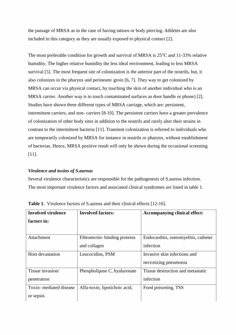
Virulence and toxins of S.aureus
Several virulence characteristics are responsible for the pathogenesis of S.aureus infection.
The most important virulence factors and associated clinical syndromes are listed in table 1.
Table 1. Virulence factors of S.aureus and their clinical effects [12-16].
Involved virulence
factors in:
Involved factors:
Accompanying clinical effect:
Attachment
Fibronectin- binding proteins
and collagen
Endocarditis, osteomyelitis, catheter
infection
Host devastation
Leucocidins, PSM Invasive skin infections and
necrotizing pneumonia
Tissue invasion/
penetration
Phospholipase C, hyaluronate Tissue destruction and metastatic
infection
Toxin- mediated disease
or sepsis
Alfa-toxin, lipotichoic acid,
Food poisoning, TSS

S.aureus has the ability to produce a plethora of toxins and some are mentioned below:
Panton-Valentine leukocidin
PVL is a staphylococcal leukocidin (cytotoxic to white blood cells) which belongs to the
pore-forming toxin family, β-barrel molecular complex. It also comprises other toxins as γ-
toxin, and α-toxin. It induces lysis of host defense cells as PMNs, monocytes and
macrophages [13]. It is encoded by lukS- PV and lukF genes-PV, which in turn encodes the
pore the cytolytic activity, LukS-PV and LukF- PV. Clinically, they are responsible for skin
abscesses [16].
Phenol-soluble modulins
PSMs are a family of amphipathic, α-helical peptides produced by staphylococci. They are
peptide toxins, which are known to attract and lyse neutrophils and erythrocytes [15].
Alpha-toxin
Most S.aureus strains produce α- toxin, which has a cytolytic property. It is not cytolytic to
neutrophils but it lyses macrophages and erythrocytes and has pro-inflammatory effects.
Finally, alpha-toxin has been shown to contribute to the penetration of the epithelial barrier
during skin infection with the MRSA strain USA300 [16].
Methicillin-Resistant Staphylococcus aureus
MRSA are defined as isolates of S.aureus which are not susceptible to antibiotics methicillin,
which used to be the first line therapy of treating S.aureus infection. Resistance to methicillin
implies resistance to all β-lactam antibiotics [17, 18]. Therefore, precision and swiftness with
detection of methicillin resistance is of key importance to ensure correct antibiotic treatment
in infected patients, as well as control of HA- MRSA isolates in hospital to avoid spreading.
The phenomenon was first reported 1961 and became a major problem to take into
consideration in many parts of the world in the late 1970s and early 1980 [2, 17].
The first MRSA was isolated by Patricia Jevons. From 1980s new strains of MRSA appeared
and led to uninterrupted pandemic infections of MRSA globally [17]. Since this bacterium is
able to produce series of toxins and cause harm [12, 13], the continuous emergence of
mutations of the specific genes responsible for the resistance of treatment, leads to difficulty
to prevent and control MRSA. MRSA infections were initially confined to the health care

environment, but have also been found in the community [19, 20]. Both HA- MRSA and CA-
MRSA share some strains but differs in antimicrobial susceptibility and potential virulence
[19, 20]. Contaminated devices, medical waste as well as medical staffs that carry MRSA is
considered as an infection source [2, 4, 21].
Genetics of MRSA
MRSA has a group of MGE genes, which is mobile within the genome and plays a significant
role in the spread of virulence factors. It is a fragment of DNA which transfers into the host
cell where it can either replicate or integrate in host DNA. The genetic component responsible
for resistance, mecA, is not native to the S.aureus genome. The MecA gene located on
SCCmec is characterized as a mobile resistance element. There are 5 SCCmec subtypes, I- V.
Subtype I, IV and V have a small size and encode recombinase genes as well as structural and
regulatory genes for resistance to methicillin [14].
MRSA have the ability to produce PBP2a protein, altered penicillin- binding protein, encoded
by the mecA gene. It has a property of low affinity for β-lactam antibiotics, and hence
minimizing its target effect [15]. This gene is regulated by two genes located upstream of the
mecA gene, called mecR1 and mecl. Together they are mentioned as mec complex. The
mecR1 gene encodes a transmembrane inducer of mecA consisting of membrane- spanning
and penicillin binding domains, while the other gene, mecl, encodes a strong repressor of
mecA [17, 22]. The resistance level differs in a spectrum from phenotypically susceptible to
highly resistant. This is dependent on the PBP2a production, which is affected by
chromosomal factors. That’s why all mecA clones are not resistant to methicillin [14].
Difference in characteristics between HA - and CA - MRSA
HA-infection is defined as an infection that occurs for a patient who was not admitted to the
hospital or other health care setting for that reason, neither was it incubating at the time of
admission [20, 23]. The infection can occur 48 hours of hospital entrance as well as three days
of discharge, as well as 30 days after an operation [23, 24]. It is known that CA- MRSA
occurs in a person with no previous history of health care exposure [20, 23, 24]. However, the
definition can be misleading. Since time- based definition regarding hospital discharge can be
ambiguous since S. aureus has the characteristics to persist as a colonizer for months or years
and the onset of CA- infection may be caused by HA- strains [19,22, 24-27]. People who were
not hospitalized but were detected with SSTI were reported to have CA- MRSA [27]. The
strains of CA- MRSA are often resistant to fewer antibiotics, compared to the HA- MRSA
strains, owing to the pressure of antibiotics that have been within hospitals [19, 22]. It usually
has IV and V subtypes of SCCmec unit and type IV usually contain the gene encoding for
PVL. They are classified as more virulent. HA- MRSA isolates classically have the subtypes’
I-III and rarely carry the gene encoding for PVL [19].
From a prospective study, it was found that CA- MRSA isolates were in a higher extent,
compared to HA- MRSA susceptible to ciprofloxacin, clindamycin, erythromycin and
Gentamicin [19]. Since the two isolates have SCCmec types which can carry various
additional genetic information, it is possible for them two develop resistance to several
antibiotic classes, which commonly occurs in HA- MRSA [19].
MIC values of HA-MRSA clones are usually higher than those of CA-MRSA clones [19].
Molecular techniques for detection of MRSA
There are many techniques available for detection of MRSA. It can for instance be performed
by either amplification of genes by the usage of PCR alternatively by phenotypic testing.
Then phenotype of resistance can vary even though genetic homogeneity depending on
culture conditions (temperature and osmolarity of the medium) [28]. This in turn can be
difficult for detection of MRSA with phenotypic susceptibility methods. Oxacillin disk
diffusion is one of the traditional methods for MRSA screening, but lately cefoxitin disks as
well as PBP2a latex agglutination have also been used as it yields high precision [29]. These
methods are categorized as antibiotic susceptibility tests, which work as culture screening
method. It works by collecting culture from colonized area as pharynx, nares and perineum
and inoculate on a MRSA- selective chromogenic agar. Most of the methods yield a result of
presumptive MRSA colonies the next day, while the method of using latex agglutination can
confirm the result after 10 min. In this method, antibodies are used against PBP2a, which are
extracted from suspensions of colonies and the detection is made by agglutination with latex
particles coated with monoclonal antibodies to PBP2a. This test may not be reliable for
colonies grown on media containing NaCl [22]. ODD test works by inoculating the suspected
MRSA colony onto MHA. 1 μg Oxacillin disks is applied and then get incubated at 35 °C for
24 h. An inhibition zone <10 mm in diameter indicates oxacillin resistance. In comparison to
CDD method, 30 μg disks are applied and the suspected MRSA isolates are inserted onto
MHA plates. The plates are then incubated at 37 °C for 18 h to allow the bacteria to grow.

Diameters of inhibition zone ≤21 mm indicated oxacillin resistance, and diameters ≥22 mm
are considered to indicate sensitivity [22].
PCR- based molecular methods is used by the aid of primers designed to amplify species-
specific targets. These targets are nuclease (nuc), coagulase (coa), protein A and surface-
associated fibrinogen- binding protein genes. However, lately, this method is mainly used for
detecting the mecA gene. It is a very efficient way to yield result quickly but yet expensive
method of choice [19].
The screening duration vary from every county council in Sweden. Positive MRSA patients
who have been in contact with the University Hospital of Örebro are screened once again after
one month after the first detection. The second screening opportunity is three months after the
first screening. If the patient continues to be positive, a new screening opportunity will be
offered again after 3 months, but if negative after 6th
months. Four negative screenings in a
row defines no longer as a carrier. MRSA screenings can be performed either by DNA testing
or by investigating bacterial culture by taking samples from for instance nares, pharynx,
wounds, perineum/ groin and urine.
β- lactam antibiotics
β- lactams are widely prescribed antibacterial drugs which is capable to lyse dividing bacteria
and therefore considered as bactericidal. Drugs within this class share the same mechanism of
action, as they all contain a β -lactam ring in their structure. Their principal function is to
inhibit the cell wall synthesis of bacteria by binding to PBPs located in their cell wall.
The conjugation leads to cell death by inhibition of peptidoglycan synthesis, and inactivating
an inhibitor of an autolytic enzyme that crosslink the peptide chains, attached to the
peptidoglycan. However, their chemical structures vary for the different agents and therefore
differ in the spectrum of action [18, 30, 31].
S.aureus has the ability to produce four PBPs which are susceptible to modification by beta-
lactam antibiotics, which allows the antibiotics to be considered bactericidal. The MRSA
bacterias additionally contain the mecA gene. A mutation in this gene produces an alternative
type of the four standard PBP, called PBP2a. This enzyme provides transpeptidase activity to
allow cell wall synthesis at beta-lactam concentrations that inhibit the β- lactam- sensitive
PBPs, and thus bestows resistance [18, 30, 31].

There are four subclasses of β- lactam agents named penicillins, cephalosporins (which are
subdivided into four generations), monobactams and carbapenems. Those vary structurally
except having the same β- lactam ring. Broad spectrum antibiotics are noted to have a broader
effect against both gram positive as well as negative bacteria while the narrow spectrum is
typically effective against Gram positive bacterias. The spectrum of action is dependent upon
two factors. Firstly, it concerns the degree of penetration of cell wall and the outer membrane,
as well as the binding ability to specific transpeptidases. Both hydrophobic and hydrophilic β-
lactams are capable to diffuse through the murein layer of gram positive bacteria. The
hydrophilic agents are additionally capable to diffuse readily through the gram negative
bacterias’ outer membrane pores in comparison to hydrophobic agents. Secondly, is the extent
to which the drug inhibits a specific transpeptidases, after accessing the periplasmic space.
Penicillin has a short plasma half- life and the elimination of most penicillin occurs via renal
elimination, 90% through tubular secretion [28, 32].
Medications for eradication of MRSA
The antibiotic Dalacin, trade brand for Clindamycin shows in some studies an appealing result
of MRSA eradication [33, 34]. However, many strains of MRSA are also resistant to it and it
should not be used when treating systemic or bacteremic infections where MRSA is the
suspected pathogen [35, 36, 37]. Rimactan which is the brand name for Rifampicin works as
bactericidal in vitro activity against MRSA [37]. Studies have proved that using this drug in
monotherapy will contribute to a rapid emergence of resistance [38, 39]. Rimactan is rather
suggested to be used in combined therapy, because it has shown to result with a synergistic
activity against staphylococci [40]. Studies have shown that Rimactan in combination with
Clindamycin yields a successful eradication of MRSA carriage [20, 41, 42]. Combined
therapy of topical antibiotics as nasal salvage and systemic antibiotic has shown to result in
good eradication result of MRSA- carriage too [43]. Nasal salvage in monotherapy has also
shown to eradicate MRSA-carriage efficiently as well as result in minimal toxicity and
approved for MRSA outbreaks [45, 46].

Aim
The aim of this research is principally to understand the outcome effect of the given treatment
for eradication of MRSA carriage by revealing the percentage of re- colonization and
eradication, as well as compare the increase of HA and CA- MRSA during a period of
October 2010- October 2013.
Material and Methods
Materials
The main material used was “Klinisk Portal”. It is a systemic computer program which can
only be used within health care settings, to view medical records. It saves information
regarding patients who have been in contact with the health care system only in Örebro
County. From this program it is possible to search patients who were positive for MRSA from
October 2010 till October 2013. In advance to the initiation, an outburst- list of all positive
patients at different time interval with their identity number was received; it was a total of 105
patients.
Method
From the outburst list received, positive patients within the time interval October 2010-
October 2013 were selected. Information regarding their medical status of MRSA and the
treatments was looked up in Klinisk portal. While reading through the patients’ medical
records, I focused on points regarding the amount of patients infected during the time interval,
source of MRSA, CA- MRSA or HA- MRSA and the sites of colonization, where MRSA
screenings from nares, pharynx, wounds, perineum/ groin showed positive result. Screenings
were performed on patients with SSTI, tracking of infected family members, adopted (during
adoption screening), wound infections, postoperative infections, patients who have been
treated abroad. Any patient with at least one MRSA positive colonization site was regarded as
MRSA positive. The screened patient that is a carrier in more than one site, have all their
positive sites included in the study too, as the preferable site of colonization. I also focused on
treatments for eradication of carriage. Different treatments were tested on different colonized
patients. However, only 70 patients out of 105 MRSA positive were treated with antibiotics
(especially short term carriers). A patient is considered to be eradicated from MRSA when
four negative MRSA screenings is resulted. Some patients that continue as MRSA carriers
after treated infection are considered re-colonized. They are included in the study as
recolonization after given treatment. After collecting the needed data, I used the program

Excel, where I could set up the data into charts. I had three charts in total for every year. First
chart represents the site and source of infection correlated with the number. The second chart
explained the given treatment correlated with the number given. The last chart represented the
reinfection site correlated with the treatment used. These charts were then converted into bar
graphs, for a visual comparison between the different years, starting from October 2010 –
October 2013. The charts referring to the site of colonization for all the three years were fused
together into one bar graph. The numbers of patients with a source of infection from CA-
MRSA and HA-MRSA or other (adoption screening or abroad stay) were made into a single
graph, and the three different years were distinctive, with its number. Patients considered as
HA- infected were those who had a post-operative infection, infected staff within the health
care setting, infected patients in the hospital. CA- MRSA infected patients are those who
appeared at the primary care and had a wound or SSTI and got screened for MRSA. Family
members who were infected were included in the category of CA- MRSA. The third graph
made was referring to the efficacy of medication given to treat patients with colonized MRSA
with topical ointment or together with antiseptics, Hibiscrub, while the severely colonized
yielded systemic antibiotic combined with antiseptic solution and topical ointment. Systemic
antibiotic refers to treating the whole body with tablets and capsules with Dalacin combined
with Rimactan per os. The infected individuals also received solely systemic antibiotic as
Dalacin and Eusaprim.
Individuals who were solely visitors in Sweden or asylum-seeking, and got in contact with the
health care system in Örebro were also registered as MRSA positive. They become a part of
the obtained data concerning the County. Frequently, the asylum- seeking patients does not
have a prolonged valid personal date of birth, which in turn restricts additional searching
regarding the points to be considered for the investigation. Therefore, those people were
excluded from this study. Registered MRSA positive patients that deceased and does no
longer have an accessible medical record, were also excluded from this study. However,
individuals who were settled in another city prior to Örebro, and were already MRSA positive
and later came in contact with the hospital care in Örebro were included in the study.
Ethical considerations
This study is approved from the operation manager of the infection center of University
Hospital of Örebro. No personal records from patients will be revealed in the academic
presentation, and I am personally well informed regarding the confidentiality within
healthcare, when reviewing medical records.
Results
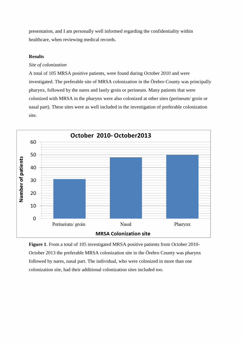
Site of colonization
A total of 105 MRSA positive patients, were found during October 2010 and were
investigated. The preferable site of MRSA colonization in the Örebro County was principally
pharynx, followed by the nares and lastly groin or perineum. Many patients that were
colonized with MRSA in the pharynx were also colonized at other sites (perineum/ groin or
nasal part). These sites were as well included in the investigation of preferable colonization
site.
Figure 1. From a total of 105 investigated MRSA positive patients from October 2010-
October 2013 the preferable MRSA colonization site in the Örebro County was pharynx
followed by nares, nasal part. The individual, who were colonized in more than one
colonization site, had their additional colonization sites included too.

Source of infection
Patients with both CA- and HA- MRSA showed an increasing trend of MRSA through the
investigated time interval. However, the dominating source of MRSA infection showed to be
CA-MRSA. There is an approximate doubling of CA- MRSA infections in comparison to
HA- MRSA, which has a threefold increase from October 2010- October 2013. The increase
of HA- MRSA is alarming and causes concerns. Other sources of MRSA infection did not
show to be a leading source (fig 2).
Figure 2. From a total of 105 patients, an increasing trend of both HA and CA- MRSA
infections during the different time intervals can be seen. Pattern of other sources of MRSA
can also be distinguished.
Tested treatments on MRSA positive patients, showed various outcome effects. The outcome
effect of the given treatment to both infected and colonized MRSA patients are elucidated by
the relation between re-colonized and eradicated MRSA carriers after given treatment. Re-
colonization is referred to MRSA colonization at any specific site after a treatment, while
eradicated refers to a total of four negative MRSA screenings in a row after given treatment.
The highest incidence of recolonization with MRSA was shown after treatment with
Hibiscrub, Bactroban nasal and systemic antibiotic. Only 25% were eradicated while 75%
were re-colonized at a colonization site. However, a minor difference was noted without the
use of systemic antibiotic. The number of eradication showed a total of 52% while re-
colonization of 47%. Bactroban Nasal demonstrated a successful result of 100% eradication.
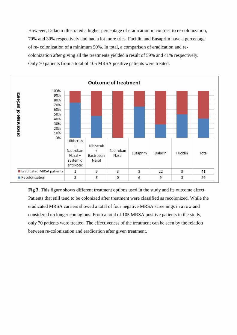
However, Dalacin illustrated a higher percentage of eradication in contrast to re-colonization,
70% and 30% respectively and had a lot more tries. Fucidin and Eusaprim have a percentage
of re- colonization of a minimum 50%. In total, a comparison of eradication and re-
colonization after giving all the treatments yielded a result of 59% and 41% respectively.
Only 70 patients from a total of 105 MRSA positive patients were treated.
Fig 3. This figure shows different treatment options used in the study and its outcome effect.
Patients that still tend to be colonized after treatment were classified as recolonized. While the
eradicated MRSA carriers showed a total of four negative MRSA screenings in a row and
considered no longer contagious. From a total of 105 MRSA positive patients in the study,
only 70 patients were treated. The effectiveness of the treatment can be seen by the relation
between re-colonization and eradication after given treatment.
Discussion
The aim of this study was to investigate the outcome of the given treatment for eradication of
MRSA carriage by revealing the percentage of re- colonization and eradication, as well as
compare the increase of HA and CA- MRSA during a period of October 2010- October 2013.
Out of 105 MRSA positive patients, only 70 people were treated with various medications. A
total of 31 trials with the antibiotic Dalacin were made on MRSA carriers and 70% were
eradicated from the bacteria. Dalacin showed to be the most effective drug per os with a lower
incidence of re-colonization in contrast to the other given treatments (figure 3). However, it
still demonstrates a high percentage of re-colonization. A hypothesis regarding the
susceptibility to antibiotics and the type of MRSA strain can be considered. Another study has
also showed an appealing result when using Dalacin for eradication of MRSA and yet a lesser
recolonization incidence was obtained [33]. Nevertheless, other studies also showed a lesser
recolonization incidence when using combined therapy with Rimactan rather than solely
monotherapy [38- 40] which was not in this case, where a total of recolonization incidence
showed a total of 75%. Rimactan is considered as an effective drug which effectively
eradicates MRSA from mucosal surfaces [38]. Bactroban Nasal, the local ointment, also used
for eradication, showed a total of 100% successful result. Several studies have also indicated
similar pleasing result [40- 42]. Nonetheless, in this study, the combination of Hibiscrub and
Bactroban nasal did not yield as tempting results as many studies have shown its efficacy to
eradicate a high number of colonized MRSA [46-48]. In this investigation it rather showed the
next highest recolonization incidence after giving this combined treatment. Probable reason
can be regarded as confusion on misunderstanding about the scrub or nasal salvage as
personal items [2, 3, 47]. Since other studies have shown to give an appealing result [46-48],
more tries with this treatment has to be tested on the carriers in order to get a more reliable
result.
The fundamental threefold increase of HA-MRSA (fig.2) is most probably due to scamp with
hygiene as many studies also indicates [49, 50]. The increase of CA-MRSA incidence leads to
increased proportion of patients who will be hospitalized, (due to MRSA- carriage).
Comparing this result to another study made in United States of America, the number of CA-
MRSA and HA- MRSA infected are also increasing. CA-MRSA is considered as the most
common pathogen for causing wound infections [50]. The hypothesis regarding the
downward slope of other sources of MRSA (fig 2) could possibly depend on more people who
tend to be more cautious while traveling abroad. The cautious note is probably due to the

increased awareness of getting bacterial infections while traveling abroad, which is well
described in the traveling brochures etc.
Many studies show the target site of MRSA colonization is in the anterior nares [29, 44] and
an insignificant distinguish can also be seen in this study between the amount of patients
colonized with MRSA in nares and in the pharynx. These preferable sites of colonization are
due to the appropriate environment; temperature and humidity that appear to be a possible
source of growth, and multiplication [2]. This can also be associated with the fact that these
parts of the human body are nearly always exposed to contagion. Touching contaminated
surfaces (with MRSA) or having physical contact with someone who is colonized with MRSA
assists colonization. Additionally, individuals practically always use hands to itch the nose or
grabbing eatable materials, which are also contributing factors [38-40].
Before starting the investigation, several points were supposed to be considered as: the site of
colonization, amount of patients infected during the investigated time interval, source of
infection, treatments for eradication of MRSA carriage as well as the outcome effect of the
treatments used by analyzing the relation between the eradicated and recolonized. The
eradicated MRSA individuals are those who have showed four negative screenings in a row.
Nevertheless, several factors were excluded from the study including the age, gender,
socioeconomic status, comorbidities, individuals who have ceased and does not have an
available medical record and the asylum- seeking patients who have been in contact with the
health care setting but does no longer have a valid personal identification number for looking
through their medical records. Visitors to Sweden who were positive for MRSA after the first
screening were excluded too. People who were settled in Örebro and were positive for MRSA
and moved to another city were also excluded from the study.
The weakness of this retrospective observational study mainly includes too few tries of
different medications on the limited number of patients. The lesser tries, the less reliable
results and thus, an improvement can be made by for instance extending the time interval that
was investigated, in order to get more available medical records, which could influence the
obtained results. Additional improvements can also be made by including a control group that
indicates whether a decrease of MRSA carriage occurs after received topical or systemic
treatment. Furthermore, verification of eradicated MRSA carriage by clinical investigation
including white blood cell count and Hb, could possibly influence the view of eradication
result differently. Another weakness concerns the irregular screening schedule for some
patients due to their case and the duration of their given treatment for eradication of MRSA.
Usually, screening routines are performed in the following manner that after detection of
MRSA after the first time, the second screening opportunity is after three months. If the
patient continues to be positive, a new screening opportunity is maintained again after 3
months, but if negative after 6th
months. Four negative screenings in a row defines as
eradicated MRSA. Changes in the standard screening routine can yield a different result and
therefore not a concrete result can be made regarding the treatment options and the results can
be misleading too. Additionally, some patients are already on other antibiotics for some other
infections and the outcome of the screening can influence the verification of eradication.
People who are having wounds or skin issues cannot start treatment without their issue being
solved, and since not many confirmed information regarding healing process was mentioned
in the medical record, the result of the eradication after the treatments could have been
different. Improvement can be done by enlightening information about healed bodily issues as
wounds in advance to a treatment. This would possibly contribute to more accurate answer
regarding eradicated number.
This retrospective study showed an increased colonization of MRSA at the pharynx, however,
a slight difference was found in the nasal part. Many studies have shown that nares are the
preferable site of colonization [9, 29, 44]. Moreover this study also showed an increased trend
amongst a source of infection from CA-MRSA as well as HA-MRSA during the time interval
from October 2010- October 2013. The threat of the rising number of infected MRSA within
a population of a county is alarming. The increasing trend occurs possibly due to the
increased number of immigrants to Örebro County, and the lack of knowledge regarding
bacterias as well as misunderstanding information received from the care systems. Another
possible point of view concerning the increased number of CA- MRSA is due to the lack of
knowledge on how to prevent the spreading of bacterias in the society amongst different
communities that are not associated with health care settings, which include athletes,
intravenous drug users and prisoners [2, 51]. The increasing amount of HA- MRSA carriage
is most likely multifactorial. The increase of CA-MRSA incidence leads to increased
proportion of patients who will be hospitalized, (due to MRSA- carriage). Lack of compliance
with hygiene routines amongst the staff also induces an increased number of HA-MRSA [49,
50]. However, the most preferable drug shown to effectively eradicate MRSA carriage is by
the use of Dalacin per os and/ or the local ointment Bactroban nasal. Many studies have also
confirmed that conclusion [43, 45, 46]. Studies have also revealed the effectiveness of
Dalacin in combination with Rimactan per os [20, 40-42], which in this study was not the
case. Informing carriers as well as their next of kin thoroughly regarding MRSA, the spread
and its resistance pattern, might increase the attention of the topic which is required for a joint
effort to prevent the inclination.
Acknowledgement
I would like to give a special thanks to Dr. Jan Källman for the support and guidance and
giving me the opportunity to work independently at the infectious center at the University
Hospital of Örebro. This project gave me an improved understanding regarding the worldwide
concern of MRSA and how eradication of MRSA-carriage can be performed by diverse
treatments. Thank you
References
[1] Peters PJ, Brooks JT, McAllister SK, Limbago B, Lowery HK, Fosheim G, et al.
Methicillin-Resistant Staphylococcus aureus Colonization of the Groin and Risk for Clinical
Infection among HIV-infected Adults. April 2013;19, ( 4): 623-629
[2] Jufeng X, Jianjun G, Norihiro K, Kiyoshi H, Wei T. Methicillin-resistant Staphylococcus
aureus antibiotic resistance and virulence. BioScience Trends. Rev. 2013; 7(3):113-121.
[3] Robinson J, Colonization and infection of the respiratory tract: What do we know?.
Paediatr Child Health. 2004 January; 9(1): 21–24
[4]- Fridkin, S.K., et al., Methicillin-resistant Staphylococcus aureus disease in three
communities. N Engl J Med, 2005. 352(14): s.1436–44.
[5] Hardbarge A. Viability of Methicillin-Resistant Staphylococcus aureus on Artificial Turf
Under Outdoor and Laboratory Environmental Conditions. Ohio: Ohio University; June 2012
[6] Wertheim, H.F., et al., The role of nasal carriage in Staphylococcus aureus infections.
Lancet Infect Dis, 2005. 5(12): s.751–62.
[7] Ringberg, H., et al., The throat: an important site for MRSA coloni-zation. Scand J Infect
Dis, 2006. 38(10): s.888–93.

[8] Kluytmans J, Belkum A, and Verbrugh H. Nasal carriage of Staphylococcus aureus:
epidemiology, underlying mechanisms, and associated risks. Clin Microbiol Rev, 1997. 10(3):
505- 20.
[9] Nouwen, J, Belkum A, and Verbrugh HA. Determinants of Staphylococcus aureus nasal
carriage. Neth J Med. 2001; 59(3): 126–33.
[10] Peacock S, De Silva I, and Lowy FD. What determines nasal carriage of Staphylococcus
aureus? Trends Microbiol. 2001; 9(12): 605–10.
[11] Wertheim H.F. The role of nasal carriage in Staphylococcus aureus infections. Lancet
Infect Dis. 2005; 5(12): 751–62.
[12] Gordon R J and Lowy F D. Pathogenesis of Methicillin-ResistantStaphylococcus aureus
Infection. Clin Infect Dis. 2008; 46 (S5): S350-S359
[13] Genestier AL, Michallet MC, Prévost G, Bellot G, Chalabreysse L, Peyrol S, Thivolet F,
Etienne J, François M. Staphylococcus aureus Panton-Valentine leukocidin directly targets
mitochondria and induces Bax-independent apoptosis of human neutrophils. J Clin Invest.
2005 Nov 1;115(11):3117–3127.
[14] Charles E Okolie. Engineering of the LukS-PV and LukF-PV subunits ofStaphylococcus
aureus Panton-Valentine leukocidin for Diagnostic and Therapeutic Applications BMC
Biotech. 2013 November 19; 13:103.
[15] Schreiner J1, Kretschmer D, Klenk J, Otto M, Bühring HJ, Stevanovic S, Wang JM,
Beer-Hammer S, Peschel A, Autenrieth SE. Staphylococcus aureus phenol-soluble modulin
peptides modulate dendritic cell functions and increase in vitro priming of regulatory T cells.
PubMed. 2013 Apr 1;190(7):3417-26.
[16] Otto M, Wittle W. Community-associated MRSA: What makes them special?
International Journal of Medical Microbiology.2013 August; 303: 324–330
[17] Alipour F, Ahmadi M, Javadi S. Evaluation of different methods to detect methicillin
resistance inStaphylococcus aureus (MRSA). Journal of Infection and Public Health. 2014
May; 7 (3): 186–191
[18] Chambers HF. Penicillin-Binding Protein-Mediated Resistance in Pneumococci and
Staphylococci. J Infect Dis. 1999; 179(S2): S353-S359.

[19] Peter C. Appelbaum. Microbiology of Antibiotic Resistance in Staphylococcus aureus.
Oxford Journals, .Clin Infect Dis. 2007; 45 (S3): S165-S170.
[20] Huang H, Neil M. Flynn, Jeff H. King, Monchaud C, Morita M, and Stuart H. Cohen
Comparisons of Community-Associated Methicillin-ResistantStaphylococcus aureus (MRSA)
and Hospital-Associated MSRA Infections in Sacramento, California. J Clin Microbiol. Jul
2006; 44(7): 2423–2427
[21] Joan Robinson, Colonization and infection of the respiratory tract: What do we know?.
Paediatr Child Health. 2004 January; 9(1): 21–24
[22] Lim TT, Geoffrey W and Warren B. Genetic organization of mecA and mecA-regulatory
genes in epidemic methicillin-resistantStaphylococcus aureus from Australia and England. J.
Antimicrob. Chemother. 2002 November 1; 50 (6): 819-824.
[23] Inweregbu K, Dave J, Pittard A. Nosocomial infections. Contin Educ Anaesth Crit Care
Pain (2005) 5 (1): 14-17.
[24] Abdallah S, Al-Asfoor K, Salama M, Al-Awadi B. Prospective Analysis Methicillin-
resistant Staphylococcus aureus and its Risk FactorsJ Glob Infect Dis. 2013 Jan-Mar; 5(1):
19–25
[25] Brown D, Edwards D, Hawkey PM, Morrison D, Ridgway G, Peter M. Hawkey,
Morrison D, Towner K J. Guidelines for the laboratory diagnosis and susceptibility testing of
methicillin-resistant Staphylococcus aureus (MRSA). Journal of Antimicrobial Chemotherapy
2005 November 17; 56 (6): 1000-1018.
[26] Stryjewski M, Chambers H. Skin and Soft-Tissue Infections Caused by Community-
Acquired Methicillin-Resistant Staphylococcus aureus. Clin Infect Dis. (2008) 46
(Supplement 5); S368-S377.
[27] Chambers, H.F. Methicillin resistance in staphylococci: Molecular and biochemical basis
and clinical implications. Rev, Clinical Microbiology. 1997 October; 10 (4):781-791
[28] Rang H, Dale MM, Ritter JM, Flower R, and Henderson G. Rang & Dale's
Pharmacology 7th Ed. London: Churchill Livingstone; 2011.

[29] Shurland SM, Colin S, Venezia R A, Johnson J K., Zhan M , Miller RR. Colonization
Sites of USA300 Methicillin-ResistantStaphylococcus aureus in Residents of Extended Care
Facilities. Chicago Journals. 2009 April; 30(4): 313–318.
[30] Lim D and Strynadka NC. Structural basis for the beta lactam resistance of PBP2a from
methicillin-resistant Staphylococcus aureus. Nat Struct Biol. 2002 Nov; 9(11):870-6.
[31] Spratt BG. Resistance to Antibiotics mediated by Target alterations. Science articles.
1994 April 15; 264: 388-392
[32] Golan DE, Tashjian Jr, Armstrong EJ, editors. Principles of Pharmacology: The
Pathophysiologic Basis of Drug Therapy. 3rd Ed. Philadelphia: Lippincott Williams &
Wilkins; 2011-06-01
[33] Fridkin, S.K., et al., Methicillin-resistant Staphylococcus aureus disease in three
communities. N Engl J Med, 2005. 352(14): s.1436–44.
[34] Smith SM, Mangia A, Eng RH, et al. Clindamycin for colonization and infection with
MRSA. Infection 1988;16:95-7
[35] Simor AE, Ofner-Agostini M, Bryce E, et al. The evolution of methicillin-
resistantStaphylococcus aureus in Canadian hospitals: 5 years of national surveillance.Can
Med Assoc J 2001;165:21-6.
[36] Maple PA, Hamilton-Miller J, Brumfitt W. World-wide antibiotic resistance in
methicillin-resistant Staphylococcus aureus .Lancet1989;1:537-40
[37] Tuazon CU, Lin MYC, Sheagren JN. In vitro activity of rifampin alone and in
combination with nafcillin and vancomycin against pathogenic strains of Staphylococcus
aureus. Antimicrob Agents Chemother1978;13:759-61
[38] Burdge D R, Nakielna EM, Noble MA. Eradication of methicillin-resistant
Staphylococcus aureus from the lower respiratory tract of patients with cystic fibrosis. The
canadian Journal of infectious diseases. 1995 Mars; 6(2): 97-101
[39] Sande MA, Mandell GL. Effect of rifampin on carriage of nasal Staphylococcus
aureus.Antimicrob Agents Chemother 1975;7:294-7

[40] Hackbarth CJ, Chambers HF, Sande MA. Serum bactericidal activity of rifampin in
combination with other antimicrobial agents against Staphylococcus aureus. Antimicrob
Agents Chemother 198629:611-3
[41] Locksley RM, Cohen ML, Quinn TC, et al. Multiply antibiotic-resistant Staphylococcus
aureus: Introduction, transmission, and evolution of nosocomial infection. Ann Intern Med.
1982;97:317–24
[42] Smith SM, Eng RH, Tecson-Tumang F. Ciprofloxacin therapy for methicillin-resistant
Staphylococcus aureus infections or colonizations. Antimicrob Agents Chemother.
1989;33:1814.
[43] Miller L, Tan J, Eells S, Benitez, E, Radnerc A. Prospective Investigation of Nasal
Mupirocin, Hexachlorophene BodyWash, and Systemic Antibiotics for Prevention of
Recurrent Community-Associated Methicillin-Resistant Staphylococcus aureus Infections.
Antimicrob. Agents Chemother. February 2012;56(2):1084-1086
[44] Bradley F. S. Eradication or Decolonization of Methicillin-Resistant Staphylococcus
aureus Carriage: What Are We Doing and Why Are We Doing It? Clin Infect Dis. 2007; 44
(2): 186-189.
[45] Boyce JM. Preventing staphylococcal infections by eradicating nasal carriage of
Staphylococcus aureus: proceeding with caution. Infect Control Hosp Epidemiol
1996;17:775-9.
[46] Smith SM1, Mangia A, Eng RH, Ruggeri P, Cytryn A, Tecson-Tumang F. Clindamycin
for colonization and infection by methicillin-resistant Staphylococcus aureus. US National
Library of Medicine National Institutes of Health, abstract. 1998 Mars; 16(2): 95-7
[47] Sandri AM, Dalarosa MG, de Alcantara LR, da Silva Elias L, Zavascki AP. Reduction in
incidence of nosocomial methicillin-resistant Staphylococcus aureus (MRSA) infection in an
intensive care unit: role of treatment with mupirocin ointment and chlorhexidine baths for
nasal carriers of MRSA. Infect Control Hosp Epidemiol 2006;27:185-7.
[48] Doebbeling BN, Breneman DL, Neu HC, et al. Elimination of Staphylococcus
aureusnasal carriage in health care workers: Analysis of six clinical trials with calcium
mupirocin ointment. Clin Infect Dis 1993;17:466-74
[49] Cosgrove S, Sakoulas G. Comparison of mortality associated with methicillin resistant
and methicillin susceptible Staphylococcus aureus bacteremia. Clin Infect Dis. 2003;36:53–59
[50] Demling RH, Waterhouse B. The Increasing Problem of Wound Bacterial Burden and
Infection in Acute and Chronic Soft-Tissue Wounds Caused by Methicillin-Resistant
Staphylococcus aureus. J Burns Wounds. 2007; 7: e8
[51] Malcolm B. The Rise of Methicillin- Resistant Staphylococcus aureus in U.S.
Correctional Population. J Correct Health Care. July 2011; 17(3): 254-265