eeg manual for residents and fellows - crossroads … · the usual bandwidth is between 0.3 hz to...
TRANSCRIPT

EEG manual for residents and fellows © Barbara C. Jobst 07/01/05
EEG manual
for residents and fellows
Dartmouth-Hitchcock Medical Center
Dartmouth Epilepsy Program
FP1 FP2
F7F3 Fz F4
F8
T3 C3 Cz C4T4
T5
P3 Pz P4T6
O1 O2
A1 A2
L R

EEG manual for residents and fellows © Barbara C. Jobst 07/01/05
Content Introduction Methods and procedure Normal EEG Normal variants Artifacts Activation procedures EEG of childhood Diffuse abnormalities Focal abnormalities Epileptiform abnormalities Seizures and epilepsy Status epilepticus Coma and brain death Special EEG procedures
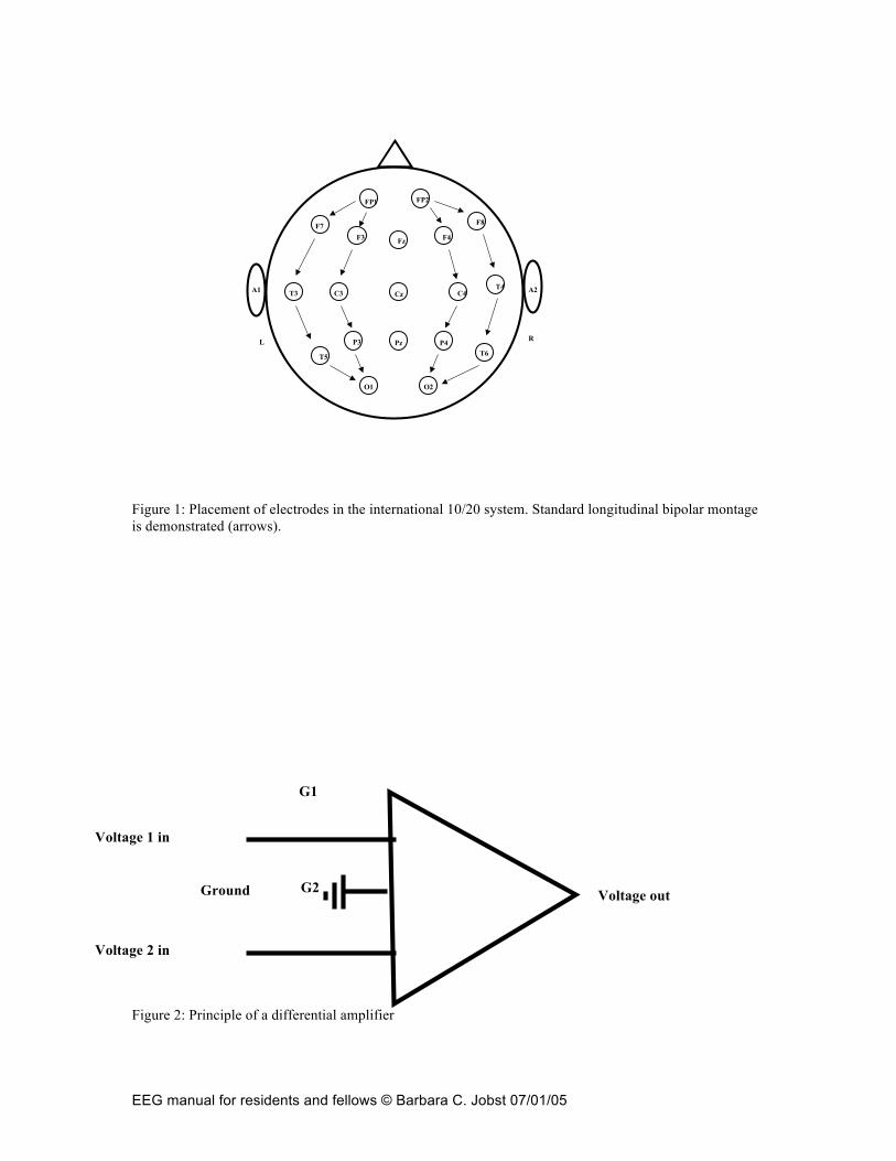
Introduction Signaling via electrical action potentials is the basis for neuronal functioning and a fundamental principle of the nervous system. Although only compounded electrical potentials can be recorded on the surface electrophysiology is one of the fundamental diagnostic tools for the neurologist. Berger in 1929 was the first to record “brain waves” in form of an electroencephalogram (EEG). Other electrophysiologic diagnostic tools like evoked potentials followed thereafter. Despite advances in MRI and neuroimaging electrophysiology remains fundamental in the evaluation of neurologic disease and more specialized applications like intraoperative monitoring and polysomnography have become indispensable diagnostic tools for the neurologist. Methods and Procedure EEG represents a limited sampling of electrical activity on the surface of the brain. Only the electrical currents in the radially oriented apical dentrites of the neocortical pyramidal neurons can be sampled. Most pyramidal cell currents are excitatory and inhibitory postsynaptic potentials (EPSPs and IPSPs) in the dentrites, but “intrinsic” membrane oscillations arising from voltage-dependent ion-channels also contribute to the EEG signal. To record EEG reversible electrodes made out of silver chloride, gold or platinum are applied in a standardized way (10/20 international system) on the scalp of the patient (figure 1). The electrodes are named by their location (F=frontal, P=parietal, O=occipital, T=temporal, C=central) and an associated number. Odd numbers are assigned to the left hemisphere, even numbers are assigned to the right hemisphere (Hughes 1994). The electrodes are wired to an EEG machine via amplifiers. Amplifiers enhance the signal as brain activity is only microvolts (µV) in amplitude. Amplifiers filter

EEG manual for residents and fellows © Barbara C. Jobst 07/01/05
environmental noise (common mode rejection). Low frequency (high pass) and high frequency (low pass) filters are then applied to exclude frequencies outside the range of interest. The usual bandwidth is between 0.3 Hz to 70 Hz. EEG machines record the voltage difference between two electrodes (figure 2). Various montages are used to identify abnormalities. Referential montages involve the difference between an active electrode on the scalp and an inactive electrode, for example, an electrode on the ear or chin. Bipolar montages record the voltage difference between two active scalp electrodes. In referential recording an abnormality is localized by identifying in which channels the abnormalities is of highest amplitude. In bipolar recording a phase reversal of an abnormal signal indicate the localization of the origin of an abnormality (Hughes 1994). By convention signals with an upward deflection are considered negative and signals with a downward deflection are considered positive. EEG can be recorded analog or digitally. Calibration is required in every EEG recording to ensure that the amplifier and filter settings are identical in each channel. Calibration differs significantly in analog and digital recording. Biocalibration is not necessary in digital recordings. Digital recording requires a sufficient sampling rate of the signal (at least twice the frequency of the desired signal) to avoid “aliasing” (=distortion) of the signal. Impedance checks of all electrodes are indispensible in digital and analog recording in order not to contaminate physiologic signal by environmental noise. Usually 16 or 18 channels of scalp EEG are recorded. Eight channels are absolutely essential. In addition EKG is recorded to recognize EKG and pulse artifacts. Two eye-leads are placed to record eye-movements that could potentially obscure the signal. A routine EEG should include referential and bipolar montages (Anonymous 1994). The EEG should be recorded with the patient awake and asleep. Epileptiform activity is enhanced with the patient being asleep. Provocation maneuvers like eye-opening- closing, hyperventilation and photic stimulation should be performed. The recording should be at least 20 min long in adults and 1 hour long in neonates. Repeat EEG recordings or prolonged EEG recordings increase the yield for detecting abnormalities (Salinsky, Kanter et al. 1987). A standard EEG report should be produced that includes an introduction, description and interpretation (Anonymous 1994). Age and state (awake or asleep) need to be known to the interpreter. Normal EEG For definition of basic EEG rhythms see table 1. During wakefulness with the eyes closed the background rhythm should be most prominent in the occipital leads and represent an alpha rhythm. The normal amplitude should vary between 15-50 µV. Frontally faster frequencies in the beta range are normal. Eye-opening results in blockage of the alpha rhythm with only beta frequencies present (figure 3). Drowsiness results in amplitude attenuation and replacement of the background rhythm with a mixture of theta, alpha and beta frequencies. With sleep stage II sleep spindles, vertex waves and K-complexes appear (figure 4). Those are most prominent centrally. With sleep stage III and IV there is increase in delta slowing and disappearance of

EEG manual for residents and fellows © Barbara C. Jobst 07/01/05
spindles and vertex waves. REM sleep is characterized by saw tooth waves and a mixture of alpha and beta frequencies, as well as frequent eye-movements. Normal Variants Apart from the above described rhythms there are normal variants that have no pathologic significance and can be difficult to distinguish from abnormal rhythms (table 2)(Westmoreland 1996). For example are EEGs that only contain beta frequencies considered a normal variant. Predominant beta frequencies are also the result of medication effects and associated with benzodiapines or barbiturate intake. For other normal variants see table 2. Artifacts Artifacts especially movement artifacts or muscle artifact due to tension in the temporal muscles can obscure the recording (figure 5 and table 3). It is essential for the electroencephalographer to recognize artifacts and distinguish those from genuine brain abnormalities. As a general rule abnormal activity generated in the brain usually has a broader field and should not only be visible in one electrode contact. Activation procedures Hyperventilation, sleep deprivation and photic stimulation are activation procedures that can elicit abnormal activity on EEG, especially epileptiform activity. During hyperventilation the patient is asked to overbreathe for three minutes. The resulting hypocapnia is responsible for slowing of the background rhythm, which is considered within normal limits during hyperventilation (figure 6). Slowing is more prominent anteriorly in adults and adolescents and can be occipitally prominent in children. Slowing during hyperventilation is overall more extensive in childhood. Slowing should subside one minute after cessation of hyperventilation. The appearance of epileptiform activity in form of spikes, sharp waves or spike wave discharges is abnormal. Hyperventilation is most useful in typical absence epilepsy of childhood. 85% of untreated children will have an absence seizure during hyperventilation with the typical 3 Hz generalized spike wave discharges (figure 7) (Dalby 1969). In patients with previous stroke or severe cardiopulomonary disease hyperventilation is contraindicated. During photic stimulation a stroboscopic light is flashed at increasing frequencies between 1 and 30 Hz. This may activate epileptiform activity. Photic stimulation can result in no change of the background rhythm, photic driving, a photomyogenic response or a photoconvulsive (=photoparoxysmal) response. With photic driving the background frequency of the EEG follows the frequency of the photic stimulus. This has no pathologic significance unless there are other background abnormalities or photic driving is asymmetric. A photomyogenic response is time locked muscle artifact to the stimulation and the abnormal activity immediately ceases at the end of the stimulus. To the contrary abnormal activity outlasts the stimulus in a

EEG manual for residents and fellows © Barbara C. Jobst 07/01/05
photoconvulsive response by at least 200 ms. Epileptiform activity in form of spike waves or polyspike waves are typical for a photoconvulsive response. A photoconvulsive response has a high association with epilepsy but can also occur in normals. About 70% of patients with a photoconvulsive response have epilepsy (Jayakar and Chiappa 1990). Especially juvenile myoclonic epilepsy is associated with photosensitivity. EEG of childhood Electrical brain activity matures during infancy and childhood. Therefore neonatal and childhood EEG significantly defers from adult recordings. In neonatal EEG a modified montage with fewer electrodes is used due to head size. In addition respirations and EMG is monitored. Electrical brain activity matures due to gestational age not legal age of the baby and EEGs of neonates have to be interpreted in relation to gestational age (Clancy, Berquist et al. 2003). During 29 and 36 weeks of gestation there is rapid evolution of brain activity (table 4). It is important whether a neonatal record is continuous or discontinuous and whether it is reactive to stimulation. At full term (36-40 weeks gestational age) the record can still be discontinous during quiet sleep, but interbust intervals should not be longer than 2-4 seconds (trace alternans pattern). Slow wave sleep in the delta range may gradually develop at full term. During wakefulness “activité moyenne” constitutes the predominent background rhythm in a full term infant, which is a mixture of low to medium amplitude mixed frequencies. “Activité moyenne” is observed during wakefulness (eyes open) and active sleep (eyes closed). Delta brushes which are high amplitude delta waves superimposed with spindle like activity are present until 44 weeks of gestation. At the age of 3- 4 months the background activity should be more organized and in the frequency range of 3-4 Hz. Slow wave sleep should be clearly present. Vertex waves appear between 4- 6 months. Sleep spindles may already appear at the age of 1-2 months but are initially asynchronous. At 12 months of age the frequency should increase to 6-7 Hz. An 8 Hz background rhythm is usually established at 3 years of age. Hypnagogic hypersynchrony is a pattern that is most prominent in childhood after the age of 12 months. It consists of bilateral high amplitude rhythmic theta waves and indicates the onset of sleep. It can easily be mistaken for epileptiform activity. ABNORMAL EEG Abnormalities on EEG especially slowing can be diffuse or focal. Diffuse abnormalities are usually seen in all channels and indicate some sort of an encephalopathy. Focal abnormalities are restricted to certain regions of the brain and often indicate a structural abnormality. If abnormalities contain spikes, spike wave discharges or paroxysmal rhythmic activity it is considered abnormal epileptiform activity. Diffuse abnormalities Diffuse irregular generalized slowing into the theta or delta range is considered an encephalopathy indicating diffuse cerebral dysfunction. There are multiple etiologies

EEG manual for residents and fellows © Barbara C. Jobst 07/01/05
(table 5). Slowing can be diffuse intermittent or diffuse continuous. Diffuse intermittent slowing indicates a mild encephalopathy. Diffuse continuous slowing indicates a moderate to severe encephalopathy. Diffuse intermittent rhythmic slow activity predominantly over the frontal regions (FIRDA=frontal intermittent rhythmic delta activity) is frequently noted with encephalopathy in adults. In children occipital diffuse intermittent rhythmic activity is more frequent (OIRDA= occipital intermittent rhythmic delta activity). FIRDA is also associated with subfrontal or diencephalic lesions and obstructive hydrocephalus. OIRDA can be associated with typical absence epilepsy. Periodic patterns are typical for more severe and acute encephalopathies. Those consist of sharp waves or triphasic waves that occur in a periodic fashion. Triphasic waves are large positive sharp waves > 70 µV that are preceded and followed by a negative wave of lower amplitude. Triphasic waves were initially described in hepatic encephalopathy but occur also in other types of encephalopathy, Creutzfeldt –Jacob disease or SSPE. Focal abnormalities Focal slowing can be rhythmic or arrhythmic (polymorphic), intermittent or continuous. Continuous focal slowing usually indicates a structural lesion. Continuous focal polymorphic delta activity is associated with tumor, stroke, abscess, hematoma. It can also occur in encephalitis and other inflammatory diseases. Focal slowing intermixed with epileptiform activity indicates epilepsy (figure 8). Voltage of delta activity usually ranges between 100-150 µV. Focal attenuation of beta frequencies can also indicate structural lesions like subdural hematomas, stroke, tumor or abscess. A breach rhythm is focal higher amplitude intermixed theta and alpha frequencies (6-11 Hz) intermixed with sharp components. It is the result of a skull defect and by itself does not have pathological significance. Epileptiform abnormalities Spikes, spike waves, sharp waves, polyspikes, polyspike waves discharges and paroxysmal rhythmic activity are considered epileptiform discharges. Epileptiform abnormalities are highly correlated with seizures and epilepsy but the association is not exclusive. Epileptiform activity can also be noted in healthy individuals and the diagnosis of epileptic seizures and epilepsy cannot be made on basis of the EEG alone. The clinical context should always be taken in consideration when interpreting an EEG. Diffuse focal slowing and FIRDA can also be associated with epilepsy but is not specific. Temporal intermittent rhythmic delta activity (TIRDA) is associated with temporal lobe epilepsy. PLEDs are periodic lateralized epileptiform discharges that most often have a frequency of 1 Hz (figure 9). They were thought to be pathognomonic for Herpes encephalitis but they also occur with stroke, epilepsy and Creutzfeldt-Jacob disease. GPEDs (generalized periodic epileptiform discharges) are similar to PLEDS, but epileptiform activity occurs in all leads. GPEDs are associated with status epilepticus.

EEG manual for residents and fellows © Barbara C. Jobst 07/01/05
A normal interictal EEG does not rule out epilepsy. In fact 50% of patient with epilepsy have a normal interictal EEG (Salinsky, Kanter et al. 1987). Repeated EEGs may increase the yield to 80-90%. In patient with a single seizure the probability to record epileptiform activity on interictal EEG is highest within 24 hours of the seizure (King, Newton et al. 1998). If ictal EEG is normal an epileptic disorder is unlikely, but some bizarre frontal lobes seizures have no changes on EEG even on ictal recordings. Seizures and Epilepsy By means of the EEG epilepsy syndromes are divided into primarily generalized and focal (partial) epilepsies (Pedley, Mendiratta et al. 2003). Primary generalized epilepsies are characterized by generalized EEG changes involving all channels on ictal recordings (figure 7 and 10). In focal or partial epilepsy syndromes epileptiform activity originates in circumscribed areas of the cortex (figure 11) and seizures may secondarily spread to the entire cortex. Primary generalized epilepsy syndromes (For discussion of the clinical features please see Dr. Thadani’s chapter) Childhood and juvenile absence epilepsy is characterized by paroxysmal typical 3Hz generalized spike wave discharges (figure 7). Spike waves discharges are frequently activated by hyperventilation (table 6). Brief episodes of spike wave discharges already seem to interfere with awareness. Absences are usually not longer than 12 seconds. OIRDA (occipital intermittent rhythmic delta activity) is seen in a large proportion of children with absence epilepsy. EEG in juvenile myoclonic epilepsy is characterized by less stereotypic generalized spike wave discharges. Polyspike wave discharges are frequent and frequency of the spike wave discharges varies between 3.5 to 6 Hz. Photosensitivity is more commonly seen than activation by hyperventilation (table 6). West syndrome is characterized by infantile spasms, hysarrhythmia and developmental delay. Hypsarrythmia is an EEG pattern of highly disorganized, chaotic delta waves of high amplitude (>300 µV), that is continuous (figure 12). Infantile spasms show an electrodecremental response on EEG. A frontally predominant generalized slow wave transient is followed by diffuse voltage attenuation. Lennox-Gastaut syndrome consists of the triad of multiple seizure types, mental retardation and slow–spike-wave discharges on EEG. The slow spike and wave discharges are large complexes of biphasic or triphasic sharp waves followed by a high amplitude negative slow wave. Those complexes appear at a frequency of 1.5-2.5 Hz. Paroxysmal fast activity of 15-20 Hz most prominent in the frontal region is another typical electrographic feature of Lennox-Gastaut syndrome. Paroxysmal fast activity is activated by sleep (table 6). Focal and multifocal epileptiform discharges are seen in addition to slow spike wave complexes and paroxysmal fast activity. Severe slowing of

EEG manual for residents and fellows © Barbara C. Jobst 07/01/05
the background is the norm and usually correlates with cognitive impairment. Tonic, atonic, atypical absence seizures, myoclonic seizures are common in addition to generalized tonic clonic seizures and focal seizures. Ictal EEG during tonic seizures consists of paroxysmal fast activity and EEG of atypical absences consists of diffuse, bisynchronous, high amplitude 1-2.5 Hz spike wave activity, which can be difficult to distinguish from interictal activity. Other ictal patterns like rhythmic theta activity or 7 Hz spike wave are also possible. Focal or localization related epilepsies Temporal lobe epilepsy can either originate in the mesial structures (hippocampus and parahippocampal gyrus) or in the lateral temporal neocortex. Mesial temporal and lateral neocortical temporal lobe seizure can be difficult to distinguish based on clinical and EEG criteria. Interictally anterior temporal sharp waves with intermittent temporal slowing are typical (figure 8). Temporal epileptiform activity can be bitemporal and occur synchronously and independently over both hemispheres (table 6). Interictal neocortical temporal sharp waves are thought to have a broader field than mesial ones (Ebersole 1996). Ictal EEG consists of lateralized temporal rhythmic theta build-up over the involved temporal lobe (figure 13). Ictal and interictal EEG in frontal, parietal and occipital lobe epilepsy can be quite variable. Interictal epileptiform activity in form of spikes or sharp waves may localize to the epileptogenic region. Frontal lobe epilepsy is recognized as having minimal ictal and interictal EEG changes, especially if seizure originate on the medial surface of the brain e.g. the supplementary motor area seizures. Benign epilepsy of Childhood with centro-temporal spike (BECTS), previously termed Rolandic epilepsy, is characterized by interictal mid-temporal and cental spikes and sharp waves. Maximal voltage of epileptiform activity is in the C3-C4 or T3-T4 region. Sharp waves and spikes are dramatically increased during slow wave sleep (table 6). Centro-temporal spikes are also seen in other neurologic diseases like perinatal hypoxia, Rett’s syndrome, fragile X- syndrome, cortical dysplasias, tumor and agenesis of the corpus callosum. Childhood epilepsy with occipital paroxysms is characterized by interictal diphasic sharp waves or spikes of high amplitude in the occipital region (figure 14). Epileptiform activity disappears with eye opening and promptly returns after eye-closure (table 6). Landau-Kleffner Syndrome (LKS) and continuous spike and waves of slow sleep (CSWS) are related syndromes. The ictal aphasia in LKS is attributed to high-voltage multifocal spikes and spike wave epileptiform activity in the language dominant temporal region. Epileptiform activity is activated by non-REM sleep and may become generalized similar to CSWS (table 6). In CSWS generalized spike wave discharges are the predominant pattern throughout NREM sleep and occupy more than 85% of the NREM record. During wakefulness spike wave discharges are present but markedly decreased at compared to the sleep record.

EEG manual for residents and fellows © Barbara C. Jobst 07/01/05
Status epilepticus Status epilepticus can be convulsive or non-convulsive. In convulsive status epilepticus seizures with associated EEG change occur initially discretely, then a period of continuous rhythmic discharge appears (figure 15) (Treiman, Walton et al. 1990). This is followed by periods of attenuation and finally GPEDs (generalized periodic epileptiform discharges) are seen against a severely attenuated background. Absence status and complex partial status epilepticus are variations of non-convulsive status epilepticus. During absence status generalized spike wave discharges at the frequency of the underlying epilepsy syndrome are present. Continuous bilateral sharp-slow activity is the hallmark of complex partial status epilepticus. Coma and brain death EEG is an important diagnostic tool in coma and can be helpful to determine the prognosis of a patient in coma. EEG is useful to determine the depth of coma. Stimulation with alerting or painful stimuli can contribute important information. Reactivity of the EEG carries a better prognosis than unreactive, monotonous EEG. Intermittent rhythmic delta activity or patterns of generalized high voltage delta activity alternating with lower voltage irregular potentials (alternating pattern) with preserved reactivity indicate less severe coma. High voltage non-reactive delta activity is associated with an unfavorable prognosis. Burst suppression patterns and periodic patterns are associated with a poor prognosis. Bursts of irregular high amplitude theta and delta waves with or without intermixed spikes or sharp waves alternate with episodes of suppression in burst suppression patterns. Burst suppression can occur with severe anoxic encephalopathy or hypothermia. Acute drug intoxication as well as anesthetic agents are reversible causes of burst suppression patterns. In severe progressive anoxic injury burst suppression precedes electrocerebral inactivity. Electrocerebral inactivity is defined as no electrical brain activity exceeding 2 µV and if sustained it indicates brain death. A slow, unreactive, low voltage (<20µV) EEG in coma indicates cortical damage. This has to be distinguished from normal low voltage variants in awake patients. Alpha coma refers to an EEG that indicates diffuse monorhythmic alpha activity in all leads. This is different from the awake state, in which there are faster mixed frequencies over the frontal regions. Alpha coma is seen in anoxic encephalopathy and pontomesenecphalic lesions and is thought to be a poor prognostic sign. If in a comatose patient the EEG contains features that are usually seen in sleep stage II it is termed spindle coma. Spindle coma is associated with high mesencephalic lesions and generally carries a better prognosis. EEG can be a confirmatory test to determine brain death, but recording has to be modified as compared to routine EEG testing. At least 30 minutes of recording are required, montages should include double spacing of electrodes and sensitivity should be

EEG manual for residents and fellows © Barbara C. Jobst 07/01/05
2 µV/ mm. Stimuli should be applied and EEG recording should not occur before 8 hours after the onset of coma (table 7) (Anonymous 1994). Somatosensory evoked potentials are another confirmatory test to diagnose brain death. Clear loss of cortical potentials confirms brain death, but recording of SSEPs in the ICU can be challenging due to electrical noise contamination in the environment. Special EEG procedures As longer recordings increase the yield of detecting epileptiform activity 24 hour ambulatory EEG monitoring is frequently performed. This can be helpful as ictal events are more likely to be captured. It is frequently utilized if absence epilepsy is suspected. In intractable epilepsy and to establish a definite diagnosis of epileptic versus non-epileptic seizures, video/ EEG monitoring is utilized. It is also essential for the presurgical evaluation if epilepsy surgery is considered. The patient is admitted to a specialized inpatient EEG monitoring unit. Antiepileptic medications are often withdrawn and seizures are captured on video with simultaneous EEG monitoring. If further epilepsy surgery is pursued the procedure may be extended and intracranial subdural strip/grid electrodes or depth electrodes may be implanted to identify the region of seizure onset in intractable epilepsy. In a functional mapping procedure those electrodes can be electrically stimulated. By functional mapping information can be obtained whether eloquent cortex is covered by the stimulated electrodes (e.g the primary motor cortex or the language areas) and whether resection of those brain regions is safe. Intracranial EEG is also utilized in the operating room to identify epileptogenic tissue (electrocorticography), but due to anesthesia effects, the use of electrocorticography is limited. References: Anonymous (1994). "Guidelines for writing EEG reports." Journal of Clinical Neurophysiology 11(1): 37-39. Anonymous (1994). "Minimum technical standards for EEG recording in suspected cerebral death." Journal of Clinical Neurophysiology 11(1): 10-13. Anonymous (1994). "A proposal for standard montages to be used in clinical EEG." Journal of Clinical Neurophysiology 11(1): 30-36. Clancy, R. R., A. G. C. Berquist, et al. (2003). Neonatal Electroencephalography. Current Practice of Electroencephalography. J. S. Ebersole and T. A. Pedley. Philadelphia, Lippincott, Williams and Wilkins: 160-234. Dalby, M. A. (1969). "Epilepsy and 3 per second spike and wave rhythms. A clinical, electroencephalographic and prognostic analysis of 346 patients." Acta Neurol Scand: Suppl 40:3+.

EEG manual for residents and fellows © Barbara C. Jobst 07/01/05
Hughes, J. (1994). EEG in clinical practice. Newton, MA, Butterworth-Heinemann. Jayakar, P. and K. H. Chiappa (1990). "Clinical correlations of photoparoxysmal responses." Electroencephalogr Clin Neurophysiol 75(3): 251-4. King, M. A., M. R. Newton, et al. (1998). "Epileptology of the first-seizure presentation: a clinical, electroencephalographic, and magnetic resonance imaging study of 300 consecutive patients." Lancet 352(9133): 1007-11. Pedley, T. A., A. Mendiratta, et al. (2003). Seizures and Epilepsy. Current Practice of Clinical Encephalography. J. S. Ebersol and T. A. Pedley. Philadelphia, Lippincott Williams and Wilkins: 506-587. Salinsky, M., R. Kanter, et al. (1987). "Effectiveness of multiple EEGs in supporting the diagnosis of epilepsy: an operational curve." Epilepsia 28(4): 331-4. Treiman, D. M., N. Y. Walton, et al. (1990). "A progressive sequence of electroencephalographic changes during generalized convulsive status epilepticus." Epilepsy Res 5(1): 49-60. Westmoreland, B. F. (1996). "Epileptiform electroencephalographic patterns." Mayo Clin Proc 71(5): 501-11.
EEG manual for residents and fellows © Barbara C. Jobst 07/01/05
Figure 1: Placement of electrodes in the international 10/20 system. Standard longitudinal bipolar montage is demonstrated (arrows). Figure 2: Principle of a differential amplifier
FP1 FP2
F7F3 Fz F4
F8
T3 C3 Cz C4T4
T5
P3 Pz P4T6
O1 O2
A1 A2
L R
Voltage out
Voltage 1 in
Voltage 2 in
Ground
G1
G2
EEG manual for residents and fellows © Barbara C. Jobst 07/01/05
Figure 3: Normal EEG with well developed alpha background with the eyes closed and a completely blocked alpha rhythm due to eye opening in the initial part of the tracing, Figure 3: Normal EEG with well developed alpha background with the eyes closed and a completely blocked alpha rhythm due to eye opening in the initial part of the tracing,
Normal alpha background
Frontal muscle artifact
Eye blink artifact
Eyes open Eyes closed

EEG manual for residents and fellows © Barbara C. Jobst 07/01/05
Figure 4: Normal stage II sleep in a 12 yo boy with vertex waves and sleep spindles in a transverse montage.
Vertex waves
Sleep spindles

EEG manual for residents and fellows © Barbara C. Jobst 07/01/05
Figure 5: Muscle artifact over the bitemporal leads

EEG manual for residents and fellows © Barbara C. Jobst 07/01/05
Figure 6: Symmetric normal slowing during hyperventilation in a 8 yo boy.

EEG manual for residents and fellows © Barbara C. Jobst 07/01/05
Figure 7: Typical generalized 3 Hz spike wave discharge as seen in typical absence epilepsy.

EEG manual for residents and fellows © Barbara C. Jobst 07/01/05
Figure 8: Focal delta slowing over the left temporal region with intermixed sharp waves. 28 yo patient with seizures.

EEG manual for residents and fellows © Barbara C. Jobst 07/01/05
Figure 9: Periodic lateralizing epileptiform discharges over the right fronto-central region (PLEDS).

EEG manual for residents and fellows © Barbara C. Jobst 07/01/05
Figure 10: Generalized isolated spike wave discharges.

EEG manual for residents and fellows © Barbara C. Jobst 07/01/05
Figure 11: Focal epileptiform activity over the right temporal region in form of sharp wave discharges.
Sleep spindles
Epileptiform activity

EEG manual for residents and fellows © Barbara C. Jobst 07/01/05
Figure 12: Hysarrhythmia-like pattern in a 8 month old infant. Note high amplitude chaotic disorganized delta waves with intermixed spikes.

EEG manual for residents and fellows © Barbara C. Jobst 07/01/05
Figure 13: Temporal lobe focal seizure with rhythmic theta build-up.

EEG manual for residents and fellows © Barbara C. Jobst 07/01/05
Figure 14: Occipital spikes in a patient with childhood epilepsy with occipital paroxysms.

EEG manual for residents and fellows © Barbara C. Jobst 07/01/05
Figure 15: Rhythmic generalized epileptiform delta activity.
EEG manual for residents and fellows © Barbara C. Jobst 07/01/05
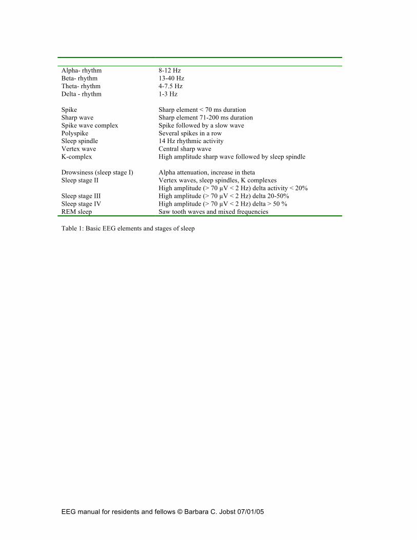
Alpha- rhythm Beta- rhythm Theta- rhythm Delta - rhythm Spike Sharp wave Spike wave complex Polyspike Sleep spindle Vertex wave K-complex Drowsiness (sleep stage I) Sleep stage II Sleep stage III Sleep stage IV REM sleep
8-12 Hz 13-40 Hz 4-7.5 Hz 1-3 Hz Sharp element < 70 ms duration Sharp element 71-200 ms duration Spike followed by a slow wave Several spikes in a row 14 Hz rhythmic activity Central sharp wave High amplitude sharp wave followed by sleep spindle Alpha attenuation, increase in theta Vertex waves, sleep spindles, K complexes High amplitude (> 70 µV < 2 Hz) delta activity < 20% High amplitude (> 70 µV < 2 Hz) delta 20-50% High amplitude (> 70 µV < 2 Hz) delta > 50 % Saw tooth waves and mixed frequencies
Table 1: Basic EEG elements and stages of sleep

EEG manual for residents and fellows © Barbara C. Jobst 07/01/05
Normal variant Description and location Occurs in Rhythmic temporal theta burst of drowsiness (RTTD) =psychomotor variant
Notched temporal rhythm Adolescents and young adults
Alpha variant (slow and fast) Harmonic relationship to alpha rhythm Slow alpha variant: half the frequency of alpha Fast alpha variant: double the frequency of alpha rhythm
Adults
Frontal arousal rhythms 7-20 Hz over frontal regions lasting up to 20 sec
Children, during arousal
Subclinical rythmic electrographic theta discharges in adults (SREDA)
High voltage monophasic sharp component, followed by one second lower amplitude rhythms, followed by 5-7 Hz sinusoidal theta, bilateral, posterior temporal 40-80 sec long
> 50 yo during drowsiness or hyperventilation
Midline theta rhythms 5-7 Hz over vertex Fourteen and Six Hertz positive bursts
Resembles sleep spindles with sharp positive phase Posterior temporal asynchronous
Children and adolescents
Small sharp spikes= benign epileptiform transients of sleep
Small spikes <50µV unilateral
Drowsiness and light sleep
Six-Hertz Spike and Wave= Phantom spike wave
Bilateral synchronus 6 Hz spike and wave
Drowsiness and relaxed wakefullness
Wicket spikes Arc like waves that look like a “wicket” temporal
> 30 yo Drowsiness and light sleep
Posterior slow waves of Youth = Youth waves
Posterior theta activity Children and adolescents
Positive occipital slow transients of sleep =POSTS
Positive repetitive sharp waves occipitally, bilateral synchronous
15-35 yo during sleep
Lamda waves Positive repetitive sharp waves occipitally
Occur with scanning eye movements, more common in children
Mu (µ) rhythm Rhythmic arc like activity of alpha frequency centrally
Young adults, disappears with hand movement
Table 2: Normal EEG variants

EEG manual for residents and fellows © Barbara C. Jobst 07/01/05
Artifact Characteristics Muscle/EMG artifact High frequency artifact often temporal Movement artifact High amplitude irregular activity often in all
channels 60 cycle artifact Sinusoidal artifact of 60 Hz Electrode pop Localizes to only one electrode Eye movement artifact Eye blinks
Retina is negative, cornea is positive. Upward movement of eye ball creates positive potential in frontal leads and “out of phase activity” in eye channels
Sweating/perspiration High amplitude slow potentials <1 Hz EKG artifact Synchronized with EKG Pulse artifact Time locked to EKG IV-drip artifact Electrostatic periodic artifact Digital artifact Due to sampling In infants: Hiccups Sucking Padding
Periodic movement artifact High amplitude sharp activity bitemporal Rhythmic artifact that follows frequency of padding
Table 3: Common EEG artifacts

EEG manual for residents and fellows © Barbara C. Jobst 07/01/05
Gestational Age Wakefulness Active sleep (eyes closed, like
REM) Quiet sleep
<29 weeks Discontinous, monorhyhtmic occipital delta, bursts of rhythmic occipital and temporal theta, bursts 90% synchronized
No state organization No state organization
30-32 weeks Monorthymic delta, posterior delta brushes, marginally reactive
More continuous, increased body movements
Trace discontinue (<25µV during suppression) IBI 5-8 sec 70% synchronous bursts Differentiation between quiet and active sleep possible
33-34 weeks Reactive, delta brushes, sharp waves temporal and central, monorhythmic delta decreases
Delta brushes Trace discontinue IBI 5-6 sec 80% synchronous bursts
35-36 weeks Activity moyenne, Anterior slow dysrhythmia Enchroches frontales (frontal sharp waves)
Delta brushes Trace alternans (>25 µV during suppression) IBI 4-6 sec 85% synchronous bursts
37-40 weeks Activity moyenne, sharp waves decrease 25% indeterminate sleep Delta brushes disappear
Trace alternans IBI < 4 sec, 100% synchronous bursts continuous slow wave sleep appears
41-44 weeks Activity moyenne 50 % of record active sleep Trace alternans, continuous slow wave sleep Table 4: Neonatal EEG. IBI=interburst interval

EEG manual for residents and fellows © Barbara C. Jobst 07/01/05
Encephalopathies Metabolic: uremic, hepatic, electrolyte imbalance, hypo-/ hyperglycemic Anoxic: Endocrine: thyroid, calcium, adrenal Nutritional: B1, B12 Infectious and inflammatory: meningitis, encephalitis, Creutzfeldt-Jacobs, AIDS, autoimmune Hypertensive: Epileptic: progressive myoclonic epilepsies Hereditary and neurodegenerative: leukodystrophies, inborn errors of metabolism Table 5: Encephalopathies Hyperventilation Photic stimulation Awake Non-REM sleep REM Absence Epilepsy 85% activation
OIRDA 20% activation Organized 3Hz SW
burst Increased SW bursts, fragmented non-REM sleep polyspikes
Like awake state, but decreased SW
Benign epilepsy with centro-temporal spikes
No effect No effect Poss. Unilateral centro-temporal spikes
Marked increase in spiking, mainly in stage III and IV
Rare spikes
Childhood epilepsy with occipital paroxsyms
No effect No effect Fixation off sensitivity (increased spiking with eye closure, blocked with eye-opening)
Increased spiking Inhibits spikes
Juvenile myoclonic epilepsy
Some activation Significant activation Eye closure activates generalized SW and polyspike wave
Suppresses SW discharges
Less SW than in awake, but more than in NREM
West-Syndrome 64% hysarrhythmia 99% hysarrhythmia in stage II, III
Hypsarrhythmia disappears
Lennox-Gastaut Syndrome
Activates slow spike wave
No effect Slow-spike wave 1.5-2.5 Hz
15-20 Hz frontal paroxysmal fast and increased slow spike wave
Reduced or absent slow spike wave
Landau-Kleffner Syndrome
Focal epileptiform discharges
Activation with increased discharges
Like awake
Continuous spike and wave of slow wave sleep
3-5 Hz intermittent SW > 85% generalized SW SW disappear < 5%
Temporal lobe epilepsy Lateralized temporal epileptiform activity
Bilateral independent temporal epileptiform activity
Table 6: EEG of certain epilepsy syndromes during various stages and activation procedures. OIRDA= occipital intermittent rhythmic delta activity, SW=spike wave
EEG manual for residents and fellows © Barbara C. Jobst 07/01/05
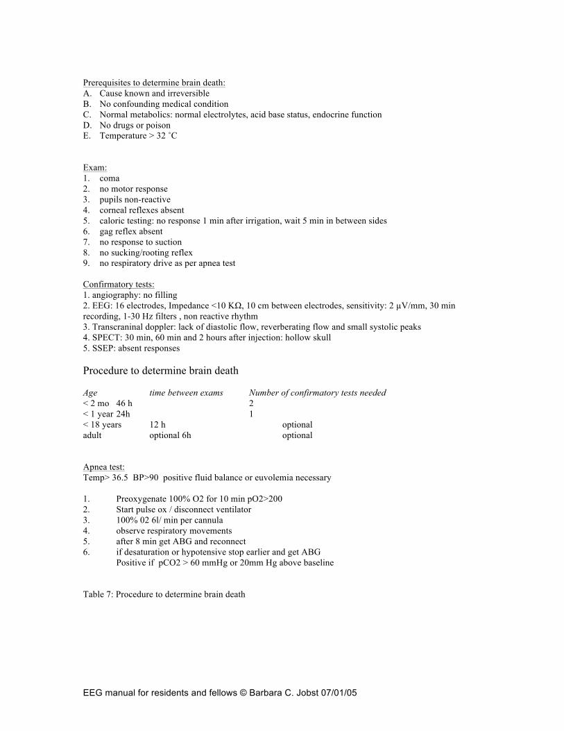
Prerequisites to determine brain death: A. Cause known and irreversible B. No confounding medical condition C. Normal metabolics: normal electrolytes, acid base status, endocrine function D. No drugs or poison E. Temperature > 32 ˚C Exam: 1. coma 2. no motor response 3. pupils non-reactive 4. corneal reflexes absent 5. caloric testing: no response 1 min after irrigation, wait 5 min in between sides 6. gag reflex absent 7. no response to suction 8. no sucking/rooting reflex 9. no respiratory drive as per apnea test Confirmatory tests: 1. angiography: no filling 2. EEG: 16 electrodes, Impedance <10 KΩ, 10 cm between electrodes, sensitivity: 2 µV/mm, 30 min recording, 1-30 Hz filters , non reactive rhythm 3. Transcraninal doppler: lack of diastolic flow, reverberating flow and small systolic peaks 4. SPECT: 30 min, 60 min and 2 hours after injection: hollow skull 5. SSEP: absent responses Procedure to determine brain death Age time between exams Number of confirmatory tests needed < 2 mo 46 h 2 < 1 year 24h 1 < 18 years 12 h optional adult optional 6h optional Apnea test: Temp> 36.5 BP>90 positive fluid balance or euvolemia necessary 1. Preoxygenate 100% O2 for 10 min pO2>200 2. Start pulse ox / disconnect ventilator 3. 100% 02 6l/ min per cannula 4. observe respiratory movements 5. after 8 min get ABG and reconnect 6. if desaturation or hypotensive stop earlier and get ABG
Positive if pCO2 > 60 mmHg or 20mm Hg above baseline
Table 7: Procedure to determine brain death

EEG manual for residents and fellows © Barbara C. Jobst 07/01/05
Questions: 1. To use EEG as a confirmatory test for brain death
A. The test should be 30 min long. B. It should be recorded with a higher sensitivity as usual (2 µV/mm). C. Only electrocerebral inactivity confirms brain death. D. Larger spaced distances between electrodes should be recorded. E. All of the above.
2. In a normal EEG of a 3 year child
A. Hypnagogic hypersynchony may occur. B. There is continuous delta slowing C. No sleep spindles or vertex waves should be present D. There is epileptiform activity E. Photic stimulation results in increased slowing
3. Hyperventilation
A. causes hypercapnia B. often activates generalized spike wave discharges in absence epilepsy C. should not be performed in patients with severe cardiovascular disease D. B and C E. A,B and C
4. Alpha coma
A. carries a good prognosis. B. is associated with anoxic brain injury. C. shows frontal beta activity on most recordings. D. is considered a reactive pattern. E. is the same as spindle coma.
5. A 15 yo boy with mental retardation and tonic, atonic and atypical absence seizures
has most likely A. generalized slow spike wave complexes on EEG B. paroxysmal fast activity on EEG C. Landau-Kleffner Syndrome D. A and B E. A,B and C
6. A 26 yo man has his first witnessed generalized tonic clonic convulsion. No
metabolic or infectious causes could explain the seizure. EEG and MRI are normal. A. It is unlikely that he has epilepsy as EEG and MRI are normal. B. The likelihood that he has another seizures is more than 80%. C. He has juvenile myoclonic epilepsy. D. A repeat EEG would increase the likelihood to identify epileptiform activity. E. He should be admitted for video/EEG monitoring to clarify the diagnosis.

EEG manual for residents and fellows © Barbara C. Jobst 07/01/05
7. While recording an EEG A. Checking of impedances is unnecessary with digital recording B. Low and high pass filters should be checked C. Hyperventilation and photic stimulation should be performed D. B and C. E. A, B and C
8. Hyparrythmia
A. is the EEG pattern of infantile spasm B. is of high voltage C. represents chaotic delta activity D. A and B E. A,B and C
9. A nine year old boy has intermittent facial twitching mainly at night
A. His EEG most likely shows centro-temporal spikes B. NREM sleep will enhance epileptiform activity on EEG C. Prognosis is generally good. D. He may also develop generalized convulsion E. All of the above
10. Loss of motor evoked potentials during surgery for scoliosis A. is always due to anesthesia effects B. is only significant if there is also loss of SSEPs C. indicates spinal cord injury D. should be ignored E. should prompt more instrumentation of the spinal column
1. E 2. A 3. D 4. C 5. A 6. D 7. D 8. E 9. E 10. C