edward p. mulligan, ms, pt, scs, atc - · pdf file1 goniometry 101 edward p. mulligan, ms, pt,...
TRANSCRIPT
1
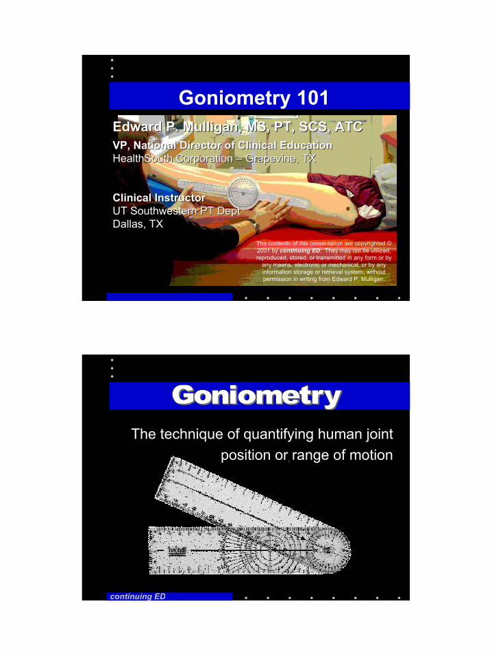
Goniometry 101Edward P. Mulligan, MS, PT, SCS, ATCEdward P. Mulligan, MS, PT, SCS, ATCVP, National Director of Clinical Education VP, National Director of Clinical Education HealthSouth Corporation HealthSouth Corporation –– Grapevine, TXGrapevine, TX
Clinical Instructor Clinical Instructor UT Southwestern PT DeptUT Southwestern PT DeptDallas, TXDallas, TX
The contents of this presentation are copyrighted © 2001 by continuing ED. They may not be utilized, reproduced, stored, or transmitted in any form or by
any means, electronic or mechanical, or by any information storage or retrieval system, without permission in writing from Edward P. Mulligan.
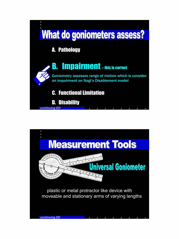
The technique of quantifying human joint position or range of motion
continuing ED
2
A. Pathology
B. Impairment – this is correct
Goniometry assesses range of motion which is consider an impairment on Nagi’s Disablement model
C. Functional Limitation
D. Disabilitycontinuing ED
plastic or metal protractor like device with moveable and stationary arms of varying lengths
continuing ED
3
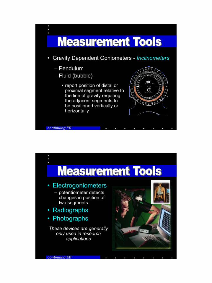
• Gravity Dependent Goniometers - Inclinometers
– Pendulum – Fluid (bubble)
• report position of distal or proximal segment relative to the line of gravity requiring the adjacent segments to be positioned vertically or horizontally
continuing ED
• Electrogoniometers– potentiometer detects
changes in position of two segments
• Radiographs• PhotographsThese devices are generally
only used in research applications
continuing ED
4
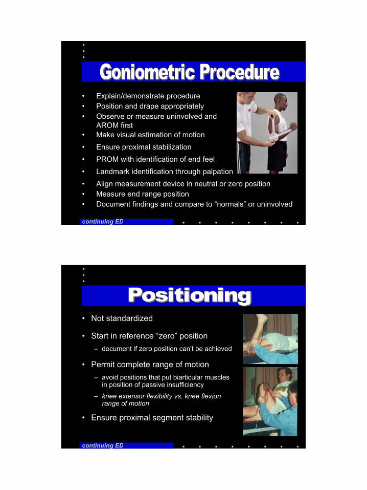
• Explain/demonstrate procedure
continuing ED
• Position and drape appropriately• Observe or measure uninvolved and
AROM first• Make visual estimation of motion• Ensure proximal stabilization• PROM with identification of end feel• Landmark identification through palpation• Align measurement device in neutral or zero position• Measure end range position• Document findings and compare to “normals” or uninvolved
• Not standardized
continuing ED
• Start in reference “zero” position – document if zero position can't be achieved
• Ensure proximal segment stability
• Permit complete range of motion – avoid positions that put biarticular muscles
in position of passive insufficiency
– knee extensor flexibility vs. knee flexion range of motion
5
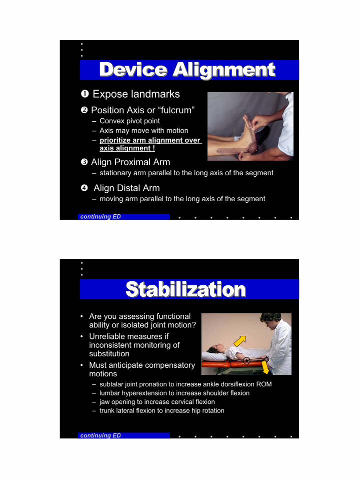
Expose landmarks
continuing ED
Align Distal Arm – moving arm parallel to the long axis of the segment
Align Proximal Arm– stationary arm parallel to the long axis of the segment
Position Axis or “fulcrum”– Convex pivot point– Axis may move with motion – prioritize arm alignment over
axis alignment !
• Are you assessing functional ability or isolated joint motion?
• Unreliable measures if inconsistent monitoring of substitution
• Must anticipate compensatory motions– subtalar joint pronation to increase ankle dorsiflexion ROM– lumbar hyperextension to increase shoulder flexion– jaw opening to increase cervical flexion– trunk lateral flexion to increase hip rotation
continuing ED

6
A. How much motion you have?
B. What is limiting the motion you have? – correct!From an intervention standpoint it may be more important to know what is limiting the motion as opposed to have much you have because that information will help you pick the appropriate intervention
continuing ED
Start
1st Stop
Final Stop
Start
Start
1st Stop
1st Stop
Final Stop
Final Stop
STA
Capsular
Bony
distinct arrest
Nature of the motion barrier that characterizes the type of tissue limiting range
abrupt halt
spongy
continuing ED

7
• Empty– Painful
• Boggy– Mushy
• Muscle spasm – painful rebound
• Springy – internal derangement
continuing ED
continuing ED

8
• Failure to read at eye level causing parallax distortion
• Incorrect landmark identification
• Failure to read proper scale
• Lack of patient cooperation
continuing ED
Consistent repeatable, and reproducible measurements have:
A. Sensitivity
B. Specificity
C. Reliability – correct!
D. Validity
continuing ED
9
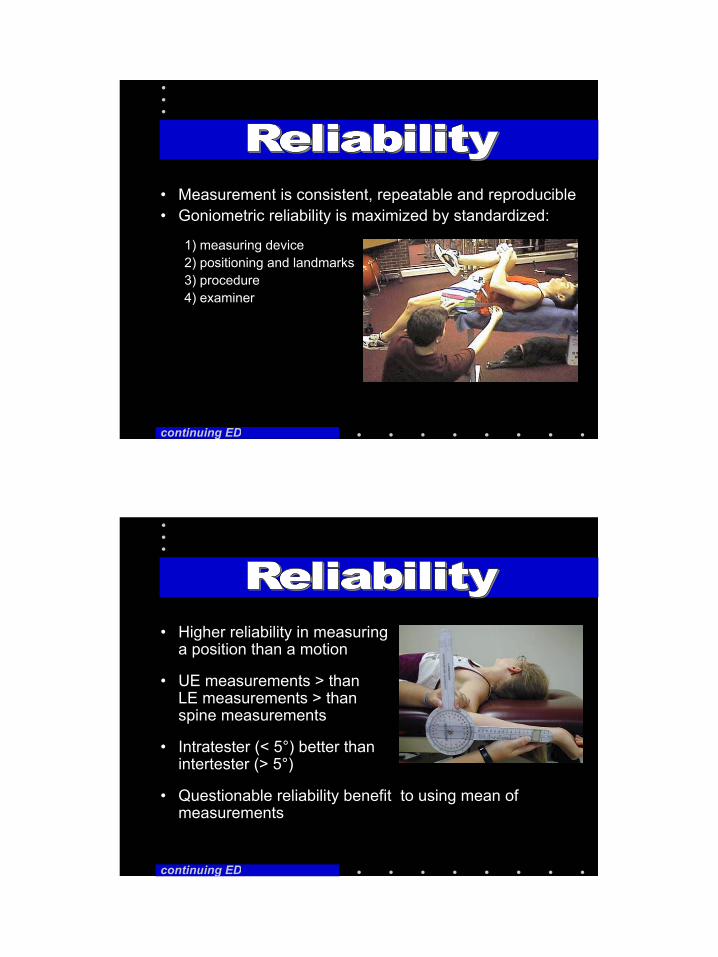
• Measurement is consistent, repeatable and reproducible• Goniometric reliability is maximized by standardized:
1) measuring device2) positioning and landmarks3) procedure 4) examiner
continuing ED
• Higher reliability in measuring a position than a motion
• UE measurements > than LE measurements > than spine measurements
• Intratester (< 5°) better than intertester (> 5°)
• Questionable reliability benefit to using mean of measurements
continuing ED
10
Measures that represent the true value are:
A. Sensitive
B. Specific
C. Reliable
D. Valid – correct!
continuing ED
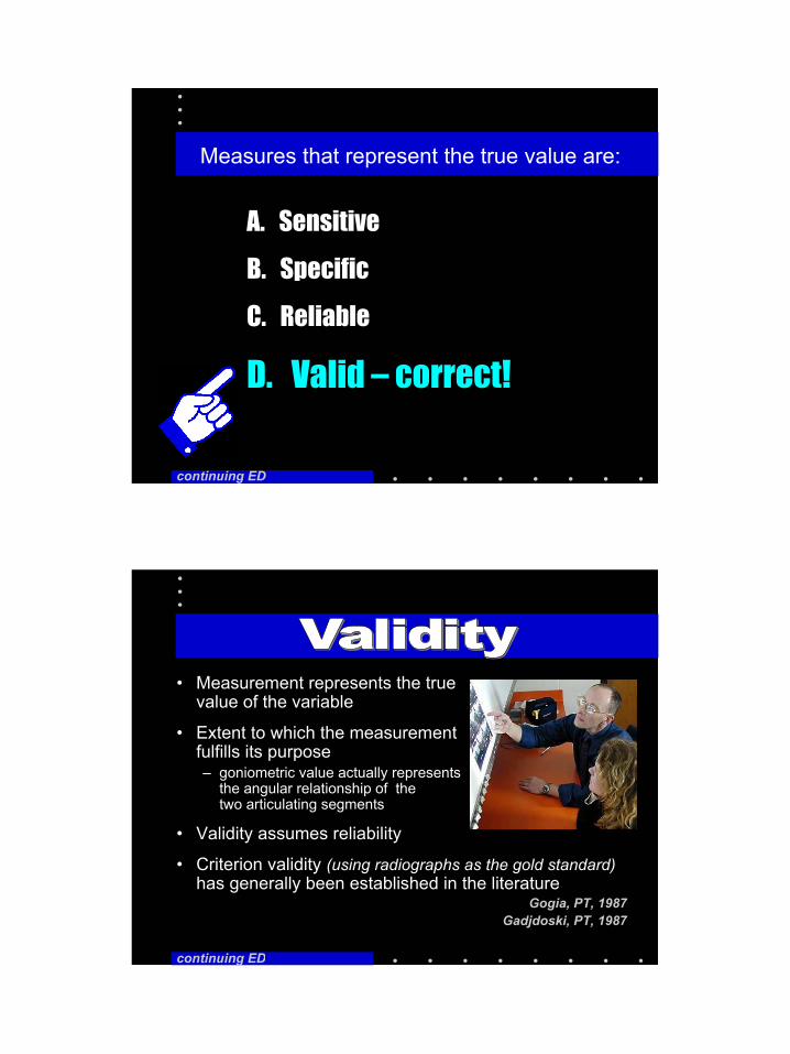
• Measurement represents the true value of the variable
• Extent to which the measurement fulfills its purpose– goniometric value actually represents
the angular relationship of the two articulating segments
• Validity assumes reliability
• Criterion validity (using radiographs as the gold standard)has generally been established in the literature
Gogia, PT, 1987Gadjdoski, PT, 1987
continuing ED

11
• WNL unacceptable unless referenced– AAOS, Kendall, Hoppenfeld, AMA
Impairment• Compare involved to uninvolved sides • Compare to subjects of similar age and gender
• 0-152º (R) pain free passive elbow flexion with soft tissue end feel
• 7-0-85° (L) passive knee motion with capsular end feel and symptom reproduction at end range flexion
• Always use a third value to indicate neutral position if motion exist on each side of the neutral positioncontinuing ED
continuing ED
12
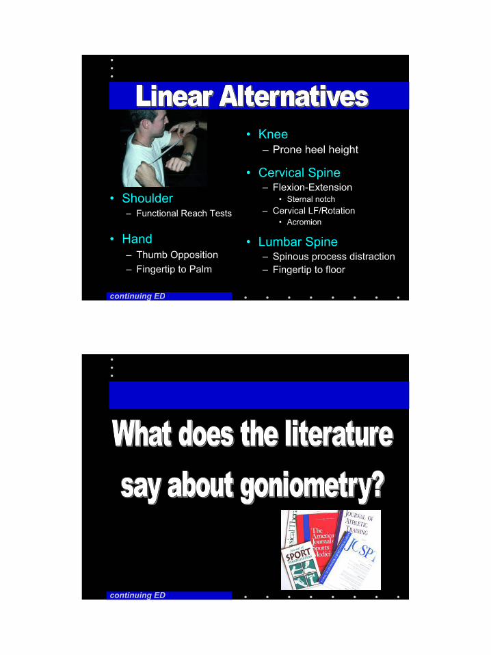
• Shoulder– Functional Reach Tests
• Hand– Thumb Opposition– Fingertip to Palm
• Knee– Prone heel height
• Cervical Spine– Flexion-Extension
• Sternal notch– Cervical LF/Rotation
• Acromion
• Lumbar Spine– Spinous process distraction– Fingertip to floor
continuing ED
continuing ED
13
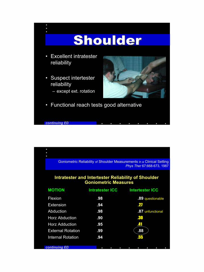
• Excellent intratester reliability
• Suspect intertester reliability– except ext. rotation
• Functional reach tests good alternative
continuing ED
Intratester and Intertester Reliability of Shoulder Goniometric Measures
MOTION Intratester ICC Intertester ICC
Flexion .98 .89 questionable
Extension .94 .27
Abduction .98 .87 unfunctional
Horz Abduction .90 .30
Horz Adduction .95 .41
External Rotation .99 .88Internal Rotation .94 .55
Goniometric Reliability of Shoulder Measurements in a Clinical SettingPhys Ther 67:668-673, 1987
continuing ED
14
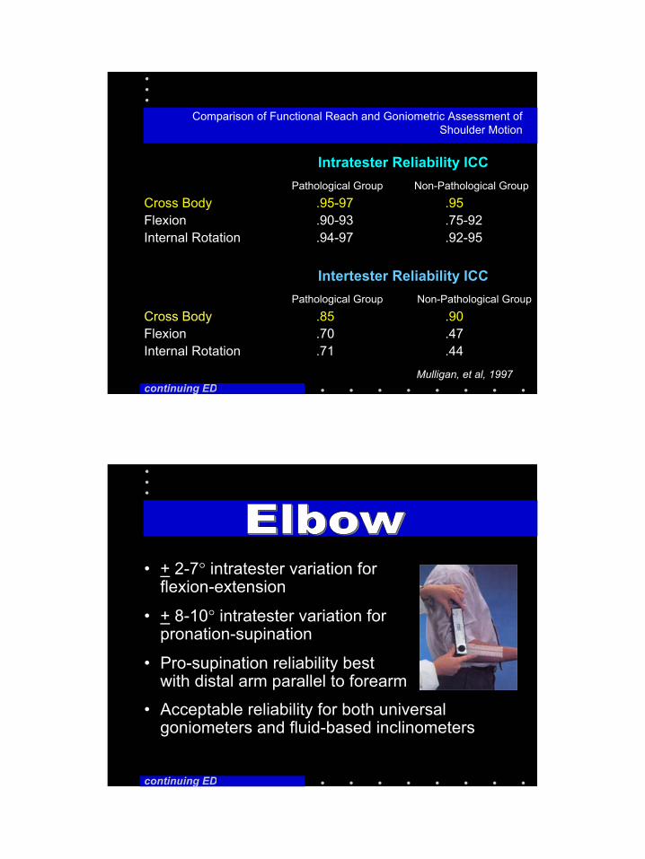
Comparison of Functional Reach and Goniometric Assessment of Shoulder Motion
Intratester Reliability ICCPathological Group Non-Pathological Group
Cross Body .95-97 .95Flexion .90-93 .75-92Internal Rotation .94-97 .92-95
Intertester Reliability ICCPathological Group Non-Pathological Group
Cross Body .85 .90Flexion .70 .47Internal Rotation .71 .44
continuing EDMulligan, et al, 1997
• + 2-7° intratester variation for flexion-extension
• + 8-10° intratester variation for pronation-supination
• Pro-supination reliability best with distal arm parallel to forearm
• Acceptable reliability for both universal goniometers and fluid-based inclinometers
continuing ED
15
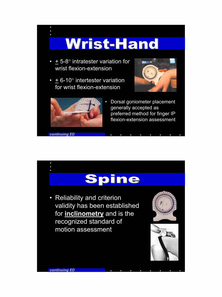
• + 5-8° intratester variation for wrist flexion-extension
• + 6-10° intertester variation for wrist flexion-extension
• Dorsal goniometer placement generally accepted as preferred method for finger IP flexion-extension assessment
continuing ED
• Reliability and criterion validity has been established for inclinometry and is the recognized standard of motion assessment
continuing ED

16
• Alternative methodologies– Universal goniometer– Flexible ruler– Tape measure attraction-
distraction– Finger tip to floor
measurements
continuing ED
• Both inclinometers and goniometers have acceptable intratester reliability but are not interchangeable
• Intertester reliability has not been established
• Lowest coefficient of variance for flexion ROM, extension ROM the highest
continuing ED

17
• Excellent intra and intertester goniometric reliability for knee flexion range of motion
continuing ED
• Tibial rotation ROM reliability has not been established
Intratester/Intertester Reliability of Measuring Knee Extension Position with a Goniometric Method
ICC
Therapist 1 involved extremity .94Therapist 1 uninvolved extremity .96Therapist 2 involved extremity .97Therapist 2 uninvolved extremity .94Therapist 1 vs. 2 involved extremity .88Therapist 1 vs. 2 uninvolved extremity .83
Mulligan 1995
continuing ED
Excellent intra and intertester reliability
18
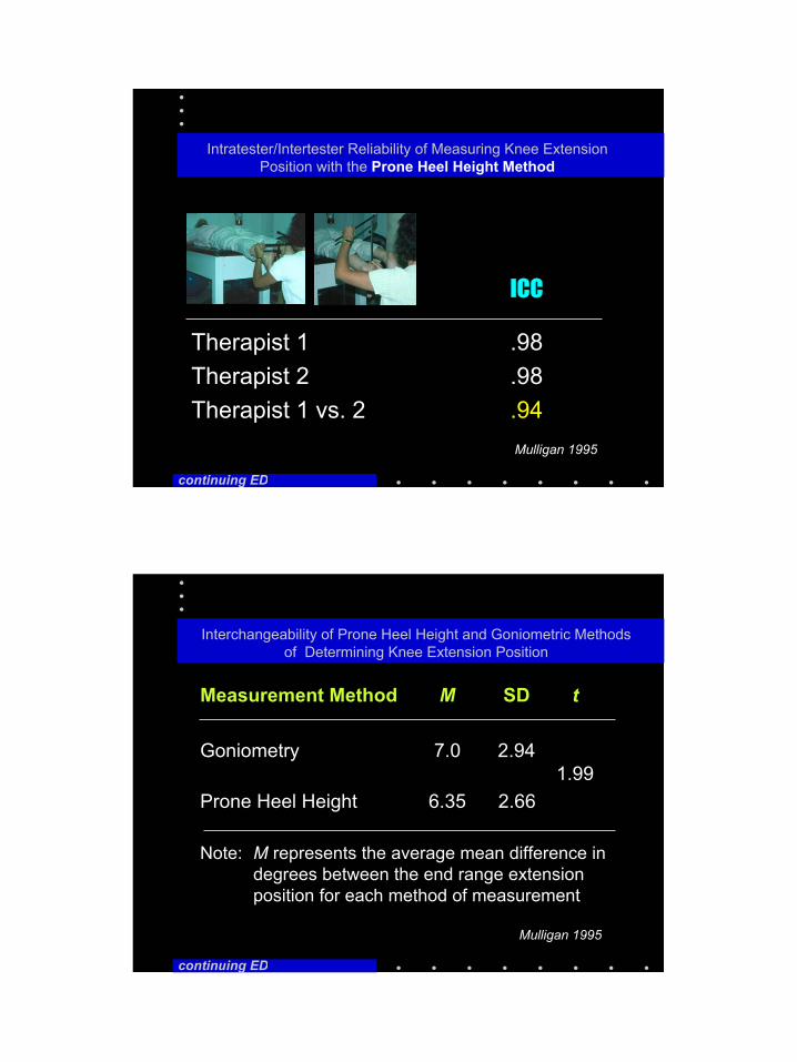
ICC
Therapist 1 .98Therapist 2 .98Therapist 1 vs. 2 .94
continuing ED
Intratester/Intertester Reliability of Measuring Knee Extension Position with the Prone Heel Height Method
Mulligan 1995
Interchangeability of Prone Heel Height and Goniometric Methods of Determining Knee Extension Position
Measurement Method M SD t
Goniometry 7.0 2.941.99
Prone Heel Height 6.35 2.66
Note: M represents the average mean difference in degrees between the end range extension position for each method of measurement
continuing ED
Mulligan 1995
19
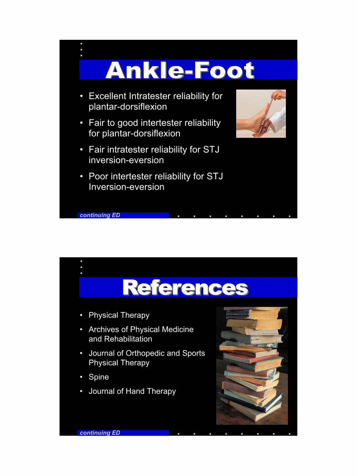
• Excellent Intratester reliability for plantar-dorsiflexion
• Fair to good intertester reliability for plantar-dorsiflexion
• Fair intratester reliability for STJ inversion-eversion
• Poor intertester reliability for STJ Inversion-eversion
continuing ED
continuing ED
• Physical Therapy
• Archives of Physical Medicine and Rehabilitation
• Journal of Orthopedic and Sports Physical Therapy
• Spine
• Journal of Hand Therapy

20
• American Academy of Orthopedic Surgeons (Green WB, Heckman JD eds.): The Clinical Measurement of Joint Motion. 6300 North River Road, Rosemont, IL 60018, 1994
• American Medical Association: Guide to the Evaluation of Permanent Impairment. AMA, Chicago, 1988.
• Magee DJ: Orthopedic Physical Assessment, 3rd ed. WB Saunders Co, Philadelphia, 1997
• Norkin CC, White DJ: Measurement of Joint Motion: A Guide to Goniometry, 2nd ed. FA Davis Co, Philadelphia, 1995
continuing ED
continuing ED
Practice and Discuss Standards with your Colleagues