edward n. trifonov - ipb trif 2013... · edward n. trifonov . university of haifa, israel . beograd...
TRANSCRIPT

Edward N. Trifonov University of Haifa, Israel
Beograd 2013
Simple physics and bioinformatics of nucleosome positioning
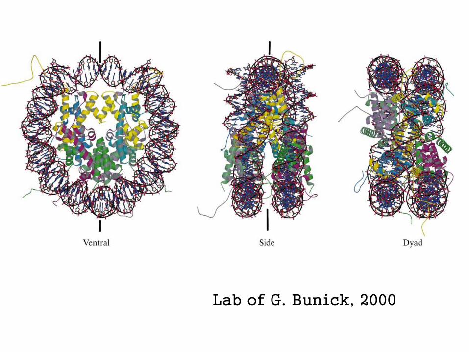
Lab of G. Bunick, 2000
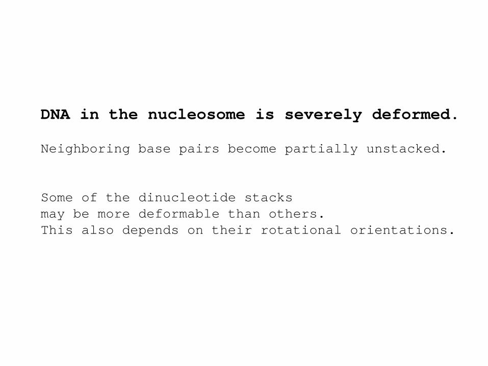
DNA in the nucleosome is severely deformed. Neighboring base pairs become partially unstacked. Some of the dinucleotide stacks may be more deformable than others. This also depends on their rotational orientations.
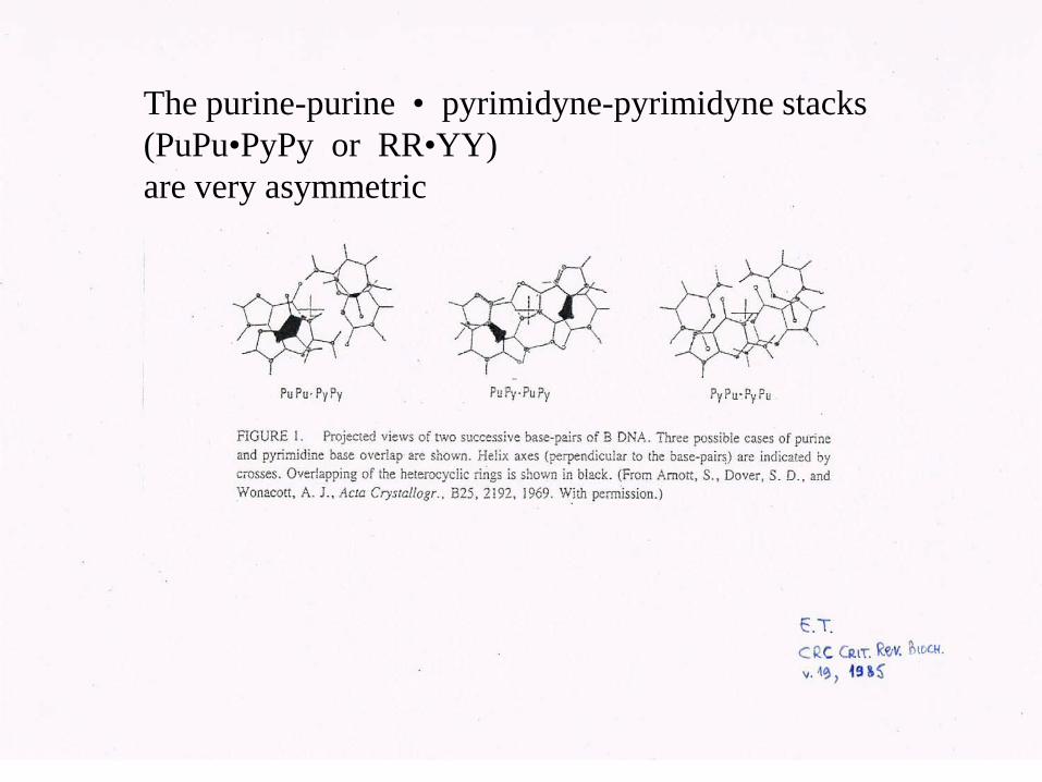
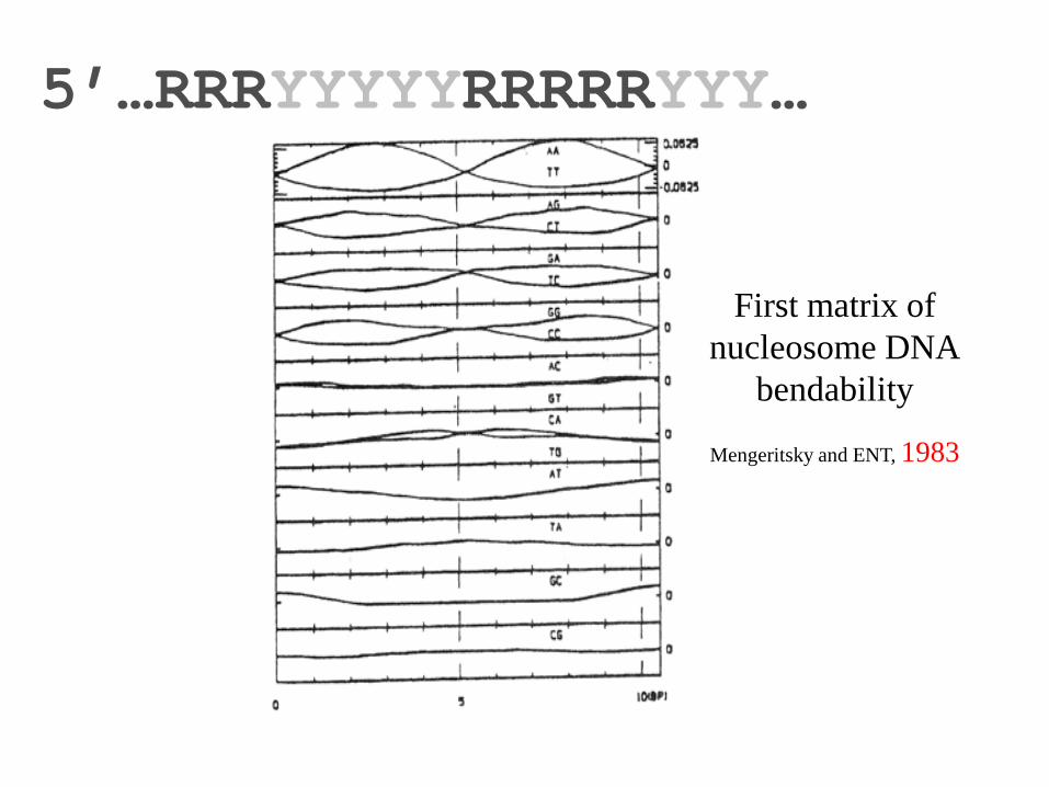
The purine-purine • pyrimidyne-pyrimidyne stacks (PuPu•PyPy or RR•YY) are very asymmetric

Deformable stacks (“wedges”) of the same kind
should be oriented on the surface
of the nucleosome the same way.
Hence – the preferred distances between
certain dinucleotides along the sequence
should be multiples of DNA period (10-11 bases)
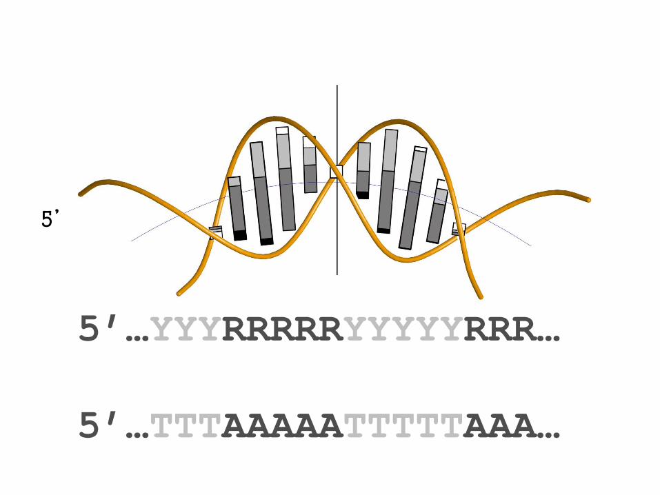
5’
5’…YYYRRRRRYYYYYRRR… 5’…TTTAAAAATTTTTAAA…
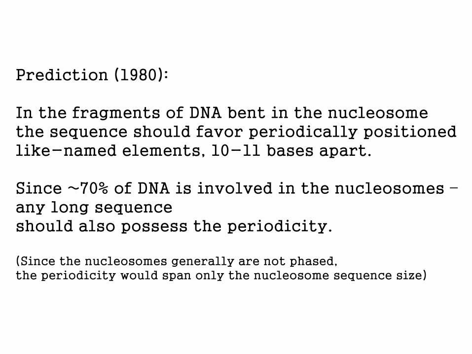
Prediction (1980): In the fragments of DNA bent in the nucleosome the sequence should favor periodically positioned like-named elements, 10-11 bases apart. Since ~70% of DNA is involved in the nucleosomes – any long sequence should also possess the periodicity. (Since the nucleosomes generally are not phased, the periodicity would span only the nucleosome sequence size)

DISTANCE ANALYSIS
(Autocorrelation)

One more important prediction:
The deformation (bending) should follow the
dyad symmetry of DNA molecule.
So should the dinucleotide elements (stacks).
Thus, within the sequence period AA and TT elements should be
on opposite sides from the axes, at the same distance
5’…TTTTAAAAATTTTTAAAA…
axis ↓
axis ↓
axis ↓
5’…RRRYYYYYRRRRRYYY…
First matrix of nucleosome DNA
bendability
Mengeritsky and ENT, 1983

The dyad symmetry of the DNA in the nucleosome has been mistaken in 1986 by a reputed team of scientists for a mirror symmetry. (“Errare humanum est”) This had catastrophic consequences for trustful naïve chromatin community (biologists), causing major confusion worldwide, still in effect

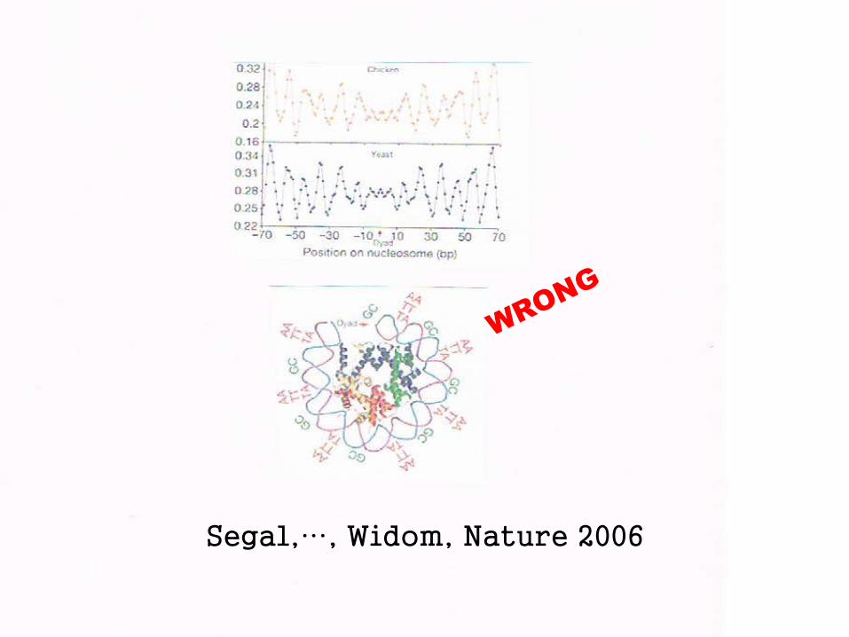
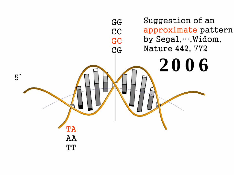
Segal,…, Widom, Nature 2006
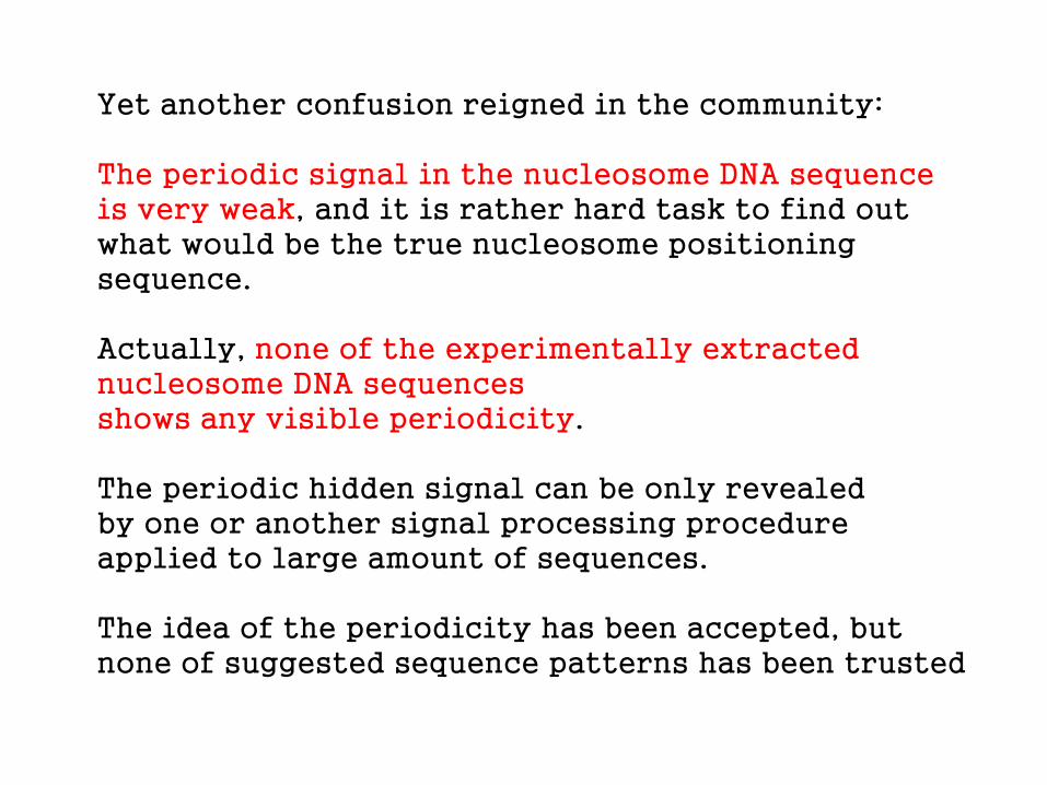
Yet another confusion reigned in the community: The periodic signal in the nucleosome DNA sequence is very weak, and it is rather hard task to find out what would be the true nucleosome positioning sequence. Actually, none of the experimentally extracted nucleosome DNA sequences shows any visible periodicity. The periodic hidden signal can be only revealed by one or another signal processing procedure applied to large amount of sequences. The idea of the periodicity has been accepted, but none of suggested sequence patterns has been trusted
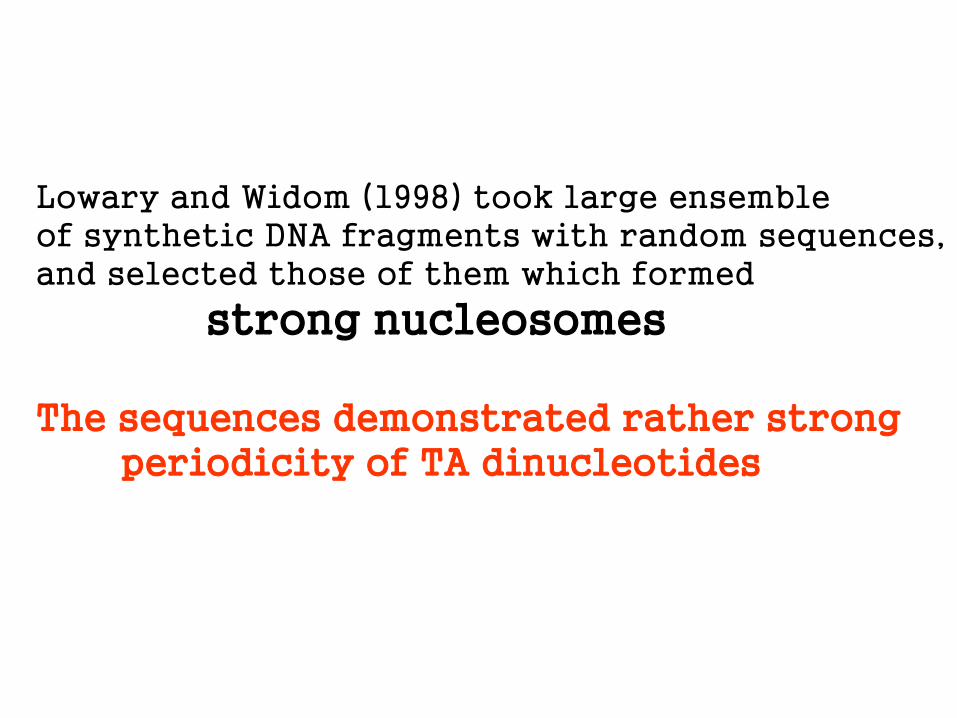
Lowary and Widom (1998) took large ensemble of synthetic DNA fragments with random sequences, and selected those of them which formed
strong nucleosomes The sequences demonstrated rather strong periodicity of TA dinucleotides

Clone 601, from collection of Lowary and Widom (1998) ...CAGCGCGTACGTGCGTTTAAGCGGTGCTAGAGCTGTCTAC... TACGTGCGTTTA TAAGCGGTGCTA TAGAGCTGTCTA We took all TAnnnnnnnnTA segments from the collection of Lowary/Widom, and analysed which dinucleotides are most frequently located in the interval between TA, and in which positions.

Bendability matrix for strong nucleosome DNAs of Lowary and Widom collection
0 1 2 3 4 5 6 7 8 9 0
AA 0 16 3 0 0 1 0 0 0 0 0 AC 0 5 2 5 2 3 5 3 1 0 0 AG 0 25 11 9 2 4 1 1 1 0 0 AT 0 2 0 3 1 1 3 1 2 0 0 CA 0 0 1 0 2 4 3 1 0 0 0 CC 0 0 0 0 5 4 7 3 6 0 0 CG 0 0 4 4 4 4 4 5 3 0 0 CT 0 0 0 2 1 2 1 9 11 22 0 GA 0 0 12 4 3 3 0 0 0 0 0 GC 0 0 4 7 6 7 5 10 5 0 0 GG 0 0 7 4 3 3 7 0 1 0 0 GT 0 0 2 7 6 4 5 6 2 6 0 TA 48 0 1 1 4 1 2 3 0 0 48 TC 0 0 0 0 1 1 1 4 10 0 0 TG 0 0 0 1 8 6 4 2 1 0 0 TT 0 0 1 1 0 0 0 0 5 20 0

C T A G A G x x x x C T A G – manually from the matrix C T A G x x x x x x C T A G – from Lowary & Widom paper C T A G A G G C C T C T A G – by dynamic programming Y R R R R R Y Y Y Y Y R
T A G A G G C C T C T A
T A G A G G C C T C T A
The periodical pattern hidden in the sequences of Lowary and Widom is selfcomplementary (that is, displays dyad symmetry), and manifests alternation of RRRRR and YYYYY

HALF

Taking the elegant idea of Lowary and Widom as a lead we extracted natural strong nucleosomes from whole genomes computationally. We looked for periodical sequences in genomes

The exact value of the period of DNA in the nucleosome has been matter of bitter argument between two schools during last 30 years: Close to 10.0 bp per turn – Crick, Klug, Richmond
(torsional constraint for unfolding of the nucleosome) Close to 10.4 bp per turn – our works
(no torsional constraint)

Structural (sequence) periodicity of nucleosome DNA
DNase I digestion of chromatin 10.30-10.40 bp Prunell, Kornberg, Lutter, Klug, Levitt, Crick, 1979
Beat effect, DNase I 10.33-10.40 bp Bettecken, 1979 Analytical geometry of nucl. DNA 10.30-10.50 bp Ulanovsky, 1983
DNA path in nucleosome crystals 10.36-10.44 bp Cohanim, 2006 CG periodicity, honey bee 10.36-10.44 bp Bettecken, 2009 DNase I digestion of chromatin 10.30-10.40 bp Boyle et al., 2008 Winter et al., 2013 Common range 10.36-10.40 bp

Magic distances, 10.4•n bases nearest integers 10.4 10 20.8 21 31.2 31 41.6 42 52.0 52 62.4 62 72.8 73 83.2 83 93.6 94 104.0 104 114.4 114 The ideal nucleosome positioning sequence would contain some periodically repeating motif, and all the distances between the same dinucleotides would be magic distances. Strong nucleosome DNA would show many magic distances.

The strongest nucleosomes of A. thaliana display very clear though still imperfect periodicity
The consensus pattern for A.thaliana is repetition of TAAAAATTTTTA, again, alternation of RRRRR and YYYYY, and complementary symmetry
TAAACTCTTTAAAAATCTTTTAAAAACCCTTGTACATATCTTAAAACCCTTTTAAAATCTCTTGTAAATCTTTAAAACCCTTTTAAAATCCCTTGTAAATCTTTTAAAACCCTTT AAATATTTTAAAACACTTTTCAAACAATTTTGAACCCTTTAAAAATCTTTATAAAACCTTTGTAAATCTTTTAAAGCCCTTTAAAATCTCTTATAAATCTTTTAAAACCCTTTTA CCCTGTAAAACTTTTAAAACCCTTTTAAAATCCCTTGTAAATCTTTTTAAACCCTTTTAAAATCCTTGTAAATATTTTAAAATCCCGTGTAATTCTTTTAAAACTCTTTTAAAAT AAATTTTAAAAAGGTTTTATAAGATTTGCAAGGGATTTTAAAGGGATTTAAAAGATTTACAAAAGTTTTTTAAAGGTTTAAAATTGTTTTAAAAGGATTTTAAAATATTTACAAG TTTTAAAAGGGTTTTAAAATATTTACATATGTTTTTTAAAGTTTTTTAAAGGGTTTAAAAGTGTTTTGCAAGATTTACAAGAGATTTTAAAAGGGTTTTAAGAGATTTACAAGAG ATCCTTTAAAAAATCATGTAAATCTTTTTAAAACCTTTTAAAATCCCTTGTAAATCTTTTAAAATCCTTTTAAAATCTCTTGTAAATGTTTAAAAACCCTTTTAAAATCTCTTGT AAGGGTTTTAAAATATTTACAAGGGATTTTAAAAGGGTTTTAAAAAATTTACAAGTGATTTTAAAAGATTTACAAGGGATTTTAAAAGGTTTTAAAAAAATTTACAAAAGTTTAT AAATCTTTTAAAACCCTTTTAAAATCCCTTGTAAATCTTTTAAAACACTTTTAAACCCTTTAAAAATCTTTAAAAAAACCTTTATAAATCTTTTAAAACTCTTTAAAATCTCTTG AAATGTTTTAAAACCTTTTTAAAATAATTTTAAACCCTTTAAAAATCGTTAAAAAACTTTTGTAAATCTTTTAAAGCCCTTTAAAATCCCTTGTAAATATTATAAAACCCTTTTA TGATTTTAAAAGGGTTTAAAAAGATTTACAAGGGATTTTAAAAGGGTTTTAAAAAATTTACAAGAGATTTTAAAAGGTTTTAAAAAGATTTACAAGAGTTTTAAAGGGTCTTCTT ATCTTTTAAAAATCCTTGTACATCTTTTAAAACCCTTTCAAACCCTTTAAAAATCTCTTGTAAATCTTTTAAAACCCTTTTAAAATCCCTTGTAAATCTTTCAAAACACTTTAAA CCTTTAAAATCCCTTGTAAATCTTTTAAAACCCTTTTCAAATCCCTTGTAAATGTTTTAAAACCCTTTTAGAACAATTTTAAACCCTTTAAAAATCTTTAAAAACCCTTTGTAAA TTTACAAAGGTTTTTAAAAGATTTTGAAAGGGTTTAAAAGTGTTTTAAAAGATTTACAAGGGATTTTAAAAGGGTTTTAAAGATTTACAAGAGATTTTAAAAGGGTTTTAAAAGA CTTGTAAATCTTTTAAAACCCTTTTAAAATCCTTTGTAAATATTTTAAAAGCCTTTTAAAATCCATTGTAAATCTTTTAAAATCCTTTGTAAATCTTTTAAAACCCTTTTAAAAT AGGATTTTAAAAATGTTTTAAAAGATTTACAATGGATTTTAAAAGGGTTTAAAATATTTATAAGGGATTTTGAAGGGCTTTCAAAGATTTATAAAGGTTTTTTAAAAATTTTTAA TTGTAAATTATTTAAAAATCTTTTAAAACTCCTTGTACATCTTTTAAAACTCTTTTAAAATTTCTTGTAAATCTTTAAAACCCTTTAAAATCCCTTGTAAATCTTTTAAAATACT ACCCTTTAAAAATCTTTTAAAAATCTTTGTAAATCTTTTAAAGCCCTTTGAAATCCCTTGTAAATATTTTAAAATCTTTTAAAATTCCTTGTAAATGTTTTAAAACCCTTTTAAA GATTTGCAAAAGATTTTAAAAGATTTACAAAGGATTTTAAAAGATTTACAATGGATTTTAAAGGGGTTTAAAAGATTTACAAAGGTTTTTTAAAGATTTTTAAAGGGTTTTAAAT
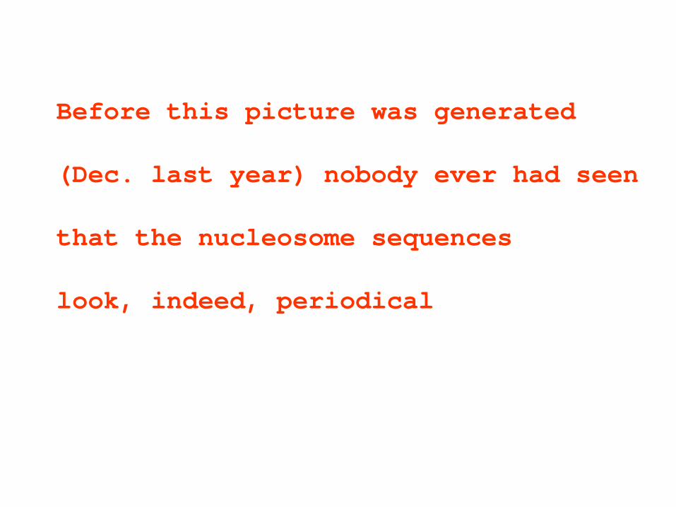
Before this picture was generated (Dec. last year) nobody ever had seen that the nucleosome sequences look, indeed, periodical

From the bendability matrices
for the strong nucleosomes: T AGAGG CCTCT A Lowary and Widom T AAAAA TTTTT A A.thaliana T AAAAA TTTTT A C.elegans T AAAAA TTTTT A H.sapiens T AAAAA TTTTT A isochores L1, L2, H1 and H2 C GGGGG CCCCC G isochores H3 Y RRRRR YYYYY R common for all (and complementarty symmetry)

Previously detected patterrns, species: species authors method C GRAAA TTTYC G C. elegans Gabdank, 2009 A C AAAAA TTTTT G C. elegans Rapoport, 2011 B C AAAAA TTTTT G A. gambiae same B C AAAAA TTTTT G C. albicans same B C AAAAA TTTTT G D. melanogaster same B C AAAAA TTTTT G S. cerevisiae same B T AAAAA TTTTT A A. mellifera same B T AAAAA TTTTT A A. thaliana same B T AAAAA TTTTT A D. discoideum same B T AAAAA TTTTT A D. rerio same B T AAAAA TTTTT A G. gallus same B T AAAAA TTTTT A H. sapiens same B T AAAAA TTTTT A M. musculus same B c GGGGG CCccc G C. reinhardtii same B Y RRRRR YYYYY R consensus A – signal regeneration, nucleosomes B – Shannon N-gram extension, whole genome

Nucleosome positioning patterns of various isochores (Frenkel et al., 2011) by N-gram extension isochores G+C %
C AGGGG CCCCT G C GGGGA TCCCC G C AGAAA TTTCT G T AAAAA TTTTT A T AAAAA TTTTT A
Y RRRRR YYYYY R

5’
5’…YYYRRRRRYYYYYRRR… TA CG TG CA
AT GC AC GT
Contact with arginines
Exposed
Nucleosome positioning pattern
2013
TA CG TG CA

start ↓
end ↓
center ↓

Example of the output from the nucleosome mapping server http://www.cs.bgu.ac.il/~nucleom (Google “finestr”)
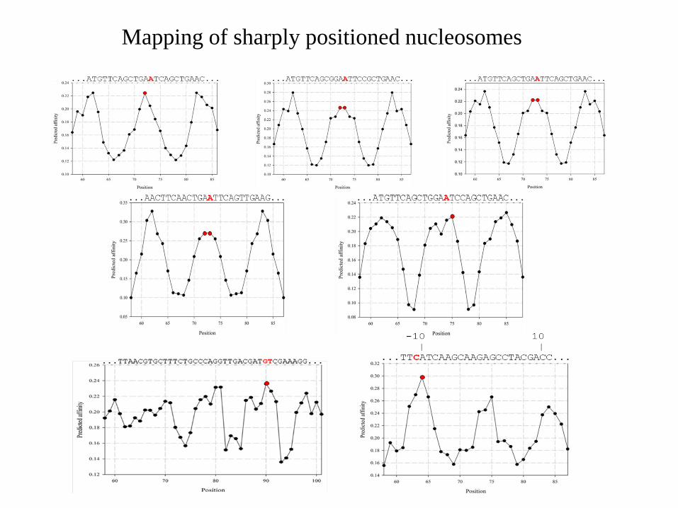
Mapping of sharply positioned nucleosomes

Nucleosomes around the GT splice junctions Hapala J, ENT, Gene 2011

GT AG
Dots • - N9 atoms of guanines
Splice junctions are hiding on the surface of histone octamers

Nucleosome DNA which carries promoter TATAAA box has two rotational settings encoded in the sequence (two peaks within one period). Jan Hapala & ET, in press

TATA-switch Two alternative positions of TATAAA box in the promoter nucleosomes are separated by 140 (220) degrees, which corresponds to exposed and inaccessible orientations of the box. By shifting the DNA along its path by 4(6) bases, the promoter is switched ON or OFF. The switch (shift) may be triggered by remodelers or transcription factors.
ACKNOWLEDGEMENTS Recent colaborators (2009-2013): Idan Gabdank (Beer Sheva, Israel) Zakharia Frenkel (Haifa, Israel) Alexandra Rapoport (Haifa, Israel) Thomas Bettecken (München, Germany) Jan Hapala (Brno, Czech Republic) Bilal Salih (Haifa, Israel) Vijay Tripathi (Haifa, Israel) Earlier colaborators (1979-2008) Thomas Bettecken Joel Sussman Galina Mengeritsky Levy Ulanovsky Alex Bolshoy Ilya Ioshikhes Amir Cohanim Fadil Salih Simon Kogan
Funding (2009-2012) Israel Science Foundation, and South Moravian Program


From FHC Crick & A Klug, Kinky helix, Nature 255, 530-533, 1975 We have found it very difficult to estimate just how much energy is required to bend DNA “smoothly” to a sma1l
radius of curvature, say 30-50A, bearing in mind that these numbers are not many times greater than the diameter of the DNA double helix, which is about 20A, and that bending a helix destroys its symmetry. We have formed the impression that the energy might be rather high. We therefore asked ourselves whether the folded DNA may consist of relatively straight stretches joined by large kinks. This paper describes a certain type of kink which can be built rather nicely and has interesting properties. In other words: Smooth bending is difficult to calculate. We therefore thought about large kinks Гладкий изгиб посчитать трудно Поэтому мы

. Possibility of nonkinked packing of DNA in chromat JL Sussman, EN Trifonov Proc Natl Acad Sci U S A 75, 103–107, 1978
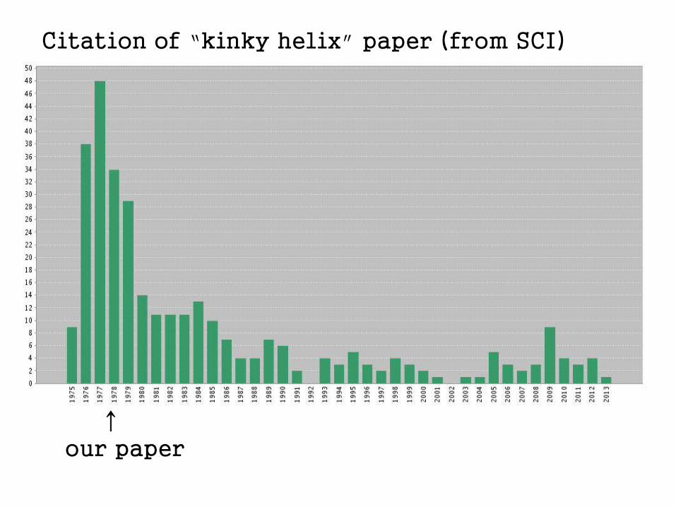
↑ our paper
Citation of “kinky helix” paper (from SCI)
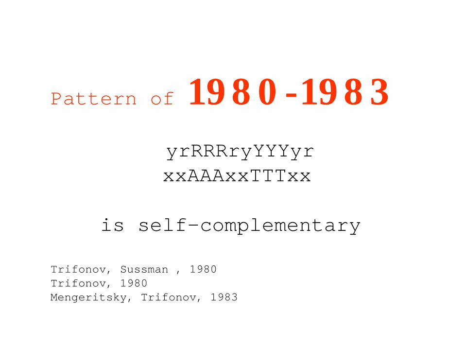
Pattern of 1980-1983 yrRRRryYYYyr xxAAAxxTTTxx is self-complementary Trifonov, Sussman , 1980 Trifonov, 1980 Mengeritsky, Trifonov, 1983

This achievement in the single-base accuracy mapping of the nucleosomes has not been accepted by chromatin research community. The reasons: 1. Mistrust. The physics of the phenomenon and multiple alternative positions of the nucleosome centers are hard to grasp for non-physicists, and the sequences did not show any obvious periodicity 2. The chromatin research community was not ready yet methodologically to conduct high resolution experimental studies
5’
TA AA TT
GG CC GC CG
Suggestion of an approximate pattern by Segal,…,Widom, Nature 442, 772
2006

The work of Segal et al., 2006, was the first high throughput whole-genome analysis. It drew a lot of attention, and the approach became very fashionable in the chromatin community. But the emphasis was still on low resolution studies, maps of “occupancy”, where the alternative positions of the nucleosomes and rotational setting of DNA are not seen. No attempts were made in that work to derive an exact nucleosome positioning sequence pattern from the whole genome sequences.


From 1979 until 2008 the value 10.0 dominated in liter It is now gradually replaced by 10.4 It was admitted by Richmond at the conference in 200that “everybody knows that the period is 10.4”
Не иначе как бес попутал кристаллографов
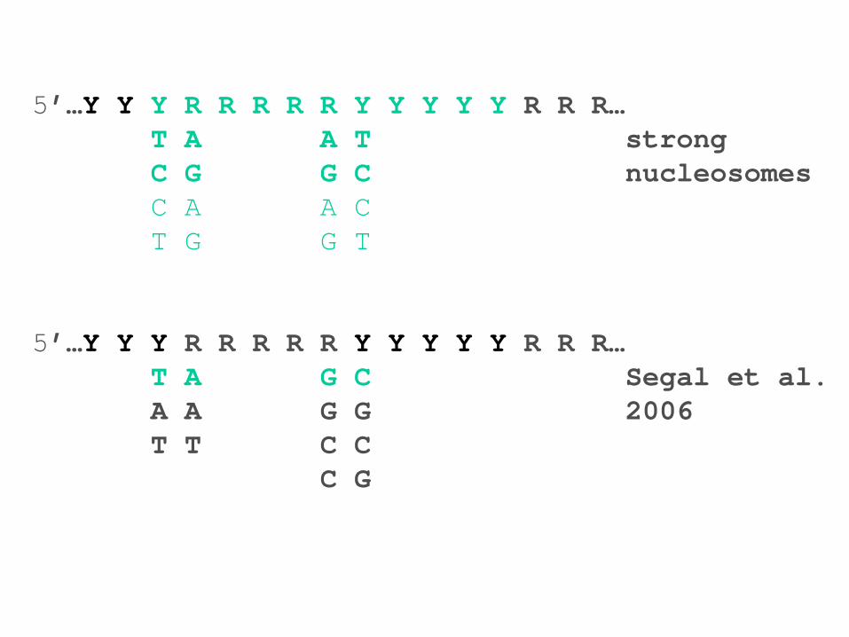
5’…Y Y Y R R R R R Y Y Y Y Y R R R… T A A T strong C G G C nucleosomes C A A C T G G T 5’…Y Y Y R R R R R Y Y Y Y Y R R R… T A G C Segal et al. A A G G 2006 T T C C C G

We now entered a new era of single-base resolution chromatin research. None of experimental techniques provides today the single-base resolution. The computational mapping of the nucleosomes, quick and accurate, is waiting for sceptic experimentalists to join and enjoy. Но поезд уходит: мы первыми пожнем плоды

5’
5’…YYYRRRRRYYYYYRRR… TA CG TG CA
AT GC AC GT
Contact with arginines
Exposed
Nucleosome positioning pattern
2013
TA CG TG CA

СПАСИБО ЗА ВНИМАНИЕ!
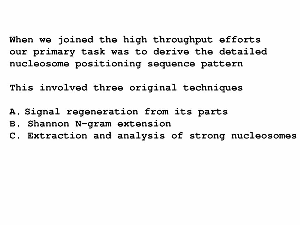
When we joined the high throughput efforts our primary task was to derive the detailed nucleosome positioning sequence pattern This involved three original techniques A. Signal regeneration from its parts B. Shannon N-gram extension C. Extraction and analysis of strong nucleosomes
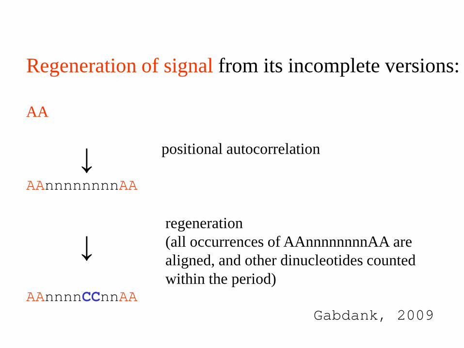
Regeneration of signal from its incomplete versions: AA positional autocorrelation AAnnnnnnnnAA regeneration (all occurrences of AAnnnnnnnnAA are aligned, and other dinucleotides counted within the period) AAnnnnCCnnAA Gabdank, 2009
↓
↓

A. thaliana T AAAAA TTTTT A strong nucleosomes T AAAAA TTTTT A Shannon extension C. elegans T AAAAA TTTTT A strong nucleosomes c grAAA TTTyc g signal regeneration isochores L1, L2 T AAAAA TTTTT A strong nucleosomes T AAAAA TTTTT A Shannon extension isochores H1 T AAAAA TTTTT A strong nucleosomes c AgAAA TTTcT g Shannon extension isochores H2 T AAAAA TTTTT A strong nucleosomes c ggggA Tcccc g Shannon extension isochores H3 C GGGGG CCCCC G strong nucleosomes C aGGGG CCCCt G Shannon extension Y RRRRR YYYYY R – all, and all with complementary symmetry

The dinucleotide stacks are placed in such positions within the nucleosome DNA period to ensure best possible bending. The better the bending – the stronger the nucleosome. But the bulk of the nucleosomes are only marginally stable. Only a fraction of properly positioned dinucleotides is present in any given nucleosome DNA sequence.

CGGAAATTTTCCGGAAATTTCCGGAAATTTCCGGGAAATTTCCGGAAATTTCCGGAAATTTTCCGGAAATTTCCGGAAATTTCCGGGAAATTTCCGGAAATTTCCGGAAATTTTCC CagaggagcttcctggggaTCCaGAcATgataagatacaTTgatGAgtTTggacaAAccacaactagAATgcagtGAAAaaaatgctttATTTgtgaAAtTTgtgatgctaTTgct YRRRRRagYYYYctRRRgaYYYRRRcRYgataRRRtacaYYgatRRRtYYggacRRRccacaactRRRRYgcagtRRRRaaaaYRctttRYYYgtRRRRtYYgtgatgctaYYgYY
Match of the BamHI nucleosome (typical semistable nucleosome) to the standard nucleosome probe (GAAAATTTTC)n

TAAACTCTTTAAAAATCTTTTAAAAACCCTTGTACATATCTTAAAACCCTTTTAAAATCTCTTGTAAATCTTTAAAACCCTTTTAAAATCCCTTGTAAATCTTTTAAAACCCTTT AAATATTTTAAAACACTTTTCAAACAATTTTGAACCCTTTAAAAATCTTTATAAAACCTTTGTAAATCTTTTAAAGCCCTTTAAAATCTCTTATAAATCTTTTAAAACCCTTTTA
CCCTGTAAAACTTTTAAAACCCTTTTAAAATCCCTTGTAAATCTTTTTAAACCCTTTTAAAATCCTTGTAAATATTTTAAAATCCCGTGTAATTCTTTTAAAACTCTTTTAAAAT AAATTTTAAAAAGGTTTTATAAGATTTGCAAGGGATTTTAAAGGGATTTAAAAGATTTACAAAAGTTTTTTAAAGGTTTAAAATTGTTTTAAAAGGATTTTAAAATATTTACAAG
TTTTAAAAGGGTTTTAAAATATTTACATATGTTTTTTAAAGTTTTTTAAAGGGTTTAAAAGTGTTTTGCAAGATTTACAAGAGATTTTAAAAGGGTTTTAAGAGATTTACAAGAG ATCCTTTAAAAAATCATGTAAATCTTTTTAAAACCTTTTAAAATCCCTTGTAAATCTTTTAAAATCCTTTTAAAATCTCTTGTAAATGTTTAAAAACCCTTTTAAAATCTCTTGT AAGGGTTTTAAAATATTTACAAGGGATTTTAAAAGGGTTTTAAAAAATTTACAAGTGATTTTAAAAGATTTACAAGGGATTTTAAAAGGTTTTAAAAAAATTTACAAAAGTTTAT AAATCTTTTAAAACCCTTTTAAAATCCCTTGTAAATCTTTTAAAACACTTTTAAACCCTTTAAAAATCTTTAAAAAAACCTTTATAAATCTTTTAAAACTCTTTAAAATCTCTTG AAATGTTTTAAAACCTTTTTAAAATAATTTTAAACCCTTTAAAAATCGTTAAAAAACTTTTGTAAATCTTTTAAAGCCCTTTAAAATCCCTTGTAAATATTATAAAACCCTTTTA
TGATTTTAAAAGGGTTTAAAAAGATTTACAAGGGATTTTAAAAGGGTTTTAAAAAATTTACAAGAGATTTTAAAAGGTTTTAAAAAGATTTACAAGAGTTTTAAAGGGTCTTCTT ATCTTTTAAAAATCCTTGTACATCTTTTAAAACCCTTTCAAACCCTTTAAAAATCTCTTGTAAATCTTTTAAAACCCTTTTAAAATCCCTTGTAAATCTTTCAAAACACTTTAAA CCTTTAAAATCCCTTGTAAATCTTTTAAAACCCTTTTCAAATCCCTTGTAAATGTTTTAAAACCCTTTTAGAACAATTTTAAACCCTTTAAAAATCTTTAAAAACCCTTTGTAAA
TTTACAAAGGTTTTTAAAAGATTTTGAAAGGGTTTAAAAGTGTTTTAAAAGATTTACAAGGGATTTTAAAAGGGTTTTAAAGATTTACAAGAGATTTTAAAAGGGTTTTAAAAGA CTTGTAAATCTTTTAAAACCCTTTTAAAATCCTTTGTAAATATTTTAAAAGCCTTTTAAAATCCATTGTAAATCTTTTAAAATCCTTTGTAAATCTTTTAAAACCCTTTTAAAAT
AGGATTTTAAAAATGTTTTAAAAGATTTACAATGGATTTTAAAAGGGTTTAAAATATTTATAAGGGATTTTGAAGGGCTTTCAAAGATTTATAAAGGTTTTTTAAAAATTTTTAA TTGTAAATTATTTAAAAATCTTTTAAAACTCCTTGTACATCTTTTAAAACTCTTTTAAAATTTCTTGTAAATCTTTAAAACCCTTTAAAATCCCTTGTAAATCTTTTAAAATACT
ACCCTTTAAAAATCTTTTAAAAATCTTTGTAAATCTTTTAAAGCCCTTTGAAATCCCTTGTAAATATTTTAAAATCTTTTAAAATTCCTTGTAAATGTTTTAAAACCCTTTTAAA GATTTGCAAAAGATTTTAAAAGATTTACAAAGGATTTTAAAAGATTTACAATGGATTTTAAAGGGGTTTAAAAGATTTACAAAGGTTTTTTAAAGATTTTTAAAGGGTTTTAAAT
The strongest nucleosomes of A. thaliana display very clear though still imperfect periodicity
The ideal pattern for A. thaliana is repetition of TAAAAATTTTTA, again, alternation of RRRRR and YYYYY, and complementary symmetry

Cat in bushes. Courtesy of I. Gabdank

Guanines of GT- and AG-ends of introns are oriented towards the surface of the histone octamer, away from exterior. Such orientation is the best for guanines to minimize spontaneous depurination and oxidation The most frequent spontaneous damages to DNA bases:
depurination of G (N9 atoms) oxidation of G deamination of C
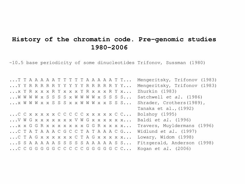
History of the chromatin code. Pre-genomic studies 1980-2006 ~10.5 base periodicity of some dinucleotides Trifonov, Sussman (1980) ...T T A A A A A T T T T T A A A A A T T... Mengeritsky, Trifonov (1983) ...Y Y R R R R R Y Y Y Y Y R R R R R Y Y... Mengeritsky, Trifonov (1983) ...x Y R x x x R Y x x x Y R x x x R Y x... Zhurkin (1983) ...W W W W x S S S S x W W W W x S S S S... Satchwell et al. (1986) ...x W W W x x S S S x x W W W x x S S S... Shrader, Crothers(1989), Tanaka et al.,(1992) ...C C x x x x x C C C C C x x x x x C C... Bolshoy (1995) ...V W G x x x x x x x V W G x x x x x x... Baldi et al. (1996) ...x x G G R x x x x x x x G G R x x x x... Travers, Muyldermans (1996) ...C T A T A A A C G C C T A T A A A C G... Widlund et al. (1997) ...C T A G x x x x x x C T A G x x x x x... Lowary, Widom (1998) ...S S A A A A A S S S S S A A A A A S S... Fitzgerald, Anderson (1998) ...C C G G G G G C C C C C G G G G G C C... Kogan et al. (2006)
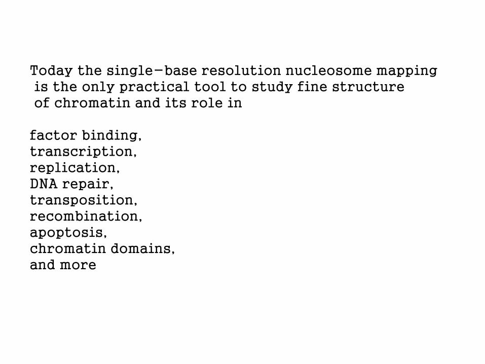
Today the single-base resolution nucleosome mapping is the only practical tool to study fine structure of chromatin and its role in factor binding, transcription, replication, DNA repair, transposition, recombination, apoptosis, chromatin domains, and more

Immediate questions: Where in genomes the strong nucleosomes are located? What they are doing there? Tentative answer: Strong nucleosomes are chromatin organizers.


Why DNA binds to histone octamers by one side? It could be either intrinsic DNA curvature or better bending in one specific direction (deformational anisotropy of DNA) Both should be sequence-dependent

Nucleosome positioning sequence pattern is very weak (as the nucleosomes should be easy to unfold) The weak pattern overlaps with other messages (“noise”). That makes the signal/noise ratio very low. VERY large database of the nucleosome DNA sequences is needed, to extract and fully describe the signal It is easy, however, to detect the signal

DISTANCE ANALYSIS
(Autocorrelation)

T.Bettecken, E.N.T., 2009
Whole-genome periodicities (distance analysis) AA TT CG GC CA TG AG CT AT GG CC GA TC AC GT TA S. cerevisiae ● ● ● ● ● ● ● ● ● ● ● ● ● - - ● C. elegans ● ● ● ● ● ● ● ● ● - - ● ● ● ● - A. thaliana ● ● - ● ● ● - - ● ● - - - - - - D. rerio ● ● - ● - - - - - ● ● - - - - - C. albicans ● ● - - ● ● - - - - - - - - - - A. mellifera ● ● ● ● - - - - - - - - - - - - D. melanogaster ● ● ● ● - - - - - - - - - - - - G. gallus - - - - - - ● ● - - - - - - - - A. gambiae ● ● - - - - - - - - - - - - - - C. reinhardtii ● ● - - - - - - - - - - - - - - D. discoideum - - ● - - - - - - - - - - - - - H. sapiens - - ● - - - - - - - - - - - - - M. musculus - - - - - - - - - - - - - - - -

Nucleosome positioning patterns, isochores (Frenkel, 2011, 2012) isochore method T AAAAA TTTTT A L1 (<37% G+C) B T AAAAA TTTTT A same A T AAAAA TTTTT A L2 (37-41% G+C) B C AGAAA TTTCT G H1 (41-46% G+C) B C GGGGA TCCCC G H2 (46-53% G+C) B C AGGGG CCCCT G H3 (>53% G+C) B C AGGGG CCCCT G same A Y RRRRR YYYYY R consensus A signal regeneration, nucleosomes B Shannon N-gram extension, whole genome

Score
Score
Score
Position Position
AAnnnnnnnnAA repeat structure (C. elegans)
Regenerated pattern (AAATTTCCGG)(AAAT…
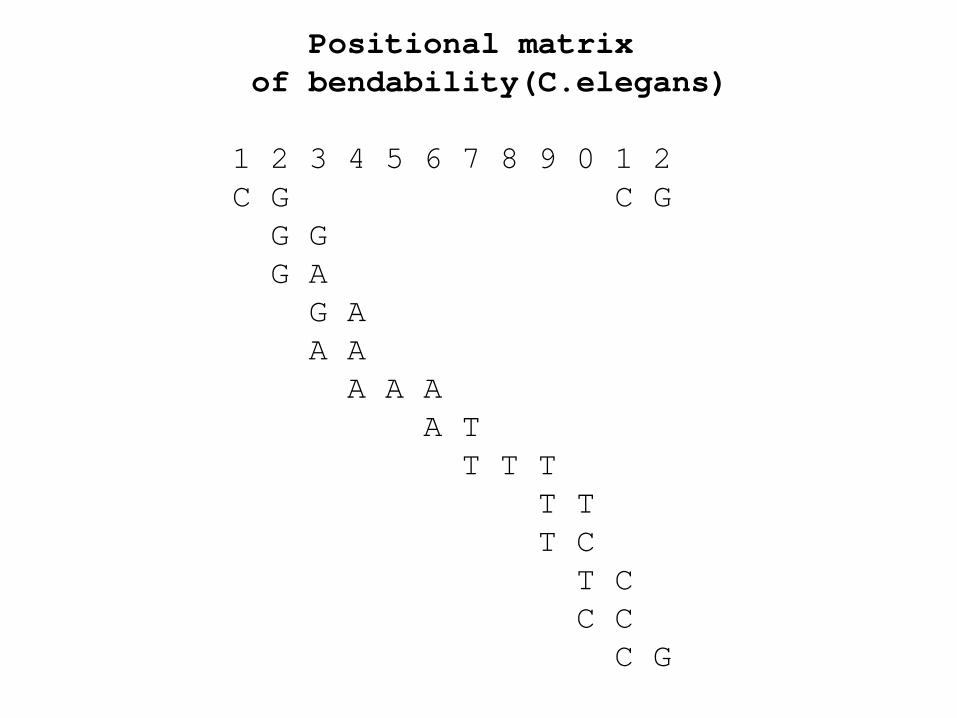
Positional matrix of bendability(C.elegans) 1 2 3 4 5 6 7 8 9 0 1 2 C G C G G G G A G A A A A A A A T T T T T T T C T C C C C G
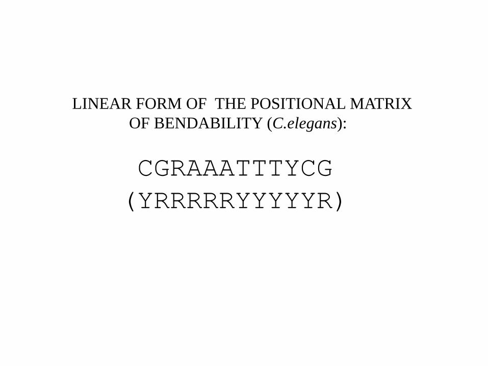
LINEAR FORM OF THE POSITIONAL MATRIX OF BENDABILITY (C.elegans):
CGRAAATTTYCG (YRRRRRYYYYYR)
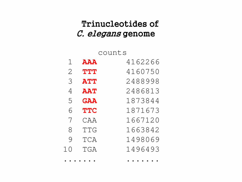
Trinucleotides of C. elegans genome counts 1 AAA 4162266 2 TTT 4160750 3 ATT 2488998 4 AAT 2486813 5 GAA 1873844 6 TTC 1871673 7 CAA 1667120 8 TTG 1663842 9 TCA 1498069 10 TGA 1496493 ....... .......

TOPMOST TRINUCLEOTIDES MAKE TOGETHER THE DOMINANT PATTERN
GAAAATTTTC:
GAAAATTTTC GAAAATTTTC GAAAATTTTC GAAAATTTTC GAAAATTTTC GAAAATTTTC GAAAATTTTC GAAAATTTTC
This technique is known since 1948 –
Shannon N-gram extension
It has been very helpful in further studies
of the nucleosome positioning patterns

Human isochores Lab of G. Bernardi, 2006
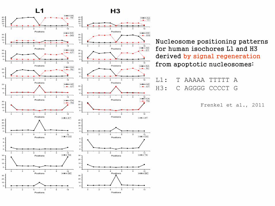
Nucleosome positioning patterns for human isochores L1 and H3 derived by signal regeneration
from apoptotic nucleosomes: L1: T AAAAA TTTTT A H3: C AGGGG CCCCT G Frenkel et al., 2011

Shannon N-gram reconstruction of linkers TTTTATTTTAAAATAAAA human linkers AAAATAAAATATTTTATTTT yeast linkers TAAAgTAcTTTA human, apoptotic cuts consensus: TAxxxTAxxxTAxxx (B. Salih, T. Bettecken, Z. Frenkel)
TTAAAAATTTTTAAAAATTTTTAA human L1 isochores, nucleosomes

BamHI nucleosome of Ponder and Crawford, 1977
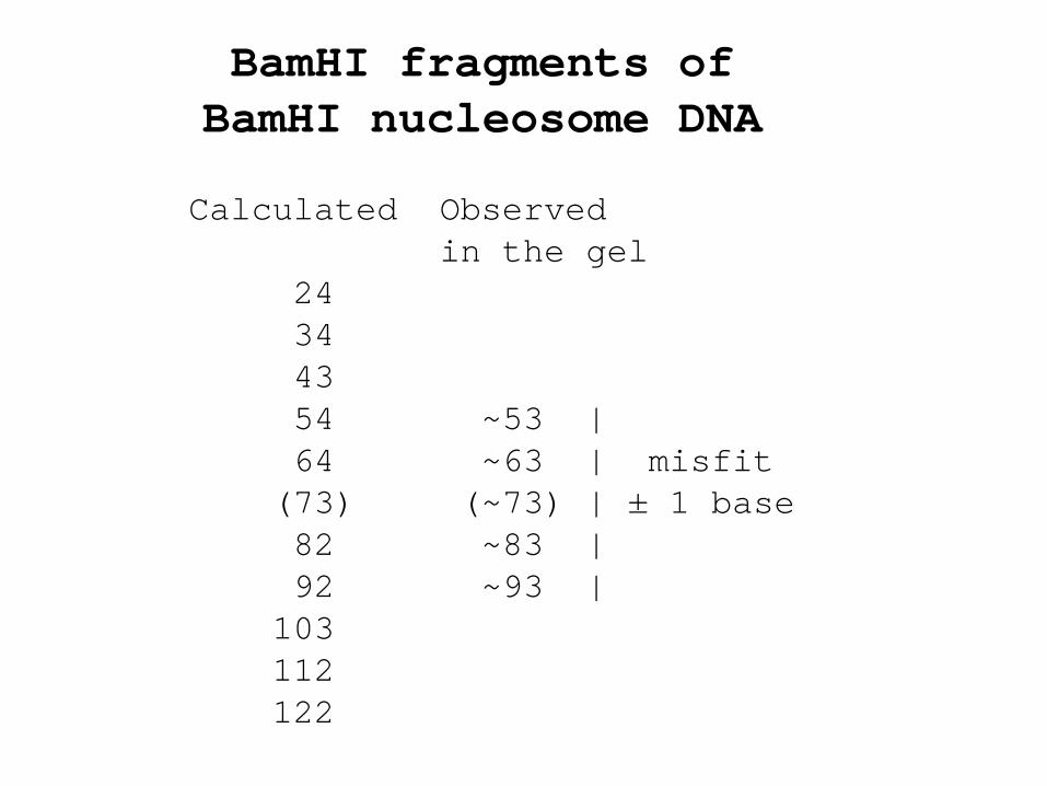
BamHI fragments of BamHI nucleosome DNA Calculated Observed in the gel 24 34 43 54 ~53 | 64 ~63 | misfit (73) (~73) | ± 1 base 82 ~83 | 92 ~93 | 103 112 122

Example of the nucleosomes at and around GT splice junction
Hapala, 2011

Plenty of various other nucleosome positioning patterns have been suggested during 30 years since the first observation of sequence periodicity. At the best they provide occupancy maps (resolution of ~15 bases). The (GRAAATTTYC)n and (RRRRRYYYYY)n are the only patterns that generate maps with single-base resolution, verified by crystal data. The future of the chromatin structure/function is with the high resolution studies.