editorial original articles review articles.pdf · for a while, the “green revolution” became a...
TRANSCRIPT

Vol.49, No.7&8 July/August 2006
Editorial
Green Revolution
Naruo Uehara ......................................................................................................................................................... 235
Original Articles
The Longitudinal Effects of the “Green Revolution” on the Infant Mortality Ratein Rural Thailand
Yukinori Okada, Susumu Wakai ......................................................................................................................... 236
Symptom Experience and Patterns of End-of-Life—Home care for elderly patients with cancer vs. those without cancer in Japan
Yoshihisa Hirakawa, Yuichiro Masuda, Masafumi Kuzuya, Takaya Kimata,Akihisa Iguchi, Kazumasa Uemura ..................................................................................................................... 243
Review Articles
Aid Coordination Mechanisms for Reconstructing the Health Sectorof Post-Conflict Countries
Hidechika Akashi, Noriko Fujita, Rumiko Kakishima Akashi ...................................................................... 251
Ocular Manifestations in Behçet’s Disease
Takahiko Hayashi, Nobuhisa Mizuki .................................................................................................................. 260
Case Reports
A Case of Pneumothorax in Which Progression of Reexpansion PulmonaryEdema Was Arrested
Makoto Nonaka, Koji Maezawa, Yasushi Koike, Toshiyuki Hatakeyama, Hitoshi Soda,Osamu Sakurai, Yasuo Ishida, Kiyoshi Hataya ................................................................................................ 269
A Case of Subclinical Central Diabetes Insipidus Unmasked by Pregnancy
Ryuji Katase, Yuji Moriwaki, Sumio Takahashi, Zenta Tsutsumi, Taku Inokuchi,Tuneyoshi Ka, Tetsuya Yamamoto ..................................................................................................................... 272
Clinical Topics in Japan
COX-2 Inhibitors and the Risk of Cardiovascular Events
Shuntaro Hara, Ichiro Kudo ................................................................................................................................ 276
Internet Addiction among Students:Prevalence and psychological problems in Japan
Takeshi Sato ........................................................................................................................................................... 279

235JMAJ, July /August 2006 — Vol. 49, No. 7 • 8
Editorial
A new variety of rice called IR8 or “miracle rice,”capable of vastly increasing the rice yield per landarea unit, was developed in 1966 at the InternationalRice Research Institute (IRRI), established in thePhilippines by a U.S. foundation. The “Green Revo-lution,” which refers to the use of new crop breedsand new farming technologies to increase cerealproductivity, has been introduced and promoted byinternational development aid to developing coun-tries in Asia, as epoch-making technology whichhelps overcome anticipated food crises to be broughtby the rapid increase of population. As a result, inChina, Pakistan and India, the supply of cereals hasbeen improved, and countries such as Indonesia, Viet-nam and Thailand have even become net exporters.For a while, the “Green Revolution” became a bywordto symbolize the power of science and the success oftechnology transfer.
However, now after the passage of over 30 years,the evaluation on the effects of “Green Revolution”remains controversial.1 In contrast to conventionalcrop varieties, the high yield of the new varieties wasdependent upon the use of large quantities of water,agricultural chemicals and chemical fertilizers, whichin turn required high levels of investment and thusenlarged the divide between rich and poor farmingareas, while creating a dependence upon the marketeconomies of advanced nations for the provision ofsuch agricultural chemicals and chemical fertilizers.Moreover, this also helped cause the desertificationof irrigated areas and pollution by agricultural chemi-cals, in some cases even producing conflict betweencommunities over water. In addition, it had beenhoped that the increase in productivity of cerealcrops would help alleviate food shortages and thuscontribute to the improvement of nutritional statusof the people, consequently benefiting public health.However, Okada’s review suggested that these hopesappear to have been betrayed.
It seems that although the “Green Revolution”seems to have been a success as far as the nationaltotal cereal production figures are concerned, a lookat it from the perspective of communities and indi-vidual humans indicates that the problems have faroutweighed the successes, leaving many lessons to belearned.
In his now classic book “Small Is Beautiful,” E.F.Schumacher said “Modern agriculture relies onapplying to soil, plants, and animals ever-increasingquantities of chemical products, the long-term effect
of which on soil fertility and health is subject to verygrave doubts.”2 Claiming that the central issue is howthe technology should be, he requested scientistsand engineers to pursue appropriate technology(“Intermediate Technology”) instead of compellingthe choice of “poison or hunger.” Appropriate tech-nology is available to everybody, not exclusively toprivileged groups, and it must be useful in providing“help to those who need it most.”
In the sense that technological innovation tookthe leading role in “development” and penetratedradically and extensively into the lives in communitiesfor the cause of achieving the benefit it seemed topromise, the lessons from the “Green Revolution”are reminiscent of “medicalization of life” of IvanIllich. Illich argued that the modern medicine hasbeen causing more harm than benefit to people,despite the series of innovation of medical tech-nologies.3 He addressed the issue of uncertainty ineffectiveness of modern medical technologies, andwarned us the danger of rendering our health andsurvival dependent on technologies, which tended tobe industry-driven, rather than human-centered.
When I encountered his admonition in 1978, Icould not accept it, as being a zealous hard-workingsurgeon, and seriously tried to contrive a way torefute it. Now, in the face of the large body of facts ofmedical adverse events in Japan, as well as the realityrevealed by the report from Institute of Medicine4
and also numerous subsequent reports from manycountries, I realistically understand the gravity of theIllich’s words at the beginning of his book titled“Medical Nemesis”—“The medical establishmenthas become a major threat to health.”3
References
1. Shiva V. The Violence of the Green Revolution: Third WorldAgriculture, Ecology and Politics. London: Zed Books Ltd.; 1992.
2. Schumacher EF. Small is Beautiful: Economics as if PeopleMattered. London: Blond and Briggs; 1973.
3. Illich I. Limits to Medicine: Medical Nemesis, the Expropriation ofHealth. London: Calder & Boyars; 1975.
4. Committee on Quality of Health Care in America, Institute ofMedicine. To Err is Human: Building a Safer Health System.Washington DC: National Academy Press; 2000.
Green Revolution
Naruo Uehara*1
*1 Division of International Health (Quality & Health System),Tohoku University Graduate School of Medicine, SendaiCorrespondence to: Naruo Uehara MD, PhD, Division ofInternational Health (Quality & Health System), Tohoku UniversityGraduate School of Medicine, 2-1 Seiryo-machi, Aoba-ku, Sendai,Miyagi 980-8575, Japan. Tel: 81-22-717-8197,Fax: 81-22-717-8198, E-mail: [email protected]

236 JMAJ, July /August 2006 — Vol. 49, No. 7 • 8
*1 Fujieda Municipal General Hospital, Fujieda*2 Department of Community Health, School of International Health, Graduate School of Medicine, the University of Tokyo, TokyoCorrespondence to: Yukinori Okada MD, Fujieda Municipal General Hospital, 4-1-11 Surugadai, Fujieda-shi, Shizuoka 426-8677, Japan.Tel: 81-5-4646-1111, Fax: 81-5-4646-1122, E-mail: [email protected]
Original Article
The Longitudinal Effects of the “Green Revolution”on the Infant Mortality Rate in Rural Thailand
JMAJ 49(7 • 8): 236–242, 2006
Yukinori Okada,*1 Susumu Wakai*2
AbstractObjectives A rapid increase in the ratio of rented farming land to total farming land and the stratification ofpeasantry has been reported in rural Thailand since the 1960s. The problem is especially severe in the centralregion, where the commercialization of agriculture called the “Green Revolution” has been rampant withenhanced integration of agricultural resources by absentee landlords. We analyzed the problem by evaluatingits effect on the health status of infants in rural Thailand.
Methods We examined three indicators; IMR, NMR and PNMR as dependent variables in relation to the ratioof rented land as independent variable. The surveyed period was between 1963 and 1998. ANCOVA analyseswere applied.
Results IMR and NMR showed a statistically significant association with the ratio of rented land (F�7.77,P�0.01; F�32.88, P�0.0001). The longitudinal time-trends of IMR and NMR also showed a significant asso-ciation with that of the ratio (F�10.97, P�0.05; F�29.87, P�0.001). PNMR did not show any significance.
Conclusion The ratio of rented land is a factor that reflects the extent of commercialization of agricultureand explains the substantial regional diversity of the mortality rates in Thailand. The existence of nutritionaldeterioration and vulnerability induced by the “Green Revolution” is suggested, since NMR reflects endogenousfactors of the area and is sensitive to nutritional and sanitary conditions rather than PNMR. Further empiricalstudies that emphasize the linkage are necessary to attain a lower IMR in Thailand.
Key words Ratio of rented land, Green revolution, Infant, neonatal, and post-neonatal mortality rate,Nutritional deterioration
problems are a major deterrent to higher pro-ductivity, resulting in the lower average riceyield of Thailand compared to that of otherAsian nations.3
On the other hand, the World Bank has con-cluded that there is little evidence of exploit-ation and indebtedness, and the studies had ex-aggerated the issue.5 Laurence surveyed twoChangwats (provinces in Thailand) of the cen-tral region from 1910–1972 concluding thatthere was no evidence of a dramatic increasein land concentration among wealthy landlordssince landlords were too heterogeneous to build
Introduction
The ratio of rented farming land to the totalfarming land in rural Thailand has increasedrapidly since the 1960s, and the effect on therural society has been studied.1–10 This problemis especially severe in the central region, wherethe commercialization of agriculture is rampant.Numerous studies have warned of the increasein the number of landless peasants and theirindebtedness to landlords.1–4 Sein and Brouseconcluded that such deteriorating land tenure

237JMAJ, July /August 2006 — Vol. 49, No. 7 • 8
typical exploiting relationships with landlesspeasants in traditional Thai rural villages,6 al-though some studies suggested that the com-mercialization of agriculture around the 1970ssymbolized as the “Green Revolution” hadrestructured traditional Thai rural villages, andthe inequitable distributions of resources mightbe expanded.4,7–9
While the effect of such problems has beendiscussed, few studies have evaluated its associ-ation with health statuses in rural Thai societies,partly because of the lack of sufficiency andcredibility of the statistical data. The purposeof this research is thus to evaluate the effectof the inequitable land tenure and the “GreenRevolution” on health statuses of infants in ruralThailand with the aid of both cross-sectionaland longitudinal analyses.
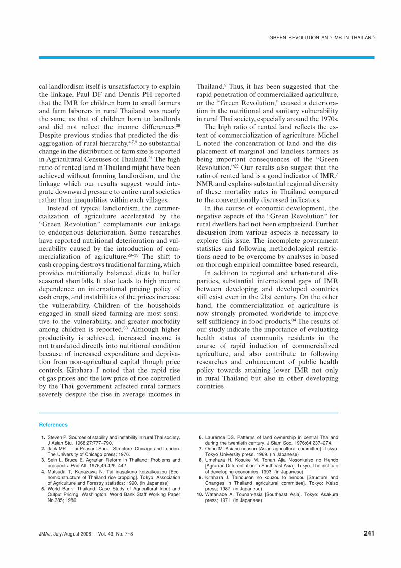
The Ratio of Rented Land
The ratio of rented farming land to total farmingland in Thailand underwent a rapid increase,particularly in northern and central regions(Fig. 1).20,21 In 1963, the ratio was less than 5%,but increased rapidly in the 1960s and 1970s,and reached as high as 15% in 1985. Althoughthe agricultural land reform law passed the Leg-islative Assembly of Thailand in 1975, the raisedratio remained high and unchanged afterward.The Thai government proclaimed that the reformwas failed.3,4 At the same time, regional diversityhad widely enlarged. The central and northernregions marked rapid increase of the ratio whilethe northeast and southern regions maintaineda low ratio of approximately 5% throughoutthe surveyed period.
Thailand has a politically stable history anda high proportion of the population is engagedin agriculture.10,22 The rice export policy enhancedthe commercial value of rice and the tenure ofrice fields, and speculative trade on agriculturallands by absentee landlords living in urban areashas been active since the 1910s.1,10 However, theheterogeneous relationships between peasantsand landlords supported by land renting per-formed mostly by relatives hampered the inequi-table accumulation of land to absentee landlords,and the increase of the ratio was represseduntil the 1960s.4–10
The ratio was dramatically increased by thecommercialization and modernization of agri-
culture in the 1970s during the so-called “GreenRevolution.”4,7,9 It provided new seeds with highharvest, mechanical cultivators, fertilizers, andsignificant progress in land productivity wasachieved. Since it demanded large-scale capitalinvestment, which was difficult to meet for smallfarmers and caused soaring land prices, integra-tion of agricultural resources (�land) by the ab-sentee landlords and stratification of peasantrywas rapidly promoted. Kitahara reported thatsmall farmers under 20 rais (1 rai�0.16 ha) wereunable to compete because of the difficulty inmeeting investment demand and indebtedness.9
The central and northern regions have wide,flat plains with abundant rainfall. Irrigation sys-tems double the rice yield. The commercializa-tion of agriculture and the effects of the “Greenrevolution” were most evident in these two re-gions, especially around Bangkok in the centralregion.1,4,7–10
IMR/NMR/PNMR in Thailand
We selected infant mortality rate (IMR), neo-natal mortality rate (NMR) and post-neonatalmortality rate (PNMR) as health indicators.These three mortality rates are good indicatorsof health statuses in developing countries, andmany previous studies have suggested strongnegative associations with socioeconomic andenvironmental factors in societies.11–19,34 Mortalityrates reflect unique characteristics of societies.While NMR is more sensitive to the sanitary
Fig. 1 The ratio of agricultural rented land to totalfarming land in four regions of Thailand:1963–1998
North Northeast Central South
0.0
0.1
0.2
0.3
0.4
1960 1970 1980 1990 2000
The
rat
io o
f ren
ted
land
GREEN REVOLUTION AND IMR IN THAILAND

238 JMAJ, July /August 2006 — Vol. 49, No. 7 • 8
and nutritional conditions of the area, PNMRis more sensitive to the social and economicdevelopment or the availability of health re-sources.14–16,19 Within the first year, endogenouscauses of death (e.g. prematurity and congenitaldisorders) occur mainly during the neonatalperiod, while exogenous causes (e.g. infectiousand parasitic diseases caused by environmental
factors) are responsible for deaths occurringduring the post-neonatal period.19
Thailand has experienced a considerable de-cline in IMR in recent decades like other devel-oping countries (Fig. 2).22,23 Not only the medicaltechnical imports from the developed world buteconomic growth, the development of healthcare systems and socioeconomic conditions havecontributed to this decline.25–27 However, sub-stantial regional and urban-rural (municipal/non-municipal) differences exist within the coun-try.24–27 Children born in urban areas have a sig-nificantly higher chance of survival. Althoughthe differences in socioeconomic developmenthave been used to explain these diversities,typical socioeconomic factors, such as primaryeducation and average income, have failed toexplain these diversities.24,27 Moreover, IMRmarked little progress, or even recession, aroundthe 1970s, when the “Green revolution” was infull swing. Thai society has unique socioeco-nomic factors. NMR and PNMR have also beenin a similar situation.
Methods
Data used in this paper were obtained fromthe statistics and censuses published by the Thaigovernment.20–23 The ratio of rented land andother agricultural data were taken from agricul-tural statistics (published annually) and censuses(conducted in 1963, 1978, 1983, 1993), and mor-tality rates and population data from the Minis-try of Public Health.
Although the Thai government has systematicalrecord systems, it has suffered under-registrationin vital statistics.26,28 Gaps occurred between IMRestimated by the Ministry of Public Health andthe Office of the Prime Minister.22,24,26,28 In ourresearch, we adopted the former because theywere the only sources that covered the basic vitalstatistics of all four regions of rural Thailand.Analytical efforts were also made to comple-ment imperfections in the data.
Thailand consists of four major regions andabout 80 Changwats (provinces). Although theseclassifications were adopted in Thai official stat-istics, provincial data were insufficient in somevariables. Since most previous studies referringto rural Thai societies put emphases on the com-parisons between regions, we adopted regionsas basic units of our analyses (n�4).
Fig. 2 Time-trends of IMR/NMR/PNMRin four regions of Thailand
IMR: Infant mortality rate; NMR: Neonatal mortality rate;PNMR: Post-neonatal mortality rate
North Northeast Central South
1960 1970 1980 1990 2000
1960 1970 1980 1990 2000
1960 1970 1980 1990 2000
IMR
0
20
40
60
80
0
20
NM
RP
NM
R
0
20
40
60
Okada Y, Wakai S

239JMAJ, July /August 2006 — Vol. 49, No. 7 • 8
Variables used in the analyses were IMR/NMR/PNMR (dependent variables), the ratioof rented farming land to total farming land(independent variable), and the ratio of popu-lation in non-municipal area (control variable).Variables were calculated from the data forevery year of the surveyed period 1963–1998.There were, however, lags in the data becauseof restrictions in the data available. Most of thefollowing analyses were applied for the yearswhen necessary data were available.
Although we firstly adopted other typicalindicators as control variables, including house-hold income and the extent of maternal educa-tion, our previous research did not show anysignificant association between these indicatorsand IMR, except for the ratio of population innon-municipal areas, as some documents pointedout.24,27 Thus, the ratio of population in non-municipal areas was finally adopted as the con-trol variable in addition to the year and region.Since the urban-rural diversity of the mortalityrates was substantial in Thailand, the Thai gov-ernment divided all country areas into municipal(�urban) and non-municipal (�rural) areas,and the ratio of population in non-municipalareas had been recognized as the most influentialfactor in determining the mortality rates in pre-vious studies.24–27
After we checked the correlation amongthe variables and certified the suitability of themodel for multi-variant analysis, we performedthe ANCOVA (analysis of co-variance) tests toanalyze a more integrated association betweenthem.
Since the ratio of rented land and the effect ofthe “Green Revolution” had not been directlyassociated with the mortality rates in Thailand,we also evaluated the association among thelongitudinal time-trends of these variables toachieve further analysis, in addition to the non-trend evaluations. To smooth data and clarifythe time-trends of the variables, we divided thewhole surveyed term, 1963–1988, into three terms(1963–1975, 1975–1985, 1985–1998) in accordancewith the situation of Thai agricultural and thegovernmental policy. The land reform law waspassed in 1975, and it was rethought in 1985 be-cause of its insufficient impact. For these threeterms, the slopes of each variable were calcu-lated from liner regression. They were definedas new variables, which reflected the longitudinal
movements of the original variables, or whetherthey were increasing or decreasing. As for themortality rates, slopes were calculated from log-linear regression to control their natural decline.The correlation check and ANCOVA tests werealso applied for these new variables in the sameway as the non-trends analysis. (Twelve measure-ments for three terms and four control variables.)We used Stat 8.0 for analyses. The study designwas approved by an ethics review board.
Results
The correlation tests showed significant asso-ciation between variables, and verified that ourmodel was appropriate for ANCOVA tests.
Table 1 and 2 show the results of the ANCOVAtests. The associations of the variables are sum-marized in Table 1, and the associations betweenthe longitudinal time-trends of the variables aresummarized in Table 2. Region and year weretreated as categorical variables, and the others asnumerical variables. In longitudinal time-trendsanalysis, region was omitted from the controlvariables because of the fitness of the models.All the models marked R2 more than 0.80.
In Table 1, the ratio of rented land had a sig-nificant association with IMR (F�7.77, P�0.01)and NMR (F�32.88, P�0.0001), but not withPNMR. The year had a significant associationwith all mortality rates. Although the controlvariables had a significant association with NMR,their F values were less than that of the ratioof rented land. This suggests that the ratio ofrented land significantly associates with IMRand NMR through the surveyed period com-pared to other control variables.
In Table 2, longitudinal time-trends analysisshows similar results. The slopes of the ratio ofrented land had a significant association withthose of IMR (F�10.97, P�0.05) and NMR(F�29.87, P�0.001), but did not have suchan association with PNMR. The period had asignificant association with all mortality rates.Although the slopes of the ratio of the popula-tion living in non-municipal areas had a signifi-cant association with those of NMR, its F valuewas less than that of the ratio of rented land,and this gap was wider in longitudinal time-trends analysis than in non-trend analysis. Thissuggests that the longitudinal movement of theratio of rented land also links more significantly
GREEN REVOLUTION AND IMR IN THAILAND

240 JMAJ, July /August 2006 — Vol. 49, No. 7 • 8
to that of IMR and NMR than to other controlvariables.
These results suggest significant longitudinalassociations between the ratio of rented land andIMR/NMR in rural Thailand, and the ratio is abetter indicator than the previous discussed con-trol variables. Although IMR and NMR markeda general progression throughout the surveyedterm, they show a temporal stagnation aroundthe 1970s especially in the central region. Ourresults suggest that this stagnation is signifi-cantly associated with the rapid rise in the ratioof rented farming land, reflecting the negativeeffects of the “Green Revolution.”
Discussion
Since NMR reflects the endogenous factors ofthe area and greater sensitivity to nutritional andsanitary conditions rather than PNMR,14,16–19 ourresults indicate substantial longitudinal linkagefrom the rapid increase of the rented land tothe temporary recession of IMR/NMR, andfinally to the nutritional and sanitary deterior-ation in the rural area of Thailand in the 1960sand 1970s.
Sociological studies on Thai rural commu-nities indicate this linkage. The formation of typi-
Table 1 Results of ANCOVA test applied for IMR/NMR/PNMR
IMR NMR PNMR
Number of observations 68 64 64
Root MSE 3.210 1.310 2.466
R-squared 0.918 0.899 0.914
Adj R-squared 0.881 0.852 0.874
F value Model 24.64** 19.1** 22.76**
The ratio of rented land 7.77** 32.88** 0.87
Year 7.36** 2.63** 9.33**
Region 0.72 9.58** 0.54
The ratio of populationin non-municipal areas 3.25 20.33** 0.09
**: P�0.01IMR: Infant mortality rate; NMR: Neonatal mortality rate; PNMR: Post-neonatalmortality rate
Table 2 Results of ANCOVA test applied for longitudinaltrends of IMR/NMR/PNMR
IMR NMR PNMR
Number of observations 12 12 12
Root MSE 0.015 0.019 0.024
R-squared 0.848 0.857 0.824
Adj R-squared 0.762 0.775 0.724
F value Model 9.78** 10.46** 8.19**
The ratio of rented land 10.97* 29.87** 4.10
Period 7.06* 14.34** 6.8*
The ratio of populationin non-municipal area 2.46 6.51* 1.00
**:P�0.01, *:P�0.05IMR: Infant mortality rate; NMR: Neonatal mortality rate; PNMR: Post-neonatalmortality rate
Okada Y, Wakai S
241JMAJ, July /August 2006 — Vol. 49, No. 7 • 8
References
1. Steven P. Sources of stability and instability in rural Thai society.J Asian Stu. 1968;27:777–790.
2. Jack MP. Thai Peasant Social Structure. Chicago and London:The University of Chicago press; 1976.
3. Sein L, Bruce E. Agrarian Reform in Thailand: Problems andprospects. Pac Aff. 1976;49:425–442.
4. Matsuda T, Kanazawa N. Tai inasakuno keizaikouzou [Eco-nomic structure of Thailand rice cropping]. Tokyo: Associationof Agriculture and Forestry statistics; 1990. (in Japanese)
5. World Bank, Thailand: Case Study of Agricultural Input andOutput Pricing. Washington: World Bank Staff Working PaperNo.385; 1980.
6. Laurence DS. Patterns of land ownership in central Thailandduring the twentieth century. J Siam Soc. 1976;64:237–274.
7. Oono M. Asiano-nouson [Asian agricultural committee]. Tokyo:Tokyo University press; 1969. (in Japanese)
8. Umehara H, Kosuke M. Tonan Ajia Nosonkaiso no Hendo[Agrarian Differentiation in Southeast Asia]. Tokyo: The instituteof developing economies; 1993. (in Japanese)
9. Kitahara J. Tainouson no kouzou to hendou [Structure andChanges in Thailand agricultural committee]. Tokyo: Keisopress; 1987. (in Japanese)
10. Watanabe A. Tounan-asia [Southeast Asia]. Tokyo: Asakurapress; 1971. (in Japanese)
cal landlordism itself is unsatisfactory to explainthe linkage. Paul DF and Dennis PH reportedthat the IMR for children born to small farmersand farm laborers in rural Thailand was nearlythe same as that of children born to landlordsand did not reflect the income differences.28
Despite previous studies that predicted the dis-aggregation of rural hierarchy,4,7,9 no substantialchange in the distribution of farm size is reportedin Agricultural Censuses of Thailand.21 The highratio of rented land in Thailand might have beenachieved without forming landlordism, and thelinkage which our results suggest would inte-grate downward pressure to entire rural societiesrather than inequalities within each villages.
Instead of typical landlordism, the commer-cialization of agriculture accelerated by the“Green Revolution” complements our linkageto endogenous deterioration. Some researcheshave reported nutritional deterioration and vul-nerability caused by the introduction of com-mercialization of agriculture.29–33 The shift tocash cropping destroys traditional farming, whichprovides nutritionally balanced diets to bufferseasonal shortfalls. It also leads to high incomedependence on international pricing policy ofcash crops, and instabilities of the prices increasethe vulnerability. Children of the householdsengaged in small sized farming are most sensi-tive to the vulnerability, and greater morbidityamong children is reported.33 Although higherproductivity is achieved, increased income isnot translated directly into nutritional conditionbecause of increased expenditure and depriva-tion from non-agricultural capital though pricecontrols. Kitahara J noted that the rapid riseof gas prices and the low price of rice controlledby the Thai government affected rural farmersseverely despite the rise in average incomes in
Thailand.9 Thus, it has been suggested that therapid penetration of commercialized agriculture,or the “Green Revolution,” caused a deteriora-tion in the nutritional and sanitary vulnerabilityin rural Thai society, especially around the 1970s.
The high ratio of rented land reflects the ex-tent of commercialization of agriculture. MichelL noted the concentration of land and the dis-placement of marginal and landless farmers asbeing important consequences of the “GreenRevolution.”29 Our results also suggest that theratio of rented land is a good indicator of IMR/NMR and explains substantial regional diversityof these mortality rates in Thailand comparedto the conventionally discussed indicators.
In the course of economic development, thenegative aspects of the “Green Revolution” forrural dwellers had not been emphasized. Furtherdiscussion from various aspects is necessary toexplore this issue. The incomplete governmentstatistics and following methodological restric-tions need to be overcome by analyses in basedon thorough empirical committee based research.
In addition to regional and urban-rural dis-parities, substantial international gaps of IMRbetween developing and developed countriesstill exist even in the 21st century. On the otherhand, the commercialization of agriculture isnow strongly promoted worldwide to improveself-sufficiency in food products.34 The results ofour study indicate the importance of evaluatinghealth status of community residents in thecourse of rapid induction of commercializedagriculture, and also contribute to followingresearches and enhancement of public healthpolicy towards attaining lower IMR not onlyin rural Thailand but also in other developingcountries.
GREEN REVOLUTION AND IMR IN THAILAND

242 JMAJ, July /August 2006 — Vol. 49, No. 7 • 8
11. Hobcraft JN, McDonald JW, Rutstein SO. Socioeconomic factorsin infant and child mortality: A cross-national comparison. Popstu. 1984;38:193–223.
12. Jayachandran J, George KJ. Socioeconomic development,medical care, and nutrition as determinants of infant mortalityin less-developed countries. Soc Bio. 1986;33:301–315.
13. Jacob AA. Infant mortality in Nigeria: Effects of place of birth,mother’s education and region of residence. J biosoc sci. 1994;26:469–477.
14. Kees M, Roland B, John SK. Socio-cultural determinants ofchild mortality in southern Peru: Including some methodologicalconsiderations. Soc Sci Med. 1993;36:317–331.
15. Bimal KP, Deborah JR. Utilization of health facilities and trainedbirth attendants for child in rural Bangladesh: an empirical study.Soc Sci Med. 2002;54:1755–1765.
16. Bimal KP. Health service resources as determinants of infantdeath in rural Bangladesh: as empirical study. Soc Sci Med.1991;32:43–49.
17. Shushum B. Patterns and causes of neonatal and postneonatalmortality in rural Bangladesh. Stud Fam Plan. 1989;20:136–146.
18. Jeffrey BG, Becky D, Susan L. Socioeconomic differentials andneonatal mortality: Racial comparison of California singletons.Pediatrics. 1989;83:181–186.
19. Dudley L, Poston Jr, Richard GR. Toward a reformulation ofthe neonatal mortality rate. Soc Bio. 1985;32:1–12.
20. Ministry of Agriculture, Government of Thailand. Agriculturalstatistics of Thailand. Thailand: Ministry of Agriculture, Govern-ment of Thailand; 1963–1998.
21. Office of the Prime Minister, Government of Thailand. Thailandcensus of agriculture. Thailand: Office of the Prime Minister,Government of Thailand; 1963,1973,1983,1993.
22. Office of the Prime Minister, Government of Thailand. Populationand Housing Census. Thailand: Office of the Prime Minister,
Government of Thailand; 1960,1970,1980,1990,2000.23. Ministry of Public Health, Government of Thailand. Public Health
Statistics. Thailand: Ministry of Public Health, Government ofThailand;1963–1998.
24. John K, Apichat C. Infant and child mortality in Thailand: levels,trends, and differentials as derived through indirect estimationtechniques. Honolulu: East-West Center; 1978.
25. Paul DF, Dennis PH. The impact of class, education and healthcare on infant mortality in a developing society: The case of ruralThailand. Demography. 1982;19:391–408.
26. Apichat C. Mortality Trends and Differentials in Thailand: 1950–1975. (Presented in Mortality in South East Asia: a reviewof changing trends and patterns, 1950–1975.) Manila: WHO/ESCAP; 1980.
27. Ogawa N, Gavin WJ, Jeffrey GW. Human Resources in Devel-opment along the Asia-Pacific Rim. New York: Oxford UniversityPress; 1993.
28. Lumbiganon P, Panamonta M, Laopaiboon M, Pothinam S,Patithat N. Why are Thai official perinatal and infant mortalityrates so low? Int J Epidemiology. 1990;19:997–1000.
29. Michel L. New seeds and poor people. London: UNIWINHYMAN; 1989.
30. Kathleen MD. Income and dietary adequacy in an agriculturalcommunity. Soc Sci Med. 1983;17:1877–1786.
31. Kathryn GD. Nutrition and the commoditization of food systemsin Latin America and the Caribbean. Soc Sci Med. 1989;28:415–424.
32. Deborah FB. Nutrition and the commoditization of food in Sub-Saharan Africa. Soc Sci Med. 1989;28:425–440.
33. Kathleen MD. Nutrition and the commoditization of agriculture:Ten years later. Soc Sci Med. 1993;36:1407–1416.
34. United Nations. Human Development Report 2003. New York:United Nations Publications; 2003.
Okada Y, Wakai S

243JMAJ, July /August 2006 — Vol. 49, No. 7 • 8
*1 Department of Geriatrics, Nagoya University Graduate School of Medicine, Nagoya*2 Center of Medical Education, Nagoya University School of Medicine, NagoyaCorrespondence to: Yoshihisa Hirakawa MD, PhD, Department of Geriatrics, Nagoya University Graduate School of Medicine, 65 Tsuruma-cho,Showa-ku, Nagoya, Aichi 466-8550, Japan. Tel: 81-52-744-2364, Fax: 81-52-744-2371, E-mail: [email protected]
Original Article
Introduction
Due to the aging of the population and longer lifespans, end-of-life care for the elderly has becomea major national problem in Japan.1,2
Due to patients’ preference for dying athome1,3,4 and rising health care costs in Japan,5
end-of-life care for the elderly at home hasreceived much attention within the long-term
care insurance system.1
The goal of palliative care is to achieve thebest possible quality of life for patients and theirfamilies, in part by providing information onnatural clinical courses and available treatmentframeworks at home and by discussing caregoals.6,7 Therefore, primary sources of informa-tion are needed to assist physicians and nurses indiscussing home end-of-life care programs withelderly patients and family members. Particu-
Symptom Experience and Patterns of End-of-Life—Home care for elderly patients with cancer
vs. those without cancer in Japan
JMAJ 49(7 • 8): 243–250, 2006
Yoshihisa Hirakawa,*1 Yuichiro Masuda,*1 Masafumi Kuzuya,*1 Takaya Kimata,*1
Akihisa Iguchi,*1 Kazumasa Uemura*2
AbstractBackground The aim of this study was to assess and compare the frequency of symptoms and the receipt ofend-of-life care between elderly patients with and those without cancer who spent the last two days of their livesat home.
Methods The present data were obtained from the Dying Elderly at Home (DEATH) project, a multicenterobservational study. The subjects of the study were 240 consecutive decedents aged 65 or older who had diedat home between October 2002 and September 2004. The following information was collected: decedentcharacteristics, observed symptoms and end-of-life care provided during the last 48 hours of life. To assess thedifferences in characteristics and clinical course among decedents, the survey data were divided into two groups:cancer and non-cancer.
Results We evaluated 173 decedents with cancer and 61 decedents without cancer. The decedents withcancer were more likely to show symptoms of pain, acute confusion, or nausea/vomiting and less likely to displayfever or cough than the decedents without cancer. In addition, decedents with cancer were more likely to receiveintravenous drip injection or narcotic analgesia and less likely to be given heart massage, sputum suction, orantibiotics. After adjustment for age and other baseline characteristics, difference in the frequency of controlledpain remained significant.
Conclusions We observed that both the dying process and end-of-life care differed between elderly patientswith cancer and those without. However, cancer itself is not an independent predictor of end-of-life symptomsor care.
Key words End-of-life care, Elderly, Cancer, Symptom, Home

244 JMAJ, July /August 2006 — Vol. 49, No. 7 • 8
Hirakawa Y, Masuda, Y, Kuzuya M, et al.
larly, the assessment and management of symp-toms are central to high-quality end-of-life care,since dying patients frequently develop symp-toms. As a number of researchers have suggested,we believe that the symptoms experienced byelderly patients with advanced cancer differ fromthose who do not have advanced cancer.2,8 It istherefore logical to assume that end-of-life carefor elderly patients should differ between thosewith and those without cancer. However, theprevalence of symptoms experienced and pat-terns of care received by elderly patients with orwithout cancer who are dying at home remainunclear.
The aim of this study was to assess and com-pare the frequency of symptoms and patterns ofend-of-life care receipt between elderly patientswith and those without cancer at home during the
last two days of their lives. This study providesprimary sources to assist medical teams, elderlypatients and their families in designing homequality end-of-life care programs.
Methods
Study populationThe Dying Elderly at Home (DEATH) projectwas a prospective study of elderly patients dyingat home of an end-stage disease. It was conductedin collaboration with the nonprofit JapaneseSociety of Hospice and Home-care, which con-sists of general physicians and other medical andsocial professionals involved in hospice andhome care. To recruit physicians for the study, wefirst selected the major members of the societywho were clinical physicians and who were expe-
Table 1 Cancer and without cancer decedent characteristics and causes of death
Cancer (n�173) Without cancer (n�61)Variable
n (%) n (%) P
Sex Female 75 (43.4) 37 (60.7) 0.02
Age (average�SD) 75.7�8.7 87.1�8.9 �0.01
ADL scale of disabled elderly J�independent 5 (2.9) 3 (4.9) 0.01
A�house-bound 15 (8.7) 4 (6.6)
B�chair-bound 40 (23.1) 8 (13.1)
C�bed-bound 90 (52.0) 45 (73.8)
Unknown 23 (13.3) 1 (1.6)
Cognitive impairment Present 50 (28.9) 44 (72.1) �0.01
Major disease Gastrointestinal 67 (38.7) 0(primary tumor or noncancer disease) Gastric 42 (24.3)
Colorectal 25 (14.5)
Pulmonary 33 (19.1) 19 (31.1)
Liver 26 (15.0) 0
Pancreas 11 (6.4) 0
Prostate 7 (4.0) 0
Breast 4 (2.3) 0 �0.01
Kidney 4 (2.3) 2 (3.3)
Uterine 3 (1.7) 0
Blood 3 (1.7) 1 (1.6)
Cerebrovascular 1 (0.6) 5 (8.2)
Cardiovascular 0 9 (14.8)
Others 9 (5.2) 21 (34.4)
Unknown 5 (2.9) 4 (6.6)
ADL: activities of daily living

245JMAJ, July /August 2006 — Vol. 49, No. 7 • 8
SYMPTOM EXPERIENCE AND PATTERNS OF END-OF-LIFE
rienced in providing end-of-life home care. Wethen sent to these physicians a prospectus on ourresearch. Sixteen clinic physicians agreed to par-ticipate, representing 16 clinics in Western Japan.The subjects of the study were 240 consecutivedecedents aged 65 or older who had used thestudy clinics while diagnosed with any disease,including advanced cancer, and who had died athome between October 2002 and September 2004.Decedents were excluded if they were transferredto a hospital immediately before death because itis difficult to obtain information from a hospital.The following information was collected: socio-demographics; activities of daily living (ADLs)(Japan’s Ministry of Welfare identifies four ranksof ADL in elderly people with disabilities9: Rank J(independence in ADLs), Rank A (house-bound),Rank B (chair-bound), and Rank C (bed-ridden));cognitive impairment, observed symptoms; andend-of-life care provided during the last 48 hoursof life. With the approval of the Japanese Society
of Hospice and Home-care, we used a question-naire that included a list of common end-of-lifesymptoms and treatments:SymptomsDyspnea, uncontrolled pain, controlled pain,coma, acute confusion, anxiety, dizziness, nauseaand vomiting, anorexia, diarrhea, constipation,fever, urinary and fecal incontinence, hemate-mesis, hemoptysis, bottom blood, other types ofhemorrhage, cough, sputum.End-of-life careHeart massage, intubation, mechanical ventila-tion, oxygen inhalation, air-way placement, spu-tum suction, hyperalimentation, intravenous dripinjection (except hyperalimentation), antibiotics,vasopressor, blood transfusion, opioids, urinarycatheter placement, mental support, religioushealing, others.
Data collectionImmediately after the death of a study patient,
Table 2 Cancer and without cancer decedent symptom experience in last two days of life
Cancer (n�173) Without cancer (n�61)Symptom
n (%) n (%) POdds ratio* 95%CI
Dyspnea 79 (45.7) 26 (42.6) 0.68 0.55 0.11– 2.81
Pain (uncontrolled) 33 (19.1) 2 (3.3) �0.01 0.82 0.06–10.61
Pain (controlled) 80 (46.2) 5 (8.2) �0.01 10.73 1.76–65.25
Coma 77 (44.5) 20 (32.8) 0.11 2.09 0.46– 9.55
Acute confusion 44 (25.4) 3 (4.9) �0.01 3.71 0.48–28.96
Anxiety 29 (16.8) 5 (8.2) 0.10 2.16 0.28–17.05
Dizziness 2 (1.2) 1 (1.6) 0.77 1.15 0.03–41.01
Nausea and Vomiting 49 (28.3) 5 (8.2) �0.01 3.31 0.31–35.95
Anorexia 101 (58.4) 31 (50.8) 0.31 1.00 0.27– 3.75
Diarrhea 10 (5.8) 3 (4.9) 0.80 — —
Constipation 12 (6.9) 4 (6.6) 0.92 1.89 0.15–23.64
Fever 41 (23.7) 26 (42.6) �0.01 0.73 0.17– 3.18
Incontinence 31 (17.9) 6 (9.8) 0.14 5.39 0.90–32.18
Hematemesis 8 (4.6) 3 (4.9) 0.93 — —
Hemoptysis 2 (1.2) 0 (0.0) 0.40 — —
Bottom blood 11 (6.4) 5 (8.2) 0.62 — —
Other hemorrhage 9 (5.2) 3 (4.9) 0.92 — —
Cough 25 (14.5) 21 (34.4) �0.01 0.57 0.11– 2.86
Sputum 51 (29.5) 23 (37.7) 0.23 0.92 0.20– 4.30
Other symptom 44 (25.4) 7 (11.5) 0.02 3.46 0.58–20.61
*: Controlling for age, sex, activities of daily living, cognitive impairment, and disease

246 JMAJ, July /August 2006 — Vol. 49, No. 7 • 8
the general practitioner (GP) who oversaw thepatient was asked to fill out a questionnairebased on the patient’s medical chart and on thephysician’s recollection of the clinical course. TheGP also asked family members or visiting nurseswho witnessed the last 48 hours of the patient’slife to provide additional information.
The GPs and other information providerswere blinded to the study hypothesis and to anyanticipated study results. For ethical consider-ations, data on all eligible participants obtainedfrom the Japanese Society of Hospice and Home-care remained anonymous.
The decedents were divided into two groupsfor analysis to confirm the differences in charac-teristics and clinical courses between subjectswith cancer and those without. For each patient,the GP in charge recorded the disease as eithercancer or noncancer. Data were collected on 173decedents with cancer and 61 without cancer. Thedistribution of patients’ characteristics is shownin Table 1. The research protocol was reviewedand approved by the Nagoya University Research
Ethics Board.
Statistical analysisThe data were analyzed using Statview-J5.0.Group differences were compared using theunpaired-t test and the chi square test. We alsoperformed multivariate logistic regression ana-lysis to identify any independent associationbetween cancer and symptoms or intervention,after adjusting for baseline characteristics. Wepresent the results as odds ratios and 95%confidence intervals. A P value of less than 0.05was considered statistically significant.
Results
As seen in Table 1, a significantly greater percent-age of decedents with cancer were male com-pared to decedents without cancer. Those withcancer were also generally younger. On thewhole, decedents with cancer scored lower inADLs and in cognitive function. Among the pri-mary sites of cancer, stomach was the most
Hirakawa Y, Masuda, Y, Kuzuya M, et al.
Table 3 Cancer and without cancer decedent care receipt in last two days of life
Cancer (n�173) Without cancer (n�61)Care
n (%) n (%) POdds ratio* 95%CI
Heart massage 2 (1.2) 4 (6.6) 0.02 — —
Intubation 0 (0.0) 0 (0.0) — —
Mechanical ventilation 0 (0.0) 1 (1.6) 0.09 — —
Oxygen inhalation 63 (36.4) 19 (31.1) 0.46 0.98 0.23– 4.17
Airway placement 4 (2.3) 1 (1.6) 0.75 1.05 0.00–411.55
Sputum suction 41 (23.7) 23 (37.7) 0.03 0.41 0.09– 1.90
Hyperalimentation 17 (9.8) 1 (1.6) 0.04 3.42 0.22– 54.00
Antibiotics 20 (11.6) 17 (27.9) �0.01 1.29 0.27– 6.21
Vasopressor 1 (0.6) 1 (1.6) 0.44 — —
Blood transfusion 1 (0.6) 0 (0.0) — —
Intravenous drip injection 60 (34.7) 20 (32.8) 0.79 3.56 0.58– 21.94
Volume (average�SD)24–48 hours before death 467.2�306.8 705.9�397.6 �0.01 — —
0–24 hours before death 360.8�299.0 666.7�343.0 �0.01 — —
Opioids 82 (47.4) 0 (0.0) �0.01 — —
Urinary catheter placement 31 (17.9) 8 (13.1) 0.39 3.43 0.50– 23.61
Mental support 5 (2.9) 0 (0.0) 0.18 — —
Religious healing 3 (1.7) 0 (0.0) 0.30 — —
Others 11 (6.4) 2 (3.3) 0.37 1.21 0.02– 95.15
*: Controlling for age, sex, activities of daily living, cognitive impairment, and disease

247JMAJ, July /August 2006 — Vol. 49, No. 7 • 8
prevalent (24.3%) followed by lung (19.1%),liver (15.0%), and colorectal area (14.5%). Pul-monary disease was the most prevalent (32.8%)disease among decedents without cancer.
Table 2 shows the decedents’ symptoms dur-ing the last two days of their lives. Decedents withcancer were more likely to display controlled oruncontrolled pain, acute confusion, or nausea/vomiting and less likely to show fever or cough.However, after controlling for age, sex, ADLs,dementia, and disease, only controlled pain wasdetermined to be a significant independent pre-dictor, with an adjusted odds ratio of 10.73 (95%CI, 1.76–65.25).
Table 3 shows end-of-life care given to dece-dents during the last two days of their lives.Decedents with cancer were more likely to receiveintravenous drip injection or narcotic analgesiaand less likely to receive heart massage, sputumsuction, or antibiotics. As for intravenous dripinjection, decedents with cancer received smallervolumes than patients without cancer (467.2 mlvs 705.9 ml, respectively). After controlling forage, sex, ADLs, dementia, and disease, no factorswere determined to be significant independentpredictors.
Discussion
Consistent with some studies,1,2,8 the presentresults revealed differences in end-of-life symp-toms experienced and care received betweenelderly patients with cancer and those without itat home, where a growing percentage of elderlypeople spend their last years. However, afteradjustment for age and other baseline character-istics, almost all of these differences in end-of-lifesymptoms and care received disappeared. Thus,our findings did not suggest that cancer itself is anindependent predictor of end-of-life symptomsor care. The results add an end-of-life perspectiveto the current knowledge of end-of-life care athome, especially in Japan.
In our study, elderly decedents without cancertended to be older and to have poorer physicaland cognitive status than elderly decedents withcancer. Elderly patients, especially those withoutcancer, are more vulnerable to chronic medicalproblems, including dementia and stroke, andare less able to perform ADLs than are youngerpatients. Therefore, aged patients often die ofnonspecific diseases as a result of old age.1,2 The
characteristics of decedents without cancer seemto reflect the situation of dying of old age. Inaddition, our results show that pulmonary diseasewas a common cause of death among patientswho did not have cancer. This may be explainedby the fact that aspiration pneumonia is preva-lent among elderly patients dying of old age.2,8
Symptoms during the last two daysIn our study, before adjustment, decedents withcancer and those without were reported to expe-rience significant differences in the frequency ofsymptoms such as pain, acute confusion, nauseaand vomiting, fever, and cough. After adjustment,differences in the frequency of controlled painremained significant.
Our results suggest that pain control was amajor problem for patients with cancer who weredying at home, compared to those without cancerwho experienced pain only occasionally. Thereare several possible explanations for these find-ings. First, although it is generally believed thatpain is less common in elderly decedents withcancer than in younger patients with cancer,8,10
pain is prevalent in patients with end-stage can-cer at home. Second, decedents without cancerwere tolerant of pain because they were cogni-tively impaired11 or older12 compared to decedentswith cancer. Third, patients without cancer couldnot inform nurses and physicians about their painbecause of cognitive impairment or difficulty incommunication.11–13 It is possible that pain con-trol is a major problem for dying community-dwelling elderly patients regardless of whetherthey have cancer or not. Thus, nurses and physi-cians should monitor and evaluate the pain ofall patients on a daily basis, regardless of thepresence or absence of cancer.
Surprisingly, acute confusion was not commonin patients without cancer, unlike the case ofpatients with cancer. It was previously suggestedthat elderly patients with noncancer diseases,especially those with dementia, often show acuteconfusion.8,14–16 Our results were inconsistentwith this finding. Further research is needed toaccount for the difference in the prevalence ofacute confusion between cancer and decedentswithout cancer at the end of life.
According to Griffie and Mckinnon,17 nauseaand vomiting affect 40 to 70% of patients withadvanced cancer. Our results were consistentwith this finding. Nausea and vomiting have com-
SYMPTOM EXPERIENCE AND PATTERNS OF END-OF-LIFE

248 JMAJ, July /August 2006 — Vol. 49, No. 7 • 8
Hirakawa Y, Masuda, Y, Kuzuya M, et al.
monly been associated with cancers of the gas-trointestinal system.17,18 The prevalence of gastricand colon cancer in primary sites may account forthese symptoms. Additionally, the use of opioidsis one of the most frequently identified causes ofnausea and vomiting in persons with end-stagediseases.17 Pain can also cause nausea and vomit-ing, and the higher prevalence of pain in patientswith cancer contributes to the higher incidence ofnausea and vomiting than in patients withoutcancer.17
Univariate analysis revealed that cough andfever were more prevalent in patients withoutcancer than in patients with cancer. Commoncauses of cough during end-stage disease includepulmonary disease such as pneumonia or lungcancer.19 This may be due to the fact that agreater percentage of patients without cancerdied of pulmonary disease (42.6%) than patientswith cancer died of lung cancer (19.1%), as ourresults indicate. Although fever is not commonlyreported among elderly patients,20 our resultssuggest that, at the end of life, the managementof fever is a major problem in elderly patientsunless they have cancer.
End-of-life care during the last two daysAs can be expected in the home setting, fewdecedents received life-sustaining interventionssuch as heart massage, intubation, or mechanicalventilation, regardless of whether they had can-cer or not. Patients without cancer, however, weremore likely to receive heart massage. This findingwas consistent with previous studies.21,22 Thereare two explanations for the difference. Becausethe process of dying of old age is not as clearlyunderstood as that of cancer,1,8 the GPs did notdiscuss end-of-life care plans with patients andtheir families. It is also possible that the conceptof “end-of-life care for the elderly” was not prop-erly understood by the study’s physician.2,23
Decedents without cancer were more likelythan those with cancer to receive sputum suction.Therefore, we can assume that some decedentswith cancer displayed mild rather than severesputum that did not need clearing. Morita et al.10
and Andrews et al.24 suggested that dehydrationin patients with advanced cancer reduces sputumproduction and improves QOL. Dehydrationmay have resulted in reduced sputum amongdecedents with cancer. How to give drip infusionsto elderly patients without cancer in the last days
of their lives is an area that needs attention. As ina previous study in Japan,25 the volume of dripinfusion given to our decedents with cancer wasapproximately 500 ml/day. Our results added tothe stock of information on the volume of end-of-life drip infusion in Japan.26
Hyperalimentation was more prevalent indecedents with cancer than in those without beforemultivariate adjustment. There are two possibleexplanations for this difference. As mentionedearlier, patients with cancer suffered from nauseaand vomiting. As a result, general practitionersresorted to hyperalimentation to meet patients’nutritional needs. In addition, we hypothesizethat a greater proportion of elderly decedentswithout cancer than with cancer received tubefeeding instead of hyperalimentation; in Japan,elderly patients who cannot eat sufficiently arelikely to receive tube feeding.14,23 Further researchis needed to determine the related factors account-ing for the difference in referral patterns forhyperalimentation between decedents with can-cer and those without.
Antibiotics were more prevalent in decedentswithout cancer before multivariate adjustment.We assume that the GPs gave antibiotics topatients without cancer who had a fever, becausepatients without cancer are generally more likelyto experience fever than patients with cancer.However, among patients with dementia, there isinsufficient evidence to support the positive effectof antibiotics on the prolongation of life14,27,28;their use also carries a risk of impaired renalfunction27 and poorer cognitive function.28 There-fore, because a greater percentage of decedentswithout cancer than those with cancer werecognitively impaired, it is not clear whether theuse of antibiotics among decedents without can-cer was appropriate.
The use of narcotic analgesics was moreprevalent in decedents with cancer than in thosewithout. Because patients with cancer were morelikely to experience pain than those without can-cer, the results do not seem surprising. However,we may also speculate that pain was not well con-trolled in decedents with cancer even thoughnarcotic analgesics were frequently used. Con-versely, none of the decedents without cancerreceived a narcotic analgesic. Because few patientswithout cancer complained of pain, it is naturalthat the GPs refrained from giving them narcoticanalgesics. However, in our study, nearly half of

249JMAJ, July /August 2006 — Vol. 49, No. 7 • 8
SYMPTOM EXPERIENCE AND PATTERNS OF END-OF-LIFE
the patients without cancer presented with dysp-nea. Because opioids can alleviate dyspnea,8,29 itis possible that the GPs did not properly under-stand the use of opioids for dyspnea.
Study limitationsThere were several important limitations to thisstudy. First, this study partly relied on familymembers’ accounts of patient symptoms, becausethe settings were the communities. This may havebiased the researchers’ evaluation. Second, theresults must be interpreted with caution since welack information about the degree of symptomsexperienced. Therefore, we cannot thoroughlyevaluate whether or not the symptoms wereproperly managed. In addition, because the dura-tion of the home end-of-life care could con-tribute to the use of intervention, the lack ofinformation about the duration weakened thepower of our results concerning the differencesin end-of-life intervention. Third, we enlistedeach clinic to perform evaluations because of thelarge number of settings. This may have biasedthe researchers’ evaluations and limited thevalidity of the results, since it is possible that thedata-collection procedures and quality varieddepending on the GPs in charge of data collec-tion. Fourth, due to the difficulty of collectingdata, we excluded patients who received homecare but were finally admitted to hospital justbefore death. We believe that such patients, ifthey have serious symptoms, are an interesting
target population. Finally, the small number ofpatients and limited variety of study settings alsolimited generalization. In addition, as mentionedabove, the Japanese Society of Hospice andHome-care is interested in hospice and homecare, and thus selection bias is also possible.Larger studies in other areas of research mayhelp further reveal the current situation of end-of-life care at home in Japan.
Conclusion
We investigated the differences in symptomexperience and end-of-life care between elderlypatients with or without cancer who were dyingat home in Japan. Our results suggested that boththe dying process and end-of-life care differedbetween elderly patients with cancer and thosewithout. However, cancer itself is not an indepen-dent predictor of end-of-life symptoms or care.Larger studies are needed to substantiate ourfindings.
Acknowledgements
This study was supported by the Ministry of Health,Labour and Welfare of Japan. We extend our appre-ciation to all members of the Japanese Society of Hos-pice and Home-care, especially Mr. Nobuyoshi Daitoand Mr. Sunchi Ryan. We also thank the followingresearch assistants: Ms. Noriko Sano and Mr. MinoruNishi.
References
1. Hashimoto H. Establishment of terminal care systems on thebasis of patients’ will. In: Iryokeizaikikou ed. Iryohakusyo. Tokyo:Nihon Iryokikaku; 2001:47–58. (in Japanese)
2. Suzuki Y, Iguchi A. Geriatric comprehensive medicine: anapproach to terminal care. J Jpn Med Assoc. 2004;93:2508–2513. (in Japanese)
3. Sauvaget C, Tsuji I, Li JH, et al. Factors affecting death at homein Japan. Tohoku J Exp Med. 1996;180:87–98.
4. Tilden VP, Drach LL, Tolle SW. Complementary and alternativetherapy use at end-of-life in community settings. J AlternComplement Med. 2000;8:897–906.
5. Hiroi Y. Future prospects of hospice and palliative care from amedical economics point of view. Taminaru Kea. 1998;8(Suppl):6–11. (in Japanese)
6. Morrison RS. Survival in end-stage dementia following acutedisease. JAMA. 2000;284:47–52.
7. Pekmezaris R, Breuer L, Zaballero A, et al. Predictors of site ofdeath of end-of-life patients: the importance of specificity inadvance directives. J Palliat Med. 2004;7:9–17.
8. Uemura K. Characteristics of end-of-life care for the elderly. In:Iguchi A ed. Future Gerontology. Nagoya: Nagoya DaigakuShuppan Kai; 2000:302–305. (in Japanese)
9. Hirakawa Y, Masuda Y, Kimata T, Uemura K, Kuzuya M, IguchiA. Effects of home massage-rehabilitation therapy for the bed-ridden elderly: a pilot trial with a three-month follow-up. ClinRehabil. 2004;18:1–8.
10. Morita T, Tsunoda J, Inoue S, Chihara S, Ichiki, T. Symptomprevalence and risk factors in terminally ill cancer patients. JpnJ Cancer Clin. 1998;44:879–884. (in Japanese)
11. Porter FL, Malhotra KM, Wolf CM, et al. Dementia and responseto pain in the elderly. Pain. 1996;68:413–421.
12. Tilden VP, Tolle SW, Drach LL, Perrin NA. Out-of-hospital death:advance care planning, decedent symptoms, and caregiver bur-den. J Am Geriatr Soc. 2004;52:532–539.
13. Shuster JL. Palliative care for advanced dementia. Clin GeriatrMed. 2000;16:373–386.
14. Hirakawa Y, Masuda Y, Kimata T, Uemura K, Kuzuya M, IguchiA. Terminal care for elderly with dementia in two long-term carehospitals. Nippon Ronen Igakkai Zasshi. 2004;41:99–104. (inJapanese)
15. Kuebler KK, Heidrich DE. Delirium/acute confusion. In: KueblerKK, Berry PH, Heidrich DE ed. End-of-Life Care—Clinical Prac-tice Guidelines. Philadelphia: WB Saunders; 2002:253–267.
16. Bedford S, Melzer D, Guralnik J. Problem behavior in the last

250 JMAJ, July /August 2006 — Vol. 49, No. 7 • 8
Hirakawa Y, Masuda, Y, Kuzuya M, et al.
year of life: prevalence, risks, and care receipt in older Ameri-cans. J Am Geriatr Soc. 2001;49:590–595.
17. Griffie J, Mckinnon S. Nausea and vomiting. In: Kuebler KK,Berry PH, Heidrich DE ed. End-of-Life Care—Clinical PracticeGuidelines. Philadelphia: WB Saunders; 2002:333–343.
18. Mercadante S, Casuccio A, Fulfaro F. The course of symptomfrequency and intensity in advanced cancer patients followed athome. J Pain Symptom Manage. 2000;20:104–112.
19. Berry PH. Cough. In: Kuebler KK, Berry PH, Heidrich DE ed.End-of-Life Care. Philadelphia: WB Saunders; 2002:235–241.
20. Kuzuya M. Characteristics of age-related illnesses. In: Iguchi Aed. Future Gerontology. Nagoya: Nagoya Daigaku Shuppan Kai;2000:51–56. (in Japanese)
21. Claessens MT, Lynn J, Zhong Z, et al. Dying with lung canceror chronic obstructive pulmonary disease: insights from SUP-PORT. J Am Geriatr Soc. 2000;48:S146–S153.
22. Lynn J, Teno JM, Phillips RS, et al. For the SUPPORT investiga-tion. Perceptions by family members of the dying experienceof older and seriously ill patients. Ann Intern Med. 1997;126:97–106.
23. Murai A. Terminal care for the elderly. Nippon Ronen IgakkaiZasshi. 1999;36:623–626. (in Japanese)
24. Andrews M, Bell ER, Smith SA, et al. Dehydration in terminallyill patients. Is it appropriate palliative care? Post Grad Med.1993;93:201–208.
25. Morita T, Tsunoda J, Inoue S, Chihara S. Characteristics ofpalliative care for elderly cancer patients—a pilot study at ourhospital. Geriatr Med. 1997;35:1505–1511. (in Japanese)
26. Morita T, Shima Y, Adachi I. Attitudes of Japanese physicianstoward terminal dehydration: a nationwide survey. J Clin Oncol.2002;20:4699–4704.
27. Evers MM, Dushyant P, Daniel P, Khalid K, Marin DB. Palliativeand aggressive end-of-life care for patients with dementia.Psychiatr Serv. 2002;53:609–613.
28. Shuster JL. Palliative care for advanced dementia. Clin GeriatrMed. 2000;16:373–386.
29. Kuebler KK. Dyspnea. In: Kuebler KK, Berry PH, Heidrich DE ed.End-of-Life Care—Clinical Practice Guidelines. Philadelphia:WB Saunders; 2002:301–315.

251JMAJ, July /August 2006 — Vol. 49, No. 7 • 8
*1 Expert Service Division, Bureau of International Cooperation, International Medical Center of Japan, Tokyo*2 Ph.D. Program, Columbia University, School of Social Work, New York, USACorrespondence to: Hidechika Akashi MD, PhD, MPH, DTMH, Department of International Health, Nagoya University Graduate School ofMedicine, 65 Tsurumai-cho, Showa-ku, Nagoya, Aichi 466-8550, Japan.Tel: 81-52-744-2109, Fax: 81-52-744-2114, E-mail: [email protected]
Review Article
Aid Coordination Mechanisms for Reconstructingthe Health Sector of Post-Conflict Countries
JMAJ 49(7 • 8): 251–259, 2006
Hidechika Akashi,*1 Noriko Fujita,*1 Rumiko Kakishima Akashi*2
AbstractSome trends have been observed in international assistance for the reconstruction of the health sector incountries that have suffered a conflict throughout their entire country. The authors, having pursued aid activitiesin Cambodia and Afghanistan and reviewed past experiences, propose useful aid coordination mechanismsfor post-conflict countries. The mechanisms include the establishment of a national coordination group betweenthe recipient country’s Ministry of Health, donors and NGOs, sub-coordinating groups for specific technical topics,and coordination groups at the provincial level. Additionally, the involvement of NGOs, such as umbrella NGOs,is indispensable to the reconstruction process. Although aid work sharing and the participation of military forcesin reconstruction activities may be necessary under certain circumstances, we still need to examine strategiesto include them. Consequently, it is time for the international community to integrate aid activities for post-conflictcountries in order to respond immediately and effectively to their immediate needs.
Key words Post-conflict, Afghanistan, Cambodia, Aid coordination, Health
The increase in conflicts since the 1950s hasincreased opportunities to assist afflicted coun-tries.4 As Launtze and her colleagues5 point out,internal and external organizations (such as civilsociety organizations, UN agencies, donors, non-governmental organizations, and internationalorganizations) have been criticized for recurringdifficulties in coordinating such activities. Theinternational aid community, therefore, needs tolearn from its experiences, formulate concreteaid strategies on coordination for post-conflictcountries, and establish an international systemfor providing guidance and technical and policyadvice to fledging governments emerging fromconflict.6 Kreczko7 extracted elements from ex-periences in Afghanistan to propose an aidframework for how international donors shouldprovide humanitarian assistance to post-conflictcountries facing complex emergencies. However,his framework did not include the necessary
Introduction
The past several decades have been character-ized by a series of conflicts around the worldas witnessed in the countries such as Cambodia,Kosovo, East Timor, and Afghanistan. Obviously,the violence resulting from conflicts increasesmorbidity, mortality and disabilities.1 Conflictsare most likely to destroy a country’s health sys-tem or undermine the quality and availability ofpublic health care owing to paralysis in decision-making, budgetary deficits, and low moraleamong government workers.2 Rebuilding thehealth care system is not an easy task,3 especiallyin post-conflict countries where the entire coun-try has been devastated, such as Cambodia andAfghanistan. Immediate and coordinated inter-national support expedites the reconstructionprocess.

252 JMAJ, July /August 2006 — Vol. 49, No. 7 • 8
Akashi H, Fujita N, Akashi RK
specific mechanisms for implementing activitiesin the field.
There are some potential difficulties with co-ordination, including, 1) how to create owner-ship by the Ministry of Health (MOH) and todevelop the capacity of the MOH, 2) how tocommunicate national policy to the peripherallevel, 3) how to coordinate donors and externalexperts, and provide advice from external actorsto the MOH, 4) how to include NGOs in policymaking, how to develop the capacity of localNGOs, and how to coordinate with NGOs, and5) how to coordinate humanitarian activitieswith other actors, such as military forces.
The authors of this paper have pursued healthreconstruction efforts in Cambodia and Afghan-istan and witnessed some trends in health aid.Based on our involvement in health assistanceand a review of earlier reports in other post-conflict countries including Rwanda, Uganda,Somalia, East-Timor, and Kosovo as well asCambodia and Afghanistan, this paper describesthe type of coordination activities and mech-anisms required from the international aid com-munity in post-conflict countries, and examines:1) the participation of national and local govern-ments, 2) the MOH and donor coordination,3) NGO involvement, 4) the involvement ofother actors.
Table 1 summarizes reports on the seven coun-tries covering coordination groups, membershipof the coordination group, issues, lessons, andrecommendations. From these experiences, wetried to extract meaningful common mechanismsfor better coordination. In addition, we tried toidentify useful approaches from the lessonslearnt and recommendations received from dif-ferent countries, although some of the detailswere unclear due to deficiencies in the reports.
Experiences
Health coordination groups—Coordinationmechanisms between the government anddonor agenciesOver the past several decades, the number ofactors involved in the reconstruction of post-conflict countries and their collaborative effortshave increased.8 Our review shows that somecoordination efforts were implemented in manypost-conflict countries, However, the method-ology is neither clear and nor established yet,
and there is still a trend for individual actors toinitiate their own activities and for coordinationnot to always occur immediately.Coordination groups at national levelIn many countries there is a coordination groupat the national level, and NGOs usually par-ticipate in this group. However, the Ministry ofHealth (MOH) is neither always included, norstrengthened. In Uganda in the 1980s, the HealthPolicy Review Commission was formulated tocoordinate actions among the recipient countryand donors. However, it served to present finan-cial aid for the programs of expertise and theinterests of donors and failed to seek to enhancethe policy-making capacity of the UgandanMOH.9 In the case of Rwanda,10 the UN RwandaEmergency Office (UNREO) was set up beforethe establishment of the new transitional govern-ment, and its roles were preparing and elabor-ating discussion and policy papers, overall infor-mation collection, supporting UN agency oper-ation, and assuming the role of secretariat forthe donor meetings. After the establishment ofthe new transitional government, UNICEF ini-tially concentrated on supporting the new gov-ernment to re-establish functioning ministries,and it expected that the government, rather thanUN agencies, could play its coordination role.However, technical coordination was outsideof mandate of UN field coordination capacity,and UNREO’s ability to provide authoritativeleadership and effective management coordina-tion was limited. Instead, two largest donorswithin the international communities led initia-tives. Consequently, the technical coordinationmechanism was unclear, and Rwandan owner-ship was not promoted.10
Even in the countries that have no MOH,efforts to involve local representatives from thedifferent factions can be tried to foster the own-ership of the recipient countries. In Kosovo,11
limited local ownership was observed in theinitial policy formulation process for “InterimHealth Policy Guidelines”. In this process, newlyformed health policy-working group appointedby Department of Health and Social Welfare ofUN Mission in Kosovo (UNIMIK) was involved,however, the weakness of ownership was rec-ognized by WHO and UNIMIK, and some re-presentatives from the different factions wereadded into this health policy-working group. InSomalia,4 a mechanism for aid coordination was

253JMAJ, July /August 2006 — Vol. 49, No. 7 • 8
AID COORDINATION MECHANISMS FOR RECONSTRUCTING THE HEALTH SECTOR OF POST-CONFLICT COUNTRIES
Cou
ntry
(Yea
r of
eve
nts/
star
t of
sup
port
)
Uga
nda
(198
7)
Cam
bodi
a(1
992)
Rw
anda
(199
4)
Som
alia
(199
4)
Eas
t-T
imor
(199
9)
Kos
ovo
(199
9)
Afg
hani
stan
(200
1)
Coo
rdin
atio
n gr
oup
(CG
)
Yes
(H
ealth
Pol
icy
Rev
iew
Com
mis
sion
)
Yes
(C
oCom
: C
oord
inat
ion
Com
mitt
ee a
t na
tiona
lle
vel,
Pro
CoC
om:
Pro
vin-
cial
Coo
rdin
atio
n C
omm
it-te
e at
pro
vinc
ial l
evel
)
Yes
(U
NR
EO
: U
N R
wan
daE
mer
genc
y O
ffice
)
Yes
(lin
ked
with
UN
OS
OM
:U
N O
pera
tion
in S
omal
ia)
Diff
eren
t fo
rm (
INT
ER
FE
T�
UN
TA
ET
�In
terim
hea
lthau
thor
ity)
beca
use
of n
oex
istin
g go
vern
men
t
Yes
(he
alth
pol
icy
wor
king
grou
p)
Yes
(Lo
cal C
GH
N:
Loca
lC
onsu
ltativ
e G
roup
for
Hea
lth a
nd N
utrit
ion
atna
tiona
l lev
el,
PC
C:
Pro
-vi
ncia
l Coo
rdin
atin
gC
omm
ittee
at
prov
inci
alle
vel
Mem
bers
hip
of C
G a
nd t
heir
role
Exp
atria
te t
echn
ical
adv
isor
s w
orki
ngw
ith M
OH
, in
clud
ing
NG
Os
Mem
bers
: MO
H &
ext
erna
l don
ors,
NG
Os
(nat
iona
l & in
tern
atio
nal)
Cha
ired
by M
OH
Rol
e:•m
onito
ring
and
eval
uatin
g al
l hea
thac
tiviti
es•ad
visi
ng M
OH
•m
akin
g re
com
men
datio
ns t
o M
OH
UN
age
ncie
sR
ole:
•pr
epar
ing
polic
y pa
pers
•co
llect
ioni
ng�
diss
emin
atio
ning
info
rmat
ion
•su
ppor
ting
UN
age
ncie
s•ac
ting
as s
ecre
taria
t fo
r th
e D
isas
ter
Man
agem
ent
Tea
m,
NG
O,
and
dono
rs
Ext
erna
l don
ors,
UN
age
ncie
s, N
GO
s,an
d S
omal
i fac
tions
UN
and
Int
erna
tiona
l NG
Os
Pol
icy
wor
king
gro
up c
ompo
sed
ofK
osov
o A
lban
ians
, w
ith t
echn
ical
assi
stan
ce f
rom
WH
OC
onsu
ltativ
e m
eetin
g w
ith lo
cal/
inte
rnat
iona
l med
ical
com
mun
ities
Mem
bers
: MO
H &
ext
erna
l don
ors,
NG
Os
(nat
iona
l & in
tern
atio
nal)
Cha
ired
by M
OH
Rol
e:•m
onito
ring
and
eval
uatin
g al
l hea
thac
tiviti
es•ad
visi
ng t
o M
OH
•m
akin
g re
com
men
datio
ns t
o M
OH
Issu
es/F
eatu
res
•Li
ttle
enha
ncem
ent
of p
olic
y m
akin
g ca
paci
tyat
gov
ernm
ent
leve
l.•D
onor
s te
nded
to
supp
ort
thei
r ow
n in
tere
sts.
•T
here
was
no
natio
nal h
ealth
fra
mew
ork.
•T
rain
ing
of M
OH
sta
ff w
as li
mite
d.
•A
n um
brel
la N
GO
(M
ED
ICA
M)
exis
ted,
with
a se
at in
CoC
om.
•In
trod
uctio
n of
sub
cont
ract
ing
to N
GO
s.
•T
echn
ical
coo
rdin
atio
n w
as in
com
plet
e.•R
espo
nsib
ility
for
tec
hnic
al c
oord
inat
ion
lay
outs
ide
UN
RE
O’s
man
date
.•U
NIC
EF
, W
HO
etc
. m
aint
aine
d re
spon
sibi
lity
for
tech
nica
l coo
rdin
atio
n in
divi
dual
ly.
•A
utho
rity
of c
oord
inat
ion
by U
NR
EO
was
uncl
ear.
•T
wo
larg
est
dono
rs m
ade
initi
ativ
es.
•U
NIC
EF
initi
ally
con
cent
rate
d on
sup
port
ing
the
new
gov
ernm
ent.
•Lo
cal N
GO
s w
ere
not
incl
uded
in U
NT
AE
T.
•U
mbr
ella
NG
O,
Eas
t-T
imor
NG
O F
orum
was
form
ulat
ed.
•U
tiliz
atio
n of
Loc
al N
GO
s in
sub
-con
trac
ting
and
trai
ning
rol
es w
as c
ondu
cted
thr
ough
Com
mun
ity E
mpo
wer
men
t P
rogr
am
•In
terim
hea
lth p
olic
y gu
idel
ines
wer
ees
tabl
ishe
d.•P
olic
y w
orki
ng g
roup
was
app
oint
ed b
yth
e D
epar
tmen
t of
Hea
lth a
nd S
ocia
l Wel
fare
for
mak
ing
owne
rshi
p (b
ut,
Ser
bian
med
ical
com
mun
ity w
as n
ot s
ucce
ssfu
lly in
clud
ed).
•T
here
is n
o um
brel
la N
GO
, ho
wev
er,
ther
eis
an
info
rmal
vol
unta
ry n
etw
ork.
•S
ubco
ntra
ctin
g to
NG
Os
to r
un p
rovi
nces
and
dist
ricts
was
wid
ely
intr
oduc
ed.
•D
onor
s an
d N
GO
s w
ho s
uppo
rt s
peci
ficac
tiviti
es (
such
as
nurs
ing
scho
ol)
have
resp
onsi
bilit
ies
for
spec
ific
sect
oral
and
geog
raph
ic a
reas
.•M
ilita
ry f
orce
s pa
rtic
ipat
e in
hea
lth d
evel
op-
men
t ac
tiviti
es
Less
ons/
Rec
omm
enda
tions
UN
TA
C w
orke
d ac
cord
ing
to t
he t
hree
key
prin
cipl
esof
sov
erei
gnty
, re
spec
t fo
r lo
cal
capa
city
, an
d ba
lanc
eof
ass
ista
nce
for
all a
reas
.
The
gov
ernm
ent
shou
ld p
lay
a co
ordi
natin
g ro
le.
With
draw
al o
f U
N f
orce
s du
e to
bad
sec
urity
The
lac
k of
inv
olve
men
t of
loc
al N
GO
s w
as c
ritic
ized
as a
new
for
m o
f co
loni
alis
m.
•T
he r
oles
of
vario
us k
ey a
ctor
s sh
ould
be
defin
ed e
arly
.•W
HO
can
pla
y th
e le
ad ro
le in
hea
lth p
olic
y de
velo
pmen
t.•T
he
lead
po
licy
orga
niza
tion
shou
ld
conc
entr
ate
onpo
licy
deve
lopm
ent
and
coor
dina
tion,
and
sup
port
the
proc
ess
of it
s im
plem
enta
tion.
Tab
le1
Co
ord
inat
ion
mec
han
ism
s in
dif
fere
nt
po
st-c
on
flic
t co
un
trie
s

254 JMAJ, July /August 2006 — Vol. 49, No. 7 • 8
established, which was linked with UNOSOM(UN Operation in Somalia) as an independentunit, to build legitimacy both nationally and in-ternationally through an ongoing process of dia-logue with the different Somali factions, interna-tional donors, UN agencies, and NGOs. However,it was ended due to the subsequent breakdownin the security situation and the withdrawal ofUN forces.
In Cambodia and Afghanistan, on the otherhand, the Ministry of Health has had a strongcoordinating role, and has established effectivepartnerships with UN agencies, donors, andacademic institutions.6,12 In addition, the MOHallocated major donors to key underserved ruralareas, and used major NGOs to deliver primaryhealth care packages in Afghanistan.6
The Coordination Committee (CoCom: thisname was subsequently changed to TechnicalWorking Group for Health.) in Cambodia andthe Local Consultative Group for Health andNutrition in Afghanistan13 as a coordinationgroup at national level, progressed with theownership of the two countries respectively. Themajor advantages in the case of both countrieswere that the representatives of the MOHchaired the group and were the major actors indecision-making, and that donors played therole of advisors to the MOH.
A coordination group at the national levelshould function to support policy-making thatencourages the active participation of a recipient
country in the process, and strengthens the cap-acity of its MOH. Such a national coordinationgroup will enhance the ownership and initiativesof a recipient government in the reconstructionprocess.Sub-coordination groups under the nationalcoordination groups for technical issuesA national coordination group often faces dif-ficulties in dealing with specific health issues,and its members are not always familiar with alltechnical issues. Thus, sub-coordination groupsunder the national coordination groups in whichthe MOH technical staff and aid agency advisorsattend, can be useful for discussing specifichealth policies and submitting technical reportsor recommendations to the national coordin-ation group. Smaller-scale working groups undersub-coordination groups may also be effectivefor discussing further detailed issues and topics.
There are few descriptions of these coordin-ation groups held under the national coordin-ation group. However, the Sub-CoComs in Cam-bodia12,14 and the Task Forces and WorkingGroups in Afghanistan operating under the na-tional coordination group which we observed,were formulated to perform these functions (seeTable 2 for topics discussed at these groups).
One matter that should be noted is that topicsto be discussed at the sub-coordination groupsshould be selected based on the needs of a recipi-ent country and not based on the political will ofdonors, as this may weaken donor coordination.
Table 2 Topics of sub-coodination meetings
Cambodia Afghanistan
Personnel Human resources Human resources
Maternal and MCH Women's health (SMI/Reproductive health)child health Child health
NutritionEPI
Other Health economics Salary and incentive standardization for NGOsMental health Essential drugs and private sectorOral health Health management information system
Prevention of blindness Hospital managementSurgical training Community based health careMedical technology IECLaboratory HIV/AIDSMedical ethics and research Communicable disease and emergency response
Total 10 topics 13 topics
Akashi H, Fujita N, Akashi RK

255JMAJ, July /August 2006 — Vol. 49, No. 7 • 8
We observed one case in which a donor tried toestablish a working group which would discussalmost the same topics as other groups, in orderto seize the political initiative in that field.Coordination groups at provincial levelCoordination at the provincial level has alsobeen little reported on, except in Cambodia andAfghanistan. In Cambodia, the Provincial Coor-dination Committees (Pro-CoCom; this namewas also changed to Provincial Technical Work-ing Group for Health) were established to sup-port the local governments’ implementation ofnational policies, and advisors from UNICEFand WHO worked with provincial health depart-ments under this scheme (see Fig. 1 for the coor-dination mechanism in Cambodia). In Afghani-stan, the process is underway to establish Pro-vincial Coordinating Committees (PCC). Suchcollaborative arrangements are effective for co-ordinating donor support of health reconstruc-tion at the provincial level.
Humanitarian agencies in countries such asKosovo, Afghanistan, and Iraq are increasinglyfacing the need to work closely with, and evenbe coordinated by and support the capacity of,national government or quasi-state entities.6
Our review of initiatives in some post-conflictcountries indicates that coordination mechan-isms at the national and provincial levels areindispensable in rebuilding the health sector.
Involvement of NGOsNGOs have been important actors in recon-structing the health sector of post-conflict coun-tries, and three types of participation mechan-
isms have been notable in providing assistance.Considering their advantages, the internationalaid community must include NGO participationas a component of future aid strategy.Involvement of NGO in coordination groupsTo benefit most from the collaboration of NGOs,the recipient government, and donors, a specificmechanism is required. The mechanism needsto function to convey the opinions of NGOs tothe MOH and the aid community as well as thenational policies and other relevant informationto NGOs. The first type of mechanism is for theNGOs to have some seats in coordination groupsat the national level and also at the provinciallevel.
In many countries that we reviewed, NGOshad opportunities to participate in national co-ordination groups, sometimes individually, andsometimes as representatives of an umbrellaNGO. At the same time, it is important that localNGOs should not be neglected. For instance,the involvement of only international NGOswas criticized as a new form of colonialism inEast Timor, when local NGOs were not includedin the national coordination groups.15,16 In thisway, NGOs’ views can be reflected in healthpolicies, and the policies can be implemented atthe grass-roots level.Umbrella NGOThe second mechanism is an umbrella NGOor NGO network in post-conflict health recon-struction efforts which can support the abovefunctions.
In the case of Cambodia, an umbrella organi-zation, MEDICAM, which coordinates various
Fig. 1 Aid coordination mechanisms in Cambodia
Working Group1
Working Group2
Working Group3
Sub-Cocom1
Sub-CoCom2
Sub-CoCom3
CoComDonorsupport
Donorsupport
Donorsupport
Pro-CoCom Pro-CoCom Pro-CoComDonorsupport
Central level
Provincial level
AID COORDINATION MECHANISMS FOR RECONSTRUCTING THE HEALTH SECTOR OF POST-CONFLICT COUNTRIES
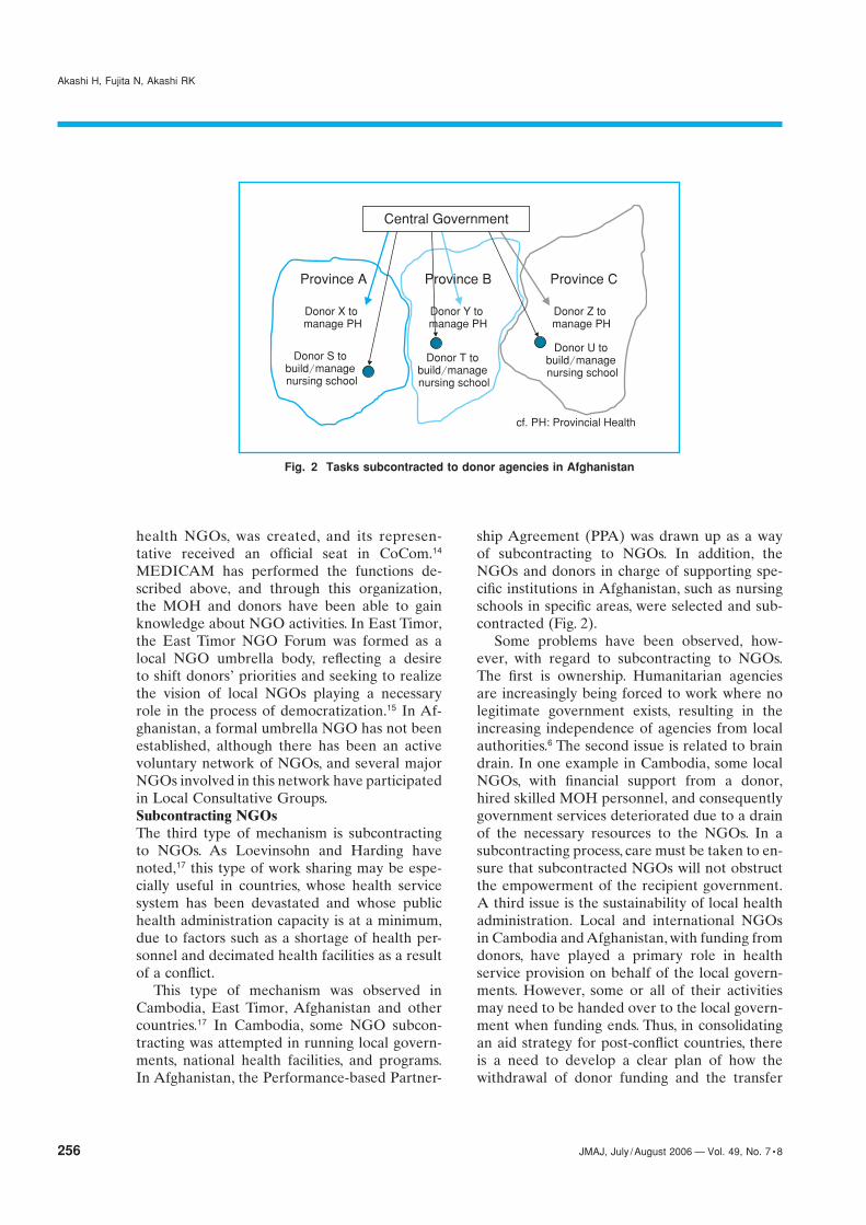
256 JMAJ, July /August 2006 — Vol. 49, No. 7 • 8
health NGOs, was created, and its represen-tative received an official seat in CoCom.14
MEDICAM has performed the functions de-scribed above, and through this organization,the MOH and donors have been able to gainknowledge about NGO activities. In East Timor,the East Timor NGO Forum was formed as alocal NGO umbrella body, reflecting a desireto shift donors’ priorities and seeking to realizethe vision of local NGOs playing a necessaryrole in the process of democratization.15 In Af-ghanistan, a formal umbrella NGO has not beenestablished, although there has been an activevoluntary network of NGOs, and several majorNGOs involved in this network have participatedin Local Consultative Groups.Subcontracting NGOsThe third type of mechanism is subcontractingto NGOs. As Loevinsohn and Harding havenoted,17 this type of work sharing may be espe-cially useful in countries, whose health servicesystem has been devastated and whose publichealth administration capacity is at a minimum,due to factors such as a shortage of health per-sonnel and decimated health facilities as a resultof a conflict.
This type of mechanism was observed inCambodia, East Timor, Afghanistan and othercountries.17 In Cambodia, some NGO subcon-tracting was attempted in running local govern-ments, national health facilities, and programs.In Afghanistan, the Performance-based Partner-
ship Agreement (PPA) was drawn up as a wayof subcontracting to NGOs. In addition, theNGOs and donors in charge of supporting spe-cific institutions in Afghanistan, such as nursingschools in specific areas, were selected and sub-contracted (Fig. 2).
Some problems have been observed, how-ever, with regard to subcontracting to NGOs.The first is ownership. Humanitarian agenciesare increasingly being forced to work where nolegitimate government exists, resulting in theincreasing independence of agencies from localauthorities.6 The second issue is related to braindrain. In one example in Cambodia, some localNGOs, with financial support from a donor,hired skilled MOH personnel, and consequentlygovernment services deteriorated due to a drainof the necessary resources to the NGOs. In asubcontracting process, care must be taken to en-sure that subcontracted NGOs will not obstructthe empowerment of the recipient government.A third issue is the sustainability of local healthadministration. Local and international NGOsin Cambodia and Afghanistan, with funding fromdonors, have played a primary role in healthservice provision on behalf of the local govern-ments. However, some or all of their activitiesmay need to be handed over to the local govern-ment when funding ends. Thus, in consolidatingan aid strategy for post-conflict countries, thereis a need to develop a clear plan of how thewithdrawal of donor funding and the transfer
Fig. 2 Tasks subcontracted to donor agencies in Afghanistan
Akashi H, Fujita N, Akashi RK
Central Government
Province A Province C
Donor Z to manage PH
Donor U to build/manage nursing school
Donor T to build/manage nursing school
Donor X to manage PH
Donor S to build/manage nursing school
cf. PH: Provincial Health
Province B
Donor Y to manage PH

257JMAJ, July /August 2006 — Vol. 49, No. 7 • 8
of NGO activities to the local governments, inaddition to the empowerment of the local gov-ernments, should be achieved.
In evaluating the effectiveness of NGO sub-contracting, therefore, it is necessary to assess thecontribution of NGOs not only in improvinghealth indicators in their field of responsibilitybut also in strengthening the capacity of localpublic health institutions,18 as well as nationalhealth systems. The capacity building of a recipi-ent country’s public health sector must be pri-oritized in aid efforts. Without an empoweredpublic sector, the health system will neither bedeveloped nor sustained when internationalpost-conflict assistance terminates.
Other aid coordination trends and issuesAs discussed above, many new attempts havebeen implemented to assist post-conflict coun-tries. In Afghanistan, two additional forms ofactivities have been observed and they haveindicated important issues for donors in formu-lating an effective health aid strategy for post-conflict countries.Stratification of aidIn Cambodia, each donor agency usually pro-vided a comprehensive aid package to healthfacilities that included the reconstruction ofbuildings, the provision of equipment, and thetraining of local human resources (Fig. 3-1).However, in Afghanistan, donor aid was rathermore stratified, with each donor supporting an
aspect of assistance, resulting in multiple donorsperforming their own aid activities and contrib-uting to a joint health aid package for a healthfacility. For example, one aid organization reno-vated hospitals or health centers, while otheragencies provided equipment, and others trainedtheir staff. In this way, one health facility wassupported by various aid agencies (Fig. 3-2).This stratification of aid activities, or aid worksharing, may have been due to the limited cap-acity of each donor or the restricted scope of aidschemes.
However, the stratification of aid creates someproblems. One of the side effects of stratificationis that aid agencies, especially bilateral donors,often face difficulties in generating domestic po-litical visibility in supporting a particular healthorganization19 and may become reluctant to pro-vide assistance. It can also cause imbalanced aid.For instance, some facilities received assistancefor building reconstruction but for neither equip-ment nor training.
To avoid this imbalance, in Afghanistan, somedonor agencies assisted building renovation onlywhen equipment was made available by otherdonors. To implement aid stratification moreefficiently, a coordination model was formulatedin the field of nursing education as a joint projectby various agencies. One NGO that is assistinga nursing school in Kabul and Japanese-sideas a donor agency agreed on a collaborativeeffort and generated an aid stratification format
Fig. 3-1 Sharing aid work by facility (former way of conducting aid prousior)
AID COORDINATION MECHANISMS FOR RECONSTRUCTING THE HEALTH SECTOR OF POST-CONFLICT COUNTRIES
Facility A
Facility CFacility B
Donor X
Donor ZDonor Y

258 JMAJ, July /August 2006 — Vol. 49, No. 7 • 8
whereby the NGO would train nurses, the donoragency would renovate facilities and provideequipment and some budgetary support, andanother selected local NGO would support themanagement of the nursing school (HIS; Insti-tute of Health Science) in one province. TheMOH approved this work sharing format andintends to replicate it in eight nursing schools.The effectiveness of this format model needs tobe examined. Once its benefit has been verified,it should be included in aid mechanisms so thatthe model can be replicated as an aid stratifi-cation strategy in other post-conflict countries.The participation of military forces in aidactivitiesAnother feature of aid to Afghanistan is thatmilitary forces have participated in aid activi-ties. The participation of military forces in aidactivities is not new, and has been observed ina series of recent emergencies in Afghanistan,Somalia, the Balkans, and Iraq. Previously therole of military forces was mainly to maintainorder. However, the U.S. military forces and theInternational Security Assistance Force (ISAF)in Afghanistan have served not only to helpmaintain order but also to reconstruct healthrelated-facilities such as hospitals and medicalschools.20
Furthermore, many American-led civil andmilitary provincial reconstruction teams areworking to rebuild hospitals and schools, providedrugs and equipment, and even deliver clinical
services, and medical specialists of the U.S. mili-tary forces have been conducting clinical trainingin Afghanistan as well.6 Actually, the main ob-stacle to reconstruction programmes is the secu-rity situation.21 Especially in the early stage ofreconstruction efforts, aid activities tended toconcentrate on the capital and surroundingareas owing to poor security in rural areas.9 Thistendency was also observed in Cambodia, andthe reconstruction process was delayed in ruralareas as well. Therefore, the deployment of mili-tary assistance may be effective for the speedierreconstruction of rural areas.
However, the direct participation of militaryforces in the health sector makes it difficult todistinguish aid organizations from armed forces,and aid workers can be more easily targetedby anti-government forces.21,22 Recent eventsactually show that foreigners, including inter-national aid workers, are being targeted by anti-government fighters.23 In addition, some NGOsare not in favor of military participation inhumanitarian activities and have even rejectedparticipation in aid activities conducted underthe supervision of peacekeeping authorities.24
The participation of armed forces is therefore acontroversial issue. The international commu-nity should clarify the role of military forces inhumanitarian assistance and endeavor to pro-mote an environment in which aid agencies donot have to be restricted in their activities forsecurity reasons.
Fig. 3-2 Stratification of aid (Sharing aid work by task)
Akashi H, Fujita N, Akashi RK
Facility A
Facility CFacility B
Donor X
Facilitybuilding
Human resourcedevelopment
Donor Z
Equipmentprovision
Donor Y

259JMAJ, July /August 2006 — Vol. 49, No. 7 • 8
References
1. Zwi A, Ugalde A. Political violence in the third world: a publichealth issue. Health Policy and Planning. 1991;6(3):203–217.
2. Ugalde A, Selva-Sutter E, Castillo C, et al. The health costs ofwar: can they be measured? Lessons from El Salvador. BMJ.2000;321:169–172.
3. Richards T. Afghanistan struggles to build post-conflict healthcare. BMJ. 2003;326:837.
4. Macrae J, Zwi A, Forsythe V. Aid policy in transition: a prelim-inary analysis of ‘post’-conflict rehabilitation of the health sector.Journal of International Development. 1995;7(4):669–684.
5. Launtze S, Leaning J, Raven-Roberts A, Kent R, Mazurana D.Assistance, protection, and governance networks in complexemergencies. Lancet. 2004;364:2134–2141.
6. Salama P, Spiegel P, Talley L, Waldman R. Lessons learnedfrom complex emergencies over past decade. Lancet. 2004;364:1801–1813.
7. Kreczko A. The Afghan experiment: the Afghan support group,principled common programming and the strategic framework.Disasters. 2003;27(3):239–258.
8. US Mission to the UN. Global humanitarian emergencies, 1998.New York: United Nations; 1998.
9. Macrae J, Zwi AB, Gilson L. A triple burden for health sectorreform: ‘post’-conflict rehabilitation in Uganda. Social Scienceand Medicine 1996;42(7):1095–1108.
10. Borton J. An account of co-ordination mechanisms for humani-tarian assistance during the international response to the 1994crisis in Rwanda. Disaster 1996;20(4):305–323.
11. Shuey DA, Qosaj FA, Shouten EJ, et al. Planning for healthsector reform in post-conflict situations: Kosovo 1999–2000.Health Policy. 2003;63:299–310.
12. Lanjouw S, Macrae J, Zwi AB. Rehabilitating health servicesin Cambodia: the challenge of coordination in chronic political
emergencies. Health Policy and Planning. 1999;14(3):229–242.13. Ministry of Health. Consultative group for health and nutrition,
Terms of reference. Kabul: Ministry of Health, Islamic Transi-tional Government of Afghanistan; 2003.
14. Akashi H. Improvement of Health Care. The Kingdom ofCambodia. —From reconstruction to sustainable development—.Tokyo: Institute for International Cooperation, Japan Interna-tional Cooperation Agency; 2002.
15. Patrick I. East Timor emerging conflict: the role of local NGOsand international assistance. Disaster. 2001;25:48–66.
16. Povey G, Mercer MA. East Timor in transition: health andhealth care. International Journal of Health Services. 2002;32(3):607–623.
17. Loevinsohn B, Harding A. Buying results? Contracting for healthservice delivery in developing countries. Lancet. 2005;366:676–681.
18. Putoto G. The role of northern NGOs in rehabilitating the healthsector in post-conflict settings: a need for a critical analysis.World Hospitals and Health Services. 1997;33(2):17–21.
19. Editorial. Afghanistan’s health challenge. Lancet. 2003;362:841.20. Kuehn BM. A long road: Army veterinarians provide humani-
tarian aid, rebuild infrastructure in Iraq and Afghanistan. Journalof the American Veterinary Medical Association. 2004;224:176–176,190.
21. Ahmad K. Warlords and bandits slow Afghanistan’s efforts torebuild. Attacks drive relief workers from some areas. Lancet.2003;362:710–711.
22. Bruderlein C, Leaning J. New challenges for humanitarianprotection. BMJ 1999;319: 430–435.
23. Turone F. Preparing for the worst. BMJ. 2003;326(7383):244.24. Weissman F. Humanitarian action and military intervention:
temptations and possibilities. Disasters. 2004;28(2):205–215.
Conclusion
Aid agencies have accumulated experience in re-construction of the health sector in post-conflictcountries. What we need for future health assis-tance is not individual action by different donorsbut coordinated action by the international aidcommunity. It is time for the international aidcommunity to evaluate aid in the light of thetrends and activities identified above, and agreeupon a strategy of integrating individual compe-tencies into coordinated assistance mechanisms,in addition to a broad framework. We suggestdevelopment of coordination mechanisms, as onemethod to help post-conflict countries rebuild
their health sector. NGOs, including local NGOs,should be involved in this process. Althoughsome issues still remain, we believe that such aconsensus will allow a quicker, more effectiveand systematic response to the rebuilding ofwar-torn countries, and that these coordinationmechanisms can be useful to other developingcountries.
Acknowledgements
This research was supported by the International Co-operation Research Grant for the “Study on strength-ening social development approach on health and dis-trict health system in developing countries” from theMinistry of Health, Labor and Welfare.
AID COORDINATION MECHANISMS FOR RECONSTRUCTING THE HEALTH SECTOR OF POST-CONFLICT COUNTRIES

260 JMAJ, July /August 2006 — Vol. 49, No. 7 • 8
*1 Department of Ophthalmology, Yokohama City University School of Medicine, YokohamaCorrespondence to: Takahiko Hayashi MD, Department of Ophthalmology, Yokohama City University School of Medicine, 3-9 Fukuura,Kanazawa-ku, Yokohama, Kanagawa 236-0004, Japan. Tel: 81-45-787-2683, Fax: 81-45-781-9755, E-mail: [email protected]
Review Article
Ocular Manifestations in Behçet’s Disease
JMAJ 49(7 • 8): 260–268, 2006
Takahiko Hayashi,*1 Nobuhisa Mizuki*1
AbstractBehçet’s disease is a refractory inflammatory disease; its cause is unknown and it presents with repeated acuteinflammation in various organs throughout the body. Its 4 major manifestations are oral aphthae, ocular manifes-tations, skin manifestations, and genital ulcers. Frequently appearing in the mongoloid population in the vicinityof the Silk Road, the disease is rare in Europeans and Americans. There are currently no laboratory findingsuseful in and specific to the diagnosis of Behçet’s disease. Behçet’s disease is diagnosed based on a combinationof clinical manifestations. In Japan, the diagnostic criteria have major and minor manifestations and the diseaseis classified as a complete, incomplete, possible, or specific type based on the combination of those manifesta-tions. This review discusses the clinical manifestations of Behçet’s disease with a focus on ocular manifestations.
Key words Behçet’s disease, HLA-B51, Uveitis, Vasculitis
pathogenesis is becoming clearer.
Demographic Characteristics/Epidemiology in Japan
EpidemiologyThe male-to-female patient ratio in Japan is 0.98.Total patients in Japan: 18,300 people (1991),19,800 people (1996).135 people out of a population of 1 million.
DistributionPrevalent along the Silk Road, 30–45 degreesnorth on the Eurasian landmass.Prevalent in the north of Japan.Widely prevalent in Turkey, Jordan, Syria, Israel,Lebanon, and Saudi Arabia. Also found in theMediterranean, Spain, Portugal, Greece, andItaly. Not found in Caucasians of NorthernEuropean decent and Sub-Sahara Africans.Not found in Southeast Asia or in Hawaiiansor Brazilians of Japanese descent.Not found in any of the races in North, Central,or South America.
Introduction
Behçet’s disease was first described by ProfessorHulusi Behçet, a Turkish dermatologist, in 1937,1
but an ailment akin to Behçet’s disease hadpreviously been described by the noted Greekphysician Hippocrates in the 5th century BC.The history of this disease is extremely old, butit was first systematically described by ProfessorBehçet in the 20th century.
Behçet’s disease is a refractory inflammatorydisease; its cause is unknown and it presents withrepeated acute inflammation in various organsthroughout the body. Its 4 major manifestationsare oral aphthae, ocular manifestations, skinmanifestations, and genital ulcers. Frequentlyappearing in the mongoloid population in thevicinity of the Silk Road, the disease is rare inEuropeans and Americans (Fig. 1). In Japan,registered patients are described as numberingabout 18,000 people,2,3 however, this number isassumed to be even higher if potential patientsare also included. The causes of Behçet’s diseaseare still unknown, but the mechanism of its

261JMAJ, July /August 2006 — Vol. 49, No. 7 • 8
Table 1 Diagnostic criteria for Behçet’s disease according to the Behçet’s Disease Research Committee of Japan
Behçet’s disease clinical diagnostic criteria (2003 revision)
(1) Major manifestations
� Recurrent aphthae of oral mucosa� Skin manifestations
(a) Erythema nodosum(b) Subcutaneous thrombophlebitis(c) Folliculitis-like rash, acne-like rash
Referential finding: enhanced skin irritability� Ocular manifestations
(a) Iridocyclitis(b) Uveoretinitis (retinochoroiditis)(c) If the following findings are present, they conform to (a) (b)
Posterior synechia, lens pigmentation, chorioretinal atrophy, optic atrophy, complicated cataract,secondary glaucoma, or phthisis bulbi considered to be the progress of (a) (b)
� Genital ulcers
(2) Minor manifestations
� Arthritis without deformation or stiffness� Epididymitis� Digestive tract sores as typified by ileocecal ulcers� Vascular lesions� Moderate or more severe CNS lesions
(3) Criteria for diagnosis of the type of disease
� Complete type4 major manifestations appear in the course of the illness
� Incomplete type(a) 3 major manifestations or 2 major and 2 minor manifestations appear in the course of the illness(b) Typical ocular manifestations and 1 major or 2 minor manifestations appear in the course of the illness
� Possible typeSome major manifestations appear but do not qualify as the incomplete type and typical minor manifestationsrepeat or worsen
� Specific lesions(a) entero-Behçet’s disease—The presence/absence of abdominal pain and occult blood test result will be listed.(b) vasculo-Behçet’s disease—Impairment of large arteries, small arteries, or large & small arteries will be listed.(c) neuro-Behçet’s disease—The presence/absence of headaches, palsy, cerebrospinal-type symptoms,
psychiatric manifestations, etc. will be listed.
(4) Test once for HLA-B51 (B5) positivity
The type of HLA should be listed
(5) Laboratory findings for reference (not mandatory)
� Negative/positive in a positive skin pathergy testA relatively large injection needle of 22–18G will be used
� Negative/positive on a prick test with a streptococcus vaccineHypersensitivity to streptococcusMany patients with Behçet’s disease display strong hypersensitivity to oral streptococci like Streptococcussanguinis, so in a prick test (fine 26G needle) with Streptococcus sanguis antigen, this can be seen as a strongflare reaction 20–24hrs later.
� Inflammatory responseEnhanced level of sedimentation, positive serum CRP, increased WBC count in peripheral blood, rise incomplement titer
� HLA-B51(5) positive� Pathological findings
With acute-stage erythema nodosum, cells infiltrating because of septal panniculitis are due to theinfiltration of polymorphonuclear and mononuclear leukocytes.Initially, polymorphonuclear leukocytes are prevalent but infiltration is mainly of mononuclear leukocytes,presenting an image of “lymphocytic vasculitis.”This may also be accompanied by necrotizing vasculitis, which suggests the possibility of systemicvasculitis, so the presence/absence of this vasculitis will be examined.
OCULAR MANIFESTATIONS IN BEHÇET’S DISEASE

262 JMAJ, July /August 2006 — Vol. 49, No. 7 • 8
Incidence of major and minormanifestations (1991)2
Recurrent aphthae of the oral mucosa mani-fested in 98.2%, skin manifestations in 87.1%(erythema nodosum, subcutaneous thrombo-phlebitis, folliculitis-like rash, and ocular mani-festations in 69.1%, and genital ulcers in 73.2%).(see Table 2)
Arthritis without deformation or stiffnessmanifested in 56.9%, epididymitis in 6.0%, di-gestive tract sores as typified by ileocecal ulcersin 15.5%, vascular lesions in 8.9%, and moder-ate or more severe CNS lesions in 11.0%. (seeTable 3)
Fig. 1 Global distribution of Behçet’s diseasePlotting the global distribution of Behçet’s disease on the chart indicates that countries with a prevalenceof this disease are concentrated in the region along the Silk Road from 30 to 45 degrees north. With itseastern end in Japan, the region covers East Asia, Central Asia, Eurasia, West Asia, and then theMediterranean before reaching its western end in Morocco, Spain, and Portugal. The �’s indicatecountries with a prevalence of Behçet’s disease and the �’s indicate regions where it is only rarely seen.
(Studies on exogenous and endogenous factors associated with various ocular diseases6)
Table 3 Minor manifestations
A) Arthritis without deformation or stiffness: 56.9%
B) Epididymitis: 6.0%
C) Digestive tract sores as typifiedby ileocecal ulcers: 15.5%
D) Vascular lesions: 8.9%
E) Moderate or more severe CNS lesions: 11.0%
(Results of a National Epidemiological Survey of Patientswith Behçet’s Disease (First report)2)
Epidemiology by type of disease (1991)2
The complete type accounts for 28.9%, the in-complete type for 55.3%, and possible casesaccount for 8.4%.
Epidemiology by manifestation (1991)2
According to a large-scale epidemiological study2
that compared cases in 1972 and 1991, manypatients having the incomplete type with ocularmanifestations were men (sex ratio: 1.87); manypatients having the incomplete type with genitalulcers were women (sex ratio: 0.42).
Table 2 Major manifestations
A) Recurrent aphthae of the oral mucosa: 98.2%
B) Skin manifestations: 87.1%Erythema nodosumSubcutaneous thrombophlebitisFolliculitis-like rash
C) Ocular manifestations: 69.1%IridocyclitisUveoretinitis, etc.
D) Genital ulcers: 73.2%
(Results of a National Epidemiological Survey of Patientswith Behçet’s Disease (First report)2)
Hayashi T, Mizuki N

263JMAJ, July /August 2006 — Vol. 49, No. 7 • 8
Pathology of Behçet’s Disease
The pathogenesis of Behçet’s disease clearlyinvolves neutrophil dysfunction,4 lymphocyteabnormalities, cytokine abnormalities,5 immuno-logical abnormalities, etc. Genetically, the diseaseis, based on an examination of HLA-B51 andrelated genes, a multifactorial illness in whichthe disease arises with some external environ-mental factors at work based on specific intrinsicgenetic factors.6 The distribution of HLA-B51is shown in Fig. 2. Traversing race, Behçet’s dis-ease is markedly correlated with the HLA-B51antigen—a specific type of HLA, the humanform of the major histocompatibility antigen—and is highly positive for HLA-B51; HLA-B51is considered to play a major role in the patho-genesis of the disease. In addition, we have prof-fered the hypothesis that asparagine at position63 and phenylalanine at position 67, amino acidsspecific to the HLA-B51 molecule, play a majorrole in the onset of this disease. In actuality,HLA-B51 carriers have a relative risk of suffer-ing Behçet’s disease of 17.1,7 which is extremelyhigh. Additionally, the MICA (MHC class I chain-related gene A) gene is located in very closeproximity to the HLA-B gene (slightly centro-
meric at 46 kb) and is in strong linkage disequi-librium with the HLA-B gene. The MICA geneis an HLA-like molecule and has 30% aminoacid homology with the HLA class I antigen.The MICA molecule is primarily expressed inintestinal epithelium, vascular endothelial cells,keratinocytes, etc. and coincides with local in-flammation in Behçet’s disease. We previouslyanalyzed the MICA gene and clarified the factthat the MICA-A6 allele (MIC-A*009 allele)increases significantly in patients with Behçet’sdisease, suggesting its secondary involvementin the onset of the disease.8–10 Recent studieshave also indicated that the A6 allele of theMICA molecule (and particularly MICA-A*009)displays a high level of affinity for HLA-B51,8–12
and further analysis is required with regard tothe involvement of the MICA molecule in theonset of this disease. Additionally, immunologi-cal abnormalities due to self- and/or bacterial/microbial heat shock proteins (HSPs) are alsocritical. T lymphocytes of patients with Behçet’sdisease who have ocular manifestations arespecific and highly reactive to self HSP and exo-genous HSP with cross-reactivity to self HSPand actively produce proinflammatory cytokinesand neutrophil-directed cytokines.13 Based onreports that TNF� production is significantly
Fig. 2 Global distribution of HLA-B51HLA-B51 is frequently seen in East Asia, e.g. Japan, Central Asia, Eurasia, West Asia, and theMediterranean. However, it is infrequent in Sub-Sahara Africa and in Europe and the US whereCaucasians live. In addition, HLA-B51 is rare in Southeast Asia. Thus, the global distribution of thegenetic factors of HLA-B51 coincides impressively with the global distribution of Behçet’s disease.
(Studies on exogenous and endogenous factors associated with various ocular diseases6)
OCULAR MANIFESTATIONS IN BEHÇET’S DISEASE

264 JMAJ, July /August 2006 — Vol. 49, No. 7 • 8
enhanced in the active stage of ocular manifest-ations14–16 and that the serum concentration ofTNF� is higher regardless of whether oral aph-thae and erythema nodosum are present, TNF�is also involved in the pathology of Behçet’s dis-ease. Though in the clinical trial stage, the efficacyof anti-TNF� chimeric antibody (infliximab), aform of anti-TNF� antibody therapy, as a thera-peutic agent has also been confirmed.17,18
Diagnostic Criteria for Behçet’s Disease
Nevertheless, there are currently no laboratoryfindings useful in and specific to the diagnosisof Behçet’s disease at this time. HLA-B51 anti-gen is positive in about 60% of patients, whichis somewhat high, and this positivity can serveas a reference, but it is positive in about 15% ofhealthy individuals, so this result cannot be con-sidered definitive.7 Thus, Behçet’s disease is diag-nosed based on a combination of clinical mani-festations. Diagnostic criteria in Japan and theWest differ. International diagnostic criteria20
by an International Study Group cite recurrentoral aphthae as a mandatory manifestation; withthis, if 2 of 4 items—genital ulcers, ocular mani-festations, skin manifestations, or a positive path-ergy test—are satisfied, then the condition canbe diagnosed as Behçet’s disease. Ocular mani-festations have a high specificity in Behçet’s dis-ease; the complete type in Japan’s diagnostic cri-teria agrees 100% with international diagnosticcriteria but incomplete Behçet’s with ocular mani-festations is excluded. In addition, the positivepathergy test, one of the international diagnostic
criteria, is 43.8%, which is not frequent enoughto be considered a major manifestation in Japan,2
so cases that cannot be definitively diagnosedas Behçet’s disease with Japan’s diagnostic cri-teria can be diagnosed as Behçet’s disease withinternational diagnostic criteria.
Thus, future reappraisal of international diag-nostic criteria is probably required.
In Japan, criteria are divided into major andminor manifestations and the disease is classifiedas a complete, incomplete, possible, or specifictype based on the combination of those mani-festations (Table 1).
Clinical Features
This section cites a 1991 Ministry of Health andWelfare Committee report and notes the inci-dence of major and minor manifestations ofBehçet’s disease (Tables 2 and 3).
Recurrent aphthae(included in major manifestations)Oral aphthae (Fig. 3) are the first manifestationto appear in Behçet’s disease. Aphthae may bepresent singly or clustered in the buccal mucosain the mouth, on the gingiva, on the tongue,inside the lips, on the palate, etc. They may alsoappear on the tonsils, pharynx, and esophagus.The aphtha is a small, round ulcer with a clearlydefined border and is painful. Characteristic ofaphthae is that they heal in 1–2 weeks without
Fig. 3 Typical oral aphtha is apparent (�)(Courtesy of [Assoc.] Professor Mitsuhiro Takeno, FirstDepartment of Surgery, Yokohama City University)
Fig. 4 Typical genital ulcer (�)(Courtesy of [Assoc.] Professor Mitsuhiro Takeno, FirstDepartment of Surgery, Yokohama City University)
Hayashi T, Mizuki N

265JMAJ, July /August 2006 — Vol. 49, No. 7 • 8
scarring but repeatedly reappear. During recur-rence, the location of the aphtha often differs.However, aphthae are not specific to Behçet’sdisease and are no different from chronic recur-rent aphthae in ordinary individuals, so cautionis required since, in the initial stages, they aresimply regarded as stomatitis and are left un-treated. Oral aphthae are the most frequentinitial manifestation of Behçet’s disease.2 Theyoften appear 10–15 years prior to definitive diag-nosis and have a final incidence of almost 100%.2
Genital ulcers(included in major manifestations)Genital ulcers (Fig. 4) are a characteristic find-ing of Behçet’s disease. The ulcers are painfulwith a clear boundary and also cause consider-able pain. They occur on the penis, scrotum, labiaminora, vaginal walls, in the anal region, etc.They heal in 1–2 weeks, but unlike oral aphthaeand erythema nodosum, scarring may remaineven after the site has healed. The occurrenceof ulcers at such sites is rare for other illnesses,so this is a specific finding that strongly suggestsBehçet’s disease.
Skin manifestations (major manifestations)With regard to skin manifestations, major mani-festations are erythema nodosum (Fig. 5), sub-cutaneous thrombophlebitis, and folliculitis-likerash, and a positive pathergy test is a referentialfinding.
Erythema nodosum is painful erythema of1-several cm that favors the extensor aspects ofthe lower extremities and is characterized bya somewhat raised area. It heals in about 1–2weeks without scarring but repeatedly recurs.
A folliculitis-like rash involves circular follic-ular sterile pustules of several mm and often ap-pears on the face, trunk, and at sites subjected tomechanical irritation. However, such skin mani-festations are not findings specific to Behçet’sdisease and are not readily differentiated fromthose due to other illnesses.
When pricked with a needle, the area formsa sterile pustule after 24–48 hours; this is calleda positive pathergy test. Occurrence in Japanis low at 43.8%,2,24 but a foreign source (Israel)did find the test to have a high sensitivity andspecificity.21
Skin irritability is observed along with puru-lence and development of a rash.
Ocular manifestationsThe age at onset of ocular manifestations isoften in the 30s, and these manifestations tendto be severe in men.
One year after onset, 40% of patients havevisual acuity of less than 0.1; after 8 years, 80% ofpatients have visual acuity of less than 0.1. After10 years, 40% of cases become blind.2,22,23
Iridocyclitis type:recurrent hypopyon iridocyclitisAcute non-granulomatous inflammation occursin the anterior eye (iris, ciliary bodies, etc.) andoften forms a hypopyon (Fig. 6). The hypopyonmay be seen by the patient himself while lookingin a mirror when an attack occurs or it may be
Fig. 5 Erythema nodosum (portion circled with a �)(Courtesy of [Assoc.] Professor Mitsuhiro Takeno, FirstDepartment of Surgery, Yokohama City University)
Fig. 6 Recurrent hypopyon iridocyclitisCiliary hyperemia (�) and a hypopyon (circledportion) are apparent.
OCULAR MANIFESTATIONS IN BEHÇET’S DISEASE

266 JMAJ, July /August 2006 — Vol. 49, No. 7 • 8
noted by family. A hypopyon also occurs withHLA-B27-associated uveitis and uveitis in dia-betes, ulcerative colitis, Crohn’s disease, and her-pes virus infection but it is naturally frequentin Behçet’s disease. A hypopyon resulting fromBehçet’s disease is facile and moves in accor-dance with gravity, but when the patient layson his side, it moves horizontally and a niveauis formed at an angle. In contrast, a hypopyonresulting from HLA-B27-associated uveitis hasa large amount of fibrin, is sticky, and does notmove, so this serves as a reference for differen-tiation of the two. At times, an angle hypopyoncan be seen with a goniolens. Posterior synechiais found in 32%22–24 and is more frequent thanwith other types of uveitis. With complete pos-terior synechia, moreover, iris bombe occurs in71%. When ciliary body function declines, rube-osis iridis and phthisis bulbi may occur. Keraticprecipitates are infrequent, but even if they aredetected, they do not become mutton-fat keraticprecipitates. Peripheral anterior synechia is alsoseen, but it does not lead to “tenting.”
In Behçet’s disease, the iridocyclitis type ac-counts for about 20% of ocular manifestationsoverall. Patients often complain of blurred visionbut may have partial whitening of the visual fieldand be unable to see. Initially, symptoms oftenclear up within several weeks but may becomeprolonged with repeated attacks.Uveoretinitis type:vasculitis with bleeding and exudateRepeated inflammation occurs in the choroidand retina, and permanently blurred vision re-mains. About 5 years later the peak of inflamma-tion occurs, after which the inflammation winds
down. Retinal vasculitis with bleeding and exu-date is found in fundoscopy of the uveoretinitistype (Fig. 7). Such fundal attacks can occur any-where in the retina. Exudate runs along retinalvessels and displays the same distribution asbleeding. In addition to Behçet’s disease, suchfindings are also seen in viral retinochoroiditis,tuberculous retinochoroiditis, etc. There are in-stances where visual acuity of 1.0 may drop toless than 0.1 with just one attack. However, evenif visual acuity drops to the hand motion level,visual acuity returns in a considerable numberof cases once an antiinflammatory is given forseveral weeks to several months. There are fre-quently instances in which 80 to 90% of the pre-vious visual acuity returns. However, visual acu-ity often gradually declines with repeated attacksand often ultimately results in blindness.2,22,23
The Ministry of Health, Labour and Welfare’sBehçet’s Disease Research Committee previouslystated that individuals in which ocular manifes-tations appeared would have visual acuity of lessthan 0.1 in about 8 years; this condition wouldresult in blindness for close to 40% of individualsin about 10 years. However, visual prognosis isgradually improving with the recent moderationof Behçet’s disease and development of newtherapies. When the condition is complicatedwith uveoretinitis, posterior sub-Tenon’s injec-tion of a steroid is added. And when frequentattacks are seen, systemic agents are also incor-porated. Colchicine and cyclosporine are sys-temic agents, but orally administered medicationis selected depending on the patient’s age, levelof disease activity, the wishes of the patient,
Fig. 7 Fundal attackBleeding and exudate are apparent.
Fig. 8 End-stage fundus photographyVessels appear as white lines due to vascular occlusion;optic and chorioretinal atrophy are apparent.
Hayashi T, Mizuki N

267JMAJ, July /August 2006 — Vol. 49, No. 7 • 8
and the type of disease. If there is a possibility ofpregnancy, colchicine, which is teratogenic, iscontraindicated. The introduction of cyclospo-rine is examined when the condition is highlyactive, but the agent must be carefully adminis-tered with an initial dose of 2–3 mg/kg/day andthe trough level should be observed periodically.With neuro-Behçet’s, however, cyclosporine iscontraindicated. In addition, concomitant use ofcolchicine and cyclosporine is contraindicatedbecause it may induce myopathy.Ocular prognosis and end-stage fundus findingsEnd-stage fundus findings for Behçet’s diseaseare chorioretinal and optic atrophy (Fig. 8).
The portions of the retina and choroid arewhere fundal attacks have occurred because ofBehçet’s disease atrophy. In addition, bloodflowis impaired by the inflammation of vessels andocclusive vasculitis occurs; as this progresses,bloodflow is disrupted; retinal vessels whitenand appear as white lines. Cells of the retina andchoroid in whitened vascular areas are damagedand regions suffering chorioretinal atrophy causea decrease in visual performance, so portionsof the visual field that cannot be seen graduallyincrease with attacks and the visual field alsonarrows. However, ocular attacks occur randomlyin the fundus, so this is fundamentally differentfrom the narrowing of the visual field that occursin glaucoma as optic nerves are damaged. Thus,narrowing of the visual field gradually progresseswith ocular attacks.Fluorescein angiographyFluorescein angiography is performed to diagnoseBehçet’s disease and determine the progress ofthe disease. Pathology involves retinal vasculitis
(and particularly thrombophlebitis); an indicativefinding is fluorescein leakage in a fern pattern,i.e. exuding of the fluorescent agent (Fig. 9). Inaddition, occlusive vasculitis, avascular areas withthat vasculitis, neovascularization, etc. are seen.Other generalized manifestationsThere are 5 types of minor manifestations. Ar-thritis appears in close to 60% of cases, meaningthat its frequency is close to that of a majormanifestation. It is characterized by a lack ofdeformation or stiffness in major joints and alack of morning stiffness, which differentiates itfrom rheumatoid arthritis. Other manifestationsinclude epididymitis, ulcers of the ileocecum(entero-Behçet’s), vascular lesions, and neuro-logical manifestations. These all have a frequencyof about 10% (Table 3). Since they can be life-threatening, entero-, vasculo-, and neuro-Behçet’sdisease require vigilance and are classified intospecific types of the disease. Neurological mani-festations appear last in Behçet’s disease andoften in men; they occur after about 5–10 yearshave passed since the onset of this disease.With characteristic CNS manifestations, lesionsof the base of the brain, i.e. the brainstem, mid-brain, and pons, occur in 80–90%. Brainstemand cerebellar manifestations such as motorparalysis of the hands and feet, hemiplegia andheadaches, staggering, gait disturbance, dysar-
Fig. 10 Neuro-Behçet’sHyperintensity presents in T2 MRIs
Fig. 9 Fluorescein angiographyVascular leakage in a fern pattern is apparent
OCULAR MANIFESTATIONS IN BEHÇET’S DISEASE
268 JMAJ, July /August 2006 — Vol. 49, No. 7 • 8
thria and diplopia and psychiatric manifestationssuch as personality changes are seen. Thus, ifpatients complain of headaches, staggering, ordysarthria, detailed examinations should bequickly performed. An MRI is useful for testing(Fig. 10). In terms of vascular manifestations,inflammatory or thrombotic occlusions occur inlarge and medium-sized vessels and sometimes
References
1. Saylan T. Life story of Dr. Hulusi Behçet. Yonsei Med J. 1997;38(6):327–332.
2. Nakae K, et al. Results of a National Epidemiological Surveyof Patients with Behçet’s Disease (First report): Estimationof Prevalence and Morbidity. Behçet’s Disease Research Com-mittee, Japanese Ministry of Health and Welfare, CollectedPapers-1992. 1993:40–46.
3. Suzuki Kurokawa M, Suzuki N. Behçet’s disease. Clin Exp Med.2004;4(1):10–20.
4. Direskeneli H. Behçet’s disease: infectious aetiology, newautoantigens, and HLA-B51. Ann Rheum Dis. 2001;60(11):996–1002.
5. Mege JL, Dilsen N, Sanguedolce V, et al. Overproduction ofmonocyte derived tumor necrosis factor alpha, interleukin (IL) 6,IL-8 and increased neutrophil superoxide generation in Behçet’sdisease. A comparative study with familial Mediterranean feverand healthy subjects. J Rheumatol. 1993;20(9):1544–1549.
6. Ohno S, Studies on exogenous and endogenous factors associ-ated with various ocular diseases. Nippon Ganka Gakkai Zasshi2005;109(12):885–916.
7. Chajek-Shaul T, Pisanty S, Knobler H, et al. HLA-B51 may serveas an immunogenetic marker for a subgroup of patients withBehçet’s syndrome. Am J Med. 1987;83(4):666–672.
8. Mizuki N, Yabuki K, Ota M, et al. Analysis of microsatellitepolymorphism around the HLA-B locus in Iranian patients withBehçet’s disease. Tissue Antigens. 2002;60(5):396–399.
9. Yabuki K, Mizuki N, Ota M, et al. Association of MICA geneand HLA-B*5101 with Behçet’s disease in Greece. InvestOphthalmol Vis Sci. 1999;40(9):1921–1926.
10. Mizuki N, Inoko H, Ohno S. Molecular genetics (HLA) of Behçet’sdisease. Yonsei Med J. 1997;38(6):333–349.
11. Park SH, Park KS, Seo YI, et al. Association of MICA polymor-phism with HLA-B51 and disease severity in Korean patientswith Behçet’s disease. J Korean Med Sci. 2002;17(3):366–370.
12. Picco P, Porfirio B, Gattorno M, et al. MICA gene polymorphismsin an Italian paediatric series of juvenile Behçet’s disease. IntJ Mol Med. 2002;10(5):575–578.
13. Direskeneli H, Saruhan-Direskeneli G. The role of heat shockproteins in Behçet’s disease. Clin Exp Rheumatol. 2003;21:
S44–S48.14. Gul A. Behçet’s disease: an update on the pathogenesis. Clin
Exp Rheumatol. 2001;19:S6–S12.15. Yamashita N, Kaneoka H, Kaneko S, et al. Role of gammadelta
T lymphocytes in the development of Behçet’s disease. ClinExp Immunol. 1997;107:241–247.
16. Mege JL, Dilsen N, Sanguedolce V, et al. Overproduction ofmonocyte-derive TNF-a, IL-6, IL-8 and increased neutrophilsuperoxide generation in Behçet’s disease. A comparativestudy with familial Mediterranean fever and healthy subjects.J Rheumatol. 1993;20:1544–1549.
17. Lanthier N, Parc C, Scavennec R, et al. Infliximab in the treat-ment of posterior uveitis in Behçet’s disease. Long term follow upin four patients. Presse Med. 2005;34(13):916–918.
18. Nakamura S, Ohno S. Anti-tumor necrosis factor alpha antibodyin the treatment of Behçet’s disease. Int Ophthalmol Clin. 2005;45(2):179–189.
19. Hamzaoui K, Hamzaoui A, Guemira F, et al. Cytokine profile inBehçet’s disease patients. Relationship with disease activity.Scand J Rheumatol. 2002;31(4):205–210.
20. International Study Group for Behçet’s disease. Criteria fordiagnosis of Behçet’s disease. Lancet. 1990;335:1078–1080.
21. Friedman-Birnbaum R, Bergman R, et al. Sensitivity and speci-ficity of pathergy test results in Israeli patients with Behçet’sdisease. Cutis. 1990;45(4):261–264.
22. Ando K, Fujino Y, Hijikata K, et al. Clinical aspects of Behçet’sdisease—epidemiological features and visual prognosis. NipponGanka Gakkai Zasshi. 1997;101(10):814–818.
23. Mishima S, Masuda K, Izawa Y, et al. The eighth FrederickH. Verhoeff Lecture. presented by Saiichi Mishima, MD.Behçet’s disease in Japan: ophthalmologic aspects. Trans AmOphthalmol Soc. 1979;77:225–279.
24. Nakae K, Hashimoto T, Inaba G, et al. Results of a NationalEpidemiological Survey of Patients with Behçet’s Disease(Second report): Relationship between clinical epidemiologicalresults and HLA-B51. Behçet’s Disease Research Committee,Japanese Ministry of Health and Welfare, Collected Papers-1992.1993:70–82.
form an aortic aneurysm; this is a deadly compli-cation. Use of cyclosporine therapy has recentlybecome widespread, often leading to problemswith the induction or promotion of neurologicalmanifestations of Behçet’s disease and differen-tiation from CNS manifestations that are adverseeffects of cyclosporine itself.
Hayashi T, Mizuki N

269JMAJ, July /August 2006 — Vol. 49, No. 7 • 8
*1 Department of Surgery, Yokohama Asahi Central & General Hospital, YokohamaCorrespondence to: Makoto Nonaka MD, Department of Surgery, Yokohama Asahi Central & General Hospital, 4-20-1 Wakabadai, Asahi-ku,Yokohama, Kanagawa 241-0801, Japan. Tel: 81-45-921-6111, Fax: 81-45-921-4931, E-mail: [email protected]
Case Report
A Case of Pneumothorax in Which Progression ofReexpansion Pulmonary Edema Was Arrested
JMAJ 49(7 • 8): 269–271, 2006
Makoto Nonaka,*1 Koji Maezawa,*1 Yasushi Koike,*1 Toshiyuki Hatakeyama,*1 Hitoshi Soda,*1
Osamu Sakurai,*1 Yasuo Ishida,*1 Kiyoshi Hataya*1
AbstractWe report a case of pneumothorax secondary to emphysema in which we arrested reexpansion pulmonaryedema. The patient was a 58-year-old male. A chest drainage tube was inserted to manage the secondary leftpneumothorax, but further aggravation of dyspnea and frothy airway secretions were observed shortly afterdrainage. Because reexpansion pulmonary edema was suspected, we intentionally chose to perform oxygenadministration and opening of the drain. As symptoms subsided, the patient was put in a right lateral decubitus,and gradual expansion of the lungs was attempted. As chest X-ray showed suspected edema in the left lung, apotent diuretic was administered. Since no exacerbation of symptoms or decrease in transcutaneous arterialoxygen pressure was noted, the patient was kept under observation without requiring further medication. In aclinical situation where a patient shows aggravation of dyspnea and appearance of frothy sputum duringreexpansion of collapsed lungs, it is critical to prevent the development of fatal reexpansion pulmonary edemaby arresting rapid expansion of the lungs and avoiding the increase in blood flow into the reexpanding lungs.
Key words Pulmonary edema, Pneumothorax, Pulmonary blood flow, Blood vessel permeability,Dyspnea, Frothy sputum
treated by a local physician for pulmonary em-physema. Precordial pain and dyspnea devel-oped about 9 a.m., and the patient visited ourhospital at about 16:00 of the same day becauseof aggravating dyspnea. Chest X-ray revealed aleft pneumothorax (Fig. 1), which was consideredto have been present for about 7 hours. A chestdrainage tube was inserted to manage the sec-ondary pneumothorax, but further aggravationof dyspnea and the sudden appearance of frothyairway secretions were observed shortly afterdrainage. Because RPE was suspected, we inten-tionally chose to open the chest tube to releasethe negative pressure in the left thoracic cavityfor the purpose of preventing mechanical dam-age to pulmonary alveoli and avoiding theincrease in pulmonary blood flow that mightresult from the rapid reexpansion of the lungs.
Introduction
Reexpansion pulmonary edema (RPE) is a typeof pulmonary edema that may develop whencollapsed lungs are reexpanded. It is a rare butfatal condition. RPE is less likely to occur duringreexpansion after a short period of pulmonarycollapse, and the incidence increases after pro-longed pulmonary collapse.1 In this article, wereport a case in which we arrested the develop-ment of RPE following chest drainage in a pa-tient with pneumothorax secondary to pulmo-nary emphysema.
The Case
The patient was a 58-year-old man, who had been

270 JMAJ, July /August 2006 — Vol. 49, No. 7 • 8
Nonaka M, Maezawa K, Koike Y, et al.
Oxygen was administered and the patient wasput in a sitting position. As the symptoms sub-sided in about 10 minutes, the patient was putin a right lateral decubitus, and gradual expan-sion of the lungs was attempted by water-sealdrainage and repeated drain clamping. Duringdrain clamping, the development of subcutane-ous emphysema was prevented by strong com-pression on the drain insertion site. As chestX-ray showed suspected edema in the left lung(Fig. 2), a potent diuretic was administered.Since no exacerbation of symptoms or decreasein transcutaneous arterial oxygen pressure wasnoted, the patient was kept under observationwithout requiring further medication. CT exam-ination on the following day did not indicateapparent aggravation of signs suggesting pul-monary edema (Fig. 3). Although the completedisappearance of ground-glass opacities in thelungs was achieved subsequently, the pulmonaryair leak did not disappear. Consequently, thora-coscopic partial lung resection under general an-esthesia was performed 11 days after drainage.Postoperative examinations demonstrated thedisappearance of pulmonary air leak and theabsence of RPE. The chest tube was removed2 days after operation. As of the time of thiswriting, 7 months after surgery, the patient hasbeen doing well without any relapse of the pneu-mothorax or the development of pulmonaryedema.
Discussion
Treatment methods for pneumothorax includerest, thoracocentesis, chest drainage, and sur-gery. The guidelines and consensuses in suchcountries as the U.S. and the U.K. state that chestdrainage is the method of choice for secondarypneumothorax resulting from pulmonary emphy-sema or pulmonary fibrosis.2,3 In the present case,we selected hospitalization and chest drainagefollowing these examples.
RPE generally tends to develop when the
Fig. 1 Chest X-ray showing a leftpneumothorax
Fig. 2 Chest X-ray after chest drainagefor the left pneumothorax
Fig. 3 Chest CT after chest drainagefor the left pneumothorax
While the left lung is expanded well, pulmonaryedema in the left lung is suspected.
While the left lung is expanded well, pulmonaryedema in the left lung is suspected.
Pleural adhesion is seen in the apical segment,and complete lung collapse has not taken place.

271JMAJ, July /August 2006 — Vol. 49, No. 7 • 8
A CASE OF PNEUMOTHORAX IN WHICH PROGRESSION OF REEXPANSION PULMONARY EDEMA WAS ARRESTED
lungs are reexpanded rapidly after a long periodof pulmonary collapse.4 This fact suggests theinvolvement of the mechanical and chemicaldamage to pulmonary microcirculation duringprolonged pulmonary collapse, resulting in en-hanced blood vessel permeability. In the presentcase, RPE developed as shortly as about 7 hoursafter the onset of symptoms. Although the causeof this early development of RPE is unknown,a reasonable explanation is that damage topulmonary alveoli and microcirculation mighthave existed as a result of pulmonary emphy-sema. Rapid reexpansion of collapsed lungs isconsidered likely to induce RPE, as it promotesthe influx of pulmonary arterial blood.5,6 In ourcase, early symptoms of RPE such as aggrava-tion of dyspnea and appearance of frothy spu-tum were observed despite the fact that the lungswere reexpanded gradually using the water-sealmethod after chest tube insertion. This observa-tion coincides with various authors reportingthat RPE can develop even during gradual reex-pansion of the lungs, as the development of RPEis dictated by blood vessel permeability and pul-monary blood flow.7 For this reason, we shouldkeep the patient under close observation for acertain period after chest tube insertion, remem-bering that RPE can develop in any situation.RPE is most likely to occur during the 24 hours,particularly during the first hour, after reexpan-sion of the lungs, and care should be taken not toleave the patient unattended during this period.8
Once RPE has become established, the con-dition should be treated with diuretics, cardio-tonics, and bronchodilators, as well as artificialrespiration management. The aggravation ofRPE may be prevented by the timely detectionof early symptoms and stopping pulmonary
blood flow into the reexpanding lungs. Afterthese measures are taken, medication to reversethe enhanced permeability of blood vessels in thelungs should be selected appropriately.9 In thepresent case, we attempted to address the aggra-vation of dyspnea shortly after chest drainageby opening the chest drain to release the nega-tive pressure in the left thoracic cavity, inducethe collapse of the left lung and blood vesselstherein as much as possible, and limit the bloodflow into the left lung. In this procedure, theopen end of the drain tube was protected withgauze to reduce the risk of retrograde infection.Subsequently, the patient was put in a sittingposition and oxygen was administered to im-prove dyspnea and lessen anxiety of the patient,and then he was put in a right lateral decubitusas promptly as possible. The purpose of thismaneuver was to use gravitation for increasingblood flow in the right lung and decreasing thatin the left lung. When complete collapse ofthe left lung is hindered by intrathoracic adhe-sion as in this case, it is impossible to stop theblood flow to the lower left lung field as longas the patient is in a sitting position.
Our judgment that the patient was about todevelop RPE was based on clinical and imagingfindings. We cannot say for sure whether or notthe patient would have developed RPE withoutthe treatment described above. However, fullydeveloped RPE is often fatal, and treatment ofthe patients after successful life saving wouldrequire much time.10 We report this case toemphasize the fact that physicians performingthoracic drainage should always consider thepossibility of RPE, and any aggravating signsor symptoms observed after drainage shouldbe treated promptly.
References
1. Genofre EH, Vargas FS, Teixeira LR, et al. Reexpansion pulmo-nary edema. J Pneumol. 2003;29:101–106.
2. Baumann MH, Strange C, Heffner JE, et al. Management ofspontaneous pneumothorax: an American College of Chest Phy-sicians Delphi consensus statement. Chest. 2001;119:590–602.
3. Henry M, Arnold T, Harvey J. BTS guidelines for the manage-ment of spontaneous pneumothorax. Thorax. 2003;58 (Suppl II):ii39–ii52.
4. Jackson RM, Veal CF. Review: re-expansion, re-oxygenation,and rethinking. Am J Med Sci. 1989;298:44–50.
5. Sivrikoz MC, Tuncozgur B, Cekmen M, et al. The role of tissuereperfusion in the reexpansion injury of the lungs. Eur JCardio-Thorac Surg. 2002;22:721–727.
6. Sprung CL, Loewenberg JW, Baier H, et al. Evidence forincreased permeability in reexpansion pulmonary edema. AmJ Med. 1981;71:497–500.
7. Aghajanzadeh M, Dadkhah-Tirani H, Shafaroodi A. Reexpansionpulmonary edema and its treatment approaches. Arch IranianMed. 2003;6:208–211.
8. Jantz MA, Sahn SA. Pleural disease in the Intensive Care Unit.J Intensive Care Med. 2000;15:63–89.
9. Spragg RG. Acute lung injury in 2003. Acta Pharmacol Sin.2003;24:1288–1291.
10. Nonaka M, Hataya K. Saibouchousei haisuishu [ReexpansionPulmonary Edema]. The Journal of the Japan Medical Asso-ciation. 2006;134:1955–1958. (in Japanese)

272 JMAJ, July /August 2006 — Vol. 49, No. 7 • 8
*1 Division of Endocrinology and Metabolism, Department of Internal Medicine, Hyogo College of Medicine, NishinomiyaCorrespondence to: Yuji Moriwaki MD, Division of Endocrinology and Metabolism, Department of Internal Medicine, Hyogo College of Medicine,1-1 Mukogawa-cho, Nishinomiya, Hyogo 663-8501, Japan. Tel: 81-798-45-6472, Fax: 81-798-45-6474, E-mail: [email protected]
Case Report
A Case of Subclinical Central Diabetes InsipidusUnmasked by Pregnancy
JMAJ 49(7 • 8): 272–275, 2006
Ryuji Katase,*1 Yuji Moriwaki,*1 Sumio Takahashi,*1 Zenta Tsutsumi,*1 Taku Inokuchi,*1
Tuneyoshi Ka,*1 Tetsuya Yamamoto*1
AbstractA 30-year-old woman was referred to our division because of polyuria and polydipsia, and was treated fortransient diabetes insipidus associated with her pregnancy. At the time of consultation, the patient was at 34weeks of gestation and being followed by an obstetrician. Her urine output during hospitalization ranged from5–8 L/day. On examination, she was apparently in good health without definite evidence of dehydration, andher blood pressure was 110/80 mmHg and pulse rate was 114/minute. Laboratory results disclosed the follow-ing values: Total protein 6.9 g/dL, serum sodium 155 mmol/L, potassium 4.1 mmol/L, chloride 123 mmol/L,and serum and urine osmolality 316 mOsm/L and 138 mOsm/L, respectively, suggesting hemoconcentration.MR-CT imaging of the pituitary gland showed disappearance of a high intensity signal from the posterior lobein a T1-weighted sequence, suggesting ADH depletion. A tentative diagnosis of central diabetes insipidus wasmade and the patient was given 1-desmino-8D-arginine-vasopressin (dDAVP), after which her urine outputdecreased significantly and serum and urine osmolality returned to normal ranges. She gave birth to a 3002 gmale infant 5 weeks later. The labor was uneventful, however, water-deprivation and hypertonic saline challengetests performed 2 and 3 weeks later, respectively, showed incomplete central diabetes insipidus. Following thebirth, urine output decreased spontaneously and dDAVP was no longer needed, although MR-CT imaging ofthe pituitary gland showed that disappearance of the high intensity signal in the posterior lobe in a T1-weightedsequence persisted.
An association between pregnancy and diabetes insipidus is unusual, and most cases are transient. In thepresent patient, it was considered that pre-existing subclinical partial diabetes insipidus became unmasked bypregnancy, although the exact mechanism remains unclear.
Key words Polyuria, Polydipsia, ADH, Pregnancy
designated as transient diabetes insipidus duringpregnancy (diabetes insipidus gravidarum). In thepresent report, we describe a patient with sub-clinical diabetes insipidus who developed tran-sient central diabetes insipidus during pregnancy,along with a review of relevant literature.
Case Presentation
A 30-year-old woman was referred to our divi-sion because of polyuria and polydipsia. At thetime of consultation, she was at 34 weeks of
Introduction
Diabetes insipidus is a rare cause of urinary fre-quency during pregnancy, with estimated ratesranging from 2 to 4 of every 100,000 pregnancies.1
This condition has been tentatively classified asassociated with pregnancy in patients with pre-existing diabetes insipidus, diabetes insipidus oc-curring during pregnancy, or diabetes insipidusoccurring after delivery. Most of the cases be-long to the second of those 3 conditions and are

273JMAJ, July /August 2006 — Vol. 49, No. 7 • 8
gestation and being followed by an obstetrician.Her family history and past history were notremarkable, though she had experienced mildpolyuria during the first pregnancy. On exami-nation, she was apparently in good health witha regular pulse rate of 114 beats per minute, arespiratory rate of 19 per minute, and bloodpressure of 110/80mmHg. Her height was 146 cmand her weight was 52.5 kg. There were no otherabnormal findings in her chest, abdomen, andextremities, however, her urine output rangedfrom 5–8 L/day during hospitalization. A labora-tory examination disclosed the following values:Total protein 6.9 g/dL, serum sodium 158 mmol/L, potassium 4.1 mmol/L, chloride 123 mmol/L,calcium 9.1 mg/dL, and serum and urine osmo-lality of 316 mOsm/L and 138 mOsm/L, re-spectively, suggesting hemoconcentration. Theresults of a liver function test were AST 50 U/L,ALT 34 U/L, LDH 302 U/L, and Alp 448 U/L.The fasting plasma glucose level was 123 mg/dL
and urinalysis findings were normal. At 35 weeksof gestation, the patient showed polyuria andher basal plasma ADH value was 0.59 pg/mL,despite high plasma osmolality (292 mOsm/L).Intravenous hydration was started, however, nei-ther a water-deprivation test nor salt challengetest was performed during pregnancy. MR-CTimaging of the pituitary gland did not showswelling of the pituitary gland or thickening ofthe pituitary stalk, however, there was evidenceof disappearance of a high intensity signal inthe posterior lobe in a T1-weighted sequence,suggesting ADH depletion, although basal hor-mone secretion from the anterior pituitary glandwas normal. A tentative diagnosis of centraldiabetes insipidus was made and she was given1-desmino-8D-arginine-vasopressin (dDAVP) 2.5�g twice intranasally, after which urine outputdecreased significantly and osmolality in serumand urine returned to normal ranges. At 39weeks of gestation, the patient spontaneouslygave birth to a 3,002 g male infant transvaginalyand the labor was uneventful. Following thebirth, urine volume decreased spontaneouslyand dDAVP was discontinued without recur-rence of polyuria. An 8-hour water-deprivationtest, according to the modified method of Miller-Moses,2 was performed 2 weeks after the deliv-ery. After an overnight dehydration, an hourlyurine collection was started, followed by 10 �gof intranasal dDAVP injection. After water de-privation, urine osmolality exceeded plasmaosmolality, however, intranasal dDAVP admin-istration increased urine osmolality by 17%.Thereafter, a hypertonic saline challenge testwas performed 3 weeks after the delivery, dur-ing which 0.05 mL/kg/minute of 5% saline wasinfused for 2 hours. At the end of the test, plasmaosmolality rose to 302 mOsm/L, however, ADHwas only 2.66 pg/mL. These results are consist-ent with our earlier diagnosis of partial centraldiabetes mellitus. MR-CT imaging of the pitu-itary gland performed 4 months after deliveryshowed that the high intensity signal in the pos-terior lobe in a T1-weighted sequence remainedabsent (Fig. 1). Therefore, a final diagnosis ofsubclinical incomplete central diabetes insipidusunmasked by pregnancy was made.
Discussion
Polyuria/pollakiuria during pregnancy is often
Fig. 1 Sagittal section of T1-weighted MR-CT imagesof the pituitary gland before (A) and 4 monthsafter (B) delivery
A high intensity signal from the posterior lobe is absent inboth, suggesting persistent ADH depletion.
A
B
TRANSIENT DIABETES INSIPIDUS DURING PREGNANCY

274 JMAJ, July /August 2006 — Vol. 49, No. 7 • 8
associated with excessive thirst due to a re-duction in the thirst threshold, hyperglycemia,urinary tract infection, or compression of theurinary bladder by the enlarged uterus. Theoccurrence of diabetes insipidus with pregnancyis considered to be rare, with the incidenceestimated to be from 2 to 4 of every 100,000pregnancies,1 but comparable with the estimatedprevalence of 1:25,000 in the general population.3
In the present patient, primary polydipsia waseliminated by the findings of increased serumosmolality and hypernatremia, while the re-sponse to intranasal dDAVP excluded nephro-genic diabetes insipidus. The present case wasdiagnosed with subclinical diabetes insipidus ofthe central type, which was not evident beforepregnancy. During the previous pregnancy, sheexperienced similar symptoms, such as polyuriaand polydipsia, thus, pregnancy might have beenan aggravating factor for the development ofovert central diabetes insipidus.
Although the causal linkage between diabetesinsipidus and pregnancy remains unclear, severalstudies have speculated an underlying mecha-nism for this association.4–6 Possible explanationsinclude: 1) increased glomerular filtration, 2) in-creased vasopressinase activity,7 3) suppressionof ADH secretion by swelling of the anteriorlobe of the pituitary gland causing compressionof the posterior lobe or by polydipsia due to adecreased thirst threshold,8 4) increased hepaticand renal clearance of ADH,9 5) hCG-inducedreduction in osmotic threshold,10 and 6) resist-ance to ADH,9 possibly via increased renal pros-taglandin production.11,12 In the present case,swelling of the pituitary gland was not observed,however, other factors such as increased vaso-pressinase activity, increased prostaglandin con-centration, increased hepatic and renal clearanceof ADH, hCG-induced reduction in osmoticthreshold, and diminished reactivity of the renaltubules to ADH during pregnancy may havecontributed to expose the pre-existing subclinicaldiabetes insipidus.
Transient diabetes insipidus during pregnancy
can be divided into that associated with preg-nancy and that associated with another pre-existing disease. The former shows increasedvasopressinase activity due to reduced degrada-tion as a result of impaired liver function,13
pre-eclampsia,14 or unknown etiology,7 therebyleading to transient diabetes insipidus, while thelatter seems to be associated with a subclinicalform of diabetes insipidus that becomes clinicallyevident only during pregnancy.1,5,12,15,16 The MR-CT imaging and postpartum water deprivationand hypertonic saline challenge test findings, inwhich no polyuria was evident without dDAVP,indicated that the present case was subclinicaldiabetes insipidus of the central type, which wasaggravated by pregnancy. In many patients withtransient diabetes insipidus during pregnancy,MR-CT imaging and water deprivation testingis not performed after delivery, therefore theoverall incidence of underlying subclinical dia-betes insipidus may be underestimated.
For a diagnosis of diabetes insipidus duringpregnancy, it is not recommended that a water-deprivation test be performed, since it will causedehydration and hemoconcentration, leadingto uteroplacental insufficiency that may be dan-gerous to both mother and fetus.15 However,dDAVP should be administered as early as pos-sible when the condition is suggested, since itdoes not possess oxytocic or pressor activities,and will not lead to premature delivery.17 More-over, since dDAVP does not appear in breastmilk or possess teratogenic activity,18 it is safeto use during and after pregnancy.19
In conclusion, the findings in the present caseindicate that pregnancy may unmask subclinicaldiabetes insipidus. Therefore, patients who de-velop polyuria and/or polydipsia during preg-nancy should be tested during the postpartumstage to detect latent diabetes insipidus, as theresults will provide invaluable information forearly diagnosis and treatment for polyuria duringa subsequent pregnancy, as cases of recurrentdiabetes insipidus during pregnancy have beendescribed.12,20
References
1. Dürr JA, Lindheimer MD. Diagnosis and management of dia-betes insipidus during pregnancy. Endocr Pract. 1996;2:353–361.
2. Miller M, Dalakos T, Moses AM, Fellerman H, Streeten DHP.Recognition of partial defects in antidiuretic hormone secretion.Ann Intern Med. 1970;73:721–729.
Katase R, Moriwaki Y, Takahashi S, et al.

275JMAJ, July /August 2006 — Vol. 49, No. 7 • 8
3. Baylis PH. Posterior pituitary. Hypothalamus and pituitary. In:Besser GM, Thorner MO ed. Comprehensive Clinical Endocri-nology. 3rd ed. Mosby; 2002;85–102.
4. Hime MC, Richardson JA. Diabetes insipidus and pregnancy:Case report, incidence and review of literature. Obstet GynecolSurv. 1978;33:375–379.
5. Baylis PH, Thompson C, Burd J, Tunbridge WMG, SnodgrassCA. Recurrent pregnancy-induced polyuria and thirst due tohypothalamic diabetes insipidus: An investigation into possiblemechanisms responsible for polyuria. Clin Endocrinol (Oxf).1986;24:459–466.
6. Davison JM, Gilmore EA, Durr J, Robertson GL, Lindheimer MD.Altered osmotic thresholds for vasopressin secretion and thirstin human pregnancy. Am J Physiol. 1984;246:F105–109.
7. Dürr JA, Hoggard JG, Hunt JM, Schrier RW. Diabetes insipidusin pregnancy associated with abnormally high circulatingvasopressinase activity. N Engl J Med. 1987;316;1070–1074.
8. Davison JM, Shiells EA, Philips PR, Lindheimer MD. Serialevaluation of vasopressin release and thirst in pregnancy.Role of human chorionic gonadotropin in the osmoregulatorychanges of gestation. J Clin Invest. 1988;81:798–806.
9. Davison JM, Sheills EA, Barron WM, Robinson AG, LindheimerMD. Changes in the metabolic clearance of vasopressinasethroughout human pregnancy. J Clin Invest. 1989;83:1313–1318.
10. Davison JM, Shiells EA, Philips PR, Lindheimer MD. Serialevaluation of vasopressin release and thirst in human preg-nancy. Role of human chorionic gonadotropin in the osmo-regulatory changes of gestation. J Clin Invest. 1988;81:798–806.
11. Bernheim J, Shapira J, David F, Behari C, Gelerntner I, RathausM. Renal prostaglandins E2 and F2� throughout normal humanpregnancy. Eur J Clin Invest. 1986;16:113–116.
12. Williams DJ, Metcalfe KA, Skingle L, Stock AI, Beedham T,Monson JP. Pathophysiology of transient cranial diabetesinsipidus during pregnancy. Clin Endocrinol (Oxf). 1993;38:595–600.
13. Cammu H, Velkeniers B, Charels K, Vinken W, Amy JJ. Idio-pathic acute fatty liver of pregnancy associated with transientdiabetes insipidus. Case report. Br J Obstet Gynaecol. 1987;94:173–178.
14. Campbell DM, MacGillivary I, Carr-Hill R. Pre-eclampsia insecond pregnancy. Br J Obstet Gynaecol. 1985;92:131–140.
15. Krege J, Katz VL, Bowes WA Jr. Transient diabetes insipidusof pregnancy. Obstet Gynecol Surv. 1989;44:789–795.
16. Iwasaki Y, Oiso Y, Kondo K, et al. Aggravation of subclinicaldiabetes insipidus during pregnancy. N Engl J Med. 1991;324:522–526.
17. Robinson AG. DDAVP in the treatment of central diabetesinsipidus. N Engl J Med. 1976;294:507–511.
18. Burrow GN, Wassenaar W, Robertson GL, Sehl H. DDAVPtreatment of diabetes insipidus during pregnancy and thepost-partum period. Acta Endocrinol. 1981;97:23–25.
19. Ray JG. DDAVP use during pregnancy: an analysis of itssafety for mother and child. Obstet Gynecol Surv. 1998;53:450–455.
20. Hughes JM, Barron WM, Vance ML. Recurrent diabetes insipi-dus associated with pregnancy: pathophysiology and therapy.Obstet Gynecol. 1989;73:462–464.
TRANSIENT DIABETES INSIPIDUS DURING PREGNANCY

276 JMAJ, July /August 2006 — Vol. 49, No. 7 • 8
*1 Department of Health Chemistry, School of Pharmaceutical Sciences, Showa University, TokyoCorrespondence to: Shuntaro Hara PhD, Ichiro Kudo PhD, Department of Health Chemistry, School of Pharmaceutical Sciences, Showa University,1-5-8 Hatanodai, Shinagawa-ku, Tokyo 142-8555, Japan. Tel: 81-3-3784-8196, Fax: 81-3-3784-8245, E-mail: [email protected]
Clinical Topics in Japan
Non-steroidal anti-inflammatory drugs (NSAIDs)like aspirin demonstrate anti-inflammatory effectssuch as antipyretic or analgesic actions, by inhibit-ing cyclooxygenase (COX), a rate-limiting enzymeproducing prostaglandin (PG), involved in feverand pain. It is known that there are two COXisozymes, designated as COX-1 and COX-2.However, while COX-1 is expressed constitu-tively in almost all organs, COX-2 is expressedin very limited fashion throughout most tissuesunless induced by inflammatory stimuli or mito-gens.1 PGs not only exacerbate inflammation, butalso have a significant effect on living organismsto protect gastric mucosa or adjust kidney func-tion. COX-1 is thought to be related to the pro-duction of PGs, which have this protective effecton the gastrointestinal tract. However, classicalNSAIDs inhibit not only COX-2, which in theinflammatory process acts to produce PGs thuscausing fever and pain, but also inhibit COX-1,causing gastrointestinal tract disorders such asgastric ulcers. Hence, both domestic and interna-tional pharmaceutical companies launched thedevelopment of COX-2 selective inhibitors, hop-ing that if it is possible to isolate and inhibitCOX-2, a new NSAID “Super aspirin” withoutadverse effects on the digestive system can becreated. As a result, celecoxib (trade name:Celebrex), valdecoxib (trade name: Bextra) byPfizer, rofecoxib (trade name: Vioxx) by Merck,and so forth were developed (Fig. 1).
Although COX-2 selective inhibitors weredeveloped as anti-inflammatory drugs with less
adverse effects, it was subsequently found to beeffective in preventing the recurrence of cancer.In spite of it being known for some time fromepidemiologic data that aspirin is effective in pre-venting recurrences of colon cancer, it becameevident that COX-2 is excessively expressed notonly in colon cancer tissues, but also in manyother cancer tissues and that COX-2 inhibitorssuppressed the occurrence and progression ofcancer in animal models.2 In response to theseresults, Merck and Pfizer carried out clinical trialsto validate the effects of preventing the recur-rence of colorectal adenoma in patients with aprevious history of the disease. However, in theclinical trial, adenomatous polyp prevention onVioxx study (APPROVe) by Merck, contrary totheir expectations, it was revealed that rofecoxibincreased the risk of cardiovascular disease.3
After a year or more of treatment, differences inthe occurrence of myocardial infarction andthrombotic apoplectic stroke gradually appearedbetween the placebo group and the rofecoxibgroup. After 18 months of treatment, with dailydoses of 25 mg of rofecoxib, the rofecoxib groupshowed an increase in the risk of critical throm-boembolic events, with a relative risk of 1.92,as compared to the placebo group. In responseto these results, Merck in the United Statesimmediately suspended the sales of this drug,which was making annual sales of 2,500 millionUS dollars as of September 2004 and ordereda recall. Furthermore, the clinical trials ofrofecoxib in Japan, which were underway at the
COX-2 Inhibitors and the Risk of CardiovascularEvents
JMAJ 49(7 • 8): 276–278, 2006
Shuntaro Hara,*1 Ichiro Kudo*1
Key words COX-2, Cyclooxygenase, Prostaglandin, Prostacyclin, Thromboxane, Cardiovascular event

277JMAJ, July /August 2006 — Vol. 49, No. 7 • 8
time, were discontinued.In fact, the risk of rofecoxib was expected
to some extent in the VIGOR trial (Vioxx Gas-trointestinal Outcomes Research), which evalu-ated its effects on the gastrointestinal tract.4 Thisresearch, which compared rofecoxib with theclassical NSAID, naproxen, indicated that therisk of occurrences of gastrointestinal tract disor-ders was half with rofecoxib, although the occur-rence of myocardial infarction was in turn 5 times
more. However, at that time, it was insisted thatnaproxen had cardioprotective effects.
On the other hand, in a long-term trial study-ing the effects of celecoxib on cancer patients,Pfizer reported no increase of cardiovascularrisk. COX-2 selective inhibitors are classified intosulfones and sulfoamides. Rofecoxib is a sulfone,while celecoxib and valdecoxib are sulfonamides(Fig. 1). Sulfone COX-2 inhibitors promote oxi-dization of lipoprotein, unlike sulfonamide COX-2inhibitors. Based on their different constitutions,COX-2 inhibitors are reported to have differenteffects in increasing the risk of cardiovasculardiseases.5 However, in the APC trial (adenomaprevention with celecoxib) by the National Can-cer Institute, the results differed from those ofthe report by Pfizer. A daily celecoxib dose of400 mg also increased the risk of cardiovasculardiseases 3.4 times.6 Although there are differ-ences in their effects, almost all of COX-2 inhibi-tors increase the risk of cardiovascular disease.
Currently, the mechanism with which COX-2inhibitors increase cardiovascular risk is thoughtto be attributed to the break-down of the produc-tion balance of thromboxane A2 (TXA2) andprostacyclin (PGI2).7 Although TXA2 and PGI2
are both produced by COX, they demonstratetotally opposite effects (Fig. 2). TXA2, which hasvasoconstrictive and platelet-aggregating effects,is produced by COX-1 from platelets. On theother hand, PGI2, which has vasodilatory effectsand inhibits platelet aggregation, is producedfrom vascular endothelial cells. In vascular endot-helial cells, PGI2 is also produced by COX-1, butin many cases, COX-2 is synthesized in responseto extracellular stimulus, for instance, in accor-dance to the alteration of blood flow from varia-tion in blood pressure, producing PGI2. In our
Fig. 1 Chemical structures of COX-2 selective inhibitors
Fig. 2 Biosynthetic pathways of PGI2 and TXA2
PGIS, prostacylin synthase; TXS, thromboxane synthase
COX-2 INHIBITORS AND THE RISK OF CARDIOVASCULAR EVENTS

278 JMAJ, July /August 2006 — Vol. 49, No. 7 • 8
bodies, the production balance of TXA2, which isproduced by COX-1 in the platelets, and PGI2,which is produced by COX-2 in vascular endot-helial cells, is kept steady and blood flow and clotformation are adjusted. Since classical NSAIDsinhibit both COX-1 and COX-2, it does not upsetthis balance. However, it is thought that when adrug which inhibits COX-2 only is used, whileTXA2 in platelets remains unaffected, PGI2 syn-thesis in vascular endothelial cells is inhibited,resulting in an increase in platelet aggregationand the risk of cardiovascular diseases such asatherosclerosis or thrombosis. For this reason,although rofecoxib demonstrates higher selec-tivity than celecoxib, some researchers think that
inhibitors with a high specificity against COX-2may indicate prominent increase in the risk ofcardiovascular diseases.
Although the use of rofecoxib has been dis-continued, other COX-2 inhibitors are still on themarket. Furthermore, some companies are stilldeveloping drugs which have COX-2 inhibitoryeffects. It is true that COX-2 has merits with-out adverse effects of gastrointestinal tract dis-orders. For the future, it is deemed important torecognize the increased risk of cardiovasculardiseases with COX-2 inhibitors and use thesedrugs properly by not administering them topatients with cardiovascular diseases like ischemicheart disease.
References
1. Turini ME, DuBois RN. Cyclooxygenase-2: a therapeutic target.Annu Rev Med. 2002;53:35–57.
2. Marnett LJ, DuBois RN. COX-2: a target for colon cancer preven-tion. Annu Rev Pharmacol Toxicol. 2002;42:55–80.
3. Bresalier RS, Sandler RS, Quan H, et al. Cardiovascular eventsassociated with rofecoxib in a colorectal adenoma chemopre-vention trial. N Engl J Med. 2005;352:1092–1102.
4. Bombardier C, Laine L, Reicin A, et al. Comparison of uppergastrointestinal toxicity of rofecoxib and naproxen in patientswith rheumatoid arthritis. N Engl J Med. 2000;343:1520–1528.
5. Walter MF, Jacob RF, Day CA, Dahlborg R, Weng Y, Mason RP.Sulfone COX-2 inhibitors increase susceptibility of human LDLand plasma to oxidative modification: comparison to sulfonamideCOX-2 inhibitors and NSAIDs. Atherosclerosis. 2004;177:235–243.
6. Solomon SD, McMurray JJV, Pfeffer MA, et al. Cardiovascularrisk associated with celecoxib in a clinical trial for colorectaladenoma prevention. N Engl J Med. 2005;352:1071–1080.
7. Fitzgerald GA. Coxibs and cardiovascular disease. N Engl JMed. 2004;351:1709–1711.
Hara S, Kudo I

279JMAJ, July /August 2006 — Vol. 49, No. 7 • 8
*1 Health Care Center, Saga University, SagaCorrespondence to: Takeshi Sato MD, PhD, Health Care Center, Saga University, 1-Honjo, Saga, Saga 840-8502, Japan.Tel: 81-952-28-8181, Fax: 81-952-28-8184, E-mail: [email protected]
Clinical Topics in Japan
The explosive growth of the Internet in the lastdecade has had a huge impact on communicationand interpersonal behavior. Psychological researchduring the same period has sought to increaseunderstanding of this impact. The Internet wasoriginally designed to facilitate communicationand research activities. However, the dramaticincrease in the use of the Internet in recent yearshas led to pathological use (Internet addiction).The effect of Internet addiction includes theimpairment of academic performance, psycho-logical well-being, and interaction with peers andfamily members.
To understand the current status in Japan, thisstudy reviews the prevalence of Internet addic-tion among students and compares it with that inforeign countries. It also introduces psychologi-cal problems concerned with Internet addictionand mobile phone E-mail encountered in a pri-mary care practice.
Definition of Internet Addiction
Addictive use of the internet is a new phenom-enon that many practitioners are unaware of andconsequently unprepared to treat. Some practi-tioners are unfamiliar with the Internet, makingits seductive powers difficult to understand.Sometimes its impact on the afflicted person’slife is underestimated as many practitioners donot recognize the legitimacy of the disorder.Hence, this paper starts with a review of the diag-nostic criteria of internet addiction to help pre-
pare clinicians for this issue.The best method clinically to detect compul-
sive use of the internet is to compare it withcriteria for other established addictions. Of allthe diagnoses referenced in the Diagnostic andStatistical Manual of Mental Disorders (DSM-IV)1 pathological gambling was viewed as mostakin to this phenomenon. Prior research definedinternet addiction as an impulse control disorderthat does not entail an intoxicant.
Using pathological gambling as a model,Young2 developed eight items of criteria ofInternet dependency that modified the DSM-IVcriteria to be used as a screening device to differ-entiate “dependent” from “non-dependent” users.The questions are shown in Table 1.
Patients are considered “addicted” when answer-ing “yes” to five or more of the questions and whentheir behavior cannot be better accounted for bya manic episode. The cut off score of “five” isconsistent with the number of criteria used forpathological gambling and seen as an adequatenumber of criteria to differentiate normal frompathological internet use.
Official criteria for the diagnosis of InternetAddiction Disorder (IAD) along with more infor-mation on IAD and subscription information forthe Internet Addiction Support Group (IASG)are available on Ivan Goldberg’s website.3
Screening Devices for Internet Addiction
How do patients know if they are already addicted
Internet Addiction among Students:Prevalence and psychological problems in Japan
JMAJ 49(7 • 8): 279–283, 2006
Takeshi Sato*1
Key words Internet addiction, Diagnostic criteria, Prevalence, Psychological problem

280 JMAJ, July /August 2006 — Vol. 49, No. 7 • 8
Sato T
Table 1 Diagnostic criteria for Internet addiction according to DSM-IV2
1. Do you feel preoccupied with the Internet (think about previous on-line activity or anticipate the next on-linesession)?
2. Do you feel the need to use the Internet with increasing amounts of time in order to achieve satisfaction?
3. Have you repeatedly made unsuccessful efforts to control, cut back, or stop Internet use?
4. Do you feel restless, moody, depressed, or irritable when attempting to cut down or stop Internet use?
5. Do you stay on-line longer than originally intended?
6. Have you jeopardized or risked the loss of a significant relationship, job, educational or career opportunitybecause of the Internet?
7. Have you lied to family members, therapist, or others to conceal the extent of your involvement with the Internet?
8. Do you use the Internet as a way of escaping from problems or of relieving a dysphoric mood (for example,feelings of helplessness, guilt, anxiety, depression)?
Table 2 Internet Addiction Test (IAT)2
To assess level of addiction, answer the following questions using this scale:1=Rarely, 2=Occasionally, 3=Frequently, 4=Often, 5=Always.
1. How often do you find that you stay on-line longer than you intended?
2. How often do you neglect household chores to spend more time on-line?
3. How often do you prefer the excitement of the Internet to intimacy with your partner?
4. How often do you form new relationships with fellow on-line users?
5. How often do others in your life complain to you about the amount of time you spend on-line?
6. How often do your grades or school work suffer because of the amount of time you spend on-line?
7. How often do you check your e-mail before something else that you need to do?
8. How often does your job performance or productivity suffer because of the Internet?
9. How often do you become defensive or secretive when anyone asks you what you do on-line?
10. How often do you block out disturbing thoughts about your life with soothing thoughts of the Internet?
11. How often do you find yourself anticipating when you will go on-line again?
12. How often do you fear that life without the Internet would be boring, empty, and joyless?
13. How often do you snap, yell, or act annoyed if someone bothers you while you are on-line?
14. How often do you lose sleep due to late-night log-ins?
15. How often do you feel preoccupied with the Internet when off-line, or fantasize about being on-line?
16. How often do you find yourself saying “just a few more minutes” when on-line?
17. How often do you try to cut down the amount of time you spend on-line and fail?
18. How often do you try to hide how long you’ve been on-line?
19. How often do you choose to spend more time on-line over going out with others?
20. How often do you feel depressed, moody, or nervous when you are off-line, which goes away once you are backon-line?

281JMAJ, July /August 2006 — Vol. 49, No. 7 • 8
INTERNET ADDICTION AMONG STUDENTS
or rapidly tumbling toward trouble? Everyone’ssituation is different, and it’s not simply a matterof time spent on-line. Some patients feel they areaddicted with only twenty hours of Internet use,while others who spent forty hours on-line insistit is not a problem to them. It’s more important tomeasure the damage their Internet use causes intheir life. What conflicts have emerged in family,relationships, work, or school?
The Internet Addiction Test (IAT) developedby Young2 appears in his book, Caught in the Net.This is a simple exercise that helps people in twoways: 1) If patients already know or stronglybelieve they are addicted to the Internet, thisguide will assist them in identifying the areasin their life most impacted by their excessiveNet use; and 2) If they are not sure whether theyare addicted or not, this will help determine theanswer and begin to assess the damage done.When answering, respondents should only con-sider the time they spent on-line for non-academicor non-job related purposes. The IAT has beenalready translated into Japanese and can be usedby clinicians for screening in their clinic if theywant to rule out Internet addiction (Table 2).After patients have answered all the questions,add the numbers they selected for each response
to obtain a final score. The higher their score, thegreater their level of addiction and the greaterthe number of problems their Internet usagecauses. Here’s a general scale to help measuretheir score:20–49 points: You are an average on-line user.You may surf the Web a bit too long at times, butyou have control over your usage.50–79 points: You are experiencing occasional orfrequent problems because of the Internet. Youshould consider its full impact on your life.80–100 points: Your Internet usage is causing sig-nificant problems in your life. You should evalu-ate the impact of the Internet on your life andaddress the problems directly caused by yourInternet usage.
Another scale includes Pathological InternetUse Scale (PIUS) developed by Morahan-Martinand Schumacher.4
Prevalence of Internet Addiction
The prevalence of Internet addiction is shownin Table 3. These studies were undertaken usingvarying instruments, and show that 8–10% of col-lege students have Internet dependence.4–14 Theseresults were obtained from many college student
Table 3 Prevalence of Internet addiction among students
Number ofPercentage
Year Authors Country SubjectsSubjects
of Internet Assessment ToolAddiction
1997 Scherer5 South America college 531 13.0 Internet dependence
2000 Chou & Hsiao6 Taiwan college 910 5.9 IAS
2000 Morahan-Martin & US college 283 8.1 PIU scaleSchumacher4
2001 Anderson7 US college 1,300 9.8 Internet dependence
2001 Wang8 Australia college 293 9.6 IAD
2002 Mingyi9 China college 500 6.4 IAD
2002 Lin & Tsai10 Taiwan high school 753 11.7 Chinese IAS
2004 Johansson & Norway 12–18 years 3,237 10.7 IAS 40 or moreGotestam11
2004 Sato12 Japan college 242 9.1 IAS 40 or more
2005 Niemz et al.13 UK college 371 18.3 PIU scale
2006 Kim et al.14 Korea high school 1,573 39.6 IAS 40 or more
(IAS: Internet Addiction Scale; PIU: Pathological Internet Use; IAD: Internet Addiction Disorder)

282 JMAJ, July /August 2006 — Vol. 49, No. 7 • 8
population studies and are similar to results inJapan, but primary care physicians may also needdata based on community samples.
It is difficult to predict the future prospects ofInternet dependence. It might be on the increase,or it may fade out. Before drawing any conclu-sions about this, it is necessary to perform aprevalence study of Internet addiction on a largescale in Japan. Case studies of Internet addictsmay also provide more insight into the specificnature of the disorder.
Characteristics of Internet Addiction
With regard to gender and addiction, Greenberget al.15 reported an interesting pattern of genderdifferences in addictive tendencies (Table 4).Analyses on the individual substances and activi-ties revealed that men reported greater levelof addiction to cigarettes, alcohol, video games,gambling, and internet use. On the other hand,women reported greater levels of addiction tochocolate and caffeine. No significant differenceswere found for exercise or television. Men tendto be addicted to the Internet.
Furthermore, low self-esteem has been linked
to addictive behaviors. Armstrong et al. investi-gated whether low self-esteem was associatedwith Internet addiction, and found that self-esteem was a good predictor of Internet addic-tion and amount of time spent on-line per week.According to Kim et al. 14 the level of depressionand suicidal ideation were highest in the Internet-addicts group. Further studies should investigatethe direct relationship between psychologicalhealth problems and Internet dependence.
Future Problems of Internet Addictionin Japan
Recently, the use of E-mail on mobile phonesamong adolescents in Japan has been increasing.New and serious psychological problems havearisen relative to the spread of mobile phones.Some Japanese adolescents with school refusaluse the mobile phone E-mail to communicatewith their friends excessively. I encountered twocases of high school students with school refusalin a primary care practice. They sent E-mail totheir friends more than 200 times a day. Theycould not stop using mobile phone E-mail, andthen were exhausted from sending mail all day,which resulted in depression. They had developedaddictive and compulsive behavior to E-mail useon mobile phones, which impaired their aca-demic performance, psychological well-being,and interpersonal relationships with friends andfamily members.
As shown in the case vignettes of these twohigh school students, new psychological prob-lems have developed as a result of the changeof communication media in Japan. We shouldpay more attention to adolescents’ feelings andthen propose possible solutions for problematicbehavior among students.
Table 4 Gender difference in addiction15
AddictionGender
AddictionGender
Difference Difference
Substances Activities
Alcohol M�W Exercise M�W
Cigarettes M�W Television M�W
Caffeine M�W Internet use M�W
Chocolate M�W Video games M�W
Gambling M�W
References
1. American Psychiatric Association. Diagnostic and StatisticalManual of the Mental Disorders, 4th ed. Washington DC: Ameri-can Psychiatric Publishing, Inc.; 1994.
2. Young KS. Caught in the Net: How to Recognize the Sign ofInternet Addiction and a Winning Strategy for Recovery. NewYork: John Wiley & Sons; 1996. (Odashima Y. Mainichi Publish-ing Co.; 1998, translated in Japanese)
3. Goldberg I. Internet Addictive Disorder (IAD) Diagnostic Criteria.Available on line at http://www.psycom.net/iadcriteria.html
4. Morahan-Martin J, Schumacher P. Incidences and correlates ofpathological Internet use among college students. Computers inHuman Behavior. 2000;16:13–29.
5. Schere K. College life on-line: healthy and unhealthy Internetuse. J of College Student Development. 1997;38:655–665.
6. Chou C, Hsiao MC. Internet addiction, usage, gratification, andpleasure experience: The Taiwan college students’ case. Com-puters and Education. 2000;35:65–80.
7. Anderson KJ. Internet use among college students: an explor-atory study. J Am Coll Health. 2001;50:21–26.
8. Wang W. Internet dependency and psychosocial maturity amongcollege students. Int J Hum Comp. 2001;55:919–938.
9. Mingyi Q. Internet abuse raises concern. Available on line athttp://www.china.org.cn/english/25467.htm
10. Lin SSJ, Tsai CC. Sensation seeking and Internet dependence
Sato T

283JMAJ, July /August 2006 — Vol. 49, No. 7 • 8
of Taiwanese high school adolescents. Comput Hum Bahav.2002;18:411–426.
11. Johansson A, Gotestam K. Internet addiction: Characteristics ofa questionnaire and prevalence in Norwegian youth (12–18years). Scand J Psychol. 2004;45:223–229.
12. Sato T. Psychiatric consultation in the Internet addiction. Jpn JPsychiat. 2004;9:345–351. (in Japanese)
13. Niemz K, Griffiths M, Banyard P. Prevalence of pathologicalInternet use among university students and correlations with
self-esteem, the General Health Questionnaire (GHQ), and Dis-inhibition. Cyberpsychol Bahav. 2005;8:562–570.
14. Kim K, Ryu E, Chon MY, et al. Internet addiction in Koreanadolescents and its relation to depression and suicidal ideation:A questionnaire survey. Int J Nurs Stud. 2006;43:185–192.
15. Greenberg JL, Lewis SE, Dodd DK. Overlapping addictions andself-esteem among college men and women. Addict Behav.1999;24:565–571.
INTERNET ADDICTION AMONG STUDENTS