editorial manager(tm) for neuroscience manuscript …kilgard/nichols_enrichment_invitro_2007.pdf ·...
TRANSCRIPT
Editorial Manager(tm) for Neuroscience
Manuscript Draft
Manuscript Number:
Title: Environmental enrichment selectively increases glutamatergic responses in layer II/III of the auditory
cortex of the rat
Article Type: Research Paper
Section/Category:
Keywords: Behavior, AI, Patch-clamp, perforated patch, excitation/inhibition balance, neuronal modeling
Corresponding Author: Dr. Marco Atzori, PhD
Corresponding Author's Institution: University of Texas at Dallas
First Author: Justin A Nichols, doctoral student
Order of Authors: Justin A Nichols, doctoral student; Vikram Jakkamsetti, doctoral student; Lu Dinh,
doctoral student; Michael P Kilgard, associate professor; Marco Atzori, PhD
Manuscript Region of Origin:
Abstract: Prolonged exposure to environmental enrichment (EE) induces behavioral adaptation
accompanied by detectable morphological and physiological changes. Auditory EE is associated with an
increased auditory evoked potential (AEP) and increased auditory gating in the primary auditory cortex. We
sought physiological correlates to such changes by comparing synaptic currents in control vs. EE-raised rats,
in a primary auditory cortex (AI) slice preparation. Pharmacologically isolated glutamatergic or -amino-
butyric acid-A (GABAA-) receptor-mediated currents were measured using perforated patch whole-cell
recordings. Glutamatergic alpha-amino-3-hydroxy-5-methyl-4-isoxazolepropionic acid-receptor- (AMPAR-)
mediated postsynaptic currents (EPSCs) displayed a large amplitude increase (64.2 ± 11.6 % in EE vs.
control) accompanied by a rise-time decrease (-29.24 ± 5.7 % in EE vs. control) and decrease in pair pulse
ratio in layer II/III but not in layer V. Changes in glutamatergic signaling were not associated with changes in
the ratio between N-methyl-D aspartate-receptor- (NMDAR-) mediated vs. AMPAR- mediated components,
in amplitude or pair pulse ratio of GABAergic transmission, or in passive neuronal properties.
A realistic computational model was used for integrating in vivo and in vitro results, and for
determining how EE synapses correct for phase error of the inputs. We found that EE not only increases the
mean firing frequency of the responses, but also improves the robustness of auditory processing by
decreasing the dependence of the output firing on the phase difference of the input signals.
We conclude that behavioral and electrophysiological differences detected in vivo in rats exposed to
an auditory EE are accompanied and possibly caused by selective changes in cortical excitatory
transmission. Our data suggest that auditory EE selectively enhances excitatory glutamatergic synaptic
transmission in layer II/III without greatly altering inhibitory GABAergic transmission.

Dear Dr. Lisberger and Dr. Quirk,
please, receive our manuscript entitled "Environmental enrichment selectively increases glutamatergic responses in layer II/III of the auditory cortex of the rat" for review and publication on "Neuroscience".
We used a model of auditory enriched environment to investigate possible neocortical synaptic changes associated with it. The model has been widely tested in other studies by some of us, who also demonstrated previously that environmental enrichment produces large changes in scalp recording from enriched rats. A naturalfollow up of those studies was to determine the cellular bases of such changes.
We used patch clamp recording for monitoring synaptic currents, overcoming the problem of the relative old age of the animals -due to the enrichment protocol- with the perforated patch technique, which allowed us to record from animals up to several-month old. Our main finding is that environmental enrichment is accompanied by several changes in glutamatergic synaptic responses, while the inhibitory, GABAergic system, did not show any measurable changes following the same treatment. Most notably, the amplitude of synaptic glutamatergic currents in enriched animals was more than 60% larger than in control animals.
Many works studied the behavioral consequences of living in a sensory enriched environment, but only a handful of them report its correlates at the neural level. Most, if not all of them, deal with changes at the level of the hippocampus, for its obvious relevance in learning and memory. To our knowledge, ours is one of the first studies to report on neural modifications at the neocortical single cell level.
We also used a Hodgkin-Huxley model of neurons for constructing two simple neural networks to better understand the computational consequences of environmental enrichment. The first part of the model suggests an explanation for the "increased gating", terminology referring to the increase in pair pulse depression observed by some of us in in vivo recordings from enriched animals. The second part of the model shows that environmental enrichment not only produces a solid increase in the responses in terms of firing frequency, but it also increases the robustness of the network by making it less sensitive to differences in the phase of the input layer.
We believe that our study will pave the ground to further studies on the cortical effects of the environmental enrichment. We did not insert the source MatLab codes used for the simulations. In case you would like to have them we will be glad of letting you and your journal have them.
The manuscript is not under revision in any other journal and the material has only been presented earlier under the form of abstract or poster. The manuscript is in accordance with the statement of ethical standards for manuscripts submitted to Neuroscience.
1 - Cover Letter
thanking you for your attention, we send you ourBest regards
Marco Atzori and co-authors
Marco AtzoriAssistant ProfessorThe University of Texas at Dallas/GR41Richardson, TX 75080tel. 972 883 4311fax 972 883 [email protected]
We would like to suggest the following reviewers:
Mark Murphy Phone: 61-3-8344 5785Fax: 61-3-9347 5219 Email: [email protected] of Anatomy and Cell Biology The University of Melbourne Victoria 3010 Australia
Hubert H R O Dinse Ruhr Univ BochumInst NeuroinformatikLehrstuhl Theoretische Biol ND04 Box 102 148D-44780 Bochum GermanyWork Phone: 492343225565Fax: 492343214209E-mail: [email protected]
Michael E Hasselmo, PhD Boston UnivPsych Ctr Mem & Brain2 Cummington St Boston MA 02215 work phone: 617-353-1397fax: 617-353-1424e-mail: [email protected]
* 2 - Reviewer Suggestions
Nichols et al. 1
Environmental enrichment selectively increases glutamatergic responses in layer II/III of the auditory cortex of the rat.
Justin Nichols, Vikram Jakkamsetti, Lu Dinh, Michael Kilgard, Marco Atzori*
The University of Texas at DallasSchool for Behavioral and Brain Sciences
*Corresponding author:
Marco Atzori2601 N. Floyd roadGR41University of Texas at DallasSchool for Behavioral and Brain SciencesRichardson, TX 75080
Field editor
Behavioral Neuroscience:Dr. G.J. Quirk, Ponce School of MedicineDepartment of PhysiologyDr. Ana Marchand Perez StreetUrb. Industrial Reparada, Ponce, 00731, Puerto Rico
Abbreviations: AI, primary auditory cortex; ACSF, artificial cerebrospinal fluid; AMPA,
α-amino-3-hydroxy-5-methyl-4-isoxazolepropionic acid; APV, D-2-amino-5-
phosphonopentanoic acid; eEPSC, evoked postsynaptic current; GABAAR, -
aminobutyric acid A receptor; NMDAR, N-methyl-D-aspartate receptor; DNQX, 6,7-
Dinitroquinoxaline-2,3-dione
* 3 - Manuscript
Nichols et al. 2
Abstract
Prolonged exposure to environmental enrichment (EE) induces behavioral
adaptation accompanied by detectable morphological and physiological changes.
Auditory EE is associated with an increased auditory evoked potential (AEP) and
increased auditory gating in the primary auditory cortex. We sought physiological
correlates to such changes by comparing synaptic currents in control vs. EE-raised rats, in
a primary auditory cortex (AI) slice preparation. Pharmacologically isolated
glutamatergic or -amino-butyric acid-A (GABAA-) receptor-mediated currents were
measured using perforated patch whole-cell recordings. Glutamatergic alpha-amino-3-
hydroxy-5-methyl-4-isoxazolepropionic acid-receptor- (AMPAR-) mediated postsynaptic
currents (EPSCs) displayed a large amplitude increase (64.2 ± 11.6 % in EE vs. control)
accompanied by a rise-time decrease (-29.24 ± 5.7 % in EE vs. control) and decrease in
pair pulse ratio in layer II/III but not in layer V. Changes in glutamatergic signaling were
not associated with changes in the ratio between N-methyl-D aspartate-receptor-
(NMDAR-) mediated vs. AMPAR- mediated components, in amplitude or pair pulse
ratio of GABAergic transmission, or in passive neuronal properties.
A realistic computational model was used for integrating in vivo and in vitro
results, and for determining how EE synapses correct for phase error of the inputs. We
found that EE not only increases the mean firing frequency of the responses, but also
improves the robustness of auditory processing by decreasing the dependence of the
output firing on the phase difference of the input signals.
Nichols et al. 3
We conclude that behavioral and electrophysiological differences detected in vivo
in rats exposed to an auditory EE are accompanied and possibly caused by selective
changes in cortical excitatory transmission. Our data suggest that auditory EE selectively
enhances excitatory glutamatergic synaptic transmission in layer II/III without greatly
altering inhibitory GABAergic transmission.
Key words
Behavior, AI, Patch-clamp, perforated patch, excitation/inhibition balance, neuronal
modeling
Nichols et al. 4
The anatomic connectivity and physiological properties of the central nervous
system are determined by a combination of genetic programs and by the type and amount
of sensory input (Bartoletti et al., 2004). Many studies report anatomical and cellular
consequences of the exposure to a sensory enriched environment (EE).
While several studies reported the effects of EE on synaptic transmission in the
hippocampus (Duffy et al., 2001; Artola et al., 2006; Irvine and Abraham, 2005; Foster
and Dumas, 2001), scant information is available on the effects of EE on the synaptic
properties of the neocortex. In a previously developed model of auditory EE Engineer et
al. demonstrated large increases in surface AEPs and in the number of action potentials
recorded at the auditory cortex (Engineer et al., 2004). EE also increased the degree of
auditory gating (paired pulse depression) recorded with both epidural and intracortical
electrodes (Percaccio et al., 2005). Physiological differences detected in the auditory
cortical responses might originate in the cortex itself, or might rather be the result of a
different subcortical processing between control and EE animals. The purpose of this
study was to identify a possible local, cortical source of differential processing between
control and EE animals. We used the same behavioral paradigm of auditory EE reported
above (Engineer et al., 2004; Percaccio et al., 2005) to investigate differences in
pharmacologically isolated excitatory and inhibitory synapses in EE vs. control-raised
animals in the auditory cortex of the rat.
Experimental Procedures
Environmental conditions
Nichols et al. 5
Twenty-three control and twenty-seven EE-raised Sprague-Dawley rats were used
in this study. All rats were housed with their mothers and littermates until they reached
an age of twenty-one days. They were then randomly separated and placed into either
enriched or standard housing conditions. Rats were given a code of colored tail stripes in
order to preserve their housing condition’s confidentiality from experimenters and avoid
any unintentional bias. All rats were provided with food and water ad libitum. A reverse
twelve hour light/dark cycle and constant humidity and temperature was maintained for
both groups. All experimental procedures were performed in accordance with the NIH
Ethical Treatment of Animals and were approved by the University Committee on
Animal Research at the University of Texas at Dallas. Housing conditions were nearly
identical to those described in previous studies (Percaccio et al., 2005; Engineer et al.,
2004). The enriched environment (EE) exposure time for this study averaged 5 weeks.
Four to eight rats were housed together in the EE cage which was located in a
separate room from the main rat colony at UTD. This cage (76 X 45 X 90 cm) had four
levels accessible by ramps. The environment’s complexity was augmented by bells, wind
chimes, and chains. Tones at 2.1 or 4.0 kHz were sounded when touch plates (located at
the bottom of two of the ramps) were depressed. Additionally a chime was sounded when
an infrared beam was broken in front of the water source and each rotation of an exercise
wheel activated a small green light emitting diode and a 3 kHz tone. These devices were
designed and positioned in such a way that their sounds provided information about
movement in a specific location within the cage at a particular time.
Other meaningful sounds were provided by a CD player. Every 2 – 60 seconds, a
randomly selected sound was played, including simple tones, amplitude and frequency
Nichols et al. 6
modulated tones, noise bursts, and other complex sounds (rat vocalizations, classical
music, rustling leaves, etc.). Seven of the seventy-four sounds activated a pellet
dispenser (Med Associates) that delivered a sugary treat intended to encourage attention
to the sounds. The rewarded tracks included interleaved tones of different carrier
frequencies (25 ms long and 4, 5, 9, 12, 14, and 19 kHz tones with inter-stimulus
intervals ranging from 50 ms to 2 seconds) and frequency modulated sweeps (1 octave up
or down in a 140 or 300 ms sweep with inter-stimulus intervals ranging from 80 to 800
ms). All sounds were < 75 dB SPL, provided 24 hours a day and spanned the entire
hearing range of the rat (1-45kHz).
Standard environment cages were 26 X 18 X 18 cm and included 1-2 rats per
cage. The standard housing environment exposed rats to vocalizations from 20 - 30 other
rats housed in the same room, in addition to general sounds (which were also heard by
rats in the EE) resulting from daily traffic, cleaning, and feeding while they were most
active. Although rats housed in both environments heard approximately the same
number of sounds each day, sounds in the EE condition were more diverse, and provided
more behaviorally significant information than the sounds in the standard condition.
Slice preparation
We followed an auditory cortex slice preparation protocol similar to one
previously described (Atzori et al., 2001). After exposure to enriched or standard
environmental conditions (as described above), six to nine week old Sprague Dawley rats
were anesthetized (evaluated by toe-pinch response) in a chamber with vaporized
isoflurane (0.2ml/100g) and immediately decapitated. The brains were carefully
Nichols et al. 7
extracted and immersed in a solution (slicing ACSF) chilled to approximately 0.5˚C
containing (mM) 130 NaCl, 3.5 KCl, 10 Glucose, 24 NaHCO3, 1.25 NaH2PO4, 0.5 CaCl2
and 1.5 MgCl2, and saturated with a mixture of 95% O2 and 5% CO2 (pH ≈ 7.35
osmolarity 301 ± 5mOsm). After removal of the cerebellum, a vibratome (VT1000,
Leica, Germany) was used to cut 270 µm-thick coronal slices from the first sixth of the
caudal part of the brain, corresponding to the primary auditory area (A1). Slices were
then placed into an incubating chamber super-fused with the ACSF solution described
above and incubated at 33˚C for approximately one hour, and then maintained at room
temperature until used for recording.
Electrophysiology
Slices were transferred to a recording chamber and immersed in a solution
(recording – ACSF) similar to the slicing - ACSF solution described above containing 1.5
mM CaCl2 rather than 0.5 mM. Pyramidal neuron selection procedures were adopted
from those described previously (Atzori et al., 2005). Cells with an obvious apical
dendrite located in layers II/III or V and dorsal to the ectosylvian region were visually
selected using a Luigs & Neumann 380 FM Workstation with Olympus BX51 WI optics
and an infrared camera system.
Perforated patch clamp recordings were performed using techniques similar to the
whole cell patch clamp technique already described (Atzori et al., 2001), with an internal
recording solution containing, in addition, the antibiotic amphotericin B (3.24 mM).
Both intracellular recording solutions contained in mM 100 CsCl, 5 1,2-bis(2-
Nichols et al. 8
aminophenoxy)ethane-N,N,N’,N’-tetraacetic acid K (BAPTA-K), 1 lidocaine N-ethyl
bromide (QX314), 1 MgCl2, 10 N-(2-hydroxyethyl)piperazine-N’-(2-ethanesulfonic acid)
(HEPES), 4 glutathione, 1.5 ATPMg2, 0.3 GTPNa2, 8 biocytin (pH ≈ 7.35 osmolarity
270 ± 10 mOsm). Amphotericin B was used to form pores in the neuronal membrane
layer allowing electrical access (perforated-patch) without the intracellular dialysis
normally associated with whole cell patch clamping. The pulled glass electrode tips (5-8
MΩ) were back filled with the intracellular recording solution after the most distal 200
µm were filled with the amphotericin B free intracellular solution in order to prevent tip
clogging during electrode – membrane seal formation. Holding current (Ih) and input
resistance (Ωin) was continuously monitored with a 2mV negative pulse delivered before
the paired pulse protocols. Recording was delayed until the voltage gated sodium
channel blocker lidocaine (QX314) reached an intracellular concentration high enough to
prevent action potentials and input resistance stabilized (15-20 min).
Electrically evoked post synaptic currents were measured by delivering two
electric stimuli (90-180 s, mean µA) 20, 40, 50, 100, 500, and 1000 ms apart every 8
seconds with a stimulus isolator (A365 triggered by a DS8000-82112 Digital Simulator,
both from World Precision Instruments) through a glass stimulation mono-polar electrode
filled with recording-ACSF, and always placed at the same distance (approximately 120
m) from the recording electrode.
Excitatory post synaptic currents (EPSCs) were measured in a bath of bicuculline
(10 M) at a holding potential of - 60 mV for inward currents and + 60 mV for outward
currents and reversibly blocked by DNXQ (10 mM) and Kynurenic acid (2 mM)
indicating a glutamatergic composition. EPSC’s amplitude was measured at the peak of
Nichols et al. 9
inward current as AMPA current (IAMPA), and current amplitude 45 ms after the outward
excitatory current peak was taken as the estimate of NMDA current (INMDA). Similar
methods were previously described (Duffy et al., 2001). We selected the ratio between
NMDA receptor mediated currents and AMPA receptor mediated currents (INMDA/IAMPA)
as an indicator of post synaptic function. Paired Pulse Ratio (PPR) was defined as the
ratio between the mean of the second inward current response and the mean of the first
inward current response (P2/P1).
Inhibitory post synaptic currents (IPSCs) were measured at a holding potential Vh
= -60 mV in a bath solution containing 10 μM 6,7-dinitroquinoxaline-2, 3-dione (DNQX)
and 2 mM kynurenic acid for blocking glutamate receptor-mediated currents. The
intracellular recording solution provided a reversal potential for Cl- of approximately 0
mV. Inhibitory postsynaptic currents (IPSCs) were blocked by bicuculline (10 M),
indicative of their GABAergic origin.
Signals were acquired via a Digidata 1322A 16 bit data acquisition system
controlled by Clampex 9.2 and Multiclamp 700B software (Axon Instr., California) and
filtered at 3200 Hz (low pass) with a Frequency Devices 900 tunable active filter. The
recording chamber was situated within a 1 m3 faraday cage on an anti-vibration table
(Technical Manufacturing Corporation) attached to a dedicated ground. DNQX and
amphotericin B were dissolved in dimethylsulphoxide (DMSO). All other drugs were
dissolved in de-ionized H2O. All drugs in this study were purchased from Sigma (St.
Louis, MO, USA) or from Tocris (Ellisville, MO, USA).
Nichols et al. 10
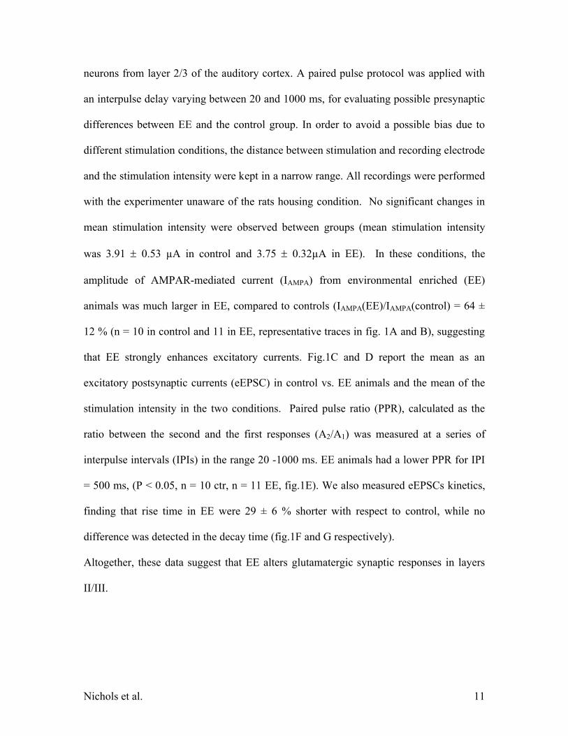
Data analysis
Statistical analysis was performed with Clampfit 9.2, SigmaPlot 8.0, and
Microsoft Excel software. A Student’s unpaired t-test was used for comparison between
different groups of cells. Data were reported as significantly different only if p < 0.05.
Neural model
We used MatLab to develop a realistic neural network model for simulating the
effects of EE on cortical processing. The model, described in more detail in the appendix,
used a three-compartment pyramidal neurons and a single compartment interneuron with
previously described voltage-gated conductances (Wang, 1998). Synaptic release elicits
-function like synaptic currents (see the Results and Appendix sections). Short-term
memory properties of the synapses (facilitation and fast and slow depression) are present
in the model as reported previously (Brunel and Wang, 2001; Tiesinga and Sejnowski,
2001; Varela et al., 1997).
Results
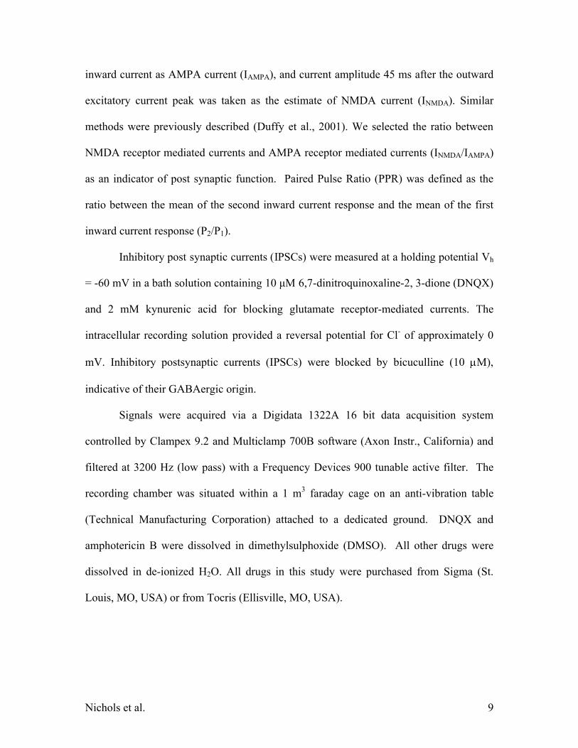
AMPAR-mediated currents in layer II/III
Since EE increases 3H AMPA binding in the hippocampus (Foster et al., 1996) and has
been suggested to enhance glutamatergic activity (Foster and Dumas, 2001), we first
measured the amplitude of electrically evoked AMPA currents in visually identified
Nichols et al. 11
neurons from layer 2/3 of the auditory cortex. A paired pulse protocol was applied with
an interpulse delay varying between 20 and 1000 ms, for evaluating possible presynaptic
differences between EE and the control group. In order to avoid a possible bias due to
different stimulation conditions, the distance between stimulation and recording electrode
and the stimulation intensity were kept in a narrow range. All recordings were performed
with the experimenter unaware of the rats housing condition. No significant changes in
mean stimulation intensity were observed between groups (mean stimulation intensity
was 3.91 0.53 µA in control and 3.75 0.32µA in EE). In these conditions, the
amplitude of AMPAR-mediated current (IAMPA) from environmental enriched (EE)
animals was much larger in EE, compared to controls (IAMPA(EE)/IAMPA(control) = 64 ±
12 % (n = 10 in control and 11 in EE, representative traces in fig. 1A and B), suggesting
that EE strongly enhances excitatory currents. Fig.1C and D report the mean as an
excitatory postsynaptic currents (eEPSC) in control vs. EE animals and the mean of the
stimulation intensity in the two conditions. Paired pulse ratio (PPR), calculated as the
ratio between the second and the first responses (A2/A1) was measured at a series of
interpulse intervals (IPIs) in the range 20 -1000 ms. EE animals had a lower PPR for IPI
= 500 ms, (P < 0.05, n = 10 ctr, n = 11 EE, fig.1E). We also measured eEPSCs kinetics,
finding that rise time in EE were 29 ± 6 % shorter with respect to control, while no
difference was detected in the decay time (fig.1F and G respectively).
Altogether, these data suggest that EE alters glutamatergic synaptic responses in layers
II/III.
Nichols et al. 12
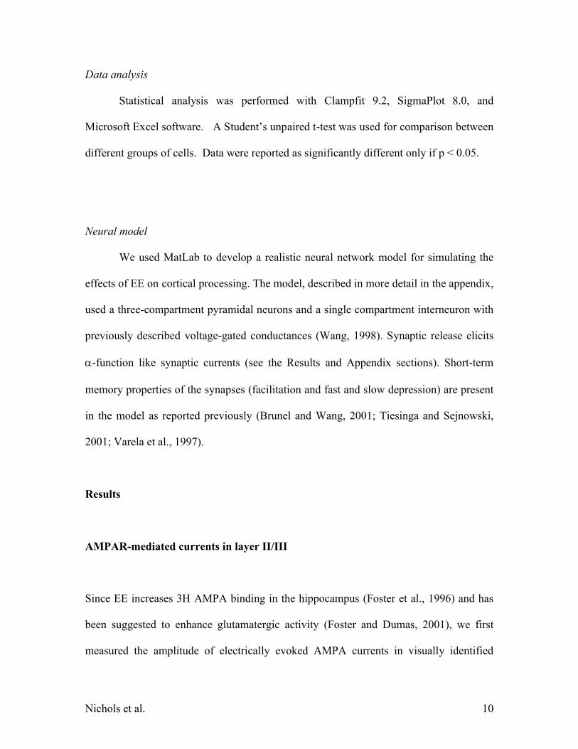
GABAergic currents in layer II/III
The changes in extracellular signal associated with EE (Percaccio et al., 2005)
might be due to an increase in excitatory drive and/or decreased inhibition. Decreased
inhibition might be a consequence of a decrease in GABAAR-mediated currents. We
tested the hypothesis of a decrease in synaptic inhibition by directly measuring
GABAergic currents (eIPSCs) from layer II/III neurons using a similar stimulation
protocol to the one described in the previous paragraph, but in the presence of the
glutamate ionotropic receptor blockers DNQX (10 M) and kynurenic acid (2mM). The
remaining currents were blocked by 10 M bicuculline, confirming their GABAergic
nature.
Evoked IPSCs amplitude was not different in control vs. EE animals (fig.2A). No
difference in rise- or decay-time were detected (Fig. 2B and C). The same pair pulse
protocol used for AMPAR-mediated currents (interpulse intervals in the range 20-1000
ms) was used to measure possible changes in PPR (Fig.2D). PPR too remained
unchanged between the two conditions (p > 0.5, n = 8 each). These data suggest that
changes in inhibition are not likely to play a major role in EE.
AMPA-mediated currents in layer V
Nichols et al. 13
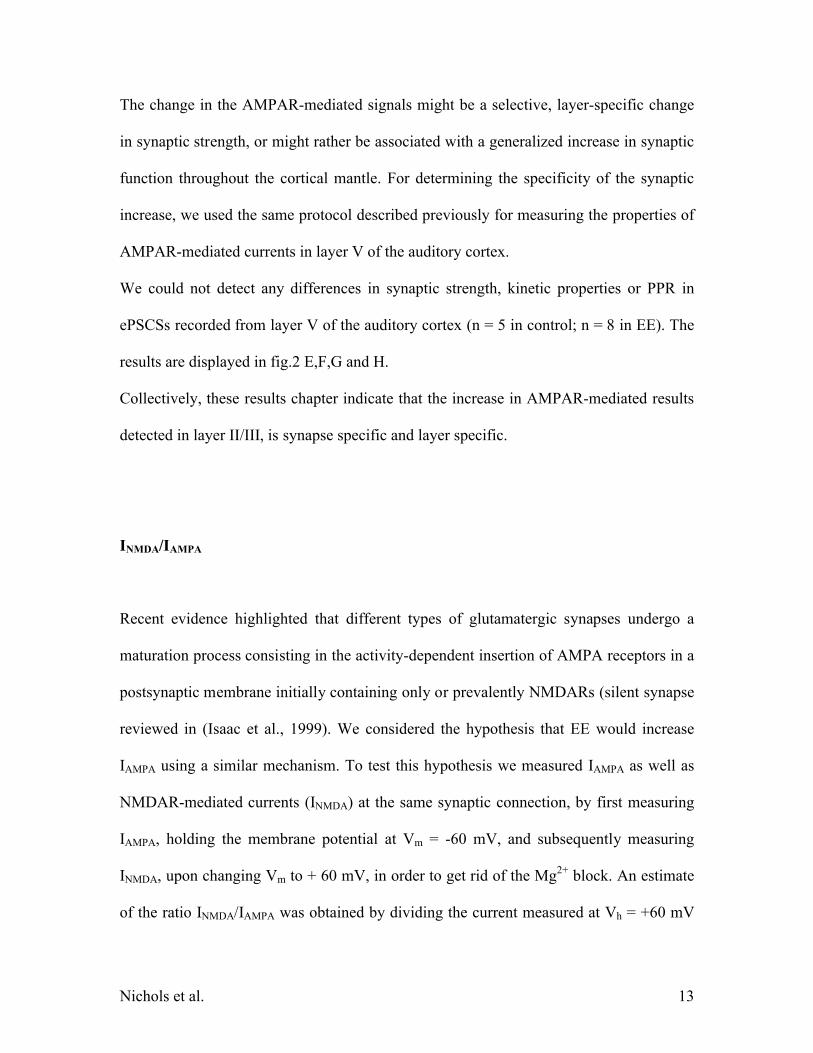
The change in the AMPAR-mediated signals might be a selective, layer-specific change
in synaptic strength, or might rather be associated with a generalized increase in synaptic
function throughout the cortical mantle. For determining the specificity of the synaptic
increase, we used the same protocol described previously for measuring the properties of
AMPAR-mediated currents in layer V of the auditory cortex.
We could not detect any differences in synaptic strength, kinetic properties or PPR in
ePSCSs recorded from layer V of the auditory cortex (n = 5 in control; n = 8 in EE). The
results are displayed in fig.2 E,F,G and H.
Collectively, these results chapter indicate that the increase in AMPAR-mediated results
detected in layer II/III, is synapse specific and layer specific.
INMDA/IAMPA
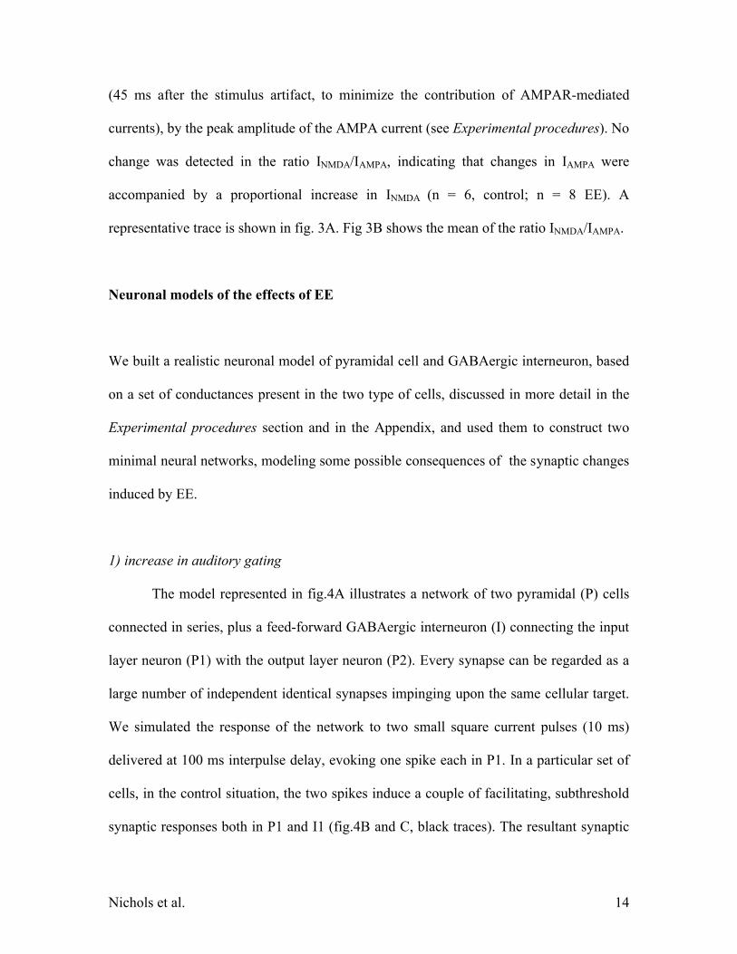
Recent evidence highlighted that different types of glutamatergic synapses undergo a
maturation process consisting in the activity-dependent insertion of AMPA receptors in a
postsynaptic membrane initially containing only or prevalently NMDARs (silent synapse
reviewed in (Isaac et al., 1999). We considered the hypothesis that EE would increase
IAMPA using a similar mechanism. To test this hypothesis we measured IAMPA as well as
NMDAR-mediated currents (INMDA) at the same synaptic connection, by first measuring
IAMPA, holding the membrane potential at Vm = -60 mV, and subsequently measuring
INMDA, upon changing Vm to + 60 mV, in order to get rid of the Mg2+ block. An estimate
of the ratio INMDA/IAMPA was obtained by dividing the current measured at Vh = +60 mV
Nichols et al. 14
(45 ms after the stimulus artifact, to minimize the contribution of AMPAR-mediated
currents), by the peak amplitude of the AMPA current (see Experimental procedures). No
change was detected in the ratio INMDA/IAMPA, indicating that changes in IAMPA were
accompanied by a proportional increase in INMDA (n = 6, control; n = 8 EE). A
representative trace is shown in fig. 3A. Fig 3B shows the mean of the ratio INMDA/IAMPA.
Neuronal models of the effects of EE
We built a realistic neuronal model of pyramidal cell and GABAergic interneuron, based
on a set of conductances present in the two type of cells, discussed in more detail in the
Experimental procedures section and in the Appendix, and used them to construct two
minimal neural networks, modeling some possible consequences of the synaptic changes
induced by EE.
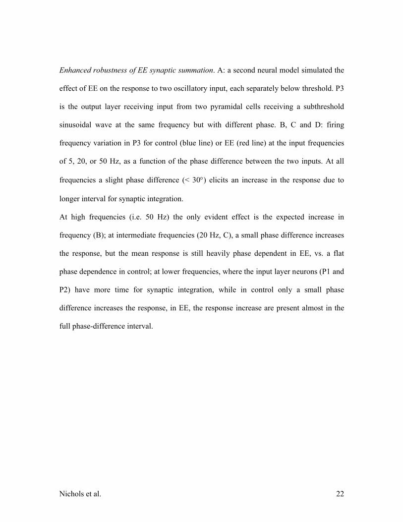
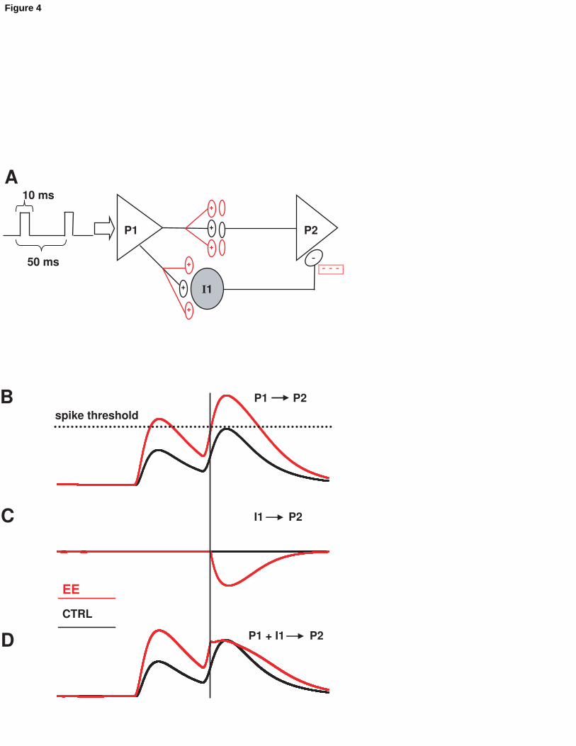
1) increase in auditory gating
The model represented in fig.4A illustrates a network of two pyramidal (P) cells
connected in series, plus a feed-forward GABAergic interneuron (I) connecting the input
layer neuron (P1) with the output layer neuron (P2). Every synapse can be regarded as a
large number of independent identical synapses impinging upon the same cellular target.
We simulated the response of the network to two small square current pulses (10 ms)
delivered at 100 ms interpulse delay, evoking one spike each in P1. In a particular set of
cells, in the control situation, the two spikes induce a couple of facilitating, subthreshold
synaptic responses both in P1 and I1 (fig.4B and C, black traces). The resultant synaptic
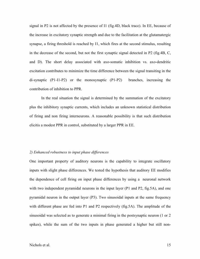
Nichols et al. 15
signal in P2 is not affected by the presence of I1 (fig.4D, black trace). In EE, because of
the increase in excitatory synaptic strength and due to the facilitation at the glutamatergic
synapse, a firing threshold is reached by I1, which fires at the second stimulus, resulting
in the decrease of the second, but not the first synaptic signal detected in P2 (fig.4B, C,
and D). The short delay associated with axo-somatic inhibition vs. axo-dendritic
excitation contributes to minimize the time difference between the signal transiting in the
di-synaptic (P1-I1-P2) or the monosynaptic (P1-P2) branches, increasing the
contribution of inhibition to PPR.
In the real situation the signal is determined by the summation of the excitatory
plus the inhibitory synaptic currents, which includes an unknown statistical distribution
of firing and non firing interneurons. A reasonable possibility is that such distribution
elicitis a modest PPR in control, substituted by a larger PPR in EE.
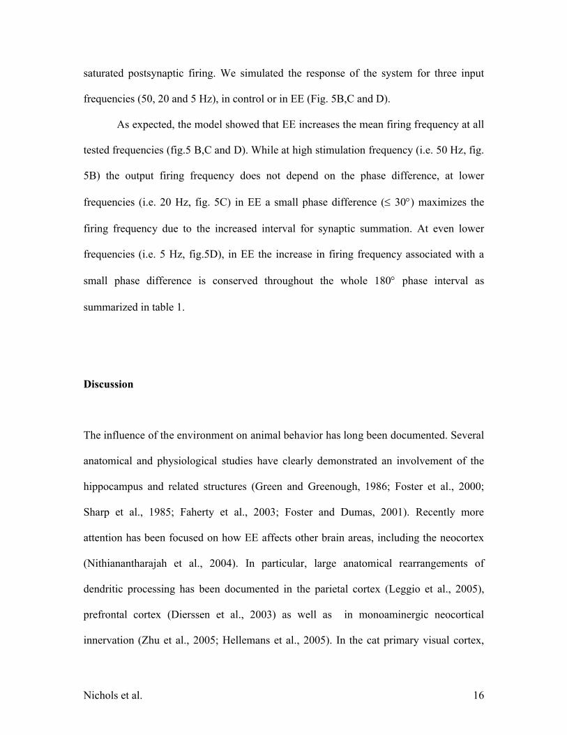
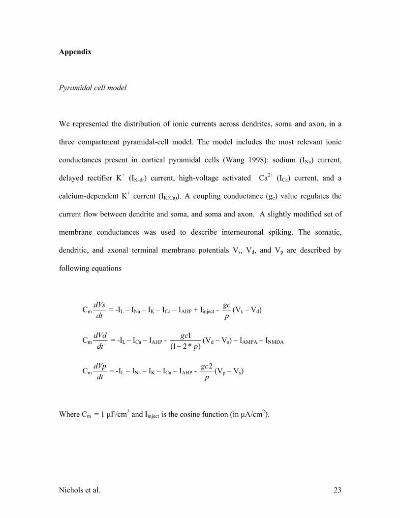
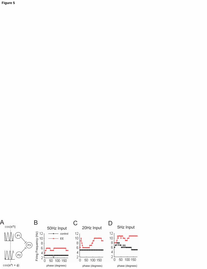
2) Enhanced robustness to input phase differences
One important property of auditory neurons is the capability to integrate oscillatory
inputs with slight phase differences. We tested the hypothesis that auditory EE modifies
the dependence of cell firing on input phase differences by using a neuronal network
with two independent pyramidal neurons in the input layer (P1 and P2, fig.5A), and one
pyramidal neuron in the output layer (P3). Two sinusoidal inputs at the same frequency
with different phase are fed into P1 and P2 respectively (fig.5A). The amplitude of the
sinusoidal was selected as to generate a minimal firing in the postsynaptic neuron (1 or 2
spikes), while the sum of the two inputs in phase generated a higher but still non-
Nichols et al. 16
saturated postsynaptic firing. We simulated the response of the system for three input
frequencies (50, 20 and 5 Hz), in control or in EE (Fig. 5B,C and D).
As expected, the model showed that EE increases the mean firing frequency at all
tested frequencies (fig.5 B,C and D). While at high stimulation frequency (i.e. 50 Hz, fig.
5B) the output firing frequency does not depend on the phase difference, at lower
frequencies (i.e. 20 Hz, fig. 5C) in EE a small phase difference ( 30) maximizes the
firing frequency due to the increased interval for synaptic summation. At even lower
frequencies (i.e. 5 Hz, fig.5D), in EE the increase in firing frequency associated with a
small phase difference is conserved throughout the whole 180 phase interval as
summarized in table 1.
Discussion
The influence of the environment on animal behavior has long been documented. Several
anatomical and physiological studies have clearly demonstrated an involvement of the
hippocampus and related structures (Green and Greenough, 1986; Foster et al., 2000;
Sharp et al., 1985; Faherty et al., 2003; Foster and Dumas, 2001). Recently more
attention has been focused on how EE affects other brain areas, including the neocortex
(Nithianantharajah et al., 2004). In particular, large anatomical rearrangements of
dendritic processing has been documented in the parietal cortex (Leggio et al., 2005),
prefrontal cortex (Dierssen et al., 2003) as well as in monoaminergic neocortical
innervation (Zhu et al., 2005; Hellemans et al., 2005). In the cat primary visual cortex,
Nichols et al. 17
EE has been found to be effective in rescuing ocular dominance columns impaired by
dark rearing (Bartoletti et al., 2004). In the primary auditory cortex, EE induces major
physiological rearrangements, detected as changes in single cell response properties as
well as in scalp recording (Engineer et al., 2004; Percaccio et al., 2005).
Our results showed a strong and selective increase in amplitude and a change in
kinetics of glutamatergic responses. These data are in agreement with the general
increase in the excitability, firing rate and decrease in latency in the AEP amplitude
observed in the response to short (25 ms) tones in vivo (Engineer et al., 2004).
The stability of the ratio between NMDAR-mediated and AMPAR-mediated
currents indicates that EE is not associated with a selective insertion of new AMPARs in
the postsynaptic membrane ("silent" synapse "awakening"). Yet, other postsynaptic
changes preserving IAMPA/INMDA might take place at EE synapses.
The dramatic increase in EPSC amplitude might simply reflect an increase in the total
number of excitatory spines, in agreement with previous findings (Dierssen et al., 2003).
Similarly, the lack of changes in eEPSC PPR at short (<500ms) II does not allow to
exclude the presence of presynaptic rearrangements preserving PPR.
Unchanged inhibitory responses do not support a major role for inhibition in the
EE-driven re-shaping of auditory cortical responses. A sharp-electrode study on
hippocampal non-pharmacologically dissected synaptic currents reported similar
conclusion (Foster and Dumas, 2001).
The increase in synaptic efficacy could be the result of a generalized synaptic
strengthening or could be a layer-specific phenomenon. The unchanged excitatory
responses from layer V would suggest the second hypothesis, indicating layer II/III as a
Nichols et al. 18
privileged substrate for the solidification of cortical plasticity. A similar conclusion was
previously reached with an anatomical-morphological study (Johansson and Belichenko,
2002), and is expected if EE is caused by spike-time-dependent plasticity, since the
threshold for action potentials in layer V is approximately 10 mV more positive than in
layer II/III (Atzori et al., 2004).
The increase in synaptic amplitude after exposure to the EE might derive from the
transformation of low-probability and small amplitude synapses into high probability,
large amplitude synapses (Atzori et al., 2001). Yet, high probability synapses possess
slower rise times and smaller PPR with respect to low-probability synapses, contrasting
with our current finding that EE decreases rise-time and leaves PPR unchanged,
suggesting a different origin for the synaptic changes in EE.
The use of a computational model corroborated the hypothesis that the increase in
auditory gating in EE (Percaccio et al., 2005) is due in part or completely to local,
cortico-cortical changes in excitatory synaptic strength.
Auditory information is largely conveyed by low frequency envelope signals
surfacing onto AI with slight phase differences within an isofrequency contour. Our
computational model showed that the synaptic changes associated with the EE not only
produce an expectable increase in firing rate but that they also decrease the dependence
of the postsynaptic firing rate on the input phase differences, in the case of two slow
frequency de-phased inputs. In fact, at low input frequencies, the increase in firing rate
corresponding to a small phase increase displayed in control is replaced in EE by a solid
enhancement almost independent on the phase difference between the two inputs, making
synaptic summation more robust in EE.
Nichols et al. 19
Although our data indicate that EE causes significant cortical plasticity, we cannot
exclude the possibility that non cortical auditory relays are also modified by EE,
introducing a further component to the EE-induced alteration of the cortical signal
detected by AEP (Percaccio et al., 2005). In conclusion, we demonstrated for the first
time that EE induces a change in the efficacy of glutamatergic synapses within the
primary auditory cortex, associated with a major postsynaptic rearrangement compatible
with an increase in the total number of synaptic connections.
Author statement
JN performed all the patch-clamp experiments, analyzed and elaborated data and figures,
and contributed to writing the final version of the manuscript, VJ raised the EE animals
and contributed to the discussion, LD wrote and ran the MatLab program using literature
and original data, MK contributed to the original idea, to the design of the experiments
and to the final version of the manuscript, MA contributed to the original idea and to the
design of the experiments, and wrote the final version of the manuscript.
Grants: NIDCD 1R01-DC005986-01A1 and NARSAD foundation/Sidney Baer Trust (M.A.) and American
Academy of Audiology to J.A.N.
Nichols et al. 20
Figure legends
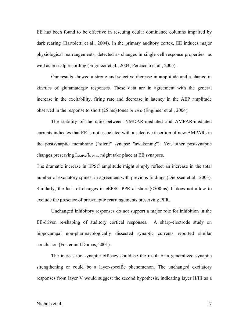
Figure 1
AMPAR-mediated currents in Layer II/III. A and B. Mean traces for control (n = 10) and
EE (n = 11) animals. C Intensity of the electrical stimulation for control (blank bar) and
EE (gray bar) did not change. D Mean of the peak amplitude of the AMPAR-mediated
current in control (blank bar, n = 10) vs. EE (gray bar, n = 11). The mean amplitude was
more than 60% larger in EE animals. E Paired Pulse Ratio (PPR =A2/A1). PPR at an
interpulse interval of 500 ms was smaller in EE animals (0.943 0.026 in control vs
0.877 0.018 in EE, n = 10 control; n = 11 in EE). F Rise time and G decay time of the
eEPSC. The rise time is shorter in EE animals while no differences are present in the
decay time. Altogether, these data suggest that EE elicits changes in glutamatergic
synaptic signals.
Figure 2
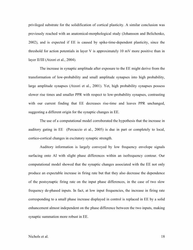
Neurotransmitter- and layer-specificity of the synaptic changes. A, B, C and D:
amplitude, rise time, decay time and PPR of GABAergic currents in layer II/III do not
differ in control or in EE (n = 8 each). E, F, G and H: amplitude, rise time, decay time
and PPR in AMPAR-mediated glutamatergic currents of layer V do not differ between
control and EE (n = 5 control; n = 8 EE).
Nichols et al. 21
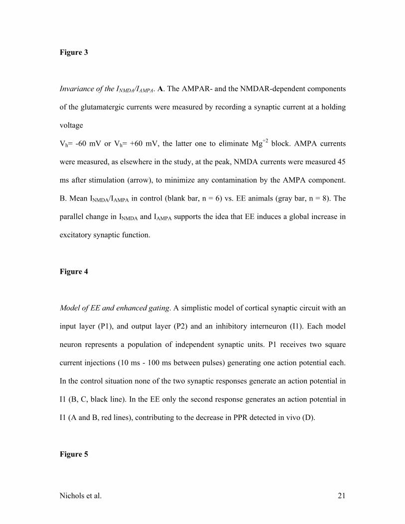
Figure 3
Invariance of the INMDA/IAMPA. A. The AMPAR- and the NMDAR-dependent components
of the glutamatergic currents were measured by recording a synaptic current at a holding
voltage
Vh= -60 mV or Vh= +60 mV, the latter one to eliminate Mg+2 block. AMPA currents
were measured, as elsewhere in the study, at the peak, NMDA currents were measured 45
ms after stimulation (arrow), to minimize any contamination by the AMPA component.
B. Mean INMDA/IAMPA in control (blank bar, n = 6) vs. EE animals (gray bar, n = 8). The
parallel change in INMDA and IAMPA supports the idea that EE induces a global increase in
excitatory synaptic function.
Figure 4
Model of EE and enhanced gating. A simplistic model of cortical synaptic circuit with an
input layer (P1), and output layer (P2) and an inhibitory interneuron (I1). Each model
neuron represents a population of independent synaptic units. P1 receives two square
current injections (10 ms - 100 ms between pulses) generating one action potential each.
In the control situation none of the two synaptic responses generate an action potential in
I1 (B, C, black line). In the EE only the second response generates an action potential in
I1 (A and B, red lines), contributing to the decrease in PPR detected in vivo (D).
Figure 5
Nichols et al. 22
Enhanced robustness of EE synaptic summation. A: a second neural model simulated the
effect of EE on the response to two oscillatory input, each separately below threshold. P3
is the output layer receiving input from two pyramidal cells receiving a subthreshold
sinusoidal wave at the same frequency but with different phase. B, C and D: firing
frequency variation in P3 for control (blue line) or EE (red line) at the input frequencies
of 5, 20, or 50 Hz, as a function of the phase difference between the two inputs. At all
frequencies a slight phase difference (< 30) elicits an increase in the response due to
longer interval for synaptic integration.
At high frequencies (i.e. 50 Hz) the only evident effect is the expected increase in
frequency (B); at intermediate frequencies (20 Hz, C), a small phase difference increases
the response, but the mean response is still heavily phase dependent in EE, vs. a flat
phase dependence in control; at lower frequencies, where the input layer neurons (P1 and
P2) have more time for synaptic integration, while in control only a small phase
difference increases the response, in EE, the response increase are present almost in the
full phase-difference interval.
Nichols et al. 23
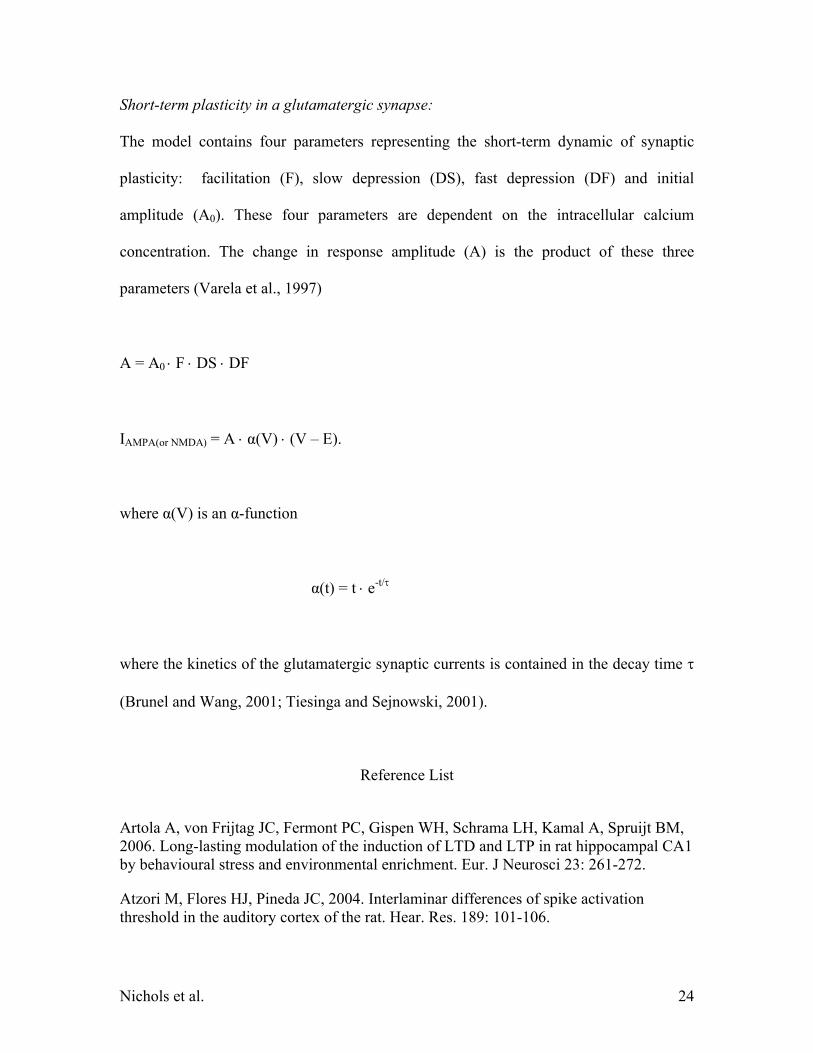
Appendix
Pyramidal cell model
We represented the distribution of ionic currents across dendrites, soma and axon, in a
three compartment pyramidal-cell model. The model includes the most relevant ionic
conductances present in cortical pyramidal cells (Wang 1998): sodium (INa) current,
delayed rectifier K+ (IK-dr) current, high-voltage activated Ca2+ (ICa) current, and a
calcium-dependent K+ current (IK(Ca)). A coupling conductance (gc) value regulates the
current flow between dendrite and soma, and soma and axon. A slightly modified set of
membrane conductances was used to describe interneuronal spiking. The somatic,
dendritic, and axonal terminal membrane potentials Vs, Vd, and Vp are described by
following equations
Cmdt
dVs= -IL – INa – IK – ICa – IAHP + Iinject -
p
gc(Vs – Vd)
Cmdt
dVd = -IL – ICa – IAHP -
)*21(
1
p
gc
(Vd – Vs) – IAMPA – INMDA
Cmdt
dVp= -IL – INa – IK – ICa – IAHP -
p
gc2(Vp – Vs)
Where Cm = 1 μF/cm2 and Iinject is the cosine function (in μA/cm2).
Nichols et al. 24
Short-term plasticity in a glutamatergic synapse:
The model contains four parameters representing the short-term dynamic of synaptic
plasticity: facilitation (F), slow depression (DS), fast depression (DF) and initial
amplitude (A0). These four parameters are dependent on the intracellular calcium
concentration. The change in response amplitude (A) is the product of these three
parameters (Varela et al., 1997)
A = A0 F DS DF
IAMPA(or NMDA) = A α(V) (V – E).
where α(V) is an α-function
α(t) = t e-t/
where the kinetics of the glutamatergic synaptic currents is contained in the decay time
(Brunel and Wang, 2001; Tiesinga and Sejnowski, 2001).
Reference List
Artola A, von Frijtag JC, Fermont PC, Gispen WH, Schrama LH, Kamal A, Spruijt BM, 2006. Long-lasting modulation of the induction of LTD and LTP in rat hippocampal CA1 by behavioural stress and environmental enrichment. Eur. J Neurosci 23: 261-272.
Atzori M, Flores HJ, Pineda JC, 2004. Interlaminar differences of spike activation threshold in the auditory cortex of the rat. Hear. Res. 189: 101-106.
Nichols et al. 25
Atzori M, Kanold PO, Pineda JC, Flores-Hernandez J, Paz RD, 2005. Dopamine prevents muscarinic-induced decrease of glutamate release in the auditory cortex. Neuroscience 134: 1153-1165.
Atzori M, Lei S, Evans DI, Kanold PO, Phillips-Tansey E, McIntyre O, McBain CJ, 2001. Differential synaptic processing separates stationary from transient inputs to the auditory cortex. Nat. Neurosci. 4: 1230-1237.
Bartoletti A, Medini P, Berardi N, Maffei L, 2004. Environmental enrichment prevents effects of dark-rearing in the rat visual cortex. Nat. Neurosci 7: 215-216.
Brunel N, Wang XJ, 2001. Effects of neuromodulation in a cortical network model of object working memory dominated by recurrent inhibition. J Comput. Neurosci 11: 63-85.
Dierssen M, avides-Piccione R, Martinez-Cue C, Estivill X, Florez J, Elston GN, DeFelipe J, 2003. Alterations of neocortical pyramidal cell phenotype in the Ts65Dn mouse model of Down syndrome: effects of environmental enrichment. Cereb. Cortex 13: 758-764.
Duffy SN, Craddock KJ, Abel T, Nguyen PV, 2001. Environmental enrichment modifies the PKA-dependence of hippocampal LTP and improves hippocampus-dependent memory. Learn. Mem. 8: 26-34.
Engineer ND, Percaccio CR, Pandya PK, Moucha R, Rathbun DL, Kilgard MP, 2004. Environmental enrichment improves response strength, threshold, selectivity, and latency of auditory cortex neurons. J Neurophysiol. 92: 73-82.
Faherty CJ, Kerley D, Smeyne RJ, 2003. A Golgi-Cox morphological analysis of neuronal changes induced by environmental enrichment. Brain Res. Dev. Brain Res. 141: 55-61.
Foster TC, Dumas TC, 2001. Mechanism for increased hippocampal synaptic strength following differential experience. J Neurophysiol. 85: 1377-1383.
Foster TC, Fugger HN, Cunningham SG, 2000. Receptor blockade reveals a correspondence between hippocampal-dependent behavior and experience-dependent synaptic enhancement. Brain Res. 871: 39-43.
Foster TC, Gagne J, Massicotte G, 1996. Mechanism of altered synaptic strength due to experience: relation to long-term potentiation. Brain Res. 736: 243-250.
Green EJ, Greenough WT, 1986. Altered synaptic transmission in dentate gyrus of rats reared in complex environments: evidence from hippocampal slices maintained in vitro. J Neurophysiol. 55: 739-750.
Nichols et al. 26
Hellemans KG, Nobrega JN, Olmstead MC, 2005. Early environmental experience alters baseline and ethanol-induced cognitive impulsivity: relationship to forebrain 5-HT1A receptor binding. Behav. Brain Res. 159: 207-220.
Irvine GI, Abraham WC, 2005. Enriched environment exposure alters the input-output dynamics of synaptic transmission in area CA1 of freely moving rats. Neurosci Lett. 391: 32-37.
Isaac JT, Nicoll RA, Malenka RC, 1999. Silent glutamatergic synapses in the mammalian brain. Can. J Physiol Pharmacol. 77: 735-737.
Johansson BB, Belichenko PV, 2002. Neuronal plasticity and dendritic spines: effect of environmental enrichment on intact and postischemic rat brain. J Cereb. Blood Flow Metab 22: 89-96.
Leggio MG, Mandolesi L, Federico F, Spirito F, Ricci B, Gelfo F, Petrosini L, 2005. Environmental enrichment promotes improved spatial abilities and enhanced dendritic growth in the rat. Behav. Brain Res. 163: 78-90.
Nithianantharajah J, Levis H, Murphy M, 2004. Environmental enrichment results in cortical and subcortical changes in levels of synaptophysin and PSD-95 proteins. Neurobiol. Learn. Mem. 81: 200-210.
Percaccio CR, Engineer ND, Pruette AL, Pandya PK, Moucha R, Rathbun DL, Kilgard MP, 2005. Environmental enrichment increases paired-pulse depression in rat auditory cortex. J Neurophysiol. 94: 3590-3600.
Sharp PE, McNaughton BL, Barnes CA, 1985. Enhancement of hippocampal field potentials in rats exposed to a novel, complex environment. Brain Res. 339: 361-365.
Tiesinga PH, Sejnowski TJ, 2001. Precision of pulse-coupled networks of integrate-and-fire neurons. Network. 12: 215-233.
Varela JA, Sen K, Gibson J, Fost J, Abbott LF, Nelson SB, 1997. A quantitative description of short-term plasticity at excitatory synapses in layer 2/3 of rat primary visual cortex. J Neurosci 17: 7926-7940.
Wang XJ, 1998. Calcium coding and adaptive temporal computation in cortical pyramidal neurons. J Neurophysiol. 79: 1549-1566.
Zhu J, Apparsundaram S, Bardo MT, Dwoskin LP, 2005. Environmental enrichment decreases cell surface expression of the dopamine transporter in rat medial prefrontal cortex. J Neurochem. 93: 1434-1443.
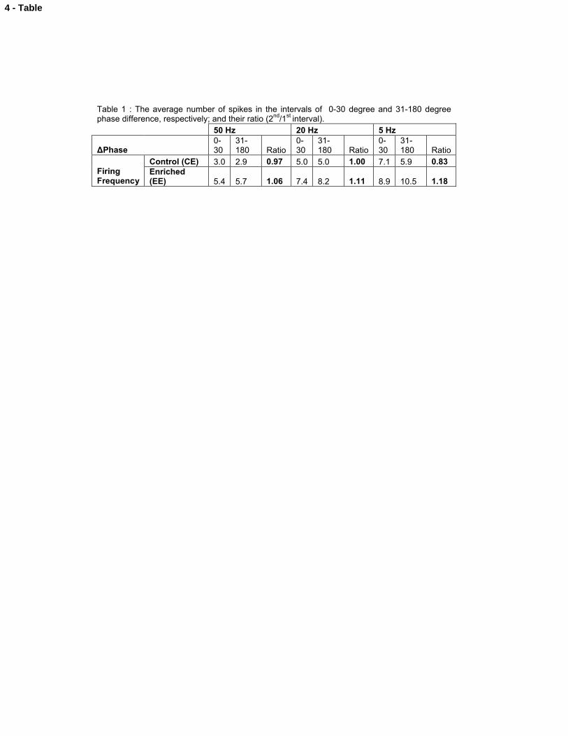
Table 1 : The average number of spikes in the intervals of 0-30 degree and 31-180 degree phase difference, respectively; and their ratio (2nd/1st interval).
50 Hz 20 Hz 5 Hz
ΔPhase0-30
31-180 Ratio
0-30
31-180 Ratio
0-30
31-180 Ratio
Control (CE) 3.0 2.9 0.97 5.0 5.0 1.00 7.1 5.9 0.83Firing Frequency
Enriched (EE) 5.4 5.7 1.06 7.4 8.2 1.11 8.9 10.5 1.18
4 - Table
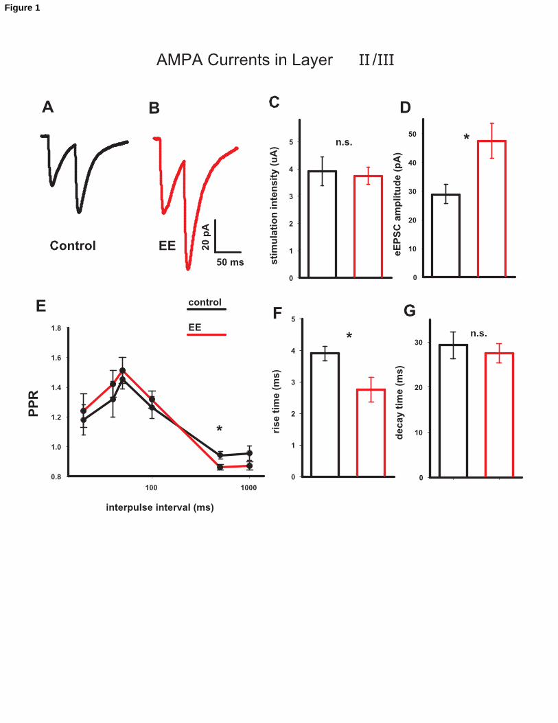
Figure 1
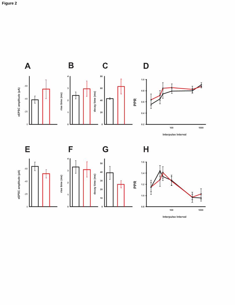
Figure 2
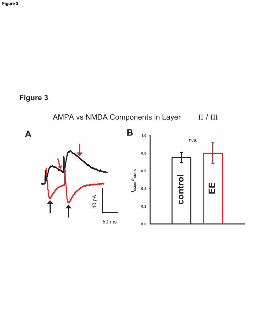
Figure 3
P1 P2
I1
50 ms
10 ms
+
+
+
+
+
+
- - -
A
EE
CTRL
spike threshold
B
C
D
P1 P2
I1 P2
P1 + I1 P2
Figure 4
Fir
ing
Fre
qu
en
cy (
Hz)
phase (degrees)
P1
P2
50Hz Input
B CA20Hz Input 5Hz Input
P3
D
phase (degrees) phase (degrees)
control
EE
Figure 5