ecologie chimique du dinoflagellé toxique ostreopsis cf
TRANSCRIPT

HAL Id: tel-03015932https://tel.archives-ouvertes.fr/tel-03015932
Submitted on 20 Nov 2020
HAL is a multi-disciplinary open accessarchive for the deposit and dissemination of sci-entific research documents, whether they are pub-lished or not. The documents may come fromteaching and research institutions in France orabroad, or from public or private research centers.
L’archive ouverte pluridisciplinaire HAL, estdestinée au dépôt et à la diffusion de documentsscientifiques de niveau recherche, publiés ou non,émanant des établissements d’enseignement et derecherche français ou étrangers, des laboratoirespublics ou privés.
Ecologie chimique du dinoflagellé toxique Ostreopsis cf.ovata en Méditerranée Nord Occidentale
Anne-Sophie Pavaux
To cite this version:Anne-Sophie Pavaux. Ecologie chimique du dinoflagellé toxique Ostreopsis cf. ovata en MéditerranéeNord Occidentale. Ecosystèmes. Sorbonne Université, 2019. Français. �NNT : 2019SORUS292�.�tel-03015932�

Sorbonne Université Ecole Doctorale des Sciences de l’Environnement d’Ile de France (ED 129)
Laboratoire d’Océanographie de Villefranche UMR 7093 – SU – CNRS
Ecologie chimique du dinoflagellé toxique
Ostreopsis cf. ovata en Méditerranée Nord Occidentale
Thèse présentée par Anne-Sophie PAVAUX
En vue de l’obtention du grade de Docteur de Sorbonne Université
Spécialité : Ecologie Marine
Soutenue publiquement le 18 novembre 2019 au Laboratoire d’Océanographie de Villefranche
Devant le jury composé de : Dr. Elisa BERDALET (CSIC, Espagne) Examinatrice
Pr. Cécile BERNARD (MNHN, France) Examinatrice
Pr. Gilles BOEUF (Sorbonne Université, France) Examinateur
Dr. Gérald CULIOLI (Université de Toulon, France) Rapporteur
Pr. Olivier THOMAS (National University of Galway, Irlande) Examinateur
Dr. Dorothée VINCENT (LOG - en détachement à l’Agence Rapportrice
Française pour la Biodiversité, France)
Pr Rodolphe LEMEE (Sorbonne Université, France) Directeur de thèse
Dr Stéphane GASPARINI (Sorbonne Université, France) Co-encadrant


REMERCIEMENTS
Je souhaite remercier ici tous ceux qui, de près ou de loin, ont contribué à cette thèse.
J’aimerai dans un premier temps remercier Anne Corval et Elisabeth Christians qui se sont
succédées au poste de Directrice de l’observatoire de Villefranche ainsi que le directoire du Laboratoire
d’Océanographie, Antoine Sciandra et son tout jeune successeur, Rodolphe Lemée. J’ai passé trois
années fantastiques à vos côtés. J’aimerai également remercier les membres de mon jury : Cécile
Bernard et Gérald Culioli d’avoir acceptés d’être les rapporteurs de ce manuscrit ainsi que Gilles Boeuf,
Dorothée Vincent, Elisa Berdalet et Olivier Thomas d’avoir acceptés d’examiner mon travail. Je souhaite
également remercier Cécile Jauzein et Michèle Tackx qui, en tant que membres de mon comité de thèse,
ont suivi de très près mon travail pendant ces trois années de thèse.
Mes premiers remerciements vont à mon directeur de thèse, le Pr. Rodolphe Lemée. Un grand
merci pour tout ce que tu m’as apporté durant ces quatre dernières années. Merci également pour tous
les moments que l’on a partagés, aussi bien en conférences (avec ou sans perruques !) qu’au labo, et
(ancestralement) sur le terrain. Ce fut un véritable plaisir de travailler avec toi et j’espère avoir la chance
de collaborer à nouveau avec toi dans le futur.
Un immense merci à toi Eva, l’aboutissement de cette thèse te revient en grande partie. Merci
pour m’avoir fait confiance dès notre premier skype en Master et pour m’avoir soutenue tout le long
depuis. J’ai énormément appris à tes côtés, aussi bien professionnellement que personnellement, et
j’espère que l’on se recroisera très vite.
Un merci particulier à toi Sophie d’avoir été un soutien sans faille, je suis très heureuse d’avoir
pu partager ces années avec toi et j’ai hâte d’enfin trouver le temps pour rencontrer Anatole. Merci
aussi pour tous les moments hors du labo, notamment en randonnées où on finissait toujours perdus
ou ensevelis sous la neige (Merci Hubert pour tes merveilleuses idées !)
Je souhaite également remercier Stéphane Gaparini pour m’avoir permis de réaliser cette
thèse. Merci à Laurence Guidi-Guilvard pour ces précieux conseils, notamment sur l’élevage et
l’identification des copépodes. Un grand merci à Olivier Thomas pour m’avoir fait confiance dès le
master 2 et pour m’avoir permis de faire des manips de chimie supplémentaires à Galway.
Merci à tous les stagiaires que j’ai encadré au cours de cette thèse et qui ont grandement
participé à l’avancement de ce travail : Julie R., Mélanie B. et Mélanie H. Un merci particulier à Louison
pour ta gentillesse et ton sérieux (à ton tour maintenant !) et à Anais et Alan, les meilleurs stagiaires
estivaux du monde ! Merci à vous deux d’avoir rendu ces deux étés moins pénibles (au fait Anais, j’ai
retrouvé la lampe torche..!)
Merci à Inalve d’avoir hébergé mes algues pendant ma fin de thèse ! Un merci particulier à
Quentin pour nos pauses déjeuner parfois trèèèèès longues, Freddy (à quand la location d’enfants ?) et
Hubert pour ta bonne humeur et ton humour sans limite.
Je tiens maintenant à adresser des remerciements tous particuliers aux personnes qui ont
partagé mon quotidien pendant cette thèse.
Un grand merci aux gestionnaires, Linda, Corinne (Merci pour le voyage aux USA !!) et Anne qui
facilitent grandement notre vie au labo. Un grand merci à toi Isa pour ta gentillesse et tes conseils.
Merci à Martine pour ta précieuse aide tout au long de la thèse, nottament bibliographique
et toutes nos discussions.
Merci à Thierry et Jocelyne pour vos formidables repas. Merci à Véronique, Katia et Didier pour
votre gentillesse (et les posters !).
Merci aux marins et particulièrement Jean-Yves pour toutes ces sorties en mer ! Un grand merci
à Guillaume (à quand la sortie à Boussole ?)
Merci à Géraldine qui a partagé mon quotidien pendant ces trois dernières années ! Merci pour
nos fous-rires, nos collectes de coquillages, nos baignades et j’en passe. Tu as rendu, à coup sûr, cette
thèse plus agréable. Bon courage pour ta dernière année !
Merci à toi Alice pour ta présence, ton écoute et ta joie de vivre.
Merci à Laurent, qui même à près de 10 000 km m’a soutenu. J’ai hâte de retourner faire du
kayak avec toi !
Merci à tous mes collègues de l’open-space ! Merci à Kévin, peuchère c’était quand même
moins drôle cette année sans toi ! Coco pour ta joie de vivre, Simon pour tes blind-test en cors de chasse,
Margaux pour tes conseils, Amélie, Ophélie et Charlotte.
Merci à François pour avoir égayé mes journées par ton exhubérance et ta bonne humeur.
Merci à toutes les personnes qui ont, à un moment ou à un autre, rendu cette thèse plus facile :
Mathilde (même si tu as émigré en Bretagne, tu me manques bcp !!), Fabio, Marin, Lolo, Maya et
Quentin, Florentine, Olivier B., Samir, Régis, Salomé, Marc B, Laure, Dr C, Mathilde, Ilan et Etienne,
Marine, Thierry B.
Même si on appartient à deux labos différents, je tiens à remercier de nombreuses personnes
du Laboratoire de Biologie de Développement notamment Guy, Stéfania, David, Yas pour sa gentillesse
mais également Marta, Maciej, Alex et Marion.
Au terme de ce parcours, je remercie enfin celles et ceux qui me sont chers et que j’ai quelque
peu délaissés ces derniers temps pour achever cette thèse. Leurs attentions et encouragements m’ont
accompagné tout au long de ces années. Je suis redevable à ma maman (Oui maman j’ai enfin fini mes
études !!), mon frère et ma sœur pour leur soutien et leur confiance indéfectibles dans mes choix. Enfin
merci à toi Axel, sans toi, je n’aurai jamais pu terminer ce travail. Merci pour m’avoir soutenu (et
supporté !) jusqu’au bout !


LISTE DES ARTICLES PUBLIES, SOUMIS ET EN PREPARATION
Ternon E., Pavaux A-S., Marro S., Thomas O.P., Lemée R., 2018. Allelopathic interactions between the benthic toxic dinoflagellate Ostreopsis cf. ovata and a co-occurring diatom. Harmful Algae 75, 35–44. https://doi.org/10.1016/j.hal.2018.04.003. Pavaux A-S., Rostan J., Guidi-Guilvard L., Marro S., Ternon E., Thomas O.P., Lemée R., Gasparini S., 2019. Effects of the toxic dinoflagellate Ostreopsis cf. ovata on survival, feeding and reproduction of a phytal harpacticoid copepod. Journal of Experimental Marine Biology and Ecology 516, 103–113. https://doi.org/10.1016/j.jembe.2019.05.004. Pavaux A-S., Ternon E., Dufour L., Marro S., Thomas O., Lemée R. Reliable and handful monitoring of Ostreopsis sp. toxicity using an Artemia sp. bioassay. En préparation. Pavaux A-S., Berdalet E., Lemée R. Chemical ecology of the benthic dinoflagellates genus Ostreopsis : à review. En préparation. Pavaux A-S., Velasquez D., Drouet K., Lebrun A., Hiroux A., Marro S., Castagnetti S., Lemée R. Vertical migration of Ostreopsis cf. ovata. En préparation.
ARTICLES REALISES EN COLLABORATION
Boisnoir A., Pavaux A-S., Schizas N. V., Marro S., Blasco T., Lemée R., Pascal P-Y. The use of stable
isotopes to measure the ingestion rate of potentially toxic benthic dinoflagellates by harpacticoid
copepods. Journal of Experimental Marine Biology and Ecology. En révision.
Gémin M-P., Réveillon D., Hervé F., Pavaux A-S., Séchet V., Bertrand S., Lemée R., Amzil Z. Toxin
content of Ostreopsis cf. ovata depends on bloom phases, depth and macroalgae in the NW
Mediterranean Sea. Harmful algae. En révision.
Ternon E., Pavaux A-S., Jauzein C., Gemin M-P., Peltekis A., Bailleul B., Lemée R., Thomas O. P.
Underestimated role of the specialized metabolism in species microalgae species dynamics using the
toxic benthic dinoflagellate Ostreopsis cf. ovata as a model. En préparation.
Jaouannet M., Pavaux A-S., Marro S., Lemée R., Martens K., Michelet C., Keller H, Coustau C. A weird
MIF coming out of the blue. En préparation.
Jauzein C., Pavaux A-S., Lemée R. Influence of nutrients and hydrodynamism on Ostreopsis cf. ovata
phenology in NW Mediterranean Sea. En préparation.

LISTE DES COMMUNICATIONS
Ternon E., Pavaux A-S., Thomas O. (2017). Metabolomics should be implemented wisely in the context
of marine chemical ecology. GdR Phycotox 2017, Mars 2017, Gif-sur-Yvette, France – Présentation
orale.
Pavaux A-S., Lemée R., Gasparini S. Impact of the toxic dinoflagellate Ostreopsis cf. ovata on coastal
fauna in NW Mediterranean Sea. GdR PHYCOTOX, Mars 2017, Gif-sur-Yvette, France – Poster.
Pavaux A-S., Ternon E., Marro S., Thomas O., Lemée R. Competition between two Mediterranean
benthic microalgae: Endo/Exo metabolome study and physiological responses. DINO11, Juillet 2017,
Bordeaux, France – Poster: Prix du meilleur poster.
Pavaux A-S., Rostan J., Guidi-Guilvard L., Marro S., Olivi J., Gasparini S., Lemée R. Impact of the toxic
dinoflagellate Ostreopsis cf. ovata on coastal fauna in N.W. Mediterranean Sea. Global HAB meeting,
Avril 2018, Villefranche-sur-mer, France – Présentation orale.
Pavaux A-S., Rostan J., Guidi-Guilvard L., Marro S., Gasparini S., Lemée R. Impact of the toxic
dinoflagellate Ostreopsis cf. ovata on coastal fauna in N.W. Mediterranean Sea. FADEX-O- Oregon
State University & SCRIPPS, Mai 2018, USA – Présentation orale.
Pavaux A-S., Rostan J., Guidi-Guilvard L., Marro S., Gasparini S., Lemée R. Chemical ecology of Benthic
HABs: the impact of NW Mediterranean Ostreopsis cf. ovata on copepods. ICHA, Octobre 2018, Nantes,
France – Poster.
Pavaux A-S., Rostan J., Guidi-Guilvard L., Marro S., Gasparini S., Lemée R. Chemical ecology of the
benthic toxic dinoflagellate Ostreopsis cf. ovata in N.W. Mediterranean Sea. Société phycologique de
France, Novembre 2018, Paris, France – Présentation orale : Obtention d’une bourse pour la
participation à la Gordon Research Conference de 2019.
Pavaux A-S., Rostan J., Guidi-Guilvard L., Marro S., Gasparini S., Lemée R. Chemical ecology of the
benthic toxic dinoflagellate Ostreopsis cf. ovata in N.W. Mediterranean Sea. Visite volontaire au
Natural History Museum of Denmark, Janvier 2019, Copenhague, Danemark – Présentation orale.
Pavaux A-S., Ternon E., Dufour L., Marro S., Thomas O., Lemée R. Artemia franscicana as a simple and
pertinent model to check Ostreopsis spp. toxicity. Projet Assemblee + APOTOX, National University of
Galway, Mars 2019, Galway, Irlande – Présentation orale.
Pavaux A-S., Ternon E., Dufour L., Rostan J., Guidi-Guilvard L., Marro S., Gasparini S., Thomas O., Lemée
R. Chemical ecology of the benthic toxic dinoflagellate Ostreopsis cf. ovata in N.W. Mediterranean Sea.
GdR PHYCOTOX, Mai 2019, Brest, France – Présentation orale : Prix de la meilleure présentation.
Pavaux A-S., Ternon E., Dufour L., Marro S., Thomas O., Lemée R. The use of Artemia franciscana to
assess the toxicity of benthic dinoflagellates genus Ostreopsis. Gordon Research Conference –
Phycotoxins and Mycotoxins, June 2019, Boston, USA – Poster.
Boisnoir A., Pascal P.Y., Pavaux A-S., Shizas N. V., Marro S., Blasco T., Lemee R. Ingestion of potentially
toxic benthic dinoflagellates by harpacticoid copepods. SeventIMCO, July 2019, Evora, Portugal –
Présentation orale.


TABLE DES MATIERES
1. Ecologie chimique marine 5
2. Application de l’écologie chimique à l’étude des efflorescences d’algues nuisibles 8
3. Cas du dinoflagellé toxique Ostreopsis cf. ovata 12 3.1. Ecologie d’Ostreopsis cf. ovata 12 3.2. Ecologie chimique d’Ostreopsis cf. ovata 15
1. Présentation de l’étude 55
2. Allelopathic interactions between the benthic toxic dinoflagellate Ostreopsis cf. ovata and a co-occurring
diatom 55 2.1. Abstract 57 2.2. Introduction 58 2.3. Methods 60 2.4. Results 65 2.5. Discussion 70 2.6. Conclusion 74 2.7. Acknowledgments 75
3. Synthèse des résultats 77
1. Présentation de l’étude 81
2. Effects of the toxic dinoflagellate Ostreopsis cf. ovata on survival, feeding and reproduction of a phytal
harpacticoid copepod 81 2.1. Abstract 83 2.2. Introduction 84 2.3. Materials and Method 85 2.4. Results 90 2.5. Discussion 98 2.6. Conclusion 102 2.7. Acknowledgements 103
3. Synthèse de l’étude 104

1. Présentation de l’étude 107
2. Etude des phases benthiques et planctoniques d’Ostreopsis cf. ovata 109 2.1. Introduction 109 2.2. Matériels et Méthodes 110 2.3. Résultats 113 2.4. Discussion 118 2.5. Conclusion 120
3. Impact d’Ostreopsis cf. ovata sur les copépodes planctoniques 121 3.1. Introduction 121 3.2. Matériels et méthodes 121 3.3. Résultats 123 3.4. Discussion 123 3.5. Conclusion 126
4. Synthèse des résultats 127
1. Présentation de l’étude 131
2. Introduction 133
3. Matériels et Méthodes 134 3.1. Expérimentations in situ 134 3.2. Expérimentations en laboratoire 135 3.3. Paramètres d’études 137 3.4. Analyse statistique 138
4. Résultats 138 4.1. Efflorescences d’Ostreopsis cf. ovata en 2017 et 2018 138 4.2. Evolution du taux d’ingestion des oursins exposés à Ostreopsis cf. ovata 140 4.3. Mesure de la taille des oursins 140 4.4. Impact d’Ostreopsis cf. ovata sur le temps de retournement des oursins 140 4.5. Impact sur le rapport gonado-somatique 141
5. Discussion 142 5.1. Absence d’effet d’Ostreopsis cf. ovata sur le comportement des oursins 142 5.2. Impact d’Ostreopsis cf. ovata sur le taux d’ingestion de Paracentrotus lividus 144 5.3. Impact d’Ostreopsis cf. ovata sur le rapport gonado-somatique 145
6. Conclusion 145
7. Synthèse de l’étude 147

1. Présentation de l’étude 151
2. Reliable and handful monitoring of Ostreopsis sp. toxicity using an Artemia sp. bioassay 151 2.1. Abstract 153 2.2. Introduction 154 2.3. Materials and Methods 155 2.4. Results 160 2.5. Discussion 168 2.6. Acknowledgement 171
3. Synthèse de l’étude 173
1. Impacts d’Ostreopsis cf. ovata sur les écosystèmes méditerranéens 177 1.1. Ostreopsis cf. ovata : toxicité aigüe vs toxicité chronique ? 178 1.2. Facteurs contrôlant la production de toxines 179 1.3. Localisation des toxines produites par Ostreopsis cf. ovata 180 1.4. Transfert des toxines dans le réseau trophique 184
2. Perturbation des écosystèmes suite aux efflorescences d’Ostreopsis cf. ovata 186 2.1. Sélection de la nourriture 186 2.2. Fuite 187 2.3. Production de composés toxiques 187
3. Bilan sur le rôle écologique des toxines produites par Ostreopsis cf. ovata 188
4. Synthèse et perspectives de recherche sur l’écologie chimique d’Ostreopsis cf. ovata 189


LISTES DES FIGURES
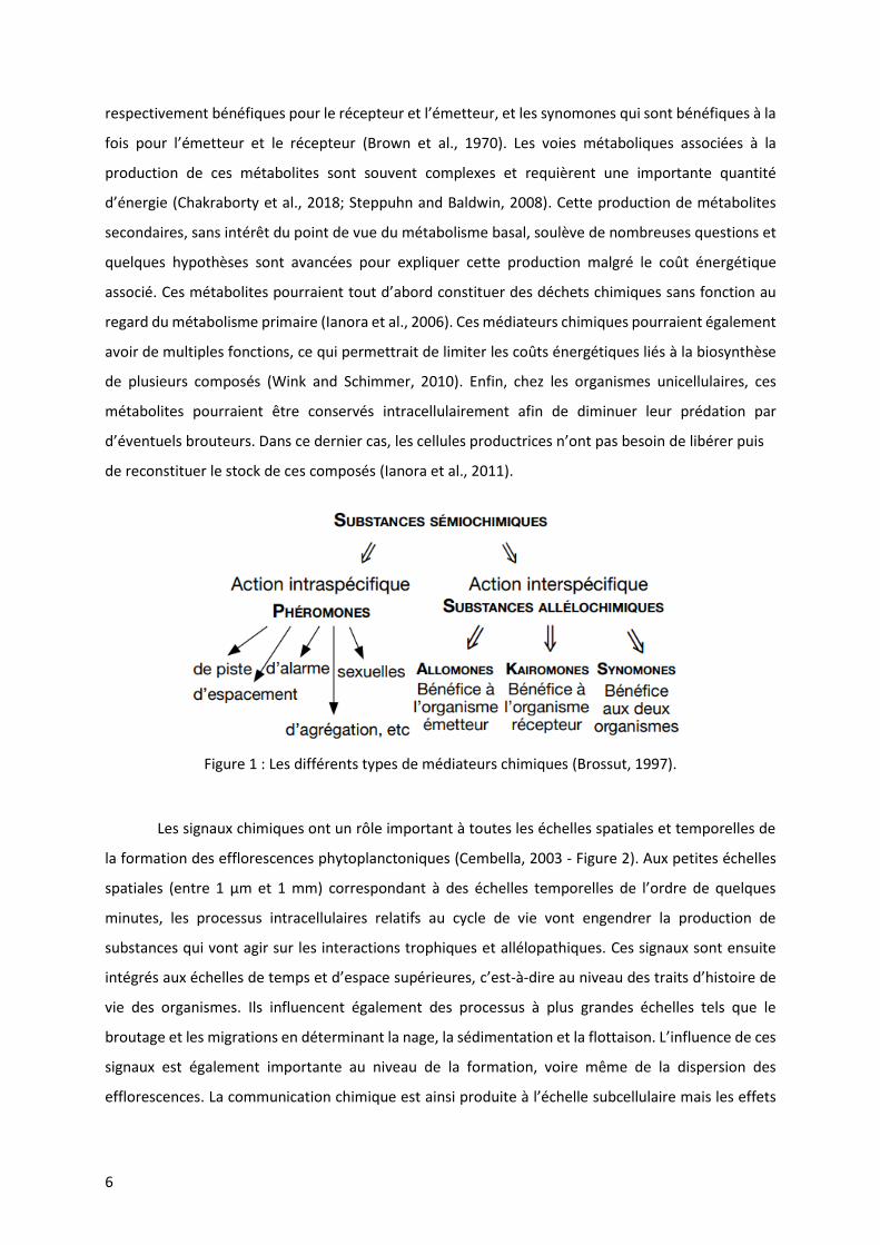
Figure 1 : Les différents types de médiateurs chimiques (Brossut, 1997). ............................................. 6
Figure 2 : Schéma représentant les niveaux d’organisation fonctionnelles régis par la communication
chimique dans les efflorescences d’algues toxiques planctoniques. L’axe des abscisses représente
l’échelle temporelle (en échelle logarithmique) et celui des ordonnées l’échelle spatiale (également en
échelle logarithmique). Les signaux chimiques sont issus de processus cellulaires mais agissent à
différents niveaux d’organisation : à l’échelle subcellulaire (ex : excrétion, croissance), à méso-échelle
(ex : broutage, parasitisme, allélopathie) ainsi qu’à l’échelle de la formation et la dispersion des
efflorescences. L’importance de cette communication dans l’évolution des traits d’histoire de vie est
également représentée (Cembella, 2003). ............................................................................................. 7
Figure 3 : Evolution du nombre d’efflorescences d’algues toxiques provoquant des intoxication par
fruits de mer de type paralysante entre 1970 et 2015 (Sources : National Oceanic and Atmospheric
Administration – NOAA and Woods Hole Oceanographic Institution - WHOI ........................................ 8
Figure 4 : Bilan des facteurs de stress climatiques qui peuvent potentiellement déterminer l’intensité
et la fréquence des efflorescences d’algues nuisibles. L’élévation globale des températures,
l’augmentation des concentrations de CO2 atmosphériques ainsi que l’eutrophisation décrites depuis
le début du 20ème siècle peuvent potentiellement favoriser de manière indépendante et interactive les
HABs (Griffith and Gobler, 2019)............................................................................................................. 9
Figure 5 : Cellules d’Ostreopsis cf. ovata (MCCV 054) en culture (A), recouvrant les macroalgues (B) et
les rochers in situ (C). Une remise en suspension des cellules est visible sur la photo B. .................... 12
Figure 6 : Co-culture device with the two flat growth chambers connected by the metabolome was
performed at day 0 using the initial 300mL clamp. .............................................................................. 61
Figure 7 : Structure of the ovatoxin-a and its major fragments A and B. ............................................. 64
Figure 8 : Effect of the EX of (i) L. paradoxa onto the in vivo fluorescence(Fv/Fm) of O. cf. ovata (left)
and (ii) O. cf. ovata onto L. paradoxa (right). Controls were performed with sterile media. Statistical
significance was assessed using a Wilcoxon-Mann-Whitney test (* p < 0.1). errors bars correspond to
standard error between samples (n=4)................................................................................................. 66
Figure 9 : Abundance of cells (cells/mL) and in vivo fluorescence (Fv/Fm ratio) of control (dark grey)
and co-cultured (light grey) chambers for O. cf. ovata (A, C; left side) and L. paradoxa (B,D; right side).
Statistical significance was assessed using a Wilcoxon-Mann-Witney test (* and · p < 0.1 and 0.2
respectively). ......................................................................................................................................... 67
Figure 10 : Toxin content in the (EN) endometabolome and in the (EX) exometabolome normalized by
the number of cells ; in controls= cultures (dark grey) and co-cultures (light grey). A palytoxin standard
was used to obtain the calibration curve and the concentrations are therefore expressed as an
equivalent palytoxin. Statistical significance was assessed using a Wilcoxon-Mann-Whitney test (*, p <
0.1). Error bars define the standard errors between samples (n=3). ................................................... 68
Figure 11 : PLS-DA plots obtained from the untargeted metabolomics analysis on the content of the EX
of the chambers containing (A) the diatom L. paradoxa and (B) the dinoflagellate O. cf. ovata. The ions
showed in the tables are the VIP over-expressed in the co-cultured chambers and correspond to
compounds secreted by either O. cf. ovata (A) and L. paradoxa (B) cultures. ..................................... 70

Figure 12 : Mortality (Means ± SE, n=6) of Artemia franciscana larvae after different exposure times
(hours) and for increasing concentrations of Ostreopsis cf. ovata. The ratio number of algal cells:
number of target animals is between 20:5 to 20 000:5. ....................................................................... 91
Figure 13 : Mortality (Means ± SE, n=6) of the copepod Sarsamphiascus cf. propinquus after different
exposure times (days) and for increasing concentrations of Ostreopsis cf. ovata. The ratio of algal cells:
number of target animals is this time between 2500:1 to 100 000:1................................................... 92
Figure 14 : Fecal pellet production (Mean ± SE, n=6) of the copepod Sarsamphiascus cf. propinquus
exposed to different feeding treatments and no-food treatment as a function of time. Number of fecal
pellets were cumulated over time. Two identical experiments were performed during 10 days (1) and
14 days (2). Letters formulate significant differences: groups that have no common letter differ
significantly. ........................................................................................................................................... 93
Figure 15 : The copepod Sarsamphiascus cf. propinquus observed under epifluorescence microscopy
(excitation in UV, fluorescence in red; left) and light (middle) after having been fed with Ostreopsis cf.
ovata. The autofluorescence of chlorophyll is visualized inside the digestive track. Ostreopsis cf. ovata
cells (Light microscopy; right) also appear attached to the body and the caudal rami. ....................... 94
Figure 16 : Kaplan-Meier survival analysis of the copepod Sarsamphiascus cf. propinquus exposed to
different feeding treatments and no-food treatment as a function of time. Two identical experiments
were performed during 10 days (1) and 14 days (2). Letters formulate significant differences: groups
that have no common letter differ significantly (p < 0.05). .................................................................. 97
Figure 17 : Site d’échantillonnage dans la rade de Villefranche au cours des étés 2018 et 2019. ..... 109
Figure 18 : Schéma représentant les différentes profondeurs échantillonnées. ............................... 110
Figure 19 : Evolution de la concentration d’Ostreopsis cf. ovata à Rochambeau (Villefranche-sur-Mer,
France) en 2018 (a) et 2019 (b). En 2018, la période d’échantillonnage à haute résolution temporelle
s’est déroulée juste après le pic de l’efflorescence et au moment du pic de l’efflorescence en 2019
(flèches rouges). .................................................................................................................................. 111
Figure 20 : Macroalgues échantillonnées en 2018 : (a) Dyctiota sp.(Lamouroux, 1809), (b) Halopteris
scoparia (Linnaeus – Sauvageau, 1904) et (c) Padina pavonica (Linnaeus – Thivy 1960). .................. 112
Figure 21 : Evolution de la concentration d’Ostreopsis cf. ovata en surface (a), planctonique (b) et
benthique (c) au cours du temps en 2018. Les dégradés d’orange et de bleu représentent
respectivement les abondances cellulaires obtenues la journée et la nuit. ....................................... 114
Figure 22 : Evolution de la concentration d’Ostreopsis cf. ovata en surface (a), planctonique (b) et
benthique (c) au cours du temps en 2019. Les dégradés d’orange et de bleu représentent
respectivement les abondances cellulaires obtenues la journée et la nuit. ....................................... 115
Figure 23 : Variation des abondances cellulaires d’Ostreopsis cf. ovata dans le benthos (vert), la
colonne d’eau (bleu) et à la surface de l’eau (gris) en 2018 (a) et 2019 (b). ...................................... 116
Figure 24 : Taux de croissance apparent des cellules d’Ostreopsis cf. ovata benthiques, planctoniques
et de surface pour les années 2018 (gauche) et 2019 (droite). .......................................................... 117
Figure 25 : Pourcentage de mortalité des copépodes planctoniques Acartia clausi (a), Temora stylifera
(b) et Lucicutia flavicornis (c) exposés à différentes concentrations d’Ostreopsis cf. ovata. ............. 124
Figure 26 : Sites d’échantillonnage dans la rade de Villefranche au cours des étés 2017 et 2018 .... 135

Figure 27 : Installation utilisée pour maintenir les oursins pour l’expérimentation en laboratoire (a).
Les oursins ont été exposés à des concentrations variables d’Ostreopsis cf. ovata (b) incorporées à la
nourriture artificielle préparée en laboratoire pour maintenir les oursins (c). .................................. 137
Figure 28 : Concentration cellulaire d’Ostreopsis cf. ovata au site de Rochambeau (en noir) à 0.5 m
(ligne pointillée) et 2 m de profondeur (ligne pleine) et du Lido à 2 m de profondeur (ligne pleine grise)
en 2017 (a) et 2018 (b). L’évolution de la taille des oursins Paracentrotus lividus (Moyenne ± erreur
standard ; n=10) prélevés à Rochambeau (ligne noire) et au Lido (ligne grise) au cours des étés 2017 (c)
et 2018 (d) est également représentée. Le temps de retournement (moyenne ± erreur standard ; n=10)
des oursins (Paracentrotus lividus) mesurées in situ en 2017 (e) et 2018 (f) au niveau du site de
Rochambeau (lignes noires) et du Lido (lignes grises) est également figuré. ..................................... 139
Figure 29 : Evolution du temps de retournement des oursins maintenus en laboratoire et nourris avec
une nourriture artificielle préparée e laboratoire enrichie en Ostreopsis cf. ovata,, en diatomées
Licmophora paradoxa ou non enrichie .............................................................................................. 141
Figure 30 : Evolution du rapport gonado-somatique (Moyenne ± erreur standard ; n= 10) des oursins
(Paracentrotus lividus) mesuré in situ aux sites de Rochambeau (ligne noire) et du Lido (ligne grise) lors
de l’été 2017. Les concentration cellulaires d’Ostreopsis cf. ovata à Rochambeau (tirets noirs) et au
Lido (tirets gris) sont également représentées. .................................................................................. 142
Figure 31 : Percentage of mortality (Mean ± ES) after 12 h (a), 24 h (b) and 48 h (c) of Artemia
franciscana larvae exposed to various concentrations of Mediterranean strains of Ostreopsis cf. ovata
(MCCV 054 and 055) and Ostreopsis fattorussoi (MCCV 057 and 058). ............................................. 162
Figure 32 : Toxin concentration (Mean ± ES) in pg eq. PLTX/cell (black line) depending on the growth
phase of Ostreopsis cf. ovata (MCCV 055 – grey bars). ...................................................................... 163
Figure 33 : Percentage of mortality (Mean ± ES) of Artemia franciscana larvae exposed to 4 (black lines),
40 (dotted lines) and 400 cells.mL-1 (dashed lines) of Ostreopsis cf. ovata (MCCV 055) after (a) 12 hours,
(b) 24 hours and (c) 48 hours exposure time. The growth curve of O. cf. ovata (MCCV 055) cultured in
300mL flasks is also represented in grey bars. .................................................................................... 164
Figure 34 : Mortality (Mean ± ES) of Artemia franciscana larvae after being exposed 24 hours to
fractions of Ostreopsis cf. ovata obtained from MCCV 054 extracts. The concentration of all fractions
was previously adjusted to 3 mg.ml-1 using DMSO and MilliQ. The polarity of the metabolites decreases
from Fraction 1 to 7 (F1-F7). The content of OVTXs in each fraction is also represented (black squares).
............................................................................................................................................................. 165
Figure 35 : Mortality (Mean ± ES) of Artemia franciscana after 24 hours exposition to 4 cells.mL-1.
Evolution of cellular concentration of Ostreopsis cf. ovata on artificial substrates is also represented in
dotted line. .......................................................................................................................................... 166
Figure 36 : Mortality (Mean ± ES) after 12 h (a), 24 h (b) and 48 h (c) of Artemia franciscana larvae
exposed to increased concentrations of dissolved PLTX and OVTX (from 0.012, 0.12, 1.2 and 12 µg/mL)
and living cells at various cell-equivalent concentrations. Letters formulate significant differences:
groups that have no common letter differ significantly (p < 0.05). .................................................... 167
Figure 37 : Schéma représentant l’ensemble des relations biotiques (connues dans la littérature)
qu’Ostreopsis cf. ovata entretient avec son environnement. Les flêches en traits entiers représentent
les interactions directes, celles en tirets représentent les interactions indirectes. Les références
bibliographiques associées sont précisées sous la forme de chiffres (référencées ci-dessous). Le
symbole triangulaire jaune représentent les voies d’intoxication humaines possibles aux toxines
produites par Ostreopsis cf. ovata. ..................................................................................................... 192


LISTES DES TABLEAUX
Table 1 : Classification des phycotoxines selon les symptômes qu’elles provoquent chez l’Homme. Les
espèces ainsi que les toxines associées sont également énumérées (Traduit depuis Sidharta, 2005). 10
Table 2 : Les toxines émergentes .......................................................................................................... 11
Table 3 : Representative information about the range of toxin content in described Ostreopsis species
from different locations. The literature review is not exhaustive. * = Identity of the isolate(s) confirmed
genetically confirming to which described or cryptic (ribotype) species they belong. Abbreviations:
Equivalents (eq.); Hemolytic Assay - ability of cellular extract to induce erythrocyte cell lysis versus a
saponin control (HA); HPLC‐ESI‐MS/MS High-performance liquid chromatography-electrospray
ionisation tandem mass spectrometry; Ovatoxin (OVTX); Liquid Chromatography – High Resolution
Mass Spectrometry (LC-HRMS); Mouse Bioassay - toxicity determined by intraperitoneal injection of
Ostreopsis cellular extracts (MBA); Mouse Units, the 24 h LD50 dose estimated using ~20 g mice (MU);
Not Detectable = below detection limits (nd); neuroblastoma cell line cytotoxicity assay (N2a); putative
palytoxin (pPLTX) like toxin = isobaric PLTX (García-Altares et al., 2015). More information on toxicity
tests as well as on other Ostreopsis strains (that can become new species) is found in Tester et al.,
2019. ...................................................................................................................................................... 22
Table 4 : Reports of mild respiratory, cutaneous and/or general malaise symptoms in humans after
exposure to aerosols in Ostreopsis cf. ovata blooms affected beaches. .............................................. 26
Table 5 : Organisms in direct interaction with Ostreopsis spp. Half maximal effective concentration
(EC50) ; Digestive Tube (DT); Hemolysis Neutralization Assay (HNA); Median Lethal concentration (LC50)
; Liquid Chromatography –Mass Spectrometry (LC-MS); Media, lethal time (LT50); Mouse Bioassay
(MBA); Remaining tissue (RT); Whole flesh (WF). ................................................................................. 36
Table 6 : Organisms in indirect interaction with Ostreopsis spp. Half maximal effective concentration
(EC50); Median lethal concentration (LC50); Median lethal time (LT50). ................................................. 42
Table 7 : Artemia bio-assay for Ostreopsis toxicity. Median lethal concentration (LC50); Median lethal
time (LT50). ............................................................................................................................................. 49
Table 8 : List of the ovatoxins investigated in this study and their major ions. .................................... 65
Table 9 : Percentage of exudation (ratio EN/EX of ovatoxin concentration) of all detected toxins in the
control and in the co-culture chambers that contained Ostreopsis cultures. ...................................... 69
Table 10 : Fecundity and fertility ratios of S. cf. propinquus exposed to different food treatments (two
different diets and a no-food control) assessed through 3 identical experiments which ran for 7 days.
Experiments started off with 30 adults and late copepodites of unknown sex. Actual number of
females, resulting ovigerous females and nauplii are indicated. .......................................................... 97
Table 11 : P-values of the Fisher exact test for the fecundity (a) and fertility ratios (b) of Sarsamphiascus
cf. propinquus exposed to different feeding treatments and no-food treatment. Significant results are
evidenced in bold; *, **, *** corresponding respectively to p < 0.05, p < 0.01 and p < 0.001. ........... 98
Table 12 : Moyenne des taux de croissance apparents par profondeurs, période du jour et année. 116
Table 13 : LC50 values and confidence limits in cells.mL-1 obtained exposing Artemia franciscana larvae
to different strains of Ostreopsis sp. and dissolved toxins. ................................................................ 161

LISTES DES ABBREVIATIONS
ACN : Acétonitrile ANR : Agence Nationale de la Recherche ANSES : Agence Nationale de Sécurité Sanitaire de l’alimentation, de l’environnement et du travail ASP : Amnesic Shellfish Poisoning AZA : Azaspiracide BPC : Base Peak Chromatogram CAT : Catalase CE : Collision Energies Cellules/gPF : nombre de cellules par gramme de poids frais de macroalgues CFP : Ciguatera Fish Poisoning DMSO : Dimethylsulfoxide DMS : Diméthylsulfide dNTP : Désoxyribonucléotides DSP : Diarrheic Shellfish Poisoning DTX : Dinophysistoxines EC50 : Median effect concentration EFSA : Autorité européenne de sécurité des aliments EN : Endométabolome Eq. : Equivalent ESI-MS/MS : ElectroSpray Ionization-tandem Mass Spectrometry EX : Exométabolome FW : Fresh Weight GdR MediatEC : Groupement de Recherche Médiation chimique dans l’environnement – Ecologie Chimique GEOHAB : Global Ecology and Oceanography of Harmful Algal Blooms GYM : Gymnodimine HA : Hemolytic Assay HAB : Harmful Algal Bloom HNA : Hemolytic Neutralization Assay HPLC-ESI-MS/MS : High Performance Liquid Chromatography – ElectroSpray Ionization tandem Mass Spectrometry HTD : Heterotrophic dinoflagellate IFREMER : Institut français de Recherche pour l’exploitation de la mer ITS : Internal Transcribed Spacer LC50 : Median lethal concentration LC-HRMS : Liquid Chromatography – High Resolution Mass Spectrometry LC-FLD : Liquid Chromatography with Florescence Detection LD50 : Median lethal dose LSU : Large ribosomal subunit LT50 : Median lethal time MBA : Mouse BioAssay MCCV : Mediterranean Culture Collection of Villefranche MCE : Marine Chemical Ecology MC-PAM : Multi-Color Pulse-Amplitude-Modulated MeOH : Méthanol MT6 : Métallothioneine 6 MU : Mouse Unit N2a : Neuroblastoma cell line cytotoxicity assay nd : not detectable
NSP : Neurotoxic Shellfish Poisoning NOAA : National Oceanic and Atmsopheric Administration OCEAN-15 : Ostreopsis Chemical Ecology and Allelopathy Network (Projet ANR) OVTX : Ovatoxine PCA : Principal Components Analysis PCR : Polymerase Chain Reaction PLS-DA : Partial Least Squares-Discriminant Analysis PLTX : Palytoxine PnTX : Pinnatoxine pPLTX : putative Palytoxine PS II : Photosystème II PSP : Paralytic Shellfish Poisoning PTX : Pectenotoxine PUAs : PolyUnsaturated Aldehydes PUFAs : PolyUnsaturated Fatty Acids PVDF : PolyVinyliDene Fluoride QC : Quality Control qPCR : PCR quantitative QTOF : Quadripole-temps de vol RT : Remaining Tissue Rt : Retention time SPX : Spirolide UHPLC : Chromatographie Liquide à Ultra Haute Performance VIP : Variable Importance in Projection v/v : volume/volume WF : Whole Flesh WHOI : Woods Hole Oceanographic Institute YTX : Yessotoxine


Préambule
Cette thèse a été financée par l’Ecole Doctorale 129 (ED 129 - Sciences de l’Environnement) de
Sorbonne Université.
Ce travail s’inscrit principalement dans le cadre du projet multipartenaires (LOV - Sorbonne
Université et CNRS, ANSES, Université de Chimie de Galway et IFREMER Nantes) ANR OCEAN-15
(Ostreopsis Chemical Ecology and Allelopathy Network) dont l’objectif principal est de comprendre les
effets des changements climatiques sur le métabolisme secondaire d’Ostreopsis cf. ovata afin
d’anticiper les modifications potentielles de la toxicité et les effets écologiques qui en découlent. Plus
précisément, cette thèse s’insère dans la tache 3 de ce projet dont l’objectif est d’étudier les impacts
écologiques de ces toxines.
Une partie du travail effectué lors de mon doctorat est également intégrée dans le projet
européen CoCliMe (https://www.coclime.eu/) dont l’objectif est de fournir des services climatiques en
relation avec les qualités des eaux marines littorales (en particulier en relation avec les microalgues
nuisibles). J’ai également pu bénéficier d’un soutien financier dans le cadre du projet européen
Assemble + (Association of European Marine Biological Laboratories Expanded -
http://www.assembleplus.eu/) afin de faire des analyses chimiques au sein du Laboratoire Marine
Biodiscovery à Galway, Irlande, pendant 3 semaines au printemps 2019.
L’objectif principal de mon travail de thèse est d’améliorer nos connaissances sur les rôles
écologiques des toxines produites par le dinoflagellé Ostreopsis cf. ovata en mer Méditerranée. Dans
un premier temps, j’ai réalisé une étude exhaustive de la bibliographie sur ce sujet, qui sera publiée
sous forme de revue. J’ai ensuite étudié les interactions chimiques avec les compétiteurs, puis j’ai évalué
l’impact des phycotoxines sur des prédateurs (copépodes benthiques et planctoniques). Enfin, puisque
la nécessité s’est rapidement fait sentir, j’ai développé un modèle biologique simple permettant
d’évaluer rapidement la toxicité des cellules d’O. cf. ovata.
Afin de répondre à mon objectif principal, j’ai combiné des études expérimentales in situ et en
laboratoire. J’ai également utilisé une approche métabolomique, technique indispensable dans l’étude
de la communication chimique, qui a permis l’étude de l’endo- et de l’exométabolome d’O. cf. ovata.
Ce manuscrit s’articule ainsi autour de 7 chapitres :
- Le premier chapitre introductif traite de l’écophysiologie et de l’écologie chimique des espèces
du genre Ostreopsis.
- Le second chapitre détaille les relations allélopathiques qu’Ostreopsis cf. ovata entretient avec
les producteurs primaires et notamment une diatomée benthique compétitrice ;
- Les chapitres 3 et 4 traitent respectivement de l’impact d’Ostreopsis cf. ovata sur les copépodes
benthiques et planctoniques ;
- Le chapitre 5 s’intéresse aux effets de ce dinoflagellé sur les consommateurs secondaires en
prenant l’exemple de l’oursin comestible Paracentrotus lividus
- Le chapitre 6 aborde la mise en place d’un test écologique pour tester facilement, rapidement
et manière pertinente, la toxicité des espèces du genre Ostreopsis.
- Le chapitre 7 constitue une conclusion des travaux et présente les perspectives envisagées.
Le titre et les résumés des articles réalisés en collaboration au cours de ma thèse, sont ajoutés en
annexe.

Introduction
Illustration of armoured dinoflagellates from Ernst Haeckel's ''Kunstformen der Natur'' (1904)

Introduction
5
1. Ecologie chimique marine
L’écologie chimique marine est une science pluridisciplinaire relativement jeune dont l’objectif
est de mieux comprendre l’implication de la signalétique chimique produite par les organismes marins
(Paul et al., 2006; Paul & Ritson-Williams, 2008) dans l’organisation et le fonctionnement des
écosystèmes marins (Hay, 1996; McClintock & Baker, 2001; Pohnert et al., 2007). L’impact de ces
signaux chimiques sont encore mal reconnus (Hay, 2009). Ils affectent pourtant les comportements
individuels et les processus à l’échelle des populations, mais également l’organisation des
communautés par des effets en cascade (Hay and Kubanek, 2002). Ils affectent ainsi les transferts
d’énergie, la dynamique des réseaux trophiques et la structure des écosystèmes (Schwartz et al., 2016)
en intervenant dans la quête de nourriture, les communications intraspécifiques, les interactions de
compétition, de prédation et d’allélopathie, mais également dans la sélection des habitats et des
partenaires sexuels (Hay, 2009; Poulson et al., 2009; Sieg et al., 2011). L’un des exemples les plus
étonnants illustrant l’importance de ces signaux est celui des oiseaux procellariiformes. Le
diméthylsulfure (DMS), composé excrété par le phytoplancton lorsqu’il est brouté par le zooplancton
(Dacey and Wakeham, 1986), agit comme un guide olfactif pour ces oiseaux et leur permet de repérer
les zones océaniques productives où leur prise de nourriture sera un succès (Nevitt, 2000; Nevitt et al.,
1995). Ils ont pour cela développé une neuroanatomie particulière leur permettant de détecter ce
métabolite à plusieurs dizaines de kilomètres (Nevitt, 2008; Nevitt and Bonadonna, 2005). Un autre
exemple montrant l’importance de ces signaux chimiques dans les écosystèmes marins est l’utilisation,
par les crabes juvéniles Libina dubia, des métabolites secondaires produits par la macroalgue Dictyota
menstrualis pour décorer leurs carapaces afin de diminuer leur prédation (Stachowicz and Hay, 1999).
Les signaux intervenant dans ces processus sont encore assez mal étudiés, mais cette discipline connait
un véritable essor ces dernières années (McClintock and Baker, 2001), essor illustré par la création de
groupements de recherche au niveau national (GdR MediatEC : Médiation Chimique dans
l’Environnement - Ecologie Chimique https://www.gdr-mediatec.cnrs.fr/) et international
(International Society of Chemical Ecology https://chemecol.org/) dont l’objectif est de fédérer des
scientifiques d’horizons variés (chimistes, biologistes et écologues) pour explorer le rôle fonctionnel
de ces signaux et leur coévolution.
La communication chimique est régie par les métabolites secondaires, c’est-à-dire des produits
métaboliques non essentiels à la croissance végétative des organismes producteurs (Amsler, 2008;
Hay, 1996). Les métabolites secondaires sont d’une incroyable diversité en termes de structure et de
fonction. Ils sont regroupés sous le terme de phéromones lorsque liés aux interactions intraspécifiques
et plutôt qualifiés d’allélochimiques pour les relations interspécifiques (Bagnères and Hossaert-Mckey,
2017 - Figure 1). Parmi ces substances allélochimiques, on distingue les kairomones et les allomones,
6
respectivement bénéfiques pour le récepteur et l’émetteur, et les synomones qui sont bénéfiques à la
fois pour l’émetteur et le récepteur (Brown et al., 1970). Les voies métaboliques associées à la
production de ces métabolites sont souvent complexes et requièrent une importante quantité
d’énergie (Chakraborty et al., 2018; Steppuhn and Baldwin, 2008). Cette production de métabolites
secondaires, sans intérêt du point de vue du métabolisme basal, soulève de nombreuses questions et
quelques hypothèses sont avancées pour expliquer cette production malgré le coût énergétique
associé. Ces métabolites pourraient tout d’abord constituer des déchets chimiques sans fonction au
regard du métabolisme primaire (Ianora et al., 2006). Ces médiateurs chimiques pourraient également
avoir de multiples fonctions, ce qui permettrait de limiter les coûts énergétiques liés à la biosynthèse
de plusieurs composés (Wink and Schimmer, 2010). Enfin, chez les organismes unicellulaires, ces
métabolites pourraient être conservés intracellulairement afin de diminuer leur prédation par
d’éventuels brouteurs. Dans ce dernier cas, les cellules productrices n’ont pas besoin de libérer puis
de reconstituer le stock de ces composés (Ianora et al., 2011).
Figure 1 : Les différents types de médiateurs chimiques (Brossut, 1997).
Les signaux chimiques ont un rôle important à toutes les échelles spatiales et temporelles de
la formation des efflorescences phytoplanctoniques (Cembella, 2003 - Figure 2). Aux petites échelles
spatiales (entre 1 µm et 1 mm) correspondant à des échelles temporelles de l’ordre de quelques
minutes, les processus intracellulaires relatifs au cycle de vie vont engendrer la production de
substances qui vont agir sur les interactions trophiques et allélopathiques. Ces signaux sont ensuite
intégrés aux échelles de temps et d’espace supérieures, c’est-à-dire au niveau des traits d’histoire de
vie des organismes. Ils influencent également des processus à plus grandes échelles tels que le
broutage et les migrations en déterminant la nage, la sédimentation et la flottaison. L’influence de ces
signaux est également importante au niveau de la formation, voire même de la dispersion des
efflorescences. La communication chimique est ainsi produite à l’échelle subcellulaire mais les effets

Introduction
7
biologiques de ces signaux chimiques peuvent couvrir de larges échelles spatiales et temporelles. Seule
la stabilité de la molécule devient alors limitante.
Figure 2 : Schéma représentant les niveaux
d’organisation fonctionnelles régis par la
communication chimique dans les
efflorescences d’algues toxiques
planctoniques. L’axe des abscisses
représente l’échelle temporelle (en échelle
logarithmique) et celui des ordonnées
l’échelle spatiale (également en échelle
logarithmique). Les signaux chimiques sont
issus de processus cellulaires mais agissent
à différents niveaux d’organisation : à
l’échelle subcellulaire (ex : excrétion,
croissance), à méso-échelle (ex : broutage,
parasitisme, allélopathie) ainsi qu’à l’échelle
de la formation et la dispersion des
efflorescences. L’importance de cette
communication dans l’évolution des traits
d’histoire de vie est également représentée
(Cembella, 2003).
Les difficultés majeures de cette discipline sont liées aux faibles quantités de métabolites
produits. En effet, l’identification structurelle et fonctionnelle de ces composés nécessite une quantité
importante de métabolites. Ces composés peuvent également agir en synergie avec d’autres
métabolites plutôt que sous la forme de composés purs, voire être dégradés rapidement pour éviter
les erreurs de communication (Hay, 2009), rendant la séparation et l’identification structurelle de ces
composés complexes. A titre d’exemple, les toxines issues du métabolisme secondaire de certains
unicellulaires sont produits à hauteur de quelques picogrammes par cellule : 0,5 à 7 pg.cellules-1
d’acide domoique, une toxine amnésiante, sont ainsi produits par les diatomées du genre Pseudo-
nitzschia (Bates et al., 1991; Lundholm et al., 2018; Tammilehto et al., 2015).
Les efflorescences d’algues toxiques illustrent parfaitement comment la production de
métabolites secondaires affecte la structure et le fonctionnement des écosystèmes. Les phycotoxines
produites par certaines espèces de microalgues peuvent affecter l’approvisionnement en eau, les

8
activités de pêcheries, voire les activités récréatives humaines (Paerl and Otten, 2013), générant des
pertes économiques pouvant se chiffrer en milliards de dollars (Hoagland et al., 2002; Hoagland and
Scatasta, 2006; Shumway et al., 2018). L’impact économique annuel des efflorescences d’algues
néfastes est ainsi estimé à plus de 83 millions de dollars aux États-Unis (Ritzman et al., 2018). Les
microalgues toxiques constituent donc un modèle pertinent pour les études en écologie chimique. Le
rôle écologique des toxines (et des métabolites secondaires en général), dans les interactions biotiques
(allélopathie, prédation, lutte contre les infections et le parasitisme, coopération intra- et
interspécifique) que ces microalgues entretiennent avec leur environnement est en effet encore peu
connu (Berry et al., 2008; Holland and Kinnear, 2013; Leflaive and Ten‐Hage, 2007; Legrand et al., 2003;
Schatz et al., 2007; Sugg and VanDolah, 1999).
2. Application de l’écologie chimique à l’étude des efflorescences d’algues
nuisibles.
Les efflorescences d’algues nuisibles (Harmful Algal Bloom en anglais – HAB) sont des
proliférations exceptionnelles d’espèces qui vont affecter les écosystèmes par des effets non
chimiques (=algues nuisibles) conduisant à l’anoxie ou causant des dommages mécaniques et
physiques préjudiciables, ou des effets chimiques (=algues toxiques) liés à la production de
phycotoxines (Granéli 2007; Smayda, 1997a). Ces efflorescences sont décrites dans la littérature
depuis plus de 150 ans (Francis, 1878), mais une augmentation de l’incidence et de l’intensité de ces
évènements aussi bien dans les eaux douces que marines a été observée ces dernières années
(Hoagland et al., 2002). Le nombre de cas d’efflorescences responsables d’intoxications de type
paralysantes causées par des fruits de mer (ou Paralytic Shellfish Poisonning – PSP en anglais) a ainsi
été multiplié par 10 entre 1970 et 2015 (Figure 3).
Figure 3 : Evolution du nombre d’efflorescences d’algues toxiques provoquant des intoxications par
fruits de mer de type paralysante entre 1970 et 2015 (Sources : National Oceanic and Atmospheric
Administration – NOAA and Woods Hole Oceanographic Institution - WHOI
https://www.whoi.edu/website/redtide/regions/world-distribution/ ).

Introduction
9
A ce jour, il reste difficile de savoir si l’intensification de ces efflorescences est plutôt due à une
fréquence d’échantillonnage plus élevée ces dernières années ou à d’autres facteurs, qu’ils soient
d’origines naturelles et/ou anthropiques. Les changements climatiques globaux et l’eutrophisation
sont souvent considérés comme des facteurs potentiellement déterminants dans l’augmentation de
ces évènements (Figure 4 : Anderson et al., 2002, 2008 ; Fu et al., 2012; GEOHAB, 2001; Glibert et al.,
2005; Glibert et al., 2010; Glibert and Burkholder, 2011; Griffith and Gobler, 2019; Heisler et al., 2008;
Moore et al., 2008; O’Neil et al., 2012; Paerl et al., 2011; Paerl and Huisman, 2008; Paerl and Paul,
2012; Visser et al., 2016). Le rôle du transport des cellules et des cystes par les eaux de ballast, ainsi
que l’augmentation de l’aquaculture et/ou de la surpêche sont également considérés car ils vont
permettre aux algues nuisibles de dominer les communautés phytoplanctoniques en modifiant les
équilibres trophiques (GEOHAB, 2001; Griffith and Gobler, 2019; Hallegraeff, 1993).
Figure 4 : Bilan des facteurs de stress climatiques qui peuvent potentiellement déterminer l’intensité
et la fréquence des efflorescences d’algues nuisibles. L’élévation globale des températures,
l’augmentation des concentrations de CO2 atmosphériques ainsi que l’eutrophisation décrites depuis
le début du 20ème siècle peuvent potentiellement favoriser de manière indépendante et interactive les
HABs (Griffith and Gobler, 2019).

10
De nombreuses études se sont focalisées sur les conséquences de ces phycotoxines au niveau
de la santé humaine (Backer et al., 2015; James et al., 2010; Munday et al., 2012; Picot et al., 2011).
Ces toxines sont d’ailleurs souvent classées selon les symptômes humains qu’elles
provoquent (Tableau 1) : on retrouve les toxines paralysantes (PSP), neurotoxiques (NSP), diarrhéiques
(DSP), amnésiques (ASP) et la ciguaterra (CFP).
Tableau 1 : Classification des phycotoxines selon les symptômes qu’elles provoquent chez l’Homme.
Les espèces ainsi que les toxines associées sont également énumérées (Traduit depuis Sidharta, 2005).
Intoxication Organismes Toxines produites Symptômes
ASP Amnesic Shellfish
Poisoning
Pseudo-nitschia spp.
Acide domoique En 24h : nausées, vomissements, maux de ventre, diarrhées. En 48h : symptômes neurologiques comme étourdissements, maux de tête, désorientation, pertes de mémoires à court terme, difficultés respiratoires et coma.
DSP Diarrheic Shellfish
Poisoning
Dinophysis spp. Prorocentrum lima
Acide okadaique En 30 minutes : diarrhées sévères (92%), nausées (80%), vomissements (79%), maux de ventres et frissons.
NSP Neurotoxic
Shellfish Poisoning
Gymnodinium breve
Brévétoxines Diarrhées, vomissements, maux de ventre, suivis par des troubles neurologiques
PSP Paralytic Shellfish
Poisoning
Alexandrium spp., Gymnodinium
catenatum, Pyrodinium
bahamense var. compressum
Saxitoxines Potentiellement fatal (14%), mort dans les 24 heures. Dans les cas non létaux, sensations de fourmillements, engourdissements, ataxie, vertiges, somnolence, fièvre, éruptions cutanées.
CFP Ciguatera Fish
Poisoning
Gambierdiscus spp., Prorocentrum spp.,
Ostreopsis spp., Coolia monotis
Ciguatoxines, maitotoxines
Initialement maux de ventre, diarrhées, vomissements, suivis de douleurs musculaires, d’étourdissements, anxiété, engourdissements et picotements au niveau de la bouche et de l’extrémité des membres
L’émergence de nouveaux outils de chimie analytique tels que la métabolomique (Fiehn et al.,
2007; Kuhlisch and Pohnert, 2015; Poulin and Pohnert, 2019; Prince and Pohnert, 2010) ont toutefois
permis la détection de nouveaux analogues voire de nouvelles toxines dites émergentes, souvent
encore non soumises aux législations européennes et mondiales (Silva et al., 2015 - Tableau 2). Le rôle
écologique des phycotoxines dans la structuration des écosystèmes est encore mal appréhendé
(Cembella, 2003).
De manière générale, l’importance même des interactions biotiques est également rarement
prise en compte dans la modélisation des dynamiques phytoplanctoniques conduisant à la formation
des efflorescences (Ianora et al., 2006). Ces toxines peuvent pourtant conférer un avantage écologique

Introduction
11
en diminuant la prédation ou en influençant la composition et la dynamique des communautés
phytoplanctoniques (Zabaglo et al., 2016).
Tableau 2 : Les toxines émergentes
Phycotoxines Organismes producteurs Symptomatologies Références
Azaspiracides (AZAs) Protoperidinium crassipes Nausées, vomissements, diarrhées
Furey et al., 2010
Dinophysistoxines (DTXs) = analogues de
l’acide okadaique
Dinophysis spp. DSP Vale and Botana, 2008
Gymnodimines (GYMs) Gymnodinium spp. NSP Seki et al., 1995
Ovatoxines/Palytoxine (OVTXs/PLTX)
Ostreopsis spp. Palythoa spp.
Nausées, vomissements, goût métallique, maux de
ventre, parasthésie des extrémités, détresse
respiratoire, convulsion, vertiges...
Alcala et al., 1988;
Ciminiello et al., 2012a;
Yasumoto et al., 1986
Pectenotoxines (PTXs) Dinophysis spp. DSP Draisci et al., 1996
Pinnatoxines (PnTXs) Peridinoid dinoflagellate Symptômes proches des gymnodimines
Hellyer et al., 2011;
Rhodes et al., 2010
Spirolides (SPXs) Alexandrium ostenfeldii A. peruvianum
Symptômes proches des gymnodimines
Munday et al., 2012
Yessotoxines (YTXs) Protoceratium reticulatum, Lingulodinium polyedrum and Gonyaulax spinifera
Symptômes apparentés aux brévétoxines et ciguatoxines
Paz et al., 2008
Une intensification des efflorescences du dinoflagellé benthique toxique Ostreopsis cf. ovata
a été signalée en mer Méditerranée depuis une quinzaine d’années (Cohu et al., 2013; Mangialajo et
al., 2011; Parsons et al., 2012; Rhodes, 2011). Des épisodes de contamination humaine, liés à ces
efflorescences, ont même été signalés notamment en Espagne, Italie et Algérie, où plus de 200
personnes ont été hospitalisées à plusieurs reprises entre 2001 et 2006 (Brescianini et al., 2006; Illoul
et al., 2012; Vila et al., 2001). Des phénomènes de mortalité de masse d’invertébrés benthiques ont
également été décrits pendant ces efflorescences toxiques (Ciminiello et al., 2006; Shears and Ross,
2009; Totti et al., 2010; Vale and Ares, 2007). Même si de nombreuses études se sont intéressées à
l’impact de ces toxines sur l’homme (Beau et al., 2017; de Haro, 2011; Kermarec et al., 2008; Tichadou
et al., 2010), le rôle écologique des toxines et leurs potentielles implications dans ces mortalités de
masse ne sont encore que très peu documentés et nécessitent plus d’études afin de comprendre les
mécanismes impliqués.

12
3. Cas du dinoflagellé toxique Ostreopsis cf. ovata
3.1. Ecologie d’Ostreopsis cf. ovata
Ostreopsis cf. ovata est un dinoflagellé benthique (Figure 5a) qui produit une substance
mucilagineuse composée d’un réseau de trichocystes et de filaments de polysaccharides (Escalera et
al., 2014; Honsell et al., 2013). Ce mucus a un rôle d’adhérence permettant aux cellules de se
développer sur des substrats biotiques (macroalgues) ou abiotiques (Figure 5b et c - Mangialajo et al.,
2011) dans des zones peu profondes et généralement abritées (Mangialajo et al., 2008). Le rôle de
défense de cette matrice mucilagineuse, notamment contre les brouteurs, est également de plus en
plus décrit (Giussani et al., 2015).
Lorsque les conditions sont favorables (généralement l’été sur les côtes méditerranéennes
françaises), O. cf. ovata prolifère et peut atteindre des concentrations benthiques de plus de 2 millions
de cellules par gramme de poids frais de macroalgue (Mangialajo et al., 2008). Les cellules vont
également être remises en suspension et atteindre des concentrations planctoniques allant jusque
200 000 cellules.mL-1 (Berdalet, comm. pers.) et former des fleurs d’eau en surface (Figure 5c). Les
mécanismes expliquant cette migration sont encore mal connus mais les phénomènes de remise en
suspension des cellules benthiques dans la colonne d’eau, sous l’effet de l’hydrodynamisme, sont
souvent avancés.
Figure 5 : Cellules d’Ostreopsis cf. ovata (MCCV 054) en culture (a), recouvrant les macroalgues (b) et
les rochers in situ (c). Une remise en suspension des cellules est visible sur la photo b.
L’intensification de la fréquence des efflorescences d’Ostreopsis cf. ovata en mer
Méditerranée a conduit à une augmentation des études portant sur l’écologie et l’écophysiologie de
ce dinoflagellé. Une meilleure connaissance des paramètres abiotiques qui influencent la croissance
de ce dinoflagellé, permet de mieux déterminer la niche écologique d’O. cf. ovata mais également de
faciliter son maintien en culture, au laboratoire.
© A. Lebrun © A.Hiroux © S. Marro
(a) (c) (b)

Introduction
13
Température
De nombreuses études affirment que le paramètre clef pour la prolifération des dinoflagellés
benthiques toxiques est la température de l’eau (Ballatine et al., 1988; Morton et al., 1982). Pourtant,
pour O. cf. ovata, aucune corrélation ne peut être observée entre l’abondance des microalgues et la
température de l’eau (Pistocchi et al., 2011). En effet, les preferenda thermiques obtenus via les études
in situ et en laboratoire sont divers, souvent compris entre 19°C et 29°C, et varient entre les années
(Accoroni et al., 2011; Ciminiello et al., 2006; Cohu et al., 2013; Granéli et al., 2008; Mangialajo et al.,
2008; Pezzolesi et al., 2012; Simoni et al., 2004; Totti et al., 2010). Ces différences sont souvent
expliquées par les variations intrapécifiques en lien avec les localisations géographiques des souches.
Salinité
Une corrélation négative entre la salinité et la croissance a été mise en évidence, in situ, pour
O. cf. ovata proliférant le long des côtes hawaïennes (Parsons and Preskitt, 2007). L’étude de Morton
et al. (1992) a plutôt montré un effet négatif de la diminution de la salinité sur la croissance. Les
conclusions inverses obtenues par les différentes études (Pezzolesi et al., 2012; Rhodes et al., 2000)
laissent d’avantage présager un rôle secondaire de la salinité dans la croissance cellulaire d’O. cf. ovata
(Pistocchi et al., 2011).
Hydrodynamisme
Les études qui se sont focalisées sur le rôle de l’hydrodynamisme dans le développement des
efflorescences d’O. cf. ovata sont unanimes et décrivent qu’O. cf. ovata se développe
préférentiellement dans des sites abrités et peu profonds (Accoroni et al., 2011, 2012; Barone, 2007;
Shears and Ross, 2009; Totti et al., 2010). Ces études ont également montré l’importance de
l’hydrodynamisme dans la régulation des efflorescences d’O. cf. ovata puisque des abondances
significativement plus importantes d’O. cf. ovata sont retrouvées dans les sites abrités. Néanmoins, les
observations réalisées à Villefranche-sur-Mer et à Nice (Lemée, comm. pers) ne sont pas si tranchées :
les sites agités peuvent supporter de très fortes abondances d’Ostreopsis cf. ovata, mais celles-ci sont
fugaces à cause de l’hydrodynamisme important qui emporte plus fréquemment les microalgues.
Nutriments
La disponibilité en nutriments est souvent considérée comme un facteur important dans
l’apparition et le contrôle des efflorescences de dinoflagellés toxiques. Le consensus existant autour
de l’enrichissement des zones côtières sous l’effet des activités anthropiques, amène à une
eutrophisation des écosystèmes côtiers considérés comme un facteur explicatif de l’intensification des
efflorescences d’algues toxiques au niveau mondial. Toutefois, même si cette relation semble claire

14
pour les dinoflagellés toxiques planctoniques (Parsons and Dortch, 2002), elle l’est beaucoup moins
pour les dinoflagellés benthiques. En effet, de manière globale, O. cf. ovata se développe aussi bien
dans des zones eutrophes (Accoroni et al., 2011) qu’oligotrophes (Shears and Ross, 2009). Aucune
relation claire ne peut être déterminée entre les abondances cellulaires en O. cf. ovata et les
concentrations en nutriments inorganiques (Parsons and Preskitt, 2007; Shears and Ross, 2009; Vila et
al., 2001). La capacité de mixotrophie (par osmotrophie) d’O. cf. ovata ne semble pas non plus pouvoir
expliquer le succès de son développement (Jauzein et al., 2017). Il reste à savoir si sa capacité de
phagotrophie de plastes de macroalgues rouges décrite par Lee and Park (2018), peut faciliter sa
prolifération.
Substrats
En terme de substrats, les études ont montré qu’O. cf. ovata était aussi bien capable de se
développer sur des substrats durs abiotiques que sur des substrats biotiques, avec une préférence
pour les macrophytes affichant une architecture tridimensionnelle complexe (Totti et al., 2010; Vila et
al., 2001). Cette affinité pour les thalles complexes est expliquée par une réponse différente de ce
morphotype de thalle à l’action des vagues limitant ainsi le décrochement des cellules d’O. cf. ovata
(Accoroni et al., 2016).
Lumière
Les études réalisées en laboratoire suggèrent une préférence pour les fortes intensités
lumineuses mais les irradiances ne sont pas toujours précisées (Monti and Cecchin, 2012; Morton et
al., 1992). En milieu naturel, les concentrations cellulaires d’O. cf. ovata diminuent rapidement avec la
profondeur, ce qui soutient une éventuelle préférence pour les fortes intensités lumineuses (Brissard
et al., 2014; Cohu et al., 2013).
En bilan, les variations des facteurs écologiques classiques (température, salinité,
hydrodynamisme, concentrations en sels nutritifs, mixotrophie, substrats) ne permettent pas, pour
l’instant, d’expliquer les proliférations d’Ostreopsis cf. ovata en mer Méditerranée. Il est par
conséquent important d’étudier plus en détails les interactions biotiques, régies par le biais des
métabolites secondaires produits par ce dinoflagellé.

Introduction
15
3.2. Ecologie chimique d’Ostreopsis cf. ovata
La description des différentes espèces d’Ostreopsis, de leur distribution ainsi que des toxines
associées sont détaillées dans la revue de la littérature qui suit. L’objectif de cette revue est de faire
un point sur les connaissances en écologie chimique pour toutes les espèces d’Ostreopsis, même si la
plupart des études concernent Ostreopsis cf. ovata. Les relations qu’Ostreopsis sp. entretient avec son
environnement biotique sont ainsi détaillées afin de comprendre les impacts d’Ostreopsis cf. ovata à
la fois sur les producteurs primaires, mais également sur les consommateurs de différents niveaux
trophiques, aussi bien benthiques que planctoniques. Les effets sur les bactéries, virus et autres
protistes hétérotrophes sont également détaillés, ainsi que la rétroaction de ces différents organismes
en réponse à l’exposition à O. cf. ovata.
La présentation de cette partie sous forme d’une revue a pour principal objectif de valoriser le
travail de synthèse effectué, sous forme d’une publication qui sera soumise à Frontiers in Marine
Science dès que possible.

16
Introduction
17
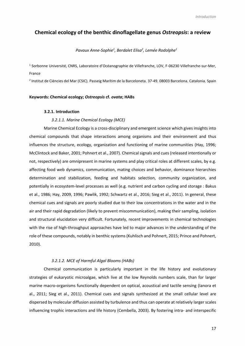
Chemical ecology of the benthic dinoflagellate genus Ostreopsis: a review
Pavaux Anne-Sophie1, Berdalet Elisa2, Lemée Rodolphe1
1 Sorbonne Université, CNRS, Laboratoire d’Océanographie de Villefranche, LOV, F-06230 Villefranche-sur-Mer,
France
2 Institut de Ciències del Mar (CSIC), Passeig Marítim de la Barceloneta, 37-49, 08003 Barcelona, Catalonia, Spain
Keywords: Chemical ecology; Ostreopsis cf. ovata; HABs
3.2.1. Introduction
3.2.1.1. Marine Chemical Ecology (MCE)
Marine Chemical Ecology is a cross-disciplinary and emergent science which gives insights into
chemical compounds that shape interactions among organisms and their environment and thus
influences the structure, ecology, organization and functioning of marine communities (Hay, 1996;
McClintock and Baker, 2001; Pohnert et al., 2007). Chemical signals and cues (released intentionally or
not, respectively) are omnipresent in marine systems and play critical roles at different scales, by e.g.
affecting food web dynamics, communication, mating choices and behavior, dominance hierarchies
determination and stabilization, feeding and habitats selection, community organization, and
potentially in ecosystem-level processes as well (e.g. nutrient and carbon cycling and storage : Bakus
et al., 1986; Hay, 2009, 1996; Pawlik, 1992; Schwartz et al., 2016; Sieg et al., 2011). In general, these
chemical cues and signals are poorly studied due to their low concentrations in the water and in the
air and their rapid degradation (likely to prevent miscommunication), making their sampling, isolation
and structural elucidation very difficult. Fortunately, recent improvements in chemical technologies
with the rise of high-throughput approaches have led to major advances in the understanding of the
role of these compounds, notably in benthic systems (Kuhlisch and Pohnert, 2015; Prince and Pohnert,
2010).
3.2.1.2. MCE of Harmful Algal Blooms (HABs)
Chemical communication is particularly important in the life history and evolutionary
strategies of eukaryotic microalgae, which live at the low Reynolds numbers scale, than for larger
marine macro-organisms functionally dependent on optical, acoustical and tactile sensing (Ianora et
al., 2011; Sieg et al., 2011). Chemical cues and signals synthesized at the small cellular level are
dispersed by molecular diffusion assisted by turbulence and thus can operate at relatively larger scales
influencing trophic interactions and life history (Cembella, 2003). By fostering intra- and interspecific

18
cooperation, or as defense mechanisms against predators, parasites or competitors (Poulson et al.,
2009; Prince et al., 2008; Schwartz et al., 2016) chemical cues and signals can play a role in the
formation of blooms of different species. However, the information of chemical signals in the domain
of HABs is very limited mainly due to the inherent technical difficulties of their study. To date, some
studies have focused on allelopathy as an effective mechanism that could reinforce high cell density
blooms but could not be a main or direct cause of HABs (Jonsson et al., 2009). Benthic marine
ecosystems are supposed to be a source of great diversity of chemical signals and cues since the
competition for space and substrate is higher than in pelagic systems (Hay, 2009). Moreover,
compared to the three-dimensional space experienced by planktonic cells, benthic systems are
essentially bi-dimensional, which imposes unique constraints with distinct advantages for the
effectiveness of chemical ecological interactions. The importance of chemical cues and signals in
benthic HAB dynamics has not been yet well studied.
3.2.1.3. Objectives of the review
The objectives of this work were to review the present state of knowledge on the MCE
concerning the biology and dynamics of the benthic dinoflagellate genus Ostreopsis. This genus has
attracted scientific and social interest due to their negative impacts on certain benthic marine fauna,
the production of toxins chemically close to the potent palytoxin (PLTX) associated to dramatic seafood
borne poisonings in tropical areas, and the link of its blooms to mild respiratory and cutaneous
irritations in beach users in temperate latitudes. The diversity, distribution and toxins produced by the
Ostreopsis species are presented first, followed by a synthesis of the knowledge on the health impacts
associated to aerosolized toxins. Next, a review of the literature on the potential chemical interactions
between Ostreopsis spp. and their environment, from virus and bacteria to large herbivorous and
carnivorous macroorganisms is presented. Finally, some key scientific aspects to improve knowledge
on MCE of Ostreopsis spp. are suggested.
3.2.2. The genus Ostreopsis
3.2.2.1. Species and distribution
The genus Ostreopsis and the type species Ostreopsis siamensis were first described a century
ago from the Gulf of Siam (Thailand) by Schmidt (1901). Since then, 9 other species of Ostreopsis have
been identified: O. ovata (Fukuyo, 1981), O. lenticularis (Fukuyo, 1981), O. heptagona (Norris et al.,
1985), O. labens (Faust and Morton, 1995), O. mascarenensis (Quod, 1994), O. belizeanus, O.
caribbeanus and O. marinus (Faust, 1999) and more recently, O. fattorussoi (Accoroni et al., 2016a). A
first characterization of Ostreopsis species is based on morphological approaches combining size,
shape and thecal plates pattern (Hoppenrath et al., 2014). Molecular approaches based on the nuclear

Introduction
19
rDNA internal transcribed spacer region (ITS1 and ITS 2), partial nuclear LSU (D1/D2 domains) and 5.8S
rDNA gene (Penna et al., 2010, 2005) are also key in the identification and determination of phylogeny
link. These methods allowed , for example, the description of Ostreopsis sp. by Tartaglione et al. (2016),
as a new species, O. fattorussoi. With the most recent progresses in chemical analysis, toxin profile can
also become a taxonomic characteristic. Nevertheless, a big part of the taxonomy of the genus
Ostreopsis remains unclear and controversial due to the high morphological and cell size variability
within the different species and the fact that original descriptions lacked genetic analysis. In particular,
there is still a debate regarding the taxonomy of the most widespread species, O. cf. ovata and O. cf.
siamensis since their first description was based on morphological features only.
Initially considered a tropical genus, Ostreopsis is increasing its biogeographic distribution to
temperate latitudes (Parsons et al., 2012 – Table 3). Indeed, Ostreopsis cf. ovata has been isolated in
Australia, Brazil, China (Malacca, South China Sea), Cook Islands, Caribbean, French Polynesia, Hawaii,
Indian Ocean, Japan, Malaysia, New Caledonia, New Zealand, Mediterranean, Venezuela, Vietnam
(Table 3). Other Ostreopsis species that appeared to be circumscribed to narrower regions, are also
found in new localities. For instance, Ostreopsis fattorussoi isolated from the Eastern Mediterranean
could be also growing in the Atlantic coast of the Iberia Peninsula although it has not been
demonstrated yet (Accoroni et al., 2016a). The wide expansion of the genus Ostreopsis is considered
a potential problem on human health and wellbeing (economy, tourism, beach quality) issues due to
the production of PLTX analogues whose mode of action is still not clear (see section 2.2.2.3.4. of this
review).
3.2.2.2. Toxins: Secondary metabolites synthesized by Ostreopsis species
Several species in the Ostreopsis genus are considered potentially harmful to humans and
other marine organisms due to the production of palytoxin (PLTX) like compounds (Table 1). PLTX is
the most potent non-bacterial toxin of biological origin, initially isolated in 1971 from the tropical soft
coral genus Palythoa, and now recognized as 42-OH-PLTX (see revision by Ciminiello et al., 2011 and
references there in Poli et al., 2018).
Progresses in chemical analysis technology, in particular on HR LC-MS/MS and NMR have
revealed the high diversity of PLTX analogues (Table 1) and eight Ostreopsis species have been
described as toxic. Ostreocin D was the first analogue isolated from O. siamensis by Usami et al. (1995)
and other PLTX analogues, namely, ovatoxins, ostreocines, mascarenotoxin or ostreotoxins sensu lato,
have been characterized for O. fattorussoi, O. lenticularis, O. mascarensis, O. cf. ovata and O. siamensis
(Deeds and Schwartz, 2010; Mercado et al., 1994; Parsons et al., 2012). However, toxicity of O.
heptagona, O. labens and O. rhodesiae has only been described using biological assays and toxins
involved in these effects have not already been identified (Table 1). In the case of O. cf. ovata, up to

20
12 ovatoxins, from a to k and the isobaric PLTX (isoPLTX, Garcia-Altares et al., 2015) have been
identified among the different isolated strains, and the toxinologic profile constitutes a taxonomic
characteristic (e.g. Accoroni et al., 2016). A main limitation to ascertain the health impacts risks
associated to Ostreopsis blooms is the lack of standards for the variety of toxins, all them complex and
with high molecular weight (molecular formulas varying between C129H223N3O55 and C131H227N3O52).
Phycotoxins in general and the palytoxin group as well, are considered secondary metabolites
and their physiological role in the cells is unknown. Environmental factors such as temperature,
salinity, light or nutrients, but also the characteristics of the strains (isolation site, age and growth
phase of the strain in culture) may influence toxin production (Ciminiello et al., 2012a; Granéli et al.,
2011; Pezzolesi et al., 2012; Pezzolesi et al., 2014; Scalco et al., 2012; Vanucci et al., 2012a). However,
it has been suggested that other microorganisms co-occurring along the Ostreopsis blooms could be
involved in the toxin production as well. Tosteson et al. (1989) and Ashton et al. (2003) found, in
cultures, that bacteria associated with the surfaces or the extracellular matrices of O. lenticularis were
correlated with the high dinoflagellate toxicity during the stationary phase of the cultures. Biological
assays and analytical chemistry analysis are both used for toxin presence detection and/or
quantification. Mouse Bioassay (prohibited in Europe since 2005 for ethical reasons), Haemolysis
Neutralization Assay (HNA) or Artemia bioassay have been long recognized for toxicity quantification
(Bignami, 1993; Botana, 2014; Delaney, 1984). Advantages given by analytical chemistry analysis (i.e.
LC-MS/MS) are the absence of restrictions in terms of reproducibility, specificity, precision and
consistency. Unfortunately, this methodology relies on very expensive technical equipment, it requires
high qualified personnel skills and it produces significant chemical waste (it is not environmentally
friendly).
3.2.2.3. Health problems in humans: an indication of the Ostreopsis Chemical Ecology
As mentioned in the introduction, chemical ecology is focused the communication among
organisms mediated by chemical compounds. Somehow, health impacts in humans due to exposure
to PLTX analogues constitute an indication of Ostreopsis chemical ecology. Health impacts can occur:
i) after consumption of potentially PLTX contaminated seafood, ii) from direct contact with zoanthids
in the natural environment or in home aquaria, or iii) after direct exposure to seawater and/or aerosol
during Ostreopsis blooms. The characterization of human poisonings potentially due to exposure to
PLTX analogues was reviewed by Tubaro et al. (2011) and it is briefly presented next. The effects of
Ostreopsis spp. in different marine organisms have also been reported and are presented in sections
2.2.3.

Introduction
21
3.2.2.3.1 Seafood borne PLTX poisonings
Severe illness and even lethal fatalities after consumption of potentially PLTX contaminated
seafood have been scarcely reported in tropical and subtropical latitudes since the 1970s so far (see
Tables 1, 2 and 3 in Tubaro et al. (2011). Among them, only a few cases were confirmed through direct
PLTX analogues detection in seafood leftovers. In many other cases, screening tests on seafood
samples collected or purchased after or before the poisoning episode combined or not with clinical
observations and symptoms, concluded that consumption of PLTX contaminated seafood was the
causative reason of the health disorders. Symptoms include nausea, tiredness, diarrhea, vomiting,
dizziness, numbness of the extremities, muscle cramps, paresthesia, restless-ness, respiratory distress
and bradichardia. In dramatic cases, patients died after 15h to 4 days following seafood poisonings,
even after hospitalization. Unusual metallic taste of the seafood is one of the common signs noted by
potential PLTX intoxication.
In the Mediterranean coasts, the proliferation of O. cf. ovata and O. cf. siamensis since the end
of the 21th century raised concern about the potential risk of seafood intoxication. Biré et al. (2013)
and Brissard et al. (2014) reported the presence of toxin content in macrofauna (mainly sea urchin,
rock shell, crab and flathead mullet) sampled during O. cf. ovata blooms. OVTX-a was the most
abundant toxin detected, and in some cases PLTX equivalents accounted on average for 223 to 392.2
µg·kg-1 of whole flesh, exceeding the threshold of 30 µg·kg-1 in shellfish recommended by the European
Food Safety Authority (EFSA). The key and unsolved questions are whether Ostreopsis was the ultimate
organism synthesizing the PLTX-like compounds that contaminated the seafood and, how toxins can
be transferred through the food webs. Fortunately, until now, seafood intoxications due to PLTX
analogues have not been occurred in the Mediterranean zone yet.
3.2.2.3.2 Health impacts by direct contact with zoanthids in the natural
environment or in home aquaria
Zoanthids have become common in hobby aquarists worldwide. Deeds and Schwartz (2010)
described the health impacts on a person that inhaled a potent irritative compound released after
pouring boiling water or bleach in aquaria with the aim to eliminate a colony of zoanthids. Within 20
min after exposure, the man experienced rhinorrhea and coughing, and difficulty breathing and light
headedness which progressed to severe fits of coughing and chest pain 4h later. At the local hospital
he was administered an anti-inflammatory corticosteroid and pain medication, an inhaled steroid
treatment and cough suppressant for one months. A follow-up examination by a pulmonary specialist
2 weeks post exposure diagnosed the patient with asthma-like symptoms of bronchial inflammation
and bronchoconstriction. Similar cases have been reported in the last years (see Tables 4 and 5 in
Tubaro et al. (2011), one last case affecting a whole family, with 3 little kids in the UK in August 2019.
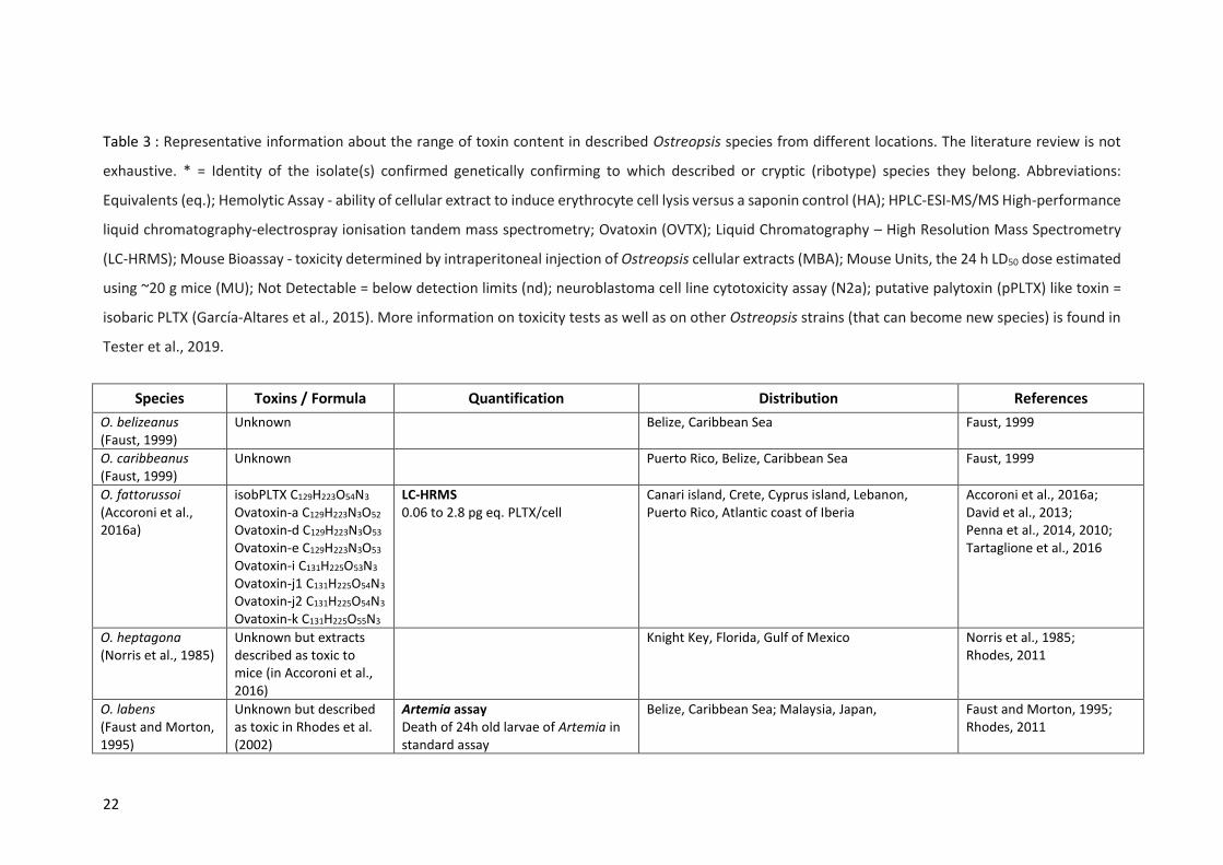
22
Table 3 : Representative information about the range of toxin content in described Ostreopsis species from different locations. The literature review is not
exhaustive. * = Identity of the isolate(s) confirmed genetically confirming to which described or cryptic (ribotype) species they belong. Abbreviations:
Equivalents (eq.); Hemolytic Assay - ability of cellular extract to induce erythrocyte cell lysis versus a saponin control (HA); HPLC‐ESI‐MS/MS High-performance
liquid chromatography-electrospray ionisation tandem mass spectrometry; Ovatoxin (OVTX); Liquid Chromatography – High Resolution Mass Spectrometry
(LC-HRMS); Mouse Bioassay - toxicity determined by intraperitoneal injection of Ostreopsis cellular extracts (MBA); Mouse Units, the 24 h LD50 dose estimated
using ~20 g mice (MU); Not Detectable = below detection limits (nd); neuroblastoma cell line cytotoxicity assay (N2a); putative palytoxin (pPLTX) like toxin =
isobaric PLTX (García-Altares et al., 2015). More information on toxicity tests as well as on other Ostreopsis strains (that can become new species) is found in
Tester et al., 2019.
Species Toxins / Formula Quantification Distribution References
O. belizeanus (Faust, 1999)
Unknown Belize, Caribbean Sea Faust, 1999
O. caribbeanus (Faust, 1999)
Unknown Puerto Rico, Belize, Caribbean Sea Faust, 1999
O. fattorussoi (Accoroni et al., 2016a)
isobPLTX C129H223O54N3 Ovatoxin-a C129H223N3O52
Ovatoxin-d C129H223N3O53
Ovatoxin-e C129H223N3O53 Ovatoxin-i C131H225O53N3
Ovatoxin-j1 C131H225O54N3
Ovatoxin-j2 C131H225O54N3
Ovatoxin-k C131H225O55N3
LC‐HRMS
0.06 to 2.8 pg eq. PLTX/cell
Canari island, Crete, Cyprus island, Lebanon, Puerto Rico, Atlantic coast of Iberia
Accoroni et al., 2016a; David et al., 2013; Penna et al., 2014, 2010; Tartaglione et al., 2016
O. heptagona (Norris et al., 1985)
Unknown but extracts described as toxic to mice (in Accoroni et al., 2016)
Knight Key, Florida, Gulf of Mexico Norris et al., 1985; Rhodes, 2011
O. labens (Faust and Morton, 1995)
Unknown but described as toxic in Rhodes et al. (2002)
Artemia assay Death of 24h old larvae of Artemia in standard assay
Belize, Caribbean Sea; Malaysia, Japan,
Faust and Morton, 1995; Rhodes, 2011

Introduction
23
O. lenticularis (Fukuyo, 1981) Includes the cryptic species Ostreopsis sp. 5 in Chomérat et al., 2019
Ostreotoxin 1 Ostreotoxin 3
N2a analysis EC50(PLTX) = 1191 ± 175 pg.mL−1 (O− condition) LC-UV-MS/MS analysis Under detection levels
Australia, Caribbean, French Polynesia, Indian Ocean, Malaysia, Mexican Pacific, New Caledonia, New Zealand, Philippines, Venezuela, Vietnam
Ashton et al., 2003; Carnicer et al., 2015b*; Chomérat et al., 2019*; Mercado et al., 1995; Sato et al., 2011; Tosteson et al., 1989
O. marinus (Faust, 1999)
Unknown No pPLTX activity by HA using sheep erythrocytes
Caribbean, Indian Ocean, Vietnam Carnicer et al., 2015a*; Faust, 1999
O. mascarenensis (Quod, 1994)
Mascarenotoxin‐A C127H221N3O50
Mascarenotoxin‐B nd
HPLC‐ESI‐MS/MS: 0.04 pg eq.PLTX/cell Mouse bioassay: LD50 = 900 μg.kg-1
(intraperitoneal injection)
Rodrigues and Reunion Islands, Indian Ocean Lenoir et al., 2004; Ramos and Vasconcelos, 2010
O. ovata (Fukuyo, 1981)
p-PLTX C129H223N3O54 Ovatoxin-a C129H223N3O52 Ovatoxin-b C131H227N3O53
Ovatoxin-c C131H227N3O54
Ovatoxin-d C129H223N3O53
Ovatoxin-e C129H223N3O53
Ovatoxin-f C131H227N3O52
Ovatoxin-g C129H223N3O51
Ovatoxin-h C129H225N3O51
Ostreol A C67H112N2O23
Mascarenotoxin-a C127H221N3O50
Mascarenotoxin-c C129H221N3O51
LC‐HRMS
pPLTX: 0.1 to 24.8 pg eq. PLTX/cell OVTX a: 0.065 to 171 pg eq. PLTX/cell OVTX b: 2 to 205 pg eq. PLTX/cell OVTX c: 0.7 to 37 pg eq. PLTX/cell OVTX d+e: 2 to 80 pg eq. PLTX/cell OVTX f: ND - 17 pg eq. PLTX/cell OVTX g: 0.35 to 24.8 pg eq. PLTX/cell
Australia, Brazil, China, Cook Islands, Caribbean, French Polynesia, Hawaii, Indian Ocean, Japan, Korea, Malaysia, New Caledonia, New Zealand, Mediterranean, Eastern Russia, Tasmania, Venezuela, Vietnam
Ben Gharbia et al., 2017; Brissard et al., 2015, 2014; Ciminiello et al., 2012, 2011, 2008, 2006; Garcia-Altares et al., 2015; Giussani et al., 2015; Granéli et al., 2011*; Guerrini et al., 2010*; Honsell et al., 2013; Hwang et al., 2013; Nascimento et al., 2012b*; Penna et al., 2005; Pezzolesi et al., 2012*; Rossi et al., 2010; Sechet et al., 2012*; Selina and Levchenko, 2011; Tawong et al., 2014*; Vanucci et al., 2012b
O. rhodesiae (Verma et al., 2016)
Unknown but described as toxic in Verma et al. (2016)
LC-MS/MS: No PLTX analogs Fish gill cell line assay for toxicity: ≥98.5% decrease of gill cell line RTgill-W1 viability
Heron Reef Lagoon, southern Great Barrier Reef, Coral Sea, Australia
Verma et al., 2016

24
O. siamensis (Schmidt, 1901)
Ostreocin A C127H219N3O54
Ostreocin‐D C127H219N3O53
Ostreocin-E1 C127H217N3O52
Ostreocin‐B C127H219N3O53
pPLTX
Haemolysis Neutralization Assay 0.3 pg PLTX eq/cells Mouse bioassay: LD50 = 0.75 μg.kg-1
(intraperitoneal injection) Mediterranean strains = sub-fg levels of PLTX/cells and no toxicity by MBA Japanese Strains contained Ostreocin-b + Ostreocin-d + pPLTX (nd–0.8 pg eq. PLTX/cell). Total: nd-16 pg PLTX eq/cells
Eastern Atlantic coast, Australia, Hawai, Japan, Madagascar, Mediterranean Sea, Mexico, New Zeland, Reunion Island, Thailand, Venezuela, Vietnam
Cagide et al., 2009; Ciminiello et al., 2013; Meunier et al., 1997; Ramos and Vasconcelos, 2010; Rhodes et al., 2002, 2000; Terajima et al., 2019, 2018; Tichadou et al., 2010; Tindall and Miller, 1988; Ukena et al., 2001; Verma et al., 2016*
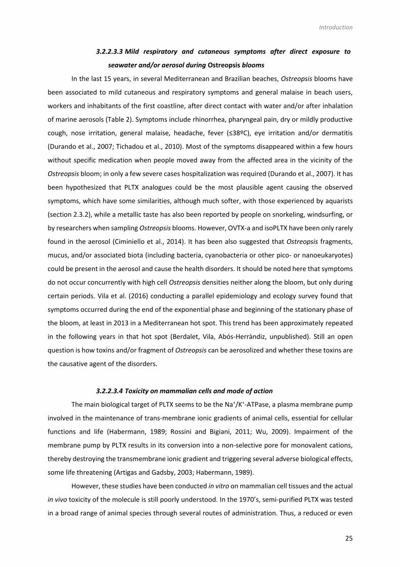
Introduction
25
3.2.2.3.3 Mild respiratory and cutaneous symptoms after direct exposure to
seawater and/or aerosol during Ostreopsis blooms
In the last 15 years, in several Mediterranean and Brazilian beaches, Ostreopsis blooms have
been associated to mild cutaneous and respiratory symptoms and general malaise in beach users,
workers and inhabitants of the first coastline, after direct contact with water and/or after inhalation
of marine aerosols (Table 2). Symptoms include rhinorrhea, pharyngeal pain, dry or mildly productive
cough, nose irritation, general malaise, headache, fever (≤38ºC), eye irritation and/or dermatitis
(Durando et al., 2007; Tichadou et al., 2010). Most of the symptoms disappeared within a few hours
without specific medication when people moved away from the affected area in the vicinity of the
Ostreopsis bloom; in only a few severe cases hospitalization was required (Durando et al., 2007). It has
been hypothesized that PLTX analogues could be the most plausible agent causing the observed
symptoms, which have some similarities, although much softer, with those experienced by aquarists
(section 2.3.2), while a metallic taste has also been reported by people on snorkeling, windsurfing, or
by researchers when sampling Ostreopsis blooms. However, OVTX-a and isoPLTX have been only rarely
found in the aerosol (Ciminiello et al., 2014). It has been also suggested that Ostreopsis fragments,
mucus, and/or associated biota (including bacteria, cyanobacteria or other pico- or nanoeukaryotes)
could be present in the aerosol and cause the health disorders. It should be noted here that symptoms
do not occur concurrently with high cell Ostreopsis densities neither along the bloom, but only during
certain periods. Vila et al. (2016) conducting a parallel epidemiology and ecology survey found that
symptoms occurred during the end of the exponential phase and beginning of the stationary phase of
the bloom, at least in 2013 in a Mediterranean hot spot. This trend has been approximately repeated
in the following years in that hot spot (Berdalet, Vila, Abós-Herràndiz, unpublished). Still an open
question is how toxins and/or fragment of Ostreopsis can be aerosolized and whether these toxins are
the causative agent of the disorders.
3.2.2.3.4 Toxicity on mammalian cells and mode of action
The main biological target of PLTX seems to be the Na+/K+-ATPase, a plasma membrane pump
involved in the maintenance of trans-membrane ionic gradients of animal cells, essential for cellular
functions and life (Habermann, 1989; Rossini and Bigiani, 2011; Wu, 2009). Impairment of the
membrane pump by PLTX results in its conversion into a non-selective pore for monovalent cations,
thereby destroying the transmembrane ionic gradient and triggering several adverse biological effects,
some life threatening (Artigas and Gadsby, 2003; Habermann, 1989).
However, these studies have been conducted in vitro on mammalian cell tissues and the actual
in vivo toxicity of the molecule is still poorly understood. In the 1970’s, semi-purified PLTX was tested
in a broad range of animal species through several routes of administration. Thus, a reduced or even
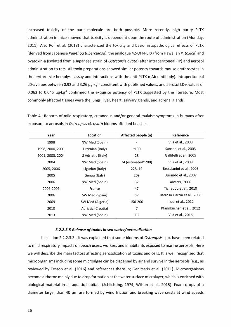
26
increased toxicity of the pure molecule are both possible. More recently, high purity PLTX
administration in mice showed that toxicity is dependent upon the route of administration (Munday,
2011). Also Poli et al. (2018) characterized the toxicity and basic histopathological effects of PLTX
(derived from Japanese Palythoa tuberculosa), the analogue 42-OH-PLTX (from Hawaiian P. toxica) and
ovatoxin-a (isolated from a Japanese strain of Ostreopsis ovata) after intraperitoneal (IP) and aerosol
administration to rats. All toxin preparations showed similar potency towards mouse erythrocytes in
the erythrocyte hemolysis assay and interactions with the anti-PLTX mAb (antibody). Intraperitoneal
LD50 values between 0.92 and 3.26 μg·kg-1 consistent with published values, and aerosol LD50 values of
0.063 to 0.045 μg·kg-1 confirmed the exquisite potency of PLTX suggested by the literature. Most
commonly affected tissues were the lungs, liver, heart, salivary glands, and adrenal glands.
Table 4 : Reports of mild respiratory, cutaneous and/or general malaise symptoms in humans after
exposure to aerosols in Ostreopsis cf. ovata blooms affected beaches.
Year Location Affected people (n) Reference
1998 NW Med (Spain) - Vila et al., 2008
1998, 2000, 2001 Tirrenian (Italy) ~100 Sansoni et al., 2003
2001, 2003, 2004 S Adriatic (Italy) 28 Gallitelli et al., 2005
2004 NW Med (Spain) 74 (estimated~200) Vila et al., 2008
2005, 2006 Ligurian (Italy) 228, 19 Brescianini et al., 2006
2005 Genoa (Italy) 209 Durando et al., 2007
2006 NW Med (Spain) 37 Àlvarez, 2006
2006-2009 France 47 Tichadou et al., 2010
2006 SW Med (Spain) 57 Barroso García et al., 2008
2009 SW Med (Algeria) 150-200 Illoul et al., 2012
2010 Adriatic (Croatia) 7 Pfannkuchen et al., 2012
2013 NW Med (Spain) 13 Vila et al., 2016
3.2.2.3.5 Release of toxins in sea water/aerosolization
In section 2.2.2.3.3., it was explained that some blooms of Ostreopsis spp. have been related
to mild respiratory impacts on beach users, workers and inhabitants exposed to marine aerosols. Here
we will describe the main factors affecting aerosolization of toxins and cells. It is well recognized that
microorganisms including some microalgae can be dispersed by air and survive in the aerosols (e.g., as
reviewed by Tesson et al. (2016) and references there in; Genitsaris et al. (2011). Microorganisms
become airborne mainly due to drop formation at the water surface microlayer, which is enriched with
biological material in all aquatic habitats (Schlichting, 1974; Wilson et al., 2015). Foam drops of a
diameter larger than 40 µm are formed by wind friction and breaking wave crests at wind speeds

Introduction
27
exceeding 7 to 11 m·s-1. Bubble bursting due to waves, rainfall, boat traffic, or supersaturation of gases
in the water produces film drops (1 µm to 10 µm diameter), projected in various directions, and
vertically emitted jet drops (6 µ to 100 µm diameter). Recreational activities (i.e., waterskiing, using
personal watercraft, swimming, or wading) can also generate aerosols. Once emitted, airborne
turbulent kinetic energy drags the microorganisms further up into the atmosphere.
The few available studies indicate that microalgae are found at very low concentrations (10-4
to 104 cells per m-3 (Mayol et al., 2014) and with heterogeneous distributions, which requires high-
volume sampling technology. High-volume sampling on filters or agar plates can be achieved by
different instruments, but high-volume sampling into liquids is more complicated, as high airflows may
lead to evaporation and reduce collection efficiency. Once collected combination of microscopy and
molecular tools are contributing to the identification of the airborne microalga. The main microalgal
groups found in bioaerosols are chlorophytes ("green algae"), bacillariophytes (diatoms, mainly small
Nitzschia) and cyanobacteria. Airborne microalgae are recognized as allergens and antigens. They are
the cause of severe medical issues, including respiratory allergies (e.g., hay fevers), asthmatic attacks,
dermatitis and skin lesions, rhinitis, and disturbances in lymphatic systems or vital organs (e.g.,
protothecosis, see review in Ashok and Gupta, 1998). In addition, certain aerosolized algal toxins are
the causes of further human illnesses, namely, microcystins produced by cyanobacteria, brevetoxins
synthesized by Karenia brevis and ovatoxins and palytoxin analogues generated by Ostreopsis species.
So far, the detection of Ostreopsis cf. ovata and its related toxins in the aerosol have been conducted
by Casabianca et al. (2013) and (Ciminiello et al., 2014). Casabianca et al. (2013) used air pump
samplers (CAV-A/mb, Barcelona, Spain) equipped with 15 cm diameter QM-A quartz filters (Whatman,
Maidstone, UK). Two air samplers located at 0.5 m and 3 m above sea level, respectively, pumped air
continuously for 3 days during an Ostreopsis cf. ovata bloom at a flow rate of 30 m3·h-1. Filters were
replaced every 6 or 7 h and were divided in two portions to analyze, respectively, the concentration of
Ostreopsis cell concentrations by qPCR and toxins by the hemolytic assay and by liquid chromatography
with fluorescence detection (LC-FLD). Ostreopsis cells were detected in the aerosol by qPCR, exhibiting
a marked variability along the bloom although not always directly related to the concentrations of the
epiphytic Ostreopsis in the water; the authors noted that wind and other environmental factors could
have affected these findings. Epiphytic Ostreopsis contained ca. 1.2 pg total PLTX analogues per cell
but toxins were not detected in the aerosol. Different technical aspects (e.g. toxin binding to glass
tubes, not efficient retention by the filters) any need to be overcome. Ciminiello et al. (2014) also
employed AirCube COM2 (Analitica Strumenti srl., Italy) equipped with 47 mm diameter glass/quartz
microfiber filters (Whatman, Maidstone, UK) and SAS PCR (VWR International (PBI, Italy). In this case,
air and a collecting fluid (50 mL distilled water in 2009) flow together through a nozzle were poured
into a vessel with a coil; the collecting fluid was circulated continuously to prolong the contact time

28
with aerosols and to enrich itself in toxins and/or cells. Both air samplers were located 10 m from the
seashore and worked continuously for 1.5−6 h. Air flow speed was 10 L.min-1 in 2009 and 30 L.min-1 in
2010. The presence of ovatoxins in the aerosol at levels of 2.4 pg of ovatoxins per liter of air was
confirmed for the first time but palytoxins by inhalation exposure toxicological data were not available.
Overall, the presence of both cells and toxins in the aerosols provided by these studies opened new
possibilities to increase the understanding of the Ostreopsis-related respiratory syndrome.
3.2.3. MCE of Ostreopsis spp
3.2.3.1. Introduction
As described in the former section, toxins, the secondary metabolites synthesized by
Ostreopsis, may be involved in some human health impacts associated to the blooms of this benthic
dinoflagellate. Here, the described and potential impacts on different marine organisms and
environments as part of the chemical ecology of Ostreopsis are reviewed.
3.2.3.2. Interactions with bacteria, viruses and parasites
Interactions between HAB species and bacteria remain still poorly studied although these
connections may play important roles in the biology and the ecology of these organisms. Bacteria may
interact with algae within the phycosphere (Bell and Mitchell, 1972) as free-living bacteria, attached
to the algal cell surface or occurring as intracellular symbionts. Algae-bacteria interactions can be
synergistic, mutualistic, as commensalism or symbiotic (e.g., by producing cytokinins - phytohormones,
Maruyama et al., 2008). Some vitamins and ion chelators produced by bacteria are involved in nutrient
recycling facilitating nutrient incorporation by algae (Jasti et al., 2005), while, in return, bacteria use
microalgae exudates (Sarmento and Gasol, 2012). Furthermore, competition, parasitism or antagonist
interactions also occur. Bacteria could interfere microalgal life cycle (Sawayama et al., 1993) and
growth of some HAB species (Doucette, 1995; Fukami et al., 1992; Hare et al., 2005; Kim et al., 2009).
Bacterial exopolymers can increase the tendency of cells to flocculate and enhance sinking rates and
degradation of decaying (harmful and innocuous) blooms (Decho, 1990). In turn, algae could also
control bacteria growth by the production of antibiotic (Trick et al., 2011), or hydroxyl radicals (Oda et
al., 1992) and other selected anti-bacterial growth substance (Bell et al., 1974).
These interactions exhibit spatio-temporal scales variability (Doucette, 1995; Mayali et al.,
2011). Phylogenetically diverse bacterial groups with different functional roles could modulate
phytoplankton and HABs bloom dynamics. While some bacteria could microalgal cells during the
initiation and exponential phases of the blooms, antagonist or parasitic bacteria would proliferate
before the declining period and saprophytic or scavenger bacteria will occur after bloom termination
when the availability of phytodetritus is high.

Introduction
29
Bacterial community associated to Ostreopsis species appear to be species and even clone
specific (Tosteson et al., 1989) and vary with the bloom phase, and likely, with the habitat.
Alphaproteobacteria (83 % relative abundance - genera Ruegeria, Jannaschia and Erythrobacter-
Gammaproteobacteria (13%) and Flavobacteria (7%) were identified during the early Ostreopsis cf.
ovata bloom period, while Alphaproteobactria (66%) and Flavobacteria (19%) were predominant in
peak bloom phases (Guidi et al., 2018; pratoci et al., 2016). Bacterial communities associated to
Ostreopsis lenticularis included single species belonging to the Cytophaga-Flavobacter-Bacteroides
complex (Pérez-Guzmán et al., 2008). Bacteria-algae interactions can be very strong given that some
microalgae are unable to grow in axenic conditions in culture confirming the predominant role of
bacteria in algal cycle. This has also been observed in some Ostreopsis cultures.
Some studies indicate that bacteria could directly or indirectly modulate production and
degradation of microalgals toxins (Albinsson et al., 2014) although the results appear study- and
species- dependent and contradictory. While associated bacteria were linked to the production of
paralytic shellfish toxin by Alexandrium spp. (Gallacher et al., 1997), higher toxin levels were found in
bacteria-free than in non-axenic cultures of Alexandrium tamarense (Hold et al., 2001) and A.
lusitanicum (Wang et al., 2004). Ashton et al. (2003) reported a toxicity loss of Ostreopsis lenticularis
when exposed to elevated temperature and explained it by the reduction of Ostreopsis associated
Pseudomonas and Alteromonas. Axenic cultures of (Vanucci et al., 2012a) were also characterized by
lower toxin content in late stationary phase compared to non-axenic cultures. Ostreopsis cf. ovata cell
concentration along a bloom was correlated with the abundance of planktonic, free-living Vibrio
(Bellés-Garulera et al., 2016) and a high proportion of Vibrio were found attached to particles,
suggesting that Ostreopsis bloom could provide biological colonizable surfaces (fragments of thecae or
mucilage) for bacteria. The study suggested that bacteria could be involved in wound infections
reported by beach users during Ostreopsis blooms. In fact, Ostreopsis blooms were reported in heavily
antropogenically impacted environments (e.g. Cencini, 1998).
The role of the mucus produced by Ostreopsis (a mucilaginous matrix, described as a network
of trichocysts embedded with acid polysaccharides filaments by Honsell et al., 2013) and associated
bacteria in the respiratory irritations associated to the microalgal bloom is not clarified yet.
Parasites infection may play critical roles in the regulation of HABs. It has been already
described for the planktonic toxic dinoflagellate Alexandrium fundyense which present an up-
regulation of genes associated to defense against the parasite Amoebophyra and thus controlling host
mortality (Lu et al., 2016, 2014). The algal metabolite dimethylsulphide (DMS) was identified as the
density-dependent cue of the presence of potential dinoflagellate host (Alexandrium minutum,
Amphidinium carterae and Karlodinium veneficum) that triggers zoospore activation and release from
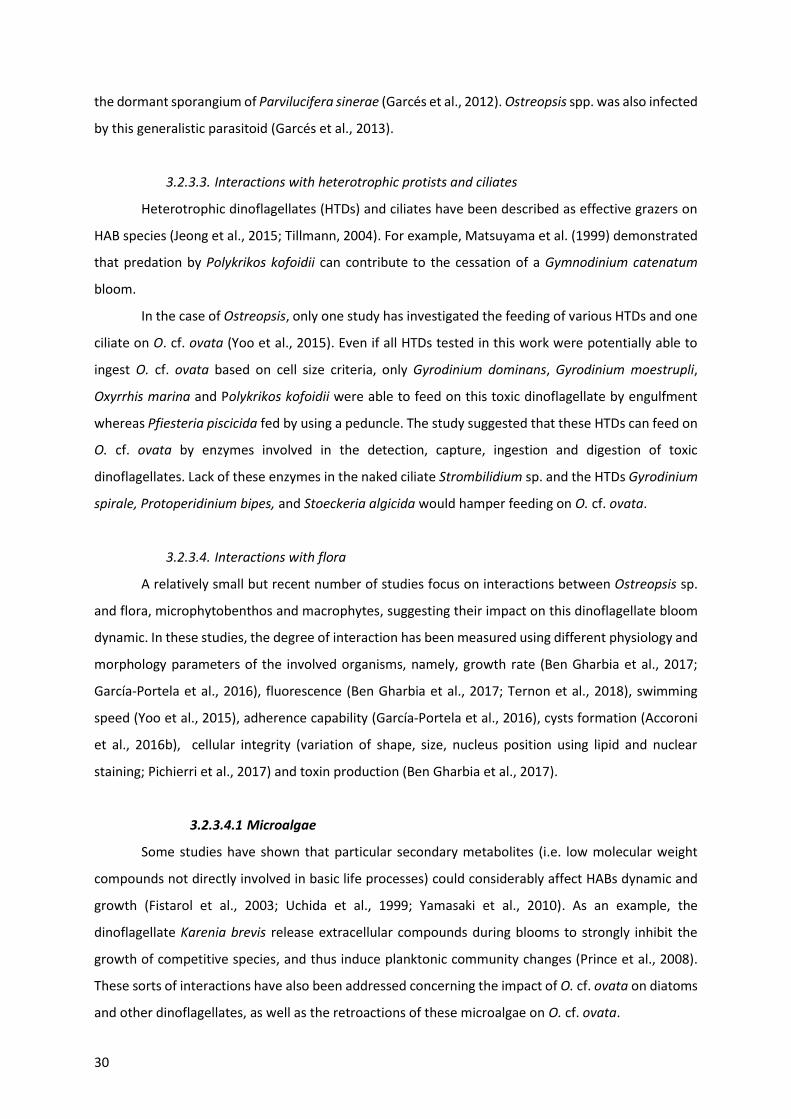
30
the dormant sporangium of Parvilucifera sinerae (Garcés et al., 2012). Ostreopsis spp. was also infected
by this generalistic parasitoid (Garcés et al., 2013).
3.2.3.3. Interactions with heterotrophic protists and ciliates
Heterotrophic dinoflagellates (HTDs) and ciliates have been described as effective grazers on
HAB species (Jeong et al., 2015; Tillmann, 2004). For example, Matsuyama et al. (1999) demonstrated
that predation by Polykrikos kofoidii can contribute to the cessation of a Gymnodinium catenatum
bloom.
In the case of Ostreopsis, only one study has investigated the feeding of various HTDs and one
ciliate on O. cf. ovata (Yoo et al., 2015). Even if all HTDs tested in this work were potentially able to
ingest O. cf. ovata based on cell size criteria, only Gyrodinium dominans, Gyrodinium moestrupli,
Oxyrrhis marina and Polykrikos kofoidii were able to feed on this toxic dinoflagellate by engulfment
whereas Pfiesteria piscicida fed by using a peduncle. The study suggested that these HTDs can feed on
O. cf. ovata by enzymes involved in the detection, capture, ingestion and digestion of toxic
dinoflagellates. Lack of these enzymes in the naked ciliate Strombilidium sp. and the HTDs Gyrodinium
spirale, Protoperidinium bipes, and Stoeckeria algicida would hamper feeding on O. cf. ovata.
3.2.3.4. Interactions with flora
A relatively small but recent number of studies focus on interactions between Ostreopsis sp.
and flora, microphytobenthos and macrophytes, suggesting their impact on this dinoflagellate bloom
dynamic. In these studies, the degree of interaction has been measured using different physiology and
morphology parameters of the involved organisms, namely, growth rate (Ben Gharbia et al., 2017;
García-Portela et al., 2016), fluorescence (Ben Gharbia et al., 2017; Ternon et al., 2018), swimming
speed (Yoo et al., 2015), adherence capability (García-Portela et al., 2016), cysts formation (Accoroni
et al., 2016b), cellular integrity (variation of shape, size, nucleus position using lipid and nuclear
staining; Pichierri et al., 2017) and toxin production (Ben Gharbia et al., 2017).
3.2.3.4.1 Microalgae
Some studies have shown that particular secondary metabolites (i.e. low molecular weight
compounds not directly involved in basic life processes) could considerably affect HABs dynamic and
growth (Fistarol et al., 2003; Uchida et al., 1999; Yamasaki et al., 2010). As an example, the
dinoflagellate Karenia brevis release extracellular compounds during blooms to strongly inhibit the
growth of competitive species, and thus induce planktonic community changes (Prince et al., 2008).
These sorts of interactions have also been addressed concerning the impact of O. cf. ovata on diatoms
and other dinoflagellates, as well as the retroactions of these microalgae on O. cf. ovata.

Introduction
31
A strong inhibitory effect of O. cf. ovata on microalgae was observed in the in situ study of
Accoroni et al. (2016b). In particular, the diversity index decreased significantly and motile diatoms
increased during O. cf. ovata bloom. Motile life diatom phase would be favored by the Ostreopsis
mucous matrix, illustrating the importance of the mucus in the interactions between O. cf. ovata and
its environment. Giussani et al. (2015) had already documented the toxicity of the mucus in biological
assays. Recently, Ternon et al. (2018) using co-cultures assays (with diatoms) suggested a contact-
toxicity of Ostreopsis cells, probably mediated by the mucus, which could retain the toxins and other
compounds and avoid their dilution in the surrounding environment.
Monti and Cecchin (2012) observed weak effects on the growth of the dinoflagellates Coolia
monotis and Prorocentrum minimum when exposed to filtrates of O. cf. ovata cultures obtained during
its exponential phase. This highlight the importance of growth stages considered in the experiments.
Indeed, variation of the toxin content among the growth stage has already been described by different
studies (Brissard et al., 2014; Pezzolesi et al., 2016; Vanucci et al., 2012b, Pavaux et al., 2019 - In prep.).
García-Portela et al. (2016), using mixed cultures of different Ostreopsis sp. strains and toxic and non-
toxic dinoflagellates, demonstrated differences of toxicity among the Ostreopsis strains. Indeed, O. cf.
ovata was more toxic than O. sp. «Lanzarote type», highlighting the necessity to study the intraspecific
variability of the interactions of Ostreopsis sp. with other organisms. The chemical compounds
involved in the negative interaction of Ostreopsis and other microalgae (specially diatoms) are poorly
unknown and secondary metabolites other than the OVTXs group could be at play, as suggested by
Ternon et al. (2018).
In turn, planktonic and benthic diatoms can affect O. cf. ovata physiology, morphology and
growth. An inhibition of O. cf. ovata growth rate by 57 % and 78 % was reported in cultures exposed
to, respectively, filtrates of the planktonic diatoms Skeletonema marinoi and Thalassiosira sp. (Pichierri
et al., 2017). Similar inhibition of growth, deleterious effects and genotoxic damages were also
observed on Ostreopsis cultures exposed to filtrates of other benthic diatoms (e.g., Tabularia affinis,
Proschkinia complanatoides and Navicula sp.). These studies suggested the production of allelopathic
compounds by all tested diatoms, especially released after cell destruction (Pohnert, 2000). These
diatoms are well known to produce a large family of polyunsaturated aldehydes (PUAs - Wichard et
al., 2005). Pichierri et al. (2017) found that exposure to a range of PUAs (2E,4E-decadienal, 2E,4E-
octadienal and 2E,4E-heptadienal) concentrations (from 3 to 36 µmol.L-1) caused O. cf. ovata growth
inhibition, decrease in photosynthesis efficiency, increase of abnormal cells (motionless, decrease in
dimensions, contraction of cytoplasm and formation of abnormal vesicle-like structure) and a decrease
in cell integrity (chromatin dispersion, lack of autofluorescence of the chlorophyll and larger lipid
bodies).

32
To conclude, there are still too few studies to understand interactions between Ostreopsis spp.
and other microalgae and they are mainly focused on O. cf. ovata. However, a strong strain/species
dependent effect of Ostreopsis on co-occuring community was demonstrated. Moreover, the
predominant role of the mucilaginous matrix, as “concentrator of secondary metabolites” was
suggested.
3.2.3.4.2 Macroalgae
Biotic substrate preferences of Ostreopsis species has already been demonstrated in several
studies (e.g. Totti et al., 2010). This preference of substrate by Ostreopsis sp. could be explained in
part, by the macroalgal thallus surface and morphology: complex, three dimensional flexible and
branched thalli favored cell attachment (Accoroni et al., 2016a; Cohu et al., 2013; Meroni et al., 2018).
These thalli would also be more suitable for O. cf. ovata (Accoroni et al., 2012; Vila et al., 2001)
resistance to wave action. Ostreopsis cells might be removed more easily by wave action on low
ramified thalli compared to complex thalli. Higher abundances of Ostreopsis cf. ovata were also found
on the turf dominated community i.e. in a complex intricate matrix of small Corallinales, Ceramiales
and other filamentous algae (Meroni et al., 2018) as previously reported in Vila et al. (2001) for
Ellissolandia elongata and in Blanfuné et al. (2015) for Jania rubens.
The production of allelopathic compounds by macroalgae could also explain substrate
preferences by Ostreopsis sp. Macroalgae are known to produce a large amount of secondary
metabolites sometimes used in biotechnology as antibiofouling coatings (Hellio et al., 2002) or as a
way to control HABs (Hu and Hong, 2008; Tang et al., 2015). Thus, it is reasonable to hypothesize that
these metabolites could selectively determine Ostreopsis spp. substrate preference. Rhodymenia
pseudopalmata (Florideophyceae) have little effects on Ostreopsis growth and only in powder
(Accoroni et al., 2015b). The seagrasses Zostera noltei and Cymodocea nodosa have also a low impact
on Ostreopsis cf. ovata growth although Zosteria noltei have been described as a producer of phenolic
acids inhibiting the growth of Alexandrium catenella (Laabir et al., 2013). Ulva rigida (Ulvophyceae)
and Dyctyota dichotoma (Phaeophyceae) had a strong effect on O. cf. ovata by decreasing its growth,
physiology, morphology, toxin production and behaviour with a decrease of cell adherence (Accoroni
et al., 2015b; Ben Gharbia et al., 2017). The potential allelochemical compounds produced by this
macroalgae are still poorly known but could mainly include phenols and polyunsaturated fatty acids
(PUFAs - Ben Gharbia et al., 2017).
In summary, macroalgae morphology is decisive for Ostreopsis spp. colonization but
allelochemicals produced by macroalgae could also play a key role. More studies are needed to
understand which compounds are involved in interactions between O. cf. ovata and macroalgae, to
identify and purify them in order to test them directly in biological assays.
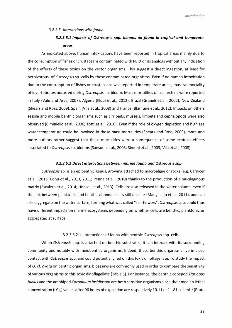
Introduction
33
3.2.3.5. Interactions with fauna
3.2.3.5.1 Impacts of Ostreopsis spp. blooms on fauna in tropical and temperate
areas
As indicated above, human intoxications have been reported in tropical areas mainly due to
the consumption of fishes or crustaceans contaminated with PLTX or its analogs without any indication
of the effects of these toxins on the vector organisms. This suggest a direct ingestion, at least for
herbivorous, of Ostreopsis sp. cells by these contaminated organisms. Even if no human intoxication
due to the consumption of fishes or crustaceans was reported in temperate areas, massive mortality
of invertebrates occurred during Ostreopsis sp. bloom. Mass mortalities of sea urchins were reported
in Italy (Vale and Ares, 2007), Algeria (Illoul et al., 2012), Brazil (Granéli et al., 2002), New Zealand
(Shears and Ross, 2009), Spain (Vila et al., 2008) and France (Blanfuné et al., 2012). Impacts on others
sessile and mobile benthic organisms such as cirripeds, mussels, limpets and cephalopods were also
observed (Ciminiello et al., 2006; Totti et al., 2010). Even if the role of oxygen depletion and high sea
water temperature could be involved in those mass mortalities (Shears and Ross, 2009), more and
more authors rather suggest that these mortalities were a consequence of some ecotoxic effects
associated to Ostreopsis sp. blooms (Sansoni et al., 2003; Simoni et al., 2003; Vila et al., 2008).
3.2.3.5.2 Direct interactions between marine fauna and Ostreopsis spp
Ostreopsis sp. is an epibenthic genus, growing attached to macroalgae or rocks (e.g. Carnicer
et al., 2015; Cohu et al., 2013, 2011; Penna et al., 2010) thanks to the production of a mucilaginous
matrix (Escalera et al., 2014; Honsell et al., 2013). Cells are also released in the water column, even if
the link between planktonic and benthic abundances is still unclear (Mangialajo et al., 2011), and can
also aggregate on the water surface, forming what was called “sea-flowers”. Ostreopsis spp. could thus
have different impacts on marine ecosystems depending on whether cells are benthic, planktonic or
aggregated at surface.
3.2.3.5.2.1. Interactions of fauna with benthic Ostreopsis spp. cells
When Ostreopsis spp. is attached on benthic substrates, it can interact with its surrounding
community and notably with meiobenthic organisms. Indeed, these benthic organisms live in close
contact with Ostreopsis spp. and could potentially fed on this toxic dinoflagellate. To study the impact
of O. cf. ovata on benthic organisms, bioassays are commonly used in order to compare the sensitivity
of various organisms to this toxic dinoflagellate (Table 5). For instance, the benthic copepod Tigriopus
fulvus and the amphipod Corophium insidiosum are both sensitive organisms since their median lethal
concentration (LC50) values after 96 hours of exposition are respectively 10.11 et 11.81 cell.mL-1 (Prato

34
et al., 2011). Other organisms, such as the harpacticoid copepod Sarsamphiascus cf. propinquus, co-
occuring with Ostreopsis cf. ovata in summer, has been demonstrated as very resistant to toxins
produced by Ostreopsis cf. ovata (Pavaux et al., 2019) with LC50(48h) values higher than 20 000
cells.mL-1 so at least 250 times higher than another benthic copepod, Tigriopus fulvus (Faimali et al.,
2012), suggesting potential acclimation phenomenon to the presence of Ostreopsis. However, it
remains complex to compare results of these studies since life stages of the studied organism varies
from one study to another. Parameters used to determine the sensitivity also varies between studies,
with sometimes the determination of LC50 values at 24, 48 or 96 hours. Moreover, the strong influence
on the determination of this parameter (LC50) has been demonstrated by Faimali et al. (2012) since
toxicity appears to increase with the temperature of culture. Indeed, LC50 values of the barnacle
Amphibalanus amphitrite are respectively equal to 1416.95 and 192.26 at 20 and 25°C. This suggests
the need of standardizing the methods to test the toxicity of Ostreopsis spp. on benthic organisms.
Moreover, a reprotoxic effect of O. cf. ovata has been suggested by the significant decrease of
number of nauplii of copepods described in the in situ study of Guidi-Guilvard et al. (2012) and
confirmed by laboratory experiments by Pavaux et al. (2019), with a decrease of fecundity and fertility
ratios of the benthic copepod S. cf. propinquus. These reprotoxic effects lead to changes in benthic
communities.
By forming aggregates that cover macroalgae, Ostreopsis spp. cells are also ingested by
macroherbivores such as sea urchins or herbivorous fish. Numerous studies have already been
quantified PLTX and analogs in these two groups (Amzil et al., 2012; Biré et al., 2013, 2015; Brissard et
al., 2014; Milandri et al., 2010) using biological assays (HNA) or toxin analysis by HPLC-MS.
Paracentrotus lividus (an edible and herbivorous sea urchin) and Sarpa salpa (an herbivorous fish)
accumulated high concentrations of PLTX-group toxins (respectively 423 and 361 µg.kg-1), making
these organisms good sentinel species for the presence of PLTXs in marine organisms (Biré et al., 2015).
Quantification of toxins seems to be organ-dependent: no toxins were found in sea urchins
gonads although it reached to 423 µg eq PLTX·kg-1 in the digestive tube (Brissard et al., 2014). The study
of Bauder et al. (2001) demonstrated that scallops exposed to Diarrheic Shellfish Toxins (DSP)
produced by Prorocentrum lima mostly accumulated toxins in viscera (76 %) but also in the gonads (12
%) with a quick detoxification. After the depuration period, toxins were only found in the gut
suggesting that the toxins were poorly bound to the tissues with the exception of the viscera. This low
affinity of toxins for tissues excepted for viscera, could explain the absence of toxins in sea urchins
gonads documented in the work of Biré et al. (2015, 2013). Neves et al. (2018) also reported that the
ingestion of O. cf. ovata cells by adult sea urchins have an impact on their offspring (dead and abnormal
larvae, arrest of embryonic development – Table 5). These development anomalies persist at least 8
months after the exposition to Ostreopsis cf. ovata and seems to be transmitted from the female
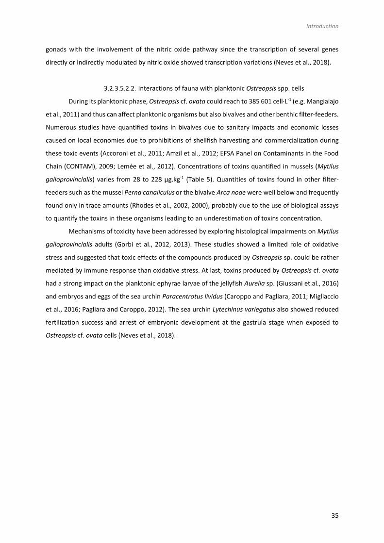
Introduction
35
gonads with the involvement of the nitric oxide pathway since the transcription of several genes
directly or indirectly modulated by nitric oxide showed transcription variations (Neves et al., 2018).
3.2.3.5.2.2. Interactions of fauna with planktonic Ostreopsis spp. cells
During its planktonic phase, Ostreopsis cf. ovata could reach to 385 601 cell·L-1 (e.g. Mangialajo
et al., 2011) and thus can affect planktonic organisms but also bivalves and other benthic filter-feeders.
Numerous studies have quantified toxins in bivalves due to sanitary impacts and economic losses
caused on local economies due to prohibitions of shellfish harvesting and commercialization during
these toxic events (Accoroni et al., 2011; Amzil et al., 2012; EFSA Panel on Contaminants in the Food
Chain (CONTAM), 2009; Lemée et al., 2012). Concentrations of toxins quantified in mussels (Mytilus
galloprovincialis) varies from 28 to 228 µg.kg-1 (Table 5). Quantities of toxins found in other filter-
feeders such as the mussel Perna canaliculus or the bivalve Arca noae were well below and frequently
found only in trace amounts (Rhodes et al., 2002, 2000), probably due to the use of biological assays
to quantify the toxins in these organisms leading to an underestimation of toxins concentration.
Mechanisms of toxicity have been addressed by exploring histological impairments on Mytilus
galloprovincialis adults (Gorbi et al., 2012, 2013). These studies showed a limited role of oxidative
stress and suggested that toxic effects of the compounds produced by Ostreopsis sp. could be rather
mediated by immune response than oxidative stress. At last, toxins produced by Ostreopsis cf. ovata
had a strong impact on the planktonic ephyrae larvae of the jellyfish Aurelia sp. (Giussani et al., 2016)
and embryos and eggs of the sea urchin Paracentrotus lividus (Caroppo and Pagliara, 2011; Migliaccio
et al., 2016; Pagliara and Caroppo, 2012). The sea urchin Lytechinus variegatus also showed reduced
fertilization success and arrest of embryonic development at the gastrula stage when exposed to
Ostreopsis cf. ovata cells (Neves et al., 2018).

36
Table 5 : Organisms in direct interaction with Ostreopsis spp. Half maximal effective concentration (EC50) ; Digestive Tube (DT); Hemolysis Neutralization Assay
(HNA); Median Lethal concentration (LC50) ; Liquid Chromatography –Mass Spectrometry (LC-MS); Media, lethal time (LT50); Mouse Bioassay (MBA); Remaining
tissue (RT); Whole flesh (WF).
Species/Strain Organisms Stage Effects Reference
Interactions with epibenthic cells of Ostreopsis spp.
Ostreopsis ovata isolated by A. Penna
Tigriopus fulvus (Fisher, 1860)
I-II stage Nauplii
Mortality LC50-48h-20°C = 1486.74 cells.mL-1
LC50-48h-25°C = 250.12 cells.mL-1 Faimali et al., 2012
Ostreopsis cf. ovata Tigriopus fulvus (Fisher, 1860)
Mortality LC50-24h-20°C > 4000 cells.mL-1
Found in Giussani et al., 2016. Unpublished data
Ostreopsis ovata isolated by A. Penna
Amphibalanus amphitrite (Darwin, 1854)
II-III stage Nauplii
Mortality LC50-48h-20°C = 1416 cells.mL-1 LC50-48h-25°C = 192 cells.mL-1
Faimali et al., 2012
Ostreopsis cf. ovata Amphibalanus amphitrite (Darwin, 1854)
Mortality LC50-24h-20°C > 4000 cells.mL-1
Found in Giussani et al., 2016. Unpublished data
Ostreopsis cf. ovata Natural samples Ligurian Sea (44°3’27’’N; 9°55’43’’E)
Dinophylus gyrociliatus (Schmidt, 1857)
Juveniles Mortality LT50-24°C (3500 cells.mL-1) = 2h LT50-24°C (1500 cells.mL-1)=3.4h LT50-24°C (200 cells.mL-1) = 12.25h ± 3.18
Simonini et al., 2011
Ostreopsis ovata isolated by A. Penna
Paracentrotus lividus (Lamarck, 1816)
Juveniles Mortality LC50-48h-20°C = 168 cells.mL-1 Privitera et al., 2012
Ostreopsis cf. ovata Natural samples Gulf of Taranto
Tigriopus fulvus (Fisher, 1860)
Nauplii Mortality LC50-96h-25°C = 10.11 cell.mL-1 Prato et al., 2011
Corophium insidiosum (Crawford, 1937)
2-4 mm body length
Mortality LC50-96h-25°C = 11.81 cell.mL-1
Sphaeroma serratum (Fabricius, 1787)
2-4 mm body length
Mortality LC50-96h-25°C = 214.81 cell.mL-1
Ostreopsis siamensis CAWD73/74/75/96
Haliotis virginea (Gmelin, 1791)
Larvae Mortality Not lethal within 24h Rhodes et al., 2000
Ostreopsis cf. ovata MCCV 054
Sarsamphiascus cf. propinquus (Sars G.O., 1906)
Adults Mortality LC50-24h-24°C > 20 000 cell.mL-1 Pavaux et al., 2019

Introduction
37
Ostreopsis cf. ovata Natural samples from French and Italian NW Med. coasts
Phytal meiofauna Community composition
72 % decrease of nauplii Guidi-Guilvard et al., 2012
Ostreopsis cf. ovata MCCV 054
Sarsamphiascus cf. propinquus (Sars G.O., 1906)
Females Reproduction Decrease of fertility and fecundity rations Pavaux et al., 2019
Ostreopsis cf. ovata Natural samples (French Mediterranean coasts)
Paracentrotus lividus (Lamarck, 1816)
Toxin quantification LC-MS/MS: 77 to 361 µg eq PLTX.kg-1 Amzil et al., 2012
Ostreopsis cf. ovata Field sample (Villefranche-sur-Mer)
Paracentrotus lividus (Lamarck, 1816)
Toxin quantification LC-MS/MS: 103 to 423 µg.kg-1 Hemolytic test: 179 to 270 µg.kg-1
Brissard et al., 2014
Sarpa salpa (Linnaeus, 1758)
Toxin quantification LC-MS/MS: 33 to 152 µg.kg-1
Ostreopsis ovata Field samples (Tyrrhenian Sea)
Sea urchins Toxin quantification HNA: 5 to 99 pPLTX µg.kg-1
LC-MS/MS: < LOQ pPLTX µg.kg-1 87 to 164 OVTXa µg.kg-1
Milandri et al., 2010
Ostreopsis cf. ovata Field samples Nice harbour and Villefranche-sur-mer
Paracentrotus lividus (Lamarck, 1816)
Toxin quantification Haemolytic assay : 256.6 µg/kg (WF) LC-MS/MS: 192.5 µg/kg (WF)
Biré et al., 2013
Sarpa salpa (Linnaeus, 1758)
Toxin quantification Haemolytic assay : 230.0 µg/kg (DT) LC-MS/MS: 191.2 µg/kg (WF)
Ostreopsis cf. ovata Field samples - Rochambeau site, Villefranche-sur-mer
Paracentrotus lividus (Lamarck, 1816)
Toxin quantification Haemolytic assay : 159.2 µg/kg (DT) Biré et al., 2015
Sarpa salpa (Linnaeus, 1758)
Toxin quantification Haemolytic assay : 361.0 µg/kg (DT)
Interactions with planktonic cells of Ostreopsis sp.
Ostreopsis ovata isolated by A. Penna
Paracentrotus lividus (Lamarck, 1816)
Competent larvae
Mortality LC50-48h-20°C = 168 cells.mL-1 Privitera et al., 2012
Ostreopsis cf. ovata CBA29-2012 (Genoa)
Aurelia sp. Polyp Reproduction/early stages
Unaffected (100 cells.mL-1) Giussani et al., 2015
Ostreopsis cf. ovata Marche coast (Italie NW Adriatic sea)
Mytilus galloprovincialis (Lamarck, 1819)
Adults Histological impairments
Immune system: - Decrease of granulocytes - Phagocytosis activity decrease - Reduced lysosomal membrane stability Histology: - Decrease of the digestive gland wall thickness - Dilatation of tubules
Gorbi et al., 2013

38
- Haemocytes infiltration into digestive gland
- Decrease of neutral lipids quantity Oxidative stress: limited role
Ostreopsis cf. ovata Field sample from Adriatic Sea (Portonevo & Passetto sites)
Mytilus galloprovincialis (Lamarck, 1819)
Adults Histological impairments
- Inhibition of Na+/K+ATPase and acetylcholine esterase activity
Immune system: Reduced lysosomal membrane stability Oxidative stress: limited role
Gorbi et al., 2012
Ostreopsis spp. Cultures + field samples (North Aegean coasts
Mytilus galloprovincialis (Lamarck, 1819) Venus verrucosa Odiolus barbatus
Toxin quantification MBA: Qualitative analysis HNA: 33.3 to 97 pPLTX µg.kg-1
Aligizaki et al., 2008
Ostreopsis cf. ovata Natural samples (French Mediterranean coasts)
Mytilus galloprovincialis (Lamarck, 1819)
Toxin quantification LC-MS/MS: 28 to 217 µg eq PLTX.kg-1 Amzil et al., 2012
Ostreopsis cf. ovata Filed samples (Adriatic Sea)
Mytilus galloprovincialis (Lamarck, 1819)
Toxin quantification MBA + HNA: Qualitative analysis Gorbi et al., 2012
Ostreopsis ovata Field samples (Tyrrhenian Sea)
Mussels Toxin quantification HNA: 103 pPLTX µg.kg-1
LC-MS/MS: < LOQ pPLTX µg.kg-1 - 131 to 228 OVTXa µg.kg-1
Milandri et al., 2010
Ostreopsis siamensis CAWD75 (New Zeland)
GreenshellTM mussels Pacific oysters Scallops
Toxin quantification MBA: qualitative analysis HNA: 0.3 pg eq. PLTX.cell-1
Rhodes et al., 2000
Ostreopsis siamensis CAWD75 (New Zeland)
Perna canaliculus (Gmelin, 1791) Pecten novaezalandiae (Reeve, 1852) Crassostrea gigas (Thunberg, 1793)
Toxin quantification MBA (qualitative analysis): none to death HNA: trace to 1 ng.g-1
Rhodes et al., 2002
Ostreopsis cf. ovata Field samples - Rochambeau site Villefranche-sur-mer
Arca noae (Linneaus, 1758)
Toxin quantification Haemolytic assay : < 10 µg/kg (WF) Biré et al., 2015

Introduction
39
Ostreopsis cf. ovata CBA29-2012 (Genoa)
Aurelia sp. Ephyrae Reproduction/early stages
% Alteration of Pulsation Frequency EC50-24h = 5.32 cells.mL-1
Immobility EC50-24h = 10.50 cells.mL-1
Giussani et al., 2015
Ostreopsis cf. ovata ITAC501
Paracentrotus lividus (Lamarck, 1816)
Eggs Reproduction/early stages
- Gametes: % fertilization decrease; less toxic for eggs than sperms
- Embryos: no segmentation
Caroppo and Pagliara, 2011
Ostreopsis cf. ovata Natural samples (Gaiola MPA)
Paracentrotus lividus (Lamarck, 1816)
Embryos Reproduction/early stages
Fertilization and developmental impairments Alteration of several marker genes
Migliaccio et al., 2016
Ostreopsis cf. ovata Strain UNR-05 Armaçao dos Bùzios (Rio de Janeiro)
Lytechinus variegatus (Lamarck, 1816)
Embryos Reproduction/early stages
Stronger effect on larvae than on fertilization success: - Reduced fertilization - Arrest of embryonic development (gastrula stages at 400 cells.mL-1) - Increase of dead and abnormal larvae
Neves et al., 2018
Ostreopsis cf. ovata (whole cells + lysate) Natrural samples (Ionian Sea)
Paracentrotus lividus (Lamarck, 1816)
Embryos Reproduction/early stages
Whole cells: Developmental delay or anomalous cell division Cell lysate: Growth inhibition at 1.87 mg.mL-1
Pagliara and Caroppo, 2012
Interactions with surface cells of Ostreopsis spp.
Ostreopsis cf. ovata Field samples Nice harbour and Villefranche-sur-mer
Patella spp Toxin quantification Haemolytic assay : 12.7 µg/kg (WF) Biré et al., 2013
Mugil cephalus (Linneaus, 1758)
Toxin quantification Haemolytic assay : 392.2 µg/kg (DT) LC-MS/MS: 359.1 µg/kg (WF)
Ostreopsis cf. ovata Field samples - Rochambeau site Villefranche-sur-mer
Patella spp. Toxin quantification Haemolytic assay : < 10 µg/kg (WF) Biré et al., 2015
Ostreopsis cf. ovata Field samples Genoa, Nice, Villefranche-sur-Mer
Paracentrotus lividus (Lamarck, 1816)
Density Any decrease of P. lividus densities related to high abundances of O. cf. ovata
Blanfuné et al., 2012
Patella spp. No significant decrease of densities in Villefranche-sur –mer and Nice ; Significant decrease in Genoa potentially linked to O. cf. ovata blooms

40
3.2.3.5.2.3. Interactions of fauna with sea-surface Ostreopsis spp. cells
Toxins quantified both in the digestive tube and in the whole flesh of the mullet Mugil cephalus
were high and reached to 392.5 µg·kg-1 which can be explained by the grazing behavior of this animal
that feeds directly on Ostreopsis cf. ovata cells aggregates on the water surface (Biré et al., 2013).
Other organism from the mediolittoral area such as the limpets Patella spp. are also exposed to surface
cells of Ostreopsis cf. ovata and up to now, studies have been mainly focused on toxin quantification
in the whole flesh that reached up to 12.7 µg.kg-1, well below the threshold value established by the
EFSA (EFSA Panel on Contaminants in the Food Chain (CONTAM), 2009). The study of Blanfuné et al.
(2012) reported a significant decrease of Patella spp. densities during the 2008 O. cf. ovata bloom in
Genoa, attributed to the poisoning of the shellfish by ingestion of O. cf. ovata cells.
To conclude, most studies have focused on the effect of O. cf. ovata and revealed reprotoxic
effect on both planktonic and meroplanktonic organisms. Toxins are mainly found in the digestive
tubes of contaminated organisms and their concentrations appeared to be independent of the way of
O. cf. ovata exposure (epibenthic, planktonic or surface cells) since toxin concentrations found in the
mullet Mugil cephalus feeding on surface cells of O. cf. ovata, were comparable to values found in sea
urchins’ digestive tubes, feeding on epibenthic cells. Lastly, toxins are found in herbivorous and
omnivorous organisms, regardless of the diet.
3.2.3.5.3 Indirect interactions between Ostreopsis spp. and marine fauna
Toxin concentrations found in carnivorous and omnivorous organisms suggested
bioaccumulation of toxins within the trophic web (Biré et al., 2013, 2015; Faimali et al., 2012; Giussani
et al., 2016; Milandri et al., 2010; Pezzolesi et al., 2012). Toxins quantified in both the crab Eriphia
verrucosa (carnivorous) and the gastropod Stramonita haemastoma (carnivorous) raised up to 58.6
and 26.4 µg·kg-1 (Fresh Weight) respectively. Octopuses also accumulated more than 446 µg.kg-1
(Milandri et al., 2010) suggesting a bioaccumulation of toxins among the trophic web which might have
played a role in the ecosystem suffering with stranding of octopuses.
Other studies reported a strong sensitivity of Dicentrarchus labrax fish juveniles to O. cf. ovata
raising the question of the impact on the structure of marine ecosystems (Faimali et al., 2012; Giussani
et al., 2016; Pezzolesi et al., 2012). Indeed, these fish are unable to eat Ostreopsis cells, the observed
toxicity was exclusively due to contact with Ostreopsis cells.
To conclude, high accumulation of Ostreopsis toxins has been documented in various marine
organisms suggesting the potential bioaccumulation among the trophic web with threshold higher
than those recommended by EFSA. More studies are needed to better understand the transfer of
toxins among the trophic web as already described for other toxic dinoflagellate such as Gambierdiscus

Introduction
41
species and ciguatoxins (Ledreux et al., 2014). Moreover, the few existing studies have reported strong
differences of sensitivity between organisms hence the need of more studies to investigate potential
acclimation and detoxification phenomena on some organisms. At last, the reprotoxicity of Ostreopsis
sp. have been demonstrated for benthic organisms which can lead to changes in benthic communities.
3.2.4. Overall vision of the impacts of Ostreopsis spp. on ecosystem structure and
functioning
3.2.4.1. Ecotoxicology studies
The studies on Ostreopsis spp. chemical ecology summarized above show negative effects of
this dinoflagellate on different trophic levels and suggest subsequent negative effects on the structure
of the ecosystems. At the level of primary producers, Ostreopsis sp. negatively impact diatoms and
other dinoflagellates with which it co-occurs, regardless of the exposure pathway (extracellular
released compounds or whole living cells) leading to changes in microphytobenthic communities.
Concerning secondary producers, a strong reprotoxic effect has also been reported. Herbivorous
organisms feeding on Ostreopsis sp. (by e.g., the benthic copepod Sarsamphiascus cf. propinquus) or
organisms feeding on macroalgae supporting its populations (e.g., sea urchins -Paracentrotus lividus-
or fish -Sarpa salpa-) could accumulate very high quantity of toxins without short term lethal effects.
However, a clear decrease of fecundation success, fertility and fecundity ratios and abnormal
development effects (that lasted for many months after the exposition), was reported for various
organisms such as benthic copepods and sea urchins (Table 5). This effect on the progeny could also
strongly affect the structure of ecosystems. Despite several studies report invertebrate mass
mortalities (Shears and Ross, 2009; Totti et al., 2010), no studies have been conducted to determine if
these mortalities could genuinely be attributed to toxins produced by Ostreopsis spp. Overall, the
transfer to toxin through the food webs poses a risk of seafood poisoning in humans through
consumption of the contaminated organisms, not reported in temperate areas yet, but, unfortunately,
with some dramatic cases in tropical areas.

42
Table 6 : Organisms in indirect interaction with Ostreopsis spp. Half maximal effective concentration (EC50); Median lethal concentration (LC50); Median
lethal time (LT50).
Species/Strain Organisms Authority Effects Reference
Ostreopsis ovata Field samples (Tyrrhenian Sea)
Octopuses Toxin quantification HNA: 446 pPLTX µg.kg-1
LC-MS/MS: 115 pPLTX µg.kg-1 971 OVTXa µg.kg-1
Milandri et al., 2010
Ostreopsis cf. ovata Field samples Nice harbour and Villefranche-sur-mer
Diplodus annularis Linnaeus, 1758 Toxin quantification Haemolytic assay : 10.6 µg/kg (DT) Biré et al., 2013
Diplodus sargus Linnaeus, 1758 Toxin quantification Haemolytic assay : 5.1 µg/kg (DT)
Eriphia verrucosa Forskål, 1775 Toxin quantification Haemolytic assay: 38.4 µg/kg (WF) LC-MS/MS: 58.6 µg/kg (WF)
Maja squinado Herbst, 1788 Toxin quantification Haemolytic assay : 8.3 µg/kg (WF)
Mullus surmuletus Linnaeus, 1758 Toxin quantification Haemolytic assay : < LOQ
Muraena helena Linnaeus, 1758 Toxin quantification Haemolytic assay : < LOQ
Octopus vulgaris Cuvier, 1797 Toxin quantification Haemolytic assay : 18.5 µg/kg (RT)
Scorpaena porcus Linnaeus, 1758 Toxin quantification Haemolytic assay : 3.7 µg/kg (DT)
Serranus scriba Linnaeus, 1758 Toxin quantification Haemolytic assay : < LOQ
Stramonita haemastoma Linnaeus, 1767 Toxin quantification Haemolytic assay: 34.1 µg/kg (WF) LC-MS/MS: 26,4 µg/kg (WF)
Symphodus tinca Linnaeus, 1758 Toxin quantification Haemolytic assay : 1.8 µg/kg (DT)
Ostreopsis cf. ovata Field samples Rochambeau site, Villefranche-sur-mer
Coris julis Linnaeus, 1758 Toxin quantification Haemolytic assay : < 10 µg/kg (WF) Biré et al., 2015
Diplodus vulgaris
Geoffroy Saint-Hilaire, 1817
Toxin quantification
Eriphia verrucosa Forskål, 1775 Toxin quantification Haemolytic assay : 13.0 µg/kg (WF)
Hexaplex trunculus Linnaeus, 1758 Toxin quantification Haemolytic assay : 40.4 µg/kg (WF)
Gobiidae Toxin quantification Haemolytic assay : < 10 µg/kg (WF)
Maja squinado Herbst, 1788 Toxin quantification Haemolytic assay : 51.3 µg/kg (WF)
Mullus surmuletus Linnaeus, 1758 Toxin quantification Haemolytic assay : < 10 µg/kg (WF)
Muraena helena Linnaeus, 1758 Toxin quantification Haemolytic assay : < 10 µg/kg (WF)
Octopus vulgaris Cuvier, 1797 Toxin quantification Haemolytic assay : 19.9 µg/kg (RT)

Introduction
43
Saurida undosquamis Richardson, 1848
Toxin quantification Haemolytic assay : < 10 µg/kg (WF)
Scorpena porcus Linnaeus, 1758 Toxin quantification Haemolytic assay : < 10 µg/kg (WF)
Serranus scriba Linnaeus, 1758 Toxin quantification Haemolytic assay : < 10 µg/kg (WF)
Symphodus melops Linnaeus, 1758 Toxin quantification Haemolytic assay : < 10 µg/kg (WF)
Symphodus ocellatus Linnaeus, 1758 Toxin quantification Haemolytic assay : < 10 µg/kg (WF)
Symphodus roissali Risso, 1810 Toxin quantification Haemolytic assay : < 10 µg/kg (WF)
Symphodus rostratus Bloch, 1791 Toxin quantification Haemolytic assay : < 10 µg/kg (WF)
Symphodus tinca Linnaeus, 1758 Toxin quantification Haemolytic assay : < 10 µg/kg (WF)
Thalassoma pavo Linnaeus, 1758 Toxin quantification Haemolytic assay : < 10 µg/kg (WF)
Xantho poressa Olivi, 1792 Toxin quantification Haemolytic assay : < 10 µg/kg (WF)
Ostreopsis ovata isolated by A. Penna
Dicentrarchus labrax
Linnaeus, 1758 Mortality LC50-48h-20°C = 540 cells.mL-1 Faimali et al., 2012
Ostreopsis cf. ovata Dicentrarchus labrax
Linnaeus, 1758 Mortality LC50-48h-20°C = 679 cells.mL-1 Found in Giussani et al., 2016. Unpublished data
Ostreopsis cf. ovata Strain OOAN0601
Dicentrarchus labrax
Linnaeus, 1758 Mortality 100% mortality after 45h to 72h exposed to O. cf. ovata (between 425 to 2367 cells.mL-1)
Pezzolesi et al., 2012

44
Some organisms are able to protect themselves to the presence of Ostreopsis spp. by different
mechanisms. Some diatoms such as the competitive diatom Licmophora paradoxa produce secondary
metabolites which negatively affect Ostreopsis cf. ovata at different levels of organization. Some
copepods commonly exposed to Ostreopsis cells have also demonstrated higher resistance to other
species, suggesting a possible acclimation to Ostreopsis spp. Acclimation processes and detoxification
and storage mechanisms of organisms exposed to Ostreopsis sp. have been rarely observed although
they have already been described for other toxic dinoflagellates. A rapid detoxification of asparacids
by Mytilus edulis (Jauffrais et al., 2012), of DSP produced by Prorocentrum lima by the scallop
Argopecten irradians (Bauder et al., 2001) or gymnodimine by the carpet shell Ruditapes decussatus
(Walid et al., 2010) has been already been demonstrated. Other studies have also demonstrated
interspecific differences in toxins accumulation linked to toxins sensitivity and determined by
neurologic, physiologic and behavior responses (Bricelj and Shumway, 1998). Toxicity also varies
according to immersion time, the size of organism, or the off-bottom position (Bricelj and Shumway,
1998). Genes involved in detoxification mechanisms of oysters exposed to Gymnodinium catenatum
have been already described and are involved in antioxidant defense, cell detoxification, intermediate
immune response activation and stress response (García-Lagunas et al., 2013).
Acclimation processes have been already demonstrated for copepods exposed to Alexandrium
spp. Indeed, populations of Acartia hudsonica from Massachussets (USA) to New Brunswick (Canada),
commonly exposed to this dinoflagellate are more resistant than naïve populations, which failed to
acclimate to the exposition to this toxic dinoflagellate (Colin and Dam, 2002).
Toxins quantified in cephalopods and carnivorous, herbivorous and omnivorous fish (Table 6)
raise the question of bioaccumulation of OVTXs along the food web. Toxins circulation through the
food webs from benthic unicellular algae to herbivorous fish has already been demonstrated for
ciguatoxins (Ledreux et al., 2014). Another hypothesis is the incorporation of toxins into higher
organisms through zooplankton (Baustian et al., 2018) and more specially through copepods or fecal
pellets (Lehtiniemi et al., 2002). Copepods and other zooplankton organisms have been already
described as vectors of the toxins up the food web for microcystins (Smith and Haney, 2006), nodularin
(Lehtiniemi et al., 2002), okadaic acid (Maneiro et al., 2000) and PSP (Teegarden and Cembella, 1996).
The presence of Ostreopsis sp. could also favor some organisms. Indeed, particles produced by
Ostreopsis sp. blooms i.e. theca fragments and mucus, respectively rich in cellulose and carbohydrates,
provide biological colonizable surfaces and a good source of organic carbon, which can favor the
growth of some bacteria such as Vibrio (Bellés-Garulera et al., 2016). This positive association for
pathogenic bacteria increases the toxicity of Ostreopsis for human populations.

Introduction
45
3.2.4.2. Importance of the mucus in Ostreopsis spp. interactions
The functional role of the mucilaginous matrix produced by Ostreopsis sp. is still unknown.
Mucus production is considered as an adaptation to benthic life (Escalera et al., 2014; Parsons et al.,
2012) and described as a complex structure composed by a network of long fibers, deriving from
trichocysts extruded trough thecal pores and by an amorphous matrix of acid polysaccharides (Honsell
et al., 2013). The production of mucilaginous matrix is common in benthic dinoflagellates (MacKenzie
et al., 2002) and plays a role in cellular adhesion to surfaces with the formation of a transparent film
embedding many cells, providing a clear example of ecosystem engineering (Escalera et al., 2014). Its
role in defense against grazers, in increasing buoyancy and metabolic self-regulation has already been
investigated in other microalgae (Reynolds, 2007). The role of the mucilaginous matrix as a method of
defense against grazers or to capture larger organisms has indeed been documented for other
mixotrophic dinoflagellates (Blossom et al., 2012; Kiϕrboe and Titelman, 1998). For Ostreopsis sp., its
role in predation and competition has been previously argued. Toxic effect of O. cf. ovata on co-
occurring diatom is mediated by contact between cells, suggesting the role of the mucus in the
competition as a storage for toxins in order to avoid dilution of toxins in the environment (Ternon et
al., 2018). The toxicity of the mucus was also documented by Giussani et al. (2015) with a significant
toxic effect of O. cf. ovata mucilage on Artemia salina and supported by images obtained by atomic
force microscopy. However, this role is poorly documented mainly due to the difficulty to separate
cells from this mucus.
The role of this mucilaginous matrix in mixotrophic behavior was also reported (Barone, 2007).
Even if mixotrophy is a common way of nutrition in harmful algal species (Burkholder et al., 2008), this
heterotrophic behavior in Ostreopsis sp. is also poorly documented. Indeed, mixotrophic behavior of
Ostreopsis sp. was first described by Faust and Morton (1995) since they observed Ostreopsis labens
cells with large ingested particles within the cytoplasm. In 2012, Fraga et al. linked the presence of
reddish round inclusions inside Ostreopsis cells through light microscopy, to a mixotrophic behavior of
Ostreopsis species and confirmed by the study of Almada et al. (2017) which showed evidence of
phagotrophy using LysoSensor staining even if the prey inside the cells were not discernable. Using
molecular approaches, Lee and Park (2018) defined these red bodies as digestive vacuoles containing
red macroalgae (Class Florideophyceae) ingested as spores.
3.2.4.3. Regulation of Ostreopsis bloom by biotic interactions
There is a growing awareness that HABs may have been increasing globally in the last 40 years
and may continue to increase under the fast coming scenarios of global warning. HABs have strong
impacts on human health and economy (Adams et al., 2018; Hoagland et al., 2002; Larkin and Adams,
2007). Hence the necessity to better understand factors and mechanisms favoring bloom formation

46
and maintenance. The formation, maintain and declining of such events is mainly attributed to biotic
factors combining chemical (salinity, nutrients..) and physicals processes (currents, upwelling, river
flow..) associated to anthropogenic inputs leading to eutrophication (Sellner et al., 2003). However,
this review highlights the number, the importance and the complexity of interactions between
Ostreopsis sp. and its environment. The question is whether or not these interactions could regulate
blooms initiation and decline. Most harmful bloom-forming species produce toxins even if adaptive
significance of this synthesis is still unclear. It has been suggested that toxins confer some advantages
in reaching very high cell densities (Anderson et al., 1998) by providing a defense against pathogens,
grazers and parasites. This hypothesis is supported by the study of Selander et al. (2006) describing
that toxin-producing species are less preferred by predators and that predator presence induces toxin
production. In the case of Ostreopsis cf. ovata, it has been noted that benthic copepods are able to
actively select non-toxic diatom in a mixture (Pavaux et al., 2019).
The role of these toxins in limiting growth and survival of competitors via allelopathic processes
is poorly documented. Ternon et al. (2018) have demonstrated the importance of the toxins in negative
effects on competitive diatom (and mediated by the mucus) and the potential involvement in chemical
control by O. cf. ovata on its competitor. However, there is no consensus on the characterization of
toxins produced by HABs as phytochemicals. Indeed, the work of Tillmann and John (2002) has rather
shown that toxins produced by Alexandrium spp. are not involved in allelochemical defense
mechanism. In view of conflicting results obtained, it is difficult to conclude about the role of the toxins
as allelochemicals.
One hypothesis is that allelopathy could be an important mechanism to favor HAB by
suppressing the growth of competitors (Smayda, 1997b). However, the importance of allelopathy is
such mechanisms is difficult to conclusively demonstrate in the field (Prince et al., 2008). Mechanisms
for allelopathy has already been proposed and included oxidative damage, loss of competitor mobility,
inhibition of photosynthesis, inhibition of enzymes and membrane damage (Legrand et al., 2003). For
Ostreopsis sp. a strong negative effect has been demonstrated on its competitor by inhibiting growth
and fluorescence rates leading to a diminution of competitors’ abundances. The production of
allelopathic compounds explain in particular the success of Karenia brevis blooms (Kubanek et al.,
2005; Prince et al., 2008). However, meta-analysis conducted by Jonsson et al. (2009) rather supported
a significant effect of allelopathic mechanisms only at high cell densities since at a beginning of a
bloom, cell abundances are too low to concentrate enough infochemicals.
Predation as a mechanism to favor Ostreopsis sp. Bloom: The balance between growth- and loss-
promoting factors determine the abundance of harmful species. Predation by zooplankton is the
largest mortality factor for phytoplankton (Calbet and Landry, 2004). Factors such as size, shape,
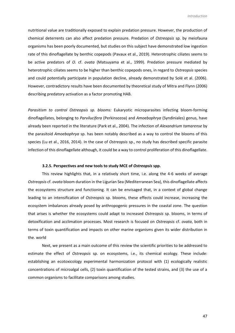
Introduction
47
nutritional value are traditionally exposed to explain predation pressure. However, the production of
chemical deterrents can also affect predation pressure. Predation of Ostreopsis sp. by meiofauna
organisms has been poorly documented, but studies on this subject have demonstrated low ingestion
rate of this dinoflagellate by benthic copepods (Pavaux et al., 2019). Heterotrophic ciliates seems to
be active predators of O. cf. ovata (Matsuyama et al., 1999). Predation pressure mediated by
heterotrophic ciliates seems to be higher than benthic copepods ones, in regard to Ostreopsis species
and could potentially participate in population decline, already demonstrated by Solé et al. (2006).
However, contradictory results have been documented by theoretical study of Mitra and Flynn (2006)
describing predatory activation as a factor promoting HAB.
Parasitism to control Ostreopsis sp. blooms: Eukaryotic microparasites infecting bloom-forming
dinoflagellates, belonging to Parvilucifera (Perkinsozoa) and Amoebophrya (Syndiniales) genus, have
already been reported in the literature (Park et al., 2004). The infection of Alexandrium tamarense by
the parasitoid Amoebophrya sp. has been notably described as a way to control the blooms of this
species (Lu et al., 2016, 2014). In the case of Ostreopsis sp., no study has described specific parasite
infection of this dinoflagellate although, it could be a way to control proliferation of this dinoflagellate.
3.2.5. Perspectives and new tools to study MCE of Ostreopsis spp.
This review highlights that, in a relatively short time, i.e. along the 4-6 weeks of average
Ostreopsis cf. ovata bloom duration in the Ligurian Sea (Mediterranean Sea), this dinoflagellate affects
the ecosystems structure and functioning. It can be envisaged that, in a context of global change
leading to an intensification of Ostreopsis sp. blooms, these effects could increase, increasing the
ecosystem imbalances already posed by anthropogenic pressures in the coastal zone. The question
that arises is whether the ecosystems could adapt to increased Ostreopsis sp. blooms, in terms of
detoxification and acclimation processes. Most research is focused on Ostreopsis cf. ovata, both in
terms of toxin quantification and impacts on other marine organisms given its wider distribution in
the. world
Next, we present as a main outcome of this review the scientific priorities to be addressed to
estimate the effect of Ostreopsis sp. on ecosystems, i.e., its chemical ecology. These include:
establishing an ecotoxicology experimental harmonization protocol with (1) ecologically realistic
concentrations of microalgal cells, (2) toxin quantification of the tested strains, and (3) the use of a
common organisms to facilitate comparisons among studies.
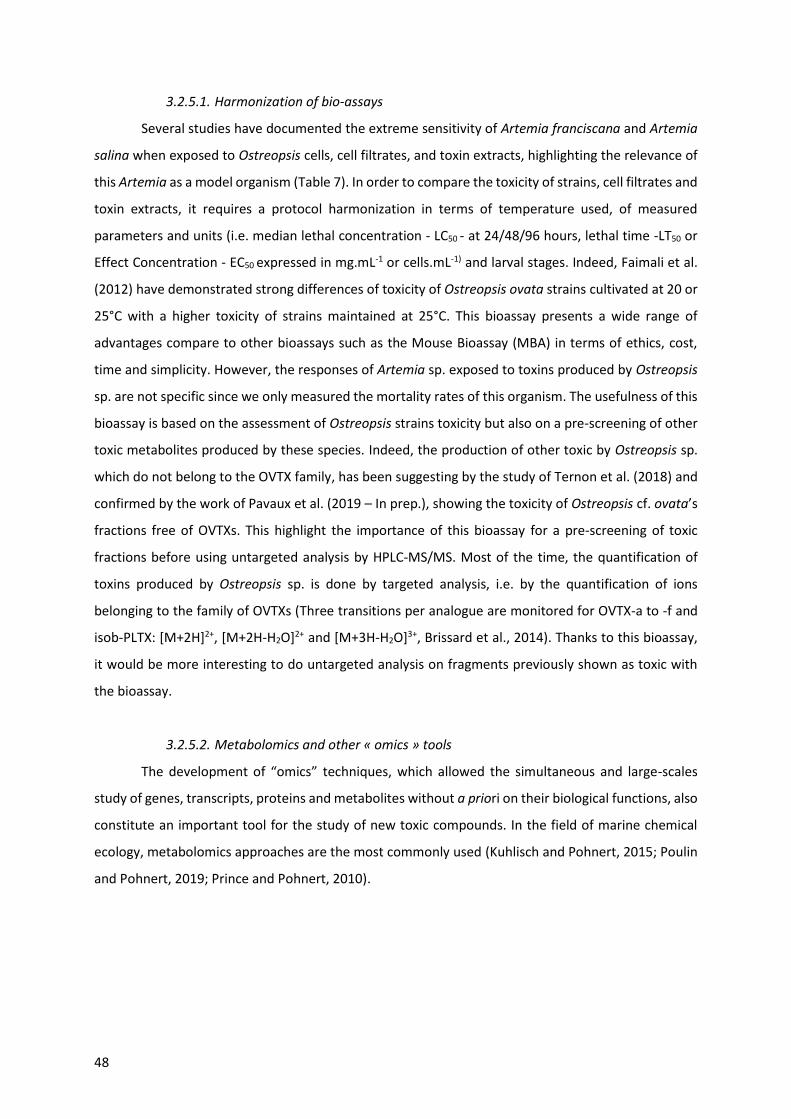
48
3.2.5.1. Harmonization of bio-assays
Several studies have documented the extreme sensitivity of Artemia franciscana and Artemia
salina when exposed to Ostreopsis cells, cell filtrates, and toxin extracts, highlighting the relevance of
this Artemia as a model organism (Table 7). In order to compare the toxicity of strains, cell filtrates and
toxin extracts, it requires a protocol harmonization in terms of temperature used, of measured
parameters and units (i.e. median lethal concentration - LC50 - at 24/48/96 hours, lethal time -LT50 or
Effect Concentration - EC50 expressed in mg.mL-1 or cells.mL-1) and larval stages. Indeed, Faimali et al.
(2012) have demonstrated strong differences of toxicity of Ostreopsis ovata strains cultivated at 20 or
25°C with a higher toxicity of strains maintained at 25°C. This bioassay presents a wide range of
advantages compare to other bioassays such as the Mouse Bioassay (MBA) in terms of ethics, cost,
time and simplicity. However, the responses of Artemia sp. exposed to toxins produced by Ostreopsis
sp. are not specific since we only measured the mortality rates of this organism. The usefulness of this
bioassay is based on the assessment of Ostreopsis strains toxicity but also on a pre-screening of other
toxic metabolites produced by these species. Indeed, the production of other toxic by Ostreopsis sp.
which do not belong to the OVTX family, has been suggesting by the study of Ternon et al. (2018) and
confirmed by the work of Pavaux et al. (2019 – In prep.), showing the toxicity of Ostreopsis cf. ovata’s
fractions free of OVTXs. This highlight the importance of this bioassay for a pre-screening of toxic
fractions before using untargeted analysis by HPLC-MS/MS. Most of the time, the quantification of
toxins produced by Ostreopsis sp. is done by targeted analysis, i.e. by the quantification of ions
belonging to the family of OVTXs (Three transitions per analogue are monitored for OVTX-a to -f and
isob-PLTX: [M+2H]2+, [M+2H-H2O]2+ and [M+3H-H2O]3+, Brissard et al., 2014). Thanks to this bioassay,
it would be more interesting to do untargeted analysis on fragments previously shown as toxic with
the bioassay.
3.2.5.2. Metabolomics and other « omics » tools
The development of “omics” techniques, which allowed the simultaneous and large-scales
study of genes, transcripts, proteins and metabolites without a priori on their biological functions, also
constitute an important tool for the study of new toxic compounds. In the field of marine chemical
ecology, metabolomics approaches are the most commonly used (Kuhlisch and Pohnert, 2015; Poulin
and Pohnert, 2019; Prince and Pohnert, 2010).

Introduction
49
Table 7 : Artemia bio-assay for Ostreopsis toxicity. Median lethal concentration (LC50); Median lethal time (LT50).
Species/Strain Organims Stage Effects References
Ostreopsis ovata osolated by A. Penna
Artemia salina (Linnaeus, 1758)
II-III stage nauplii LC50-48h-20°C = 12 cells.mL-1 LC50-48h-25°C < 4 cells.mL-1
(Faimali et al., 2012)
Ostreopsis cf. ovata Isolated from Lazio & Marche
Artemia sp. Nauplii EC50 calculated but not expressed (Guerrini et al., 2008)
Ostreopsis cf. ovata Artemia salina (Linnaeus, 1758)
Nauplii LC50-24h-20°C = 15 cells.mL-1
Found in Giussani et al., 2016. Unpublished data
Ostreopsis cf. ovata CBA29-2012
Artemia salina (Linnaeus, 1758)
II – III stages larvae LC50-48h-20°C = 24.83 / 16.67/ <4 cells.mL-1 (exponential, stationary and late stationary phase)
(Giussani et al., 2015)
Ostreopsis cf. ovata Artemia salina (Linnaeus, 1758)
Nauplii Immobility EC50-48h-20°C < 4 cells.mL-1 Found in Giussani et al., 2016. Unpublished data
Ostreopsis cf. ovata UNR-05
Artemia salina (Linnaeus, 1758)
Adults LT50-24°C (200 cells.mL-1) = 3h ( Neves et al., 2017)
Ostreopsis cf. ovata (cell lysate) Natural samples (Ionian Sea)
Artemia salina (Linnaeus, 1758)
Nauplii (24h after hatching)
LC50-24h-25°C = 0.03 mg.mL-1 (Pagliara and Caroppo, 2012)
Ostreopsis cf. ovata Strain OOAN0601 (Marche Region)
Artemia sp.
Nauplii
LC50-24h-20°C (live cells) = 8 cells.mL-1 ± 5 = 115 pg.mL-1 ± 72 LC50-24h-20°C (lysed cells) = 96 cells.mL-1 ± 6 = 1376 pg.mL-1 ± 86 LC50-24h-20°C (extract) = 80 cells.mL-1 ± 7 = 1146 pg.mL-1 ± 272
(Pezzolesi et al., 2012)
Ostreopsis cf. ovata Natural samples Gulf of Taranto
Artemia franciscana (Kellogg, 1906)
II – III stages larvae
LC50-48h-25°C = 1.63 cell.mL-1 (Prato et al., 2011)
Ostreopsis siamensis CAWD73/74/75/96
Artemia salina (Linnaeus, 1758)
LT50 (250 cells.mL-1) = 24h Time until Morbidity50 (250 cells.mL-1) = 4h
(Rhodes et al., 2000)
Ostreopsis ovata Mediterranean Sea
Artemia salina (Linnaeus, 1758)
Correlation (p<1%) between O. cf. ovata concentration and death of A. salina
(Simoni et al., 2004)
Ostreopsis ovata Mediterranean Sea
Artemia salina (Linnaeus, 1758)
Correlation between A. salina mortality after 24 h and the number of O. ovata cells
Vale et al., 2007 in Ramos and Vasconcelos, 2010
Ostreopsis cf. ovata Ostreopsis fattorussoi Mediterranean Sea
Artemia franciscana (Kellogg, 1906)
II – III stages larvae LC50 – 24h (MCCV 054) = 81.96 ± 63.3 cells.mL-1
LC50 – 24h (MCCV 055) = 0.25 ± 0.90 cells.mL-1
LC50 – 24h (MCCV 057) = 0.0012± 0.00033 cells.mL-1
LC50 – 24h (MCCV 058) = 0.023 ± 0.039 cells.mL-1
LC50 – 24h (Dissolved OVTXs) = 5.96 ± 2.12 cells.mL-1
LC50 – 24h (Dissolved PLTX) = 48.42 ± 14.23 cells.mL-1
Pavaux et al., 2019. In prep.

This recent discipline allows the analysis of a wide range of low molecular weight metabolites
of a cell, an organism or excreted, in a given context (Fiehn et al., 2007). One of the distinctive features
of the metabolomics approach is that, due to strong diversity of physico-chemical properties of
molecules produced by an organism, it is today impossible to describe exhaustively the metabolome
with the actual analytic techniques. A method currently used in metabolomic is an approach combining
LC-MS/MS and molecular networking and employed as a rapid analytical method to detect toxins
(Allard et al., 2016; Quinn et al., 2017; Watrous et al., 2012; Yang et al., 2013). Mass spectrometry-
based molecular networking relies on the principle that structurally similar molecules share similar
MS/MS fragmentation patterns, thus giving a visual representation of molecular relatedness of
compounds in a sample (Watrous et al., 2012). For instance, this technic allowed the quantitative
identification of the lipopeptide malyngamide C by different strains of cyanobacteria (Winnikoff et al.,
2014). More and more studies use metabolomic approaches to investigate biotic interactions between
HAB and their environment (Giacometti et al., 2013; Poulin et al., 2018; Poulson-Ellestad et al., 2014)
but this approach is still barely applied to Ostreopsis spp. study. The production of organic compounds
not linked to OVTXs family, using notably untargeted metabolomics approach, reported by Ternon et
al. (2018) underline the urgent need to characterize Ostreopsis spp. metabolome.
The review of McLean (2013) on “omics” tools have demonstrating the relevance of genomics,
transcriptomics and proteomics approaches to advance our understanding on the ecology of HAB.
These techniques could give insights in metabolic and regulatory pathways involved in toxins and other
secondary metabolites production and the mechanisms of action of these metabolites in the target
organisms. These techniques are still not very widely applied to Ostreopsis spp. although proteomic
and metabolomics coupled-analyses lead to a better understanding of allelopathy interactions
between Karenia brevis and co-occurring species (Poulson-Ellestad et al., 2014).
3.2.5.3. Isolation and toxicity evaluation of each PLTX analogs
To date, only pure palytoxine standard is is commercialized, no OVTX standard are available
which restricts our knowledges on bioactivity and structure of PLTX analogs. Indeed, these studies
need large amount of pure toxins. Poli et al. (2018) have reported differences of toxicity between PLTX
and some of its congeners (42-OH-PLTX and OVTXa) by exposing rats to these toxins trough
intraperitoneal routes and aerosol exposures and confirmed results already published by others
colleagues (Ciminiello et al., 2010; Pelin et al., 2016; Suzuki et al., 2012; Tubaro et al., 2011). Suzuki et
al., 2012 have evaluated huge differences of toxicity between OVTX a and PLTX: OVTX-a is 10 to 100
times less potent than the other but data obtained by (Poli et al., 2018) showed only minimal
differences. Such differences confirmed difficulties associated with the use of the toxins due to the use
of different analogs between Mediterranean and Adriatic strains.


Interactions allélopathiques entre Ostreopsis cf. ovata et une diatomée co-occurente
Illustration of diatoms. The 84th plate from Ernst Haeckel's ''Kunstformen der Natur'' (1904)


Interactions allélopathiques entre Ostreopsis cf. ovata et une diatomée compétitrice
55
1. Présentation de l’étude
Dans le cadre de mes travaux de Master 2, j’ai étudié les interactions chimiques existant entre
Ostreopsis cf. ovata et la diatomée benthique Licmophora paradoxa. Des expériences in vitro associées
à des observations in situ datant de l’été 2015 suggèrent une compétition entre ces deux microalgues
(Marro et Lemée, comm. pers.). Elles possèdent des niches écologiques proches mais L. paradoxa
disparait avec la prolifération des cellules d’O. cf. ovata. L’hypothèse de travail était alors que les
toxines produites par O. cf. ovata pouvaient affecter le développement de la diatomée.
Afin de confirmer cette hypothèse, des tests biologiques permettant de d’évaluer l’effet des
filtrats (exométabolome) sur les cultures algales, ont été couplés avec des co-cultures. Un système de
co-culture innovant a été développé afin de permettre la culture sans contact des deux partenaires
grâce à une séparation membranaire autorisant uniquement l’échange de métabolites secondaires.
Les paramètres étudiés afin d’estimer le potentiel effet néfaste d’O. cf. ovata sur la diatomée et la
rétroaction de celle-ci, sont la croissance cellulaire, l’activité photosynthétique du photosystème II
ainsi que la croissance des communautés bactériennes associées à chacune des macroalgues. Une
approche de métabolomique a également été utilisée afin d’étudier l’endo- ainsi que l’exométabolome
des deux partenaires.
2. Allelopathic interactions between the benthic toxic dinoflagellate Ostreopsis cf.
ovata and a co-occurring diatom
L’article qui suit a été publié dans Harmful Algae en mai 2018 (Volume 75, pages 35-44).

56

Interactions allélopathiques entre Ostreopsis cf. ovata et une diatomée compétitrice
57
Allelopathic interactions between the benthic toxic dinoflagellate Ostreopsis
cf. ovata and a co-occurring diatom
Eva Ternon 1, Anne-Sophie Pavaux 1,2, Sophie Marro 2, Olivier P. Thomas 1,3, Rodolphe Lemée 2
1 Université Côte d’Azur, CNRS, OCA, IRD, Géoazur, 250 rue Albert Einstein, 06560 Valbonne, France
2 Sorbonne Universités, Université Pierre et Marie-Curie Paris 6, INSU-CNRS, Laboratoire d’Océanographie de
Villefranche, 06234 Villefranche sur mer, France.
3 Marine Biodiscovery, School of Chemistry and Ryan Institute, National University of Ireland Galway, University
Road, H91 TK33 Galway, Ireland
Keywords: O. cf. ovata, benthic diatom, exometabolome, allelochemistry, photosynthetic activity
2.1. Abstract
For decades the microphytobenthos assemblage in the coastal Mediterranean Sea has been
regularly colonized by the toxic benthic dinoflagellate Ostreopsis cf. ovata. This harmful algal species
is a toxin producer and occupies the same ecological niche as various diatoms. Surprisingly, there are
only few insights reported on the physiological responses of diatoms to blooms of O. cf. ovata. The
chemical interactions of O. cf. ovata with the co-occurring diatom Licmophora paradoxa was studied
using a bioassay (measuring impact of cell-free culture filtrate) and a co-culture approach (separate by
a membrane) to investigate the effects of the exometabolome and its mode of action. Bioassays
highlighted a toxic effect of the exometabolome of O. cf. ovata on the diatom photosynthetic activity.
However, the co-cultures revealed that these toxic effects do not occur through remote allelopathy.
Contact or close interactions between cells of the two species is most likely needed to impair the
diatom growth. Ovatoxins are suspected to be the toxic metabolites secreted by O. cf. ovata although
the current set of data did not give confirmation of this assumption. Interestingly, the exometabolome
of L. paradoxa impaired the growth and the photochemistry of O. cf. ovata in both bioassays and co-
cultures. Some biomarkers possibly involved for the effect were identified using a metabolomic
approach and may correspond to oxylipins, however a bacterial source of the bioactive metabolites is
also considered.

58
2.2. Introduction
The concept of planktonic allelopathy originated from the “paradox of the plankton” by
(Hutchinson, 1961) who outlined that the large diversity of planktonic species in an ecosystem with
limiting resources could only coexist thanks to a close balance among cell concentrations, nutrients
and light availability. Later, as the roles of swimming, mixotrophy and allelopathy were better
understood, Hutchinson’s theory was revised (Smayda, 1997b).
Allelopathy is the release of chemical compounds, the so-called allelochemicals, by individual of a
population, which can have negative effects on individuals of another species in the community (Ianora
et al., 2011; Legrand et al., 2003; Rice, 2012) Allelochemicals are specialized metabolites
biosynthesized by the cells likely involved in a chemical competition that contribute to the success of
certain species (Wolfe, 2000). Consequently, allelopathy can exhibit a major impact driving the
succession of species in the planktonic communities (Keating, 1977), and is now recognized as a key
component of the competition between microalgae (Schwartz et al., 2016 for a review). Nevertheless,
our understanding of those interactions is still very limited, especially due to the difficulty of identifying
the allelochemicals involved, their structure complexity, but also their fates in the water medium
(Gillard et al., 2013; Selander et al., 2015).
Among microalgae, dinoflagellates can form blooms (Harmful Algal Bloom, HAB) usually
associated with important toxicities. The oceans are currently facing an increase of HABs events,
inducing serious socioeconomic impacts in many maritime regions (Anderson et al., 2012; Zingone and
Oksfeldt Enevoldsen, 2000). The factors facilitating HABs are still controversial although environmental
factors like an increase in temperature, the availability of nutrients or the stratification of the water
column are frequently suggested (Pitcher et al., 2010). The toxins produced by these micro-algal
species may be a key-factor explaining their ecological success. They are suspected to act as
allelochemicals influencing biotic interactions and may, therefore, be involved in structuring the
community. Even if poorly understood, the ecological role of toxins may be deterrence against grazers
as (Adolf et al., 2006; Hong et al., 2012; Kubanek et al., 2007; Selander et al., 2006), help for prey
catching (Blossom et al., 2012; Breckels et al., 2011) or structuration of microbial communities (Sipler
et al., 2014; Weissbach et al., 2010). One major question in HAB research has been whether these algal
toxins play a role in establishing or maintaining the bloom events (Kubanek et al., 2005; Prince et al.,
2008).
Benthic dinoflagellates of the genus Ostreopsis Schmidt are known to be the origin of seafood
poisoning in tropical areas and some species are toxic by direct contact or inhalation of bioaerosols in
temperate regions (Berdalet et al., 2017). In the Mediterranean Sea, blooms of Ostreopsis cf. ovata
have been related to several respiratory and dermatological symptoms together with general
weakness, headache, fever and eye irritations Vila et al. (2016) for a review). These toxic episodes led

Interactions allélopathiques entre Ostreopsis cf. ovata et une diatomée compétitrice
59
to a thorough investigation of the chemical content of these species which revealed the presence of
palytoxin analogs, named ovatoxins a to h; (Brissard et al., 2015; Ciminiello et al., 2010b, 2012a).
Ovatoxins were thought to be responsible for the observed toxicity (Amzil et al., 2012; Ciminiello et
al., 2008). The ecological impact of O. cf. ovata has not been fully assessed despite preliminary studies
on members of the benthic and planktonic communities such as macro-algae (Accoroni et al., 2015b),
microalgae (García-Portela et al., 2016; Monti and Cecchin, 2012; Vila et al., 2001), crustaceans (Faimali
et al., 2012), sea urchins (Amzil et al., 2012; Brissard et al., 2014; Privitera et al., 2012; Shears and Ross,
2009), bivalves (Amzil et al., 2012), jellyfish (Giussani et al., 2016), Artemia (Neves et al., 2017) and
cephalopods (Lopes et al., 2013). Whilst various benthic microalgae were shown to co-occur in the
same ecological niche as O. cf. ovata, like diatoms (Coscinodiscus sp. Ehrenberg, 1844 ; Vila et al., 2001)
or dinoflagellates (Coolia monotis Meunier, Prorocentrum lima Ehrenberg, 1844 ; (García-Portela et al.,
2016; Vila et al., 2001); the growth of other species seem to be affected by the presence of O. cf. ovata
(Licmophora paradoxa Lyngbye, 1828, Coscinodiscus spp.; Lemee, pers. observations). Allelopathic
interactions may be involved in the ability of some species to co-occur (or not) with O. cf. ovata. The
chemical interactions between toxic dinoflagellates and competitors are very specific with either
reduced or enhanced growth observed among species (Kubanek et al., 2005; Poulson-Ellestad et al.,
2014; Prince et al., 2008). Weak to moderate allelopathic interactions have been observed between
O. cf. ovata and Coolia monotis but also Prorocentrum minimu m (Pavillard) Schiller in the Atlantic and
Mediterranean areas respectively(García-Portela et al., 2016; Monti and Cecchin, 2012). Other
competitors should be investigated together along with the nature and the mode of action of their
potential allelochemicals.
Since microalgal toxins are not necessarily involved in allelopathic interactions (Kubanek et al.,
2005; Poulson-Ellestad et al., 2014; Prince et al., 2008), the analysis of the extracellular toxin content
may be insufficient to fully understand the mechanisms of allelopathy. Metabolomic approaches
represent a potent analytical and statistical tool that can lead to the identification of biomarkers
possibly involved in the chemical communication (see for a review Kuhlisch and Pohnert, 2015) as they
enable the untargeted detection of a large panel of specialized metabolites. The recent identification
of L-diproline as a kairomone secreted by diatoms (Gillard et al., 2013) demonstrated the efficiency of
metabolomics in the investigation of allelochemicals. In this context, we studied the allelopathic
interactions between O. cf. ovata and one of its potential competitor, the diatom Licmophora
paradoxa. The choice of the diatom species was motivated by its co-occurrence in the benthic
assemblage of the northwestern Mediterranean Sea, as well as a marked decrease in its abundance
observed when the dinoflagellate bloom starts (Lemée, pers. obs.). Bioassays (measuring impact of
cell-free culture filtrate) and a co-culture (microalgae separated by a membrane) were both used to
investigate a putative chemical interaction, followed by targeted and untargeted metabolomics

60
analyses tofurther determine the metabolites responsible for this interaction, focusing first on
ovatoxins, and then on other potential allelochemicals.
2.3. Methode
2.3.1. Microalgal cultures
Strains of Ostreopsis cf. ovata (MCCV 054) and Licmophora paradoxa (MCCV 033) originally
isolated from France (bay of Villefranche-sur-mer in 2014, Rochambeau site), were cultured in 100 mL
and 300 mL flasks in the laboratory (for the bioassay and co-culture, respectively) in autoclaved and
filtered aged-seawater, at a salinity of 38 g.L-1, adding nutrients at L1 concentration. All flasks were
incubated at 24 °C under a 14:10 D/L cycle (250 µmol m-2 s-1) for 7 days (exponential phase) before the
transfer of the cells for the experiments.
2.3.2. Bioassays
A first assessment of a chemical mediation between both species was undertaken by observing
the responses of the algal cells to the addition of the cell-free filtrate obtained from the other
microalgal culture: (i) L. paradoxa cells versus O. cf. ovata cell-free filtrate, (ii) O. cf. ovata cells versus
L. paradoxa cell-free filtrate. A control was also performed for both species adding sterile L1 medium
instead.
An aliquot of 2 mL was collected from the 100 mL culture flasks for cell counting. Then, the
homogenized remaining algal culture was split into a 70 mL volume of culture to obtain the cell-free
filtrate and 30 mL to perform the bioassay. The 70 mL aliquots were centrifuged (1 800 g for 15 min)
to retrieve the cells and the supernatant was filtered through a 0.2 µm membrane (Whatman RC 58)
to remove any remaining bacteria.
Using the remaining 30 mL of culture, twelve culture tubes were inoculated with 2 mL of L1
medium and 2.5 mL of O. cf ovata culture (3 505 cells.mL-1) and the same was done with L. paradoxa
(7 886 cells.mL-1). All tubes were incubated for 3 days at the conditions previously described to reach
the log phase. Cell-free filtrates were then added to the culture tubes at a 1:10 ratio (500 µL) and the
cultures were further incubated for 48 h. The physiological state of the cells was assessed by measuring
the in-vivo fluorescence (see details of the method below) at +24 h.

Interactions allélopathiques entre Ostreopsis cf. ovata et une diatomée compétitrice
61
2.3.3. Co-cultures
2.3.3.1. Device
The co-culture device was home-designed and based on the system developed by (Paul et al.,
2013) and consists of two flat-bottom glass bottles with a 100 mm flat edge opening on their median
part and a 30 mm round neck on the top (Figure 6). The flat-bottom bottles were chosen to allow an
optimal growth of the benthic microalgae. Both bottles are connected together by holding a clamp and
a 0.22 µm hydrophilic PolyVinyliDene Fluoride (PVDF) membrane filter (142 mm diameter, Durapore ®
Millipore GVWP14250, Merck Millipore, Cork Ireland) was placed to separate the two growth
chambers. The top opening was used for filling and sampling purpose and was closed using porous
foam. All items of this device (bottles, clamp, membrane filters and foam) were autoclaved (120 °C, 20
min) before use and were assembled under a laminar hood.
2.3.3.2. Experiments
Following the assemblage of all parts, the co-culture chambers were filled up with 700 mL of
sterile L1 medium before cells inoculation. A total of 9 devices (18 chambers) were used in order to
have the following conditions in triplicate: (i) control 1 (C1) consisted of one chamber free of cells
(Blank chamber) and the other inoculated with O. cf. ovata, (ii) control 2 (C2) consisted of one chamber
free of cells (Blank chamber) and the other inoculated with L. paradoxa, and (iii) treatment 3 (T3)
consisted of one chamber inoculated with O. cf. ovata and the other with L. paradoxa. Cell
concentrations at the time of inoculation were measured as 1 147 cell mL-1 for the dinoflagellate and
7 215 cell mL-1 for the diatom, corresponding to a ratio of 6:1 (O. cf. ovata: L. paradoxa) that is
consistent with a smaller biovolume of L. paradoxa regarding O. ovata. The cells inoculation was
performed at Day 0 and no measurements were performed before day 3 to allow a period of
adaptation. The experiments lasted for 13 days and aliquots of seawater were sampled in all chambers
Figure 6 : Co-culture device with the two flat growth chambers connected
by the metabolome was performed at day 0 using the initial 300mL clamp.

62
at days 0, 3, 5, 7, 10, 12 and 13 for further analysis. Manual agitation was performed twice a day for
homogenization by gently stirring each device to ensure diffusion through the membrane.
2.3.3.3. Cell counting
An aliquot of 11 mL was collected at each sampling day and fixed with acidic lugol (HCl 4 % v/v)
before being stored at 4 °C until analysis. Even if a more acidic solution (4 mM HCl) was reported to
ensure mucus dissolution (Guerrini et al., 2010), it was not used in our case since preliminary results
indicated cell lysis at this pH. Cell counting was performed using a HIAC (Hiac/Royco, Pacific Scientific
Instruments) following a size range of 2-80 µm (Stramski et al., 2002).
2.3.3.4. Photosynthetic activity
PSII (PhotoSystem II) function was assessed by fast repetition rate fluorometry (Kolber et al.,
1998) using a MC-PAM (Multi-Color Pulse-Amplitude-Modulated, Heinz Walz Gmbh, Effeltrich,
Germany). For a full description of the method see Cosgrove and Borowitzka (2006). In this study, only
the maximum quantum yield of PSII (Fv/Fm ratio) was used (Kolber et al., 1998). The Fv/Fm ratio gives
insight into the photosynthetic capacity and the current light adaptation state of a sample. An aliquot
of 1.5 mL was collected in all chambers of the co-cultures at each sampling day and all aliquots were
quickly transferred to a glass cuvette containing a stir bar to avoid cell sedimentation. Prior to
fluorescence measurement, samples were dark-acclimated at 24 °C for 15 min to re-open PSII reaction
centers and relax non-photochemical quenching. The fluorescence measurement protocol consisted
of saturation pulses of light (intensity 15, width 300 ms, gain 3). Curve-fitting software provided with
the instrument (PAMwin V3.20W) was used to obtain Fv/Fm. All curve fits and fluorescence transients
were manually inspected in real time.
2.3.3.5. Microbial community
Analysis of the abundance of heterotrophic bacteria was performed at each sampling day. The
methods and the results are presented and further discussed in the Supplemental Information.
2.3.3.6. Study of the metabolomes
2.3.3.6.1 Extraction of the endo- and exo-metabolome of both microalgae
Sampling for the study of the endo- and the exo-metabolome was performed at day 0 using
the initial 300 mL culture flasks (used for inoculation) and at day 13 using the content of all chambers
(700 mL). The volume of culture was gently passed through two filters of decreasing porosity (8 µm
Nitrocellulose SCWP, Millipore, and 0.2 µm regenerated cellulose RC 58, Whatman) using a manual

Interactions allélopathiques entre Ostreopsis cf. ovata et une diatomée compétitrice
63
pump to subsequently remove algal and bacterial cells from the medium. The cell-free filtrate and the
8 µm filters were used for the analysis of the exometabolome (EX) and the endometabolome (EN)
respectively. The 8 µm filters (EN) were placed in Teflon tubes and further extracted in an ultrasonic
bath using 3 x 5 mL of MeOH in an ice bath for 40 min (O. cf. ovata) and 3 x 5 mL of MeOH:CH2Cl2 (1:1)
for 20 min (L. paradoxa) respectively. Extracts were passed through a Whatman filter paper and
evaporated under a stream of nitrogen after transfer to a 20 mL glass vial. The dry extracts were
solubilized in MeOH at a concentration of 10 mg.L-1 and further stored at -20 °C until analysis. The cell-
free filtrate (EX) was extracted onto C18 discs (Restek®) under vacuum to trap dissolved organic
compounds (i.e. potential allelochemicals). The extraction and elution using the C18 disc was performed
following the procedure provided by the supplier with MeOH as the eluent (2 mL for activation, 8 mL
for conditioning and 2 x 10 mL for elution). The organic extracts were evaporated to dryness under a
stream of nitrogen and solubilized in 500 µL of MeOH before being stored at -20 °C until analysis.
2.3.3.6.2 Analysis of the separating PVDF membrane
To test any adsorption of the ovatoxins and other metabolites onto the PVDF membranes, the
separating membranes were cut into pieces and introduced in a 20 mL vial for extraction with 5 mL of
MeOH in an ultra-sonic bath for 15 min. The resulting extracts were further filtered on 0.2 µm and
evaporated using a SPD111 SpeedVac (ThermoSavant, Model RH12-28) before resolubilization into 300
µL of MeOH. All samples were stored at -20 °C until analysis.
2.3.3.7. UHPLC-HRMS analyses
On-line UHPLC-UV-HRMS analysis was performed using a Dionex system Ultimate 3000
equipped with an autosampler and a Dionex Ultimate 3000 diode array dual absorbance wavelength
detector, connected to a QTof mass spectrometer fitted with an electrospray ionization source (Bruker
Impact II). Mass spectra were recorded in positive mode due to the presence of a primary amine in
ovatoxins (Brissard et al., 2015; Ciminiello et al., 2012a). UHPLC separation was achieved on a
FluoroPhenyl column (ACQUITY UPLC CSH 1.7 µm, 130 Å, 2.1 x 100 mm, Waters) using a linear elution
gradient of H2O/CH3CN/formic acid from 90:10:0.1 (v/v/v, isocratic from 0 to 2 min) to 0:100:0.1 (v/v/v,
isocratic from 12 to 15 min) at a flow rate of 0.4 mL min-1. The injected volume was 10 µL and detection
set at 210, 236, and 263 nm. The spectrometer analyzer parameters were set as follows: nebulizer
sheath gas, N2 (5.8 psi); dry gas, N2 (4 L.min-1); capillary temperature, 180 °C; capillary voltage, 4,500
V; end plate offset, 500 V; collision gas, He; collision energy, 7 eV. Data were acquired in the positive
mode, at the 200 to 3000 m/z range.
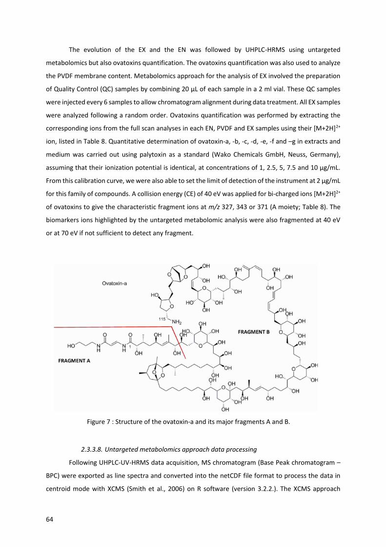
64
The evolution of the EX and the EN was followed by UHPLC-HRMS using untargeted
metabolomics but also ovatoxins quantification. The ovatoxins quantification was also used to analyze
the PVDF membrane content. Metabolomics approach for the analysis of EX involved the preparation
of Quality Control (QC) samples by combining 20 µL of each sample in a 2 ml vial. These QC samples
were injected every 6 samples to allow chromatogram alignment during data treatment. All EX samples
were analyzed following a random order. Ovatoxins quantification was performed by extracting the
corresponding ions from the full scan analyses in each EN, PVDF and EX samples using their [M+2H]2+
ion, listed in Table 8. Quantitative determination of ovatoxin-a, -b, -c, -d, -e, -f and –g in extracts and
medium was carried out using palytoxin as a standard (Wako Chemicals GmbH, Neuss, Germany),
assuming that their ionization potential is identical, at concentrations of 1, 2.5, 5, 7.5 and 10 µg/mL.
From this calibration curve, we were also able to set the limit of detection of the instrument at 2 µg/mL
for this family of compounds. A collision energy (CE) of 40 eV was applied for bi-charged ions [M+2H]2+
of ovatoxins to give the characteristic fragment ions at m/z 327, 343 or 371 (A moiety; Table 8). The
biomarkers ions highlighted by the untargeted metabolomic analysis were also fragmented at 40 eV
or at 70 eV if not sufficient to detect any fragment.
2.3.3.8. Untargeted metabolomics approach data processing
Following UHPLC-UV-HRMS data acquisition, MS chromatogram (Base Peak chromatogram –
BPC) were exported as line spectra and converted into the netCDF file format to process the data in
centroid mode with XCMS (Smith et al., 2006) on R software (version 3.2.2.). The XCMS approach
Figure 7 : Structure of the ovatoxin-a and its major fragments A and B.

Interactions allélopathiques entre Ostreopsis cf. ovata et une diatomée compétitrice
65
involved several steps necessary to generate a final matrix: (1) Peak picking (peakwidth= c(2, 20),
ppm=10) without threshold prefilter (Patti et al., 2012), (2) retention time correction
(method=”obiwarp”), (3) grouping (bw=10, minfrac=0.3, minsamp=1), (4) Fillpeaks using a parameter
of “noise level” that sets the minimum intensity for a centroid data point to be considered as part of a
peak (noise level = 103), and finally (5) report and data matrix generation (m/z / Retention time) that
was exported into Microsoft Excel. Two successive filtering steps were applied to the matrix on R in
order to suppress the data of high analytical variability: (i) variables are not considered if their mean
intensities in the samples extracts is less than 10-fold the values obtained for the blank samples
(culture medium extracts), and (ii) coefficient of variation in the intensity of the variables for the QC
samples > 20 %. Each peak area was further normalized, mean-centered and finally log-transformed
using on-line MetaboAnalyst 3.0 (Xia et al., 2015). Multivariate analysis was conducted (Principal
components analysis (PCA) and Partial Least Squares-Discriminant Analysis (PLS-DA) and the
discriminant PLS-DA was further chosen for further discussion and identification of the variable
important for the discrimination. Variable Importance in Projection (VIP) are identified by PLS-DA
together with the relative concentration of the corresponding metabolites for each group of samples
under study.
2.3.3.9. Statistical analysis
The statistical significance of the variation of the various parameters measured (ovatoxin
concentration, photosynthetic activity, cell abundance) between conditions was evaluated applying a
Wilcoxon-Mann-Whitney test on the data set, using the R software.
2.4. Results
2.4.1. Exometabolomes activity assessed by bioassays
The bioactivity of the exometabolomes (EX) of both Ostreopsis cf. ovata and Licmophora
paradoxa was tested on the other species. A small aliquot of the EX (10% of the final volume) was
added to the culture at early log phase and the in vivo fluorescence of the microalgae was measured
after 24 h of incubation (Figure 8) as a representative measurement of the cells physiological state.
Table 8 : List of the ovatoxins investigated in this study and their major ions.

66
A significant decrease of the in vivo fluorescence (Fv/Fm<0.1) of L. paradoxa was measured after 24 h
in the presence of EX of O. cf. ovata. Interestingly, a decrease in the in vivo fluorescence was also
measured for Ostreopsis cells after addition of the EX produced by Licmophora (Fv/Fm < 0.430).
2.4.2. Physiological effects of distant chemical interactions using co-cultures
The first results on the toxicity of each EX obtained from the bioassays guided the co-culture
experiment set up. A distant deleterious allelopathic effect of O. cf. ovata onto L. paradoxa physiology
was therefore expected in co-culture experiments. Interestingly, the foreseen effect was not observed
while the presence of L. paradoxa in the second chamber affected the abundance and the physiological
state of O. cf. ovata cells in the co-cultured chamber. The growth exponential phase lasted 10 and 7
days for O. cf. ovata and L. paradoxa respectively and remained stationary until the end of the
experiment at day 13 (Figure 9 A and B). Both the cell abundance and the physiological state, assessed
by the in vivo fluorescence, showed no significant differences between the mono-culture control and
the co-culture for most of the duration of the experiment for L. paradoxa (Figure 9 B, D) except at day
10. On the contrary, O. cf. ovata growth was reduced throughout the experiment as shown by the
significant decrease in both the cell abundance and the in vivo fluorescence at days 5, 12 and 13 (Figure
9 A and C). This effect was consistent with the decrease of the in vivo fluorescence of O. cf. ovata cells
in presence of the EX of Licmophora observed in the bioassays. The diatom culture contains
metabolite(s) that presents deleterious effects on O. cf. ovata photosynthetic apparatus. These
metabolites can diffuse through the PVDF membrane whereas the toxic metabolites produced in O. cf
ovata cultures possibly did not diffuse through the separating PVDF membrane.
Figure 8 : Effect of the EX of (i) L. paradoxa onto the in vivo fluorescence(Fv/Fm) of O. cf. ovata (left)
and (ii) O. cf. ovata onto L. paradoxa (right). Controls were performed with sterile media. Statistical
significance was assessed using a Wilcoxon-Mann-Whitney test (* p < 0.1). errors bars correspond to
standard error between samples (n=4).

Interactions allélopathiques entre Ostreopsis cf. ovata et une diatomée compétitrice
67
2.4.3. Ovatoxin content in EN and EX
The concentration of ovatoxins in O. cf. ovata cells (EN) was assessed in both the control and
the co-cultured samples to investigate the influence of the presence of other species on the production
of toxins. Using a palytoxin standard for mass spectrometry calibration, we evidenced that
concentrations of ovatoxins in the control were quite similar to values published on other strains (18
against 15 pg/cell for 3 000 000 cells; Pezzolesi et al., 2012). Figure 10 EN shows a clear increase in the
production of ovatoxins by O. cf. ovata cells when co-cultured with L. paradoxa, and this is especially
intense for the ovatoxin-a, from 6.6 ± 0.5 to 17.8 ± 6.6 pg. cell-1 eq. pltx (factor 2.7).
Figure 9 : Abundance of cells (cells/mL) and in vivo fluorescence (Fv/Fm ratio) of control (dark grey)
and co-cultured (light grey) chambers for O. cf. ovata (A, C; left side) and L. paradoxa (B,D; right side).
Statistical significance was assessed using a Wilcoxon-Mann-Witney test (* and · p < 0.1 and 0.2
respectively).

68
In the same manner, the toxin content in the EX followed a similar trend with an increase of all
ovatoxins concentration in the medium of co-cultured samples (from 0.52 to 1.03 pg.cell-1 eq pltx for
ovatoxin-a) although these data were not significant due to high inter-samples variability (Figure 10
EX). The measurement of the toxin content in the EN and the EX further allowed an estimation of the
percentage of toxins released by the cells in the medium (exudation) for both conditions, control and
co-culture (Table 9). Interestingly, the presence of L. paradoxa did not enhance the exudation of
ovatoxins in the medium despite the increased production in the EN (except for the ovatoxins d/e –
Increase by a factor 2).
2.4.4. Exchange of allelochemicals trough the membrane
Ovatoxins – Since no deleterious effects of the EX of O. cf. ovata was observed on the development of
L. paradoxa in co-cultures, an absence of diffusion through the PVDF membrane was considered.
Figure 10 (part EX) reveals the presence of ovatoxins in the EX of all chambers containing O. cf. ovata
(1.7 pg. cell-1 eq pltx) whereas on the other side of the PVDF membrane of these co-cultures, no
ovatoxins were quantifiable. Ovatoxins did not or just weakly diffuse through the PVDF membrane into
the blank or co-culture chambers and we could show this absence of diffusion was not due to an
adsorption on the PVDF membrane since no ovatoxins were detected in the PVDF extracts (data not
shown).
ovtx a ovtx b ovtx c ovtx a ovtx b ovtx c
Figure 10 : Toxin content in the (EN) endometabolome and in the (EX) exometabolome normalized
by the number of cells ; in controls= cultures (dark grey) and co-cultures (light grey). A palytoxin
standard was used to obtain the calibration curve and the concentrations are therefore expressed as
an equivalent palytoxin. Statistical significance was assessed using a Wilcoxon-Mann-Whitney test
(*, p < 0.1). Error bars define the standard errors between samples (n=3).

Interactions allélopathiques entre Ostreopsis cf. ovata et une diatomée compétitrice
69
Other allelochemicals from O. cf. ovata – Interestingly, the untargeted metabolomics approach
revealed that other compounds of large molecular weight (2120 Da) also present in O. cf. ovata cells
were exuded and then diffused through the PVDF membrane (Table in Figure 11A). Indeed, the diatom
culture EX contains those compounds when co-cultured with the dinoflagellate (Figure 11A). The three
ions at m/z 1046.0720; 1065.0463 and 1080.0515 were identified as dicharged adducts of the
monocharged [M+H]+=2121.1523: [M+H+K]2+=1080.07615, [M+2Na-2H2O]2+=1065.07615 and
[M+2Ca-6H2O]2+=1046.07615. It is worth noting that fragmentation experiments were performed on
those ions using a collision energy of 40 and 70 eV but no fragmentation was evidenced. Therefore,
the compounds diffusing through the membrane do not share the major fragments found for the
compounds belonging to the ovatoxin family (327.1923; 343.1861 and 371.2174) and these
compounds clearly differ for the part A of the molecule.
Allelochemicals from L. paradoxa – Likewise, the untargeted metabolomics approach revealed the
production of metabolites by the diatoms’ culture that were able to diffuse through the PVDF
membrane (as shown by the presence of several ions listed in the table of Figure 11B for EX of O. cf.
ovata when co-cultured with the L. paradoxa). Those ions showed a mid-polarity (Rt 6.8 min) and a
molecular formula was proposed for each of them by the software Data Analysis® and could
correspond to small oxylipins (C11, C15 and C18). About 20% of the concentration of those compounds
detected in the EX of the diatom culture were retrieved on the PVDF membrane. Interestingly, those
ions were not found in the EN of L. paradoxa, suggesting that either those compounds are exuded but
not stored by the diatom or they have a bacterial origin.
Table 9 : Percentage of exudation (ratio EN/EX of ovatoxin concentration) of all detected toxins in the
control and in the co-culture chambers that contained Ostreopsis cultures.
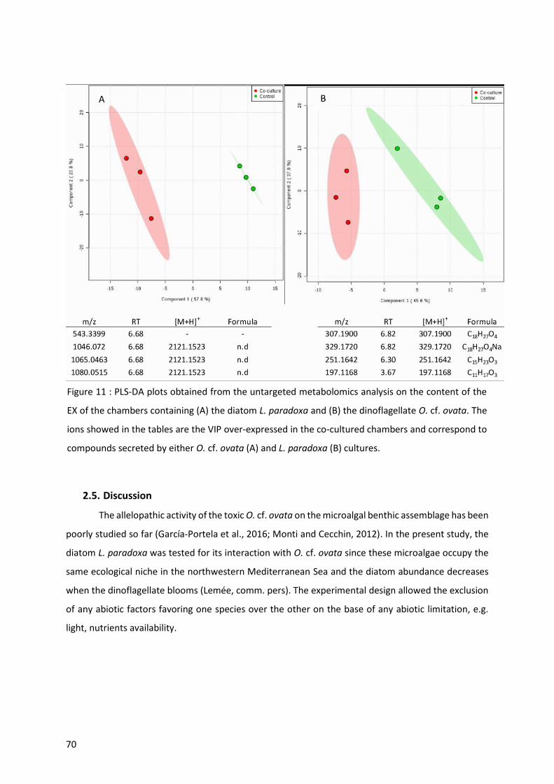
70
2.5. Discussion
The allelopathic activity of the toxic O. cf. ovata on the microalgal benthic assemblage has been
poorly studied so far (García-Portela et al., 2016; Monti and Cecchin, 2012). In the present study, the
diatom L. paradoxa was tested for its interaction with O. cf. ovata since these microalgae occupy the
same ecological niche in the northwestern Mediterranean Sea and the diatom abundance decreases
when the dinoflagellate blooms (Lemée, comm. pers). The experimental design allowed the exclusion
of any abiotic factors favoring one species over the other on the base of any abiotic limitation, e.g.
light, nutrients availability.
Figure 11 : PLS-DA plots obtained from the untargeted metabolomics analysis on the content of the
EX of the chambers containing (A) the diatom L. paradoxa and (B) the dinoflagellate O. cf. ovata. The
ions showed in the tables are the VIP over-expressed in the co-cultured chambers and correspond to
compounds secreted by either O. cf. ovata (A) and L. paradoxa (B) cultures.

Interactions allélopathiques entre Ostreopsis cf. ovata et une diatomée compétitrice
71
2.5.1. Ostreopsis cf. ovata chemical interactions may need cell-cell contact
A weak allelopathic activity of O. cf. ovata on the benthic dinoflagellate Coolia monotis has
been observed by testing fresh exudates devoid of cells (García-Portela et al., 2016; Monti and Cecchin,
2012). Conducting similar experiments on the benthic diatom L. paradoxa in log phase (e.g. bioassays),
the exudate of O. cf. ovata clearly impaired its photosystem II (PS II) in less than 24 h, suggesting a
stress induced by putative allelochemicals. Allelopathy may be used by O. cf. ovata to outcompete
other microalgal species as observed for Karenia brevis (Kubanek et al., 2005; Prince et al., 2008) or
Microcystis aeruginosa (Dunker et al., 2017). However, co-occurring species can have a different
response to allelochemicals (Kubanek et al., 2005; Poulson-Ellestad et al., 2014; Prince et al., 2008) and
more investigation should be undertaken using various benthic microalgae in order to obtain a full
comprehension of allelopathic activity of O. cf. ovata.
Interestingly, the second set of experiments using co-culture devices did not confirm the
allelopathic character of O. cf. ovata. The experimental set up prevented the contact between cells of
the two species but allowed the diffusion of compounds from one species to the other. As a result,
neither the growth nor the PS II of L. paradoxa was impaired throughout this 13-days experiment by
the presence of O. cf. ovata. The potential for exchange of the allelochemicals through PVDF
membrane-separated culture chambers is thus not sufficient for an allelopathic control of O. cf. ovata
onto L. paradoxa. This unexpected result indicates the inhibitory compounds exuded by O. cf. ovata
are not diffused through the filtering membrane and are suspected to remain in the close vicinity of
the cells. Indeed, in bioassays, the exudates were obtained by a soft centrifugation and filtration of the
cultures. The allelochemicals, if adsorbed onto the cells or the mucus, may have been extracted with
the aqueous phase. This would increase the concentration of their free-dissolved form in the exudate,
leading to the observed toxicity. In the co-culture experiment, the gentle homogenization of the co-
culture chambers performed three times a day might not have been sufficient to dissolve the active
compounds from the cells or the mucus precluding from any diffusion through the PVDF membrane.
Therefore, contact interaction between cells is most likely needed to impair the growth of the diatom
L. paradoxa. The existence of aggregates (e.g. mucus) produced by O. cf. ovata in which competitors
could be maintained at a close distance is a strong indication towards this conclusion. Similar contact
interactions were already observed for cyanobacteria (Dunker et al., 2017) or dinoflagellates
(Fernández-Herrera et al., 2016; Kubanek et al., 2005; Tillmann, 2003) and is supported by simple
mathematical models (Jonsson et al., 2009).
72
2.5.2. Ovatoxins may be in involved in Ostreopsis cf. ovata allelochemistry
Although ovatoxins synthesized by O. cf. ovata (Ciminiello et al., 2012a) might be good
candidates as allelochemicals, no experimental result has supported this hypothesis so far (García-
Portela et al., 2016; Giussani et al., 2015). It is worth noting the absence of toxicity of the exudate
found by Giussani et al. (2015) was linked to an absence of ovatoxins in the 0.2 µm filtered exudate.
Our findings were slightly different with low levels of ovatoxin-a found in the EX (<2 pg.cell-1 eq pltx),
using a different protocol for chemical extraction. Therefore, the involvement of the ovatoxins in the
deleterious effects observed on the diatom cells cannot be ruled out at this stage. From this
perspective, the absence of allelopathic effect of O. cf. ovata onto L. paradoxa in the co-culture
chambers was actually a good opportunity to test ovatoxins as the compounds responsible for the
growth impairment observed in the bioassays. Indeed, the barely detectable concentrations of
ovatoxins in the diatoms’ EX when co-cultured with O. cf. ovata indicated a weak potential of diffusion
of the ovatoxins far from the secreting cells. The reason for the absence of diffusion of the ovatoxins
through the membrane is likely due to their physico-chemical properties. Ovatoxins are amphiphilic
compounds (Botana, 2014) that can be stored in the mucus (Giussani et al., 2015). Any adsorption of
ovatoxins on the PVDF membrane hampered their diffusion and several studies have shown that
complete exchange of substances (heptadienal, dimethylsulfonopropionate and Microcystin-LR)
through the 0.2 µm PVDF membrane would happen by diffusion within 24 h (Dunker et al., 2017; Paul
et al., 2013). In conclusion, the absence of diffusion of ovatoxins associated to healthy diatom cells in
co-culture experiments suggests that if ovatoxins are involved in growth impairment of competitors,
it would most likely be through contact interactions.
The question whether toxins from microalgae act as distance or contact allelochemicals
remains controversial since experimental evidences showed on one hand some toxins, for instance
karlotoxin-1 and -2 (K. veneficum; Van Wagoner et al., 2008), are involved in prey capture (Sheng et
al., 2010), allelopathy (Adolf et al., 2006) and mechanistic interactions with other phytoplankton
species (Deeds and Place, 2006). On the other hand, the exudation of other toxins such as brevetoxins
(K. brevis, Kubanek et al., 2005) are unlikely involved in growth control of co-occurring microalgae
(Poulson-Ellestad et al., 2014; Prince et al., 2008). Finally, exudates of K. brevis were shown to stimulate
the growth of several of its diatoms competitors (Poulson-Ellestad et al., 2014).
Interestingly, other organic compounds were secreted by O. cf. ovata cells and diffused
through the PVDF membrane as revealed by the untargeted metabolomics approach. Those putative
allelochemicals are assumed to be adducts of one single compound having a molecular weight of 2120
Da, highlighting the potential for O. cf. ovata to produce other large molecules than ovatoxins. Indeed,
the fragmentation experiment did not allow to establish a link between this unidentified compound
biosynthesized by O. cf. ovata and the ovatoxins family since the ovatoxins’ fragment-A (Figure 7,

Interactions allélopathiques entre Ostreopsis cf. ovata et une diatomée compétitrice
73
Brissard et al., 2015; Ciminiello et al., 2008, 2012a) was not evidenced, and no other internal fragment
matched (Brissard et al., 2015; Garcia-Altares et al., 2015). Thus, the combination of the two
experiments underlines the urgent need for a characterization of both the endo- and the exo-
metabolome of O. cf. ovata in order to better understand the role of its specialized metabolome in
chemical interactions.
2.5.3. Allelochemistry of Licmophora paradoxa
Unexpectedly, the exudates of L. paradoxa cultures also slightly but significantly inhibited the
O. cf. ovata’s PSII in the bioassays. Some allelochemicals exuded by the diatom culture inhibited the
growth of the dinoflagellate. Interestingly the co-cultures revealed similar effects on the development
of O. cf. ovata since both its growth and PSII were modified in the presence of the diatom culture
although contact between cells was not allowed. A diffusion of chemicals affecting the development
of the dinoflagellate cells supports the existence of a distant allelopathic effect by L. paradoxa culture
on O. cf. ovata. This result is in accordance with the very recent findings of Pichierri et al., (2017) who
showed a growth inhibiting effect of several diatoms filtrates onto the DNA content of O. cf. ovata.
The identity of the compound (s) involved in this interaction has not been investigated so far although
an untargeted metabolomics analysis suggests a role of several compounds (C18H27O4, C15H23O3,
C11H17O3) enriched in the EX of O. cf. ovata when co-cultured with the diatoms. Nevertheless, there is
no clue so far whether those compounds can be held liable for the impairment of the dinoflagellate
growth. Recent research on diatoms exo-metabolome showed that compounds belonging to the
polyunsaturated fatty acids (PUFAs) as well as the polyunsaturated aldehydes (PUAs) families are, in
some cases, responsible for allelopathy (Ianora et al., 2011; Pohnert, 2005). Wichard et al. (2005)
evaluated over 71 diatom studied species, and 38% are PUA-producers. Although L. paradoxa species
are not reported in their study, they may also synthesize one or several of those compounds. The
chemical markers highlighted by the metabolomics analysis show similar molecular composition to the
PUFAs and PUAs involved in diatoms allelochemistry (see Pohnert, 2005 for a review). In particular,
C16, C17 and C18 fatty acids were shown to be biosynthetic precursors of bioactive short-chain aldehydes
and oxylipins (Ackman et al., 1968; Domergue et al., 2003; Pohnert, 2005). The extraction procedure
of the EX employed in our study precluded the detection of any potential aldehydes, known to be
highly reactive, but would have allowed the detection of fatty acids and oxylipins. A control of O. cf.
ovata growth by PUAs or PUFAs is thus highly plausible since a recent study shows the addition of three
PUAs (2E, 4E-decadienal, 2E, 4E-octadenial and 2E, 4E-hepatdenial) in O. cf. ovata cultures inhibited
the PS II after 48 to 72 h of exposure (Pichierri et al., 2016). Yet, the concentrations tested by the
authors of this study were 5 to 10 times higher than levels encountered in situ (Ribalet et al., 2014).
Since no thorough identification of the allelochemical(s) inhibiting the growth of O. cf. ovata was

74
attempted, its synthesis by the diatom L. paradoxa cannot be secured and a potential bacterial origin
can also be considered. Algicidal bacteria are widely distributed in the marine environment and can
exert a control on the growth and the lysis of microalgae (Meyer et al., 2017 for a review). Diatoms
blooms were shown to provide habitat for algicidal bacteria (Park et al., 2010) and a control of
dinoflagellate growth by algicidal compounds has often been reported (Park et al., 2010; Pokrzywinski
et al., 2012; Roth et al., 2008). Future research should be directed towards the understanding of the
potential role of algicidal bacteria in the chemical inhibition of O. cf. ovata growth by the culture of L.
paradoxa, using axenic cultures.
2.6. Conclusion
The chemical interactions between O. cf. ovata and one of its competitors observed with these
laboratory experiments give some insights on the mechanisms that may occur in the field. The
detection of the presence of a competitor clearly stimulated the production of ovatoxins by O. cf. ovata
most likely by activating the corresponding biosynthetic pathway. Therefore, a chemical control
performed by the toxins of O. cf. ovata on its competitors is plausible and may impact the species
succession during its bloom. A debate is currently ongoing to understand whether bloom initiation of
HABs species could be led by allelopathy (Jonsson et al., 2009). The latest authors estimate that at the
beginning of a bloom, cell densities are too low to concentrate enough infochemicals and that cell-cell
interactions offer an alternative transmission for allelopathic compounds, and may therefore allow
chemical compounds such as toxins to control bloom initiation. The results obtained in the present
study support this hypothesis, cell-cell contact or a close interaction is most likely needed to strongly
impair the growth of competitor species. This hypothesis is particularly relevant for benthic microalgae
species that accumulate on the substrate and would therefore concentrate their toxins nearby.
Nevertheless, the nature of the O. cf. ovata allelochemicals and their role in shaping microalgae
communities still remain to be addressed. Further work should concentrate on identifying the
allelochemicals as well as defining their mode of action.
A control of the competitor growth appeared to be intense at a sensitive growth stage (early
log phase) of L. paradoxa cell development, and more research is needed to evaluate an eventual
control on other growth stages. In order to verify the hypothesis of a chemical control on species
succession, waterborne exudates should be sampled at different phases of the bloom (early log, log
and stationary phases) and tested against several co-occurring competitor species since effects could
differ among species (García-Portela et al., 2016; Monti and Cecchin, 2012). Additionally, cell-cell
chemical interaction between O. cf. ovata and its competitors should be investigated taking into
account the difficulty of such a detection with the available chemical imaging.

Interactions allélopathiques entre Ostreopsis cf. ovata et une diatomée compétitrice
75
2.7. Acknowledgments
This study was funded by the french ANR: project OCEAN15 (2016-2020). The authors also
thank the French GDR Phycotox for enabling fruitful discussions and meetings. The metabolomics
study was supported by the MALLABAR platform (IMBE, Marseille France).

76
Interactions allélopathiques entre Ostreopsis cf. ovata et une diatomée compétitrice
77
3. Synthèse des résultats
La diatomée est inhibée par le filtrat cellulaire d’Ostreopsis cf. ovata ; toutefois, aucun effet
néfaste n’est observé, pour la diatomée, lors des co-cultures. La production d’OVTXs par O. cf. ovata
est pourtant avérée (par étude métabolomique ciblée) mais il semblerait qu’en raison de leurs
propriétés intrinsèques, les OVTXs ne traversent pas la membrane utilisée dans les systèmes de co-
culture suggérant ainsi une toxicité d’O. cf. ovata par contact. Ceci confirme également l’implication
des toxines produites par O. cf. ovata dans les relations allélopathiques qu’il entretient avec son
environnement.
Cette étude a également permis de mettre en évidence, par l’utilisation d’une approche
métabolomique non ciblée, la production de nouveaux composés par O. cf. ovata. Ces composés
n’appartiennent pas à la famille des OVTXs mais sont impliqués dans les relations allélopathiques que
ce dinoflagellé entretient avec son environnement.
Enfin, la croissance et l’activité photosynthétique du PSII d’O. cf. ovata ont été affectés par la
présence de la diatomée. Une augmentation de la production de toxines est également notée. Ces
effets sont certainement liés à la production, par la diatomée, des nouveaux composés identifiés dans
cette étude.
Cette étude met en évidence la complexité des interactions qu’O. cf. ovata entretient avec son
environnement et la méconnaissance des mécanismes associés.


Interactions entre Ostreopsis cf. ovata et consommateurs primaires : cas du copépode harpacticoide Sarsamphiascus cf. propinquus
Copepoda. Plate of Wilhelm Giesbrecht (1892)

80

Interactions entre Ostreopsis cf. ovata et prédateurs : cas des copépodes benthiques
81
1. Présentation de l’étude
Une étude réalisée in situ en Méditerranée Nord Occidentale a mis en évidence une diminution
significative du nombre de nauplii de copépodes benthiques lors des efflorescences d’Ostreopsis cf.
ovata (Guidi-Guilvard et al., 2012). Les auteurs ont suggéré un impact de ce dinoflagellé sur les
copépodes benthiques, amenant à un changement des communautés méiobenthiques.
L’objectif de la présente étude est de vérifier, en laboratoire, l’impact d’Ostreopsis cf. ovata
sur les copépodes benthiques en étudiant différents paramètres tels que la survie, l’ingestion, ou la
reproduction. Pour cela, le copépode benthique Sarsamphiascus cf. propinquus, a été isolé en rade de
Villefranche-sur-Mer depuis des macroalgues sur lesquelles O. cf. ovata, a été sélectionné comme
modèle d’étude. Maintenu en culture en laboratoire, ce copépode a ensuite été exposé à différentes
concentrations d’O. cf. ovata afin d’étudier la toxicité aigüe d’O. cf. ovata sur cette espèce. L’ingestion
des cellules de dinoflagellé a été vérifiée à travers des observations microscopiques et l’estimation de
la production de pelotes fécales. L’impact des métabolites produits par O. cf. ovata, sur la reproduction
de ce copépode, a été étudié via la mesure des taux de fécondité et de fertilité.
2. Effects of the toxic dinoflagellate Ostreopsis cf. ovata on survival, feeding and
reproduction of a phytal harpacticoid copepod
L’article qui suit a été publié dans Journal of Experimental Marine Biology and Ecology en juillet
2019 (Volume 516, pages 103-113).

82

Interactions entre Ostreopsis cf. ovata et prédateurs : cas des copépodes benthiques
83
Effects of the toxic dinoflagellate Ostreopsis cf. ovata on survival, feeding and
reproduction of a phytal harpacticoid copepod
Pavaux Anne-Sophie1, Rostan Julie1, Guidi-Guilvard Laurence1, Marro Sophie1, Ternon Eva², Thomas
Olivier P.3, Lemée Rodolphe1, Gasparini Stéphane1
1 Sorbonne Université, CNRS, Laboratoire d’Océanographie de Villefranche, LOV, F-06230 Villefranche-sur-Mer,
France
² Université Côte d’Azur, CNRS, OCA, IRD, Géoazur, 250 rue Albert Einstein, 06560 Valbonne, France
3 Marine Biodiscovery, School of Chemistry and Ryan Institute, National University of Ireland Galway, University
Road, H91 TK33 Galway, Ireland
Key words: Benthic HABs, Ostreopsis cf. ovata, chemical ecology, meiobenthic copepods,
reprotoxicity.
2.1. Abstract
Harmful algal blooms are a source of increasing concern within the health, economic and
ecological sectors. In the Mediterranean Sea, severe blooms of the benthic dinoflagellate Ostreopsis
cf. ovata have been occurring since the beginning of the century, causing human intoxications by
inhalation of bio-aerosols or direct contact with cells. The toxicity of this dinoflagellate is attributed to
the presence of palytoxin and several of its analogs called ovatoxins, palytoxin being one of the most
potent marine toxins. While mass mortalities of marine invertebrates have already been reported in
relation with O. cf. ovata blooms, the toxic effects of this dinoflagellate on benthic organisms is still
poorly documented. In the present study, laboratory experiments were performed on a meiobenthic
copepod (Sarsamphiascus cf. propinquus), which naturally lives on macrophytes in close contact to O.
cf. ovata, in order to assess its potential toxic effects on mortality, fecal pellet production (as a proxy
of feeding), as well as fecundity and fertility ratios. Both, O. cf. ovata as well as a non-toxic competitive
diatom (Licmophora paradoxa), were used as food in the experiments. Regarding acute toxicity
evaluation, this copepod proved to be the most tolerant organism to O. cf. ovata reported to date.
Nevertheless, its fecundity and fertility ratios were lower when fed with the toxic dinoflagellate,
indicating a possible reprotoxic effect. Moreover, although fecal pellet production decreased
significantly when the copepod was fed with a mono-diet of O. cf. ovata, epifluorescence microscopy
observations revealed the presence of the toxic cells inside the digestive track, hence suggesting that
these primary grazers could be a vector of toxins through the marine food web.

84
2.2. Introduction
Over the past decades, the occurrence of Harmful Algal Blooms (HABs) has been increasing in
frequency, intensity and geographic distribution (Berdalet et al., 2017; Glibert et al., 2005). These
blooms negatively impact human health and wellbeing, by affecting coastal ecosystem services
(Berdalet et al., 2015) as well as marine organisms and ecosystems (Dolah et al., 2001). In this regard,
the strong geographical expansion of the toxic dinoflagellate Ostreopsis cf. ovata in the Mediterranean
Sea constitutes an emerging problem (Parsons et al., 2012). This dinoflagellate has long been regarded
as a potential vector involved in ciguatera fish poisoning (Glaziou and Legrand, 1994; Hallegraeff,
1993), and it has more recently been shown to produce palytoxin (PLTX) and palytoxin-like compounds
(Ciminiello et al., 2006) named ovatoxins (OVTXs) a potential cause of toxic bio-aerosols causing human
respiratory illnesses (Ciminiello et al., 2014). Ostreopsis cf. ovata is a benthic dinoflagellate usually
described as epiphytic on macroalgae and seagrasses growing on rocky shallow seabeds (Mangialajo
et al., 2008; Totti et al., 2010) but has also been reported to have a planktonic phase (Vila et al., 2016).
Several studies on invertebrate mass mortalities during Ostreopsis cf. ovata blooms (Granéli et
al., 2002; Shears and Ross, 2009) suggest that this dinoflagellate may be harmful to benthic organisms
living nearby even if its effects may be confounded with other stress factors such as pollution or hypo-
/anoxic conditions (Shears and Ross, 2009; Shears and Ross, 2010). The toxic effect of O. cf. ovata has
previously been investigated on several organisms using ecotoxicological bioassays. For instance, the
jellyfish Aurelia sp., has been shown to be particularly sensitive at the ephyra (or larval) stage (Giussani
et al., 2016) and the mussel Mytilus galloprovincialis reacted to O. cf. ovata exposures by modifying
immunological, histological and oxidative levels (Gorbi et al., 2013). Also, fertilization and early
development of the sea urchin Lytechinus variegatus were affected by O. cf. ovata (Neves et al., 2018).
Moreover, a decrease of more than 98.5% of gill cell viability was observed in response to O. cf. ovata
exposure (Verma et al., 2016). Even though meiofauna living on macrophytes play important roles in
benthic biochemical and ecological processes, little is known on the effects of this toxic microalga on
the surrounding community. Only one in situ study has previously investigated the impact of O. cf.
ovata on phytal meiofauna, and results showed a severe decrease in the number of nauplii during the
toxic blooms, suggesting a reprotoxic effect of the dinoflagellate on harpacticoid copepods (Guidi-
Guilvard et al., 2012).
The aim of the present study was to provide new insights on the potential effect of the toxic
benthic dinoflagellate Ostreopsis cf. ovata on meiofauna, and more specifically on benthic copepods.
A harpacticoid copepod belonging to the genus Sarsamphiascus (formerly Amphiascus) was isolated
from the natural environment where O. cf. ovata summer blooms occur with densities reaching 4. 106
cells.gFW-1 (Cohu et al., 2013) and was here used as a model organism. Another copepod species

Interactions entre Ostreopsis cf. ovata et prédateurs : cas des copépodes benthiques
85
belonging to the same genus has already been widely used as a model in toxicity tests (Bejarano and
Chandler, 2003; Cary et al., 2004; Chandler et al., 2004). This organism was shown to be relevant for
ecotoxicological assays due to their small size, short life cycle, simplicity to maintain and manipulate
in laboratory conditions and sensitivity to a wide array of toxic substances. In the present laboratory
study, the selected copepod was exposed to ecologically relevant concentrations of the dinoflagellate
O. cf. ovata (Accoroni et al., 2012; Cohu et al., 2013) as well as Licmophora paradoxa, a diatom
frequently found associated with O. cf. ovata in benthic assemblages (Lemée, personal
communication), which will here be used as a control. The effects of O. cf. ovata were investigated by
recording three biological parameters of the copepod: survival, food uptake (assessed through the
number of fecal pellets produced) and reproduction (fertility and fecundity ratios). The toxicity of the
strain used in these experiments was evaluated using Artemia franciscana toxicity bio-assay and
confirmed by UHPLC-HRMS quantification of ovatoxins.
2.3. Materials and Method
Algal cultures
The dinoflagellate Ostreopsis cf. ovata (MCCV 054) and the diatom Licmophora paradoxa
(MCCV 033) used as a control microalga were isolated from macroalgal samples collected at the
Rochambeau site (Bay of Villefranche-sur-mer, N-W Mediterranean, 43°41’35.64’’N-7°18’31.54’’E).
The MCCV is the Mediterranean Culture Collection of Villefranche, where strains are maintained and
available for the scientific community. Both strains were cultured in 150 mL flasks containing L1
medium (Guillard and Hargraves, 1993) prepared with autoclaved aged and 0.2 µm filtered seawater,
adjusted to a salinity of 38. All the cultures were maintained at 24°C, under a 14:10 light/dark cycle
(light intensity 250 µmol.m-2.s-1). Cells of O. cf. ovata used for the experiments were collected from the
cultures during the exponential phase. They were counted in 12 mL aliquots (triplicates) fixed with
acidic lugol (4% v/v) using a liquid particle counter (HIAC/Royco 9703, Pacific Scientific Instruments)
following a size range of 2-80 µm (Stramski et al., 2002).
Cell diameter range of O. cf. ovata and L. paradoxa in culture were 20-40 µm and 15-20 µm,
respectively. Biovolumes were calculated assuming O. cf. ovata (1) had the shape of a cone with a half
sphere, and L. paradoxa (2) was a truncated cone, using the following equations (Olenina et al., 2006):
(1) V = π
12× h × 𝐷2 (2) V =
𝜋
12× (𝑑1
2 + 𝑑1𝑑2 + 𝑑22)
where V = volume, h = height, D = diameter, d1 = large diameter, d2 = small diameter. The biovolume
ratio between the two microalgae was 1:6, i.e. O. cf. ovata (average biovolume = 17 360 µm3) was 6
times larger than L. paradoxa (average biovolume = 2 968 µm3). These biovolumes were calculated
using 15 cells for each strain.
86
2.3.2. Toxicity of the strain
2.3.2.1. Artemia franciscana toxicity bioassay
The level of toxicity of the O. cf. ovata strain used for this study was checked by performing a
standard Artemia franciscana test (Faimali et al., 2012; Neves et al., 2017). The high sentivity of
Artemia to toxic substances makes it suitable for ecotoxicological assessment (Kalčíková et al., 2012;
Nunes et al., 2006). Such bio-assays, described as simple, inexpensive and convenient, can be used to
test the toxicity of harmful algal strains (Neves et al., 2017). Cysts of A. franciscana (Ocean Nutrition
Sep-art) maintained at 4°C in the dark, were incubated in 2 L of filtered seawater (salinity 38), at 20°C
with vigorous and continuous aeration until reaching the most sensitive development stages (Stages
2-3, Kerster and Schaeffer, 1983). The larvae were collected using a glass pipette and phototactism
and further transferred to 6-well plates. Five larvae were placed in each wells, using one 6-well plate
per experimental condition. A total of 4 well-plates were used to investigate the effects of the following
cell concentrations of O. cf. ovata: 4, 40, 400 and 4000 cells.mL-1.
The ratio number of algal cells: number of target animals was then between 20:5 to 20 000:5.
The number of dead larvae was estimated after 2, 4, 24 and 48 h exposure times. Larvae were
considered dead when there was no reaction to strong light or to glass pipette aspiration or when a
shift to white coloration of the body was observed. LC50 and LT50 values were calculated, corresponding
to the concentration which induced the mortality of 50% of the organisms and the lethal time (in hours)
to reach 50% mortality, respectively.
2.3.2.2. UHPLC-HRMS analyses
The toxicity of the strain was confirmed using chemical analyses. Ostreopsis cf. ovata cells were
grown in 300 mL flasks at previously described conditions and harvested at day 10. The culture was
further centrifuged at 600 g during 10 min at ambient temperature. The cell pellet was flash-freezed
using liquid nitrogen and stored at – 80 °C until extraction. The metabolites were extracted using 4 mL
of MeOH/H2O (80:20; v:v) and sonicating during 5 min in a cooled ultra-sonic bath. The extracts were
centrifuged at 2500 g during 10 min at ambient temperature and the supernatants were transferred
into 20 mL vials. These steps (MeOH, ultra-sonic bath and centrifugation) were repeated 3 times. The
resulting organomethanolic extracts were evaporated using a stream of nitrogen avoiding dryness; a
volume of 500 µl of DMSO was added in each extract and the remaining H2O was fully evaporated. All
samples were therefore stored in 100% DMSO (500 µl) at -20°C until UHPLC-HRMS analysis.
Ovatoxins quantification by UHPLC-UV-HRMS were performed using an Agilent 1290 system (Agilent
Technologies, USA) equipped with a diode array detector and coupled to an Agilent 6540 Qtof mass
spectrometer (Agilent Technologies, USA) by the injection of 10µL on T3 column (Acquity UPLC HSS T3
1.8µm, 2.1mm x 100mm, Waters). Separation was achieved using a linear elution gradient of

Interactions entre Ostreopsis cf. ovata et prédateurs : cas des copépodes benthiques
87
H2O:MeOH (80:20, v:v)/MeOH with 0.1mM of ammonium formate and 0.1% formic acid from 90:10
(v:v, isocratic from 0 to 2mins) to 0:100 (v:v, isocratic from 12 to 13 mins) with a 0.4 mL.min-1 flow rate.
UV detection was set at 210, 233 and 263 nm. Ions detections was recorded in positive mode (ESI +) in
the range 60 -3000 Da. Collision energies (CE) of 30, 70 and 110 eV were applied to confirm the
presence of ovatoxins moieties (Brissard et al., 2014; Ciminiello et al., 2012a). The spectrometer
analyzer parameters were set as follows: nebulizer sheath gas, N2 (35 psig); drying gas, N2 (11 L.min−1);
Gas Temperature, 300°C; capillary, 4.129 µA; Vaporizer/Sheat Gas Temp, 350°C.
Ovatoxins quantification was performed by extracting and integrating all tri-charged ions (from 858 to
910 m/z) from the base peak chromatogram. The concentration of ovatoxins is given in palytoxin
equivalent as commercially available standard of palytoxin (Wako Chemicals GmbH, Neuss, Germany)
was used to perform a calibration curve, assuming their ionization pattern is similar.
The model copepod
The harpacticoid copepod used in the experiments thrives in the shallow rocky shore f the
Marinières site (Bay of Villefranche-sur-Mer, N-W Mediterranean, 43°42'21.51"N – 7°19'07.44"E)
where it was collected in 2012 using a WP2 net towed over the macroalgal cover. In the laboratory,
samples were maintained in 10 L tanks together with a planktonic copepod (Acartia clausi) in 0.2-µm
filtered aged seawater (salinity 38) at 22°C, in the dark to avoid phototactism, and fed three times a
week with a mixture of the microalgae Dunaliella salina (MCCV 20) and Tisochrysis lutea (CCAP
927/14). The copepod belongs to the family Miraciidae Dana, 1946 (formerly Diosaccidae), and is part
of the varians-Group. Within this group, the genus Amphiascus, renamed Sarsamphiascus by Huys
(2009), is well known for taxonomic discrepancies and extreme difficulty in species identification
(Hicks, 1971; Wells, 2007). In the present study, the species name remains uncertain due to inter-
individual variation in the parapodial setation. However, among the possible varians-Group species,
only Amphiascus propinquus had been previously reported in areas close to the Bay of Villefranche-
sur-mer (i.e. the Italian Ligurian shore, Ceccherelli, personal communication). For all these reasons,
this species was named Sarsamphiascus cf. propinquus (Sars, 1906) in our study.
Experimental setup
All experiments were run in 6-well plates, each well containing 4 mL of autoclaved aged and
0.2-µm filtered seawater (salinity 38), at 24°C, in the dark. One milliliter of microalgae at the
appropriate cell concentration was added to the wells as food, except in the no-food controls which
received instead 1mL of clean culture medium. The content of each well was renewed every 48 hours
and wells were checked daily under a binocular microscope (ZEISS, SteREO Discovery V12).

88
Experiments involving the copepod S. cf. propinquus started with adults and late copepodites. Prior to
the experiments, individuals were collected from the culture tanks, sorted with an elongated Pasteur
pipette, flushed twice in seawater and transferred to the 6-well plates. They were left for 2 days
without food to allow gut clearance, except in the reproduction experiments where the food was
added from the start. At the end of some of the experiments, copepods were fixed with formalin (4%
v/v) to determine sex and overall size (from the tip of the cephalosome, excluding the rostrum, to the
end of the last body somite, excluding the caudal rami) using a NIKON AZ100 microscope coupled to a
Digital Imaging System equipped with the NIS-Elements software (Nikon Instruments Inc., New-York,
U.S.A.).
Acute toxicity of Ostreopsis cf. ovata on Copepods
To estimate and compare the level of toxicity of O. cf. ovata, the copepods were exposed to
five increasing cell concentrations (500, 1 000, 2 000, 4 000 and 20 000 cells.mL-1) and one no-food
control in six-well plates, as previously described for Artemia bioassay. The test involved 72 individuals
(12 per concentration and no-food control, 1 per well) and lasted 9 days. The ratio of algal cells/
number of target animals was between 2500:1 to 100 000:1. Mortality (as defined above for Artemia
bio-assay) was daily recorded in order to calculate LC50 and LT50.
Impact of Ostreopsis cf. ovata on copepod survival and food uptake
To investigate potential toxic effects of O. cf. ovata cells, mono- and mixed diets were fed to
the copepod. Five different treatments were applied: (i) O. cf. ovata at a concentration of 100 cells.mL-
1 reflecting the realistic bloom alert threshold measured in the natural environment (Lemée et al.,
2012; Tichadou et al., 2010) ; (ii) L. paradoxa at a concentration of 600 cells.mL-1 corresponding to the
microalgae supplied in treatment (i) in terms of bio-volumes ; (iii) a mixed diet of O. cf. ovata and L.
paradoxa (i.e. treatments (i) and (ii) combined) to investigate if the presence of the two microalgae
could change the response of the copepods; (iv) the mixed diet (iii) at half concentrations (i.e. O. cf.
ovata at 50 cells.mL-1 + L. paradoxa at 300 cells.mL-1) ; (v) a no-food treatment. To study both mortality
and ingestion of S. cf. propinquus, two experiments were performed during 10 days and 14 days, each
involving 150 individuals (30 per treatment, 1 per well). Mortality (as previously defined) was assessed
daily, while feeding (assessed through the number of fecal pellets produced by alive copepods ; Souza-
santos et al., 1995) every 48 h, at the time of medium renewal. Copepods that had been in contact
with O. cf. ovata alone were observed under an epifluorescence inversed microscope (Axio Scope.A1,
UV excitation, red fluorescence) to check for the presence of O. cf. ovata cells in the digestive track.

Interactions entre Ostreopsis cf. ovata et prédateurs : cas des copépodes benthiques
89
Impact of Ostreopsis cf. ovata on reproductive performances of Copepods
The potential effects of O. cf. ovata on the reproduction capacity of S. cf. propinquus were
assessed through 3 identical experiments, which ran for 7 days with only mono-diets. The treatments
were the same as (i), (ii) and (v) in the previous experiments, i.e. O. cf. ovata at 100 cells/mL-1, L.
paradoxa at 600 cells/mL-1 and a no-food control. For each experiment, 120 males and females were
first equally distributed in 3 glass crystallizing dishes and acclimatized to the corresponding treatment
for 72h. Then, copepods were randomly transferred to 6-well plates (10 individuals per well to facilitate
fecundation). The formation of egg sacs was checked every day to estimate fecundity ratios, i.e.
number of ovigerous females to total number of females. Ovigerous females were isolated
immediately after egg sacs were observed, but were still exposed to algal treatments described above.
Hatching was monitored daily by counting the number of nauplii. Fertility ratios, i.e. number of nauplii
to number of ovigerous females, were subsequently calculated, assuming that egg mortality and
cannibalism were negligible. Indeed, we assumed that no egg mortality occurred only in control
condition because the effect of the toxins on egg mortality was not tested. It is noteworthy that no
treatment was used to induce reproduction in this experiment. At the end of the experiments, sex and
size of all adult individuals were determined. Body length (Mean ± Standard Deviation) of adult S. cf.
propinquus reared in the laboratory and used in the experiments was 479 ± 65 µm (n=68) for females
and 407 ± 43 µm (n = 79) for males. No mortality was observed in this experiment since it only lasted
7 days and with very low concentrations of O. cf. ovata.
Statistical analyses
Mean fecal pellet production (number of pellets.cop-1) at a given time was calculated only for
the copepods that had survived at that given time. Mean egestion rate (number of pellets.cop-1.day-1)
for a given treatment and experiment was the slope of the regression line between the corresponding
mean fecal pellet production values and time. The R package ecotoxicology was used to calculate
median lethal time (LT50) and concentration (LC50). Note that median lethal times were only calculated
for the second experiment since a mortality of 100% of the copepods is required to estimate these
values.
Kruskal-Wallis tests were used to assess the influence of O. cf. ovata on egestion; a Dunn’s post
hoc test was used a posteriori to identify which experimental treatment differed from the others when
the Kruskal-Wallis test showed significant differences (p<0.05). Kaplan Meier curves and a log-rank test
were applied to evaluate the influence of O. cf. ovata on mortality of S. cf. propinquus. The Fisher Exact
Test was used to assess the potential toxicity of O. cf. ovata on the reproduction performances of the
copepod. All tests were performed using the PAST software (Hammer et al., 2001).

90
2.4. Results
Quantification of the toxins
The Ostreospis cf. ovata strain used in the present study (MCCV 054) was shown to produce
44 ± 17 pg PLTX eq/cell at the end of its exponential phase (day 10).
2.4.2. Acute toxicity of the Ostreopsis cf. ovata on Artemia franciscana
Mortality of Artemia franciscana larvae in the control conditions (without Ostreopsis cf. ovata)
remained below 10% even after 48 hours of experiment (Figure 12), following standard Artemia
toxicity tests which is less than 10% of mortality in the control.
Results showed that O. cf. ovata induced mortalities of A. franciscana larvae and values
increased rapidly with exposure time and concentration of the dinoflagellate. Two hours exposure at
the lowest concentration (4 cells.mL-1) were sufficient to impact 13% of the larvae and more than 70%
were dead at the highest concentration (4 000 cells.mL-1). After 4 hours of exposure, larval mortality
almost reached 50% at the lowest concentration of microalgae, and more than 86% at the highest
concentration. After 48 hours of exposure, irrespective of the cell concentration tested, all the larvae
died while more than 80% survived in the no-food control. Standard measures of toxicity showed a
median lethal concentration (LC50) below 4 cells.mL-1 after a 48 hour exposure, and a median lethal
time (LT50) of 1.3 hours when exposed to the highest cell concentration (4 000 cells.mL-1).

Interactions entre Ostreopsis cf. ovata et prédateurs : cas des copépodes benthiques
91
Figure 12 : Mortality (Means ± SE, n=6) of Artemia franciscana larvae after different exposure times
(hours) and for increasing concentrations of Ostreopsis cf. ovata. The ratio number of algal cells:
number of target animals is between 20:5 to 20 000:5.
Acute toxicity of Ostreopsis cf. ovata on Copepods
Ostreopsis cf. ovata exposures induced mortality of Sarsamphiascus cf. propinquus and the
impact increased with exposure time and cell concentration (Figure 13). However, results were highly
variable and remained relatively low irrespective of the microalgal concentration tested. Even after 9
days of exposure at the highest cell concentration, mortality did not exceed 50% (on average), while it
reached more than 58% in the no-food control. However mortality was higher at low concentration
(i.e. below 1 000 cells.mL-1) than at medium concentration (i.e. between 1 000 cells.mL-1 to 4 000
cells.mL-1 ). Indeed after 9 days of experiment, 40% of the copepods died at 500 cells.mL-1 although
only 20% died at 4 000 cells.mL-1. Median lethal concentration (LC50) calculated after an exposure time
of 48 hours was more than 20 000 cells.mL-1, and half lethal time (LT50) for the highest cell
concentration (20 000 cells.mL-1) was 120 hours. This latter value was lower compared to that found
in the no-food control, i.e. LT50 (no-food) = 192 h.

92
Figure 13 : Mortality (Means ± SE, n=6) of the copepod Sarsamphiascus cf. propinquus after different
exposure times (days) and for increasing concentrations of Ostreopsis cf. ovata. The ratio of algal cells:
number of target animals is this time between 2500:1 to 100 000:1.
Impact of Ostreopsis cf. ovata on food intake of Copepods
Fecal pellet production (or egestion) was used as a proxy to estimate food intake. In both the
10-day and 14-day experiments, the number of fecal pellets increased with time in the four feeding
treatments, except in the no food treatment (Figure 14). The Kruskal-Wallis analysis of variance
showed that mean values differed significantly between treatments (p < 0.001, for the two
experiments). In the absence of food, egestion was extremely low, and ceased shortly after the start
of the experiments. Only 3 and 6 fecal pellets were collected in total from the 10-day and 14-day
controls, respectively. When the copepods were fed with O. cf. ovata alone, fecal pellet production
first increased sharply, and slowed down after a few days. A total of 189 and 45 fecal pellets were
collected from the 10-day and 14-day experiments with mean egestion rates reaching 0.62 and 0.19
pellets.copepod-1.day-1, respectively.brnbrrrrrrrrrrrrrrrrrrrrrrrrrrrrrrrrrrrrrrrrrrrrrrrrrrrrrrrrrrrrrrrrrryrr
Day 0 Day 1 Day 2 Day 3 Day 4 Day 5 Day 6 Day 7 Day 8 Day 9
0%
10%
20%
30%
40%
50%
60%
70%
80%
90%
100%%
Mo
rtal
ity
0 cell/mL
500 cell/mL
1000 cell/mL
2000 cell/mL
4000 cell/mL
20 000 cell/mL

Interactions entre Ostreopsis cf. ovata et prédateurs : cas des copépodes benthiques
93
Figure 14 : Fecal pellet production (Mean ± SE, n=6) of the copepod Sarsamphiascus cf. propinquus
exposed to different feeding treatments and no-food treatment as a function of time. Number of fecal
pellets were cumulated over time. Two identical experiments were performed during 10 days (1) and
14 days (2). Letters formulate significant differences: groups that have no common letter differ
significantly.

94
The Dunn’s post hoc test showed that the mean pellet production measured in the O. cf. ovata
14-day experiment was not significantly different from the corresponding no-food control, but was
significantly different from measurements in the 10-day experiment. Mean fecal pellet production was
significantly different between all treatments (including controls), except for the above stated
treatment. It was also significantly different between the O. cf. ovata fed as mono-diet and the half
concentration of both microalgae mixed-diet as well as between both mixed diets. The highest fecal
pellet production values were measured with the diatom mono-diet. Egestion increased linearly with
time (α < 0.005) with a mean production rate of 5.1 and 4.0 pellets.copepod-1.day-1 in the 10-day and
14-day experiments, respectively. When the diatom was supplemented together with O. cf. ovata in
the food (mixed diets), copepod egestion followed the same trend, but mean egestion rates were
lower. With the full concentration of both microalgae in the mixed diet, copepods produced on average
2.5 and 3.8 pellets.copepod-1.day-1, while with the half-mixed diets the mean pellet production
decreased to 1.6 and 1.4 pellets.copepod-1.day-1 in the 10-day and 14-day experiments, respectively.
Copepods fed with O. cf. ovata alone (mono-diet) were observed under an epifluorescence
microscope and images revealed an intense fluorescence of their digestive tracks due to the
autofluorescence of chlorophyll indicating the presence of dinoflagellate chloroplast (Figure 15).
Figure 15 : The copepod Sarsamphiascus cf. propinquus observed under epifluorescence microscopy
(excitation in UV, fluorescence in red; left) and light (middle) after having been fed with Ostreopsis cf.
ovata. The autofluorescence of chlorophyll is visualized inside the digestive track. Ostreopsis cf. ovata
cells (Light microscopy; right) also appear attached to the body and the caudal rami.
Impact of Ostreopsis cf. ovata on survival of Copepods
Results obtained in the 10-day and 14-day experiments showed similar trends (Figure 16).
Mortality of S. cf. propinquus increased with the duration of the experiment, but the magnitude of this
increase differed significantly between treatments (log rank test, p < 0.001 for the two experiments).
Interactions entre Ostreopsis cf. ovata et prédateurs : cas des copépodes benthiques
95
The highest mortalities occurred in the absence of food, while when O. cf. ovata alone was fed to the
copepods (mono-diet), mortalities were less pronounced. Feeding a mixed diet by introducing the
diatom L. paradoxa with O. cf. ovata in the food, reduced mortalities. Finally, when the diatom alone
was used as food, mortality along time was almost null and only slightly increased towards the end of
the experiments. The log-rank test showed that mortality associated to the diatom mono-diet did not
significantly differ from mortality measured when copepods were fed with the mixed diet (iii) (i.e. O.
cf. ovata at 100 cells.mL-1 + L. paradoxa at 600 cells.mL-1). On the other hand, mortality values with O.
cf. ovata differed significantly from mortality values observed in all other treatments. Moreover,
mortality due to the absence of food differed significantly from mortality observed in all the other
treatments. In other words, O. cf. ovata alone offered to the copepods and the absence of food, both,
strongly affected their survival.
However, mortality associated to O. cf. ovata in the food always remained lower compared to
starvation, excepted during the first 7 days. This difference is reflected in the median lethal time values,
i.e. LT50 under starvation was 9 days, while LT50 with O. cf. ovata (at 100 cells.mL-1) was 11 days. When
the copepods were fed with O. cf. ovata alone at 100 cells.mL-1, mortality was significantly different
from results obtained with mixed diets. For example, after 14 days, ca. 67% of the copepods died with
the O. cf. ovata mono-diet, while they were only ca. 13% when fed with the same concentration of O.
cf. ovata supplemented with the diatom at 600 cells.mL-1. However, when reducing the cell
concentrations of both micro-algae by two fold in the mixed diet (i.e. O. cf. ovata at 50 cells.mL-1 + L.
paradoxa at 300 cells .mL-1), copepod mortality increased to ca. 37%. Finally, when the diatom was fed
alone to the copepods, mortality values remained below 10%.

96

Interactions entre Ostreopsis cf. ovata et prédateurs : cas des copépodes benthiques
97
Figure 16 : Kaplan-Meier survival analysis of the copepod Sarsamphiascus cf. propinquus exposed to
different feeding treatments and no-food treatment as a function of time. Two identical experiments
were performed during 10 days (1) and 14 days (2). Letters formulate significant differences: groups
that have no common letter differ significantly (p < 0.05).
Impact of Ostreopsis cf. ovata on reproductive performances of Copepods
Depending on the experiment, when the copepods were fed with the diatom L. paradoxa, 55
to 88% of the females were gravid (Table 10). On the other hand, when O. cf. ovata was used as food,
only 13 to 18% of the females were gravid, and in the absence of food (starvation) gravid females did
not exceed 10%. The Fisher test showed that fecundity ratios obtained with the O. cf. ovata mono-diet
were actually not significantly different from values in the no-food controls, but significantly lower
than with the diatom mono-diet (Table 11).
Table 10 : Fecundity and fertility ratios of S. cf. propinquus exposed to different food treatments (two
different diets and a no-food control) assessed through 3 identical experiments which ran for 7 days.
Experiments started off with 30 adults and late copepodites of unknown sex. Actual number of
females, resulting ovigerous females and nauplii are indicated.
Treatments Females Ovigerous
females Fecundity
Ratio Total
nauplii Fertility
ratio
Experiment 1 O. cf. ovata 11 2 0.18 2 1.0 L. paradoxa 26 23 0.88 210 9.1
No food 16 0 0 0 0
Experiment 2 O. cf. ovata 14 2 0.14 3 1.5 L. paradoxa 18 10 0.55 88 8.8
No food 10 1 0.10 0 0
Experiment 3 O. cf. ovata 15 2 0.13 11 5.5 L. paradoxa 13 9 0.69 90 10.0
No food 17 1 0.06 5 5.0
Depending on the experiment, between 88 and 210 nauplii hatched in the presence of the
diatom L. paradoxa (Table 10). Only 2 to 11 nauplii hatched in the presence of the dinoflagellate O. cf.
ovata, and between 0 and 5 nauplii hatched in the absence of food. More specifically, each gravid
female produced on average 9.3 nauplii with the diatom mono-diet, 2.7 nauplii with the O. cf. ovata
mono-diet, and 1.7 nauplii when not fed. Here again, the Fisher test showed no significant difference
in the fertility ratios derived from the two latter treatments (Table 11). Moreover, the number of
nauplii per ovigerous female was significantly lower when the copepods were exposed to O. cf. ovata
compared to copepods fed L. paradoxa. Irrespective of the experimental treatment, all the nauplii that
hatched were viable, at least until the end of the experimental period.

98
Table 11 : P-values of the Fisher exact test for the fecundity (a) and fertility ratios (b) of Sarsamphiascus
cf. propinquus exposed to different feeding treatments and no-food treatment. Significant results are
evidenced in bold; *, **, *** corresponding respectively to p < 0.05, p < 0.01 and p < 0.001.
A O. cf. ovata / L. paradoxa O. cf. ovata / No food L. paradoxa / No food
Experiment 1 7.91 x 10⁻⁵ *** 0.20 6.90 x 10⁻⁸ ***
Experiment 2 2.76 x 10-2 * 1 4.17 x 10⁻² *
Experiment 3 5.58 x 10⁻⁵ *** 0.60 9.85 x 10⁻⁴ ***
B O. cf. ovata / L. paradoxa O. cf. ovata / No food L. paradoxa / No food
Experiment 1 1.18 x 10⁻¹⁰ *** 0.21 3.35 x 10⁻¹⁶ ***
Experiment 2 4.82 x 10⁻¹³ *** 0.54 2.64 x 10⁻⁹ ***
Experiment 3 3.06 x 10⁻⁷ *** 0.23 5.35 x 10⁻¹⁰ ***
2.5. Discussion
The physiological state of the copepod Sarsamphiascus cf. propinquus was altered when exposed
to the dinoflagellate Ostreopsis cf. ovata. Although this copepod appeared as very resistant to the toxic
dinoflagellate during acute toxicity evaluation tests, changes in fecal pellet production as well as
reduced fecundity and fertility indexes were observed.
2.5.1. Acute toxicity of Ostreopsis cf. ovata
The toxicity of the dinoflagellate strain used in this study was estimated by a simple
ecotoxicological assay using Artemia franciscana larvae and the content in ovatoxins was further
assessed by UHPLC-UV-HRMS. Using a palytoxin standard for calibration, we estimated that the
ovatoxin content of the dinoflagellate strain used in this study was comparable to other Mediterranean
strains (Brissard et al., 2014), although more enriched than previously assessed for this same strain (18
pg PLTX eq/cell, Ternon et al., 2018). This enrichment can be easily explained by the protocol for cell
extraction and data analysis that differed from the previous study.
The Artemia larvae were highly sensitive to O. cf. ovata with a half lethal concentration (LC50)
below 4 cells.mL-1 and a median lethal time of 2 hours at a 4 000 cells.mL-1 cell concentration. These
crustacean larvae have previously shown high sensitivity towards other Mediterranean strains of O. cf.
ovata (Faimali et al., 2012; Pezzolesi et al., 2012) with median lethal concentrations varying from 4 to
8 cells.mL-1. The results obtained in the present study were consistent with previous studies
undertaken on other toxic dinoflagellates such as Gambierdiscus excentricus, Prorocentrum lima
(Neves et al., 2017) and Ostreopsis siamensis (Rhodes et al., 2002). The extreme sensitivity exhibited
by the Artemia larvae in the present study confirms that Artemia is a suitable model for assessing the
toxicity of O. cf. ovata.
Interactions entre Ostreopsis cf. ovata et prédateurs : cas des copépodes benthiques
99
The toxicity of O. cf. ovata has previously been investigated on benthic and pelagic model
organisms. Regarding crustaceans, previous studies have shown clear differences between species. For
instance, the isopod Sphaeroma serratum exhibited lower acute toxicity compared to the amphipod
Corophium insidiosum (Prato et al., 2011). Our study highlighted (1) a higher mortality of the copepod
S. cf. propinquus at low concentration due to a starvation. At medium concentration, the mortality was
the lowest suggesting an optimal balance between food needs and toxicity effects due to O. cf. ovata;
(2) a strong resistance of S. cf. propinquus, with a median lethal concentration (LC50), reaching more
than 20 000 cells.mL-1 after 48 hours of exposure. Indeed, S. cf. propinquus was at least 80 times more
tolerant compared to Tigriopus fulvus, for which the LC50 after a 48 hour exposure was 250 cells.mL-1
(Faimali et al., 2012). Such differences could be explained by variable environmental conditions
experienced by each species (Prato et al., 2011) or strains used for toxicity assays. We suggest that S.
cf. propinquus might have adapted to the toxicity of O. cf. ovata due to the natural co-occurrence of
both organisms. Resistance of grazers facing toxic dinoflagellates has already been reported for
calanoid copepod populations that have frequently experienced blooms of the highly toxic
Alexandrium spp. These copepods were reported to be more resistant to toxic blooms compared to
naive populations and had a relatively better fitness in the presence of the toxic dinoflagellates (Colin
and Dam, 2005). The difference of sensitivity to O. cf. ovata exposure observed between the two
benthic copepods could also be explained by the strains used for toxicity assays. Indeed, very few
studies have tested the variability among strains (see Faimali et al., 2012; Giussani et al., 2016) and it
was impossible to realise this test for technical constraints.
In some studies, the toxicity of O. cf. ovata was also evaluated by using lysed cells or cellular
extracts. For exemple, Faimali et al., (2012) compared the effects of different states of O. ovata
cultures (such as filtered and resuspended O. ovata cells, growth medium devoid of algal cells and
sonicated cells) on crustacean and fish larvae and found a significant toxic effect only when the whole
algal cells were physically present. However, it is difficult to compare such treatments as little is known
about released PLTX and OVTXs toxins which are released, their solubility as well as their stability in
seawater. Moreover, the lack of harmonization between extraction procedures applied within studies
also make comparisons difficult. It is extremely difficult to identify which chemical cues induced these
toxic effects.
2.5.2. Was Ostreopsis cf. ovata ingested by Sarsamphiascus cf. propinquus?
Although fecal pellet production is rarely used to evaluate the food uptake in copepods (Souza-
santos et al., 1995), the method is simple, fast and quite satisfying when only estimates are needed. In
our study, mean egestion rates was between 0.19 and 0.62 pellets.copepod-1.day-1 when copepods

100
were fed with O. cf. ovata. This suggests a true ingestion of the toxic dinoflagellate by S. cf. propinquus
even if the egestion rates were significantly lower than those obtained with all other food treatments.
This ingestion was confirmed by epifluorescence microscopy observations showing the
presence of algal chlorophyll in the gut content. The copepods even seemed to have obtained an
energetic advantage since mortality rates were lower when they were fed with O. cf. ovata compared
to the no-food control. Nevertheless, O. cf. ovata alone was not enough to sustain the energy
requirements of the copepods as mortality rates were higher in the mono-diet experiment compared
to the mixed diet.
Negative impacts of Ostreopsis cf. ovata on the copepod physiology
The food uptake results show a clear decrease of fecal pellet production when the copepods
fed on O. cf. ovata. Significant differences of fecal pellet production observed when the diatom was
offered in mono-diet and in mixed-diet clearly, show that the presence of O. cf. ovata is deleterious.
This difference could be due to the higher bio-volume of O. cf. ovata (compared to the control diatom
L. paradoxa) or to its toxicity. Copepods are known to actively select the size of their prey (Mullin,
1963). However, some studies suggest that prey size might not be the main factor controlling ingestion.
For example, De Troch et al. (2006) showed that the harpacticoid copepod Harpacticus obscurus had
no clear preference for small diatoms. In the present study, the difference in fecal pellet production
by S. cf. propinquus between the feeding treatments could then rather be explained by the presence
of toxic secondary metabolites produced by O. cf. ovata since mortality was also higher in the presence
of O. cf. ovata. The addition of the toxic dinoflagellate to the diet clearly contributed to increase
copepods mortality. It might hence be suggested that the copepod could avoid toxic cells by
chemosensory detection of toxic cues. Some studies have previously shown the ability of pelagic
copepods to select a non-toxic prey in a mixed algal culture including the toxic dinoflagellate
Alexandrium excavatum (Turriff et al., 1995). Such a selection capacity, as suggested for S. cf.
propinquus, is highlighted by a difference in fecal pellet production between mixed- and mono-diets
of the diatom L. paradoxa. Even if the concentration of the diatom was the same in both diets, the
decrease of egestion might be due to an effort to select non-toxic preys, leading to a lower ingestion
rate of the algal particles. In addition to the decrease in fecal pellet production of copepods fed O. cf.
ovata, hatching success was also significantly lower. In fact, compared to the no-food treatment, the
presence of the toxic dinoflagellate reduced the fecundity and fertility ratios by 4.7-fold and 3.5-fold,
respectively. These results agree with in situ observations of Guidi-Guilvard et al (2012) who showed
changes in the composition of the meiobenthic community when O. cf. ovata blooms occurred in
summer 2008, with a 72% decrease in the number of nauplii, hence suggesting that the dinoflagellate
affected benthic harpacticoid copepods reproduction. These impacts on reproduction are comparable
Interactions entre Ostreopsis cf. ovata et prédateurs : cas des copépodes benthiques
101
to results obtained in previous studies for planktonic copepod species, such as Temora stylifera fed
Prorocentrum micans, Gymnodinium sanguinium, and Gonyaulax polyedra, (Ianora et al., 1999; Laabir
et al., 2001), as well as Calanus finmarchicus (Roncalli et al., 2016) and Calanus sinicus (Liu and Wang,
2002) fed Alexandrium fundyense and Alexandrium tamarense respectively. The reprotoxicity of
microalgae on copepods could have various origins. For Temora stylifera fed on Prorocentrum micans,
the reduced fertilization was due to an effect on the sperm. Maternal effects or male age are excluded
since hatching rates returned to normal when new males were introduced in the experiment (Ianora
et al., 1999).
It is worth noting that, due to technical constraints, our results were obtained for only a single
O. cf. ovata strain. These effects on survival and reproduction should be ascertained using other strains
from different sites even if these effects of O. cf. ovata on reproduction have already been
demonstrated on sea urchin (Migliaccio et al., 2016).
Were these negative effects due to toxins produced by Ostreopsis cf. ovata?
After ingestion of O. cf. ovata cells, the release of toxins could have caused a gradual physical
incapacitation of the copepod leading to a reduced feeding efficiency. Indeed, during the first 7 days
mortality rates were higher with O. cf. ovata than in the no-food control. Moreover, during the first 4
days of the “food uptake” experiments, there was no significant difference between ingestion when
the diatom was offered alone or when it was mixed with O. cf. ovata, suggesting again that the
ingestion decrease observed after the first 5 days could be due to an alteration of the copepod’s
physiology due to chemical cues produced by the dinoflagellate. This is supported by a previous study
by Sykes and Huntley (1987), reporting that ingestion of Gonyaulax grindleyi and Karenia brevis (ex.
Ptychodiscus brevis) cells can cause physiological reactions such as regurgitation or elevated heart rate.
Other studies have moreover demonstrated an induction of domoic acid production by Pseudo-
nitzschia sp. exposed to herbivorous copepods (Lundholm et al., 2018) which could act as zooplankton
grazing deterrents. The question arises about the role of toxins produced by O. cf. ovata in the bloom
formation and maintenance by deterring copepods. The ability of Alexandrium minutum to increase
its toxin production in response to the exposition to copepods has already been demonstrated as a
way to facilitate its bloom formation by being more resistant to grazers (Selander et al., 2006).
However, the question concerning the digestibility of O. cf. ovata arises. Acartia clausi fed with the
dinoflagellate Prorocentrum micans, showed a longer gut transit time suggesting that the digestibility
of these cells was difficult, probably because of the presence of cellulose-rich compounds in the theca
hence requiring additional enzymes to break down these compounds (Tirelli and Mayzaud, 2005).
Very little information is available regarding the effects of PLTX and OVTXs on sperm viability
or as antimitotic compounds. Nonetheless, this impairment on reproductive ability caused by O. cf.

102
ovata has previously been described in two species of sea urchins. In Paracentrotus lividus, fertilization
success and progeny development processes were both compromised (Migliaccio et al., 2016) and in
Lytechinus variegatus, fertilization and early stage development were also affected (Neves et al.,
2018). The mode of action of toxins (i.e. OVTXs) produced by O. cf. ovata is poorly documented. PLTX,
for instance, is known as a potent inhibitor of sperm motility in several species including the sea urchin
Tripneustes gratilla (Morton et al., 1982). However, PLTX represents only 8 % of the known toxins
contained in Villefranche O. cf. ovata (Brissard et al., 2014) and OVTXs toxicity still needs to be
evaluated.
Reprotoxicity could also be related to the inadequate nutritive values of the dinoflagellate. A
previous study has shown that the number of copepod nauplii depends on the quality of the ingested
food, suggesting that the biochemical composition of the toxic dinoflagellate Cochlodinium
polykrikoides may significantly affect Acartia omorii reproduction (Kyoungsoon et al., 2003). Such a
hypothesis needs to be explored in detail by performing an assessment of the reproductive impacts of
O. cf. ovata on other copepods or meiobenthic organisms, to confirm if the reproductive effect was
due to an inadequate or poor nutritive value or to the toxicity of O. cf. ovata secondary metabolites.
Interaction between O. cf. ovata and its environment could also be mediated by the presence of an
extracellular mucilage (Giussani et al., 2015). Indeed, this dinoflagellate produces an abundant
mucilaginous matrix, which helps the cells to adhere to substrates. This mucus, which plays diverse
roles such as to improve competition with other micro-organisms (for space and nutrients) or as a
physical barrier against predators, is actually increasingly reported for dinoflagellates (Barone, 2007;
Giussani et al., 2015). During the present study, we often observed an intense production of mucus
when O. cf. ovata concentrations were high. This mucilage helped the algal cells to adhere to the body
of the copepods, mostly on their furca. This could have compromised the copepod’s movements, and
hence their ability to find food, which furthermore could explain the clear decrease in fecal pellet
production when the toxic dinoflagellate was used as food. It could also have altered fertilization
(Gasparini et al., 2000).
2.6. Conclusion
This study highlights the high resistance of S. cf. propinquus to acute toxicity from O. cf. ovata,
compared to other tested animals. The observed resistance can result from an acclimatization process
developed by this copepod species after frequent exposures to the toxic algae.
The present study, together with the previous in situ analyses undertaken by Guidi-Guilvard et
al (2012), clearly shows the impact of O. cf. ovata on the reproduction of benthic copepods, and more
precisely on the fecundity and fertility ratios. These deleterious effects on reproduction and

Interactions entre Ostreopsis cf. ovata et prédateurs : cas des copépodes benthiques
103
consequently on population growth, will modify the composition of benthic communities and can also
suggest the existence of chemical defence mechanisms exposed by O. cf. ovata as a survival strategy
against grazers.
2.7. Acknowledgements
This work was supported by the French ANR project (OCEAN 15: Ostreopsis Chemical Ecology
and Allelopathy Network; project ANR-15-CE35-0002) and the project COCLIME (which is part of
ERA4CS, an ERA-NET initiated by JPI Climate, and funded by EPA (IE), ANR (FR), BMBF (DE), UEFISCDI
(RO), RCN (NO) and FORMAS (SE), with co-funding by the European, Union; Grant 6904462). The
research leading to these results received funding from the European Union’s Horizon 2020 research
and innovation programme under grant agreement No 730984, ASSEMBLE Plus project. We gratefully
thank Professor V.-U. Checcerelli (retired from the University of Bologna, Italy) for his kind taxonomic
assistance and advice concerning the copepod. Authors are members of the French National GDR
PhycoTox.

104
3. Synthèse de l’étude
La toxicité de la souche d’Ostreopsis cf. ovata utilisée dans cette étude a été vérifiée suivant le
protocole de test biologique mis en place au chapitre 6 et confirmée par des analyses en UHPLC-
HRMS/MS.
Le copépode benthique Sarsamphiascus cf. propinquus est très résistant à la souche d’O. cf.
ovata utilisée dans cette étude. Ce résultat suggère même l’existence potentielle de mécanisme de
résistance voire d’acclimatation. L’ingestion des cellules d’O. cf. ovata a été démontrée via le comptage
des pelotes fécales et vérifiée par des observations microscopiques en épifluorescence, même si les
taux d’ingestion restent faibles. Ces faibles taux d’ingestion peuvent être expliqués par la taille des
cellules ou la présence d’une thèque protégeant les cellules. Néanmoins, l’hypothèse privilégiée est
plutôt la production de toxines qui pourrait à la fois expliquer les effets reprotoxiques et la diminution
de la production de pelotes fécales.
L’effet néfaste des toxines produites par O. cf. ovata observé sur les taux de fécondité et de
fertilité confirme les résultats obtenus in situ par Guidi-Guilvard et al. (2012) et pourrait amener à un
changement, voire un déséquilibre des communautés méiobenthiques.

Migration verticale d’Ostreopsis cf. ovata et interactions avec les copépodes planctoniques
Illustration of Sapphirina darwinii. Plate of Wilhelm Giesbrecht (1892)


Interactions entre Ostreopsis cf. ovata et prédateurs : cas des copépodes planctoniques
107
1. Présentation de l’étude
Ostreopsis cf. ovata est décrit comme un dinoflagellé benthique pourtant, l’étude de
Mangialajo et al., (2011) a mis en évidence une corrélation entre les concentrations benthiques et
planctoniques de ce dinoflagellé. Cette remise en suspension suggère des intéractions potentielles
entre les cellules d’O. cf. ovata et les organismes planctoniques
Nous nous sommes donc intéressés à la remise en suspension des cellules d’O. cf. ovata en
réalisant un suivi des abondances à haute résolution temporelle. Pour cela, des prélèvements ont été
réalisés in situ, à trois profondeurs différentes et ce toutes les 4 heures pendant 3 jours. La migration
verticale des cellules d’O. cf. ovata ainsi quantifiée dans cette première partie, a permis d’évaluer la
pertinence d’étudier les interactions entre O. cf. ovata et les organismes planctoniques, étude réalisée
dans la deuxième partie de ce chapitre. Les espèces de copépodes planctoniques ciblées dans cette
seconde partie sont Acartia clausi, Temora stylifera et Lucicutia flavicornis, trois espèces très
fréquentes en Méditerranée. Ces espèces ont été exposées, en laboratoire, à différentes
concentrations cellulaires d’O. cf. ovata afin de comparer leur sensibilité vis-à-vis de ce dinoflagellé
toxique.

108

Interactions entre Ostreopsis cf. ovata et prédateurs : cas des copépodes planctoniques
109
2. Etude des phases benthiques et planctoniques d’Ostreopsis cf. ovata
2.1. Introduction
Les différentes espèces d’Ostreopsis sont souvent décrites comme benthiques (Lenoir et al.,
2004; Mangialajo et al., 2008; Penna et al., 2010) et les précédents chapitres de cette thèse se sont
focalisés sur son impact sur d’autres organismes benthiques. Mais Ostreopsis cf. ovata est également
décrit comme tychoplanctonique, c’est-à-dire occasionnellement planctonique : des cellules
benthiques vont être remises en suspension dans la colonne d’eau et peuvent également former des
agrégats à la surface, communément appelés fleurs d’eau (Carnicer et al., 2015a; Vila et al., 2016). Les
mécanismes à l’origine de cette remise en suspension ne sont pas encore très clairs, même si le rôle
de l’hydrodynamisme associé aux tempêtes ou à la brise thermique estivale, est souvent avancé
(Mangialajo et al., 2011; Vila et al., 2016). Deux autres hypothèses sont actuellement discutées : (1) les
bulles d’oxygènes produites durant la journée seraient séquestrées dans le mucilage exsudé par les
microagues benthiques et finirait par faire remonter les agrégats et (2) il existerait une migration
verticale intrinsèque de cette espèce, selon un rythme nycthéméral. A notre connaissance, ce
phénomène de partage entre zones benthique et planctonique, des cellules d’Ostreopsis cf. ovata, n’a
jamais été étudié ; la plupart des publications ne font qu’observer et quantifier les cellules dans chacun
des compartiments.
Figure 17 : Site d’échantillonnage dans la rade de Villefranche au cours des étés 2018 et 2019.

110
Au cours des étés 2018 et 2019, les variations d’abondances benthiques et planctoniques d’O. cf.
ovata ont été étudiées à haute résolution temporelle. Des prélèvements à trois profondeurs ont été
réalisés toutes les 4 heures, jour et nuit pendant 72h, afin d’estimer les concentrations benthiques,
planctoniques et de surface d’O. cf. ovata. Cette étude a été réalisée en collaboration avec David
Velasquez, doctorant au Laboratoire du Biologie du Développement, qui s’intéresse à la reproduction
asexuée d’O. cf. ovata, en utilisant des techniques de biologie cellulaire et de transcriptomique.
2.2. Matériels et Méthodes
2.2.1. Site d’échantillonnage
L’échantillonnage a été effectué dans une crique de la rade de Villefranche (lieu-dit
Rochambeau) sur la côte méditerranéenne française (Figure 17 - 43°41′34.83″ N and 7°18′31.66″ E).
Cette crique est connue pour présenter des efflorescences récurrentes d’O. cf. ovata, qui sont suivies
depuis plus de 5 ans par l’équipe de R. Lemée (Sites A, B, C – Figure 17) et qui menacent
périodiquement les activités récréatives pendant la saison estivale. La zone est caractérisée par une
côte rocheuse abritée et protégée du vent thermique estival. Trois stations, séparées d’un mètre les
unes des autres au niveau du site B (Figure 17), ont été sélectionnées en raison (i) de la facilité d’accès,
même la nuit, (ii) de la qualité et de la quantité de macroalgues disponibles et (iii) du fait que qu’il
s’agit déjà d’un site régulièrement échantillonné dans le cadre d’un suivi estival d’O. cf. ovata réalisé
dans la baie de Villefranche-sur-mer.
2.2.2. Période d’échantillonnage
L’échantillonnage s’est déroulé au cours de deux étés
consécutifs. En 2018, les prélèvements ont été réalisés entre le
18 juillet à 8h et le 21 juillet 8h, soit juste après le pic de
l’efflorescence d’O. cf. ovata sur ce site (Figure 19 a). En 2019,
l’expérimentation a été réalisée au moment du pic de
l’efflorescence soit entre le 5 juillet et le 8 juillet (Figure 19 b).
Les prélèvements ont été réalisés toutes les 4 heures pendant
72h, à trois profondeurs (Figure 18) : (i) à 50 cm de profondeur,
une macroalgue (poids frais environ 3,57 ± 2,1 g) a été prélevée
dans un flacon de 250 mL, comme décrit par Cohu et al. (2013)
afin d’estimer les concentrations benthiques d’O. cf. ovata. En
2018, les macroalgues prélevées (Figure 20) majoritairement
(42%) furent Dictyota sp. (Lamouroux, 1809), mais Padina
Figure 18 : Schéma représentant
les différentes profondeurs
échantillonnées.

Interactions entre Ostreopsis cf. ovata et prédateurs : cas des copépodes planctoniques
111
pavonica (Linnaeus – Thivy 1960) et Halopteris scoparia (Linnaeus – Sauvageau, 1904) ont également
été prélevées. En 2019, seule Dictyota sp. a été prélevée (Figure 20a); (ii) à 30 cm de profondeur, c’est
à dire 20 cm au-dessus de la macroalgue citée précédemment, afin d’estimer les concentrations
planctoniques ; et (iii) à la surface de l’eau afin de suivre l’apparition potentielle de fleurs d’eau. L’ordre
des prélèvements est le suivant : surface, planctonique puis benthique, afin d’éviter toute remise en
suspension pendant les prélèvements. La température de l’eau a été mesurée à chacun des
prélèvements.
2.2.3. Traitement et comptages des échantillons
De retour au laboratoire, la salinité a été mesurée et les échantillons fixés au lugol acide (1 %
v/v). Les échantillons planctoniques et de surface ont été conservés à 4°C jusqu’à leurs comptages,
réalisés à l’aide d’un microscope inversé en utilisant la méthode d’Utermöhl et une colonne de
sédimentation de 50 mL (Utermöhl, 1958). Les abondances planctoniques et de surface sont exprimées
en nombre de cellules par mL. Les échantillons de macroalgues ont été vigoureusement agités afin de
décrocher les cellules d’O. cf. ovata épiphytes de la macroalgue puis filtrés à travers un tamis de 500
µm pour séparer la macroalgue de l’eau de mer contenant les cellules d’O. cf. ovata. La macroalgue a
été ensuite rincée à 2 reprises avec 100 mL d’eau de mer (filtrée sur 0.2 µm) et pesée. L’abondance
des cellules épiphytes a été estimée à l’aide d’une lame de Sedgwick Rafter, qui permet de déterminer
le nombre de cellules dans un volume de 1mL. L’abondance benthique est exprimée en nombre de
cellules par gramme de poids frais de macroalgue (cell/gPF).
Figure 19 : Evolution de la concentration d’Ostreopsis cf. ovata à Rochambeau (Villefranche-sur-Mer,
France) en 2018 (a) et 2019 (b). En 2018, la période d’échantillonnage à haute résolution temporelle
s’est déroulée juste après le pic de l’efflorescence et au moment du pic de l’efflorescence en 2019
(flèches rouges).
0
200 000
400 000
600 000
800 000
1 000 000
1 200 000
1 400 000(a) (b)
0
200 000
400 000
600 000
800 000
1 000 000
1 200 000
1 400 000
Ost
reo
psi
scf
. ova
ta(c
ell/
gPF)

112
Trois prélèvements de macroalgues supplémentaires ont été réalisés en 2019 pour des
expérimentations en relation avec la reproduction asexuée de la microalgue (5g de macroalgues ont
été prélevés sur le site le 5 juillet à 12h, 20h, et 4h). De retour en laboratoire, ces échantillons de
macroalgues ont été agités pendant 10 secondes afin de décrocher les microalgues, et un sous-
échantillon de 10 mL a été prélevé et fixé en paraformaldéhyde (PFA).
2.2.4. Taux de croissance apparent
Un taux de croissance apparent (taux résultant de la croissance, de la dispersion, de la
prédation, de la mort cellulaire, etc…) d’O. cf. ovata, exprimé en nombre de cellules par heure
(cellules.h-1) a été calculé selon la formule suivante :
Taux de croissance apparent =𝑙𝑛(𝑎𝑏𝑜𝑛𝑑𝑎𝑛𝑐𝑒 𝑐𝑒𝑙𝑙𝑢𝑙𝑎𝑖𝑟𝑒𝑡1) − 𝑙𝑛 (𝑎𝑏𝑜𝑛𝑑𝑎𝑛𝑐𝑒 𝑐𝑒𝑙𝑙𝑢𝑙𝑎𝑖𝑟𝑒𝑡0)
𝑡1 − 𝑡0
Ce taux de croissance apparent a été calculé entre chacun des prélèvements (soit entre 8h et 12h par
exemple).
2.2.5. Analyse statistique
Le test de student a été réalisé afin de mettre en évidence des différences entre les taux de croissance
apparents mesurés le jour et la nuit.
Figure 20 : Macroalgues échantillonnées en 2018 : (a) Dyctiota sp.(Lamouroux, 1809), (b)
Halopteris scoparia (Linnaeus – Sauvageau, 1904) et (c) Padina pavonica (Linnaeus – Thivy 1960).
(a) (c)
(b)

Interactions entre Ostreopsis cf. ovata et prédateurs : cas des copépodes planctoniques
113
2.3. Résultats .
2.3.1. Variations des abondances benthiques, planctoniques et de surface
L’abondance des cellules benthiques suit une tendance sinusoïdale (Figure 21 et 22) avec des
abondances maximales en journée et minimales la nuit. En 2018, les abondances maximales
benthiques sont reportées à 12h alors qu’elles se situent aux alentours de 8h en 2019. Le 19 juillet
2018, l’abondance cellulaire était de 44 269 cellules/gPF à 8h et atteint 136 338 cellules/gPF à 12h.
L’abondance diminue ensuite pour atteindre 1 571 cellules/gPF à 20h. Une augmentation de
l’abondance est observée à minuit et atteint 31 826 cellules/gPF avant de rediminuer à 7 961
cellules/gPF à 04h. Cette tendance sinusoïdale est également suivie par les concentrations cellulaires
planctoniques et en surface aussi bien en 2018 qu’en 2019 et l’abondance maximale est plutôt atteinte
aux alentours de 16h dans ces deux compartiments. La même tendance est suivie sur les deux années
échantillonnées malgré des prélèvements réalisés à des périodes différentes de l’efflorescences.

114
Figure 21 : Evolution de la concentration d’Ostreopsis cf. ovata en surface (a), planctonique (b) et
benthique (c) au cours du temps en 2018. Les dégradés d’orange et de bleu représentent respectivement
les abondances cellulaires obtenues la journée et la nuit.
(a)
(b)
(c)
Ost
reo
psi
s cf
. ova
ta (
cell/
L)
Ost
reo
psi
s cf
. ova
ta (
cell/
gPF)
O
stre
op
sis
cf. o
vata
(ce
ll/L)

Interactions entre Ostreopsis cf. ovata et prédateurs : cas des copépodes planctoniques
115
Figure 22 : Evolution de la concentration d’Ostreopsis cf. ovata en surface (a), planctonique (b) et
benthique (c) au cours du temps en 2019. Les dégradés d’orange et de bleu représentent
respectivement les abondances cellulaires obtenues la journée et la nuit.
Ost
reo
psi
s cf
. ova
ta (
cell/
L)
Ost
reo
psi
s cf
. ova
ta (
cell/
gPF)
Ost
reo
psi
s cf
. ova
ta (
cell/
L)
(a)
(c)
(b)

116
2.3.2. Variations des abondances cellulaires à l’échelle d’une journée
Les abondances cellulaires ont également été moyennées par heure et par compartiments puis
exprimées sous forme de proportions (Figure 23) afin d’obtenir les variations dans les différents
compartiments pour une journée « type ». Une évolution cyclique est observée au cours de la journée :
au niveau benthique, les abondances maximales sont atteintes à 12h et 8h en 2018 et 2019
respectivement. Les abondances maximales des cellules plaantoniques et de surfaces sont obtenues
simulténement mais plus tardivement, vers 16 h aussi bien en 2018 qu’en 2019.
2.3.3. Taux de croissance apparent
Le taux de croissance apparent a été calculé pour toutes les profondeurs échantillonnées sur
les deux années (Figure 24). Pour les cellules en surface et dans la colonne d’eau, les taux de croissance
apparents sont significativement différents entre le jour et la nuit (p < 0,01) quelle que soit l’année
étudiée. Ils sont toujours positifs la journée et négatifs la nuit. En 2018, en surface par exemple, les
taux de croissance apparents sont compris entre 0,060 et 0,30 cellules.h-1 la journée alors qu’ils sont
compris entre -0,31 et -0,12 cellules.h-1 la nuit.
Table 12 : Moyenne des taux de croissance apparents par profondeurs, période du jour et année.
Surface Colonne d’eau Benthos
2018
Jour 0,28 ± 0,078
0,23 ± 0,068
0,043 ± 0,091
Nuit -0,23 ± 0,047
-0,19 ± 0,054
-0,10 ± 0,17
2019
Jour 0,20 ± 0,067
0,13 ± 0,046
0,036 ± 0,066
Nuit -0,18 ± 0,087
-0,13 ± 0,053
-0,025 ± 0,037
Figure 23 : Variation des abondances cellulaires d’Ostreopsis cf. ovata dans le benthos (vert), la colonne
d’eau (bleu) et à la surface de l’eau (gris) en 2018 (a) et 2019 (b).
08:00
12:00
16:00
20:00
00:00
04:00
08:00
12:00
16:00
20:00
00:00
04:00
(a) (b)

Interactions entre Ostreopsis cf. ovata et prédateurs : cas des copépodes planctoniques
117
En moyenne, les taux de croissance apparents sont plus élevés en surface et dans la colonne
d’eau (Tableau 12) aussi bien en 2018 qu’en 2019. Au niveau benthique, les taux de croissance
apparents ne suivent pas cette même tendance et aucune différence significative n’est observée entre
le jour et la nuit (p > 0,05). En 2019, les taux de croissance apparents sont très faibles voire nuls (ils
sont compris entre -0,15 et 0,043) ; en 2018, ces taux restent également très faibles avec des
variabilités très fortes.
Figure 24 : Taux de croissance apparent des cellules d’Ostreopsis cf. ovata benthiques, planctoniques et de
surface pour les années 2018 (gauche) et 2019 (droite).
Tau
x d
e cr
ois
san
ce a
pp
aren
t (c
ell/
h)
Surface
P
lancto
niq
ue
B
enth
iqu
e

118
2.4. Discussion
2.4.1. Ostreopsis cf. ovata est une microalgue tychoplanctonique
Ostreopsis spp. est décrite comme une microalgue benthique depuis la première description
d’Ostreopsis siamensis dans le golfe de Siam par Schmidt en 1901. Pourtant plusieurs études ont
reporté la présence de cellules d’Ostreopsis cf. ovata dans la colonne d’eau (Carnicer et al., 2015a;
Mangialajo et al., 2011). Les mécanismes à l’origine de ces mouvements dans la colonne d’eau sont
peu étudiés mais un rôle de la brise thermique et de son effet sur l’hydrodynamisme a déjà été avancé
(Mangialajo et al., 2011; Vila et al., 2016). Dans cette étude, une variation cyclique des abondances
benthiques a été observée au cours du temps, aussi bien lors du pic de l’efflorescence (année 2019)
qu’à la fin de l’efflorescence (année 2018). Le maximum des abondances cellulaires est atteint le jour
alors que les abondances minimales sont plutôt observées la nuit. Un décalage des abondances
maximales est observé entre les trois compartiments : les abondances benthiques maximales sont
atteintes en début de journée, entre 8h et 12h en 2019 et 2018 respectivement, alors que les
abondances maximales sont atteintes plus tardivement dans la colonne d’eau et en surface (le plus
souvent à 16h), suggérant une migration des cellules benthiques vers les compartiments planctoniques
et de surface au cours de la journée. Cette migration est indépendante de la phase du bloom
puisqu’elle est aussi bien observée à la fin de l’efflorescence en 2018, qu’au pic de celle-ci en 2019.
Le calcul des taux de croissance apparents moyens met en évidence des différences de taux de
croissance fortes entre les compartiments. Pour les cellules planctoniques et de surface, les taux de
croissance apparents sont les plus élevés et des différences significatives sont observées entre jour et
nuit. Les taux de croissance sont positifs le jour alors qu’ils sont négatifs la nuit. Au niveau benthique,
les taux de croissance sont très faibles, voire nuls et aucune différence significative n’est observé entre
le jour et la nuit. Pourtant une nette augmentation des observations tôt dans la journée. Ces taux de
croissance apparents faibles peuvent s’expliquer par une croissance et une division forte au début de
la journée expliquant l’augmentation de l’abondance observée à 8h et 12h en 2019 et 2018
respectivement et une migration forte de ces cellules. Un grand nombre de cellules vont migrer vers
les compartiments planctoniques et de surface expliquant (1) un faible taux de croissance apparent
dans le compartiment benthique en journée puisque la migration des cellules est plus forte que la
division cellulaire et (2) des abondances cellulaires qui augmentent au cours de la journée en surface
et dans la colonne d’eau associées à des taux de croissance apparents positifs et forts la journée dans
ces deux compartiments. Si cette hypothèse est avérée, elle sera facilement confirmée par les
expérimentations de David Velasquez dont le but est de dénombrer le nombre de cellules en divisant
à chacun des prélèvements via le marquage au Hoechst des noyaux. Une migration verticale
intrinsèque a déjà été observée chez d’autres dinoflagellés toxiques planctoniques comme Karenia
brevis (Schofield et al., 2006) ou Gymnodinium catenatum (Doblin et al., 2006), mais jamais décrite

Interactions entre Ostreopsis cf. ovata et prédateurs : cas des copépodes planctoniques
119
pour les dinoflagellés benthiques. Cette étude permet de démontrer pour la première fois une
migration des cellules d’O. cf. ovata au cours du jour et de la nuit, certainement en raison de la
complexité des expérimentations et des mécanismes expliquant cette migration. Même si la migration
est avérée, il est difficile de savoir si elle est active ou passive. L’hydrodynamisme associé à la brise
thermique pourrait favoriser la remise en suspension des cellules benthiques dans la colonne d’eau.
Mais d’autres mécanismes peuvent également intervenir à des échelles plus fines. La prolifération des
cellules en profondeur engendre la production de fortes quantités de mucus, dans lequel des bulles
d’oxygène provenant de la photosynthèse sont emprisonnées, pouvant favoriser la remise en
suspension des cellules dans la colonne d’eau voire en surface. Toutefois, des mécanismes actifs
peuvent également entrer en jeu comme c’est le cas pour G. catenatum dont la migration active vers
le fond à la tombée de la nuit et vers la surface au début du jour, est liée à la prise de nitrate en
profondeur la nuit (Doblin et al., 2006).
2.4.2. Variabilité des abondances cellulaires au cours du jour dans les différents
compartiments : quels impacts pour les suivis sanitaires ?
Cette étude a démontré des variations importantes de l’abondance cellulaire d’Ostreopsis cf.
ovata au cours de la journée aussi bien au niveau des cellules benthiques, planctoniques que des
cellules en surface. Ceci aura donc des conséquences sur la qualité des suivis d’O. cf. ovata en mer
Méditerranée et dans le monde. Malgré les efforts d’harmonisation des protocoles d’échantillonnage
et de suivi en cours, il existe de très nombreuses sources de variations dans l’estimation des
abondances d’O. cf. ovata. L’identification des cellules au microscope notamment est complexe,
surtout quand différentes espèces co-occurent et nécessite une grande expertise taxonomique
(Battocchi et al., 2010) même si de nouvelles méthodes de comptages faisant intervenir des techniques
de biologie moléculaire (qPCR - Battocchi et al., 2010; Casabianca et al., 2013) sont mises en place. Les
variabilités spatiales à long terme (avec des blooms plus précoces en mer Ligure (Mangialajo et al.,
2011; Vila et al., 2012) que sur la côte Catalane (Vila et al., 2016)) et à court terme des blooms
notamment due aux variations dans la composition et la distribution des macroalgues dans le temps
et l’espace constituent également une source de variabilité forte dans l’estimation des abondances. La
diversité des méthodes de prélèvement utilisées (vacuum apparetus (Parsons et al., 2010), seringues
(Abbate et al., 2012), substrats artificiels (Faust, 2009; Jauzein et al., 2016; Tester et al., 2014) ou
macrophytes) sont également un facteur de variations, tout comme les différentes unités utilisées (de
poids ou de surface - Jauzein et al., 2016). La migration des cellules d’O. cf. ovata, qu’elle soit active ou
passive, complexifie encore les efforts d’harmonisation des données puisque selon l’heure des
prélèvements, les concentrations obtenues sont différentes. Les seuils d’alerte français et italiens
(Tichadou et al., 2010) sont basés sur les abondances planctoniques même si cela est critiqué

120
notamment en raison de la forte variabilité dans le temps et dans l’espace de ces concentrations
(Lemée et al., 2012). Des prélèvements benthiques et planctoniques conjoints sont ainsi fortement
indiqués pour obtenir une vision plus représentative des abondances cellulaires (Lemée et al., 2012;
Vila et al., 2012). Concernant l’échantillonnage, les prélèvements sont régulièrement réalisés en début
de journée, avant le début des activités récréatives. La variation des abondances planctoniques au
cours de la journée amène à revoir les stratégies d’échantillonnage utilisées en intégrant ce nouveau
paramètre.
2.5. Conclusion
Cette analyse a permis de mettre en évidence une variation importante des abondances
cellulaires d’Ostreopsis cf. ovata dans les compartiments benthiques, planctoniques et de surface au
cours de la journée, potentiellement en lien avec une migration verticale des cellules d’O. cf. ovata.
Les prélèvements supplémentaires réalisés conjointement avec D. Velasquez pour la quantification des
cellules en division sont toujours en cours d’analyse, mais apporteront des informations importantes
quant à la période de division des cellules d’O. cf. ovata.
Ces variations d’abondances au cours de la journée sont à intégrer dans les stratégies
d’échantillonnage d’O. cf. ovata car elles peuvent avoir un impact sanitaire prépondérant.
De part ces migrations, O. cf. ovata va donc pouvoir interagir avec les organismes
planctoniques d’où la nécessité d’étudier l’impact de ce dinoflagellé sur les organismes planctoniques
et notamment les copépodes.
Associée aux résultats obtenus par David Velasquez sur le cycle cellulaire d’Ostreopsis cf.
ovata, cette partie de ma thèse fera l’objet d’une publication en cours de rédaction.

Interactions entre Ostreopsis cf. ovata et prédateurs : cas des copépodes planctoniques
121
3. Impact d’Ostreopsis cf. ovata sur les copépodes planctoniques
3.1. Introduction
Ostreopsis cf. ovata n’est pas uniquement un organisme benthique. Les cellules de cette
microalgue peuvent se retrouver en grande quantité dans la colonne d’eau et même en surface (cf.
chapitre précédent). Il est par conséquent logique de penser qu’O. cf. ovata peut interagir sur les
organismes planctoniques. Certaines études ont déjà montré un impact de cette algue sur des
méduses et des larves d’oursins (Giussani et al., 2016; Migliaccio et al., 2016; Neves et al., 2018).
Néanmoins, aucune étude ne s’est focalisée sur les copépodes planctoniques, alors qu’ils représentent
une part importante de la biomasse du zooplancton, même en zone côtière (Christou, 1998).
Dans cette étude, nous nous sommes intéressés à l’impact (toxicité aiguë, évaluation de la
mortalité) d’O. cf. ovata sur trois espèces de copépodes planctoniques prélevés en zone côtière, dans
la rade de Villefranche, durant la période de développement d’O. cf. ovata. Il s’agit des espèces
suivantes : Acartia clausi, Temora stylifera et Lucicutia flavicornis, espèces très fréquentes en mer
méditerranée (Christou, 1998). Il est nécessaire de mieux comprendre l’impact d’O. cf. ovata sur les
copépodes planctoniques car ces organismes sont considérés comme des vecteurs de transfert des
toxines dans la chaine alimentaire (Kozlowsky-Suzuki et al., 2006; Maneiro et al., 2000; Teegarden and
Cembella, 1996). L’étude de Kozlowsky-Suzuki et al. (2006) a par exemple, mis en évidence une
accumulation d’acide okadaique produit par Dinophysis spp., par Temora longicornis et a quantifié un
transport potentiel de 0,16 kg d’acide okadaique quotidiennement vers les populations de sprat dans
la Mer Baltique.
3.2. Matériels et méthodes
3.2.1. Échantillonnage des copépodes
Les copépodes ont été prélevés au niveau du site des Marinières (43°42'21.7"N 7°18'58.2"E –
Villefranche-sur-Mer, France) à l’aide d’un filet WP2 de 200 µm de vide de maille, réalisé en sub-
surface. De retour au laboratoire, les copépodes ont été séparés dans deux enceintes de culture de 20
L, placées dans des conditions contrôlées (température de l’eau de 24°C, salinité de 38 et obscurité)
pendant 48h. Les prédateurs potentiels (ex : salpes, copépodes carnivores) ont été éliminés par une
filtration douce à travers un tamis de 500 µm. Après cette acclimatation, les copépodes ont été triés
et l’espèce majoritaire identifiée, soit Lucicutia flavicornis (Claus, 1863) le 21 août 2018, Acartia clausi
(Giesbrecht, 1889) le 28 août 2018 et Temora stylifera (Dana, 1849) le 11 septembre 2018. Les
copépodes ont été nourris avec un mélange de Dunaliella salina (Teodoresco, 1905 - MCCV 020) et
Tisochrysis lutea (Bendif & Probert, 2013 - CCAP 927/14) pendant ces premières 48h d’acclimatation.

122
3.2.2. Culture cellulaire d’Ostreopsis cf. ovata
La souche d’Ostreopsis cf. ovata utilisée dans cette étude est la MCCV 054 dont le profil
toxinique est connu (cf chapitre 3) et conservée dans du milieu L1 préparé à partir d’eau de mer vieillie
filtrée et autoclavée et dont la salinité est ajustée à 38. Cette souche est maintenue à 24°C suivant une
photopériode 16:8 (lumière : obscurité).
3.2.3. Evaluation de la toxicité
Afin d’évaluer la toxicité d’Ostreopsis cf. ovata sur les trois espèces de copépodes, 8 conditions
expérimentales correspondant à 7 concentrations différentes d’O. cf. ovata (2, 5, 10, 25, 50, 100 et
1000 cellules.mL-1) ont été réalisées. Une condition contrôle a également été réalisée pour laquelle la
concentration d’O. cf. ovata ajoutée est nulle. Cent vingt individus adultes (soit 15 individus par
condition expérimentale) de chaque espèce ont été transférés à l’aide de pipettes pasteur dans des
béchers remplis avec 20 mL d’eau de mer filtrée et autoclavée dont la salinité a été ajustée à 38. Les
enceintes de culture (béchers de 20mL) utilisées dans cette expérimentation diffèrent de celles
utilisées pour les copépodes benthiques (chapitre 3), car le maintien des copépodes planctoniques
nécessite une certaine hauteur d’eau compte tenu de la nature de leurs mouvements). L’utilisation
d’une roue à plancton a été envisagée mais les cultures d’Ostreopsis cf. ovata conservées en
laboratoire supportent mal l’agitation (pavaux, comm. pers.), c’est pourquoi l’utilisation de béchers
fixes, c’est-à-dire sans agitation, a été privilégiée. Les copépodes ainsi transférés dans les béchers ont
été placés 48 h dans les mêmes incubateurs que ceux utilisés pour maintenir O. cf. ovata afin de les
acclimater aux conditions utilisées durant l’expérimentation. Après cette seconde période
d’acclimatation, les cellules d’O. cf. ovata ont été ajoutées afin d’obtenir les 7 concentrations finales
suivantes : 2, 5, 10, 25, 50, 100 et 1000 cellules.mL-1. La mortalité a été observée toutes les 2 heures
pendant les 12 premières heures d’exposition puis après 24h et 48h d’exposition. Les copépodes sont
considérés comme morts lorsqu’ils ne présentent plus aucune réaction à l’aspiration et à l’exposition
la lumière pendant 10 secondes. La concentration létale médiane (ou LC50) a été estimée comme la
concentration correspondant à une mortalité de 50% de la population d’une espèce de copépodes à
l’aide du package ecotoxicology disponible sous R (Gama, 2015).
3.2.4. Analyses statistiques
Un test de Kruskal-Wallis a été réalisé afin d’évaluer l’effet d’O. cf. ovata sur la mortalité des
trois espèces de copépodes. En cas de différences significatives (p < 0.05), un test post-hoc de Dunn a
été réalisé a posteriori pour identifier quelles conditions expérimentales étaient significativement
différentes les unes des autres.

Interactions entre Ostreopsis cf. ovata et prédateurs : cas des copépodes planctoniques
123
3.3. Résultats
La mortalité dans les conditions contrôles est supérieure à 10% : une mortalité de 13% est
observée après respectivement 2h et 24h d’expérimentation pour Lucicutia flavicorni et Acartia clausi
dans la condition contrôle (Figure 25). Selon les critères d’acceptabilité de ce test décrit par Faimali et
al. (2012), les résultats obtenus sont difficilement exploitables. Néanmoins, Ostreopsis cf. ovata induit
une mortalité des copépodes, quelle que soit l’espèce testée et cette mortalité augmente avec le
temps et la concentration testée. Après 2 heures d’exposition de Lucicutia flavicornis à O. cf. ovata, la
mortalité des copépodes atteint en effet 26,7% et 86,7% lorsqu’ils sont exposés respectivement à 5 et
1 000 cellules.mL-1. La mortalité d’Acartia clausi est plus tardive : une mortalité de 13,4% et 73,3% des
copépodes exposés à 5 et 1 000 cellules.mL-1 est observée après 24h d’exposition.
Des différences significatives sont mises en évidence entre la condition contrôle et lorsque les
copépodes sont exposés à 100 et 1000 cellules.mL-1 d’O. cf. ovata, quelle que soit l’espèce de
copépodes testée (p < 0.05).
Les LC50 ont également été estimées après 24h d’exposition et sont de 1,15 cellules.mL-1, 1,38
cellules.mL-1 et 445,73 cellules.mL-1 pour Lucicutia flavicornis, Temora stylifera et Acartia clausi
respectivement.
3.4. Discussion
Cette étude a permis de tester la sensibilité de trois espèces de copépodes planctoniques
Acartia clausi, Temora stylifera et Lucicutia flavicornis, prélevés en été dans rade de Villefranche-sur-
Mer, et exposés à Ostreopsis cf. ovata. Les résultats obtenus montrent des mortalités importantes,
même dans les conditions contrôle, mettant en évidence que le protocole expérimental utilisé dans
cette étude est perfectible. La difficulté est de mettre en contact des organismes planctoniques et
benthiques. Les études précédentes concernant des organismes planctoniques se sont portées sur
l’exposition de larves de méduses et d’œufs d’oursins à O. cf. ovata (Giussani et al., 2016; Neves et al.,
2018), par l’intermédiaire de boites de pétri et plaques multi-puits qui n’offrent pas de meilleures
conditions expérimentales que celles utilisées dans notre étude. L’utilisation d’enceintes de culture
plus grandes et/ou rotatives est probablement à envisager pour améliorer et diminuer les mortalités
liées aux conditions expérimentales.
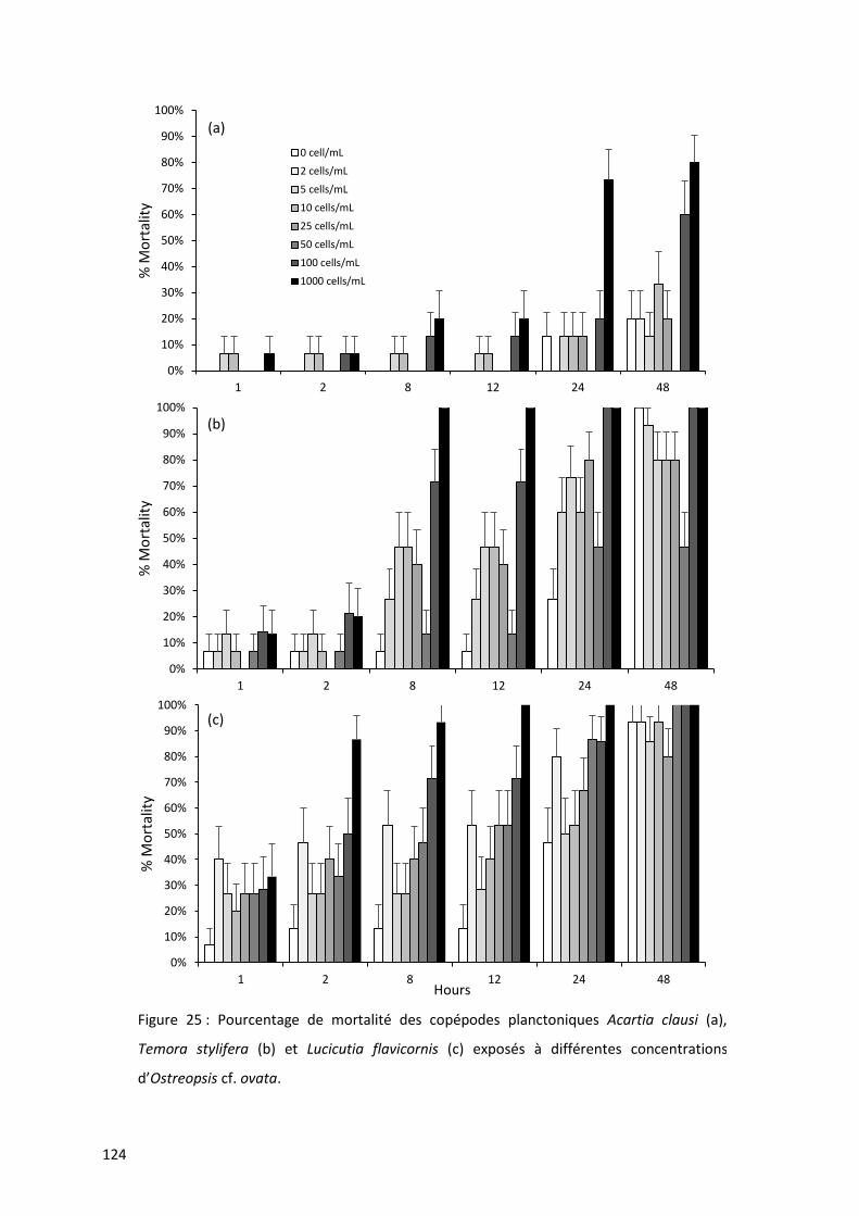
124
Figure 25 : Pourcentage de mortalité des copépodes planctoniques Acartia clausi (a),
Temora stylifera (b) et Lucicutia flavicornis (c) exposés à différentes concentrations
d’Ostreopsis cf. ovata.
0%
10%
20%
30%
40%
50%
60%
70%
80%
90%
100%
1 2 8 12 24 48
% M
ort
alit
y0 cell/mL
2 cells/mL
5 cells/mL
10 cells/mL
25 cells/mL
50 cells/mL
100 cells/mL
1000 cells/mL
0%
10%
20%
30%
40%
50%
60%
70%
80%
90%
100%
1 2 8 12 24 48
% M
ort
alit
y
(b)
0%
10%
20%
30%
40%
50%
60%
70%
80%
90%
100%
1 2 8 12 24 48
% M
ort
alit
y
Hours
(c)
(a)

Interactions entre Ostreopsis cf. ovata et prédateurs : cas des copépodes planctoniques
125
Les trois espèces de copépodes testées montrent des sensibilités différentes à O. cf. ovata :
Acartia clausi est la moins sensible des trois espèces de copépodes (LC50 = 445,73 cellules.mL-1). Temora
stylifera et Lucicutia flavicornis sont très sensibles à la présence de ce dinoflagellé puisque 1,38 et 1,15
cellules.mL-1 sont suffisantes pour observer 50% de mortalité après 24h de présence du dinoflagellé.
La mortalité d’A. clausi est plus tardive et sa moindre sensibilité est confirmée par le calcul des LC50
(24h) qui est près de 450 fois supérieures à celle de L. flavicornis et T. stylifera. Les trois espèces de
copépodes planctoniques testées sont plus sensibles que le copépode benthique Sarsamphiascus cf.
propinquus (cf. chapitre 3 de cette thèse). Ils sont également plus sensibles que Tigriopus fulvus, un
autre copépode benthique dont la LC50 (24h) est comprise entre 1486 cellules.mL-1 et des valeurs
supérieures à 4 000 cellules.mL-1 (Faimali et al., 2012; Giussani et al., 2016). Ces différences pourraient
s’expliquer par le développement de mécanismes adaptatifs plus efficaces chez les espèces
potentiellement plus exposées (comme les copépodes benthiques) tandis que les copépodes
pourraient opter pour une stratégie de fuite afin d’éviter toute exposition à O. cf. ovata. Il a déjà été
démontré qu’en réponse à des efflorescences de Phaeocystis globosa, Temora longicornis est capable
d’adapter son comportement natatoire en réalisant des sauts plus petits et plus complexes, favorisant
sa survie et à sa prise de nourriture en période d’efflorescence de cette Prymnésiophycées (Seuront
and Vincent, 2008). Néanmoins, des processus d’acclimatation ont déjà été montré pour d’autres
espèces de copépodes planctoniques exposés au dinoflagellé toxique Alexandrium fundyense (Colin
and Dam, 2005). La population d’Acartia hudsonica originaire du Maine, qui a vécu des efflorescences
récurrentes d’Alexandrium spp. depuis des décennies présentaient, lors d’une exposition à A.
fundyense en laboratoire, une meilleure croissance somatique, des tailles plus importantes ainsi
qu’une production d’œuf et une survie supérieure à la population d’A. hudsonica de New Jersey, qui
n’avait jamais été exposée aux efflorescences d’Alexandrium spp. Cette adaptation des populations
locales locale a été confirmée par des expériences de sélection génétique en laboratoire montrant
notamment une augmentation de l’ingestion et la production d’œufs après 3 générations de
copépodes naïfs maintenus en élevage avec un régime alimentaire à base de A. fundyense.
La réponse des copépodes exposés à des algues toxiques est également espèce dépendante
puisque l’ingestion d’algues toxiques n’est pas néfaste pour certains copépodes planctoniques (Turner,
2014). C’est le cas d’A. clausi et Temora longicornis qui se nourrissent respectivement de Pseudo-
nitzschia mulitseries (Maneiro et al., 2005) et Dinophysis norvegica et Dinophysis acuta (Kozlowsky-
Suzuki et al., 2006) sans présenter aucun effet néfaste. Pourtant, des effets négatifs sont observés
lorsque A. clausi se nourrit d’Alexandrium tamarense (Wohlrab et al., 2010).

126
3.5. Conclusion
La portée des résultats est malheureusement limitée par les difficultés méthodologiques
induites par la mise en contact d’organismes planctoniques avec un dinoflagellé toxique considéré
comme tychoplanctonique. Toutefois, les résultats montrent clairement que la sensibilité des
copépodes à Ostreopsis cf. ovata est espèce dépendante et que les copépodes planctoniques sont
beaucoup plus sensibles que les copépodes benthiques. Le développement de mécanismes adaptifs
en raison d’une exposition plus importante et plus fréquente des copépodes benthiques, pourrait
expliquer ces observations.

Interactions entre Ostreopsis cf. ovata et prédateurs : cas des copépodes planctoniques
127
4. Synthèse des résultats
Longtemps décrite comme benthique, cette étude a pourtant permis de décrire le caractère
tychoplanctonique d’Ostreopsis cf. ovata. Des variations fortes d’abondance cellulaires ont été mises
en évidence au cours de la journée, aussi bien pour les concentrations benthiques, planctoniques que
de surface, suggérant une potentielle migration verticale d’O. cf. ovata. Ces variations d’abondances
cellulaires au cours de la journée sont à intégrer dans les stratégies d’échantillonnage de ce dinoflagellé
en particulier pour les suivis sanitaires.
La remise en suspension d’O. cf. ovata confirme également la nécessité de mieux comprendre
l’impact de ce dinoflagellé sur les espèces vivant dans la colonne d’eau et notamment les copépodes
planctoniques. Les trois espèces testées dans cette étude présentent des sensibilités différentes vis-à-
vis d’O. cf. ovata mais demeurent beaucoup plus sensibles que les copépodes benthiques. Des études
supplémentaires sont nécessaires afin de savoir si cette plus forte sensibilité est due (1) à la capacité
de fuite des copépodes planctoniques et ainsi l’absence de processus d’acclimatation à cette algue ou
(2) à des processus de résistance inhérents aux copépodes benthiques.

128

Impacts d’Ostreopsis cf. ovata sur la macrofaune herbivore : cas de l’oursin adulte
Paracentrotus lividus
Echinoidea. Plate from Ernst Haeckel's ''Kunstformen der Natur'' (1904)

130

Impacts d’Ostreopsis cf. ovata sur la macrofaune herbivore : cas de l’oursin
131
1. Présentation de l’étude
En Méditerranée, l’oursin comestible Paracentrotus lividus est considéré comme un des
principaux macroherbivores avec la Saupe (poisson ; Sarpa salpa). En se nourrissant des macroalgues
qui constituent un substrat privilégié pour Ostreopsis cf. ovata, ces oursins vont être en contact direct
avec ce dinoflagellé toxique. Des études antérieures ont reporté (i) des épisodes de mortalités de
masse des oursins au cours des efflorescences d’Ostreopsis spp., notamment en Nouvelle Zélande et
(ii) une accumulation des toxines au niveau du tube digestif jusqu’à 15 fois supérieures aux limites
préconisées par l’Agence Européenne de Sécurité des Aliments, confirmant ainsi la pertinence de ce
modèle, aussi bien d’un point de vue écosystémique que d’un point de vue de santé publique.
Afin d’étudier les effets d’O. cf. ovata sur les oursins adultes de l’espèce Paracentrotus lividus,
des études in situ réalisées au cours de la période d’efflorescence d’O. cf. ovata ont été couplées à des
études en milieu contrôlé, en laboratoire. Les paramètres utilisés pour estimer les potentiels effets de
ce dinoflagellé sont le temps de retournement, un paramètre comportemental couramment utilisé
pour estimer l’état de santé des oursins, le taux d’ingestion, ainsi que le rapport gonado-somatique.

132

Impacts d’Ostreopsis cf. ovata sur la macrofaune herbivore : cas de l’oursin
133
2. Introduction
Au cours des quinze dernières années, une intensification mondiale des efflorescences
d’Ostreopsis sp. a été reportée (Mangialajo et al., 2011). Même s’il est difficile de savoir si ce
phénomène est directement lié à une augmentation réelle du nombre d’événements, ou uniquement
lié à un effort d’échantillonnage plus important, le nombre de cas d’hospitalisations dû à une
intoxication à Ostreopsis cf. ovata a considérablement augmenté (Durando et al., 2007; Gallitelli et al.,
2005). Plus de 200 personnes ont ainsi été hospitalisées à plusieurs reprises en Espagne, en Algérie et
en Italie entre 2001 et 2006 suite à une exposition à O. cf. ovata (Brescianini et al., 2006; Illoul et al.,
2012; Vila et al., 2008). Les intoxications, probablement liées à l’aérosolisation des cellules d’O. cf.
ovata (Ciminiello et al., 2014; Scalco et al., 2012), provoquent des irritations cutanées et oculaires, des
nausées, des diarrhées et des problèmes respiratoires. Les efflorescences d’O. cf. ovata sont
également liées à des mortalités fortes d’organismes benthiques, comme c’est le cas en Nouvelle
Zélande, en Italie et en Espagne, où des mortalités accrues d’invertébrés (dont des échinodermes) ont
été reportées (Ciminiello et al., 2006; Shears and Ross, 2009; Totti et al., 2010; Vale and Ares, 2007).
Dans ce travail, l’effet d’Ostreopsis cf. ovata sur l’oursin Paracentrotus lividus (Lamarck, 1816),
l’un des principaux macroherbivore en Méditerranée (Ruitton et al., 2000), a été étudié. Cette espèce
est comestible et largement pêchée en Méditerranée (Lawrence, 2013). Elle est également une espèce
clef, en constituant le brouteur dominant des zones benthiques peu profondes de la Mer Méditerranée
(Ruitton et al., 2000), zone de prédilection pour le développement du dinoflagellé O. cf. ovata. Des
études antérieures ont montré une forte accumulation des toxines produites par O. cf. ovata par cette
espèce d’oursin (Amzil et al., 2012; Biré et al., 2015, 2013; Brissard et al., 2014; Milandri et al., 2010) :
jusque 423 µg eq.PLTX.kg-1 ont été mesurés dans le tube digestif, soit des taux 15 fois supérieurs aux
limites préconisées par l’Agence Européenne de Sécurité des Aliments (EFSA Panel on Contaminants
in the Food Chain (CONTAM), 2009). Les études réalisées montrent que les toxines sont uniquement
retrouvées au sein tube digestif, mais pas au niveau des gonades, normalement les seules parties
consommées par l’Homme. Des études in vitro ont mis en évidence un impact important des toxines
produites par O. cf. ovata sur la fécondation et les premiers stades larvaires de ce macroherbivore
(Caroppo and Pagliara, 2011; Pagliara and Caroppo, 2012). Neves et al. (2018) ont montré une
diminution significative de la fécondation ainsi qu’une augmentation des malformations des larves
issues des gamètes exposés aux toxines d’O. cf. ovata. L’étude menée par Migliaccio et al. (2016) a
montré un effet néfaste des toxines produites par O. cf. ovata sur la fécondation et le développement
larvaire jusque 8 mois après l’exposition des adultes à une efflorescence de ce dinoflagellé. Toutefois,
aucune étude ne s’est penchée sur l’impact de ce dinoflagellé sur la physiologie de l’oursin adulte,
alors qu’il est potentiellement soumis à des abondances très fortes d’O. cf. ovata, pouvant atteindre
134
plus de 2 541 000 cellules par gramme de poids frais d’algue (Mangialajo et al., 2008). Plusieurs études
ont pourtant déjà mis en évidence une forte mortalité d’organismes benthiques, et notamment
d’oursins adultes, au cours d’efflorescences d’O. cf. ovata (Accoroni et al., 2011; Illoul et al., 2012; ,
ainsi qu’une perte des épines, indicateur d’un stress vécu par ces oursins (Sansoni et al., 2003; Shears
and Ross, 2009). Le lien entre ces épisodes de mortalité de masse et les efflorescences d’O. cf. ovata
n’a jamais été démontré. Il est aujourd’hui difficile de savoir si ces mortalités de masse sont dues
uniquement à la toxicité des composés produit par O. cf. ovata, ou aux conditions
anoxiques/hypoxiques générées indirectement par la respiration bactérienne dégradant l’abondante
biomasse d’O. cf. ovata.
Afin d’étudier les effets directs de ce dinoflagellé sur les oursins, deux types
d’expérimentation, in situ et en laboratoire, ont été réalisées. Un suivi in situ des oursins a tout d’abord
été réalisé, au niveau de deux sites localisés dans la baie de Villefranche, où les concentrations d’O. cf.
ovata sont totalement différentes : le Lido où les concentrations sont très faibles, voire nulles, et
Rochambeau, où les concentrations peuvent atteindre 1 665 873 cellules par gramme de poids frais
d’algues. Au cours de ce suivi, le temps de retournement, le rapport gonado-somatique des oursins
ainsi que la concentration d’O. cf. ovata au cours des été 2017 et 2018 ont été étudiés. L’impact d’O.
cf. ovata sur les oursins a également été étudié en laboratoire, en conditions contrôlées, en exposant
les oursins à des concentrations réalistes d’O. cf. ovata, ajoutées à la nourriture artificielle préparée
pour maintenir les oursins en aquarium.
Cette étude combine donc des méthodes d’études in situ et d’autres en conditions totalement
contrôlées afin de suivre la réponse des oursins adultes à l’exposition saisonnière au dinoflagellé
toxiques Ostreopsis cf. ovata.
3. Matériels et Méthodes
3.1. Expérimentations in situ
Deux sites ont été choisis pour étudier l’effet d’Ostreopsis cf. ovata sur l’oursins Paracentrotus
lividus (Figure 26) : le site de Rochambeau (43°41′35.64”N - 7°18′31.54″E, Villefranche-sur-Mer,
France), un site peu profond où de fortes concentrations d’Ostreopsis cf. ovata sont reportées depuis
une dizaine d’année l’été (Lemée, comm. pers.), ainsi que le site du Lido (43°41'36.3"N - 7°19'26.1"E,
Saint-Jean-Cap-Ferrat, France) où les concentrations d’O. cf. ovata sont au contraire faibles, voire
nulles, et constituant de cette manière un site témoin. Un suivi spécifique des concentrations de ce
dinoflagellé a été effectué au niveau de ces deux sites, au cours des années 2017 et 2018 (soit du 14
juin au 13 août 2017 et du 9 juin au 23 août 2018). Des échantillons de macroalgues ont été prélevées
à 2 m de profondeur (au lieu de 0,5 m de profondeur réalisé lors d’un suivi classique) afin d’estimer la
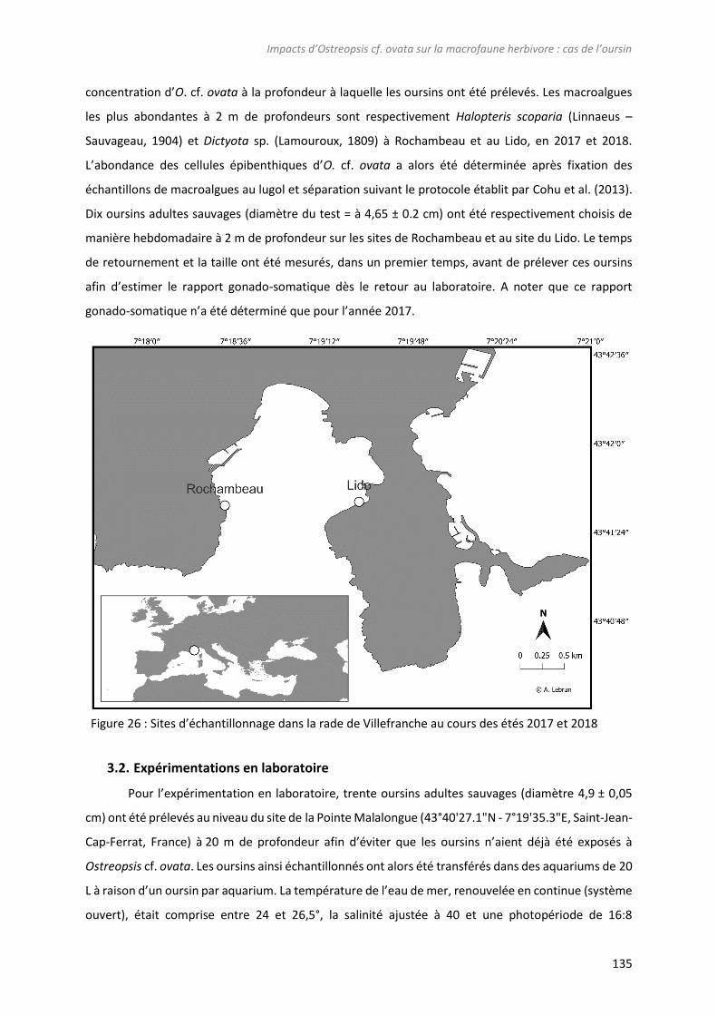
Impacts d’Ostreopsis cf. ovata sur la macrofaune herbivore : cas de l’oursin
135
concentration d’O. cf. ovata à la profondeur à laquelle les oursins ont été prélevés. Les macroalgues
les plus abondantes à 2 m de profondeurs sont respectivement Halopteris scoparia (Linnaeus –
Sauvageau, 1904) et Dictyota sp. (Lamouroux, 1809) à Rochambeau et au Lido, en 2017 et 2018.
L’abondance des cellules épibenthiques d’O. cf. ovata a alors été déterminée après fixation des
échantillons de macroalgues au lugol et séparation suivant le protocole établit par Cohu et al. (2013).
Dix oursins adultes sauvages (diamètre du test = à 4,65 ± 0.2 cm) ont été respectivement choisis de
manière hebdomadaire à 2 m de profondeur sur les sites de Rochambeau et au site du Lido. Le temps
de retournement et la taille ont été mesurés, dans un premier temps, avant de prélever ces oursins
afin d’estimer le rapport gonado-somatique dès le retour au laboratoire. A noter que ce rapport
gonado-somatique n’a été déterminé que pour l’année 2017.
3.2. Expérimentations en laboratoire
Pour l’expérimentation en laboratoire, trente oursins adultes sauvages (diamètre 4,9 ± 0,05
cm) ont été prélevés au niveau du site de la Pointe Malalongue (43°40'27.1"N - 7°19'35.3"E, Saint-Jean-
Cap-Ferrat, France) à 20 m de profondeur afin d’éviter que les oursins n’aient déjà été exposés à
Ostreopsis cf. ovata. Les oursins ainsi échantillonnés ont alors été transférés dans des aquariums de 20
L à raison d’un oursin par aquarium. La température de l’eau de mer, renouvelée en continue (système
ouvert), était comprise entre 24 et 26,5°, la salinité ajustée à 40 et une photopériode de 16:8
Figure 26 : Sites d’échantillonnage dans la rade de Villefranche au cours des étés 2017 et 2018
136
(Lumière/obscurité). Une arrivée d’air permettait une homogénéisation continue des paramètres
physico-chimiques des aquariums (Figure 27 a). En raison de la potentielle aérosolisation des toxines
d’O. cf. ovata, il nous était impossible de nourrir les oursins avec des macroalgues couvertes d’O. cf.
ovata prélevées directement in situ. Les oursins ont donc été nourris avec une nourriture artificielle
préparée à base d’un mélange de spiruline (7g – GreenSea, France), de paillettes pour poissons (30g –
Tetra, Allemagne) et d’un œuf cuit (Shpigel et al., 2004 - Figure 27 c). L’agar liquide (60°C - 17g - Fisher
Scientific, Angleterre) est ensuite ajouté et le mélange obtenu est coulé dans des boites de pétri
(diamètre de 55 mm) tarés afin de connaître la quantité de nourriture préparée et ensuite ingérée par
les oursins. Les 30 oursins ont alors été nourris pendant une semaine avec cette nourriture artificielle
avant d’être divisé en trois groupes correspondant à trois conditions expérimentales : 10 oursins ont
été nourris avec la nourriture artificielle à laquelle ont été ajoutés des concentrations d’O. cf. ovata
réalistes i.e. des concentrations atteintes naturellement par O. cf. ovata en période d’efflorescences
(Cohu et al., 2013; Mangialajo et al., 2011; Vila et al., 2008), soit d’abord 500 000 cellules.g-1, puis
1 000 000 cellules.g-1, avant de redescendre à 500 000 cellules.g-1 (Figure 27 b). La souche d’O. cf. ovata
(MCCV 054) utilisée dans cette étude, provient de la collection de culture de Villefranche
(Mediterranean Culture Collection of Villefranche – MCCV - http://www.embrc-france.fr/fr/our-
services/supply-biological-resources/microorganisms/microorganismes-unicellulaires). Elle a été
cultivée en laboratoire à 24°C dans du milieu L1 (Andersen, 2005) dont la salinité est ajustée à 38 et
avec une photopériode de 12h/8h (Lumière/obscurité) et utilisée pour enrichir la nourriture artificielle
en O. cf. ovata. La toxicité de cette souche a été vérifiée via l’utilisation du test biologique mis en place
dans le chapitre 6 ainsi que par l’utilisation de méthode de chimie analytique (UHPLC-HR-MS/MS ; voir
résultats page 6 de cette thèse). Un autre lot de 10 oursins a été nourris avec la même nourriture
artificielle à laquelle la diatomée Licmophora paradoxa (MCCV 033) a été ajoutée. Cette diatomée, co-
occurente avec O. cf. ovata (Ternon et al., 2018), est également cultivée en laboratoire dans les mêmes
conditions d’O. cf. ovata. Les concentrations de L. paradoxa sont équivalentes à celle d’O. cf. ovata en
termes de biovolume (comme estimé dans le chapitre 3) soit d’abord 1 000 000 cellules.g-1 puis
2 000 000 cellules.g-1et enfin 1 000 000 cellules.g-1. Enfin, les 10 derniers oursins ont été nourris avec
la nourriture artificielle sans aucun ajout. Après trois semaines nourris de cette façon, les 30 oursins
ont tous été à nouveau nourris avec une nourriture artificielle sans ajout (Figure 27b).

Impacts d’Ostreopsis cf. ovata sur la macrofaune herbivore : cas de l’oursin
137
La température de l’eau ainsi que la salinité ont été mesurées quotidiennement et les
aquariums ont été nettoyés tous les 2 jours afin de limiter l’ingestion des fèces par les oursins. Lors de
ces nettoyages, la nourriture artificielle restante a été récupérée puis pesées afin d’estimer les
quantités ingérées. Le temps de retournement a été mesuré une fois par semaine, à chaque
changement de condition. A la fin de l’expérimentation, soit après 5 semaines, les oursins ont été
sacrifiés afin de mesurer le rapport gonado-somatique.
3.3. Paramètres d’études
Afin d’étudier l’effet d’O. cf. ovata sur l’état de stress des oursins, le temps de retournement
des oursins a été mesuré. Cet indicateur est régulièrement utilisé chez ces échinodermes puisqu’il
reflète le bon fonctionnement du système neuromusculaire régulant la locomotion (Bayed et al., 2005;
Percy, 1973) et a déjà été utilisé dans différentes études pour mettre en évidence un stress en réponse
à des conditions environnementales variables (Lawrence and Cowell, 1996) ou la présence de
Sem 1 Sem 4 Sem 3 Sem 2 Sem 5
0 cell.gPF‐1
1 000 000
cells.gPF‐1
500 000
cells.gPF‐1
(a)
(b) (c)
Figure 27 : Installation utilisée pour maintenir les oursins pour l’expérimentation en laboratoire (a).
Les oursins ont été exposés à des concentrations variables d’Ostreopsis cf. ovata (b) incorporées à la
nourriture artificielle préparée en laboratoire pour maintenir les oursins (c).
138
macroalgues toxiques comme Caulerpa taxifolia (Boudouresque et al., 1996). Cette mesure a été
réalisée en retournant un oursin sur un carrelage de 1 000 cm², situé entre 2 et 3 m de profondeur
pour éviter l’effet des vagues. Le temps de retournement est le temps nécessaire pour qu’un oursin,
mis sur sa face aborale, reprenne sa position normale. Le rapport gonado-somatique a également été
calculé de la manière suivante :
Rapport Gonado − somatique =Poids des gonades sèches(𝑎)
Diamètre de l′oursin sans épine3∗ 100
(a) Les gonades ont été séchées pendant 24h dans une étuve à 60°C.
Le taux d’ingestion des oursins (uniquement pour l’expérimentation en aquarium) a également été
mesuré en estimant la quantité de nourriture artificielle ingérée par jour et par oursin.
3.4. Analyse statistique
Le test de Kruskal-Wallis a été utilisé pour évaluer l’influence d’Ostreopsis cf. ovata sur les
temps de retournement, l’ingestion et le rapport gonado-somatique des oursins.
4. Résultats
4.1. Efflorescences d’Ostreopsis cf. ovata en 2017 et 2018
Lors de l’étude in situ des oursins, un suivi des abondances cellulaires d’Ostreopsis cf. ovata à
2 m de profondeur (soit la profondeur à laquelle les oursins ont été échantillonnés) a été réalisé, en
plus de celui classiquement réalisé à 0.5 m de profondeur lors des suivis à but sanitaire réalisé chaque
année au site de Rochambeau (Figure 28a et b). Les efflorescences de 2017 et 2018 à Rochambeau
sont différentes en terme de périodicité, d’intensité et de durée. Les concentrations atteintes en 2017
à 0.5 m de profondeur sont plus faibles (maximum de 457 267 cellules par gramme de poids frais de
macroalgues ou cellules/gPF, contre 848 561 cellules/gPF en 2018). A 2 m de profondeur, les
abondances cellulaires atteintes au pic de l’efflorescence sont similaires pour les deux années
échantillonées (respectivement 357 510 cellules/gPF et 284 701 cellules/gPF en 2017 et 2018).
L’efflorescence à 2 m de profondeur est plus précoce en 2017 avec un maximum de concentrations
atteint le 27 juin en 2017 et un déclin le 11 juillet, alors que le pic est atteint plus tardivement en 2018
(le 11 juillet), avec un déclin le 25 juillet. Au site du Lido, les concentrations sont très faibles, que ce
soit en 2017 ou en 2018, et ne dépasse pas 66 002 cellules/gPF.
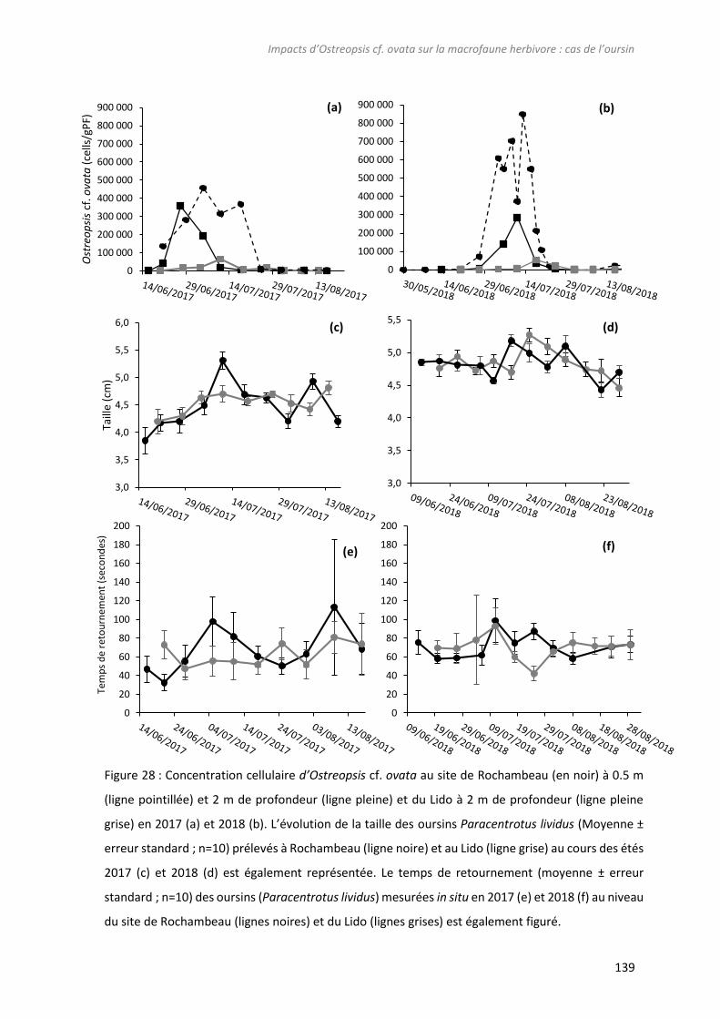
Impacts d’Ostreopsis cf. ovata sur la macrofaune herbivore : cas de l’oursin
139
(b)
3,0
3,5
4,0
4,5
5,0
5,5
6,0
Taill
e (c
m)
(c)
3,0
3,5
4,0
4,5
5,0
5,5(d)
0
100 000
200 000
300 000
400 000
500 000
600 000
700 000
800 000
900 000
Ost
reo
psi
scf
. ova
ta(c
ells
/gP
F)
0
100 000
200 000
300 000
400 000
500 000
600 000
700 000
800 000
900 000(a)
0
20
40
60
80
100
120
140
160
180
200
0
20
40
60
80
100
120
140
160
180
200
Tem
ps
de
reto
urn
emen
t (s
eco
nd
es)
(f) (e)
Figure 28 : Concentration cellulaire d’Ostreopsis cf. ovata au site de Rochambeau (en noir) à 0.5 m
(ligne pointillée) et 2 m de profondeur (ligne pleine) et du Lido à 2 m de profondeur (ligne pleine
grise) en 2017 (a) et 2018 (b). L’évolution de la taille des oursins Paracentrotus lividus (Moyenne ±
erreur standard ; n=10) prélevés à Rochambeau (ligne noire) et au Lido (ligne grise) au cours des étés
2017 (c) et 2018 (d) est également représentée. Le temps de retournement (moyenne ± erreur
standard ; n=10) des oursins (Paracentrotus lividus) mesurées in situ en 2017 (e) et 2018 (f) au niveau
du site de Rochambeau (lignes noires) et du Lido (lignes grises) est également figuré.

140
4.2. Evolution du taux d’ingestion des oursins exposés à Ostreopsis cf. ovata
Lorsque les oursins sont nourris avec la nourriture artificielle enrichie en cellules d’Ostreopis cf.
ovata, le taux d’ingestion de 0,68 g.jour-1 (±0,10), alors que les oursins nourris avec la nourriture
enrichie en diatomées ou non enrichie présentent respectivement un taux d’ingestion de 0,61 (± 0,13)
et 0.60 (± 0.14) g. jour-1. Aucune différence significative d’ingestion n’est observée entre ces conditions
expérimentales (p<0.05).
4.3. Mesure de la taille des oursins
Une augmentation de taille de 10% en cinq semaines est observée pour tous les oursins maintenus
en laboratoire (p<0.05). Néanmoins, aucune différence de taille significative n’est observée pour les
oursins dont la nourriture a été enrichie en Ostreopsis cf. ovata, ainsi que pour les oursins nourris avec
la nourriture artificielle enrichie en diatomées ou non enrichie (p<0.05).
Les paramètres étudiés, soit le temps de retournement et le rapport gonado-somatique,
pouvant varier avec la taille, la taille des oursins prélevés in situ a également été mesurée (Figure 28c
et d). Aucune évolution significative de la taille n’est observée au cours de l’été : les oursins prélevés
au cours de étés 2017 et 2018 ne présentent pas de différence significative de taille, quel que soit le
site étudié. La taille moyenne des oursins est de respectivement 4,5 ± 0,4 et 4,5 ± 0,2 à Rochambeau
et au Lido.
4.4. Impact d’Ostreopsis cf. ovata sur le temps de retournement des oursins
Le temps de retournement des oursins maintenus en laboratoire et nourris avec une nourriture
artificielle enrichie ou non en O. cf. ovata a été mesuré deux fois par semaine durant les 5 semaines
d’expérimentation. Une diminution des temps de retournement de 20 et 44 % a été observé pour les
oursins nourris respectivement avec la nourriture artificielle enrichie en O. cf. ovata et en diatomée.
Le temps de retournement des oursins nourris avec la nourriture sans ajout est constant tout au long
de l’expérimentation et aucune différence significative ne peut être mise en évidence entre les
différentes conditions expérimentales (Figure 29).

Impacts d’Ostreopsis cf. ovata sur la macrofaune herbivore : cas de l’oursin
141
Figure 29 : Evolution du temps de retournement des oursins maintenus en laboratoire et nourris avec
une nourriture artificielle préparée e laboratoire enrichie en Ostreopsis cf. ovata , en diatomées
Licmophora paradoxa (carrés ou non enrichie
Le temps de retournement a également été mesuré in situ sur des oursins prélevés à
Rochambeau, donc exposés à de fortes abondances d’O. cf. ovata, et au Lido, peu exposés à O. cf.
ovata. Les oursins à Rochambeau sont exposés à une abondance cellulaire de 350 000 cellules/gPF en
2017, mais aucune différence significative n’est observée avant, après ou pendant le bloom d’O. cf.
ovata (Figure 28e). De la même manière, aucune différence n’est observée au niveau des temps de
retournement des oursins prélevés à Rochambeau ou au Lido en 2017 (Figure 28e). Pour l’été 2018,
les résultats sont similaires : aucune différence significative du temps de retournement n’est observée
entre les oursins exposés ou non à O. cf. ovata (Figure 28f).
4.5. Impact sur le rapport gonado-somatique
A la fin des 5 semaines d’expérimentation en laboratoire, le rapport gonado-somatique des 30
oursins utilisés dans cette expérimentation a été mesuré. Aucune différence significative ne peut alors
être mise en évidence entre les trois conditions expérimentales : lorsque les oursins sont nourris avec
la nourriture artificielle enrichie en O. cf. ovata, le rapport gonado-somatique moyen est de 0,44 (±
0,05) alors qu’il est de respectivement 0,35 (±0,05) et 0,45 (±0,06) lorsque la nourriture est enrichie en
diatomée ou non enrichie. Lors du suivi in situ des oursins en 2017, le rapport gonado-somatique des
oursins a également été mesuré (Figure 30). L’évolution de ce rapport est similaire pour les oursins
prélevés à Rochambeau et ceux prélevés au Lido. Il semble y avoir un cycle dans les deux sites, avec
0
10
20
30
40
50
60
70
80
90
100
Tem
ps
de
reto
urn
emen
t (s
eco
nd
es)

142
des variations allant d’environ 4 à 7.5, mais ce cycle n’est pas en relation avec les abondances d’O. cf.
ovata.
Figure 30 : Evolution du rapport gonado-somatique (Moyenne ± erreur standard ; n= 10) des oursins
(Paracentrotus lividus) mesuré in situ aux sites de Rochambeau (ligne noire) et du Lido (ligne grise) lors
de l’été 2017. Les concentration cellulaires d’Ostreopsis cf. ovata à Rochambeau (tirets noirs) et au
Lido (tirets gris) sont également représentées.
5. Discussion
5.1. Absence d’effet d’Ostreopsis cf. ovata sur le comportement des oursins
Le comportement est souvent étudié afin d’estimer l’état de santé des échinodermes (Dumont
et al., 2007; Percy, 1973). Parmi ces indicateurs comportementaux, le temps de retournement est
couramment étudié puisqu’il est en relation avec la coordination du système neuromusculaire en
réponse à un changement d’orientation (Kleitman, 1941) et reflète, par exemple, la capacité d’un
oursin à survivre, une fois déplacés par un prédateur (Percy, 1973). Ce paramètre a déjà été utilisé
pour estimer l’état de santé d’oursins exposés à divers stress tels que des polluants chimiques (Böttger
et al., 2001), des variations de salinité ou de température (Brothers and McClintock, 2015; Lawrence,
1975), de pCO2 (Challener and McClintock, 2013) ou encore des toxines (Canty et al., 2009). Cet
indicateur a donc été utilisé dans le cadre de la présente étude afin d’estimer l’effet toxique potentiel
des cellules d’O. cf. ovata sur les oursins adultes. Aucune influence de ce dinoflagellé toxique n’a pu
être en évidence, aussi bien in situ qu’en laboratoire. La première hypothèse pouvant expliquer cette
absence d’effet serait que ce paramètre comportemental n’est pas suffisamment sensible pour mettre
en évidence un effet néfaste des cellules d’O. cf. ovata. Cet indicateur a déjà mis en évidence une
0
50 000
100 000
150 000
200 000
250 000
300 000
350 000
400 000
0
1
2
3
4
5
6
7
8
9
10
Rap
po
rt g
on
ado
som
atq
ue

Impacts d’Ostreopsis cf. ovata sur la macrofaune herbivore : cas de l’oursin
143
toxicité de la macroalgue Caulerpa taxifolia sur la même espèce d’oursins adultes (Boudouresque et
al., 1996), avec des temps de retournement des oursins dépassant 30 minutes. Ces effets sont observés
après une exposition de plus de 70 jours, alors que le temps de retournement est uniquement multiplié
par 2, en moyenne, après 20 à 40 jours d’exposition à C. taxifolia. Cette dernière expérience suggère
qu’un temps assez long est nécessaire avant de mettre en évidence un effet sur le temps de
retournement des oursins. Lors de la présente étude, ce temps est volontairement réduit à un mois,
pour être en accord avec la réalité écologique des floraisons d’O. cf. ovata. En mer Méditerranée, au
niveau de la rade de Villefranche-sur-Mer, O. cf. ovata n’atteint des abondances importantes qu’en
été et pour des périodes variant de 3 semaines à 1 mois, soit équivalentes des durées testées dans
cette étude. Les premières cellules sont souvent observées à la fin juin et diminuent très fortement
début août (Cohu et al., 2011), même si une efflorescence tardive peut se produire en automne
(Pavaux, com. pers.). Tester de fortes abondances d’O. cf. ovata sur des périodes plus longues
permettrait potentiellement d’observer un effet toxique chronique d’O. cf. ovata, mais cela n’aurait
aucune réalité écologique vis-à-vis des conditions rencontrées dans la baie de Villefranche-sur-mer.
Dans d’autres régions de Méditerranée, la durée des efflorescences est différente. C’est le cas à Sant
Andreu de Llavaneres, sur la côte catalane de l’Espagne, où les efflorescences d’O. cf. ovata débutent
plutôt fin juin et se terminent fin septembre et durent ainsi près de 4 mois (Vila et al., 2016).
Les concentrations d’O. cf. ovata choisies dans cette étude font référence à des concentrations
cellulaires reportées in situ (au niveau du site de Rochambeau, Villefranche-sur-Mer), pour garder une
certaine réalité écologique c’est-à-dire des concentrations cellulaires comprises entre 500 000 et
1 000 000 cells/gPF. Néanmoins, des concentrations bien plus fortes sont observées dans d’autres
régions méditerranéennes : c’est le cas à Gênes où les abondances cellulaires pouvaient atteindre
2 541 000 ± 588 000 cell/gPF le 27 juillet 2006. L’absence de toxicité d’O. cf. ovata sur les oursins
adultes pourrait donc également être due à l’exposition à des concentrations d’O. cf. ovata, réalistes
vis-à-vis des conditions observées à Rochambeau, mais insuffisantes pour observer un effet toxique.
La seconde hypothèse pouvant être émise pour expliquer les résultats obtenus est l’absence
d’effet toxique des cellules d’O. cf. ovata pour les oursins adultes ; les mortalités de masse ainsi
observées en Nouvelle Zélande et en Méditerranée (Shears and Ross, 2009; Totti et al., 2010; Vale and
Ares, 2007) pourraient être plutôt dues à des phénomènes d’anoxie et/ou d’hypoxie des milieux. Des
études précédentes ont en effet montré que les oursins sont capables contenir de grandes quantités
d’O. cf. ovata sans effet létal. Plusieurs études ont montré que les oursins avaient des quantités de
toxines supérieures à 400 µg eq PLTX/kg-1 sans effet mortel (Amzil et al., 2012; Biré et al., 2015, 2013;
Brissard et al., 2014; Milandri et al., 2010). Il est également important de noter que ces toxines sont
localisées uniquement dans le système digestif et que les quantités augmentent puis diminuent, en
suivant le cycle de l’efflorescence d’O. cf. ovata (Amzil et al., 2012). Les oursins adultes sont donc
144
capables de résister à des concentrations très fortes d’O. cf. ovata et les toxines ne sont a priori pas
stockées puisqu’elles ne sont pas retrouvées dans les gonades, organe ayant un double rôle de
stockage et de reproduction (Lawrence, 1975). Cette diminution de la concentration de toxines au
cours du temps suggère (i) une nouvelle fois que les oursins n’accumulent pas les phycotoxines et que
les quantités mesurées ne reflètent que la quantité de microalgues ingérées, ou (ii) qu’il existe un
mécanisme de détoxification rapide. Des mécanismes de détoxification sont déjà connus chez les
oursins et impliquent un certain nombre de gènes tels que les métallothioneine 6 (MT6) ou la catalase
(CAT), dont l’expression est régulée lorsque les larves d’oursins sont exposées aux dérivés aldéhydes
tératogènes produits par les diatomées (Varrella et al., 2014).
Les résultats obtenus dans cette étude suggèrent une absence d’effets néfastes d’O. cf. ovata
sur le temps de retournement des oursins adultes. Des études complémentaires sont néanmoins
nécessaires afin de vérifier si des floraisons plus longues d’O. cf. ovata (comme celles observées en
Espagne) ou des concentrations plus fortes, pourraient avoir un effet sur le comportement des oursins
adultes.
5.2. Impact d’Ostreopsis cf. ovata sur le taux d’ingestion de Paracentrotus lividus
Les taux d’ingestion mesurés en laboratoire ne montrent aucun impact de la présence O. cf.
ovata dans la nourriture. En ingérant des macroalgues sur lesquelles se développent O. cf. ovata, les
oursins peuvent être exposés à des concentrations supérieures à 900 000 cellules/gPF. Mais très peu
d’études se sont intéressées à l’impact d’O. cf. ovata sur les oursins adultes (alors qu’ils sont
comestibles ; Addis et al., 2009), la plupart des études étant focalisées sur les stades larvaires (Caroppo
and Pagliara, 2011; Migliaccio et al., 2016; Neves et al., 2018; Pagliara and Caroppo, 2012; Privitera et
al., 2012).
Plusieurs paramètres intrinsèques aux macroalgues peuvent agir sur leurs taux d’ingestion par
les oursins (Cook and Kelly, 2007) : la forme des macroalgues les rendant plus ou moins facile à être
ingérées par les oursins, leur qualité nutritionnelle, leur défense mécanique (en particulier la
calcification) et enfin leur défense chimique, en relation avec la présence de métabolites secondaires
plus ou moins abondants et efficaces. Il est important de noter qu’à notre connaissance, aucune
publication traitant de la défense chimique des macroalgues vis-à-vis des oursins ne fait mention des
potentiels métabolites secondaires des épiphytes de ces macroalgues. La présente étude ne montre
pas d’impact des métabolites secondaires d’Ostreopsis cf. ovata, mais il est important de prendre en
compte ce paramètre.
Impacts d’Ostreopsis cf. ovata sur la macrofaune herbivore : cas de l’oursin
145
5.3. Impact d’Ostreopsis cf. ovata sur le rapport gonado-somatique
L’évolution du rapport gonado-somatique est similaire pour tous les oursins prélevés in situ,
qu’ils aient été exposés ou non à O. cf. ovata. Ces résultats sont confirmés par les expérimentations
en laboratoire au cours desquelles aucune différence n’a pu être mise en évidence entre les rapports
gonado-somatiques des oursins nourris avec O. cf. ovata ou non. Des études précédentes ont reporté
des effets reprotoxiques de ce dinoflagellé chez l’oursin. Une diminution significative du succès de
fécondation ainsi que le développement de larves anormales a été mis en évidence lorsque les œufs
et gamètes d’oursins sont exposés à O. cf. ovata directement (Caroppo and Pagliara, 2011; Neves et
al., 2018; Pagliara and Caroppo, 2012). Cet effet reprotoxique a également été observé chez des
adultes exposés à O. cf. ovata : le succès de reproduction est de 47 % jusque 5 mois après l’exposition
(Migliaccio et al., 2016). Ces études se sont focalisées sur la qualité des gamètes formées et non sur la
quantité. Plus précisément, ces études ont montré que les gamètes produits étaient moins fertiles
mais aucune ne s’est focalisées sur la quantité de gamètes produites, qui est en lien direct avec le
rapport gonado-somatique. Ceci pourrait donc expliquer les divergences de résultats obtenus entre
notre étude et les études précédentes qui se sont plutôt intéressées au succès de reproduction.
6. Conclusion
Les résultats obtenus dans cette étude n’ont pas montré de lien entre la toxicité liée aux
efflorescences d’Ostreopsis cf. ovata en Mer Méditerranée et les épisodes de mortalité de masse
d’oursins. Aucune variation du temps de retournement, des taux d’ingestion et du rapport gonado-
somatique n’a pu être mise en évidence par les expériences en laboratoire couplées aux études in situ.
Ces résultats sont en accord avec l’étude in situ de Blanfuné et al. (2012) qui reporte l’absence de lien
entre la diminution de densité des oursins et les efflorescences d’O. cf. ovata à Gênes (Italie), à
Villefranche-sur-Mer et à Nice (France). La diminution de la densité des oursins a plutôt été reliée à
des variabilités naturelles ou aux activités de pêche existant en France. Les résultats obtenus par
Shears and Ross (2009) ont pourtant reporté une diminution de 60 % de la densité des oursins pendant
les efflorescences d’Ostreopsis siamensis en Nouvelle Zélande. Ces mortalités pourraient s’expliquer
par une combinaison de facteurs environnementaux (augmentation des températures, anoxie,
pollution anthropogénique) qui fragiliseraient les oursins, les rendant plus vulnérables à de fortes
concentrations d’Ostreopsis spp.
Les concentrations et les durées d’exposition utilisées dans cette étude reflètent les conditions
observées in situ dans la rade de Villefranche-sur-Mer, mais sont peut-être inappropriés pour observer
un effet néfaste sur les oursins adultes. Il pourrait être intéressant d’étudier des expositions plus

146
longues, avec de plus fortes concentrations d’Ostreopsis cf. ovata, en se référant par exemple aux
efflorescences se produisant en Espagne.
Enfin, même si cette expérience n’a pas montré d’effet des métabolites secondaires
d’Ostreopsis cf. ovata sur les adultes de Paracentrotus lividus, il est important de noter que les
épiphytes pourraient avoir un effet sur la consommation des macroalgues par les oursins, alors que ce
paramètre n’est jamais considéré dans les publications traitant des préférences alimentaires de ces
échinodermes.

Impacts d’Ostreopsis cf. ovata sur la macrofaune herbivore : cas de l’oursin
147
7. Synthèse de l’étude
Les résultats obtenus dans cette étude, couplant des approches in situ et en laboratoire, n’ont
pas permis d’établir de liens entre les efflorescences d’Ostreopsis cf. ovata et des changements dans
la physiologie et le comportement les oursins adultes. Ces résultats sont en contradiction avec ceux
obtenus avec l’étude in situ de (Shears and Ross, 2009) identifiant un lien entre le pourcentage de
recouvrement des macroalgues par Ostreopsis siamensis et le déclin des populations d’oursins adultes.
L’absence d’effets néfastes lors de l’exposition des oursins adultes à O. cf. ovata peut s’expliquer par
une exposition potentiellement trop courte à des concentrations trop faibles pour observer des effets
toxiques. Dans l’état actuel des connaissances, et des résultats obtenus dans cette étude, l’hypothèse
de de l’hypoxie et/ou de l’anoxie comme cause de mortalité semble à privilégier.

148
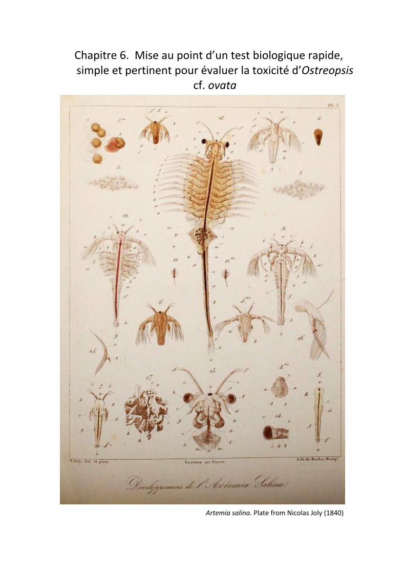
Mise au point d’un test biologique rapide, simple et pertinent pour évaluer la toxicité d’Ostreopsis
cf. ovata
Artemia salina. Plate from Nicolas Joly (1840)

150

Mise en place d’un test biologique pour évaluer la toxicité d’Ostreopsis spp.
151
1. Présentation de l’étude
La toxicité des cellules d’Ostreopsis cf. ovata est souvent évaluée par deux méthodes
majeures : (i) les méthodes biologiques comme le « Mouse Bioassay » (MBA) ou le « Hemolysis
Neutralization Assay » (HNA) et (ii) les techniques de chimie analytiques (HPLC-HR MS/MS). Les
méthodes biologiques posent des préoccupations éthiques et politiques et des problèmes de mise en
œuvre ; les techniques de chimie analytique présentent également des désavantages en raison de la
complexité des analyses et de leurs couts mais également des équipements et compétences
nécessaires à ces analyses.
Au cours de mon travail de thèse, j’ai donc mis au point un test biologique rapide pour tester
simplement et de manière précise la toxicité de différentes souches d’Ostreopsis spp. Ce test repose
sur la sensibilité des larves d’Artemia franciscana, qui est un modèle couramment utilisé pour tester
la toxicité de pesticides et d’autres contaminants chimiques et biologiques. Pour cela, la toxicité de
différentes souches d’O. cf. ovata a été évaluée, ainsi que les variations de toxicité inhérentes au cycle
de croissance cellulaires. Le protocole ainsi mis en place a ensuite été appliqué à un suivi in situ de la
toxicité des cellules d’O. cf. ovata. Les résultats de toxicité obtenus par ce test ont été validés par des
mesures quantitatives réalisées à la plateforme HPLC-HRMS/MS du laboratoire de Marine Biodiscovery
de Galway.
2. Reliable and handful monitoring of Ostreopsis sp. toxicity using an Artemia sp.
bioassay
L’article qui suit sera soumis prochainement à Marine Environmental Research.

152

Mise en place d’un test biologique pour évaluer la toxicité d’Ostreopsis spp.
153
Reliable and handful monitoring of Ostreopsis sp. toxicity using
an Artemia sp. bioassay
Pavaux Anne-Sophie1, Ternon Eva2, Dufour Louison1, Marro Sophie1, Gémin Marin-Pierre3, Thomas
Olivier4, Lemée Rodolphe1
1 Sorbonne Université, CNRS, Laboratoire d’Océanographie de Villefranche, LOV, F-06230, Villefranche-sur-Mer,
France
2 Université Côte d’Azur, CNRS, OCA, IRD, Géoazur, 250 rue Albert Einstein, 06560 Valbonne, France
3 IFREMER - Laboratoire Phycotoxines (PHYC), Rue de l'Île d'Yeu, BP 21105, F- 44311 Nantes, France
4 Marine Biodiscovery, School of Chemistry and Ryan Institute, National University of Ireland Galway, University
Road, H91 TK33 Galway, Ireland
Corresponding author: Anne-Sophie Pavaux: [email protected]
Keywords: Ostreopsis sp., Artemia franciscana, bioassay, ecotoxicity, HPLC-HRMS.
2.1. Abstract
Blooms of the benthic toxic dinoflagellate Ostreopsis sp. have increased in intensity and
frequency in the Mediterranean Sea during the past decades. These blooms are linked to adverse
effects on Human health (skin and eye irritations, fever, headaches, breath difficulties) by direct
contact with contaminated seawater or bio aerosols and deleterious effects on marine fauna. HPLC-
MS analysis are commonly used to quantify the toxicity of Ostreopsis cells although these analyses are
time-consuming and expensive. Hence there is a need to find a relevant ecotoxicity test to set toxic
threshold during Ostreopsis blooms. The crustacean species Artemia is widely used to test chemical
and biological contaminants and thus could be reasonably used to test Ostreopsis toxicity. The
relevance of this was first assessed by investigating the variability of the toxic effect among Ostreopsis
strains and throughout the dinoflagellate life cycle, in combination with chemical analysis of the toxin
content performed by UHPLC-HRMS highlighting toxicity variations among strains and growth stages.
Testing the toxicity of Ostreopsis’ endometabolome, including pure palytoxin and ovatoxins, revealed
that these toxins are not the only toxic compounds to Artemia franciscana, indicating that several toxic
compounds synthesized by Ostreopsis sp. still remain to be identified. To allow transferring the
bioassay from lab to in situ monitoring, the toxicity of the whole benthic microalgal consortium was
tested during a natural bloom of Ostreopsis cf. ovata. The results obtained highlight the accuracy and

154
sensitivity of Artemia franciscana to assess the toxicity of blooms of Ostreopsis sp. and may also be
relevant for other toxic species.
2.2. Introduction
Ostreopsis sp. blooms become more intense in temperate areas and regularly occur in the
Mediterranean Sea during summer (Accoroni et al., 2011; Accoroni and Totti, 2016; Battocchi et al.,
2010; Mangialajo et al., 2011, 2008; Totti et al., 2010; Vila et al., 2001). In these regions, Ostreopsis cf.
ovata is well known to cause adverse effects on human health (Durando et al., 2007; Gallitelli et al.,
2005; Kermarec et al., 2008; Tichadou et al., 2010; Vila et al., 2016) associated to hospitalization of
hundreds of people, like in Genova in 2005 (Brescianini et al., 2006) and in Algeria in 2009 (Illoul et al.,
2012). Toxic effects on vertebrates and invertebrates have also been documented (Accoroni et al.,
2016b; Blanfuné et al., 2012; Faimali et al., 2012; Prato et al., 2011; Privitera et al., 2012): reprotoxicity
on benthic copepods (Guidi-Guilvard et al., 2012; Pavaux et al., 2019) and sea urchins (Migliaccio et
al., 2016; Pagliara and Caroppo, 2012), as well as harmful effects on the polychaete Dinophylus
gyrociliatus and the jellyfish Aurelia sp. (Privitera et al., 2012; Simonini et al., 2011). The toxicity of
Ostreopsis cf. ovata is being attributed to the production of palytoxin (PLTX) and its analogs (named
ovatoxins, OVTX-a, -b, -c, -d, -e, -f, -g ; Ciminiello et al., 2012a, 2012b, 2010), known to be strong NA+/K+
ATPase pump inhibitors (Poli et al., 2018; Rouzaire-Dubois and Dubois, 1990).
Due to these sanitary impacts, the monitoring of Ostreopsis cf. ovata toxicity has been the
focus of most research the past decades (Battocchi et al., 2010; Casabianca et al., 2014, 2013).
Analytical chemistry (HPLC-MS) constitutes the most widespread approach to monitor OVTX
concentrations in the ecosystem (Ciminiello et al., 2006; Pierce and Kirkpatrick, 2001), despite its
various constrains: (1) cutting edge facilities (HPLC-HRMS) and skills are needed; (2) relatively high
quantification limit (2.5 µg PLTX/mL is reached); (3) involve time- and solvent-consuming analysis.
Therefore, there is an urgent need to develop an efficient (i.e rapid, simple, inexpensive and sensitive)
bioassay to allow assessing the toxicity of Ostreopsis strains, and further improve the early detection
of toxic events in concerned areas.
In the early 70s, the brine shrimp Artemia sp. was proposed as a bio-indicator for chemical
contamination (Michael et al., 1956; Vanhaecke et al., 1981) owing to its sensitivity to sodium lauryl
sulfate and various insecticides. Up to now Artemia sp. has been widely used to test fungal toxins
(Harwig and Scott, 1971), plant extract toxicity (Meyer et al., 1982), heavy metals (Martınez et al.,
1999; Sarabia et al., 2008), cyanobacteria toxins (Jaki et al., 1999; Tokodi et al., 2018) and pesticides
(Comeche et al., 2017; Gambardella et al., 2018; Jawahar et al., 2018). Artemia sp. was also shown to
be useful for marine natural products screening as shown by the work of Carballo et al. (2002) who

Mise en place d’un test biologique pour évaluer la toxicité d’Ostreopsis spp.
155
compared the cytotoxicity of the specialized metabolites produced by 20 marine organisms (mostly
invertebrates) against 2 human cell lines (lung carcinoma A-549 et colon carcinoma HT29). Therefore,
Artemia fransciscana bioassay may constitute an excellent way to assess the toxicity of Ostreopsis
blooms.
In this context, a protocol investigating the variability of the toxicity between Ostreopsis’
strains and throughout its life cycle was first set up using living cells of two Mediterranean strains (O.
cf. ovata and O. fatturossoi). As a second step, the compounds responsible for Ostreopsis toxicity were
seek using chemical extracts of O. cf. ovata cells and pure PLTX and OVTX. Finally, the implemented
bioassay was successfully used to monitor a natural bloom of O. cf. ovata in 2018.
2.3. Materials and Methods
2.3.1. Bioassays
Four experiments were performed that will be referred as Experiment 1 to 5 hereinafter.
Experiment 1 investigates the variability of the toxicity among species and strains of Ostreopsis at a
given stage of growth, using two french O. cf. ovata and two Lebanese O. fattorussoi strains;
Experiment 2 follows the toxicity throughout the growth of O. cf. ovata; Experiment 3 investigates the
nature of the toxic compound using cell extracts and pure OVTX and PLTX; Experiment 4 evaluates A.
franciscana as a tool for biomonitoring the toxicity of a natural bloom.
2.3.2. Micro-algal culture
The four strains used in this study were obtained from the Mediterranean Culture Collection
of Villefranche (MCCV,http://www.embrc-france.fr/fr/our-services/supply-biological-
resources/microorganisms/microorganismes-unicellulaires). Ostreopsis cf. ovata (MCCV 054 and
MCCV 055) were collected at the Rochambeau site in 2014 (Bay of Villefranche-sur-Mer, N-W
Mediterranean, 43°41’35.64’’N-7°18’31.54’’E) and Ostreopsis fattorussoi (MCCV 057 and MCCV 058)
were isolated in 2015 from Bat 14 site (Lebanon, Batroun, 34°15’9.0’’N-35°39’41.3’’E). All microalgae
are grown in L1 and L1/2 medium (Andersen, 2005) prepared with autoclaved aged and filtered
seawater, adjusted to a salinity of 38. Cultures are maintained at 24°C, under a 14:10 light/dark cycle
with a light intensity of 250 µmol.m-2.s-1.
2.3.3. Taxonomic Molecular Identification by a PCR Based Assay
A volume of 5 mL of each MCCV clonal culture was centrifuged for 6 min at 2000 rpm and 4°C.
The supernatant was removed, and the cell pellet was resuspended with 1 mL of sterile water
(autoclaved MilliQ) and centrifuged again for 2 min to remove salt and media. The cell pellet was kept

156
and homogenized with 50 µL of sterile water and a fraction of 10 µL of was further transferred to 0.2
µL polymerase chain reaction (PCR) tubes. Then, PCR tubes were stored at -20°C until further analysis.
Approximatively 300 base pairs of the internal transcribed spacer region (ITS1-5.8s-ITS2) rDNA were
amplified by PCR using the primer 329-F (5’-GTGAACCTGCRGAAGGATCA-3’) which is the inverse
complementary sequence of the universal eukaryote primer 329-R (Moon-van der Staay et al., 2001)
and the designed primer D1R-R (5’-TATGCTTAAAATTCAGCAGGT-3’) which is the inverse
complementary sequence of the primer DIR-F (Scholin et al., 1994). A mix of 1µl of each primer, 1 µl
of dNTP at 10mM, 5 µl of 10X Advantage 2 PCR Buffer, 31µl of sterile water and 1µl of 50X Advantage
2 Taq polymerase (Clontech) was added in each PCR tube containing the cell pellet. The PCR was
performed using a MasterGradiant thermocycler (Eppendorf) with the following conditions: one initial
denaturation at 94°C for 10 min followed by 35 cycles each consisting of 1 min at 94°C, 1 min at 53°C,
1 min at 68°C and a final elongation for 10 min at 68°C. The PCR products were purified using QIAGEN
MinElute PCR Purification kit according to the recommendations. After sequencing by Genewiz,
sequences were treated using BioEdit software and were compared to NCBI database using BLASTn
tool to ensure the taxonomy of the strains.
2.3.4. Cells sampling
2.3.4.1. Cultures
All strains used in Experiment 1, 2 and 3 were grown in triplicates, in 150-mL and 300-mL flasks,
respectively, at an initial cell concentration of 1000 cell.mL-1. A volume of 12 mL was sampled and fixed
with acidic lugol (4% v/v) for cell counting at day 10 in Experiment 1 (150 mL-flasks) and every other
day during 14 days in Experiment 2 (300-mL flasks). Cell counting was performed in triplicates using a
liquid particle counter (HIAC/Royco 9703, Pacific Scientific Instruments) following a size range of 2-80
µm (Stramski et al., 2002). Aliquots of living cells from each flasks were subsequently diluted using L1
medium to a final volume of 20 mL and a cell concentration of 20 000 cell.mL-1 to perform Artemia
bioassays.
2.3.4.2. In situ cells
Living cells used in Experiment 4 were sampled on artificial substrates (Jauzein et al., 2016)
during summer 2018 (27th of June to 23rd of August) to cover the bloom of Ostreopsis cf. ovata . Three
screens composed by a rigid frame and a 1 mm mesh were deposed in the morning at a depth of 50
cm depth at 3 sampling sites, separated from each other by few meters. After 24 hours, the screens
were carefully collected in 1 L plastic bottles containing seawater and gently homogenized to
resuspend all microalgae. For each screen, a volume of 20 mL was subsampled in triplicates fixed with
acidified lugol (4% v/v) and observed under microscope using Sedgewick rafter counting chamber to

Mise en place d’un test biologique pour évaluer la toxicité d’Ostreopsis spp.
157
measure both the abundance and the diversity. After counting, dilutions were applied to each sample
obtained from the screens to reach a final concentration of 20 000 cells.mL-1 to perform Artemia
bioassays. It is noteworthy that these samples are natural samples and therefore contain the whole
benthic micro-algal assemblage, including Ostreopsis cells.
2.3.5. Obtention of Ostreopsis extracts, fractions and pure ovatoxins
A volume of 1.4 L of a culture of Ostreopsis that aged 10 days was centrifuged at 600 g at 21°C
for 10 min. The supernatant was discarded and the cell pellet flash frozen using liquid nitrogen to stop
the cells metabolism, and further stored at -20°C before being freeze dried. An amount of 85 mg of
dried cells was extracted using 4 ml of MeOH:H2O (80:20) in an ultrasonic bath for 10 min. The extract
was centrifuged at 1000 g during 10 min and the supernatant was collected and transferred into a glass
vial. The procedure was repeated two more times to obtain 12 mL of extract. The extract was then
evaporated under a gentle flow of N2 at 35°C until full evaporation of the MeOH (leaving about 2-3 ml
of water). The aqueous extract was deposited on a C18 SPE cartridge (500 mg, Supelco) previously
washed with MeOH (6 mL) and equilibrated with MilliQ-water (6 mL). Before elution, the cartridge was
rinsed with 6 mL of MilliQ-water. A step gradient elution was thus applied to the cartridge, from
H2O:MeOH (100:0) to H2O:MeOH (0:100), using steps of 20% and volumes of 6 mL. An additional
elution with 6 mL of acetonitrile (ACN 100%) was performed, yielding seven fractions (F1 to F7, from
the most polar to apolar metabolites). Aliquots of 100 µl sampled from each fraction were evaporated
to dryness under a N2 stream (35°C) to assess their respective weight. A volume of 1 mL of
dimethylsulfoxide (DMSO) was added to the remaining fractions before evaporating other volatile
organic solvents (MeOH, ACN) and water. Based on the fractions weight, their volume of solubilisation
were further adjusted using DMSO to reach a concentration of 3 mg.mL-1. All fractions were stored at
-20°C until further bioassays and chemical analysis.
A purified pool of ovatoxins was obtained in the frame of the OCEAN15 project, from IFREMER
partner (Amzil group), following a protocol previously published (Brissard et al., 2015). Briefly, MeOH
50% raw extracts were obtained from 75 g of O. cf. ovata (strain MCCV 054) and fractionated by size-
exclusion chromatography (Sephadex LH20). Methanolic fractions containing OVTXs were pooled,
concentrated and purified by semi-preparative chromatography (Uptisphere C18-TF, 250 mm × 4.6
mm, 5 µm; λ= 233 and 266 nm). We decided to purifiy a pool of OVTXs as the separation of all
analogues is highly challenging. The final pool was concentrated in DMSO and contained a mixture of
OVTX-a (61 %), -b (25 %), -c (3.4 %), -d (5.3 %), -e (3.9 %) and -f (1.1 %) at 7.8 mg/mL, based on LC-
MS/MS quantification. As important losses may arise from evaporation to dryness (Brissard et al.,
2015), the purity has not been assessed. In the meantime, pure palytoxin was purchased from Wako
Chemicals (GmbH, Neuss, Germany). All toxins were solubilized in DMSO at a concentration of 12

158
µg/mL that would allow an addition of 5 µl for the Artemia bioassay without exceeding 0.1% of DMSO
in final volume of seawater.
2.3.6. Experimental bioassays
0.15 g of dehydrated Artemia franciscana cysts were hatched in 1 L of filtered and aged sea
water at a salinity of 38, at 20°C under a continuous light of 250 µmol.m-2.s-1 intensity and a continuous
and strong aeration. After 40 hours of incubation, all larvae moulted to metanauplii stage (2-3 stages),
considered as the most sensitive stages to be used in ecotoxicological bioassay (Nunes et al., 2006;
Sorgeloos et al., 1978). For each experiments (1 to 4), Artemia larvae were isolated from empty cysts
using a Pasteur Pipette and transferred in 6-well plates (5 larvae per well); each well containing 4 mL
of 0.2 µm filtered and aged sea-water adjusted to salinity 38 and kept at a temperature of 24°C (except
for Experiments 3 that contained 5 mL of seawater). All tests were performed using 30 metanauplii
larvae per condition. Experiments 1, 2 and 4 were conducted using the four following cellular
concentrations: 4, 40, 400 and 4000 cells.mL-1. Experiment 3 was performed using four concentrations
of ovatoxins and palytoxin (0.012, 0.12, 1.2 and 12 µg/mL) corresponding to theoretical concentrations
that would be reached with 0.4, 4, 40 and 400 cells.mL-1 of Ostreopsis cells (Brissard et al., 2014;
Ciminiello et al., 2012b; Nascimento et al., 2012a; Pistocchi et al., 2011). Also, all seven chemical
fractions obtained from cell pellet were added in the wells at a final concentration of 10 µg/ml.
Artemia franciscana larvae’s mortality was measured every hour during 12 hours, then at 24
and 48 h exposure times (only at 24 h for chemical fraction, Experiment 3). Larvae were considered
dead when there was no movement during 10 seconds and no reaction to strong light. A negative
control was also performed for all experiments, using seawater + fresh L1 medium, 80: 20 (v:v) for
Experiments 1, 2 and 4 and 0.1% DMSO (final volume) for Experiments 3. Bioassays are considered as
acceptable when mortalities rate in the control condition are lower than 10% as described in Faimali
et al. (2012).
2.3.7. Toxin profiles
2.3.7.1. Cell extraction
To assess the variability of the toxin content (i) among strains and species, and (ii) throughout
the growth phase, chemical extractions of MCCV 054, 055, 057 and 058 were performed. For inter-
strain variability, each strain was inoculated with 1000 cell.mL-1 in triplicates 50-mL flasks and grown
during ten days before harvesting the whole culture. For growth-dependent variability, the MCCV 055
was inoculated at 1000 cell.mL-1 in twenty-one 300-mL flasks and grown for fourteen days. Every other
day, three flasks were sacrificed for cell abundance (subsample of 1.2 mL) and chemical extraction
(about 300 mL). Samples for cell abundance were fixed and counted as previously described.

Mise en place d’un test biologique pour évaluer la toxicité d’Ostreopsis spp.
159
All flasks content (50 mL and 300 mL) were transferred to 50 mL falcon tubes before being
centrifuged at 600 g at ambient temperature, for 10 min. After the supernatant being removed, the
cell pellets were flash frozen using liquid nitrogen to stop any metabolic reaction, and further stored
at -20°C. A volume of 4 mL of MeOH: H2O (80:20, v/v) was poured in the falcon tubes before they were
being placed in ultra-sonic bath for 10 min, while cooling in an ice batch. The falcon tubes were then
again centrifuged at 2500 g for 10 min at ambient temperature and the supernatants were transferred
in 20 mL vials.
These steps (organic extraction, ultra-sonic bath and centrifugation) were repeated 2 more
times and the resulting extracts were evaporated under a gentle stream of N2 until full evaporation of
the MeOH. To avoid full dryness of the samples, 500 µl of DMSO was then added to the extracts and
the remaining H2O further evaporated. All extracts were stored at -20°C until UHLC-HRMS analysis.
2.3.7.2. UHPLC-HRMS
UHPLC-UV-HRMS experiments were performed using an Agilent 1290 system (Agilent
Technologies, USA) equipped with a diode array detector and coupled to an Agilent 6540 Qtof mass
spectrometer (Agilent Technologies, USA). Mass spectrum detection was carried out in positive mode
(ESI +) between 60 to 3000 Da. 10µL of each samples were injected and separated on a T3 column
(Acquity UPLC HSS T3 1.8µm, 2.1mm x 100mm, Waters) with a linear elution gradient of H2O:MeOH
(80:20, v:v)/MeOH with 0.1mM of ammonium formate and 0.1% formic acid from 90:10 (v:v, isocratic
from 0 to 2mins) to 0:100 (v:v, isocratic from 12 to 13 mins) at a 0.4 mL.min-1 flow rate. Collision
energies (CE) of 30, 70 and 110 eV were applied to confirm the presence of OVTXs. UV detection was
set at 210, 233 and 263 nm. The spectrometer analyzer parameters were set as follow: nebulizer
sheath gas, N2 (35 psig); drying gas, N2 (11 L.min−1); Gas Temperature, 300°C; capillary, 4.129 µA;
Vaporizer/Sheat Gas Temp, 350°C.
The quantification of the OVTXs in the samples was done using commercial PLTX as a reference,
considering identical ionization of these compounds. A calibration curve for PLTX was set using
concentrations between 1 and 10 mg.mL-1, using the tricharged adduct [M+3H-4H2O]3+ for
quantification. This same adduct was used to quantify the OVTX a in all samples.
2.3.8. Statistical analysis
The statistical significance of the mortality variation between the different experimental
conditions tested was evaluated using Kruskal-Wallis analyses. Dunn’s post hoc tests were used a
posteriori to identify which experimental conditions differed from the others when significant
differences were showed by the Kruskal-Wallis test (p<0.05). These tests were realized using the PAST

160
software (Hammer et al., 2001) The R package ecotoxicology was used to assess the median lethal
concentration (LC50) at 24 and 48 hours of exposition.
2.4. Results
2.4.1. Molecular analysis
PCR amplification based assay of the strains using genus and species-specific primers
confirmed the morphological analysis. The sequences obtained after PCR analysis showed a perfect
alignment between the strains MCCV 054/MCCV 055 and fully matched (100% of confidence)
Ostreopsis cf. ovata (E-value: 2e-157, accession number: MG551865.1). Similarly, the strains MCCV
057/MCCV 058 were identified as Ostreopsis fattorussoi (level of confidence 100%, E-value: 1e-129,
accession number: LT220222.1 ; Accoroni et al. 2016).
2.4.2. Inter-strain variability of the toxicity - Experiment 1
Mortality rates in controls were lower than 10% throughout the experiment, meeting the
acceptability threshold of this bioassay as defined by Faimali et al. (2012). Mortality was significantly
higher (p-value <0.05) in presence of all Ostreopsis cells suggesting all strains are toxic to Artemia
fransiscana larvae, whilst slight variability owed to the time of exposure, the cell concentration and
the nature of the strain used were found (Figure 31).
A positive correlation between mortality and exposure time (p < 0.001) was observed, and
twelve hours of exposure was sufficient to induce significant mortality of A. franciscana (p < 0.05).
Mortality reached its maximum after 48 hours (96,6%) for all strains and concentrations tested.
Several concentrations of Ostreopsis cells were tested to explore their effects on A. fransiscana.
Despite inter-strains variability, a dose-dependent response is observed with a strong increase of
mortality until a plateau of 96% of mortality was reached at 400 cells.mL-1 at all exposure time and for
most strains.
Inter-strains variability was mostly observed for low exposure times (12 and 24 hours) and low
cell concentrations (4 to 40 cell.mL-1). Looking at a concentration of Ostreopsis cells of 4 cell.mL-1, the
toxicity of the strains can be ranked as following: MCCV 054<055=057<058 (Cf. LC50-24h values – Table
13). By increasing the concentration of Ostreopsis cells, this distribution is smoothed and no significant
differences are observed between strains for 400 and 4000 cell.mL-1 (except MCCV 058 that shows the
most toxic behaviour, 96,6 %).
Interestingly, the toxin content determined by UHPLC-HRMS did not support the toxicity
pattern highlighted by the Artemia bioassay since no ovatoxins were found in neither MCCV 057 nor
058. On the other hand, an amount of 18.0 (± 4.9) and 23.7 (± 9.2) pg PLTX eq/cell were measured for

Mise en place d’un test biologique pour évaluer la toxicité d’Ostreopsis spp.
161
MCCV 054 and MCCV 055, respectively, therefore matching the level of toxicity highlighted in the
Artemia bioassay.
Table 13 : LC50 values and confidence limits in cells.mL-1 obtained exposing Artemia franciscana
larvae to different strains of Ostreopsis sp. and dissolved toxins.
Treatment LC50 – 24h (cells.mL-1) LC50 – 48h (cells.mL-1)
MCCV 054 81.96 (± 63.33) Nan
MCCV 055 0.25 (± 0.90) Nan
MCCV 057 0.0012 (± 0.00033) Nan
MCCV 058 0.023 (± 0.039) Nan
OVTX 5.96 (± 2.12) 0.42 (± 0.092)
PLTX 48.42 (± 14.23) 0.42 (± 0.085)
Living cells 9.01 (± 3.30) 1.16 (± 0.40)
2.4.3. Ostreopsis cf. ovata toxicity throughout growth - Experiment 2
After inoculation, Ostreopsis cf. ovata (MCCV 055) showed a linear and exponential growth
during nine days, corresponding to an increase of cells concentration by a factor 2, up to 6 621 cells.mL-
1 at day 10 (growth rate of 0.24 day-1), before declining on day 14 (3 637 cells.mL-1; Figure 32).
Meanwhile, the toxin content quickly stabilized around 10 pg eq. PLTX/cell between day 4 and 10,
followed by a decrease towards the end of the experiment (Figure 32). The high concentrations
obtained at day 0 were likely influenced by the fact that the cells that were extracted had been sampled
in a culture that aged 10 days.
Interestingly, the variability of the toxin content did not reflect the pattern of mortality
observed for Artemia franscicana (Figure 33 a, b and c). No clear trend emerged from the mortality
measured after 12 hours of exposure, although it increased with cell concentrations. Likewise, at high
cell concentration of Ostreopsis (40 and 400 cell.mL-1) and for an exposure time of 48 hours, mortality
varied little (Figure 33b – 6c ; p>0.05) and was close to 100 % during the whole duration of the
experiment, precluding drawing any relationship between mortality and the growth phase.

162
Figure 31 : Percentage of mortality (Mean ± ES) after 12 h (a), 24 h (b) and 48 h (c) of Artemia
franciscana larvae exposed to various concentrations of Mediterranean strains of Ostreopsis cf. ovata
(MCCV 054 and 055) and Ostreopsis fattorussoi (MCCV 057 and 058).
(b)
(c)
(a)
0%
10%
20%
30%
40%
50%
60%
70%
80%
90%
100%
110%
0 4 40 400 4000
% M
ort
alit
y
MCCV 054
MCCV 055
MCCV 057
MCCV 058
a
a
ab
a
b aab
bb
a
b
0%
10%
20%
30%
40%
50%
60%
70%
80%
90%
100%
110%
0 4 40 400 4000
% M
ort
alit
y
MCCV 054
MCCV 055
MCCV 057
MCCV 058
a
a
b
a
ab
b a
aa
abb
0%
10%
20%
30%
40%
50%
60%
70%
80%
90%
100%
110%
0 4 40 400 4000
% M
ort
alit
y
Cells.mL-1
MCCV 054
MCCV 055
MCCV 057
MCCV 058
a
a
b
ab
a
b
a a a
(a)

Mise en place d’un test biologique pour évaluer la toxicité d’Ostreopsis spp.
163
Only an exposure time of 24 and 48 hours at low concentrations of Ostreopsis cells (4 cell.mL-
1) allowed to draw a pattern of mortality for Artemia larvae that followed both the cellular growth and
the toxin content (Figure 33A) with a minimum between days 2 and 4 (10% and 43 % for 24h and 48h
respectively, 0.40 pg eq. PLTX/cell, 1436 cells.mL-1) and a maximum reached at day 10 (26% and 83%
for 24h and 48h, respectively, 13.1 pg eq. PLTX/cell, 6389 cells.mL-1).
Figure 32 : Toxin concentration (Mean ± ES) in pg eq. PLTX/cell (black line) depending on the growth
phase of Ostreopsis cf. ovata (MCCV 055 – grey bars).
2.4.4. Toxic metabolite responsible for Artemia mortality – Experiments 3
The nature of the toxic metabolite(s) responsible for the toxicity of Ostreopsis cells on Artemia
was further investigated comparing the toxic behaviour of (i) living Ostreopsis cells, (ii) chemical
fractions obtained from Ostreopsis cells extracts, (iii) purified OVTX, and (iv) commercial PLTX.
Interestingly, living cells induced an earlier response than equivalent amount of dissolved PLTX and
OVTX (mortality of 46%, 13% and 13%, respectively after 12h of exposure at a concentration of 400
cells.mL-1), although these differences tend to be reduced with increasing exposure time (Figure 36).
After 48h, mortality is significantly higher with the dissolved toxins (100 % for both OVTXs and PLTX
and 76,7 % for living cells at a cellular concentration of 4 cells.mL-1), as also confirmed by the LC50 values
given in Table 13.
0
1000
2000
3000
4000
5000
6000
7000
0
2
4
6
8
10
12
14
16
18
20
0 2 4 6 8 10 12 14
Ost
reo
psi
scf
. ova
ta(c
ells
.mL-1
)
OV
TX (
pg
eq P
LTX
/cel
l)
Days

164
Figure 33 : Percentage of mortality (Mean ± ES) of Artemia franciscana larvae exposed to 4 (black lines),
40 (dotted lines) and 400 cells.mL-1 (dashed lines) of Ostreopsis cf. ovata (MCCV 055) after (a) 12 hours,
(b) 24 hours and (c) 48 hours exposure time. The growth curve of O. cf. ovata (MCCV 055) cultured in
300mL flasks is also represented in grey bars.
0
1000
2000
3000
4000
5000
6000
7000
0%
10%
20%
30%
40%
50%
60%
70%
80%
90%
100%
0 2 4 6 8 10 12 14
Ost
reo
psi
scf
. ova
ta(c
ells
.mL-1
)
% M
ort
alit
y
0
1000
2000
3000
4000
5000
6000
7000
0%
10%
20%
30%
40%
50%
60%
70%
80%
90%
100%
0 2 4 6 8 10 12 14O
stre
op
sis
cf. o
vata
(cel
ls.m
L-1)
% M
ort
alit
y
0
1000
2000
3000
4000
5000
6000
7000
0%
10%
20%
30%
40%
50%
60%
70%
80%
90%
100%
0 2 4 6 8 10 12 14
Ost
reo
psi
scf
. ova
ta(c
ells
.mL-1
)
% M
ort
alit
y
Days
(a)
(c)
(b)

Mise en place d’un test biologique pour évaluer la toxicité d’Ostreopsis spp.
165
To test whether the OVTX and PLTX are the only toxic metabolites produced by Ostreopsis, the
activity of seven fractions obtained from the separation of the various metabolites synthesized by the
dinoflagellate, was tested over 24 hours on Artemia larvae (Figure 34). No toxicity was found for
fractions 1, 2 and 3 whereas all fractions from 4 to 7 were toxic showing mortalities of 13%, 30%, 33%
and 71% for fractions 6, 4, 7 and 5, respectively. The analysis of the chemical content of each fraction
by UHPLC-HRMS revealed that only fractions 5 and 6 contained detectable amounts of OVTX (29 and
7.5 µg, respectively), that yield a final concentration in the wells of 305 and 15 ng eq. PLTX.mL-1,
respectively. Taken altogether, these results indicate that Artemia larvae are sensitive to amount of
OVTX and PLTX as low as 12 ng.ml-1 but also to other compounds produced by Ostreopsis.
Figure 34 : Mortality (Mean ± ES) of Artemia franciscana larvae after being exposed 24 hours to
fractions of Ostreopsis cf. ovata obtained from MCCV 054 extracts. The concentration of all fractions
was previously adjusted to 3 mg.ml-1 using DMSO and MilliQ. The polarity of the metabolites decreases
from Fraction 1 to 7 (F1-F7). The content of OVTXs in each fraction is also represented (black squares).
2.4.5. Application to in situ monitoring - Experiment 4
Since Artemia franciscana larvae mortality was shown to be a good proxy for the toxicity of
Ostreopsis cells from lab cultures, the application of the Artemia bioassay to in situ monitoring of
Ostreopsis cf. ovata blooms was further investigated. Cells sampled in situ were tested for their toxicity
throughout the bloom, in July 2018. The bloom of Ostreopsis cf. ovata lasted a month (June 27th to July
24th), with a peak the 13rd of July. The cell concentration increased from 100 000 cells.mL-1 to 800 000
cells.mL-1 before decreasing to 8000 cells.mL-1 at the end of the bloom, showing no other increase
before the end of August (Figure 35).
305
150
50
100
150
200
250
300
350
0
10
20
30
40
50
60
70
80
90
100
F1 F2 F3 F4 F5 F6 F7 ControlO
vato
xin
s(n
g eq
.PLT
X/m
l)
% M
ort
alit
y
Ostreopsis cf. ovata fractions

166
Based on the results obtained from experiments 1 and 2, we decided to only monitor the
mortality of Artemia larvae after a 24-hours exposure to Ostreopsis cells (at a cellular concentration of
4 cells.mL-1). The mortality of A. franciscana correlated the concentration of O. cf. ovata (r=-0.6448,
p<0.05) at all cell concentrations tested (Figure 35). The mortality of Artemia larvae exhibited a pattern
that followed the bloom of O. cf. ovata, with a minimum at the end of the bloom (between 10 to 25 %
after the 3rd of August) and a maximum at the peak of the bloom (87% on the 14th of July – Figure 35).
Interestingly, an increase of mortality was observed in August while low abundance of Ostreopsis was
detected (from 8th of August to 22th of August; Figure 35). A quick survey of the diversity of the
microalgae showed that several species of diatoms (Licmophora paradoxa, Naviculaceae sp. and
Cylindrotheca closterium) were blooming at that time (data not shown).
Figure 35 : Mortality (Mean ± ES) of Artemia franciscana after 24 hours exposition to 4 cells.mL-1.
Evolution of cellular concentration of Ostreopsis cf. ovata on artificial substrates is also represented in
dotted line.
0
100000
200000
300000
400000
500000
600000
700000
800000
900000
0%
10%
20%
30%
40%
50%
60%
70%
80%
90%
100%
24/6 4/7 14/7 24/7 3/8 13/8 23/8 2/9
Ost
reo
psi
scf
. ova
ta(c
ells
/gFW
)
% M
ort
alit
y

Mise en place d’un test biologique pour évaluer la toxicité d’Ostreopsis spp.
167
Figure 36 : Mortality (Mean ± ES) after 12 h (a), 24 h (b) and 48 h (c) of Artemia franciscana larvae
exposed to increased concentrations of dissolved PLTX and OVTX (from 0.012, 0.12, 1.2 and 12 µg/mL)
0%
10%
20%
30%
40%
50%
60%
70%
80%
90%
100%
0 0,4 4 40 400
% M
ort
alit
y
OVTX
PLTX
Cells
b
b
aaa
0%
10%
20%
30%
40%
50%
60%
70%
80%
90%
100%
0 0,4 4 40 400
% M
ort
alit
y
OVTX
PLTX
Cells
a
a
b
a
a
0%
10%
20%
30%
40%
50%
60%
70%
80%
90%
100%
0 0,4 4 40 400
% M
ort
alit
y
Cells.mL-1
OVTX
PLTX
Cells
a aa
b
a
(a)
(b)
(c)

168
and living cells at various cell-equivalent concentrations. Letters formulate significant differences:
groups that have no common letter differ significantly (p < 0.05).
2.5. Discussion
2.5.1. Validating Artemia franscicana as a relevant model to evaluate Ostreopsis sp. toxicity
To ensure monitoring of Harmful Algal Blooms and consumer protection, the usefulness of
biological assays has been long recognized for toxicity determination of samples (Botana, 2014). For
instance, mouse bioassay (MBA) has been widely used as primary tool for HABs monitoring, notably
for shellfish toxicity (Delaney, 1984). This biological assay however presents a lack of sensitivity, is
prone to interferences with various metals and salts resulting in false positives and negatives, and
often raises major ethical and political concerns (McCulloch et al., 1989; Wiberg and Stephenson,
1961). Hence developing more sensitive and ethic bioassays is currently of high importance. The
crustacean species Artemia is very useful to evaluate the toxicity of diverse biological and chemical
contaminants (Michael et al., 1956; Nunes et al., 2006; Vanhaecke et al., 1981) and its relevance for
assessing the toxicity of Ostreopsis cells was successfully investigated in this study. First, the
replicability of this bioassay was satisfactory with coefficients of variation between 0 and 6.1%,
therefore demonstrating the excellent replicability of this test. The significant mortality of the Artemia
larvae induced by the two Ostreopsis species (ovata and fatturossoi) and the four strains (MCCV 054,
055, 057 and 058) is in accordance with previous results obtained with Ostreopsis siamensis and ovata,
isolated from New Zealand (strains CAWD 73, 74, 75 and 96) and the Marche Region (Italy – Strain
OOAN0601), respectively (Pezzolesi et al., 2012; Rhodes et al., 2000), supporting the suitability of this
bioassay to monitor the toxicity of any Ostreopsis species from any origin.
The Artemia larvae were shown to be extremely sensitive to the presence of Ostreopsis cells
since concentrations as low as 0.4 cell. mL-1 induced significant mortality (Experiment 3). In the context
of in situ biomonitoring, this sensitivity could greatly facilitate an early detection of blooms.
Nevertheless, it should be considered that differences in toxicity exist according to the growth stage
of the dinoflagellate (Experiment 2), following a pattern that correlated the cellular growth: toxicity
was maximal at the end of the exponential phase (43%, day 10) and minimal at the beginning of the
growth (83%, day 3). Thus, for standardization aspects, our data suggest that the toxicity of newly
isolated strains/species should be tested using cells that reached the end of their exponential phase
(~10 days after inoculation).
Several exposure time (12, 24 and 48 hours) and cell concentrations (0.4, 4, 40, 400 and 4000
cell.mL-1) were tested in Experiments 1, 2 and 3 to evaluate the best conditions that must be observed
to run the Artemia bioassay. The data obtained suggest that exposure lower than 12 hours is not

Mise en place d’un test biologique pour évaluer la toxicité d’Ostreopsis spp.
169
relevant whereas exposure over 24 hours provides the best accuracy (Experiment 1 and 2) and allows
determining the LC50 values. Also, cell concentrations ranging from 0.4 to 400 cell.mL-1 gives the best
results (Experiment 1 and 3).
However, results obtained in the experiment 3 highlighted earlier action of living cells
compared to dissolved toxins that could go unnoticed with only one check. Standardized conditions
that should be used to run an Artemia bioassay in the context of evaluating the toxicity of an Ostreopsis
species are summarized in the Appendices 1 and 2 of the supplemental information.
2.5.2. Compounds leading to Ostreopsis sp. toxicity
Despite no OVTX was quantified, the faturrossoi species was surprisingly highly toxic to the
Artemia larvae (93,3%), suggesting the production of toxic compounds by this Ostreopsis species
(Experiment 1). Although the production of OVTXs by O. fattorussoi has been already reported
(Accoroni et al., 2016a) the experimental conditions used in this study did not provide any
quantification of OVTXs. This suggest the variability of toxin quantity among this specie (already
documented for O. cf. ovata (Accoroni et al., 2011; Brissard et al., 2014; Ciminiello et al., 2012b; Hwang
et al., 2013) and the potential production of other toxic compounds. Indeed, several fractions of the
cells of the ovata species were also shown to be toxic although devoid of OVTX (e.g. F4 and F7,
Experiment 3), and supporting the production of unknown toxic metabolites by Ostreopsis cf. ovata.
Historically, the identification of the toxic compounds produced by the Mediterranean Ostreopsis
species were undertaken by LC-MS, searching for known metabolites (Ciminiello et al., 2006, 2008 and
2011). Other metabolites have been identified in Ostreopsis species (O. heptagona: Accoroni et al.,
2016; O. labens: Rhodes et al., 2002; O. cf. ovata: Ternon et al., 2018), underlining the utility of bio-
guided fractionation and isolation approach used in classic natural product chemistry. The OVTX are
nevertheless likely to be the most toxic metabolites produced by O. cf. ovata given the high mortality
reached when testing the most OVTX-concentrated fraction (F5). The activity of both OVTX and PLTX
was further confirmed by testing purified compounds on Artemia larvae using theoretical
concentrations that would be carried by 0.4, 4, 40 and 400 cells.mL-1. This dose-dependent experiment
highlighted the extreme sensitivity of the Artemia larvae to the presence of both OVTX and PLTX since
concentrations as low as 12 pg induced 100% mortality. Therefore, the Artemia bioassay allows the
detection of the OVTX 230 000 times more sensitive than UHPLC-HRMS, that could greatly help the
early detection of toxic events.
Despite a high similarity in their molecular formulae, PLTX and OVTX have been previously
shown to display different toxic activity on HaCaT cell lines and rats (Pelin et al., 2016; Poli et al., 2018),
which is contradictory with the results obtained at all concentrations and exposure times (80 and 93%,
respectively) with the Artemia larvae. Interestingly, living cells induced a higher mortality than pure

170
compounds at low exposure times (12 and 24 hours), confirming the toxic role of other metabolites
produced by O. cf. ovata highlighted by the fractions’ activity. Several metabolites of high molecular
weight displaying the same ionization pattern than OVTX (tri-charged ions) were shown to be released
by O. cf. ovata in the medium when co-cultured with the competitive diatom Licmophora paradoxa
(Ternon et al. 2018), although they were not toxic for the diatom. Indeed, living cells are ingested by
A. fransciscana (as previously described by Faimali et al., 2012) although dissolved toxins affected the
larvae by direct contact. However, this difference of toxicity could also be explained by the production
of other toxic metabolites by Ostreopsis sp. also suggested by all the experiments of this study. Taken
altogether, these results highlight the complementarity of using the Artemia assay together with
chemical analysis since they promote a rapid and highly sensitive screening of toxic fractions before
an identification of the toxic metabolites.
2.5.3. Application to in situ monitoring of Ostreopsis cf. ovata
The efficiency of the Artemia bioassay to monitor a real bloom of Ostreopsis cf. ovata was
investigated in 2018 at the Rochambeau site (Villefranche-sur-mer, France). Consistently with the
results obtained in vitro, the Artemia mortality was closely correlated with the concentration of
Ostreopsis cells. Although the toxin content was not analyzed during this experiment, previous
monitoring showed that the OVTX concentrations is maximal at the peak of the bloom at this site (e.g.
highest cell concentration; Gémin et al., in prep.). Various toxin profiles have been obtained in other
Mediterranean areas, suggesting a variability in the species/strains and environmental parameters,
that could easily be highlighted using the Artemia bioassay. Indeed, this simple monitoring tool is
relevant to evaluate the toxicity provoked by the OVTX and other metabolites the Ostreopsis cells
produce in the field. Interestingly, a slight increase of mortality of Artemia larvae was measured in
August while Ostreopsis exhibited low cell abundance. Other benthic microalgae could also induce a
mortality of Artemia larvae such as other dinoflagellates. Indeed, chemical extracts of Prorocentrum
sp. were shown to induce 100 % mortality of Artemia whereas no mortality was observed testing
diatoms chemical extracts (Naviculacae arenaria, Licmophora paradoxa, Ternon, pers. data).
The sensitivity of this test, its speed, its reproducibility, its ease of use and its low cost are
major assets for routine evaluation of the toxicity of Ostreopsis sp. in the field, and potentially of other
toxic dinoflagellates, but also as a tool to investigate the presence of toxins in microalgae using bio-
guided fractionation approach. This would greatly facilitate the implementation of this bioassay for
the assessment of HABs species toxicity by city halls, water and coastal managers. One potential
limitation of the Artemia bioassay is the information it provides is only qualitative and not specific.

Mise en place d’un test biologique pour évaluer la toxicité d’Ostreopsis spp.
171
2.6. Acknowledgement
This work was supported by the French ANR project (OCEAN 15: Ostreopsis Chemical Ecology
and Allelopathy Network; project ANR-15-CE35-0002) and the project COCLIME (which is part of
ERA4CS, an ERA-NET initiated by JPI Climate, and funded by EPA (IE), ANR (FR), BMBF (DE), UEFISCDI
(RO), RCN (NO) and FORMAS (SE), with co-funding by the European, Union; Grant 6904462). The
research leading to these results received funding from the European Union's Horizon 2020 research
and innovation programme under grant agreement No 730984, ASSEMBLE Plus project. Authors are
members of the French National GdR PhycoTox. We thank the Molecular Biology Platform and the
Mediterranean Culture Collection of Villefranche (MCCV) that is supported by IR EMBRC-France of the
Institut de la Mer de Villefranche (IMEV), 181 Chemin du Lazaret, 06230 Villefranche-sur-Mer, France
for the PCR analysis and the Ostreopsis sp. strains respectively.

172
Appendix 1: Hatching conditions used in all the toxicity experiments
Incubation Glassware Media Volume Light Temperature Salinity pH Oxygen G cysts Food
40 hours Erlenmeyer 500 Ml
Filtered autoclaved sea water
300 mL 250 µE.m-
2.s-1 20°C 38 Continuous
aeration 0.15 g No food
Appendix 2: Experimental conditions for toxicity assay
Experiment 1: strain comparison Experiment 2: Growth vs toxicity Experiment 3: PLTX/OVTX/living cells Experiment In situ bloom
Experimental treatment
MCCV 054/055/057/058 4, 40, 400, 4000 cells .mL-1
MCCV 055 every 2 days during 14 days
PLTX/OVTX/MCCV055 living cells In situ Ostreopsis cf. ovata cells / 1 toxicity assay per week
Time exposition
48 hours 48 hours 48 hours 48 hours
Time observation
0,1,2,3,4,5,6,7,8,9,10,11,12,24,48 hour exposition time
0,1,2,3,4,5,6,7,8,9,10,11,12,24,48 hour exposition time
0,1,2,3,4,5,6,7,8,9,10,11,12,24,48 hour exposition time
0,1,2,3,4,5,6,7,8,9,10,11,12,24,48 hour exposition time
Temperature 24°C 24°C 24°C 24°C
Light 250 µE.m-2.s-1 250 µE.m-2.s-1 250 µE.m-2.s-1 250 µE.m-2.s-1
Salinity 38 38 38 38
Glassware 6 well plates 6 well plates 6 well plates 6 well plates
Replicates 30 (5 larvae per well / 6 wells per experimental condition)
30 (5 larvae per well / 6 wells per experimental condition)
30 (5 larvae per well / 6 wells per experimental condition)
30 (5 larvae per well / 6 wells per experimental condition)
Medium Filtered and autoclaved sea water + cells culture (80:20, v:v)
Filtered and autoclaved sea water + cells culture (80:20, v:v)
Filtered and autoclaved sea water + cells culture (80:20, v:v)
Filtered and autoclaved sea water + cells culture (80:20, v:v)
Volume 5 Ml 5 mL 5 mL 5 mL
Control Filtered and autoclaved sea water + fresh L1 medium (80:20, v:v)
Filtered and autoclaved sea water + fresh L1 medium (80:20, v:v)
Filtered and autoclaved sea water + fresh L1 medium (80:20, v:v)
Filtered and autoclaved sea water + fresh L1 medium (80:20, v:v)
Quality Control
Mortality < 10% Mortality < 10% Mortality < 10% Mortality < 10%
Parameters calculated
LC50-24h
LC50-48h LC50-24h
LC50-48h LC50-24h
LC50-48h LC50-24h
LC50-48h

Mise en place d’un test biologique pour évaluer la toxicité d’Ostreopsis spp.
173
3. Synthèse de l’étude
Les résultats ont montré la pertinence de ce modèle biologique pour évaluer la toxicité
d’Ostreopsis spp. La production de métabolites secondaires inconnus mais toxiques, à la fois par O. cf.
ovata et par O. fattorussoi, a été mise en évidence par l’utilisation de ce test, soulignant la sensibilité
de ce modèle biologique. Ceci met également en évidence l’utilité du fractionnement bio-guidé et
suggère un couplage de ce test biologique aux méthodes de chimie analytiques pour l’identification de
nouveaux composés bio-actifs chez Ostreopsis spp.
L’application de ce test biologique au suivi in situ confirme la pertinence de ce modèle pour
évaluer la toxicité des efflorescences d’O. cf. ovata voire même d’autres espèces toxiques.

174

Discussion et perspectives.
Discussion et perspectives
Ostreopsis spp. from Hoppenrath et al. (2014)


Discussion et perspectives.
177
Une grande partie de mes résultats de thèse a déjà été discutée dans les publications ou les
chapitres en français de mon manuscrit. J’ai souhaité ici confronter mes résultats avec des
considérations actuelles en relation avec l’écologie chimique marine, comme la localisation des toxines
et l’impact de cette localisation sur leurs effets, le transfert possible de ces métabolites dans les
réseaux trophiques et les facteurs contrôlant la production de toxines. Je terminerai par une
conclusion sur le rôle écologique des métabolites secondaires d’Ostreopsis cf. ovata et présenterai
quelques voies de recherche afin de poursuivre ce travail.
1. Impacts d’Ostreopsis cf. ovata sur les écosystèmes méditerranéens
De nombreuses études ont déjà décrit l’impact néfaste des efflorescences d’Ostreopsis cf.
ovata sur les écosystèmes (Del Favero et al., 2012; Mangialajo et al., 2011; Parsons et al., 2012). Ces
efflorescences vont impacter les écosystèmes à tous les niveaux trophiques puisque de fortes
concentrations de toxines ont été quantifiées aussi bien chez des organismes herbivores
qu’omnivores, ainsi que chez certains carnivores se nourrissant majoritairement de crustacés et de
mollusques (Biré et al., 2015, 2013). Ces quantités dépassent parfois même le seuil de 30 µg.kg-1 établi
par l’Agence européenne de sécurité des Aliments (EFSA Panel on Contaminants in the Food Chain
(CONTAM), 2009) comme c’est le cas de l’oursin Paracentrotus lividus qui peut accumuler jusqu’à 108
µg·kg-1 d’équivalent palytoxine (Amzil et al., 2012; Biré et al., 2015, 2013). La présence de ces toxines
dans des organismes dont les régimes alimentaires diffèrent, soulève la question du transfert des
toxines dans les réseaux trophiques supérieurs ainsi que le risque de contamination des populations
humaines via l’ingestion d’organismes contaminés.
Les efflorescences d’Ostreopsis cf. ovata affectent également les communautés benthiques et
notamment le polychaete Dinophilus gyrociliatus (Simonini et al., 2011) ou les bivalves filtreurs comme
la moule Mytilus edulis (Aligizaki et al., 2008; Amzil et al., 2012; Gorbi et al., 2013, 2012; Milandri et
al., 2010; Rhodes et al., 2002). Des mortalités de masse d’oursins ont été décrites, notamment en
Nouvelle Zélande et en mer Méditerranée (Illoul et al., 2012; Shears and Ross, 2009; Totti et al., 2010).
Néanmoins, le rôle des toxines (ou autres métabolites secondaires) produites par O. cf. ovata dans ces
épisodes de mortalité de masse, est encore sujet à controverse car les conditions anoxiques et/ou
hypoxiques générées par ces efflorescences pourraient également expliquer ce phénomène.
Les organismes adultes ne sont pas les seuls à être impactés par ces toxines puisque de
nombreuses études ont décrit des effets néfastes pour la fécondation mais également pour les
premiers stades de développement embryonnaires et larvaires (Caroppo and Pagliara, 2011; Giussani
et al., 2016; Migliaccio et al., 2016; Neves et al., 2018; Pagliara and Caroppo, 2012).

178
Enfin, ces efflorescences n’affectent pas seulement les écosystèmes à l’échelle individuelle
mais également à l’échelle des communautés. Des changements au niveau des communautés
méiobenthiques ont, par exemple, été décrits par Guidi-Guilvard et al. (2012).
1.1. Ostreopsis cf. ovata : toxicité aigüe vs toxicité chronique ?
L’une des premières interrogations soulevées face aux efflorescences d’O. cf. ovata est de
savoir si ces efflorescences sont à considérer comme des phénomènes de toxicité chronique ou aigüe.
En raison de la durée de ces efflorescences en rade de Villefranche (généralement un mois, en juillet -
Cohu et al., 2011), ces épisodes pourraient être considérés comme des épisodes de toxicité aigüe,
c’est-à-dire provoquant une exposition rapide à de fortes concentrations qui pourrait engendrer des
réponses biochimiques et cellulaires pouvant conduire à un changement physiologique ou
comportemental (Landsberg, 2002). Lorsque l’on considère la capacité de certains copépodes
planctoniques à sélectionner leur nourriture, on peut effectivement penser à un effet aigu. C’est le cas
de l’espèce Acartia tonsa qui, exposée à Karenia brevis, va modifier son comportement de broutage
(Hong et al., 2012). Cette capacité de sélection a également été observée pour le copépode benthique
Sarsamphiascus cf. propinquus testé au cours de cette thèse (chapitre 3). Les copépodes planctoniques
exposés à O. cf. ovata (chapitre 4) étaient très sensibles à la présence d’O. cf. ovata et une mortalité
de 100% est observée en moins de 48h. La notion de seuil apparait : un effet mortel intervient à partir
d’un certain seuil (dépendant de la sensibilité de l’organisme testé) mais en dessous de ce seuil, on
observe uniquement des réponses comportementales, physiologiques voire pathologiques (Cembella,
2003; Landsberg, 2002).
La récurrence anneulles des efflorescences, au site de Rochambeau (Villefranche-sur-mer),
laisse présager l’existence d’un effet chronique. Aujourd’hui, il existe peu d’études sur les effets à long
terme des algues toxiques sur la faune, que ce soit en termes d’acclimatation, c’est-à-dire de plasticité
phénotypique, ou d’adaptation (qui conceptuellement est le résultat de la sélection naturelle
d’individus selon leurs traits génétiques (Edholm, 1966; Mazess, 1975; Meier et al., 2011). L’étude de
sélection génétique de Colin and Dam (2005) a reporté l’existence de mécanismes adaptatifs au sein
d’un élevage d’Acartia hudsonica naïfs exposés chroniquement à Alexandrium fundyense permettant
une augmentation de l’ingestion et de la production d’œufs après 3 générations. A long terme, cette
exposition à O. cf. ovata pourrait augmenter la sensibilité de la faune aux infections virales et
bactériennes, favoriser l’immunosuppression ou le développement d’embryons anormaux (Landsberg,
2002). L’étude de Guidi-Guilvard et al. (2012) a noté une diminution significative du nombre de nauplii
de copépodes benthiques lors des efflorescences d’O. cf. ovata qui pourrait s’expliquer par une
Discussion et perspectives.
179
diminution des indices de fécondité et de fertilité (Pavaux et al., 2019), voire par un développement
embryonnaire anormal.
Ce travail de thèse s’est focalisé sur les effets aigus liés à des expositions courtes (variant de
48h à 1 mois) à de fortes concentrations. La réponse des copépodes benthiques (chapitre 3) mais
également l’absence de réponse des oursins exposés à O. cf. ovata (chapitre 5) soulignent la nécessité
d’étudier à long terme, les effets d’une exposition chronique aux efflorescences d’O. cf. ovata.
1.2. Facteurs contrôlant la production de toxines
Ostreopsis cf. ovata et les dinoflagellés en général, présentent une plus faible affinité
d’absorption des nutriments que les diatomées, et ont dû mettre en place des adaptations majeures
pour faire face à ce désavantage écologique. Il s’agit par exemple de la mixotrophie, l’allélopathie et
la défense contre les prédateurs (Smayda, 1997b). La production de toxines peut ainsi faire partie des
avantages évolutifs adoptés par les dinoflagellés pour devenir plus compétitifs. Le rôle des toxines dans
le cycle de vie d’O. cf. ovata, et des autres microalgues toxiques en général, reste pourtant encore peu
décrit. L’influence des facteurs environnementaux sur la production de toxines a été mise en évidence
chez les dinoflagellés toxiques (Granéli and Flynn, 2006) comme c’est le cas notamment pour
Alexandrium minutum (Hwang and Lu, 2000). Chez O. cf. ovata, les facteurs environnementaux
influencent également la production de toxines (Pistocchi et al., 2011) : une diminution de la
production de toxines de 53% et de 40% est en effet observée lors de limitations respectives en azote
et phosphore (Vanucci et al., 2012b).
Aucune étude ne s’est focalisée sur l’impact des relations biotiques sur la production de
toxines par O. cf. ovata. Chez Pseudo-nitschia, la production d’acide domoique est pourtant
augmentée par l’exposition à des prédateurs (Bergkvist et al., 2008; Lundholm et al., 2018; Selander
et al., 2006). Cette augmentation est décrite comme une réponse à la présence de prédateurs et à la
sécrétion de signaux chimiques par ces prédateurs. Cette réponse spécifique permet ainsi d’éviter
d’allouer de l’énergie à la défense lorsque cela est inutile (Karban, 2011). Dans le cadre de notre étude
(chapitre 2), une augmentation de la production de toxines par O. cf. ovata a été mise en évidence en
présence d’une diatomée compétitrice (Ternon et al., 2018). Ceci confirme le contrôle de la production
des toxines par les relations biotiques. Cette production serait ainsi induite par la présence de
prédateurs et/ou compétiteurs, afin de minimiser les coûts métaboliques générés par la production de
ces composés (Pohnert, 2000). Ces mécanismes restent cependant encore peu étudiés et l’analyse de
cette stratégie dynamique pourrait contribuer à une compréhension plus précise des relations qu’O.
cf. ovata entretient avec son environnement.

180
1.3. Localisation des toxines produites par Ostreopsis cf. ovata
La toxicité des souches d’O. cf. ovata utilisées au cours de cette étude a été vérifiée par la mise
en place d’un test biologique faisant intervenir Artemia franciscana (chapitre 6). Ce test permet
d’évaluer qualitativement et de manière simple et rapide, la toxicité des souches d’O. cf. ovata. Le
profil toxinique de cette souche a également été déterminé de manière quantitative par des méthodes
chimiques faisant intervenir les techniques de chromatographie liquide à haute résolution (UHPLC-
HRMS/MS - chapitre 3 et 6). Ces deux méthodes (biologiques et chimiques) ont permis de mettre en
évidence la présence de toxines intracellulaires (endométabolome) mais également leur excrétion
(exométabolome). Les toxines produites par O. cf. ovata vont ainsi pouvoir agir selon différentes voies
d’exposition, en étant intracellulaires ou excrétées à l’extérieur de manière active ou via la lyse
cellulaire.
1.3.1. Toxines intracellulaires et leurs impacts
La présence de toxines intracellulaires, au niveau du cytomplasme (Honsell et al., 2011), a déjà
été démontrée par immunolocalisation via l’utilisation d’anticorps antipalytoxine. Au cours de mes
travaux de thèse, les toxines appartenant à l’endométabolome (soit les toxines à l’intérieur des
cellules) ont été quantifiées (chapitre 2). Les concentrations mesurées varient entre 18,0 et 23,7 pg
eq. PLTX/cell et sont en accord avec les valeurs précédemment publiées par Brissard et al. (2014) et
Pezzolesi et al. (2012). Les organismes peuvent donc être exposés directement à ces toxines en les
ingérant (que ce soit par prédation directe ou par filtration). Les copépodes benthiques testés dans
cette étude sont directement exposés à ces toxines puisque la présence de cellules d’O. cf. ovata a été
observée dans leur tractus digestif par microscopie à épifluorescence (chapitre 3). Artemia salina est
également capable d’ingérer ce dinoflagellé. En effet, la présence d’O. cf. ovata dans le système
digestif a été démontrée par l’utilisation de la microscopie confocale (Faimali et al., 2012). L’ingestion
des cellules d’O. cf. ovata a également été démontrée pour différents organismes filtreurs tels que les
moules et les éponges par la quantification des toxines produites par O. cf. ovata dans le système
digestif de ces organismes. Enfin, les protistes hétérotrophes et notamment Gyrodinium dominans,
Gyrodinium moestrupli, Oxyrrhis marina et Polykrikos kofoidii, sont également capables d’ingérer les
cellules d’O. cf. ovata (Yoo et al., 2015). Les conséquences liées à l’ingestion des cellules d’O. cf. ovata
diffèrent selon les organismes. L’ingestion de ces cellules semble sans effet pour ces protistes alors
qu’elle est mortelle Artemia franciscana.
Très peu d’études se sont focalisées sur les kystes d’O. cf. ovata. Les études sur le sujet se sont
surtout intéressées à la description de ces kystes (Accoroni et al., 2014; Bravo et al., 2012) et à leur
processus de germination (Accoroni et al., 2015a). Pourtant des études réalisées sur les kystes

Discussion et perspectives.
181
d’Alexandrium sp. ont mis en évidence la toxicité de ceux-ci (Dale and Yentsch, 1978). Les kystes en
dormance pendant plusieurs mois sont considérés comme 10 fois plus toxiques que les cellules
végétatives et peuvent même être 1000 fois plus toxiques quelques heures après leur formation. Ceci
suggère une nouvelle voie d’exposition jamais envisagées pour O. cf. ovata.
Une capacité de sélection des proies a été mise en évidence chez le copépode benthique S. cf.
propinquus (chapitre 3) et a déjà été reportée pour des copépodes planctoniques exposés à d’autres
algues toxiques (Teegarden et al., 2001). Ceci suggère que ces brouteurs évitent d’ingérer ces cellules
car ils sont capables de détecter des signaux qui vont dissuader le broutage (Colin and Dam, 2005) ou
encore que ces cellules sont indigestes (Breier and Buskey, 2007). Pourtant de nombreux copépodes
ingèrent ces microalgues toxiques sans conséquence apparente sur leur physiologie ou leur
comportement (Turner, 2014).
1.3.2. Toxines libérées activement dans l’eau et leurs impacts
L’étude réalisée dans le chapitre 2 a permis de quantifier les concentrations de toxines
excrétées. Les quantités retrouvées dans l’exométabolome varient entre 0,52 et 1,03 pg eq.
PLTX/cellules, et sont inférieures à celle quantifiées dans l’endométabolome dans les mêmes
conditions expérimentales. Les mécanismes d’excrétion des toxines sont encore peu connus chez O.
cf. ovata même si le rôle des tricocystes et mucocystes a été avancé (Honsell et al., 2013). La libération
des toxines a déjà été décrite chez d’autres dinoflagellés tels que Dinophysis acuta et quantifiée à 90%
(Nielsen et al., 2013) alors qu’elle est estimée à moins de 10% chez O. cf. ovata. Les faibles quantités
de toxines retrouvées dans l’exométabolome peut s’expliquer par une concentration des toxines
excrétées dans le mucus comme suggéré par l’étude des relations allélopathiques entre O. cf. ovata et
une diatomée co-occurrente présentée au chapitre 2. En effet, cette étude n’a mis en évidence aucun
effet toxique d’O. cf. ovata lors de l’absence de contact entre les deux compétiteurs. Cette
séquestration des toxines dans le mucus permettrait (i) d’éviter les phénomènes de dilution des
toxines dans le milieu et (ii) d’agir directement sur les compétiteurs et prédateurs d’O. cf. ovata. Le
rôle du mucus dans la prédation a déjà été avancé pour O. cf. ovata et d’autres dinoflagellés toxiques.
Très peu d’études se sont intéressées au rôle écologique de ce mucus, notamment en raison de la
difficulté à séparer les cellules d’O. cf. ovata de leur mucus.
Les toxines libérées dans l’eau vont impacter les organismes par contact, mais également par
ingestion. C’est le cas des copépodes, qui, en plus d’être exposés aux toxines intracellulaires, vont
également être exposés aux toxines exudées, tout comme les organismes filtreurs. Le rôle des toxines
pourrait ainsi être de limiter le broutage par des prédateurs potentiels. Ces propriétés dissuasives ont
déjà été décrites pour d’autres dinoflagellés toxiques comme Karlodinium veneficum (Waggett et al.,

182
2008), Gymnodinium catenatum (Costa et al., 2012) ou Alexandrium minutum (Guisande et al., 2002)
même si elles n’agissent pas chez tous les copépodes (Turner, 2014). Ceci indique clairement
l’existence de phénomènes de chémoréception par lesquels les copépodes sont capables de détecter
des signaux chimiques produits par les algues toxiques. Ces propriétés dissuasives peuvent constituer
un mécanisme favorisant l’installation et le maintien des efflorescences d’algues toxiques. Le rôle des
toxines dans ces phénomènes de dissuasion a souvent été avancé mais aucune étude ne confirme que
ce soit les toxines qui aient ces propriétés dissuasives. L’étude métabolomique réalisée au cours mes
travaux de thèse (chapitre 2) a mis en évidence la production d’un grand nombre de métabolites par
O. cf. ovata dont les propriétés biologiques ne sont pas encore connues. En effet, le métabolome
complet d’O. cf. ovata est encore peu connu et aucune étude ne s’est encore intéressée aux réponses
métaboliques d’O. cf. ovata en la présence de prédateurs.
L’excrétion des toxines va également avoir un rôle important dans les relations allélopathiques
(chapitre 2). Ces interactions sont dépendantes des propriétés physicochimiques des toxines qui vont
déterminer la stabilité des ces composés, une fois libérés. Ces toxines relarguées vont également avoir
un rôle prépondérant dans les relations allélopathiques, en empêchant le développement d’autres
microalgues compétitrices pour la lumière, l’espace ou les nutriments par exemple. Toutefois, le rôle
des toxines comme agent allélopathique est encore sujet à débat dans la communauté scientifique.
Certaines études montrent le rôle prédominant de ces toxines dans les relations que les microalgues
entretiennent avec leur environnement comme c’est le cas pour Karlodinium micrum par exemple
(Adolf et al., 2006; Deeds and Place, 2006) alors que d’autres études montrent que les brevetoxines
produites par Karenia brevis n’ont aucun effet sur les algues co-occurrentes (Poulson-Ellestad et al.,
2014; Prince et al., 2008) voire même qu’elles favorisent la croissance d’algues compétitrices (Poulson-
Ellestad et al., 2014). L’étude des relations allélopathiques qu’O. cf. ovata entretient avec son
environnement que nous avons réalisée (Chapitre 2 - Ternon et al., 2018) suggère également que ce
rôle allélopathique pourrait être assuré par d’autres composés que les ovatoxines. L’analyse
métabolomique a mis en évidence la production d’un composé organique à haut poids moléculaire
(2120 Da), indépendant de la famille des ovatoxines, lors d’un contact avec une micro-algue
compétitive. Ceci souligne la nécessité d’étudier ces relations allélopathiques notamment pour leurs
rôles dans le maintien des efflorescences d’algues toxiques. La méta-analyse réalisée par Jonsson et
al. (2009) a démontré que la formation des efflorescences d’algues toxiques n’était pas déterminée
par les relations allélopathiques mais que celles-ci favorisent leur maintien.
Le rôle des toxines dans les relations qu’O. cf. ovata entretient avec la sphère microbienne est
également peu étudié. Les bactéries vivent en relation étroite avec ce dinoflagellé et sont
indispensables à sa croissance, c’est pourquoi il est aussi difficile de maintenir en O. cf. ovata en culture
axénique (Pavaux, com. pers.). Les communautés bactériennes sont probablement importantes au sein
Discussion et perspectives.
183
de la matrice mucilagineuse produite par O. cf. ovata. Les seules études existantes à ce sujet ont décrit
les principales communautés bactériennes associées à O. cf. ovata et la variation de celles-ci au cours
de la croissance cellulaire du dinoflagellé (Guidi et al., 2018; Vanucci et al., 2016). Les interactions
existantes sont encore mal connues chez O. cf. ovata même si des relations de mutualisme ont déjà
été démontrées pour d’autres algues néfastes comme les cyanobactéries (Paerl et al., 2001). Ces
relations permettent entre autre la dominance de ces cyanobactéries par l’échange de métabolites,
de facteurs de croissance ou un rôle détoxifiant des bactéries appartenant à cette phycosphère.
Les toxines peuvent également jouer un rôle dans la lutte contre les infections parasitaires et
virales. Aujourd’hui, très peu de d’informations sont disponibles à ce sujet si ce n’est que la présence
d’O. cf. ovata est propice à la présence des bactéries du genre Vibrio (Bellés-Garulera et al., 2016). La
présence de parasites chez O. cf. ovata n’a pas encore été démontrée, alors que cette infection est
considérée comme une manière de limiter la prolifération d’autres dinoflagellés toxiques (Lu et al.,
2014, 2016).
1.3.3. Toxines libérées suite à la lyse cellulaire
Des conditions environnementales stressantes telles que des changements de salinité ou des
courants forts, mais également la sénescence cellulaire et le déclin d’une efflorescence, peuvent
amener à la lyse cellulaire induisant un relargage des toxines dans le milieu. Ce relargage de toxines
peut également avoir un impact sur les écosystèmes notamment en phase de déclin des efflorescences
au cours desquelles les quantités libérées pourront être très importantes. Certaines méthodes de
mitigation ont été proposées et consistaient à contenir les efflorescences en entrainant la lyse
cellulaire (Sun et al., 2004b, 2004a). Toutefois ces méthodes engendrent la dispersion des toxines dans
le milieu. Les débris formés par la lyse des cellules fournissent un substrat à de nombreuses bactéries
et virus qui peuvent impacter les écosystèmes et l’homme en favorisant par exemple la dispersion du
virus du genre Vibrio (Bellés-Garulera et al., 2016).
Cette libération de toxines extracellulaires peut également influencer la communication intra-
spécifique. Même si ces mécanismes sont encore peu étudiés chez O. cf. ovata, certaines études ont
mis en évidence le rôle prépondérant des signaux chimiques libérés lors de la lyse cellulaire dans la
communication intraspécifique (Griffin et al., 2019). L’étude de Senft‐Batoh et al. (2015) p
1.3.4. Aérosolisation
L’aérosolisation des cellules d’O. cf. ovata est à l’origine de nombreux épisodes de
contamination amenant à l’hospitalisation de plusieurs centaines de personnes à cause de l’apparition
de symptômes de type allergiques , comme des nausées ou des irritations cutanées et oculaires (Illoul

184
et al., 2012; Vila et al., 2016). La présence de ces composés dans les aérosols est certainement due à
la présence d’O. cf. ovata et des toxines dans la couche supérieure des eaux de surface. La présence
des algues dans les aérosols a été vérifiée par qPCR (Casabianca et al., 2014, 2013) et par des méthodes
de chimie analytiques qui ont permis de quantifier 2,4 pg d’ovatoxines par litre d’air (Ciminiello et al.,
2014). L’aérosolisation est déjà connue pour d’autres algues toxiques (Backer and McGillicuddy, 2006;
Cheng et al., 2005; Pierce et al., 2003). Les conditions météorologiques ont un rôle important dans les
processus d’aérosolisation (Sialve et al., 2015). La pluie ainsi que la force, la direction et la vitesse du
vent permettent une dispersion importante des cellules aérosolisées ; la température et l’humidité ont
une incidence sur le temps des cellules passé dans l’atmosphère avant leur dépôt. Mais outre ces
variables météorologiques et les processus physiques associés, la structure de la communauté
microbienne planctonique peut influer la qualité et la quantité des bioaérosols émis. En effet, les
espèces, la taille, la forme, l’organisation cellulaire et l’adhésion des particules (poussières, sels ou
matrices organiques) peuvent favoriser une dispersion aérienne des microalgues (Sialve et al., 2015).
L’aérosolisation des cellules d’O. cf. ovata est souvent décrite comme la méthode de contamination
humaine la plus probable dans les régions tempérées (Ciminiello et al., 2014). La dangerosité liée à ce
phénomène est également l’échelle spatiale que peut atteindre ces intoxications.
1.4. Transfert des toxines dans le réseau trophique
1.4.1. Transfert direct
L’ingestion des cellules d’O. cf. ovata engendre un transfert des toxines dans l’organisme qui
s’en nourri ; c’est le cas de l’oursin Paracentrotus lividus (Lamarck, 1816) qui, en ingérant les
macroalgues sur lesquelles se développent O. cf. ovata, accumule de fortes quantités de toxines
pouvant atteindre 423 µg.kg-1 (Brissard et al., 2014) , soit des valeurs 14 fois supérieures aux limites
préconisées par l’Agence européenne de sécurité des Aliments (EFSA Panel on Contaminants in the
Food Chain (CONTAM), 2009). Des quantités de toxines variant de 230 µg.kg-1 à 392 µg.kg-1 ont
respectivement été quantifiés chez la saupe Sarpa salpa (Linnaeus, 1758) et le mulet Mugil cephalus
(Linnaeus, 1758), deux poissons herbivores qui se nourrissent respectivement de macroalgues servant
de substrat à O. cf. ovata et des fleurs d’eau formées à la surface (Biré et al., 2015, 2013). Les cellules
d’O. cf. ovata sont également ingérées par des organismes filtreurs tels que les éponges ou les bivalves
qui accumulent de fortes concentrations d’O. cf. ovata (chapitre 1).
Les toxines s’accumulent quasi exclusivement au niveau des tissus digestifs (Biré et al., 2015)
et la présence de toxines dans d’autres tissus, comme c’est le cas pour les céphalopodes, reste une
exception. Chez les oursins, les toxines sont quantifiées au niveau du système digestif uniquement, ce
qui pourrait expliquer pourquoi, malgré les fortes concentrations retrouvées, aucun cas d’intoxication

Discussion et perspectives.
185
humaine, suite à l’ingestion d’oursins, n’a été décrit (Biré et al., 2013). Il est difficile de savoir s’il existe
un réel stockage des toxines car ces quantifications concernent le système digestif entier et les toxines
quantifiées ne pourraient être que celles retrouvées dans les cellules algales présentes dans le contenu
du tube digestif (pré-fèces) et donc excrétées immédiatement, sans période de stockage. Néanmoins,
toutes les études ayant quantifiées les toxines d’O. cf. ovata dans différents organismes marins (Amzil
et al., 2012; Biré et al., 2015, 2013; Brissard et al., 2014) montrent une accumulation préférentielle des
toxines dans le tube digestif, à l’exception du céphalopode Octopus vulgaris. L’éviscération peut ainsi
constituer une méthode efficace pour protéger les consommateurs contre l’exposition aux toxines
produites par O. cf. ovata. Des études supplémentaires restent nécessaires pour étudier les taux de
dépuration et ainsi la détermination des cinétiques de détoxification afin de permettre la mise en place
des processus de remédiation pour réduire le niveau de toxines dans les organismes marins
comestibles. Le problème de l’oursin est que, même si sa pêche est interdite l’été c’est-à-dire pendant
les périodes d’efflorescences d’O. cf. ovata, le braconnage et les habitudes alimentaires locales
(ingetsion à la fois des gonades et du contenu du système digestif) peuvent entrainer des intoxications
alimentaires.
1.4.2. Transfert indirect
Le transfert des toxines produites par O. cf. ovata dans les réseaux trophiques est encore mal
connu, bien que ce soit la manière la plus commune d’intoxication des plus hauts niveaux trophiques
(Landsberg, 2002). Les études réalisées par Biré et al. (2015, 2013) ont montré la présence de fortes
quantités de toxines dans des organismes omnivores voire carnivores tels que le céphalopode et
suggèrent un transfert des toxines vers les niveaux trophiques supérieurs. Ce transfert de toxines est
déjà connus pour d’autres dinoflagellés toxiques notamment pour les espèces du genre Gambierdiscus
(Ledreux et al., 2014; Yogi et al., 2011). Le transfert des ciguatoxines dans le réseau trophique
s’accompagne d’une biotransformation de celles-ci, conduisant notamment à une oxydation des
toxines (Ikehara et al., 2017). La quantification des toxines produites par O. cf. ovata dans les
organismes marins est souvent réalisée via une analyse ciblée, c’est-à-dire une recherche des toxines
déjà connues. Une analyse non ciblée des métabolites est donc à envisager au vu de l’existence
possible de phénomène de biotransformation des toxines au sein du réseau trophique.
Le zooplancton constitue un élément essentiel des réseaux trophiques marins notamment par
le transfert d’énergie vers les niveaux trophiques supérieurs. En se nourrissant sur des algues toxiques,
il devient un vecteur de transfert des toxines dans les réseaux trophiques (Engström-Öst et al., 2002;
Lehtiniemi et al., 2002; Teegarden and Cembella, 1996; Tester et al., 2000). Il a ainsi été démontré que
le copépode planctonique Calanus finmarchicus est le vecteur principal de transfert de l’acide

186
domoique dans les réseaux trophiques (Leandro et al., 2010). La principale difficulté dans l’observation
de ce transfert réside en la capacité de détection de faibles quantités de toxines dans les organismes
planctoniques qui est rendue possible par les avancées récentes dans les techniques analytiques telles
que l’HPLC (Tester et al., 2000). Le copépode benthique Sarsamphiascus cf. propinquus en se
nourrissant des cellules d’O. cf. ovata (retrouvées par épifluorescence dans son tractus digestif -
Pavaux et al., 2019), peut ainsi devenir un vecteur de transfert des toxines produites par O. cf. ovata
dans le réseau trophique puisqu’il constitue une source de nourriture pour les larves de poissons ou
les crustacés, par exemple. De la même manière, certains poissons comme la saupe (Sarpa salpa) qui
se nourrit des macroalgues sur lesquelles se développent O. cf. ovata, ou les mulets (Mugil cephalus)
qui se nourrissent directement des fleurs d’eau d’O. cf. ovata à la surface, peuvent constituer des
vecteurs de transfert des toxines produites par O. cf. ovata. Néanmoins, les quantités de toxines
mesurées dans les prédateurs en fin de chaîne alimentaire, comme les murènes (Muraena helena)
prélevées directement dans des zones de proliférations d’O. cf. ovata, ne sont pas détectables (Biré et
al., 2015).
2. Perturbation des écosystèmes suite aux efflorescences d’Ostreopsis cf. ovata
En parallèle de l’effet néfaste reporté sur la santé et les activités humaines, les efflorescences
d’algues nuisibles peuvent également perturber la structure et le fonctionnement des écosystèmes
(Michaloudi et al., 2009). On parle même de « Ecosystem Disruptive Algal Blooms » soit
d’efflorescences algales qui vont perturber les écosystèmes (Sunda et al., 2006). Ces efflorescences
d’algues toxiques et/ou néfastes, vont diminuer le taux de broutage par les herbivores aussi bien
benthiques que planctoniques, et vont ainsi perturber le transfert de matières vers les niveaux
trophiques supérieurs, tout en diminuant le recyclage des nutriments. Se pose alors la question de
comment les écosystèmes vont s’adapter à ces efflorescences qui tendent à augmenter dans un
contexte de changement climatique (Paerl and Huisman, 2008) ? Ces phénomènes sont aujourd’hui
peu étudiés dans le domaine des algues toxiques.
2.1. Sélection de la nourriture
Une des premières manières de s’adapter à ces efflorescences et de lutter contre les effets
toxiques potentiels est la capacité de sélectionner sa nourriture. Cette capacité de sélection de la
nourriture a déjà été mise en évidence pour certaines epsèces de copépodes (Cowles et al., 1988;
Donaghay and Small, 1979; Schultz and Kiorboe, 2009). L’étude de Witt et al. (2019) a par ailleurs mis
en évidence un changement des communautés zooplanctoniques en période d’efflorescence de

Discussion et perspectives.
187
Prymnesium parvum : l’abondance des rotifères (omnivores non sélectifs) diminuait de 28% alors que
l’abondance des copépodes augmentait de 25% en période d’efflorescence de cette haptophyte.
Dans le cadre de notre étude, le copépode benthique Sarsamphiascus cf. propinquus est capable de
sélectionner la diatomée non toxique dans une nourriture mixte composée de cellules d’O. cf. ovata.
(Chapitre 3 - Pavaux et al., 2019). Néanmoins, cette capacité de sélection peut avoir un impact négatif
à plus long terme. En se nourrissant sur d’autres microalgues, les brouteurs facilitent la domination
des communautés phytoplanctoniques par O. cf. ovata (Ger et al., 2019).
2.2. Fuite
Les copépodes planctoniques ont montré une sensibilité aux toxines produites par O. cf. ovata
plus forte que les copépodes benthiques (chapitre 4). Cette différence de sensibilité pourrait
s’expliquer par la capacité de fuite des copépodes planctoniques lors des efflorescences d’O. cf. ovata.
Des copépodes planctoniques comme Temora longicornis sont en effet capables d’adapter leurs
comportements natatoires en réalisant des sauts plus petits et plus complexes, favorisant la prise de
nourriture en période d’efflorescence de Phaeocystis globosa et donc leur survie (Seuront and Vincent,
2008). Les copépodes planctoniques pourraient ainsi s’enfuir et ne plus être exposés à O. cf. ovata
contrairement aux copépodes benthiques qui ne peuvent pas fuir les fortes concentrations auxquelles
ils sont exposés.
2.3. Production de composés toxiques
La dernière stratégie qui peut être mise en place est la production de composés
potentiellement toxiques qui vont diminuer la croissance, voire la toxicité d’O. cf. ovata. Les diatomées
sont par exemple connues pour produire des aldéhydes polyinsaturés (PUAs - Wichard et al., 2005).
Ces métabolites secondaires cytotoxiques, également appelés oxylipines, sont le résultat de la
décomposition d’acides gras insaturés, dont les PUAs constituent le groupe le plus étudié (Miralto et
al., 1999). Ils sont notamment décrits comme ayant des effets négatifs sur la reproduction et le
développement des invertébrés marins comme les copépodes, les oursins ou les étoiles de mer
(Caldwell, 2009; Ianora et al., 2004; Lauritano et al., 2012; Romano et al., 2010). Ils semblent également
avoir un effet sur O. cf. ovata en diminuant son efficacité photosynthétique et en augmentant le
nombre de cellules anormales (Pichierri et al., 2017). Dans le cadre de notre étude, la diatomée
benthique Licmophora paradoxa exposée à O. cf. ovata était également capable de produire des
métabolites secondaires toxiques pour le dinoflagellé (Chapitre 2).

188
3. Bilan sur le rôle écologique des toxines produites par Ostreopsis cf. ovata
D’un point de vue évolutif,l e rôle écologique des toxines est encore mal connu. Cependant,
compte tenu du coût métabolique élevé de leur production supportée par les cellules, il est logique de
présumer que les toxines ont un rôle écologique essentiel. Initialement, ces toxines étaient décrites
comme ayant un rôle prépondérant dans la physiologie des cellules, rôle potentiellement aujourd’hui
perdu (Carmichael, 1994). Au vu du coût métabolique lié aux réactions enzymatiques complexes
impliquées dans leur production, il semble plus probable que ces toxines aient conservé une fonction
réelle dans la physiologie et l’écologie des algues toxiques (Leflaive and Ten‐Hage, 2007). Une autre
hypothèse est l’utilisation des toxines comme une défense contre les prédateurs en constituant des
composés toxiques dissuasifs. Notre étude a en effet montré des effets reprotoxiques des toxines
produites par O. cf. ovata sur les copépodes benthiques (Chapitre 3) et un effet mortel sur les
copépodes planctoniques (Chapitre 4). Ces effets sont également observés pour d’autres dinoflagellés
toxiques comme Karlodinium veneficum dont les karlotoxines dissuadent son broutage par Acartia
tonsa (Waggett et al., 2008). La toxicité des composés produits par O. cf. ovata induit même une
sélection de la nourriture en faveur d’autres algues non toxiques. Ceci va aussi favoriser indirectement
le maintien des efflorescences d’O. cf. ovata en permettant la dominance de ce dinoflagellé dans les
assemblages de microalgues (Ger et al., 2019). Ceci confirme le rôle écologique des toxines dans le
maintien des efflorescences d’O. cf. ovata en diminuant les effets de prédation. Néanmoins, il est
difficile de savoir si ces effets sont uniquement dus aux toxines libérées dans le milieu ou si les toxines
présentes dans l’endométabolome jouent également ce rôle. La plupart des études concernant
l’écologie chimique se focalise en effet uniquement sur les toxines libérées dans le milieu. Très peu
prennent en compte l’effet des toxines présentes dans les cellules, alors qu’elles peuvent jouer un rôle
déterminant dans la communication chimique.
Une question subsiste : peut-on considérer les toxines comme des agents allélopathiques ?
Certaines études ont montré que les toxines peuvent constituer des agents allélopathiques : les
microcystines purifiées de la cyanobactérie Microcystis aeruginosa inhibent par exemple la croissance
de plusieurs algues vertes et cyanobactéries (Singh et al., 2001). Chez O. cf. ovata, le rôle des toxines
dans les relations allélopathiques est encore méconnu. L’étude menée dans le cadre de cette thèse
suggère que les toxines peuvent avoir une influence dans les relations allélopathiques mais
uniquement lors d’un contact entre O. cf. ovata et son compétiteur (chapitre 2). Néanmoins, cette
étude a également reporté la production d’un autre composé majoritaire par O. cf. ovata,
n’appartenant pas à la famille des ovatoxines, qui pourrait potentiellement être impliqué dans les
relations allélopathiques. De nombreux auteurs excluent un rôle allélopathique des toxines car de
fortes concentrations de toxines sont parfois nécessaires pour observer un effet néfaste. Cependant,

Discussion et perspectives.
189
la différence de toxicité entre les extraits cellulaires et rapport aux toxines purifiées (en faveur des
extraits cellulaires) suggère une action synergique des toxines avec d’autres métabolites secondaires
expliquant ainsi une toxicité à des concentrations plus faibles (Leflaive and Ten‐Hage, 2007).
Il est difficile de conclure sur le rôle écologique des toxines produites par O. cf. ovata. Elles
semblent jouer un rôle important dans la dissuasion de la prédation mais il reste difficile de savoir si
elles rentrent en compte dans les relations allélopathiques. Néanmoins, la production de ces toxines
est affectée par les paramètres biotiques et notamment par la présence de compétiteurs, ce qui laisse
présager un rôle des toxines dans les interactions biotiques qu’O. cf. ovata entretient avec son
environnement.
4. Synthèse et perspectives de recherche sur l’écologie chimique d’Ostreopsis cf.
ovata
Ce travail de thèse combinant suivi in situ, approche expérimentale et chimie analytique, a
permis d’étoffer les connaissances se rapportant à l’écologie chimique d’Ostreopsis cf. ovata.
Le premier résultat majeur concerne les différences de sensibilité des copépodes exposés à O.
cf. ovata. Cette étude est la première à décrire les effets combinés d’O. cf. ovata sur l’ingestion, la
mortalité et la reproduction des copépodes. Elle suggère l’existence de phénomènes
d’adaptation/acclimatation à la présence d’O. cf. ovata pour le copépode harpacticoide
Sarsamphiascus cf. propinquus. Même si des phénomènes d’adaptation ont déjà été reportés pour
d’autres espèces de copépodes planctoniques exposées à d’autres dinoflagellé toxiques (Colin and
Dam, 2005), cette hypothèse nécessite plus d’investigations et notamment l’exposition de copépodes
benthiques naïfs, c’est-à-dire jamais exposés, à O. cf. ovata. La comparaison de la réponse et de la
sensibilité de ces copépodes naïfs aux réponses des copépodes benthiques (forte résistance) et
planctoniques (forte sensibilité) permettrait de savoir si la résistance de S. cf. propinquus est
potentiellement due à l’existence de phénomène d’acclimatation ou à des processus de résistance
inhérents aux copépodes benthiques, voire à cette espèce. Il pourrait également être intéressant de
coupler cette étude à des expériences d’adaptation à long terme. En exposant à de faibles
concentrations ces copépodes naïfs, une éventuelle évolution de l’ingestion, de la mortalité voire des
taux de fécondité et de fertilité pourrait être observée et validerait l’existence de phénomène
d’adaptation des copépodes aux efflorescences d’O. cf. ovata. Ces processus d’adaptation menant à
une résistance des brouteurs d’O. cf. ovata sont prépondérants car ils peuvent constituer un moyen
de rétroaction des copépodes pouvant conduire à un contrôle de la prolifération des efflorescences
d’O. cf. ovata. Il est également nécessaire d’évaluer le transfert éventuel des toxines produites par O.
cf. ovata dans le réseau trophique. Les seules études existantes sur le sujet ont quantifié de fortes
concentrations de toxines dans des organismes omnivores et carnivores mais aucune étude ne

190
démontre un véritable transfert des toxines alors que cela pourrait avoir des conséquences sanitaires
importantes. De plis, il faut considérer que les copépodes peuvent constituer un vecteur de transfert
important des toxines dans les niveaux trophiques supérieurs.
Ces travaux ont également souligné les méconnaissances du métabolome d’O. cf. ovata. Ce
travail a en effet permis de mettre en évidence (i) la production de nouveaux composés toxiques
indépendants de la famille des OVTXs jamais décrits dans la littérature (Chapitres 2 et 6) et (ii) la
production de nombreux autres métabolites secondaires dont le rôle dans les relations allélopathiques
qu’O. cf. ovata entretient avec son environnement restent à investiguer (Chapitre 2). Une meilleure
connaissance de ces métabolites pourrait aider à mieux comprendre les successions
phytoplanctoniques et ainsi l’importance de ces mécanismes dans le maintien et l’apparition des
efflorescences d’O. cf. ovata. Il existe un véritable essor des techniques « omics », notamment en
écologie chimique car ils permettent la découverte, sans a priori, de nouvelles molécules actives, ainsi
que l’identification des interactions chimiques entre les organismes (Kuhlisch and Pohnert, 2015;
Prince and Pohnert, 2010). Il serait donc être intéressant de combiner ces technologies notamment les
approches de transcriptomique et de métabolomique pour accélérer les découvertes concernant le
métabolome et le transcriptome d’O. cf. ovata, potentiellement impliqués dans les réactions biotiques.
Aussi il serait également intéressant d’étudier les conséquences des interactions O. cf.
ovata/copépodes sur la physiologie et le comportement du dinoflagellé toxique. Des études
précédentes ont montré que l’exposition de différentes espèces d’algues toxiques à des copépodes ou
uniquement à l’eau de mer utilisée pour l’élevage de copépodes, engendrait notamment un
changement du profil toxinique (Bergkvist et al., 2008; Lundholm et al., 2018; Selander et al., 2006).
Ces résultats suggèrent que la présence des copépodes et la pression de broutage seraient synonymes
de stress pour l’algue toxique.
Dans le cadre de ces travaux de thèse, l’impact d’O. cf. ovata sur les oursins adultes a
également été investigué à travers des approches expérimentales en laboratoire et des suivis in situ
(Chapitre 5). Dans les conditions expérimentales utilisées, l’exposition d’oursins adultes à des
concentrations réalistes d’O. cf. ovata n’a eu aucun effet néfaste visible sur le temps de retournement,
l’ingestion ni même le rapport gonado-somatique. Dans l’état actuel des connaissances, l’hypothèse
de l’hypoxie et /ou de l’anoxie est à privilégier pour expliquer les mortalités de masse précédemment
décrites.
Enfin la migration verticale des cellules d’O. cf. ovata a été décrite dans ces travaux (Chapitre
4). Sans pouvoir déterminer si cette migration est due à des rythmes endogènes ou des paramètres
extérieurs comme l’hydrodynamisme, cette étude a permis de démontrer un transfert des cellules et
l’existence de variations des abondances cellulaires à l’échelle de la journée. Ces informations sont
cruciales dans la mise en place de protocole de suivi in situ de ce dinoflagellé toxique.

Discussion et perspectives.
191
En conclusion, la plupart des études s’intéressant aux facteurs expliquant la formation, le
maintien et le déclin des efflorescences d’O. cf. ovata se sont focalisées sur les paramètres
abiotiques. Les résultats de ces études sont souvent contradictoires et aucun paramètre abiotique
ne semble être prépondérant dans la formation des efflorescences d’O. cf. ovata. La complexité et
l’omniprésence des relations biotiques dans le cycle de vie d’O. cf. ovata (Figure 37) suggèrent que
ces relations biotiques peuvent avoir un rôle crucial dans la formation, le maintien voire même le
déclin des efflorescences d’O. cf. ovata en Méditerranée nord occidentale. De nombreux points sont
encore à explorer comme le transfert des toxines dans la chaine trophique, les relations
intraspécifiques, les relations de parasitisme ainsi que le métabolome d’O. cf. ovata pour mieux
comprendre les interactions que ce dinoflagellé entretient avec son environnement et comment
elles sont impliquées dans le contrôle et la formation des efflorescences de ce dinoflagellé.

192
1
Figure 37 : Schéma représentant l’ensemble des relations biotiques (connues dans la littérature) qu’Ostreopsis cf. ovata entretient avec son
environnement. Les flêches en traits entiers représentent les interactions directes, celles en tirets représentent les interactions indirectes. Les
références bibliographiques associées sont précisées sous la forme de chiffres (référencées ci-dessous). Le symbole triangulaire jaune représente
les voies d’intoxication humaines possibles aux toxines produites par Ostreopsis cf. ovata.

193
[1] Ciminiello, P., Dell’Aversano, C., Iacovo, E.D., Fattorusso, E., Forino, M., Tartaglione, L., Benedettini, G., Onorari, M., Serena, F., Battocchi, C., Casabianca, S., Penna, A. (2014). First finding of Ostreopsis cf. ovata toxins in marine aerosols. Environmental Science & Technology 48, 3532–3540. https://doi.org/10.1021/es405617d
[2] Casabianca, S., Casabianca, A., Riobó, P., Franco, J.M., Vila, M., Penna, A. (2013). Quantification of the toxic dinoflagellate Ostreopsis spp. by qpcr assay in marine aerosol. Environmental Science & Technology 47, 3788–3795. https://doi.org/10.1021/es305018s
[3] Biré, R., Trotereau, S., Lemée, R., Delpont, C., Chabot, B., Aumond, Y., Krys, S. (2013). Occurrence of palytoxins in marine organisms from different trophic levels of the French Mediterranean coast harvested in 2009. Harmful Algae 28, 10–22. https://doi.org/10.1016/j.hal.2013.04.007
[4] Biré, R., Trotereau, S., Lemée, R., Oregioni, D., Delpont, C., Krys, S., Guérin, T. (2015). Hunt for palytoxins in a wide variety of marine organisms harvested in 2010 on the French Mediterranean coast. Marine Drugs 13, 5425–5446. https://doi.org/10.3390/md13085425
[5] Privitera, D., Giussani, V., Isola, G., Faimali, M., Piazza, V., Garaventa, F., Asnaghi, V., Cantamessa, E., Cattaneo-Vietti, R., Chiantore, M. (2012). Toxic effects of Ostreopsis ovata on larvae and juveniles of Paracentrotus lividus. Harmful Algae 18, 16–23. https://doi.org/10.1016/j.hal.2012.03.009
[6] Giussani, V., Costa, E., Pecorino, D., Berdalet, E., De Giampaulis, G., Gentile, M., Fuentes, V., Vila, M., Penna, A., Chiantore, M., Garaventa, F., Lavorano, S., Faimali, M. (2016). Effects of the harmful dinoflagellate Ostreopsis cf. ovata on different life cycle stages of the common moon jellyfish Aurelia sp. Harmful Algae 57, 49–58. https://doi.org/10.1016/j.hal.2016.05.005
[7] Gorbi, S., Bocchetti, R., Binelli, A., Bacchiocchi, S., Orletti, R., Nanetti, L., Raffaelli, F., Vignini, A., Accoroni, S., Totti, C., Regoli, F. (2012). Biological effects of palytoxin-like compounds from Ostreopsis cf. ovata: A multibiomarkers approach with mussels Mytilus galloprovincialis. Chemosphere 89, 623–632. https://doi.org/10.1016/j.chemosphere.2012.05.064
[8] Gorbi, S., Avio, G.C., Benedetti, M., Totti, C., Accoroni, S., Pichierri, S., Bacchiocchi, S., Orletti, R., Graziosi, T., Regoli, F. (2013). Effects of harmful dinoflagellate Ostreopsis cf. ovata exposure on immunological, histological and oxidative responses of mussels Mytilus galloprovincialis. Fish & Shellfish Immunology 35, 941–950. https://doi.org/10.1016/j.fsi.2013.07.003
[9] Aligizaki, K., Katikou, P., Nikolaidis, G., Panou, A. (2008). First episode of shellfish contamination by palytoxin-like compounds from Ostreopsis species (Aegean Sea, Greece). Toxicon 51, 418–427. https://doi.org/10.1016/j.toxicon.2007.10.016
[10] Amzil, Z., Sibat, M., Chomerat, N., Grossel, H., Marco-Miralles, F., Lemee, R., Nezan, E., Sechet, V. (2012). Ovatoxin-a and palytoxin accumulation in seafood in relation to Ostreopsis cf. ovata blooms on the French Mediterranean coast. Marine Drugs 10, 477–496. https://doi.org/10.3390/md10020477
[11] Milandri, A., Ceredi, A., Riccardi, E., Gasperetti, L., Susini, F., Casotti, M., Faiman, L., Pigozzi, S. (2010). Impact of Ostreopsis ovata on marine benthic communities: accumulation of palytoxins in mussels, sea urchins and octopuses from Italy, in: Proceedings of the 14th International Conference on Harmful Algae. p. 23.
[12] Rhodes, L., Adamson, J., Suzuki, T., Briggs, L., Garthwaite, I. (2000). Toxic marine epiphytic dinoflagellates, Ostreopsis siamensis and Coolia monotis (Dinophyceae), in New Zealand. New Zealand Journal of Marine and Freshwater Research 34, 371–383. https://doi.org/10.1080/00288330.2000.9516939

194
[13] Rhodes, L., Towers, N., Briggs, L., Munday, R., Adamson, J. (2002). Uptake of palytoxin‐like compounds by shellfish fed Ostreopsis siamensis (Dinophyceae). New Zealand Journal of Marine and Freshwater Research 36, 631–636. https://doi.org/10.1080/00288330.2002.9517118
[14] Caroppo, C., Pagliara, P. (2011). Effects of Ostreopsis cf. ovata (Dinophyceae) toxicity on Paracentrotus lividus development. Biol Mar Med 18, 74–76.
[15] Migliaccio, O., Castellano, I., Di Cioccio, D., Tedeschi, G., Negri, A., Cirino, P., Romano, G., Zingone, A., Palumbo, A. (2016). Subtle reproductive impairment through nitric oxide-mediated mechanisms in sea urchins from an area affected by harmful algal blooms. Scientific Reports 6. https://doi.org/10.1038/srep26086
[16] Neves, R.A.F., Contins, M., Nascimento, S.M. (2018). Effects of the toxic benthic dinoflagellate Ostreopsis cf. ovata on fertilization and early development of the sea urchin Lytechinus variegatus. Marine Environmental Research 135, 11–17. https://doi.org/10.1016/j.marenvres.2018.01.014
[17] Pagliara, P., Caroppo, C. (2012). Toxicity assessment of Amphidinium carterae, Coolia cfr. monotis and Ostreopsis cfr. ovata (Dinophyta) isolated from the northern Ionian Sea (Mediterranean Sea). Toxicon 60, 1203–1214. https://doi.org/10.1016/j.toxicon.2012.08.005
[18] Faimali, M., Giussani, V., Piazza, V., Garaventa, F., Corrà, C., Asnaghi, V., Privitera, D., Gallus, L., Cattaneo-Vietti, R., Mangialajo, L., Chiantore, M. (2012). Toxic effects of harmful benthic dinoflagellate Ostreopsis ovata on invertebrate and vertebrate marine organisms. Marine Environmental Research 76, 97–107. https://doi.org/10.1016/j.marenvres.2011.09.010
[19] Pezzolesi, L., Guerrini, F., Ciminiello, P., Dell’Aversano, C., Iacovo, E.D., Fattorusso, E., Forino, M., Tartaglione, L., Pistocchi, R. (2012). Influence of temperature and salinity on Ostreopsis cf. ovata growth and evaluation of toxin content through HR LC-MS and biological assays. Water Research 46, 82–92. https://doi.org/10.1016/j.watres.2011.10.029
[20] Simonini, R., Orlandi, M., Abbate, M. (2011). Is the toxic dinoflagellate Ostreopsis cf. ovata harmful to Mediterranean benthic invertebrates? Evidences from ecotoxicological tests with the polychaete Dinophilus gyrociliatus. Marine Environmental Research 72, 230–233. https://doi.org/10.1016/j.marenvres.2011.08.009
[21] Privitera, D., Giussani, V., Isola, G., Faimali, M., Piazza, V., Garaventa, F., Asnaghi, V., Cantamessa, E., Cattaneo-Vietti, R., Chiantore, M. (2012). Toxic effects of Ostreopsis ovata on larvae and juveniles of Paracentrotus lividus. Harmful Algae 18, 16–23. https://doi.org/10.1016/j.hal.2012.03.009
[22] Prato, E., Biandolino, F., Bisci, A.P., Caroppo, C. (2011). Preliminary assessment of Ostreopsis cf. ovata acute toxicity by using a battery bioassay. Chemistry and Ecology 27, 117–125. https://doi.org/10.1080/02757540.2011.625930
[23] Guidi-Guilvard, L.D., Gasparini, S., Lemée, R. (2012). The negative impact of Ostreopsis cf. ovata on phytal meiofauna from the coastal NW Mediterranean. Cryptogamie, Algologie 33, 121–128. https://doi.org/10.7872/crya.v33.iss2.2011.121
[24] Pavaux, A.-S., Rostan, J., Guidi-Guilvard, L., Marro, S., Ternon, E., Thomas, O.P., Lemée, R., Gasparini, S. (2019). Effects of the toxic dinoflagellate Ostreopsis cf. ovata on survival, feeding and reproduction of a phytal harpacticoid copepod. Journal of Experimental Marine Biology and Ecology 516, 103–113. https://doi.org/10.1016/j.jembe.2019.05.004
[25] Brissard, C., Herrenknecht, C., Séchet, V., Hervé, F., Pisapia, F., Harcouet, J., Lémée, R., Chomérat, N., Hess, P., Amzil, Z. (2014). Complex toxin profile of french mediterranean Ostreopsis cf. ovata strains, seafood accumulation and ovatoxins prepurification. Marine Drugs 12, 2851–2876. https://doi.org/10.3390/md12052851

195
[26] Yoo, Y.D., Jeong, H.J., Lee, S.Y., Yoon, E.Y., Kang, N.S., Lim, A.S., Lee, K.H., Jang, S.H., Park, J.Y., Kim, H.S. (2015). Feeding by heterotrophic protists on the toxic dinoflagellate Ostreopsis cf. ovata. Harmful Algae 49, 1–9. https://doi.org/10.1016/j.hal.2015.08.001
[27] Guidi, F., Pezzolesi, L., Vanucci, S. (2018). Microbial dynamics during harmful dinoflagellate Ostreopsis cf. ovata growth: Bacterial succession and viral abundance pattern. Microbiology Open 7, e00584. https://doi.org/10.1002/mbo3.584
[28] Vanucci, S., Guidi, F., Pistocchi, R., Long, R.A. (2016). Phylogenetic structure of bacterial assemblages co-occurring with Ostreopsis cf. ovata bloom. Harmful Algae 55, 259–271. https://doi.org/10.1016/j.hal.2016.04.003
[29] Ashton, M., Rosado, W., Govind, N.S., Tosteson, T.R. (2003). Culturable and nonculturable bacterial symbionts in the toxic benthic dinoflagellate Ostreopsis lenticularis. Toxicon 42, 419–424. https://doi.org/10.1016/S0041-0101(03)00174-0
[30] Pichierri, S., Accoroni, S., Pezzolesi, L., Guerrini, F., Romagnoli, T., Pistocchi, R., Totti, C. (2017). Allelopathic effects of diatom filtrates on the toxic benthic dinoflagellate Ostreopsis cf. ovata. Marine Environmental Research 131, 116–122. https://doi.org/10.1016/j.marenvres.2017.09.016
[31] Accoroni, S., Romagnoli, T., Pichierri, S., Totti, C. (2016). Effects of the bloom of harmful benthic dinoflagellate Ostreopsis cf. ovata on the microphytobenthos community in the northern Adriatic Sea. Harmful Algae 55, 179–190. https://doi.org/10.1016/j.hal.2016.03.003
[32] Ternon, E., Pavaux, A.-S., Marro, S., Thomas, O.P., Lemée, R. (2018). Allelopathic interactions between the benthic toxic dinoflagellate Ostreopsis cf. ovata and a co-occurring diatom. Harmful Algae 75, 35–44. https://doi.org/10.1016/j.hal.2018.04.003
[33] Monti, M., Cecchin, E. (2012). Comparative growth of three strains of Ostreopsis ovata at different light intensities with focus on inter-specific allelopathic interactions. Cryptogamie, Algologie 33, 113–119. https://doi.org/10.7872/crya.v33.iss2.2011.113
[34] García-Portela, M., Riobó, P., Franco, J.M., Bañuelos, R.M., Rodríguez, F. (2016). Genetic and toxinological characterization of North Atlantic strains of the dinoflagellate Ostreopsis and allelopathic interactions with toxic and non-toxic species from the genera Prorocentrum, Coolia and Gambierdiscus. Harmful Algae 60, 57–69. https://doi.org/10.1016/j.hal.2016.10.007
[35] Accoroni, S., Colombo, F., Pichierri, S., Romagnoli, T., Marini, M., Battocchi, C., Penna, A., Totti, C. (2012). Ecology of Ostreopsis cf. ovata Blooms in the Northwestern Adriatic Sea. Cryptogamie, Algologie 33, 191–198. https://doi.org/10.7872/crya.v33.iss2.2011.191
[36] Accoroni, S., Percopo, I., Cerino, F., Romagnoli, T., Pichierri, S., Perrone, C., Totti, C. (2015). Allelopathic interactions between the HAB dinoflagellate Ostreopsis cf. ovata and macroalgae. Harmful Algae 49, 147–155. https://doi.org/10.1016/j.hal.2015.08.007
[37] Ben Gharbia, H., Yahia, O.K.-D., Cecchi, P., Masseret, E., Amzil, Z., Herve, F., Rovillon, G., Nouri, H., M’Rabet, C., Couet, D., Triki, H.Z., Laabir, M. (2017). New insights on the species-specific allelopathic interactions between macrophytes and marine HAB dinoflagellates. PLOS ONE 12, e0187963. https://doi.org/10.1371/journal.pone.0187963
[38] Blanfuné, A., Boudouresque, C.F., Grossel, H., Thibaut, T. (2015). Distribution and abundance of Ostreopsis spp. and associated species (Dinophyceae) in the northwestern Mediterranean: the region and the macroalgal substrate matter. Environnmental Science Pollution Research 22, 12332–12346. https://doi.org/10.1007/s11356-015-4525-4
196
[39] Cohu, S., Mangialajo, L., Thibaut, T., Blanfuné, A., Marro, S., Lemée, R. (2013). Proliferation of the toxic dinoflagellate Ostreopsis cf. ovata in relation to depth, biotic substrate and environmental factors in the North West Mediterranean Sea. Harmful Algae 24, 32–44. https://doi.org/10.1016/j.hal.2013.01.002
[40] Meroni, L., Chiantore, M., Petrillo, M., Asnaghi, V., (2018). Habitat effects on Ostreopsis cf. ovata bloom dynamics. Harmful Algae 80, 64–71. https://doi.org/10.1016/j.hal.2018.09.006
[41] Totti, C., Accoroni, S., Cerino, F., Cucchiari, E., Romagnoli, T. (2010). Ostreopsis ovata bloom along the Conero Riviera (northern Adriatic Sea): Relationships with environmental conditions and substrata. Harmful Algae 9, 233–239. https://doi.org/10.1016/j.hal.2009.10.006
[42] Vila, M., Abós-Herràndiz, R., Isern-Fontanet, J., Àlvarez, J., Berdalet, E. (2016). Establishing the link between Ostreopsis cf. ovata blooms and human health impacts using ecology and epidemiology. Scientia Marina 80, 107–115. https://doi.org/10.3989/scimar.04395.08A

197
Bibliographie
Abbate, M., Bordone, A., Cerrati, G., Di Festa, T., Melchiorre, N., Pastorelli, A. M., Peiran, A., Petruzzelli,
M.R., Ungaro, N. (2012). A new method for sampling potentially toxic benthic dinoflagellates.
Cryptogamie, Algologie, 33(2), 165‑ 170. https://doi.org/10.7872/crya.v33.iss2.2011.165
Accoroni, S., Colombo, F., Pichierri, S., Romagnoli, T., Marini, M., Battocchi, C., Penna, A., Totti, C.
(2012). Ecology of Ostreopsis cf. ovata blooms in the northwestern Adriatic Sea. Cryptogamie,
Algologie, 33(2), 191‑ 198. https://doi.org/10.7872/crya.v33.iss2.2011.191
Accoroni, S., Glibert, P. M., Pichierri, S., Romagnoli, T., Marini, M., Totti, C. (2015a). A conceptual model
of annual Ostreopsis cf. ovata blooms in the northern Adriatic Sea based on the synergic effects
of hydrodynamics, temperature, and the N:P ratio of water column nutrients. Harmful Algae,
45, 14‑ 25. https://doi.org/10.1016/j.hal.2015.04.002
Accoroni, S., Percopo, I., Cerino, F., Romagnoli, T., Pichierri, S., Perrone, C., Totti, C. (2015b).
Allelopathic interactions between the HAB dinoflagellate Ostreopsis cf. ovata and macroalgae.
Harmful Algae, 49, 147‑ 155. https://doi.org/10.1016/j.hal.2015.08.007
Accoroni, S., Romagnoli, T., Colombo, F., Pennesi, C., Di Camillo, C. G., Marini, M., Battochi, C.,
Ciminiello, P., Dell'Aversano, C., Dello Iacovo, E., Fattorusso, E., Tartaglione, L., Penna, A., Totti,
C. (2011). Ostreopsis cf. ovata bloom in the northern Adriatic Sea during summer 2009:
Ecology, molecular characterization and toxin profile. Marine Pollution Bulletin, 62(11),
2512‑ 2519. https://doi.org/10.1016/j.marpolbul.2011.08.003
Accoroni, S., Romagnoli, T., Penna, A., Capellacci, S., Ciminiello, P., Dell’Aversano, C., Tartaglione, L.,
Abboud-Abi Saab, M., Giussani, V., Asnaghi, V., Chiantore, M., Totti, C. (2016). Ostreopsis
fattorussoi sp. Nov. (Dinophyceae), a new benthic toxic Ostreopsis species from the eastern
Mediterranean Sea. Journal of Phycology. https://doi.org/10.1111/jpy.12464
Accoroni, S., Romagnoli, T., Pichierri, S., Totti, C. (2014). New insights on the life cycle stages of the
toxic benthic dinoflagellate Ostreopsis cf. ovata. Harmful Algae, 34, 7‑ 16.
https://doi.org/10.1016/j.hal.2014.02.003
Accoroni, S., Romagnoli, T., Pichierri, S., Totti, C. (2016). Effects of the bloom of harmful benthic
dinoflagellate Ostreopsis cf. ovata on the microphytobenthos community in the northern
Adriatic Sea. Harmful Algae, 55, 179‑ 190. https://doi.org/10.1016/j.hal.2016.03.003
Accoroni, S., Totti, C. (2016). The toxic benthic dinoflagellates of the genus Ostreopsis in temperate
areas : A review. Advances in Oceanography and Limnology, 7(1), 1‑ 15.
https://doi.org/10.4081/aiol.2016.5591
Ackman, R. G., Tocher, C. S., McLachlan, J. (1968). Marine Phytoplankter Fatty Acids. Journal of the
Fisheries Research Board of Canada, 25(8), 1603‑ 1620. https://doi.org/10.1139/f68-145

198
Adams, C. M., Larkin, S. L., Hoagland, P., Sancewich, B. (2018). Assessing the economic consequences
of harmful algal blooms. A summary of existing literature, Research methods, data and
informations gaps. In Harmful Algla Blooms : A compendium desk reference.
Addis, P., Secci, M., Manunza, A., Corrias, S., Niffoi, A., Cau, A. (2009). A geostatistical approach for the
stock assessment of the edible sea urchin, Paracentrotus lividus, in four coastal zones of
Southern and West Sardinia (SW Italy, Mediterranean Sea). Fisheries Research, 100(3),
215‑ 221. https://doi.org/10.1016/j.fishres.2009.07.008
Adolf, J. E., Bachvaroff, T. R., Krupatkina, D. N., Nonogaki, H., Brown, P. J. P., Lewitus, A. J., Harvey, H.R.,
Place, A. R. (2006). Species specificity and potential roles of Karlodinium micrum toxin. African
Journal of Marine Science, 28(2), 415‑ 419. https://doi.org/10.2989/18142320609504189
Albinsson, M. E., Negri, A. P., Blackburn, S. I., Bolch, C. J. S. (2014). Bacterial community affects toxin
production by Gymnodinium catenatum. PLOS ONE, 9(8).
https://doi.org/10.1371/journal.pone.0104623
Alcala, A. C., Alcala, L. C., Garth, J. S., Yasumura, D., Yasumoto, T. (1988). Human fatality due to
ingestion of the crab Demania reynaudii that contained a palytoxin-like toxin. Toxicon: Official
Journal of the International Society on Toxinology, 26(1), 105‑ 107.
Aligizaki, K., Katikou, P., Nikolaidis, G., Panou, A. (2008). First episode of shellfish contamination by
palytoxin-like compounds from Ostreopsis species (Aegean Sea, Greece). Toxicon, 51(3),
418‑ 427. https://doi.org/10.1016/j.toxicon.2007.10.016
Allard, P.-M., Péresse, T., Bisson, J., Gindro, K., Marcourt, L., Pham, V.C., Roussi, F., Litaudon, M.,
Wolfender, J.-L. (2016). Integration of molecular networking and in-silico MS/MS
fragmentation for natural products Dereplication. Analytical Chemistry, 88(6), 3317‑ 3323.
https://doi.org/10.1021/acs.analchem.5b04804
Almada, E. V. C., Carvalho, W. F. de, Nascimento, S. M. (2017). Investigation of phagotrophy in natural
assemblages of the benthic dinoflagellates Ostreopsis, Prorocentrum and Coolia. Brazilian
Journal of Oceanography, 65(3), 392‑ 399. https://doi.org/10.1590/s1679-
87592017140706503
Àlvarez, J. (2006). Internal document of the Public Health Surveillance System of the Autonomous
Government in Catalonia.
Amsler, C. D. (2008). Algal chemical ecology. Springer, Berlin, Heidelberg, 313pp.
https://doi.org/10.1007/978-3-540-74181-7
Amzil, Z., Sibat, M., Chomerat, N., Grossel, H., Marco-Miralles, F., Lemee, R., Nezan, E., Sechet, V.
(2012). Ovatoxin-a and palytoxin accumulation in seafood in relation to Ostreopsis cf. ovata
blooms on the French Mediterranean coast. Marine Drugs, 10(12), 477‑ 496.
https://doi.org/10.3390/md10020477
Andersen, R. A. (2005). Algal Culturing Techniques. Academic Press, 596 pp.

199
Anderson, D. M., Cembella, A. D., Hallegraeff, G. M. (1998). Physiological Ecology of Harmful Algal
Blooms. Springer, Verlag, Heidelberg.
Anderson, D. M., Cembella, A. D., Hallegraeff, G. M. (2012). Progress in understanding harmful algal
blooms : paradigm shifts and new Technologies for Research, Monitoring, and Management.
Annual Review of Marine Science, 4, 143‑ 176. https://doi.org/10.1146/annurev-marine-
120308-081121
Anderson, Donald M., Burkholder, J.M., Cochlan, W.P., Glibert, P.M., Gobler, C.J., Heil, C.A., Kudela, R.,
Parsons, M.L., Rensel, J.E.J., Townsend, D.W., Trainer, V.L., Vargo, G.A. (2008). Harmful algal
blooms and eutrophication : Examining linkages from selected coastal regions of the United
States. Harmful algae, 8(1), 39‑ 53. https://doi.org/10.1016/j.hal.2008.08.017
Anderson, Donald M., Glibert, P. M., Burkholder, J. M. (2002). Harmful algal blooms and
eutrophication : Nutrient sources, composition, and consequences. Estuaries, 25(4), 704‑ 726.
https://doi.org/10.1007/BF02804901
Artigas, P., Gadsby, D. C. (2003). Na+/K+-pump ligands modulate gating of palytoxin-induced ion
channels. Proceedings of the National Academy of Sciences, 100(2), 501‑ 505.
https://doi.org/10.1073/pnas.0135849100
Ashok, K. J., Gupta, M. (1998). Role of airborne bioparticles with special reference to algal components.
in Perspectives in environment (p. 93‑ 105).
Ashton, M., Rosado, W., Govind, N. S., Tosteson, T. R. (2003). Culturable and nonculturable bacterial
symbionts in the toxic benthic dinoflagellate Ostreopsis lenticularis. Toxicon, 42(4), 419‑ 424.
https://doi.org/10.1016/S0041-0101(03)00174-0
Backer, L. C., Manassaram-Baptiste, D., LePrell, R., Bolton, B. (2015). Cyanobacteria and algae blooms :
Review of health and environmental data from the harmful algal bloom-related illness
surveillance system (HABISS) 2007–2011. Toxins, 7(4), 1048‑ 1064.
https://doi.org/10.3390/toxins7041048
Backer, L. C., McGillicuddy, D. J. (2006). Harmful algal blooms : At the interface between coastal
oceanography and human health. Oceanography, 19(2), 94‑ 106.
https://doi.org/10.5670/oceanog.2006.72
Bagnères, A.-G., Hossaert-Mckey, M. (2017). Ecologie chimique. ISTE Editions Ltd, Londres, 240 pp.
Bakus, G. J., Targett, N. M., Schulte, B. (1986). Chemical ecology of marine organisms : An overview.
Journal of Chemical Ecology, 12(5), 951‑ 987. https://doi.org/10.1007/BF01638991
Ballatine, D. L., Tosteson, T. R., Bardales, A. T. (1988). Population dynamics and toxicity of natural
populations of benthic dinoflagellates in southwestern Puerto Rico. Journal of Experimental
Marine Biology and Ecology, 119(3), 201‑ 212. https://doi.org/10.1016/0022-0981(88)90193-
1
200
Barone, R. (2007). Behavioural trait of Ostreopsis ovata (Dinophyceae) in Mediterranean rock pools :
The spider’s strategy. Harmful algae news, 33, 1–3.
Barroso García, P., de la Puerta, P. R., Parrón Carreño, T., Marín Martínez, P., Guillén Enríquez, J. (2008).
Brote con síntomas respiratorios en la provincia de Almería por una posible exposición a
microalgas tóxicas. Gaceta Sanitaria, 22(6), 578‑ 584. https://doi.org/10.1016/S0213-
9111(08)75357-3
Bates, S. S., Freitas, A. S. W. de, Milley, J. E., Pocklington, R., Quilliam, M. A., Smith, J. C., Worms, J.
(1991). Controls on domoic acid production by the diatom Nitzschia pungens cf. Multiseries in
culture: Nutrients and irradiance. Canadian Journal of Fisheries and Aquatic Sciences, 48(7),
1136‑ 1144. https://doi.org/10.1139/f91-137
Battocchi, C., Totti, C., Vila, M., Masó, M., Capellacci, S., Accoroni, S., Reñé, A., Scardi, M., Penna, A.
(2010). Monitoring toxic microalgae Ostreopsis (dinoflagellate) species in coastal waters of the
Mediterranean Sea using molecular PCR-based assay combined with light microscopy. Marine
Pollution Bulletin, 60(7), 1074‑ 1084. https://doi.org/10.1016/j.marpolbul.2010.01.017
Bauder, A., Cembella, A., Bricelj, V., Quilliam, M. (2001). Uptake and fate of diarrhetic shellfish
poisoning toxins from the dinoflagellate Prorocentrum lima in the bay scallop Argopecten
irradians. Marine Ecology Progress Series, 213, 39‑ 52. https://doi.org/10.3354/meps213039
Baustian, M. M., Bargu, S., Morrison, W., Sexton, C., Rabalais, N. N. (2018). The polychaete,
Paraprionospio pinnata, is a likely vector of domoic acid to the benthic food web in the
northern Gulf of Mexico. Harmful Algae, 79, 44‑ 49. https://doi.org/10.1016/j.hal.2018.06.002
Bayed, A., Quiniou, F., Benrha, A., Guillou, M. (2005). The Paracentrotus lividus populations from the
northern Moroccan Atlantic coast : Growth, reproduction and health condition. Journal of the
Marine Biological Association of the United Kingdom, 85(4), 999‑ 1007.
https://doi.org/10.1017/S0025315405012026
Beau, N., Allemand, D., Molinatti, E., Claessens, Y.-E. (2017). Intoxication à la palytoxine ou la légende
de Limu-make-o’hana. Annales françaises de médecine d’urgence, 7(1), 52‑ 54.
https://doi.org/10.1007/s13341-017-0708-6
Bejarano, A. C., Chandler, G. T. (2003). Reproductive and developmental effects of atrazine on the
estuarine meiobenthic copepod Amphiascus tenuiremis. Environmental toxicology and
chemistry, 22(12), 3009-3016. https://doi.org/10.1897/03-40
Bell, W. H., Lang, J. M., Mitchell, R. (1974). Selective stimulation of marine bacteria by algal extracellular
products. Limnology and Oceanography, 19(5), 833‑ 839.
https://doi.org/10.4319/lo.1974.19.5.0833
Bell, W., Mitchell, R. (1972). Chemotactic and growth responses of marine bacteria to algal
extracellular products. The Biological Bulletin, 143(2), 265‑ 277.
https://doi.org/10.2307/1540052

201
Bellés-Garulera, J., Vila, M., Borrull, E., Riobó, P., Franco, J. M., Sala, M. M. (2016). Variability of
planktonic and epiphytic vibrios in a coastal environment affected by Ostreopsis blooms.
Scientia Marina, 80(S1), 97‑ 106. https://doi.org/10.3989/scimar.04405.01A
Ben Gharbia, H., Yahia, O.K.-D., Cecchi, P., Masseret, E., Amzil, Z., Herve, F., Rovillon, G., Nouri, H.,
M’Rabet, C., Couet, D., Triki, H.Z., Laabir, M. (2017). New insights on the species-specific
allelopathic interactions between macrophytes and marine HAB dinoflagellates. PLOS ONE,
12(11), e0187963. https://doi.org/10.1371/journal.pone.0187963
laabir, E., Fleming, L.E., Gowen, R., Davidson, K., Hess, P., Backer, L.C., Moore, S.K., Hoagland, P.,
Enevoldsen, H. (2015). Marine harmful algal blooms, human health and wellbeing : Challenges
and opportunities in the 21st century. Journal of the Marine Biological Association of the
United Kingdom, 96, 61-91. https://doi.org/10.1017/S0025315415001733
Berdalet, E., Tester, P.A., Chinain, M., Fraga, S., Lemée, R., Litaker, W., Penna, A., Usup, G., Vila, M.,
Zingone, A. (2017). Harmful Algal Blooms in Benthic Systems : Recent Progress and Future
Research. Oceanography, 30, 36‑ 45. https://doi.org/10.2307/24897840
Bergkvist, J., Selander, E., Henrik, P. (2008). Induction of toxin production in dinoflagellates : The grazer
makes a difference. Oecologia, 156(1), 147‑ 154. https://doi.org/10.1007/s00442-008-0981-6
Berry, J. P., Gantar, M., Perez, M. H., Berry, G., Noriega, F. G. (2008). Cyanobacterial toxins as
allelochemicals with potential applications as algaecides, herbicides and insecticides. Marine
Drugs, 6(2), 117‑ 146. https://doi.org/10.3390/md6020117
Bignami, G. S. (1993). A rapid and sensitive hemolysis neutralization assay for palytoxin. Toxicon:
Official Journal of the International Society on Toxinology, 31(6), 817‑ 820.
https://doi.org/10.1016/0041-0101(93)90389-Z
Biré, R., Trotereau, S., Lemée, R., Delpont, C., Chabot, B., Aumond, Y., Krys, S. (2013). Occurrence of
palytoxins in marine organisms from different trophic levels of the French Mediterranean
coast harvested in 2009. Harmful Algae, 28, 10‑ 22. https://doi.org/10.1016/j.hal.2013.04.007
Biré, Ronel, Trotereau, S., Lemée, R., Oregioni, D., Delpont, C., Krys, S., Guérin, T. (2015). Hunt for
palytoxins in a wide variety of marine organisms harvested in 2010 on the French
Mediterranean coast. Marine Drugs, 13(8), 5425‑ 5446. https://doi.org/10.3390/md13085425
Blanfuné, A., Boudouresque, C. F., Grossel, H., Thibaut, T. (2015). Distribution and abundance of
Ostreopsis spp. and associated species (Dinophyceae) in the northwestern Mediterranean: The
region and the macroalgal substrate matter. Environmental Science and Pollution Research,
22(16), 12332‑ 12346. https://doi.org/10.1007/s11356-015-4525-4
Blanfuné, A., Cohu, S., Mangialajo, L., Lemée, R., Thibaut, T. (2012). Preliminary assessments of the
impact of Ostreopsis cf. ovata (dinophyceae) development on macroinvertebrates in the north
western mediterranean sea. Cryptogamie, Algologie, 33(2), 129‑ 136.
https://doi.org/10.7872/crya.v33.iss2.2011.129

202
Blossom, H. E., Daugbjerg, N., Hansen, P. J. (2012). Toxic mucus traps : A novel mechanism that
mediates prey uptake in the mixotrophic dinoflagellate Alexandrium pseudogonyaulax.
Harmful Algae, 17, 40‑ 53. https://doi.org/10.1016/j.hal.2012.02.010
Botana, L. M. (2014). Seafood and Freshwater Toxins: Pharmacology, Physiology, and Detection. Third
Edition. CRC Press. 1215 pp. https://doi.org/10.1201/b16662
Böttger, S. A., McClintock, J. B., Klinger, T. S. (2001). Effects of inorganic and organic phosphates on
feeding, feeding absorption, nutrient allocation, growth and righting responses of the sea
urchin Lytechinus variegatus. Marine Biology, 138(4), 741‑ 751.
https://doi.org/10.1007/s002270000476
Boudouresque, C.-F., Lemée, R., Mari, X., Meinesz, A. (1996). The invasive alga Caulerpa taxifolia is not
a suitable diet for the sea urchin Paracentrotus lividus. Aquatic Botany, 53(3), 245‑ 250.
https://doi.org/10.1016/0304-3770(96)01021-2
Bravo, I., Vila, M., Casabianca, S., Rodriguez, F., Rial, P., Riobó, P., Penna, A. (2012). Life cycle stages of
the benthic palytoxin-producing dinoflagellate Ostreopsis cf. ovata (Dinophyceae). Harmful
Algae, 18, 24‑ 34. https://doi.org/10.1016/j.hal.2012.04.001
Breckels, M. N., Roberts, E. C., Archer, S. D., Malin, G., Steinke, M. (2011). The role of dissolved
infochemicals in mediating predator–prey interactions in the heterotrophic dinoflagellate
Oxyrrhis marina. Journal of Plankton Research, 33(4), 629‑ 639.
https://doi.org/10.1093/plankt/fbq114
Breier, C. F., Buskey, E. J. (2007). Effects of the red tide dinoflagellate, Karenia brevis, on grazing and
fecundity in the copepod Acartia tonsa. Journal of Plankton Research, 29(2), 115‑ 126.
https://doi.org/10.1093/plankt/fbl075
Brescianini, C., Grillo, C., Melchiorre, N., Bertolotto, R., Ferrari, A., Vivaldi, B., Icardi, G., Gramaccioni,
L., Funari, E., Scardala, S. (2006). Ostreopsis ovata algal blooms affecting human health in
Genova, Italy, 2005 and 2006. Weekly Releases (1997–2007), 11(36), 3040.
https://doi.org/10.2807/esw.11.36.03040-en
Bricelj, V. M., Shumway, S. E. (1998). Paralytic Shellfish Toxins in Bivalve Molluscs : Occurrence,
Transfer Kinetics, and Biotransformation. Reviews in Fisheries Science, 6(4), 315‑ 383.
https://doi.org/10.1080/10641269891314294
Brissard, C., Herrenknecht, C., Séchet, V., Hervé, F., Pisapia, F., Harcouet, J., Lémée, R., Chomérat, N.,
Hess, P., Amzil, Z. (2014). Complex toxin profile of french mediterranean Ostreopsis cf. ovata
strains, seafood accumulation and ovatoxins prepurification. Marine Drugs, 12(5), 2851‑ 2876.
https://doi.org/10.3390/md12052851
Brissard, C., Hervé, F., Sibat, M., Séchet, V., Hess, P., Amzil, Z., & Herrenknecht, C. (2015).
Characterization of ovatoxin-h, a new ovatoxin analog, and evaluation of chromatographic
columns for ovatoxin analysis and purification. Journal of Chromatography A, 1388, 87‑ 101.
https://doi.org/10.1016/j.chroma.2015.02.015

203
Brossut, R. (1997). Phéromones : La communication chimique chez les animaux. Belin. 140 pp.
Brothers, C. J., McClintock, J. B. (2015). The effects of climate-induced elevated seawater temperature
on the covering behavior, righting response, and Aristotle’s lantern reflex of the sea urchin
Lytechinus variegatus. Journal of Experimental Marine Biology and Ecology, 467, 33‑ 38.
https://doi.org/10.1016/j.jembe.2015.02.019
Brown, W. L., Eisner, T., Whittaker, R. H. (1970). Allomones and kairomones : Transspecific chemical
messengers. BioScience, 20(1), 21‑ 21. https://doi.org/10.2307/1294753
Burkholder, J. M., Glibert, P. M., Skelton, H. M. (2008). Mixotrophy, a major mode of nutrition for
harmful algal species in eutrophic waters. Harmful Algae, 8(1), 77‑ 93.
https://doi.org/10.1016/j.hal.2008.08.010
Cagide, E., Louzao, M.C., Espiña, B., Vieytes, M.R., Jaen, D., Maman, L., Yasumoto, T., Botana, L.M.,
(2009). Production of functionally active palytoxin-like compounds by mediterranean
Ostreopsis cf. Siamensis. Cellular Physiology and Biochemistry, 23(4‑ 6), 431‑ 440.
https://doi.org/10.1159/000218190
Calbet, A., Landry, M. R. (2004). Phytoplankton growth, microzooplankton grazing, and carbon cycling
in marine systems. Limnology and Oceanography, 49(1), 51‑ 57.
https://doi.org/10.4319/lo.2004.49.1.0051
Caldwell, G. S. (2009). The influence of bioactive oxylipins from marine diatoms on invertebrate
reproduction and development. Marine Drugs, 7(3), 367‑ 400.
https://doi.org/10.3390/md7030367
Canty, M. N., Hutchinson, T. H., Brown, R. J., Jones, M. B., Jha, A. N. (2009). Linking genotoxic responses
with cytotoxic and behavioural or physiological consequences : Differential sensitivity of
echinoderms (Asterias rubens) and marine molluscs (Mytilus edulis). Aquatic Toxicology, 94(1),
68‑ 76. https://doi.org/10.1016/j.aquatox.2009.06.001
Carballo, J. L., Hernández-Inda, Z. L., Pérez, P., García-Grávalos, M. D. (2002). A comparison between
two brine shrimp assays to detect in vitrocytotoxicity in marine natural products. BMC
Biotechnology, 2(1), 1‑ 8. https://doi.org/10.1186/1472-6750-2-17
Carmichael, W. W. (1994). The toxins of cyanobacteria. Scientific American, 270(1), 78‑ 86.
https://doi.org/10.1038/scientificamerican0194-78
Carnicer, O., Guallar, C., Andree, K. B., Diogène, J., Fernández-Tejedor, M. (2015a). Ostreopsis cf. ovata
dynamics in the NW Mediterranean Sea in relation to biotic and abiotic factors. Environmental
Research, 143, 89‑ 99. https://doi.org/10.1016/j.envres.2015.08.023
Carnicer, O., Tunin-Ley, A., Andree, K. B., Turquet, J., Diogène, J., Fernández-Tejedor, M. (2015b).
Contribution to the genus Ostreopsis in reunion island (indian ocean) : Molecular, morphologic
and toxicity characterization. Cryptogamie, Algologie, 36(1), 101‑ 119.
https://doi.org/10.7872/crya.v36.iss1.2015.101

204
Caroppo, C., Pagliara, P. (2011). Effects of Ostreopsis cf. ovata (Dinophyceae) toxicity on Paracentrotus
lividus development. Biologia marina mediterranea, 18, 74–76.
Cary, T. L., Chandler, G. T., Volz, D. C., Walse, S. S., Ferry, J. L. (2004). Phenylpyrazole Insecticide Fipronil
Induces Male Infertility in the Estuarine Meiobenthic Crustacean Amphiascus tenuiremis.
Environmental Science & Technology, 38(2), 522‑ 528. https://doi.org/10.1021/es034494m
Casabianca, S., Casabianca, A., Riobó, P., Franco, J. M., Vila, M., Penna, A. (2013). Quantification of the
toxic dinoflagellate Ostreopsis spp. By qpcr assay in marine aerosol. Environmental Science &
Technology, 47(8), 3788‑ 3795. https://doi.org/10.1021/es305018s
Casabianca, S., Perini, F., Casabianca, A., Battocchi, C., Giussani, V., Chiantore, M., Penna, A. (2014).
Monitoring toxic Ostreopsis cf. ovata in recreational waters using a qPCR based assay. Marine
Pollution Bulletin, 88(1), 102‑ 109. https://doi.org/10.1016/j.marpolbul.2014.09.018
Cembella, A. D. (2003). Chemical ecology of eukaryotic microalgae in marine ecosystems. Phycologia,
42(4), 420‑ 447. https://doi.org/10.2216/i0031-8884-42-4-420.1
Cencini, C. (1998). Physical processes and human activities in the evolution of the Po Delta, Italy.
Journal of Coastal Research, 14(3), 774‑ 793.
Chakraborty, S., Pančić, M., Andersen, K., Kiørboe, T. (2018). The cost of toxin production in
phytoplankton : The case of PST producing dinoflagellates. The ISME Journal, 13, 64‑ 75.
https://doi.org/10.1038/s41396-018-0250-6
Challener, R. C., McClintock, J. B. (2013). Exposure to extreme hypercapnia under laboratory conditions
does not impact righting and covering behavior of juveniles of the common sea urchin
Lytechinus variegatus. Marine and Freshwater Behaviour and Physiology, 46(3), 191‑ 199.
https://doi.org/10.1080/10236244.2013.800759
Chandler, G. T., Cary, T. L., Volz, D. C., Walse, S. S., Ferry, J. L., Klosterhaus, S. L. (2004). Fipronil effects
on estuarine copepod (Amphiascus tenuiremis) development, fertility, and reproduction: A
rapid life-cycle assay in 96-well microplate format. Environmental toxicology and chemistry,
23(1), 117‑ 124. https://doi.org/10.1897/03-124
Cheng, Y. S., Zhou, Y., Irvin, C.M., Pierce, R.H., Naar, J., Backer, L.C., Fleming, L.E., Kirkpatrick, B., Baden,
D.G. (2005). Characterization of marine aerosol for assessment of human exposure to
brevetoxins. Environmental Health Perspectives, 113(5), 638‑ 643.
https://doi.org/10.1289/ehp.7496
Chomérat, N., Bilien, G., Derrien, A., Henry, K., Ung, A., Viallon, J., Darius, H.T., Gatti, C.M., Roué, M.,
Hervé, F., Réveillon, D., Amzil, Z., Chinain, M. (2019). Ostreopsis lenticularis Y. Fukuyo
(Dinophyceae, Gonyaulacales) from French Polynesia (South Pacific Ocean) : A revisit of its
morphology, molecular phylogeny and toxicity. Harmful Algae, 84, 95‑ 111.
https://doi.org/10.1016/j.hal.2019.02.004

205
Christou, E. D. (1998). Interannual variability of copepods in a Mediterranean coastal area (Saronikos
Gulf, Aegean Sea). Journal of Marine Systems, 15(1), 523‑ 532. https://doi.org/10.1016/S0924-
7963(97)00080-8
Ciminiello, P., Dell’Aversano, C., Dello Iacovo, E., Fattorusso, E., Forino, M., Grauso, L., Tartaglione, L.,
Guerrini, F., Pezzolesi, L., Pistocchi, R., Vanucci, S. (2012a). Isolation and structure elucidation
of ovatoxin-a, the major toxin produced by Ostreopsis ovata. Journal of the American Chemical
Society, 134(3), 1869‑ 1875. https://doi.org/10.1021/ja210784u
Ciminiello, P., Dell’Aversano, C., Fattorusso, E., Forino, M., Grauso, L., Tartaglione, L. (2011). A 4-
decade-long (and still ongoing) hunt for palytoxins chemical architecture. Toxicon, 57(3),
362‑ 367. https://doi.org/10.1016/j.toxicon.2010.09.005
Ciminiello, P., Dell’Aversano, C., Fattorusso, E., Forino, M., Magno, G.S., Tartaglione, L., Grillo, C.,
Melchiorre, N. (2006). The Genoa 2005 Outbreak. Determination of putative palytoxin in
mediterranean Ostreopsis ovata by a new liquid chromatography tandem mass spectrometry
method. Analytical Chemistry, 78(17), 6153‑ 6159. https://doi.org/10.1021/ac060250j
Ciminiello, P., Dell’Aversano, C., Fattorusso, E., Forino, M., Tartaglione, L., Grillo, C., Melchiorre, N.
(2008). Putative palytoxin and its new analogue, ovatoxin-a, in Ostreopsis ovata collected
along the ligurian coasts during the 2006 toxic outbreak. Journal of the American Society for
Mass Spectrometry, 19(1), 111‑ 120. https://doi.org/10.1016/j.jasms.2007.11.001
Ciminiello, P., Dell’Aversano, C., Iacovo, E.D., Fattorusso, E., Forino, M., Grauso, L., Tartaglione, L.,
Guerrini, F., Pistocchi, R. (2010). Complex palytoxin-like profile of Ostreopsis ovata.
Identification of four new ovatoxins by high-resolution liquid chromatography/mass
spectrometry. Rapid Communications in Mass Spectrometry, 24(18), 2735‑ 2744.
https://doi.org/10.1002/rcm.4696
Ciminiello, P., Dell’Aversano, C., Iacovo, E.D., Fattorusso, E., Forino, M., Tartaglione, L., Battocchi, C.,
Crinelli, R., Carloni, E., Magnani, M., Penna, A. (2012b). Unique toxin profile of a mediterranean
Ostreopsis cf. ovata strain : HR LC-MS characterization of ovatoxin-f, a new palytoxin congener.
Chemical Research in Toxicology, 25(6), 1243‑ 1252. https://doi.org/10.1021/tx300085e
Ciminiello, P., Dell’Aversano, C., Iacovo, E.D., Fattorusso, E., Forino, M., Tartaglione, L., Yasumoto, T.,
Battocchi, C., Giacobbe, M., Amorim, A., Penna, A. (2013). Investigation of toxin profile of
Mediterranean and Atlantic strains of Ostreopsis cf. siamensis (Dinophyceae) by liquid
chromatography–high resolution mass spectrometry. Harmful Algae, 23, 19‑ 27.
https://doi.org/10.1016/j.hal.2012.12.002
Ciminiello, P., Dell’Aversano, C., Iacovo, E.D., Fattorusso, E., Forino, M., Tartaglione, L., Benedettini, G.,
Onorari, M., Serena, F., Battocchi, C., Casabianca, S., Penna, A. (2014). First finding of
Ostreopsis cf. ovata toxins in marine aerosols. Environmental Science & Technology, 48(6),
3532‑ 3540. https://doi.org/10.1021/es405617d
Cohu, S., Mangialajo, L., Thibaut, T., Blanfuné, A., Marro, S., Lemée, R. (2013). Proliferation of the toxic
dinoflagellate Ostreopsis cf. ovata in relation to depth, biotic substrate and environmental
206
factors in the North West Mediterranean Sea. Harmful Algae, 24, 32‑ 44.
https://doi.org/10.1016/j.hal.2013.01.002
Cohu, Stéphanie, Thibaut, T., Mangialajo, L., Labat, J.-P., Passafiume, O., Blanfuné, A., Simon, N.,
Cottalorda, J.-M., Lemée, R. (2011). Occurrence of the toxic dinoflagellate Ostreopsis cf. ovata
in relation with environmental factors in Monaco (NW Mediterranean). Marine Pollution
Bulletin, 62(12), 2681‑ 2691. https://doi.org/10.1016/j.marpolbul.2011.09.022
Colin, S. P., Dam, H. G. (2002). Latitudinal differentiation in the effects of the toxic dinoflagellate
Alexandrium spp. On the feeding and reproduction of populations of the copepod Acartia
hudsonica. Harmful Algae, 1(1), 113‑ 125. https://doi.org/10.1016/S1568-9883(02)00007-0
Colin, S. P., Dam, H. G. (2005). Testing for resistance of pelagic marine copepods to a toxic
dinoflagellate. Evolutionary Ecology, 18(4), 355‑ 377. https://doi.org/10.1007/s10682-004-
2369-3
Comeche, A., Martín-Villamil, M., Picó, Y., Varó, I. (2017). Effect of methylparaben in Artemia
franciscana. Comparative Biochemistry and Physiology Part C: Toxicology & Pharmacology,
199, 98‑ 105. https://doi.org/10.1016/j.cbpc.2017.04.004
Cook, E. J., Kelly, M. S. (2007). Effect of variation in the protein value of the red macroalga Palmaria
palmata on the feeding, growth and gonad composition of the sea urchins Psammechinus
miliaris and Paracentrotus lividus (Echinodermata). Aquaculture, 270(1), 207‑ 217.
https://doi.org/10.1016/j.aquaculture.2007.01.026
Cosgrove, J., Borowitzka, M. (2006). Applying Pulse Amplitude Modulation (PAM) fluorometry to
microalgae suspensions : Stirring potentially impacts fluorescence. Photosynthesis Research,
88(3), 343‑ 350. https://doi.org/10.1007/s11120-006-9063-y
Costa, R. M. da, Pereira, L. C. C., Ferrnández, F. (2012). Deterrent effect of Gymnodinium catenatum
Graham PSP-toxins on grazing performance of marine copepods. Harmful Algae, 17, 75‑ 82.
https://doi.org/10.1016/j.hal.2012.03.002
Cowles, T. J., Olson, R. J., Chisholm, S. W. (1988). Food selection by copepods: Discrimination on the
basis of food quality. Marine Biology, 100(1), 41‑ 49. https://doi.org/10.1007/BF00392953
Dacey, J. W. H., Wakeham, S. G. (1986). Oceanic dimethylsulfide : Production during zooplankton
grazing on phytoplankton. Science, 233(4770), 1314‑ 1316.
https://doi.org/10.1126/science.233.4770.1314
Dale, B., Yentsch, C. M. (1978). Red tide and paralytic shellfish poisoning. Oceanus, 21, 41‑ 49.
David, H., Laza-Martínez, A., Miguel, I., Orive, E. (2013). Ostreopsis cf. siamensis and Ostreopsis cf.
ovata from the Atlantic Iberian Peninsula: Morphological and phylogenetic characterization.
Harmful Algae, 30, 44‑ 55. https://doi.org/10.1016/j.hal.2013.08.006
de Haro, L. (2011). Nouveautés en toxicologie marine. Annales de Toxicologie Analytique, 23(3),
113‑ 117. https://doi.org/10.1051/ata/2011117

207
De Troch, M., Chepurnov, V., Gheerardyn, H., Vanreusel, A., Ólafsson, E. (2006). Is diatom size selection
by harpacticoid copepods related to grazer body size? Journal of Experimental Marine Biology
and Ecology, 332(1), 1‑ 11. https://doi.org/10.1016/j.jembe.2005.10.017
Decho, A.W. (1990). Microbial exopolymer secretions in ocean environments : Their role(s) in food
webs and marine processes. Oceanography and marine biology annual review, 28, 73‑ 154.
Deeds, J. R., Place, A. R. (2006). Sterol-specific membrane interactions with the toxins from Karlodinium
micrum (Dinophyceae)—A strategy for self-protection? African Journal of Marine Science,
28(2), 421‑ 425. https://doi.org/10.2989/18142320609504190
Deeds, Jonathan R., Schwartz, M. D. (2010). Human risk associated with palytoxin exposure. Toxicon,
56(2), 150‑ 162. https://doi.org/10.1016/j.toxicon.2009.05.035
Del Favero, G., Sosa, S., Pelin, M., D’Orlando, E., Florio, C., Lorenzon, P., Poli, M., Tubaro, A. (2012).
Sanitary problems related to the presence of Ostreopsis spp. In the Mediterranean Sea : A
multidisciplinary scientific approach. Annali Dell’Istituto Superiore Di Sanità, 48, 407‑ 414.
https://doi.org/10.4415/ANN_12_04_08
Delaney, J. E. (1984). Bioassay procedures for shellfish toxins (64-80). In Greenburg A., Hunt D., eds,
Laboratory Procedures for the Examination of Seawater and Shellfish. American Public Health
Association, Washington, DC.
Doblin, M. A., Thompson, P. A., Revill, A. T., Butler, E. C. V., Blackburn, S. I., Hallegraeff, G. M. (2006).
Vertical migration of the toxic dinoflagellate Gymnodinium catenatum under different
concentrations of nutrients and humic substances in culture. Harmful Algae, 5(6), 665‑ 677.
https://doi.org/10.1016/j.hal.2006.02.002
Dolah, F. M. Van, Roelke, D., Greene, R. M. (2001). Health and Ecological Impacts of Harmful Algal
Blooms : Risk Assessment Needs. Human and Ecological Risk Assessment, 7(5), 1329‑ 1345.
https://doi.org/10.1080/20018091095032
Domergue, F., Abbadi, A., Ott, C., Zank, T. K., Zähringer, U., Heinz, E. (2003). Acyl carriers used as
substrates by the desaturases and elongases involved in very long-chain polyunsaturated fatty
acids biosynthesis reconstituted in yeast. Journal of Biological Chemistry, 278(37),
35115‑ 35126. https://doi.org/10.1074/jbc.M305990200
Donaghay, P. L., Small, L. F. (1979). Food selection capabilities of the estuarine copepod Acartia clausi.
Marine Biology, 52(2), 137‑ 146. https://doi.org/10.1007/BF00390421
Doucette, G. J. (1995). Interactions between bacteria and harmful algae : A review. Natural Toxins,
3(2), 65‑ 74. https://doi.org/10.1002/nt.2620030202
Draisci, R., Lucentini, L., Giannetti, L., Boria, P., Poletti, R. (1996). First report of pectenotoxin-2 (PTX-
2) in algae (Dinophysis fortii) related to seafood poisoning in Europe. Toxicon, 34(8), 923‑ 935.
https://doi.org/10.1016/0041-0101(96)00030-X

208
Dumont, C. P., Drolet, D., Deschênes, I., Himmelman, J. H. (2007). Multiple factors explain the covering
behaviour in the green sea urchin, Strongylocentrotus droebachiensis. Animal Behaviour,
73(6), 979‑ 986. https://doi.org/10.1016/j.anbehav.2006.11.008
Dunker, S., Althammer, J., Pohnert, G., Wilhelm, C. (2017). A fateful meeting of two phytoplankton
species—Chemical vs. Cell-cell-interactions in co-cultures of the green algae Oocystis marsonii
and the cyanobacterium Microcystis aeruginosa. Microbial Ecology, 74(1), 22‑ 32.
https://doi.org/10.1007/s00248-016-0927-1
Durando, P., Ansaldi, F., Oreste, P., Moscatelli, P., Marensi, L., Grillo, C., Gasparini, R., Icardi, G. (2007).
Ostreopsis ovata and human health : Epidemiological and clinical features of respiratory
syndrome outbreaks from a two-year syndromic surveillance, 2005-06, in north-west Italy.
Weekly Releases (1997–2007), 12(23), 3212. https://doi.org/10.2807/esw.12.23.03212-en
Edholm, O. G. (1966). The physiology of adaptation. The Eugenics Review, 58(3), 136‑ 142.
EFSA Panel on Contaminants in the Food Chain (CONTAM). (2009). Scientific Opinion on marine
biotoxins in shellfish (1 - 38). Palytoxin group. EFSA Journal, 7(12), 1‑ 38.
https://doi.org/10.2903/j.efsa.2009.1393
Engström-Öst, J., Lehtiniemi, M., Green, S., Kozlowsky-Suzuki, B., Viitasalo, M. (2002). Does
cyanobacterial toxin accumulate in mysid shrimps and fish via copepods? Journal of
Experimental Marine Biology and Ecology, 276(1), 95‑ 107. https://doi.org/10.1016/S0022-
0981(02)00241-1
Escalera, L., Benvenuto, G., Scalco, E., Zingone, A., Montresor, M. (2014). Ultrastructural features of
the benthic dinoflagellate Ostreopsis cf. ovata (Dinophyceae). Protist, 165(3), 260‑ 274.
https://doi.org/10.1016/j.protis.2014.03.001
Faimali, M., Giussani, V., Piazza, V., Garaventa, F., Corrà, C., Asnaghi, V., Privitera, D., Gallus, L.,
Cattaneo-Vietti, R., Mangialajo, L., Chiantore, M. (2012). Toxic effects of harmful benthic
dinoflagellate Ostreopsis ovata on invertebrate and vertebrate marine organisms. Marine
Environmental Research, 76, 97‑ 107. https://doi.org/10.1016/j.marenvres.2011.09.010
Faust, M. A. (1999). Three new Ostreopsis species (Dinophyceae) : O. marinus sp. Nov., O. belizeanus
sp. Nov., and O. caribbeanus sp. Nov. Phycologia, 38(2). https://doi.org/10.2216/i0031-8884-
38-2-92.1
Faust, M. A. (2009). Ciguatera-causing dinoflagellates in a coral-reef mangrove ecosystem, Belize. Atoll
Research Bulletin, (569), 1‑ 32. https://doi.org/10.5479/si.00775630.569.1
Faust, M. A., Morton, S. L. (1995). Morphology and ecology of the marine dinoflagellate Ostreopsis
labens sp. Nov. (Dinophyceae). Journal of Phycology, 31(1), 456‑ 463.
https://doi.org/10.1111/j.0022-3646.1995.00456.x
Fernández-Herrera, L. J., Band-Schmidt, C. J., López-Cortés, D. J., Hernández-Guerrero, C. J., Bustillos-
Guzmán, J. J., Núñez-Vázquez, E. (2016). Allelopathic effect of Chattonella marina var. Marina
209
(Raphidophyceae) on Gymnodinium catenatum (Dinophycea). Harmful Algae, 51, 1‑ 9.
https://doi.org/10.1016/j.hal.2015.10.009
Fiehn, O., Robertson, D., Griffin, J., van der Werf, M., Nikolau, B., Morrison, N., Sumner, L.W.,
Goodacre, R., Hardy, N.W., Taylor, C., Fostel, J., Kristal, B., Kaddurah-Daouk, R., Mendes, P.,
van Ommen, B., Lindon, J.C., Sansone, S.-A. (2007). The metabolomics standards initiative
(MSI). Metabolomics, 3(3), 175‑ 178. https://doi.org/10.1007/s11306-007-0070-6
Fistarol, G. O., Legrand, C., Granéli, E. (2003). Allelopathic effect of Prymnesium parvum on a natural
plankton community. Marine Ecology Progress Series, 255, 115‑ 125.
https://doi.org/10.3354/meps255115
Fraga, S., Rodriguez, F., Bravo, I., Zapata, M., Marañon, E. (2012). Review of the main ecological
features affecting benthic dinoflagellate blooms. Cryptogamie, Algologie, 33(2), 171‑ 179.
Francis, G. (1878). Poisonous Australian Lake. Nature, 18, 11‑ 12.
Fu, F. X., Tatters, A. O., Hutchins, D. A. (2012). Global change and the future of harmful algal blooms in
the ocean. Marine Ecology Progress Series, 470, 207‑ 233.
https://doi.org/10.3354/meps10047
Fukami, K., Yuzawa, A., Toshitaka, N., Hata, Y. (1992). Isolation and properties of a bacterium inhibiting
the growth of Gymnodinium nagasakiense. Nippon Suisan Gakkaishi, 58(6), 1073‑ 1077.
Fukuyo, Y. (1981). Taxonomical study on benthic dinoflagellates collected in coral reefs. Bulletin of the
Japanese Society of Scientific Fisheries, 47(8), 967‑ 978.
https://doi.org/doi:10.2331/suisan.47.967
Furey, A., O’Doherty, S., O’Callaghan, K., Lehane, M., James, K. J. (2010). Azaspiracid poisoning (AZP)
toxins in shellfish : Toxicological and health considerations. Toxicon, 56(2), 173‑ 190.
https://doi.org/10.1016/j.toxicon.2009.09.009
Gallacher, S., Flynn, K. J., Franco, J. M., Brueggemann, E. E., Hines, H. B. (1997). Evidence for production
of paralytic shellfish toxins by bacteria associated with Alexandrium spp. (Dinophyta) in
culture. Applied and Environmental Microbiology, 63(1), 239‑ 245.
Gallitelli, M., Ungaro, N., Addante, L. M., Procacci, V., Silver, N. G., Sabbà, C. (2005). Respiratory illness
as a reaction to tropical algal blooms occurring in a temperate climate. JAMA, 293(21),
2595‑ 2600. https://doi.org/10.1001/jama.293.21.2599-c
Gama, J. (2015). Ecotoxicology : Methods for ecotoxicology. R Package Version 1(1).
https://www.rdocumentation.org/packages/ecotoxicology
Gambardella, C., Nichino, D., Iacometti, C., Ferrando, S., Falugi, C., Faimali, M. (2018). Long term
exposure to low dose neurotoxic pesticides affects hatching, viability and cholinesterase
activity of Artemia sp. Aquatic Toxicology, 196, 79‑ 89.
https://doi.org/10.1016/j.aquatox.2018.01.006

210
Garcés, E, Alacid, E., Bravo, I., Figueroa, R., Fraga, S. (2012). Parvilucifera sinerae (Alveolata, Myzozoa)
is a generalist parasitoid of dinoflagellates. Protist, 164(2), 245‑ 260.
https://doi.org/doi:10.1016/j.protis.2012.11.004
Garcés, Esther, Alacid, E., Reñé, A., Petrou, K., Simó, R. (2013). Host-released dimethylsulphide
activates the dinoflagellate parasitoid Parvilucifera sinerae. The ISME Journal, 7(5),
1065‑ 1068. https://doi.org/10.1038/ismej.2012.173
Garcia-Altares, M., Tartaglione, L., Dell’Aversano, C., Carciner, O., De la Iglesia, P., Forino, M., Diogène,
J., Ciminiello, P. (2015). The novel ovatoxin-g and isobaric palytoxin (so far referred to as
putative palytoxin) from Ostreopsis cf. ovata (NW Mediterranean Sea): Structural insights by
LC-high resolution MSn. Analytical and Bioanalytical Chemestry, 407(4), 1191‑ 1204.
https://doi.org/doi: 10.1007/s00216-014-8338-y
García-Lagunas, N., Romero-Geraldo, R., Hernández-Saavedra, N. Y. (2013). Genomics study of the
exposure effect of Gymnodinium catenatum, a paralyzing toxin producer, on crassostrea gigas’
defense system and detoxification genes. PLOS ONE, 8(9), e72323.
https://doi.org/10.1371/journal.pone.0072323
García-Portela, M., Riobó, P., Franco, J. M., Bañuelos, R. M., Rodríguez, F. (2016). Genetic and
toxinological characterization of North Atlantic strains of the dinoflagellate Ostreopsis and
allelopathic interactions with toxic and non-toxic species from the genera Prorocentrum,
Coolia and Gambierdiscus. Harmful Algae, 60, 57‑ 69.
https://doi.org/10.1016/j.hal.2016.10.007
Gasparini, S., Daro, M. H., Antajan, E., Tackx, M., Rousseau, V., Parent, J.-Y., Lancelot, C. (2000).
Mesozooplankton grazing during the Phaeocystis globosa bloom in the southern bight of the
North Sea. Journal of Sea Research, 43(3), 345‑ 356. https://doi.org/10.1016/S1385-
1101(00)00016-2
Genitsaris, S., Kormas, K., Moustaka-Gouni, M. (2011). Airborne algae and cyanobacteria : Occurrence
and related health effects. Frontiers in Bioscience, 3(2), 772‑ 787.
https://doi.org/10.2741/e285
GEOHAB. (2001). Global Ecology and Oceanography of Harmful Algal Blooms: Science Plan. P. Glibert
and G. Pitcher, eds, SCOR and IOC Baltimore and Paris, 86 pp.
Ger, K. A., Naus‐Wiezer, S., Meester, L. D., Lürling, M. (2019). Zooplankton grazing selectivity regulates
herbivory and dominance of toxic phytoplankton over multiple prey generations. Limnology
and Oceanography, 64(3), 1214‑ 1227. https://doi.org/10.1002/lno.11108
Giacometti, J., Tomljanović, A. B., Josić, D. (2013). Application of proteomics and metabolomics for
investigation of food toxins. Food Research International, 54(1), 1042‑ 1051.
https://doi.org/10.1016/j.foodres.2012.10.019
Gillard, J., Frenkel, J., Devos, V., Sabbe, K., Paul, C., Rempt, M., Inzé, D., Pohnert, G., Vuylsteke, M.,
Vyverman, W. (2013). Metabolomics enables the structure elucidation of a diatom sex

211
pheromone. Angewandte Chemie International Edition, 52(3), 854‑ 857.
https://doi.org/10.1002/anie.201208175
Giussani, V., Costa, E., Pecorino, D., Berdalet, E., De Giampaulis, G., Gentile, M., Fuentes, V., Vila, M.,
Penna, A., Chiantore, M., Garaventa, F., Lavorano, S., Faimali, M. (2016). Effects of the harmful
dinoflagellate Ostreopsis cf. ovata on different life cycle stages of the common moon jellyfish
Aurelia sp. Harmful Algae, 57, 49‑ 58. https://doi.org/10.1016/j.hal.2016.05.005
Giussani, V., , Sbrana, F., Asnaghi, V., Vassalli, M., Faimali, M., Casabianca, S., Penna, A., Ciminiello, P.,
Dell’Aversano, C., Tartaglione, L., Mazzeo, A., Chiantore, M. (2015). Active role of the mucilage
in the toxicity mechanism of the harmful benthic dinoflagellate Ostreopsis cf. ovata. Harmful
Algae, 44, 46‑ 53. https://doi.org/10.1016/j.hal.2015.02.006
Glaziou, P., Legrand, A.-M. (1994). The epidemiology of ciguatera fish poisoning. Toxicon, 32(8),
863‑ 873. https://doi.org/10.1016/0041-0101(94)90365-4
Glibert, P. M., Allen, J. I., Bouwman, A. F., Brown, C. W., Flynn, K. J., Lewitus, A. J., Madden, C. J. (2010).
Modeling of HABs and eutrophication : Status, advances, challenges. Journal of Marine
Systems, 83(3), 262‑ 275. https://doi.org/10.1016/j.jmarsys.2010.05.004
Glibert, P. M., Anderson, D. M., Gentien, P., Granéli, E., & Sellner, K. G. (2005). The global complex
phenomena of Harmful Algal Blooms. Oceanography, 18(2), 136‑ 147.
https://doi.org/10.5670/oceanog.2005.49
Glibert, P. M., & Burkholder, J. M. (2011). Harmful algal blooms and eutrophication : “Strategies” for
nutrient uptake and growth outside the Redfield comfort zone. Chinese Journal of Oceanology
and Limnology, 29(4), 724‑ 738. https://doi.org/10.1007/s00343-011-0502-z
Gorbi, S., Avio, G.C., Benedetti, M., Totti, C., Accoroni, S., Pichierri, S., Bacchiocchi, S., Orletti, R.,
Graziosi, T., Regoli, F. (2013). Effects of harmful dinoflagellate Ostreopsis cf. ovata exposure
on immunological, histological and oxidative responses of mussels Mytilus galloprovincialis.
Fish & Shellfish Immunology, 35(3), 941‑ 950. https://doi.org/10.1016/j.fsi.2013.07.003
Gorbi, S., Bocchetti, R., Binelli, A., Bacchiocchi, S., Orletti, R., Nanetti, L., Raffaelli, F., Vignini, A.,
Accoroni, S., Totti, C., Regoli, F. (2012). Biological effects of palytoxin-like compounds from
Ostreopsis cf. ovata : A multibiomarkers approach with mussels Mytilus galloprovincialis.
Chemosphere, 89(5), 623‑ 632. https://doi.org/10.1016/j.chemosphere.2012.05.064
Granéli, E., Ferreira, C., Yasumoto, T., Rodrigues, E., Neves, M. (2002). Sea urchins poisoning by the
benthic dinoflagellate Ostreopsis ovata on the Brazilian Coast. In book of abstract of the Xth
International Conference on Harmful Algae (p. 113). St. Petersburg, Florida, EUA.
Granéli, E. (2006). Ecology of harmful algae (Vol. 189). J. T. Turner (Ed.). Berlin: Springer.
Granéli, E., Flynn, K. (2006). Chemical and physical factors influencing toxin content. In Ecology of
harmful algae. Springer, Berlin, Heidelberg. 229-241.

212
Granéli, E., Vidyarathna, N. K., Funari, E., Cumaranatunga, P. R. T., Scenati, R. (2011). Can increases in
temperature stimulate blooms of the toxic benthic dinoflagellate Ostreopsis ovata? Harmful
Algae, 10(2), 165‑ 172. https://doi.org/10.1016/j.hal.2010.09.002
Granéli, E., Weberg, M., Salomon, P. S. (2008). Harmful algal blooms of allelopathic microalgal species :
The role of eutrophication. Harmful Algae, 8(1), 94‑ 102.
https://doi.org/10.1016/j.hal.2008.08.011
Griffin, J. E., Park, G., Dam, H. G. (2019). Relative importance of nitrogen sources, algal alarm cues and
grazer exposure to toxin production of the marine dinoflagellate Alexandrium catenella.
Harmful Algae, 84, 181‑ 187. https://doi.org/10.1016/j.hal.2019.04.006
Griffith, A. W., Gobler, C. J. (2019). Harmful algal blooms : A climate change co-stressor in marine and
freshwater ecosystems. Harmful Algae, In press. https://doi.org/10.1016/j.hal.2019.03.008
Guerrini, F., Feller, A., Pezzolesi, L., Sangiorgi, V., Bianco, I., Ciminiello, P., Dell’Aversano, C., Forino, M.,
Tartaglione, L., Fattorusso, E. (2008). Growth and toxicity characteristics of two strains of
Ostreopsis ovata (Dinophyceae). Biologia marina mediterranea, 15(1), 32–33.
Guerrini, F., Pezzolesi, L., Feller, A., Riccardi, M., Ciminiello, P., Dell’Aversano, C., Tartaglione, L., Iacovo,
E.D., Fattorusso, E., Forino, M., Pistocchi, R. (2010). Comparative growth and toxin profile of
cultured Ostreopsis ovata from the Tyrrhenian and Adriatic Seas. Toxicon, 55(2‑ 3), 211‑ 220.
https://doi.org/10.1016/j.toxicon.2009.07.019
Guidi, F., Pezzolesi, L., Vanucci, S. (2018). Microbial dynamics during harmful dinoflagellate Ostreopsis
cf. ovata growth : Bacterial succession and viral abundance pattern. Microbiology Open, 7(4),
e00584. https://doi.org/10.1002/mbo3.584
Guidi-Guilvard, L. D., Gasparini, S., Lemée, R. (2012). The negative impact of Ostreopsis cf. ovata on
phytal meiofauna from the coastal NW Mediterranean. Cryptogamie, Algologie, 33(2),
121‑ 128. https://doi.org/10.7872/crya.v33.iss2.2011.121
Guillard, R. R. L., Hargraves, P. E. (1993). Stichochrysis immobilis is a diatom, not a chrysophyte.
Phycologia, 32(3), 234‑ 236. https://doi.org/10.2216/i0031-8884-32-3-234.1
Guisande, C., Frangópulos, M., Maneiro, I., Vergara, A., Riveiro, I. (2002). Ecological advantages of toxin
production by the dinoflagellate Alexandrium minutum under phosphorus limitation. Marine
Ecology Progress Series, 225, 169‑ 176. https://doi.org/10.3354/meps225169
Habermann, E. (1989). Palytoxin acts through Na+, K+-ATPase. Toxicon, 27(11), 1171‑ 1187.
https://doi.org/10.1016/0041-0101(89)90026-3
Hallegraeff, G. M. (1993a). A review of harmful algal blooms and their apparent global increase.
Phycologia, 32(2), 79‑ 99. https://doi.org/10.2216/i0031-8884-32-2-79.1
Hammer, O., Harper, D. A. T., Ryan, P. D. (2001). PAST: Paleontological Statistics Software Package for
Education and Data Analysis. Paleontologia Electronia, 4, 1-9.
https://palaeoelectronica.org/2001_1/past/issue1_01.htm

213
Hare, C. E., Demir, E., Coyne, K. J., Craig Cary, S., Kirchman, D. L., Hutchins, D. A. (2005). A bacterium
that inhibits the growth of Pfiesteria piscicida and other dinoflagellates. Harmful Algae, 4(2),
221‑ 234. https://doi.org/10.1016/j.hal.2004.03.001
Harwig, J., & Scott, P. M. (1971). Brine shrimp (Artemia salina) larvae as a screening system for fungal
toxins. Appl. Environ. Microbiol., 21(6), 1011‑ 1016.
Hay, M. E. (1996). Marine chemical ecology : What’s known and what’s next? Journal of Experimental
Marine Biology and Ecology, 200(1), 103‑ 134. https://doi.org/10.1016/S0022-
0981(96)02659-7
Hay, M. E. (2009). Marine Chemical Ecology: Chemical signals and cues structure marine populations,
communities, and ecosystems. Annual Review of Marine Science, 1(1), 193‑ 212.
https://doi.org/10.1146/annurev.marine.010908.163708
Hay, M. E., Kubanek, J. (2002). Community and ecosystem level consequences of chemical cues in the
plankton. Journal of Chemical Ecology, 28(10), 2001‑ 2016.
https://doi.org/10.1023/A:1020797827806
Heisler, J., Glibert, P., Burkholder, J., Anderson, D., Cochlan, W., Dennison, W., Gobler, C., Dortch, Q.,
Heil, C., Humphries, E., Lewitus, A., Magnien, R., Marshall, H., Sellner, K., Stockwell, D.,
Stoecker, D., Suddleson, M. (2008). Eutrophication and harmful algal blooms: a scientific
consensus. Harmful Algae, 8(1), 3‑ 13. https://doi.org/10.1016/j.hal.2008.08.006
Hellio, C., Berge, J. P., Beaupoil, C., Le Gal, Y., Bourgougnon, N. (2002). Screening of marine algal
extracts for anti-settlement activities against microalgae and macroalgae. Biofouling, 18(3),
205‑ 215. https://doi.org/10.1080/08927010290010137
Hellyer, S. D., Selwood, A. I., Rhodes, L., Kerr, D. S. (2011). Marine algal pinnatoxins E and F cause
neuromuscular block in an in vitro hemidiaphragm preparation. Toxicon, 58(8), 693‑ 699.
https://doi.org/10.1016/j.toxicon.2011.09.006
Hicks, G. R. F. (1971). Some littoral harpacticoid copepods, including five new species, from Wellington,
New Zealand. New Zealand Journal of Marine and Freshwater Research, 5(1), 86‑ 119.
https://doi.org/10.1080/00288330.1971.9515371
Hoagland, P., Anderson, D. M., Kaoru, Y., White, A. W. (2002). The economic effects of harmful algal
blooms in the United States : Estimates, assessment issues, and information needs. Estuaries,
25(4), 819‑ 837. https://doi.org/10.1007/BF02804908
Hoagland, P., Scatasta, S. (2006). The economic effects of harmful algal blooms. In Edna Granéli & J. T.
Turner (Éd.), Ecology of Harmful Algae (p. 391‑ 402). https://doi.org/10.1007/978-3-540-
32210-8_30
Hold, G. L., Smith, E. A., Birkbeck, T. H., Gallacher, S. (2001). Comparison of paralytic shellfish toxin
(PST) production by the dinoflagellates Alexandrium lusitanicum NEPCC 253 and Alexandrium

214
tamarense NEPCC 407 in the presence and absence of bacteria. FEMS Microbiology Ecology,
36(2‑ 3), 223‑ 234. https://doi.org/10.1111/j.1574-6941.2001.tb00843.x
Holland, A., Kinnear, S. (2013). Interpreting the possible ecological role(s) of cyanotoxins : Compounds
for competitive advantage and/or physiological aide? Marine Drugs, 11(7), 2239‑ 2258.
https://doi.org/10.3390/md11072239
Hong, J., Talapatra, S., Katz, J., Tester, P. A., Waggett, R. J., Place, A. R. (2012). Algal toxins alter copepod
feeding behavior. PLOS ONE, 7(5), e36845. https://doi.org/10.1371/journal.pone.0036845
Honsell, G., Bonifacio, A., De Bortoli, M., Penna, A., Battocchi, C., Ciminiello, P., Dell’Aversano, C.,
Fattorusso, E., Sosa, S., Yasumoto, T., Tubaro, A. (2013). New insights on cytological and
metabolic features of Ostreopsis cf. ovata Fukuyo (Dinophyceae) : A multidisciplinary
approach. PLOS ONE, 8(2), e57291. https://doi.org/10.1371/journal.pone.0057291
Honsell, G., De Bortoli, M., Boscolo, S., Dell’Aversano, C., Battocchi, C., Fontanive, G., Penna, A., Berti,
F., Sosa, S., Yasumoto, T., Ciminiello, P., Poli, M., Tubaro, A. (2011). Harmful dinoflagellate
Ostreopsis cf. ovata Fukuyo: Detection of ovatoxins in field samples and cell
immunolocalization using antipalytoxin antibodies. Environmental Science & Technology,
45(16), 7051‑ 7059. https://doi.org/10.1021/es201373e
Hoppenrath, M., Murray, S. A., Chomérat, N., Horiguchi, T. (2014). Marine benthic dinoflagellates-
unveiling their worldwide biodiversity. Schweizerbart Science Publishers. 276 pp.
Hu, H., Hong, Y. (2008). Algal-bloom control by allelopathy of aquatic macrophytes-A review. Frontiers
of Environmental Science & Engineering, 2(4), 421‑ 438. https://doi.org/10.1007/s11783-008-
0070-4
Hutchinson, G. E. (1961). The paradox of the plankton. The American Naturalist, 95(882), 137‑ 145.
https://doi.org/10.1086/282171
Huys, R. (2009). Unresolved cases of type fixation, synonymy and homonymy in harpacticoid copepod
nomenclature (Crustacea: Copepoda). Zootaxa, 2183, 1‑ 99.
Hwang, B. S., Yoon, E. Y., Kin, H. S., Wonho, Y., Park, J. Y., Jeong, H. J., Rho, J.-R. (2013). Ostreol A: a
new cytotoxic compound isolated from the epiphytic dinoflagellate Ostreopsis cf. ovata from
the coastal waters of Jeju Island, Korea. Bioorganic and Medicinal Chemestry Metters, 23(10),
3023‑ 3027. https://doi.org/10.1016/j.bmcl.2013.03.020
Hwang, D. F., & Lu, Y. H. (2000). Influence of environmental and nutritional factors on growth, toxicity,
and toxin profile of dinoflagellate Alexandrium minutum. Toxicon, 38(11), 1491‑ 1503.
https://doi.org/10.1016/S0041-0101(00)00080-5
Ianora, A., Boersma, M., Casotti, R., Fontana, A., Harder, J., Hoffmann, F., Pavia, H., Potin, P., Poulet,
S.A., Toth, G. (2006). New trends in marine chemical ecology. Estuaries and Coasts, 29(4),
531‑ 551. https://doi.org/10.1007/BF02784281

215
Ianora, A., Miralto, A., Buttino, I., Romano, G., Poulet, S. A. (1999). First evidence of some
dinoflagellates reducing male copepod fertilization capacity. Limnology and Oceanography,
44(1), 147‑ 153. https://doi.org/10.4319/lo.1999.44.1.0147
Ianora, A., Bentley, M.G., Caldwell, G.S., Casotti, R., Cembella, A.D., Engström-Öst, J., Halsband, C.,
Sonnenschein, E., Legrand, C., Llewellyn, C.A., Paldavičienë, A., Pilkaityte, R., Pohnert, G.,
Razinkovas, A., Romano, G., Tillmann, U., Vaiciute, D. (2011). The relevance of marine chemical
ecology to plankton and ecosystem function: an emerging field. Marine Drugs, 9(9),
1625‑ 1648. https://doi.org/10.3390/md9091625
Ianora, A., Miralto, A., Poulet, S.A., Carotenuto, Y., Buttino, I., Romano, G., Casotti, R., Pohnert, G.,
Wichard, T., Colucci-D’Amato, L., Terrazzano, G., Smetacek, V. (2004). Aldehyde suppression
of copepod recruitment in blooms of a ubiquitous planktonic diatom. Nature, 429(6990),
403‑ 407. https://doi.org/10.1038/nature02526
Ikehara, T., Kuniyoshi, K., Oshiro, N., Yasumoto, T. (2017). Biooxidation of ciguatoxins leads to species-
specific toxin profiles. Toxins, 9(7), 205. https://doi.org/10.3390/toxins9070205
Illoul, H., Hernández, F.R., Vila, M., Adjas, N., Younes, A.A., Bournissa, M., Koroghli, A., Marouf, N.,
Rabia, S., Ameur, F.L.K. (2012). The genus Ostreopsis along the Algerian coastal waters (SW
Mediterranean Sea) associated with a human respiratory intoxication episode. Cryptogamie,
Algologie, 33(2), 209‑ 216. https://doi.org/10.7872/crya.v33.iss2.2011.209
Jaki, B., Orjala, J., Bürgi, H.-R., Sticher, O. (1999). Biological screening of cyanobacteria for antimicrobial
and molluscicidal activity, brine shrimp lethality, and cytotoxicity. Pharmaceutical Biology,
37(2), 138‑ 143. https://doi.org/10.1076/phbi.37.2.138.6092
James, K. J., Carey, B., O’halloran, J., Pelt, Škrabáková, Z. (2010). Shellfish toxicity : Human health
implications of marine algal toxins. Epidemiology & Infection, 138(7), 927‑ 940.
https://doi.org/10.1017/S0950268810000853
Jasti, S., Sieracki, M. E., Poulton, N. J., Giewat, M. W., Rooney-Varga, J. N. (2005). Phylogenetic diversity
and specificity of bacteria closely associated with Alexandrium spp. and other phytoplankton.
Applied and Environmental Microbiology, 71(7), 3483‑ 3494.
https://doi.org/10.1128/AEM.71.7.3483-3494.2005
Jauffrais, T., Marcaillou, C., Herrenknecht, C., Truquet, P., Séchet, V., Nicolau, E., Tillmann, U., Hess, P.
(2012). Azaspiracid accumulation, detoxification and biotransformation in blue mussels
(Mytilus edulis) experimentally fed Azadinium spinosum. Toxicon, 60(4), 582‑ 595.
https://doi.org/10.1016/j.toxicon.2012.04.351
Jauzein, C., Couet, D., Blasco, T., Lemée, R. (2017). Uptake of dissolved inorganic and organic nitrogen
by the benthic toxic dinoflagellate Ostreopsis cf. ovata. Harmful Algae, 65, 9‑ 18.
https://doi.org/10.1016/j.hal.2017.04.005
216
Jauzein, C., Fricke, A., Mangialajo, L., Lemée, R. (2016). Sampling of Ostreopsis cf. ovata using artificial
substrates : Optimization of methods for the monitoring of benthic harmful algal blooms.
Marine Pollution Bulletin, 107(1), 300‑ 304. https://doi.org/10.1016/j.marpolbul.2016.03.047
Jawahar, A. A., Jamal, M., Arun M.S., Akbar, J. (2018). Organophosphorus pesticides toxicity on brine
shrimp Artemia. Journal CleanWAS, 2(1), 23‑ 26.
https://doi.org/10.26480/jcleanwas.01.2018.23.26
Jeong, H. J., im, A.S., Franks, P.J.S., Lee, K.H., Kim, J.H., Kang, N.S., Lee, M.J., Jang, S.H., Lee, S.Y., Yoon,
E.Y., Park, J.Y., Yoo, Y.D., Seong, K.A., Kwon, J.E., Jang, T.Y. (2015). A hierarchy of conceptual
models of red-tide generation : Nutrition, behavior, and biological interactions. Harmful Algae,
47, 97‑ 115. https://doi.org/10.1016/j.hal.2015.06.004
Jonsson, P. R., Pavia, H., Toth, G. (2009). Formation of harmful algal blooms cannot be explained by
allelopathic interactions. Proceedings of the National Academy of Sciences of the United States
of America, 106(27), 11177‑ 11182. https://doi.org/10.1073/pnas.0900964106
Kalčíková, G., Zagorc-Končan, J., Žgajnar Gotvajn, A. (2012). Artemia salina acute immobilization test :
A possible tool for aquatic ecotoxicity assessment. Water Science & Technology, 66(4), 903-
906. https://doi.org/10.2166/wst.2012.271
Karban, R. (2011). The ecology and evolution of induced resistance against herbivores. Functional
Ecology, 25(2), 339‑ 347. https://doi.org/10.1111/j.1365-2435.2010.01789.x
Keating, K. I. (1977). Allelopathic influence on blue-green bloom sequence in a eutrophic lake. Science,
196, 885‑ 887. https://doi.org/10.1126/science.196.4292.885
Kermarec, F., Dor, F., Armengaud, A., Charlet, F., Kantin, R., Sauzade, D., de Haro, L. (2008). Les risques
sanitaires liés à la présence d’Ostreopsis ovata dans les eaux de baignade ou d’activités
nautiques. Environnement, Risques & Santé, 7(5), 357‑ 363.
https://doi.org/10.1684/ers.2008.0167
Kerster, H. W., Schaeffer, D. J. (1983). Brine shrimp (Artemia salina) nauplii as a teratogen test system.
Ecotoxicology and Environmental Safety, 7(3), 342‑ 349. https://doi.org/10.1016/0147-
6513(83)90079-9
Kim, Y. S., Lee, D.-S., Jeong, S.-Y., Lee, W. J., Lee, M.-S. (2009). Isolation and characterization of a marine
algicidal bacterium against the harmful raphidophyceae Chattonella marina. The Journal of
Microbiology, 47(1), 9‑ 18. https://doi.org/10.1007/s12275-008-0141-z
Kiϕrboe, T., & Titelman, J. (1998). Feeding, prey selection and prey encounter mechanisms in the
heterotrophic dinoflagellate Noctiluca scintillans. Journal of Plankton Research, 20(8),
1615‑ 1636. https://doi.org/10.1093/plankt/20.8.1615
Kleitman, N. (1941). The effect of temperature on the righting of echinoderms. The Biological Bulletin,
80(3), 292‑ 298. https://doi.org/10.2307/1537716
217
Kolber, Z. S., Prášil, O., Falkowski, P. G. (1998). Measurements of variable chlorophyll fluorescence
using fast repetition rate techniques : Defining methodology and experimental protocols.
Biochimica et Biophysica Acta (BBA) - Bioenergetics, 1367(1), 88‑ 106.
https://doi.org/10.1016/S0005-2728(98)00135-2
Kozlowsky-Suzuki, B., Carlsson, P., Rühl, A., Granéli, E. (2006). Food selectivity and grazing impact on
toxic Dinophysis spp. By copepods feeding on natural plankton assemblages. Harmful Algae,
5(1), 57‑ 68. https://doi.org/10.1016/j.hal.2005.05.002
Kubanek, J., Hicks, M. K., Naar, J., Villareal, T. A. (2005). Does the red tide dinoflagellate Karenia brevis
use allelopathy to outcompete other phytoplankton? Limnology and Oceanography, 50(3),
883‑ 895. https://doi.org/10.4319/lo.2005.50.3.0883
Kubanek, J., Snell, T. W., Pirkle, C. (2007). Chemical defense of the red tide dinoflagellate Karenia brevis
against rotifer grazing. Limnology and Oceanography, 52(3), 1026‑ 1035.
https://doi.org/10.4319/lo.2007.52.3.1026
Kuhlisch, C., Pohnert, G. (2015). Metabolomics in chemical ecology. Natural Product Reports, 32,
937‑ 955. https://doi.org/10.1039/C5NP00003C
Kyoungsoon, S., Min-Chul, J., Pung-Kuk, J., Se-Jong, J., Tea-Kyun, L., Man, C. (2003). Influence of food
quality on egg production and viability of the marine planktonic copepod Acartia omorii.
Progress in Oceanography, 57(3‑ 4), 265‑ 277. https://doi.org/10.1016/S0079-
6611(03)00101-0
Laabir, M., Buttino, I., Ianora, A., Kattner, G., Poulet, S.A., Romano, G., Carotenuto, Y., Miralto, A.
(2001). Effect of specific dinoflagellate and diatom diets on gamete ultrastructure and fatty
acid profiles of the copepod Temora stylifera. Marine Biology, 138(6), 1241‑ 1250.
https://doi.org/10.1007/s002270100547
Laabir, M., Grignon-Dubois, M., Masseret, E., Rezzonico, B., Soteras, G., Rouquette, M., Rieuvilleneuve,
F., Cecchi, P. (2013). Algicidal effects of Zostera marina L. and Zostera noltii Hornem. Extracts
on the neuro-toxic bloom-forming dinoflagellate Alexandrium catenella. Aquatic Botany, 111,
16‑ 25. https://doi.org/10.1016/j.aquabot.2013.07.010
Landsberg, J. H. (2002). The effects of harmful algal blooms on aquatic organisms. Reviews in Fisheries
Science, 10(2), 113‑ 390. https://doi.org/10.1080/20026491051695
Larkin, S. L., Adams, C. M. (2007). Harmful algal blooms and coastal business : Economic consequences
in Florida. Society and Natural Ressources, 20(9), 849‑ 859.
https://doi.org/10.1080/08941920601171683
Lauritano, C., Carotenuto, Y., Miralto, A., Procaccini, G., Ianora, A. (2012). Copepod population-specific
response to a toxic diatom diet. PLOS ONE, 7(10), e47262.
https://doi.org/10.1371/journal.pone.0047262

218
Lawrence, J. M. (1975). The effect of temperature-salinity combinations on the functional well-being
of adult Lytechinus variegatus (Lamarck) (Echlnodermata, Echinoldea). Journal of Experimental
Marine Biology and Ecology, 18(3), 271‑ 275. https://doi.org/10.1016/0022-0981(75)90111-2
Lawrence, John M. (2013). Sea Urchins : Biology and Ecology. Third edition. 532 pp.
Lawrence, John M., Cowell, B. C. (1996). The righting response as an indication of stress in Stichaster
striatus (Echinodermata, asteroidea). Marine and Freshwater Behaviour and Physiology, 27(4),
239‑ 248. https://doi.org/10.1080/10236249609378969
Leandro, L. F., Teegarden, G. J., Roth, P. B., Wang, Z., Doucette, G. J. (2010). The copepod Calanus
finmarchicus : A potential vector for trophic transfer of the marine algal biotoxin, domoic acid.
Journal of Experimental Marine Biology and Ecology, 382(2), 88‑ 95.
https://doi.org/10.1016/j.jembe.2009.11.002
Ledreux, A., Brand, H., Chinain, M., Bottein, M.-Y. D., Ramsdell, J. S. (2014). Dynamics of ciguatoxins
from Gambierdiscus polynesiensis in the benthic herbivore Mugil cephalus : Trophic transfer
implications. Harmful Algae, 39, 165‑ 174. https://doi.org/10.1016/j.hal.2014.07.009
Lee, B., Park, M. G. (2018). Genetic analyses of the rbcL and psaA genes from single cells demonstrate
a rhodophyte origin of the prey in the toxic benthic dinoflagellate Ostreopsis. Frontiers in
Marine Science, 6, 1‑ 11. https://doi.org/10.3389/fmars.2018.00217
Leflaive, J., Ten‐Hage, L. (2007). Algal and cyanobacterial secondary metabolites in freshwaters : A
comparison of allelopathic compounds and toxins. Freshwater Biology, 52(2), 199‑ 214.
https://doi.org/10.1111/j.1365-2427.2006.01689.x
Legrand, C., Rengefors, K., Fistarol, G., Granéli, E. (2003). Allelopathy in phytoplankton—Biochemical,
ecological and evolutionary aspects. Phycologia, 42, 406‑ 419. https://doi.org/10.2216/i0031-
8884-42-4-406.1
Lehtiniemi, M., Engström-Öst, J., Karjalainen, M., Kozlowsky-Suzuki, B., Viitasalo, M. (2002). Fate of
cyanobacterial toxins in the pelagic food web : Transfer to copepods or to faecal pellets?
Marine Ecology Progress Series, 241, 13‑ 21. https://doi.org/10.3354/meps241013
Lemée, R., Mangialajo, L., Cohu, S., Amzil, Z., Blanfune, A., Chomerat, N., Ganzin, N., Gasparini, S.,
Grossel, H., Guidi-Guivard, L., Hoareau, L., Duff, F. le, Marro, S., Simon, N., Nezan, E., Pedrotti,
M.-L., Sechet, V., Soliveres, O., Thibaut, T. (2012). Interactions between Scientists, Managers
and Policy Makers in the Framework of the French MediOs Project on Ostreopsis (2008–2010).
Cryptogamie, Algologie, 33(2), 137‑ 142. https://doi.org/10.7872/crya.v33.iss2.2011.137
Lenoir, S., Ten-Hage, L., Turquet, J., Quod, J. P., Bernard, C., Hennion, M.-C. (2004). First evidence of
palytoxin analogues from an Ostreopsis mascarenensis (dinophyceae) benthic bloom in
Southwestern Indian Ocean. Journal of Phycology, 40(6), 1042‑ 1051.
https://doi.org/10.1111/j.1529-8817.2004.04016.x
219
Liu, S., Wang, A. W.-X. (2002). Feeding and reproductive responses of marine copepods in South China
Sea to toxic and nontoxic phytoplankton. Marine Biology, 140, 595‑ 603.
https://doi.org/10.1007/s00227-001-0714-4
Lopes, V. M., Lopes, A. R., Costa, P., Rosa, R. (2013). Cephalopods as vectors of harmful algal bloom
toxins in marine food webs. Marine Drugs, 11(9), 3381‑ 3409.
https://doi.org/10.3390/md11093381
Lu, Y., Wohlrab, S., Glöckner, G., Guillou, L., John, U. (2014). Genomic insights into processes driving
the infection of Alexandrium tamarense by the parasitoid Amoebophrya sp. Eukaryotic Cell,
13(11), 1439‑ 1449. https://doi.org/10.1128/EC.00139-14
Lu, Y., Wohlrab, S., Groth, M., Glöckner, G., Guillou, L., John, U. (2016). Transcriptomic profiling of
Alexandrium fundyense during physical interaction with or exposure to chemical signals from
the parasite Amoebophrya. Molecular Ecology, 25(6), 1294‑ 1307.
https://doi.org/10.1111/mec.13566
Lundholm, N., Krock, B., John, U., Skov, J., Cheng, J., Pančić, M., Wohlrab, S., Rigby, K., Nielsen, T.G.,
Selander, E., Harðardóttir, S. (2018). Induction of domoic acid production in diatoms—Types
of grazers and diatoms are important. Harmful Algae, 79, 64‑ 73.
https://doi.org/10.1016/j.hal.2018.06.005
MacKenzie, L., Sims, I., Beuzenberg, V., Gillespie, P. (2002). Mass accumulation of mucilage caused by
dinoflagellate polysaccharide exudates in Tasman Bay, New Zealand. Harmful Algae, 1(1),
69‑ 83. https://doi.org/10.1016/S1568-9883(02)00006-9
Maneiro, I., Frangópulos, M., Guisande, C., Fernández, M., Reguera, B., Riveiro, I. (2000). Zooplankton
as a potential vector of diarrhetic shellfish poisoning toxins through the food web. Marine
Ecology Progress Series, 201, 155‑ 163. https://doi.org/10.3354/meps201155
Maneiro, I., Iglesias, P., Guisande, C., Riveiro, I., Barreiro, A., Zervoudaki, S., Granéli, E. (2005). Fate of
domoic acid ingested by the copepod Acartia clausi. Marine Biology, 148(1), 123‑ 130.
https://doi.org/10.1007/s00227-005-0054-x
Mangialajo, L., Bertolotto, R., Cattaneo-Vietti, R., Chiantore, M., Grillo, C., Lemee, R., Melchiorre, N.,
Moretto, P., Povero, P., Ruggieri, N. (2008). The toxic benthic dinoflagellate Ostreopsis ovata :
Quantification of proliferation along the coastline of Genoa, Italy. Marine Pollution Bulletin,
56(6), 1209‑ 1214. https://doi.org/10.1016/j.marpolbul.2008.02.028
Mangialajo, L., Ganzin, N., Accoroni, S., Asnaghi, V., Blanfuné, A., Cabrini, M., Cattaneo-Vietti, R.,
Chavanon, F., Chiantore, M., Cohu, S., Costa, E., Fornasaro, D., Grossel, H., Marco-Miralles, F.,
Masó, M., Reñé, A., Rossi, A.M., Sala, M.M., Thibaut, T., Totti, C., Vila, M., Lemée, R. (2011).
Trends in Ostreopsis proliferation along the Northern Mediterranean coasts. Toxicon, 57(3),
408‑ 420. https://doi.org/10.1016/j.toxicon.2010.11.019

220
Martınez, M., Del Ramo, J., Torreblanca, A., Dıaz-Mayans, J. (1999). Effect of cadmium exposure on
zinc levels in the brine shrimp Artemia parthenogenetica. Aquaculture, 172(3), 315‑ 325.
https://doi.org/10.1016/S0044-8486(98)00431-1
Maruyama, A., Maeda, M., Simidu, U. (2008). Occurrence of plant hormone (cytokinin)‐producing
bacteria in the sea. Journal of Applied Microbiology, 61, 569‑ 574.
https://doi.org/10.1111/j.1365-2672.1986.tb01731.x
Matsuyama, Y., Miyamoto, M., & Kotani, Y. (1999). Grazing impacts of the heterotrophic dinoflagellate
Polykrikos kofoidii on a bloom of Gymnodinium catenatum. Aquatic Microbial Ecology, 17(1),
91‑ 98. https://doi.org/10.3354/ame017091
Mayali, X., Franks, P. J. S., Burton, R. S. (2011). Temporal attachment dynamics by distinct bacterial
taxa during a dinoflagellate bloom. Aquatic Microbial Ecology, 63(2), 111‑ 122.
https://doi.org/10.3354/ame01483
Mayol, E., Jiménez, M. A., Herndl, G. J., Duarte, C. M., Arrieta, J. M. (2014). Resolving the abundance
and air-sea fluxes of airborne microorganisms in the North Atlantic Ocean. Frontiers in
Microbiology, 5. https://doi.org/10.3389/fmicb.2014.00557
Mazess, R. B. (1975). Biological adaptation : Aptitudes and acclimatization. In Watts, E.S., F.E. Johnston,
and G.W. Lasker. Biosocial Interrelations in Population Adaptation. The Hague: Mouton
Publishers. 918 pp.
McClintock, J. B., Baker, B. J. (2001). Marine Chemical Ecology. Boca Raton, CRC Press. 624 pp.
https://doi.org/10.1201/9781420036602
McCulloch, A., Boyd, R., De Freitas, A., Foxall, R., Jamieson, W., Laycock, M., Quilliam, M., Wright, J.,
Boyko, V., McLaren, J., Miedema, M. (1989). Zinc from oyster tissue as causative factor in
mouse deaths in official bioassay for paralytic shellfish poison. Journal of Association of Official
Analytical Chemists, 72(2), 384‑ 386.
McLean, T. I. (2013). “Eco-omics”: a Review of the application of genomics, transcriptomics, and
proteomics for the study of the ecology of harmful algae. Microbial Ecology, 65(4), 901‑ 915.
https://doi.org/10.1007/s00248-013-0220-5
Meier, R. J., Otten, C. M., Abdel-Hameed, F. (2011). Evolutionary models and studies in human diversity.
De Gruyter Mouton. 376 pp.
Mercado, J. A., Viera, M., Tosteson, T. R., Gonzalez, I., Silva, W., Escalona de Motta, G. (1995).
Differences in the toxicity and biological activity of Ostreopsis lenticularis observed using
different extraction procedures. Lavoisier, 321–326.
Mercado, J., Rivera-Rentas, A., González, I., Tosteson, T. R., Molgó, J., Escalona de Motta, G. (1994).
Neuro-and myo-toxicity of extracts from the benthic dinoflagellate Ostreopsis lenticularis is
sensitive to μ-conotoxin. Society of Neurosciences, Abstract, 20.

221
Meroni, L., Chiantore, M., Petrillo, M., Asnaghi, V. (2018). Habitat effects on Ostreopsis cf. ovata bloom
dynamics. Harmful Algae, 80, 64‑ 71. https://doi.org/10.1016/j.hal.2018.09.006
Meunier, F. A., Mercado, J. A., Molgó, J., Tosteson, T. R., Escalona de Motta, G. (1997). Selective
depolarization of the muscle membrane in frog nerve-muscle preparations by a
chromatographically purified extract of the dinoflagellate Ostreopsis lenticularis. British
Journal of Pharmacology, 121(6), 1224‑ 1230. https://doi.org/10.1038/sj.bjp.0701256
Meyer, B. N., Ferrigni, N. R., Putnam, J. E., Jacobsen, L. B., Nichols, D. E., McLaughlin, J. L. (1982). Brine
shrimp : A convenient general bioassay for active plant constituents. Planta Medica, 45(5),
31‑ 34. https://doi.org/10.1055/s-2007-971236
Meyer, N., Bigalke, A., Kaulfuß, A., &Pohnert, G. (2017). Strategies and ecological roles of algicidal
bacteria. FEMS Microbiology Reviews, 41(6), 880‑ 899.
https://doi.org/10.1093/femsre/fux029
Michael, A. S., Thompson, C. G., Abramovitz, M. (1956). Artemia salina as a test organism for bioassay.
Science, 123(3194), 464‑ 464. https://doi.org/10.1126/science.123.3194.464
Michaloudi, E., Moustaka-Gouni, M., Gkelis, S., Pantelidakis, K. (2009). Plankton community structure
during an ecosystem disruptive algal bloom of Prymnesium parvum. Journal of Plankton
Research, 31(3), 301‑ 309. https://doi.org/10.1093/plankt/fbn114
Migliaccio, O., Castellano, I., Di Cioccio, D., Tedeschi, G., Negri, A., Cirino, P., Romano, G., Zingone, A.,
Palumbo, A. (2016). Subtle reproductive impairment through nitric oxide-mediated
mechanisms in sea urchins from an area affected by harmful algal blooms. Scientific Reports,
6, 26086. https://doi.org/10.1038/srep26086
Milandri, A., Ceredi, A., Riccardi, E., Gasperetti, L., Susini, F., Casotti, M., Faiman, L., Pigozzi, S. (2010).
Impact of Ostreopsis ovata on marine benthic communities : Accumulation of palytoxins in
mussels, sea urchins and octopuses from Italy, p23-25. In: Pagou, P. and Hallegraeff, G. (eds).
Proceedings of the 14th International Conference on Harmful Algae. International Society for
the Study of Harmful Algae and Intergovernmental Oceanographic Commission of UNESCO
2013
Miralto, A., Barone, G., Romano, G., Poulet, S.A., Ianora, A., Russo, G.L., Buttino, I., Mazzarella, G.,
Laabir, M., Cabrini, M., Giacobbe, M.G. (1999). The insidious effect of diatoms on copepod
reproduction. Nature, 402(6758), 173‑ 176. https://doi.org/10.1038/46023
Mitra, A., Flynn, K. J. (2006). Promotion of harmful algal blooms by zooplankton predatory activity.
Biology Letters, 2(2), 194‑ 197. https://doi.org/10.1098/rsbl.2006.0447
Monti, M., Cecchin, E. (2012). Comparative growth of three strains of Ostreopsis ovata at different light
intensities with focus on inter-specific allelopathic interactions. Cryptogamie, Algologie, 33(2),
113‑ 119. https://doi.org/10.7872/crya.v33.iss2.2011.113
222
Moon-van der Staay, S. Y., De Wachter, R., Vaulot, D. (2001). Oceanic 18S rDNA sequences from
picoplankton reveal unsuspected eukaryotic diversity. Nature, 409, 607‑ 610.
https://doi.org/10.1038/35054541
Moore, S. K., Trainer, V. L., Mantua, N. J., Parker, M. S., Laws, E. A., Backer, L. C., Fleming, L. E. (2008).
Impacts of climate variability and future climate change on harmful algal blooms and human
health. Environmental Health, 7(2), 1‑ 12. https://doi.org/10.1186/1476-069X-7-S2-S4
Morton, B., Fraser, C., Thenawidjaja, M., Albagli, L., Rayner, M. (1982). Potent inhibition of sperm
motility by palytoxin. Experimental Cell Research, 140(2), 261‑ 265.
https://doi.org/10.1016/0014-4827(82)90113-6
Morton, S. L., Norris, D. R., Bomber, J. W. (1992). Effect of temperature, salinity and light intensity on
the growth and seasonality of toxic dinoflagellates associated with ciguatera. Journal of
Experimental Marine Biology and Ecology, 157(1), 79‑ 90. https://doi.org/10.1016/0022-
0981(92)90076-M
Mullin, M. M. (1963). Some factors affecting the feeding of marine copepods of the genus Calanus.
Limnology and Oceanography, 8(2), 239‑ 250. https://doi.org/10.4319/lo.1963.8.2.0239
Munday, R. (2011). Palytoxin toxicology : Animal studies. Toxicon, 57(3), 470‑ 477.
https://doi.org/10.1016/j.toxicon.2010.10.003
Munday, R., Quilliam, M.A., LeBlanc, P., Lewis, N., Gallant, P., Sperker, S.A., Ewart, H.S., MacKinnon,
S.L. (2012). Investigations into the toxicology of spirolides, a group of marine phycotoxins.
Toxins, 4(1), 1‑ 14. https://doi.org/10.3390/toxins4010001
Nascimento, S. M., Corrêa, E. V., Menezes, M., Varela, D., Paredes, J., Morris, S. (2012a). Growth and
toxin profile of Ostreopsis cf. ovata (Dinophyta) from Rio de Janeiro, Brazil. Harmful Algae, 13,
1‑ 9. https://doi.org/10.1016/j.hal.2011.09.008
Nascimento, S. M., França, J. V., Gonçalves, J. E. A., Ferreira, C. E. L. (2012b). Ostreopsis cf. ovata
(Dinophyta) bloom in an equatorial island of the Atlantic Ocean. Marine Pollution Bulletin,
64(5), 1074‑ 1078. https://doi.org/10.1016/j.marpolbul.2012.03.015
Neves, R. A. F., Contins, M., & Nascimento, S. M. (2018). Effects of the toxic benthic dinoflagellate
Ostreopsis cf. ovata on fertilization and early development of the sea urchin Lytechinus
variegatus. Marine Environmental Research, 135, 11‑ 17.
https://doi.org/10.1016/j.marenvres.2018.01.014
Neves, R. A. F., Fernandes, T., Dos Santos, L. N., Nascimento, S. M. (2017). Toxicity of benthic
dinoflagellates on grazing, behavior and survival of the brine shrimp Artemia salina. PLOS ONE,
12(4), 1‑ 17. https://doi.org/10.1371/journal.pone.0175168
Nevitt, G. (2000). Olfactory foraging by Antarctic procellariiform seabirds : Life at high Reynolds
numbers. The Biological Bulletin, 198(2), 245‑ 253. https://doi.org/10.2307/1542527
223
Nevitt, G. A. (2008). Sensory ecology on the high seas : The odor world of the procellariiform seabirds.
Journal of Experimental Biology, 211(11), 1706‑ 1713. https://doi.org/10.1242/jeb.015412
Nevitt, G. A., Bonadonna, F. (2005). Sensitivity to dimethyl sulphide suggests a mechanism for olfactory
navigation by seabirds. Biology Letters, 1(3), 303‑ 305.
https://doi.org/10.1098/rsbl.2005.0350
Nevitt, G. A., Veit, R. R., Kareiva, P. (1995). Dimethyl sulphide as a foraging cue for Antarctic
Procellariiform seabirds. Nature, 376(6542), 680‑ 682. https://doi.org/10.1038/376680ao
Nielsen, L. T., Krock, B., Hansen, P. J. (2013). Production and excretion of okadaic acid, pectenotoxin-2
and a novel dinophysistoxin from the DSP-causing marine dinoflagellate Dinophysis acuta –
Effects of light, food availability and growth phase. Harmful Algae, 23, 34‑ 45.
https://doi.org/10.1016/j.hal.2012.12.004
Norris, D. R., Bomber, J. W., & Balech, E. (1985). Benthic dinoflagellates associated with ciguatera from
the Florida Keys. I. Ostreopsis heptagona sp. nov. In Toxic dinoflagellates, 39-44.
Nunes, B. S., Carvalho, F. D., Guilhermino, L. M., Stappen, G. Van. (2006). Use of the genus Artemia in
ecotoxicity testing. Environmental Pollution, 144, 453‑ 462.
https://doi.org/10.1016/j.envpol.2005.12.037
Oda, T., Akaike, T., Sato, K., Ishimatsu, A., Takeshita, S., Muramatsu, T., Maeda, H. (1992). Hydroxyl
radical generation by red tide algae. Archives of Biochemistry and Biophysics, 294(1), 38‑ 43.
Olenina, I., Hajdu, S., Edler, L., Andersson, A., Wasmund, N., Busch, S., Göbel, J., Gromisz, S., Huseby,
S., Huttunen, M., Jaanus, A., Kokkonen, P., Ledaine, I., Niemkiewicz, E. (2006). Biovolumes and
size-classes of phytoplankton in the Baltic Sea. Helsinki Commission Baltic Marine Environment
Proceedings. No. 106, 144 pp.
O’Neil, J. M., Davis, T. W., Burford, M. A., Gobler, C. J. (2012). The rise of harmful cyanobacteria
blooms : The potential roles of eutrophication and climate change. Harmful Algae, 14,
313‑ 334. https://doi.org/10.1016/j.hal.2011.10.027
Paerl, H. W., Fulton, R. S., Moisander, P. H., Dyble, J. (2001). Harmful freshwater algal blooms, with an
emphasis on cyanobacteria. The Scientific World Journal, 1, 76‑ 113.
https://doi.org/10.1100/tsw.2001.16
Paerl, H. W., Hall, N. S., Calandrino, E. S. (2011). Controlling harmful cyanobacterial blooms in a world
experiencing anthropogenic and climatic-induced change. Science of The Total Environment,
409(10), 1739‑ 1745. https://doi.org/10.1016/j.scitotenv.2011.02.001
Paerl, H. W., Huisman, J. (2008). Blooms like it hot. Science, 320, 57‑ 58.
https://doi.org/10.1126/science.1155398
Paerl, H. W., Otten, T. G. (2013). Harmful cyanobacterial blooms : causes, consequences, and controls.
Microbial Ecology, 65(4), 995‑ 1010. https://doi.org/10.1007/s00248-012-0159-y

224
Paerl, H. W., Paul, V. J. (2012). Climate change : Links to global expansion of harmful cyanobacteria.
Water Research, 46(5), 1349‑ 1363. https://doi.org/10.1016/j.watres.2011.08.002
Pagliara, P., Caroppo, C. (2012). Toxicity assessment of Amphidinium carterae, Coolia cf. monotis and
Ostreopsis cf. ovata (Dinophyta) isolated from the northern Ionian Sea (Mediterranean Sea).
Toxicon, 60(6), 1203‑ 1214. https://doi.org/10.1016/j.toxicon.2012.08.005
Park, J.-H., Yoshinaga, I., Nishikawa, T., Imai, I. (2010). Algicidal bacteria in particle-associated form and
in free-living form during a diatom bloom in the Seto Inland Sea, Japan. Aquatic Microbial
Ecology, 60(2), 151‑ 161. https://doi.org/10.3354/ame01416
Park, M. G., Yih, W., Coats, W. D. (2004). Parasites and phytoplankton, with special emphasis on
dinoflagellate infections. The Journal of Eukaryotic Microbiology, 51(2), 145‑ 155.
https://doi.org/10.1111/j.1550-7408.2004.tb00539.x
Parsons, M. L., Aligizaki, K., Bottein, M.-Y. D., Fraga, S., Morton, S. L., Penna, A., Rhodes, L. (2012).
Gambierdiscus and Ostreopsis : Reassessment of the state of knowledge of their taxonomy,
geography, ecophysiology, and toxicology. Harmful Algae, 14, 107‑ 129.
https://doi.org/10.1016/j.hal.2011.10.017
Parsons, M. L., Dortch, Q. (2002). Sedimentological evidence of an increase in Pseudo‐nitzschia
(Bacillariophyceae) abundance in response to coastal eutrophication. Limnology and
Oceanography, 47(2), 551‑ 558. https://doi.org/10.4319/lo.2002.47.2.0551
Parsons, M. L., Preskitt, L. B. (2007). A survey of epiphytic dinoflagellates from the coastal waters of
the island of Hawai‘i. Harmful Algae, 6(5), 658‑ 669.
https://doi.org/10.1016/j.hal.2007.01.001
Parsons, M. L., Settlemier, C. J., Bienfang, P. K. (2010). A simple model capable of simulating the
population dynamics of Gambierdiscus, the benthic dinoflagellate responsible for ciguatera
fish poisoning. Harmful Algae, 10(1), 71‑ 80. https://doi.org/10.1016/j.hal.2010.07.002
Patti, G. J., Tautenhahn, R., Siuzdak, G. (2012). Meta-analysis of untargeted metabolomic data from
multiple profiling experiments. Nature Protocols, 7(3), 508‑ 516.
https://doi.org/10.1038/nprot.2011.454
Paul, C., Mausz, M. A., Pohnert, G. (2013). A co-culturing/metabolomics approach to investigate
chemically mediated interactions of planktonic organisms reveals influence of bacteria on
diatom metabolism. Metabolomics, 9(2), 349‑ 359. https://doi.org/10.1007/s11306-012-
0453-1
Paul, V. J., Puglisi, M. P., Ritson-Williams, R. (2006). Marine chemical ecology. Natural Product Reports,
23(2), 153‑ 180. https://doi.org/10.1039/B404735B
Paul, V. J., Ritson-Williams, R. (2008). Marine chemical ecology. Natural Product Reports, 25(4),
662‑ 695. https://doi.org/10.1039/B702742G

225
Pavaux, A.-S., Rostan, J., Guidi-Guilvard, L., Marro, S., Ternon, E., Thomas, O.P., Lemée, R., Gasparini,
S. (2019). Effects of the toxic dinoflagellate Ostreopsis cf. ovata on survival, feeding and
reproduction of a phytal harpacticoid copepod. Journal of Experimental Marine Biology and
Ecology, 516, 103‑ 113. https://doi.org/10.1016/j.jembe.2019.05.004
Pawlik, J. R. (1992). Chemical ecology of the settlement of benthic marine invertebrates. Oceanography
and Marine Biology: An Annual Review.
Paz, B., Daranas, A. H., Norte, M., Riobó, P., Franco, J. M., Fernández, J. J. (2008). Yessotoxins, a group
of marine polyether toxins : An overview. Marine Drugs, 6(2), 73‑ 102.
https://doi.org/10.3390/md6020073
Pelin, M., F Forino, M., Brovedani, V., Tartaglione, L., Dell’Aversano, C., Pistocchi, R., Poli, M., Sosa, S.,
Florio, C., Ciminiello, P., Tubaro, A. (2016). Ovatoxin-a, a palytoxin analogue isolated from
Ostreopsis cf. ovata fukuyo : Cytotoxic activity and Elisa detection. Environmental Science &
Technology, 50(3), 1544‑ 1551. https://doi.org/10.1021/acs.est.5b04749
Penna, A., Battocchi, C., Capellacci, S., Fraga, S., Aligizaki, K., Lemée, R., Vernesi, C. (2014).
Mitochondrial, but not rDNA, genes fail to discriminate dinoflagellate species in the genus
Ostreopsis. Harmful Algae, 40, 40‑ 50. https://doi.org/10.1016/j.hal.2014.10.004
Penna, A., Fraga, S., Battocchi, C., Casabianca, S., Giacobbe, M. G., Riobó, P., Vernesi, C. (2010). A
phylogeographical study of the toxic benthic dinoflagellate genus Ostreopsis Schmidt. Journal
of Biogeography, 37(5), 830‑ 841. https://doi.org/10.1111/j.1365-2699.2009.02265.x
Penna, A., Vila, M., Fraga, S., Giacobbe, M. G., Andreoni, F., Riobó, P., Vernesi, C. (2005).
Characterization of Ostreopsis and Coolia (dinophyceae) isolates in the western mediterranean
sea based on morphology, toxicity and internal transcribed spacer 5.8s rdna sequences.
Journal of Phycology, 41(1), 212‑ 225. https://doi.org/10.1111/j.1529-8817.2005.04011.x
Percy, J. A. (1973). Thermal adaptation in the boreo-arctic echinoid Strongylocentrotus droebachiensis
(O. F. müller, 1776). Physiological Zoology, 46(2), 129‑ 138.
https://doi.org/10.1086/physzool.46.2.30155593
Pérez-Guzmán, L., Pérez-Matos, A. E., Rosado, W., Tosteson, T. R., Govind, N. S. (2008). Bacteria
associated with toxic clonal cultures of the dinoflagellate Ostreopsis lenticularis. Marine
Biotechnology, 10(5), 492‑ 496. https://doi.org/10.1007/s10126-008-9088-7.
Pezzolesi, L., Guerrini, F., Ciminiello, P., Dell’Aversano, C., Dello Iacovo, E., Fattorusso, E., Forino, M.,
Tartaglione, L., Pistocchi, R. (2012) Influence of temperature and salinity on Ostreopsis cf.
ovata growth and evaluation of toxin content through HR LC-MS and biological assays. Water
Research, 46(1), 82‑ 92. https://doi.org/10.1016/j.watres.2011.10.029
Pezzolesi, L., Pistocchi, R., Fratangeli, F., Dell’Aversano, C., Dello Iacovo, E., Tartaglione, L. (2014).
Growth dynamics in relation to the production of the main cellular components in the toxic
dinoflagellate Ostreopsis cf. ovata. Harmful Algae, 36, 1‑ 10.

226
Pezzolesi, Laura, Vanucci, S., Dell’Aversano, C., Dello Iacovo, E., Tartaglione, L., Pistocchi, R. (2016).
Effects of N and P availability on carbon allocation in the toxic dinoflagellate Ostreopsis cf
ovata. Harmful Algae, 55, 202‑ 212. https://doi.org/10.1016/j.hal.2016.02.011
Pfannkuchen, M., Godrijan, J., Marić Pfannkuchen, D., Iveša, L., Kružić, P., Ciminiello, P., Dell’Aversano,
C., Dello Iacovo, E., Fattorusso, E., Forino, M., Tartaglione, L., Godrijan, M. (2012). Toxin-
producing Ostreopsis cf. ovata are likely to bloom undetected along coastal areas.
Environmental Science & Technology, 46(10), 5574‑ 5582. https://doi.org/10.1021/es300189h
Pichierri, S., Accoroni, S., Pezzolesi, L., Guerrini, F., Romagnoli, T., Pistocchi, R., Totti, C. (2017).
Allelopathic effects of diatom filtrates on the toxic benthic dinoflagellate Ostreopsis cf. ovata.
Marine Environmental Research, 131, 116‑ 122.
https://doi.org/10.1016/j.marenvres.2017.09.016
Pichierri, S., Pezzolesi, L., Vanucci, S., Totti, C., Pistocchi, R. (2016). Inhibitory effect of polyunsaturated
aldehydes (PUAs) on the growth of the toxic benthic dinoflagellate Ostreopsis cf. ovata.
Aquatic Toxicology, 179, 125‑ 133. https://doi.org/10.1016/j.aquatox.2016.08.018
Picot, C., Nguyen, T. A., Roudot, A. C., Parent-Massin, D. (2011). A preliminary risk assessment of
human exposure to phycotoxins in shellfish : A review. Human and Ecological Risk Assessment:
An International Journal, 17(2), 328‑ 366. https://doi.org/10.1080/10807039.2011.552393
Pierce, R. H., Henry, M. S., Blum, P. C., Lyons, J., Cheng, Y. S., Yazzie, D., Zhou, Y. (2003). Brevetoxin
concentrations in marine aerosol : Human exposure levels during a Karenia brevis harmful
algal bloom. Bulletin of environmental contamination and toxicology, 70(1), 161‑ 165.
https://doi.org/10.1007/s00128-002-0170-y
Pierce, Richard H., Kirkpatrick, G. J. (2001). Innovative techniques for harmful algal toxin analysis.
Environmental Toxicology and Chemistry, 20(1), 107‑ 114.
https://doi.org/10.1002/etc.5620200110
Pistocchi, R., Pezzolesi, L., Guerrini, F., Vanucci, S., Dell’Aversano, C., Fattorusso, E. (2011). A review on
the effects of environmental conditions on growth and toxin production of Ostreopsis ovata.
Toxicon, 57(3), 421‑ 428. https://doi.org/10.1016/j.toxicon.2010.09.013
Pitcher, G. C., Figueiras, F. G., Hickey, B. M., Moita, M. T. (2010). The physical oceanography of
upwelling systems and the development of harmful algal blooms. Progress in Oceanography,
85(1), 5‑ 32. https://doi.org/10.1016/j.pocean.2010.02.002
Pohnert, G. (2000). Wound‐activated chemical defense in unicellular planktonic algae. Angewandte
International Edition Chemie, 39(23), 4352‑ 4354. https://doi.org/10.1002/1521-
3773(20001201)39:23<4352::AID-ANIE4352>3.0.CO;2-U
Pohnert, G. (2005). Diatom/copepod interactions in plankton : The indirect chemical defense of
unicellular algae. ChemBioChem, 6(6), 946‑ 959. https://doi.org/10.1002/cbic.200400348

227
Pohnert, G., Steinke, M., Tollrian, R. (2007). Chemical cues, defence metabolites and the shaping of
pelagic interspecific interactions. Trends in Ecology & Evolution, 22(4), 198‑ 204.
https://doi.org/10.1016/j.tree.2007.01.005
Pokrzywinski, K. L., Place, A. R., Warner, M. E., & Coyne, K. J. (2012). Investigation of the algicidal
exudate produced by Shewanella sp. IRI-160 and its effect on dinoflagellates. Harmful Algae,
19, 23‑ 29. https://doi.org/10.1016/j.hal.2012.05.002
Poli, M., Ruiz-Olvera, P., Nalca, A., Ruiz, S., Livingston, V., Frick, O., Dyer, D., Schellhase, C., Raymond,
J., Kulis, D., Anderson, D., McGrath, S., Deeds, J. (2018). Toxicity and pathophysiology of
palytoxin congeners after intraperitoneal and aerosol administration in rats. Toxicon, 150,
235‑ 250. https://doi.org/10.1016/j.toxicon.2018.06.067
Poulin, R. X., Pohnert, G. (2019). Simplifying the complex : Metabolomics approaches in chemical
ecology. Analytical and Bioanalytical Chemistry, 411(1), 13‑ 19.
https://doi.org/10.1007/s00216-018-1470-3
Poulin, R. X., Poulson-Ellestad, K. L., Roy, J. S., Kubanek, J. (2018). Variable allelopathy among
phytoplankton reflected in red tide metabolome. Harmful Algae, 71, 50‑ 56.
https://doi.org/10.1016/j.hal.2017.12.002
Poulson, K. L., Sieg, R. D., Kubanek, J. (2009). Chemical ecology of the marine plankton. Natural Product
Reports, 26(6), 729‑ 745. https://doi.org/10.1039/B806214P
Poulson-Ellestad, K. L., Jones, C. M., Roy, J., Viant, M. R., Fernandez, F. M., Kubanek, J., Nunn, B. L.
(2014). Metabolomics and proteomics reveal impacts of chemically mediated competition on
marine plankton. Proceedings of the National Academy of Sciences, 111(24), 9009‑ 9014.
https://doi.org/10.1073/pnas.1402130111
Poulson-Ellestad, K., Mcmillan, E., Montoya, J. P., Kubanek, J. (2014). Are offshore phytoplankton
susceptible to Karenia brevis allelopathy? Journal of Plankton Research, 36(5), 1344‑ 1356.
https://doi.org/10.1093/plankt/fbu064
Prato, E., B., F., Bisci, A. P., Caroppo, C. (2011). Preliminary assessment of Ostreopsis cf. avata acute
toxicity by using a battery bioassay. Chemistry and Ecology, 27(sup2), 117‑ 125.
https://doi.org/10.1080/02757540.2011.625930
Prince, E. K., Myers, T. L., Kubanek, J. (2008). Effects of harmful algal blooms on competitors :
Allelopathic mechanisms of the red tide dinoflagellate Karenia brevis. Limnology and
Oceanography, 53(2), 531‑ 541. https://doi.org/10.4319/lo.2008.53.2.0531
Prince, E. K., Pohnert, G. (2010). Searching for signals in the noise : Metabolomics in chemical ecology.
Analytical and Bioanalytical Chemistry, 396(1), 193‑ 197. https://doi.org/10.1007/s00216-
009-3162-5
Privitera, D., Giussani, V., Isola, G., Faimali, M., Piazza, V., Garaventa, F., Asnaghi, V., Cantamessa, E.,
Cattaneo-Vietti, R., Chiantore, M. (2012). Toxic effects of Ostreopsis ovata on larvae and

228
juveniles of Paracentrotus lividus. Harmful Algae, 18, 16‑ 23.
https://doi.org/10.1016/j.hal.2012.03.009
Quinn, R. A., Nothias, L.-F., Vining, O., Meehan, M., Esquenazi, E., Dorrestein, P. C. (2017). Molecular
networking as a drug discovery, drug metabolism, and precision medicine strategy. Trends in
Pharmacological Sciences, 38(2), 143‑ 154. https://doi.org/10.1016/j.tips.2016.10.011
Quod, J. P. (1994). Ostreopsis mascarenensis sp. Nov. (Dinophyceae) dinoflagellé toxique associé á la
ciguatéra dans l’Ocean Indien. Cryptogamie, Algologie.
Ramos, V., Vasconcelos, V. (2010). Palytoxin and Analogs : Biological and Ecological Effects. Marine
Drugs, 8(7), 2021‑ 2037. https://doi.org/10.3390/md8072021
Reynolds, C. S. (2007). Variability in the provision and function of mucilage in phytoplankton :
Facultative responses to the environment. Hydrobiologia, 578(1), 37‑ 45.
https://doi.org/10.1007/s10750-006-0431-6
Rhodes, L., Smith, K., Selwood, A., McNabb, P., van Ginkel, R., Holland, P., Munday, R. (2010).
Production of pinnatoxins by a peridinoid dinoflagellate isolated from Northland, New
Zealand. Harmful Algae, 9(4), 384‑ 389. https://doi.org/10.1016/j.hal.2010.01.008
Rhodes, Lesley. (2011). World-wide occurrence of the toxic dinoflagellate genus Ostreopsis Schmidt.
Toxicon, 57(3), 400‑ 407. https://doi.org/10.1016/j.toxicon.2010.05.010
Rhodes, Lesley, Adamson, J., Suzuki, T., Briggs, L., Garthwaite, I. (2000). Toxic marine epiphytic
dinoflagellates, Ostreopsis siamensis and Coolia monotis (Dinophyceae), in New Zealand. New
Zealand Journal of Marine and Freshwater Research, 34(2), 371‑ 383.
https://doi.org/10.1080/00288330.2000.9516939
Rhodes, Lesley, Towers, N., Briggs, L., Munday, R., Adamson, J. (2002). Uptake of palytoxin‐like
compounds by shellfish fed Ostreopsis siamensis (Dinophyceae). New Zealand Journal of
Marine and Freshwater Research, 36(3), 631‑ 636.
https://doi.org/10.1080/00288330.2002.9517118
Ribalet, F., Bastianini, M., Vidoudez, C., Acri, F., Berges, J., Ianora, A., Miralto, A., Pohnert, G., Romano,
G., Wichard, T., Casotti, R. (2014). Phytoplankton cell lysis associated with polyunsaturated
aldehyde release in the Northern Adriatic Sea. PLOS ONE, 9(1), e85947.
https://doi.org/10.1371/journal.pone.0085947
Rice, E. L. (2012). Allelopathy. Academic Press.
Ritzman, J., Brodbeck, A., Brostrom, S., McGrew, S., Dreyer, S., Klinger, T., Moore, S. K. (2018).
Economic and sociocultural impacts of fisheries closures in two fishing-dependent
communities following the massive 2015 U.S. West Coast harmful algal bloom. Harmful Algae,
80, 35‑ 45. https://doi.org/10.1016/j.hal.2018.09.002

229
Romano, G., Miralto, A., Ianora, A. (2010). Teratogenic effects of diatom metabolites on sea urchin
Paracentrotus lividus embryos. Marine Drugs, 8(4), 950‑ 967.
https://doi.org/10.3390/md8040950
Roncalli, V., Turner, J. T., Kulis, D., Anderson, D. M., Lenz, P. H. (2016). The effect of the toxic
dinoflagellate Alexandrium fundyense on the fitness of the calanoid copepod Calanus
finmarchicus. Harmful Algae, 51, 56‑ 66. https://doi.org/10.1016/j.hal.2015.11.003
Rossi, R., Castellano, V., Scalco, E., Serpe, L., Zingone, A., Soprano, V. (2010). New palytoxin-like
molecules in Mediterranean Ostreopsis cf. ovata (dinoflagellates) and in Palythoa tuberculosa
detected by liquid chromatography-electrospray ionization time-of-flight mass spectrometry.
Toxicon, 56(8), 1381‑ 1387. https://doi.org/10.1016/j.toxicon.2010.08.003
Rossini, G. P., Bigiani, A. (2011). Palytoxin action on the Na+,K+-ATPase and the disruption of ion
equilibria in biological systems. Toxicon, 57(3), 429‑ 439.
https://doi.org/10.1016/j.toxicon.2010.09.011
Roth, P. B., Twiner, M. J., Mikulski, C. M., Barnhorst, A. B., Doucette, G. J. (2008). Comparative analysis
of two algicidal bacteria active against the red tide dinoflagellate Karenia brevis. Harmful
Algae, 7(5), 682‑ 691. https://doi.org/10.1016/j.hal.2008.02.002
Rouzaire-Dubois, B., Dubois, J. M. (1990). Characterization of palytoxin-induced channels in mouse
neuroblastoma cells. Toxicon: Official Journal of the International Society on Toxinology,
28(10), 1147‑ 1158.
Ruitton, S., Francour, P., Boudouresque, C. F. (2000). Relationships between algae, benthic herbivorous
invertebrates and fishes in rocky sublittoral communities of a temperate sea (mediterranean).
Estuarine, Coastal and Shelf Science, 50, 217‑ 230. https://doi.org/10.1006/ecss.1999.0546
Sansoni, G., Borghini, B., Camici, G., Casotti, M., Righini, P., Rustighi, C. (2003). Fioriture algali di
Ostreopsis ovata (Gonyaulacales : Dinophyceae) : Un problema emergente. Biologia
Ambientale, 17, 17‑ 23.
Sarabia, R., Del Ramo, J., Varó, I., Díaz-Mayans, J., Torreblanca, A. (2008). Sublethal zinc exposure has
a detrimental effect on reproductive performance but not on the cyst hatching success of
Artemia parthenogenetica. Science of The Total Environment, 398(1), 48‑ 52.
https://doi.org/10.1016/j.scitotenv.2008.03.002
Sarmento, H., Gasol, J. M. (2012). Use of phytoplankton-derived dissolved organic carbon by different
types of bacterioplankton. Environmental Microbiology, 14(9), 2348‑ 2360.
https://doi.org/10.1111/j.1462-2920.2012.02787.x
Sars, G. O. (1911). An Account of the Crustacea of Norway. Vol. V: Copepoda (Harpacticoida). Bergen,
Bergen museum. Christiania, A. Cammermeyer. 396 pp.
Sato, S., Nishimura, T., Uehara, K., Sakanari, H., Tawong, W., Hariganeya, N., Smith, K., Rhodes, L.,
Yasumoto, T., Taira, Y., Suda, S., Yamaguchi, H., Adachi, M. (2011). Phylogeography of
230
Ostreopsis along west Pacific coast, with special reference to a novel clade from Japan. PLOS
ONE, 6(12), e27983. https://doi.org/10.1371/journal.pone.0027983
Sawayama, S., Sako, Y., Ishida, Y. (1993). Bacterial inhibitors for the mating reaction of Alexandrium
catenella (Dinophyceae). In Toxic Phytoplankton Blooms in the Sea. Elsevier Sci. Pub.,
Amsterdam ( p. 177–181).
Scalco, E., Brunet, C., Marino, F., Rossi, R., Soprano, V., Zingone, A., Montresor, M. (2012). Growth and
toxicity responses of Mediterranean Ostreopsis cf. ovata to seasonal irradiance and
temperature conditions. Harmful Algae, 17, 25‑ 34. https://doi.org/10.1016/j.hal.2012.02.008
Schatz, D., Keren, Y., Vardi, A., Sukenik, A., Carmeli, S., Börner, T., Dittmann, E., Kaplan, A. (2007).
Towards clarification of the biological role of microcystins, a family of cyanobacterial toxins.
Environmental Microbiology, 9(4), 965‑ 970. https://doi.org/10.1111/j.1462-
2920.2006.01218.x
Schlichting, H. E. (1974). Ejection of microalgae into the air via bursting bubbles. Journal of Allergy and
Clinical Immunology, 53(3), 185‑ 188. https://doi.org/10.1016/0091-6749(74)90006-2
Schmidt, J. (1901). Flora of Koh Chang. Contributions to the knowledge of the vegetation in the Gulf of
Siam. Preliminary Report on the Botanical Results of the Danish Expedition to Siam 1899-1900.
Bianco Luno.
Schofield, O., Kerfoot, J., Mahoney, K., Moline, M., Oliver, M., Lohrenz, S., Kirkpatrick, G. (2006).
Vertical migration of the toxic dinoflagellate Karenia brevis and the impact on ocean optical
properties. Journal of Geophysical Research: Oceans, 111, 1‑ 11.
https://doi.org/10.1029/2005JC003115
Scholin, C. A., Herzog, M., Sogin, M., Anderson, D. M. (1994). Identification of group- and strain-specific
genetic markers for globally distributed Alexandrium (dinophyceae). II. Sequence analysis of a
fragment of the lsu rRNA gene. Journal of Phycology, 30(6), 999‑ 1011.
https://doi.org/10.1111/j.0022-3646.1994.00999.x
Schultz, M., Kiorboe, T. (2009). Active prey selection in two pelagic copepods feeding on potentially
toxic and non-toxic dinoflagellates. Journal of Plankton Research, 31(5), 553‑ 561.
https://doi.org/10.1093/plankt/fbp010
Schwartz, E., Poulin, R. X., Mojib, N., Kubanek, J. (2016). Chemical ecology of marine plankton. Natural
Product Reports, 33(7), 843‑ 860. https://doi.org/10.1039/C6NP00015K
Sechet, V., Sibat, M., Chomérat, N., Nézan, E., Grossel, H., Lehebel-Peron, J.-B., Jauffrais, T., Ganzin, N.,
Marco-Miralles, F., Lemée, R., Amzil, Z. (2012). Ostreopsis cf. ovata in the french
mediterranean coast : molecular characterisation and toxin profile. Cryptogamie, Algologie,
33(2), 89‑ 98. https://doi.org/10.7872/crya.v33.iss2.2011.089
Seki, T., Satake, M., Mackenzie, L., Kaspar, H. F., Yasumoto, T. (1995). Gymnodimine, a new marine
toxin of unprecedented structure isolated from New Zealand oysters and the dinoflagellate,
231
Gymnodinium sp. Tetrahedron Letters, 36(39), 7093‑ 7096. https://doi.org/10.1016/0040-
4039(95)01434-J
Selander, E., Kubanek, J., Hamberg, M., Andersson, M. X., Cervin, G., Pavia, H. (2015). Predator lipids
induce paralytic shellfish toxins in bloom-forming algae. Proceedings of the National Academy
of Sciences, 112(20), 6395‑ 6400. https://doi.org/10.1073/pnas.1420154112
Selander, E., Thor, P., Toth, G., Pavia, H. (2006). Copepods induce paralytic shellfish toxin production
in marine dinoflagellates. Proceedings of the Royal Society B: Biological Sciences, 273(1594),
1673‑ 1680. https://doi.org/10.1098/rspb.2006.3502
Selina, M. S., Levchenko, E. V. (2011). Species composition and morphology of dinoflagellates
(Dinophyta) of epiphytic assemblages of Peter the Great Bay in the Sea of Japan. Russian
Journal of Marine Biology, 37(1), 23‑ 32. https://doi.org/10.1134/S1063074011010135
Sellner, K. G., Doucette, G. J., Kirkpatrick, G. J. (2003). Harmful algal blooms: Causes, impacts and
detection. Journal of Industrial Microbiology and Biotechnology, 30(7), 383‑ 406.
https://doi.org/10.1007/s10295-003-0074-9
Senft‐Batoh, C. D., Dam, H. G., Shumway, S. E., Wikfors, G. H., & Schlichting, C. D. (2015). Influence of
predator–prey evolutionary history, chemical alarm‐cues, and feeding selection on induction
of toxin production in a marine dinoflagellate. Limnology and Oceanography,60(1), 318-328.
https://doi.org/10.1002/lno.10027
Seuront, L., Vincent, D. (2008). Increased seawater viscosity, Phaeocystis globosa spring bloom and
Temora longicornis feeding and swimming behaviours. Marine Ecology Progress Series, 363,
131‑ 145. https://doi.org/10.3354/meps07373
Shears, N. T., Ross, P. M. (2009). Blooms of benthic dinoflagellates of the genus Ostreopsis; an
increasing and ecologically important phenomenon on temperate reefs in New Zealand and
worldwide. Harmful Algae, 8(6), 916‑ 925. https://doi.org/10.1016/j.hal.2009.05.003
Shears, N. T., Ross, P. M. (2010). Toxic cascades : Multiple anthropogenic stressors have complex and
unanticipated interactive effects on temperate reefs. Ecology Letters, 13(9), 1149‑ 1159.
https://doi.org/10.1111/j.1461-0248.2010.01512.x
Sheng, J., Malkiel, E., Katz, J., Adolf, J. E., Place, A. R. (2010). A dinoflagellate exploits toxins to
immobilize prey prior to ingestion. Proceedings of the National Academy of Sciences, 107(5),
2082‑ 2087. https://doi.org/10.1073/pnas.0912254107
Shpigel, M., McBride, S. C., Marciano, S., Lupatsch, I. (2004). The effect of photoperiod and
temperature on the reproduction of European sea urchin Paracentrotus lividus. Aquaculture,
232(1), 343‑ 355. https://doi.org/10.1016/S0044-8486(03)00539-8
Shumway, S. E., Burkholder, J. M., Morton, S. L. (2018). Harmful Algal Blooms : A Compendium Desk
Reference. 668 pp. https//:doi.org/10.1002/9781118994672.
232
Sialve, B., Gales, A., Hamelin, J., Wery, N., Steyer, J.-P. (2015). Bioaerosol emissions from open
microalgal processes and their potential environmental impacts : What can be learned from
natural and anthropogenic aquatic environments? Current Opinion in Biotechnology, 33,
279‑ 286. https://doi.org/10.1016/j.copbio.2015.03.011
Sieg, R. D., Poulson-Ellestad, K. L., Kubanek, J. (2011). Chemical ecology of the marine plankton. Natural
Product Reports, 28(2), 388‑ 399. https://doi.org/10.1039/C0NP00051E
Silva, M., Pratheepa, V. K., Botana, L. M., Vasconcelos, V. (2015). Emergent toxins in north atlantic
temperate waters : A challenge for monitoring programs and legislation. Toxins, 7(3),
859‑ 885. https://doi.org/10.3390/toxins7030859
Simoni, F., Di Paolo, C., Gori, L., Lepri, L., Mancino, A., Falaschi, A. (2004). Further investigation on
blooms of Ostreopsis ovata, Coolia monotis, Prorocentrum lima on the macroalgae of artificial
and natural reefs in the Northern Tyrrhenian Sea. Harmful Algae News, 26, 6–7.
Simoni, F., Gaddi, A., Di Paolo, C., Lepri, L. (2003). Harmful epiphytic dinoflagellate on Tyrrhenian Sea
reefs. Harmful Algae News.
Simonini, R., Orlandi, M., & Abbate, M. (2011). Is the toxic dinoflagellate Ostreopsis cf. ovata harmful
to Mediterranean benthic invertebrates ? Evidences from ecotoxicological tests with the
polychaete Dinophilus gyrociliatus. Marine Environmental Research, 72(4), 230‑ 233.
https://doi.org/10.1016/j.marenvres.2011.08.009
Singh, D. P., Tyagi, M. B., Kumar, A., Thakur, J. K., Kumar, A. (2001). Antialgal activity of a hepatotoxin-
producing cyanobacterium, Microcystis aeruginosa. World Journal of Microbiology and
Biotechnology, 17(1), 15‑ 22. https://doi.org/10.1023/A:1016622414140
Sipler, R. E., McGuinness, L. R., Kirkpatrick, G. J., Kerkhof, L. J., Schofield, O. M. (2014). Bacteriocidal
effects of brevetoxin on natural microbial communities. Harmful Algae, 38, 101‑ 109.
https://doi.org/10.1016/j.hal.2014.04.009
Smayda, T. J. (1997a). Harmful algal blooms : Their ecophysiology and general relevance to
phytoplankton blooms in the sea. Limnology and Oceanography, 42(5), 1137‑ 1153.
https://doi.org/10.4319/lo.1997.42.5_part_2.1137
Smayda, T. J. (1997b). What is a bloom? A commentary. Limnology and Oceanography, 42(5),
1132‑ 1136. https://doi.org/10.4319/lo.1997.42.5_part_2.1132
Smith, C. A., Want, E. J., O’Maille, G., Abagyan, R., Siuzdak, G. (2006). XCMS : processing mass
spectrometry data for metabolite profiling using nonlinear peak alignment, matching, and
identification. Analytical Chemistry, 78(3), 779‑ 787. https://doi.org/10.1021/ac051437y
Smith, J. L., Haney, J. F. (2006). Foodweb transfer, accumulation, and depuration of microcystins, a
cyanobacterial toxin, in pumpkinseed sunfish (Lepomis gibbosus). Toxicon, 48(5), 580‑ 589.
https://doi.org/10.1016/j.toxicon.2006.07.009

233
Solé, J., Garcia-Ladona, E., Estrada, M. (2006). The role of selective predation in harmful algal blooms.
Journal of Marine Systems, 62(1), 46‑ 54. https://doi.org/10.1016/j.jmarsys.2006.04.002
Sorgeloos, P., Remiche-Van Der Wielen, C., Persoone, G. (1978). The use of Artemia nauplii for toxicity
tests—A critical analysis. Ecotoxicology and Environmental Safety, 2(3), 249‑ 255.
https://doi.org/10.1016/S0147-6513(78)80003-7
Souza-santos, L. P., Castel, J., dos Santos, P. J. P. (1995). Feeding rate cycle of the epibenthic
harpacticoid copepod Harpacticus flexus: Laboratory experiments using fecal pellets counts.
Vie milieu, 45(1), 75‑ 83.
Stachowicz, J. J., Hay, M. E. (1999). Reducing predation through chemically mediated camouflage :
Indirect effects of plant defenses on herbivores. Ecology, 80(2), 495‑ 509.
https://doi.org/10.1890/0012-9658(1999)080[0495:RPTCMC]2.0.CO;2
Steppuhn, A., Baldwin, I. T. (2008). Induced defenses and the cost-benefit paradigm. In A. Schaller (Éd.),
Induced Plant Resistance to Herbivory (p. 61‑ 83). https://doi.org/10.1007/978-1-4020-8182-
8_3
Stramski, D., Sciandra, A., Claustre, H. (2002). Effects of temperature, nitrogen, and light limitation on
the optical properties of the marine diatom Thalassiosira pseudonana. Limnology and
Oceanography, 47(2), 392‑ 403. https://doi.org/10.4319/lo.2002.47.2.0392
Sugg, L. M., VanDolah, F. M. (1999). No evidence for an allelopathic role of okadaic acid among
ciguatera-associated dinoflagellates. Journal of Phycology, 35(1), 93‑ 103.
https://doi.org/10.1046/j.1529-8817.1999.3510093.x
Sun, X.-X., Choi, J.-K., Kim, E.-K. (2004). A preliminary study on the mechanism of harmful algal bloom
mitigation by use of sophorolipid treatment. Journal of Experimental Marine Biology and
Ecology, 304(1), 35‑ 49. https://doi.org/10.1016/j.jembe.2003.11.020
Sun, X.-X., Han, K.-N., Choi, J.-K., Kim, E.-K. (2004). Screening of surfactants for harmful algal blooms
mitigation. Marine Pollution Bulletin, 48(9), 937‑ 945.
https://doi.org/10.1016/j.marpolbul.2003.11.021
Sunda, W. G., Graneli, E., Gobler, C. J. (2006). Positive feedback and the development and persistence
of ecosystem disruptive algal blooms. Journal of Phycology, 42(5), 963‑ 974.
https://doi.org/10.1111/j.1529-8817.2006.00261.x
Suzuki, T., Watanabe, R., Uchida, H., Matsushima, R., Nagai, H., Yasumoto, T., Yoshimatsu, T., Sato, S.,
Adachi, M. (2012). LC-MS/MS analysis of novel ovatoxin isomers in several Ostreopsis strains
collected in Japan. Harmful Algae, 20, 81‑ 91. https://doi.org/10.1016/j.hal.2012.08.002
Sykes, P. F., Huntley, M. E. (1987). Acute physiological reactions of Calanus pacificus to selected
dinoflagellates : Direct observations. Marine Biology, 94(1), 19‑ 24.
https://doi.org/10.1007/BF00392895

234
Tammilehto, A., Nielsen, T. G., Krock, B., Møller, E. F., Lundholm, N. (2015). Induction of domoic acid
production in the toxic diatom Pseudo-nitzschia seriata by calanoid copepods. Aquatic
Toxicology, 159, 52‑ 61. https://doi.org/10.1016/j.aquatox.2014.11.026
Tang, Y. Z., Kang, Y., Berry, D., Gobler, C. J. (2015). The ability of the red macroalga, Porphyra purpurea
(Rhodophyceae) to inhibit the proliferation of seven common harmful microalgae. Journal of
Applied Phycology, 27(1), 531‑ 544. https://doi.org/10.1007/s10811-014-0338-y
Tartaglione, L., Mazzeo, A., Dell’Aversano, C., Forino, M., Giussani, V., Capellacci, S., Penna, A., Asnaghi,
V., Faimali, M., Chiantore, M., Yasumoto, T., Ciminiello, P. (2016). Chemical, molecular, and
eco-toxicological investigation of Ostreopsis sp. From Cyprus Island : Structural insights into
four new ovatoxins by LC-HRMS/MS. Analytical and Bioanalytical Chemestry, 408(3), 915‑ 932.
https://doi.org/doi: 10.1007/s00216-015-9183-3
Tawong, W., Nishimura, T., Sakanari, H., Sato, S., Yamaguchi, H., Adachi, M. (2014). Distribution and
molecular phylogeny of the dinoflagellate genus Ostreopsis in Thailand. Harmful Algae, 37,
160‑ 171. https://doi.org/10.1016/j.hal.2014.06.003
Teegarden, G. J., Campbell, R. G., Durbin, E. G. (2001). Zooplankton feeding behavior and particle
selection in natural plankton assemblages containing toxic Alexandrium spp. Marine Ecology
Progress Series, 218, 213‑ 226. https://doi.org/10.3354/meps218213
Teegarden, G. J., & Cembella, A. D. (1996). Grazing of toxic dinoflagellates, Alexandrium spp., by adult
copepods of coastal Maine: Implications for the fate of paralytic shellfish toxins in marine food
webs. Journal of Experimental Marine Biology and Ecology, 196(1), 145‑ 176.
https://doi.org/10.1016/0022-0981(95)00128-X
Terajima, T., Uchida, H., Abe, N., Yasumoto, T. (2018). Simple structural elucidation of ostreocin‐B, a
new palytoxin congener isolated from the marine dinoflagellate Ostreopsis siamensis, using
complementary positive and negative ion liquid chromatography/quadrupole time‐of‐flight
mass spectrometry. Rapid Communications in Mass Spectrometry, 32(12), 1001‑ 1007.
https://doi.org/10.1002/rcm.8130
Terajima, T., Uchida, H., Abe, N., Yasumoto, T. (2019). Structure elucidation of ostreocin-A and
ostreocin-E1, novel palytoxin analogs produced by the dinoflagellate Ostreopsis siamensis,
using LC/Q-TOF MS. Bioscience, Biotechnoloy and Biochemestry, 83(3), 381‑ 390.
https://doi.org/10.1080/09168451.2018.1550356
Ternon, E., Pavaux, A.-S., Marro, S., Thomas, O. P., Lemée, R. (2018). Allelopathic interactions between
the benthic toxic dinoflagellate Ostreopsis cf. ovata and a co-occurring diatom. Harmful
Algae, 75, 35‑ 44. https://doi.org/10.1016/j.hal.2018.04.003
Tesson, S. V. M., Skjøth, C. A., Šantl-Temkiv, T., Löndahl, J. (2016). Airborne microalgae : Insights,
opportunities, and challenges. Applied and Environmental Microbiology, 82(7), 1978‑ 1991.
https://doi.org/10.1128/AEM.03333-15

235
Tester, P. A., Kibler, S.R., Holland, W.C., Usup, G., Vandersea, M.W., Leaw, C.P., Teen, L.P., Larsen, J.,
Mohammad-Noor, N., Faust, M.A., Litaker, R.W. (2014). Sampling harmful benthic
dinoflagellates : Comparison of artificial and natural substrate methods. Harmful Algae, 39,
8‑ 25. https://doi.org/10.1016/j.hal.2014.06.009
Tester, P. A., Turner, J. T., Shea, D. (2000). Vectorial transport of toxins from the dinoflagellate
Gymnodinium breve through copepods to fish. Journal of Plankton Research, 22(1), 47‑ 62.
https://doi.org/10.1093/plankt/22.1.47
Tichadou, L., Glaizal, M., Armengaud, A., Grossel, H., Lemée, R., Kantin, R., Lasalle, J.-L., Drouet, G.,
Rambaud, L., Malfait, P., de Haro, L. (2010). Health impact of unicellular algae of the Ostreopsis
genus blooms in the Mediterranean Sea : Experience of the French Mediterranean coast
surveillance network from 2006 to 2009. Clinical Toxicology, 48(8), 839‑ 844.
https://doi.org/10.3109/15563650.2010.513687
Tillmann, U. (2003). Kill and eat your predator : A winning strategy of the planktonic flagellate
Prymnesium parvum. Aquatic Microbial Ecology, 32(1), 73‑ 84.
https://doi.org/10.3354/ame032073
Tillmann, U. (2004). Interactions between planktonic microalgae and protozoan grazers. The Journal of
Eukaryotic Microbiology, 51(2), 156‑ 168. https://doi.org/10.1111/j.1550-
7408.2004.tb00540.x
Tillmann, U., John, U. (2002). Toxic effects of Alexandrium spp. on heterotrophic dinoflagellates: An
allelochemical defense mechanism independent of PSP-toxin content. Marine Ecology
Progress Series, 230, 47‑ 58. https://doi.org/10.3354/meps230047
Tindall, D., Miller, D. (1988). 2 potent toxins from Ostreopsis lenticularis, a dinoflagellate common to
Ciguatera endemic regions of the Caribbean and Tropical Atlantic. The Journal of Federation of
American Societies for Experimental Biology, 2(4).
Tirelli, V., Mayzaud, P. (2005). Relationship between functional response and gut transit time in the
calanoid copepod Acartia clausi: Role of food quantity and quality. Journal of Plankton
Research, 27(6), 557‑ 568. https://doi.org/10.1093/plankt/fbi031
Tokodi, N., Drobac, D., Lazić, G., Petrović, T., Marinović, Z., Lujić, J., Malešević, T.P., Meriluoto, J.,
Svirčev, Z. (2018). Screening of cyanobacterial cultures originating from different
environments for cyanotoxicity and cyanotoxins. Toxicon, 154, 1‑ 6.
https://doi.org/10.1016/j.toxicon.2018.09.001
Tosteson, T. R., Ballantine, D. L., Tosteson, C. G., Hensley, V., Bardales, A. T. (1989). Associated bacterial
flora, growth, and toxicity of cultured benthic dinoflagellates Ostreopsis lenticularis and
Gambierdiscus toxicus. Applied and Environmental Microbiology, 55(1), 137‑ 141.
Totti, C., Accoroni, S., Cerino, F., Cucchiari, E., & Romagnoli, T. (2010). Ostreopsis ovata bloom along
the Conero Riviera (northern Adriatic Sea) : Relationships with environmental conditions and
substrata. Harmful Algae, 9(2), 233‑ 239. https://doi.org/10.1016/j.hal.2009.10.006

236
Trick, C., J. Andersen, R., & Harrison, P. (2011). Environmental factors influencing the production of an
antibacterial metabolite from a marine dinoflagellate, Prorocentrum minimum. Canadian
Journal of Fisheries and Aquatic Sciences, 41, 423‑ 432. https://doi.org/10.1139/f84-050
Tubaro, A., Durando, P., Del Favero, G., Ansaldi, F., Icardi, G., Deeds, J. R., Sosa, S. (2011). Case
definitions for human poisonings postulated to palytoxins exposure. Toxicon, 57(3), 478‑ 495.
https://doi.org/10.1016/j.toxicon.2011.01.005
Turner, J. T. (2014). Planktonic marine copepods and harmful algae. Harmful Algae, 32, 81‑ 93.
https://doi.org/10.1016/j.hal.2013.12.001
Turriff, N., Runge, J. A., & Cembella, A. D. (1995). Toxin accumulation and feeding behaviour of the
planktonic copepod Calanus finmarchicus exposed to the red-tide dinoflagellate Alexandrium
excavatum. Marine Biology, 123(1), 55‑ 64. https://doi.org/10.1007/BF00350323
Uchida, T., Toda, S., Matsuyama, Y., Yamaguchi, M., Kotani, Y., Honjo, T. (1999). Interactions between
the red tide dinoflagellates Heterocapsa circularisquama and Gymnodinium mikimotoi in
laboratory culture. Journal of Experimental Marine Biology and Ecology, 241(2), 285‑ 299.
https://doi.org/10.1016/S0022-0981(99)00088-X
Ukena, T., Satake, M., Usami, M., Oshima, Y., Naoki, H., Fujita, T., Kan, Y., Yasumoto, T. (2001). Structure
elucidation of ostreocin d, a palytoxin analog isolated from the dinoflagellate Ostreopsis
siamensis. Bioscience, Biotechnology, and Biochemistry, 65(11), 2585‑ 2588.
https://doi.org/10.1271/bbb.65.2585
Usami, M., Satake, M., Ishida, S., Inoue, A., Kan, Y., Yasumoto, T. (1995). Palytoxin analogs from the
dinoflagellate Ostreopsis siamensis. Journal of the American Chemical Society, 117(19),
5389‑ 5390. https://doi.org/10.1021/ja00124a034
Utermöhl, H. (1958). Zur Vervoll Kommung der Quantitativen Phytoplankton-Methodik. Mitteilungen.
Internationale Vereinigung fur theoretische und angewandte Limnologie, 9, 1‑ 38.
Vale, C., Ares, I. R. (2007). Biochemistry of Palytoxins and Ostreocins. In Phycotoxins : Chemistry and
Biochemistry (p. 95‑ 118). https://doi.org/10.1002/9780470277874.ch6
Vale, C., Botana, L. M. (2008). Marine toxins and the cytoskeleton : Okadaic acid and dinophysistoxins.
The FEBS Journal, 275(24), 6060‑ 6066. https://doi.org/10.1111/j.1742-4658.2008.06711.x
Van Wagoner, R. M., Deeds, J. R., Satake, M., Ribeiro, A. A., Place, A. R., Wright, J. L. C. (2008). Isolation
and characterization of karlotoxin 1, a new amphipathic toxin from Karlodinium veneficum.
Tetrahedron Letters, 49(45), 6457‑ 6461. https://doi.org/10.1016/j.tetlet.2008.08.103
Vanhaecke, P., Persoone, G., Claus, C., Sorgeloos, P. (1981). Proposal for a short-term toxicity test with
Artemia nauplii. Ecotoxicology and Environmental Safety, 5(3), 382‑ 387.
Vanucci, S., Guerrini, F., Pezzolesi, L., Dell’Aversano, C., Ciminiello, P., Pistocchi, R. (2012a). Cell growth
and toxins’ content of Ostreopsis cf. ovata in presence and absence of associated bacteria.
Cryptogamie, Algologie, 33(2), 105‑ 112. https://doi.org/10.7872/crya.v33.iss2.2011.105

237
Vanucci, S., Guidi, F., Pistocchi, R., Long, R. A. (2016). Phylogenetic structure of bacterial assemblages
co-occurring with Ostreopsis cf. ovata bloom. Harmful Algae, 55, 259‑ 271.
https://doi.org/10.1016/j.hal.2016.04.003
Vanucci, S., Pezzolesi, L., Pistocchi, R., Ciminiello, P., Dell’Aversano, C., Iacovo, E.D., Fattorusso, E.,
Tartaglione, L., Guerrini, F. (2012b). Nitrogen and phosphorus limitation effects on cell growth,
biovolume, and toxin production in Ostreopsis cf. ovata. Harmful Algae, 15, 78‑ 90.
https://doi.org/10.1016/j.hal.2011.12.003
Varrella, S., Romano, G., Ianora, A., Bentley, M. G., Ruocco, N., Costantini, M. (2014). Molecular
response to toxic diatom-derived aldehydes in the sea urchin Paracentrotus lividus. Marine
Drugs, 12(4), 2089‑ 2113. https://doi.org/10.3390/md12042089
Verma, A., Hoppenrath, M., Dorantes-Aranda, J. J., Harwood, D. T., Murray, S. A. (2016). Molecular and
phylogenetic characterization of Ostreopsis (Dinophyceae) and the description of a new
species, Ostreopsis rhodesae sp. Nov., from a subtropical Australian lagoon. Harmful Algae, 60,
116‑ 130. https://doi.org/10.1016/j.hal.2016.11.004
Vila, M, Garcés, E., Masó, M. (2001). Potentially toxic epiphytic dinoflagellate assemblages on
macroalgae in the NW Mediterranean. Aquatic Microbial Ecology, 26, 51‑ 60.
https://doi.org/10.3354/ame026051
Vila, M., Masó, M., Sampedro, N., Illoul, H., Arin, L., Garcés, E., Giacobbe, M.G., Alvarez, J., Camp, J.
(2008). The genus Ostreopsis in recreational waters of the Catalan Coast and Balearic Islands
(NW Mediterranean Sea) : Is this the origin of human respiratory difficulties. Proceedings of
the 12th International Conference on Harmful Algae, 334–336. International Society for the
Study of Harmful Algae and Intergovernmental Oceanographic Commission of UNESCO:
Copenhagen.
Vila, M., Abós-Herràndiz, R., Isern-Fontanet, J., Àlvarez, J., Berdalet, E. (2016). Establishing the link
between Ostreopsis cf. ovata blooms and human health impacts using ecology and
epidemiology. Scientia Marina, 80(S1), 107‑ 115. https://doi.org/10.3989/scimar.04395.08A
Vila, M., Arin, L., Battocchi, C., Bravo, I., Fraga, S., Penna, A., Reñé, A., Riobó, P., Rodriguez, F., Sala,
M.M., Camp, J., de Torres, M., Franco, J.M. (2012). Management of Ostreopsis blooms in
recreational waters along the Catalan Coast (NW Mediterranean Sea): Cooperation between a
research project and a monitoring program. Cryptogamie, Algologie, 33(2), 143‑ 152.
https://doi.org/10.7872/crya.v33.iss2.2011.143
Visser, P. M., Verspagen, J.M.H., Sandrini, G., Stal, L.J., Matthijs, H.C.P., Davis, T.W., Paerl, H.W.,
Huisman, J. (2016). How rising CO2 and global warming may stimulate harmful cyanobacterial
blooms. Harmful Algae, 54, 145‑ 159. https://doi.org/10.1016/j.hal.2015.12.006
Waggett, R. J., Tester, P. A., Place, A. R. (2008). Anti-grazing properties of the toxic dinoflagellate
Karlodinium veneficum during predator–prey interactions with the copepod Acartia tonsa.
Marine Ecology Progress Series, 366, 31‑ 42. https://doi.org/10.3354/meps07518

238
Walid, M., Marielle, G., Patrick, L., Michèle, B., Philippe, T., Manoella, S., Néjib, M., Philippe, S.,
Mejdeddine, K., Zouher, A. (2010). Detoxification enhancement in the gymnodimine-
contaminated grooved carpet shell, Ruditapes decussatus (Linné). Harmful Algae, 9(2),
200‑ 207. https://doi.org/10.1016/j.hal.2009.10.002
Wang, C., Ho, A. Y. T., Qian, P., Wong, P., Hsieh, D. P. H. (2004). Antibiotic treatment enhances C2 toxin
production by Alexandrium tamarense in batch cultures. Harmful Algae, 3(1), 21‑ 28.
https://doi.org/10.1016/j.hal.2003.08.002Get
Watrous, J., Roach, P., Alexandrov, T., Heath, B.S., Yang, J.Y., Kersten, R.D., van der Voort, M., Pogliano,
K., Gross, H., Raaijmakers, J.M., Moore, B.S., Laskin, J., Bandeira, N., Dorrestein, P.C. (2012).
Mass spectral molecular networking of living microbial colonies. Proceedings of the National
Academy of Sciences of the United States of America, 109(26), 1743‑ 1752.
Weissbach, A., Tillmann, U., Legrand, C. (2010). Allelopathic potential of the dinoflagellate Alexandrium
tamarense on marine microbial communities. Harmful Algae, 10(1), 9‑ 18.
https://doi.org/10.1016/j.hal.2010.05.007
Wells, J. B. J. (2007). An annotated checklist and keys to the species of Copepoda Harpacticoida
(Crustacea). Zootaxa, 1568, 1-872. https://doi.org10.11646/zootaxa.1568.1.1 ... 4.
Wiberg, G. S., Stephenson, N. R. (1961). The effect of metal ions on the toxicity of paralytic shellfish
poison. Toxicology and Applied Pharmacology, 3(6), 707‑ 712.
Wichard, T., Poulet, S. A., Halsband-Lenk, C., Albaina, A., Harris, R., Liu, D., Pohnert, G. (2005). Survey
of the chemical defense potential of diatoms: Screening of fifty species for α,β,γ,δ-unsaturated
aldehydes. Journal of Chemical Ecology, 31(4), 949‑ 958. https://doi.org/10.1007/s10886-005-
3615-z
Wilson, J. K., Kessler, A., Woods, H. A. (2015). Noisy communication via airborne infochemicals.
BioScience, 65(7), 667‑ 677. https://doi.org/10.1093/biosci/biv062
Wink, M., Schimmer, O. (2010). Molecular modes of action of defensive secondary metabolites. In
Functions and Biotechnology of Plant Secondary Metabolites (Vol. 39, p. 21‑ 161).
https://doi.org/10.1002/9781444318876.ch2
Winnikoff, J. R., Glukhov, E., Watrous, J., Dorrestein, P. C., Gerwick, W. H. (2014). Quantitative
molecular networking to profile marine cyanobacterial metabolomes. The Journal of
Antibiotics, 67(1), 105‑ 112. https://doi.org/10.1038/ja.2013.120
Witt, B. A., Beyer, J. E., Hallidayschult, T. C., Hambright, K. D. (2019). Short-term toxicity effects of
Prymnesium parvum on zooplankton community composition. Aquatic Sciences, 81(4), 55.
https://doi.org/10.1007/s00027-019-0651-2
Wohlrab, S., Iversen, M. H., John, U. (2010). A molecular and co-evolutionary context for grazer
induced toxin production in Alexandrium tamarense. PLOS ONE, 5(11).
https://doi.org/10.1371/journal.pone.0015039

239
Wolfe, G. (2000). The chemical defense ecology of marine unicellular plankton : Constraints,
mechanisms, and impacts. The Biological Bulletin, 198(2), 225‑ 244.
https://doi.org/10.2307/1542526
Wu, C. H. (2009). Palytoxin : Membrane mechanisms of action. Toxicon, 54(8), 1183‑ 1189.
https://doi.org/10.1016/j.toxicon.2009.02.030
Xia, J., Sinelnikov, I. V., Han, B., Wishart, D. S. (2015). MetaboAnalyst 3.0—Making metabolomics more
meaningful. Nucleic Acids Research, 43(W1), 251‑ 257. https://doi.org/10.1093/nar/gkv380
Yamasaki, Y., Nagasoe, S., Tameishi, M., Shikata, T., Zou, Y., Jiang, Z., Matsubara, T., Shimasaki, Y.,
Yamaguchi, K., Oshima, Y., Oda, T., Honjo, T. (2010). The role of interactions between
Prorocentrum minimum and Heterosigma akashiwo in bloom formation. Hydrobiologia,
641(1), 33‑ 44. https://doi.org/10.1007/s10750-009-0052-y
Yang, J. Y., Sanchez, L.M., Rath, C.M., Liu, X., Boudreau, P.D., Bruns, N., Glukhov, E., Wodtke, A., de
Felicio, R., Fenner, A., Wong, W.R., Linington, R.G., Zhang, L., Debonsi, H.M., Gerwick, W.H.,
Dorrestein, P.C. (2013). Molecular networking as a dereplication strategy. Journal of Natural
Products, 76(9), 1686‑ 1699. https://doi.org/10.1021/np400413s
Yasumoto, T., Yasumura, D., Ohizumi, Y., Takahashi, M., Alcala, A. C., Alcala, L. C. (1986). Palytoxin in
two species of xanthid crab from the Philippines. Agricultural and Biological Chemistry, 50(1),
163‑ 167. https://doi.org/10.1080/00021369.1986.10867344
Yogi, K., Oshiro, N., Inafuku, Y., Hirama, M., Yasumoto, T. (2011). Detailed LC-MS/MS analysis of
ciguatoxins revealing distinct regional and species characteristics in fish and causative alga
from the pacific. Analytical Chemistry, 83(23), 8886‑ 8891. https://doi.org/10.1021/ac200799j
Yoo, Y. D., Jeong, H.J., Lee, S.Y., Yoon, E.Y., Kang, N.S., Lim, A.S., Lee, K.H., Jang, S.H., Park, J.Y., Kim,
H.S. (2015). Feeding by heterotrophic protists on the toxic dinoflagellate Ostreopsis cf. ovata.
Harmful Algae, 49, 1‑ 9. https://doi.org/10.1016/j.hal.2015.08.001
Zabaglo, K., Chrapusta, E., Bober, B., Kaminski, A., Adamski, M., Bialczyk, J. (2016). Environmental roles
and biological activity of domoic acid : A review. Algal Research, 13, 94‑ 101.
https://doi.org/10.1016/j.algal.2015.11.020
Zingone, A., Oksfeldt Enevoldsen, H. (2000). The diversity of harmful algal blooms: a challenge for
science and management. Ocean & Coastal Management, 43(8), 725‑ 748.
https://doi.org/10.1016/S0964-5691(00)00056-9

240

241
Titres et résumés des articles réalisés en collaboration.
L’article qui suit a été soumis à Journal of Experimental Marine Biology and Ecology.
En révision.
The use of stable isotopes to measure the ingestion rate of potentially toxic benthic dinoflagellates by harpacticoid copepods
Boisnoir Aurélie 1,2, Pavaux Anne-Sophie2, Schizas Nikolaos 3, Marro Sophie 2, Blasco Thierry 2, Lemée
Rodolphe 2, Pascal Pierre-Yves 1
1 Institut de Systématique, Évolution, Biodiversité, ISYEB, UMR 7205, Université des Antilles. Équipe Biologie de
la mangrove. UFR SEN. 97100 Pointe-à-Pitre. France
² Sorbonne Université, CNRS, Laboratoire d’Océanographie de Villefranche, LOV, F-06230, Villefranche-sur-Mer,
France
3 Department of Marine Sciences, University of Puerto Rico at Mayagüez, CALL Box 908, Mayagüez. 00681, USA
Corresponding author: [email protected]
Keywords: Ostreopsis cf. ovata, Amphidinium cf. carterae, grazing rate, isotope, meiofauna, food
web
Abstract
Phycotoxins synthesized by benthic dinoflagellates are known to bioaccumulate in macrofauna
and hence represent a risk for the human health. However, the presence of toxins synthesized by
benthic dinoflagellates in smaller marine organisms, compared to macrofauna, have not been
considered despite the fact that such organisms have an important ecological role in the benthic food
web. This present study quantified, for the first time, the trophic relationship between benthic
dinoflagellates and meiofauna by using stable isotope enriched dinoflagellates during ingestion
experiments. Results showed that harpacticoid copepods did not distinguish potentially toxic cells of
Ostreopsis cf. ovata from the non-toxic cells of Amphidinium cf. carterae, even when another food
resource, such as diatoms (e.g. Odontella sp.), was provided (Kruskal Wallis test, p > 0.05). These results
suggest that i) meiobenthic organisms are liable to introduce phycotoxins synthesized by benthic
dinoflagellates in the food web and ii) meiofauna could consequently concentrate these toxins and
thereby contaminate their predators.

242

243
L’article qui suit a été soumis à Harmful Algae. En révision.
Toxin content of Ostreopsis cf. ovata depends on bloom phases, depth and
macroalgae in the NW Mediterranean Sea
Gémin Marin-Pierre 1, Réveillon Damien 1, Hervé Fabienne 1, Pavaux Anne-Sophie 2, Tharaud Maxime1,
Séchet Véronique 1 , Bertrand Samuel 3 , Lemée Rodolphe 2, Amzil Zouher 1
1 IFREMER - Laboratoire Phycotoxines (PHYC), Rue de l'Île d'Yeu, BP 21105, F- 44311 Nantes, France
2 Sorbonne Université, CNRS, Laboratoire d’Océanographie de Villefranche, LOV, F-06230, Villefranche-sur-Mer,
France
3 Groupe Mer, Molécules, Santé-EA 2160, UFR des Sciences Pharmaceutiques et Biologiques, Université de
Nantes, Nantes-cedex 1 44035, France
Corresponding author: [email protected]
Keywords: Dinoflagellate, Benthic HABs, Ostreopsis cf. ovata, Ovatoxins, Mediterranean Sea.
Abstract
For the last fifteen years, blooms of the genus Ostreopsis have been reported more frequently and
with higher abundances in the Mediterranean area where Ostreopsis cf. ovata is known to produce
ovatoxins (OVTXs), structural analogues of palytoxin which is one of the most potent non-polymeric toxin.
However, the production of OVTXs is poorly characterized in situ. The present study therefore focused on
toxin content and profile depending on the bloom phases during summer 2017 in Villefranche-sur-Mer,
France (NW Mediterranean Sea), the depth (from 0.5 to 5 m) and three macroalgae substrates of this
epiphytic dinoflagellate (Padina pavonica, Dictyota spp. and Halopteris scoparia). Ovatoxin quantification
of all samples was performed by liquid chromatography coupled to tandem mass spectrometry (LC-
MS/MS). The bloom started at the end of June and declined mid-July confirming the typical seasonal pattern
in the NW Mediterranean Sea area. The peak was observed the 10th of July with 1.8 x 10⁶ cells/g FW and
1.7 x 10⁴ cells/L for benthic and planktonic cells, respectively. Total toxin content of cells, collected using
artificial substrates, increased during the exponential and stationary growth phases. After reaching a
maximum concentration of 9.2 pg/cell the 18th of July, toxin concentration decreased and remained stable
from the 25th of July until the end of the monitoring. Concerning depth, a decreasing trend of the abundance
and of the associated total toxin content was noted. Finally, the decreasing order of maximal epiphytic
concentration of O. cf. ovata was: Dictyota spp. (8.3 x 10⁵ cells/g FW), H. scoparia (3.1 x 10⁵ cells/g FW) and
P. pavonica (1.6 x 10⁵ cells/g FW). Interestingly, the highest OVTX quota was obtained in the cells present
on Halopteris scoparia, then on Dictyota spp. and Padina pavonica. This suggested that the nature of the
macroalgae substrate influenced both growth and toxin production of O. cf. ovata and further work will be
required to understand the underlying mechanisms (e.g. competition for nutrition, pH, allelopathic
interaction). However, the toxin profiles (i.e. the proportion of each ovatoxin analogue) was not affected
by either the phase of the bloom, the depth or the macroalgae and the use of artificial substrate.

244

Abstract
Toxic microalgae are known to produce a large panel of secondary metabolites whose effect
on the environment remains so far poorly understood. To better understand this ecological
problematic, this thesis aimed to decipher the chemical ecology of the benthic, toxic dinoflagellate
Ostreopsis cf. ovata in the Northern Occidental Mediterranean Sea.
First, we realized a monitoring of O. cf. ovata abundances on biotic substrates (macroalgae)
and in the water column to confirme the tychoplanctonic lifestyle of O. cf. ovata, meaning that this
dinoflagellate alternatively switches between benthic and planktonic phase within a single day.
Because of these daily changes, it thus impacts both benthic and planktonic ecosystems.
In order to study the interactions between O. cf. ovata and its environment during both benthic
and planktonic phases, we evaluated its impact on survival, growth, and metabolome of various
biologic models, including competitors (diatoms), several direct (benthic and planktonic copepods) and
indirect (sea urchins) predators during in situ and laboratory assays. Both in situ and laboratory
monitoring could not reveal any effect of O. cf. ovata on the physiology and behavior of sea urchins,
suggesting that the invertebrate mass mortality episodes that have been previously documented
rather result of the hypoxia linked to O. cf ovata blooms. By contrast, our results showed that O. cf.
ovata inhibits the growth of competitor diatoms, while the later in return inhibits photosynthetic
efficiency and growth of O. cf. ovata. Finally, our results exhibited species-related effects on copepods,
with most importantly reprotoxic effects on benthic species.
To further analyze toxic effects of O. cf. ovata strains during blooms in a faster, easier and
steadier way, we then adapted to this dinoflagellate an Artemia fransciscana based toxicity assay.
Finally, this work was complemented by a metabolomic approach, which allowed us to identify
the nature of the secondary metabolites produced by O. cf. ovata. Among these metabolites, toxins
are likely to take part in the biological interactions mentioned above. In addition, our results further
suggested that other metabolites of unknown nature also contribute to the chemical ecology of this
species.
In sum, our work demonstrates that O. cf. ovata is a valuable biological model to study
chemical ecology of toxic marine microalgae, because of its frequent blooms, its tychoplanctonic
lifestyle as well as the great diversity of secondary metabolites it produces.
Key words: Ostreopsis cf. ovata, dinoflagellates, toxic microalgae, chemical marine ecology,
allopathy, Mediterranean Sea.

Résumé
Les microalgues toxiques sont connues pour produire un grand nombre de métabolites, dont
les effets sur l’environnement demeurent relativement méconnus à ce jour. Dans ce contexte, le travail
présenté ici s’est focalisé sur l’écologie chimique du dinoflagellé benthique toxique Ostreopsis cf. ovata
en Méditerranée nord occidentale.
Dans un premier temps, un suivi de l’abondance cellulaire d’O. cf. ovata sur les substrats
biotiques (macroalgues) ainsi que dans la colonne d’eau, a été réalisé à différentes échelles de temps
et d’espace, permettant de confirmer la nature tychoplanctonique de ce dinoflagellé. En raison de ces
migrations journalières, il va donc impacter aussi bien les écosystèmes benthiques que planctoniques.
Afin d’étudier les interactions qu’O. cf. ovata entretient avec son environnement au cours de
ces différentes phases, nous avons évalué l’effet d’O. cf. ovata sur la survie, la croissance et le
métabolisme secondaire de différents modèles biologiques incluant un compétiteur (diatomée) et
plusieurs espèces de prédateurs directs (copépodes benthiques et planctoniques) et indirects (oursins)
au cours d’analyses in situ et en laboratoire. Les suivis réalisés in situ et les expériences en laboratoire
n’ont pas permis de mettre en évidence un effet néfaste d’O. cf ovata sur la physiologie et le
comportement des oursins, suggérant que c’est l’hypoxie engendrée par les efflorescences d’O. cf.
ovata qui est le facteur à l’origine des mortalités de masse d’invertébrés décrites en milieu naturel. En
revanche, nos résultats montrent qu’O. cf. ovata impacte négativement la croissance des diatomées
compétitrices, tandis que ces dernières inhibent en retour sa croissance et son efficacité
photosynthétique. Concernant les interactions avec les copépodes, nos résultats montrent une
réponse espèce-dépendante, avec en particulier un effet reprotoxique sur les copépodes benthiques.
Afin d’analyser plus rapidement, facilement et de façon reproductible la toxicité des
différentes souches d’O. cf. ovata ainsi que l’évolution de cette toxicité au cours d’une efflorescence,
nous avons ensuite adapté à ce dinoflagellé un test de toxicité utilisant le crustacé Artemia
fransciscana.
Enfin, ces expérimentations ont été couplées à une approche métabolomique qui a permis
d’étudier la nature des métabolites secondaires produits par O. cf ovata. Parmi ces métabolites, les
toxines semblent participer aux interactions biotiques mises en évidences au cours des expériences
décrites précédemment. En outre, nos résultats suggèrent que d’autres métabolites, dont la nature
n’est pas encore connue, contribuent également à l’écologie chimique de cette espèce.
En conclusion, notre travail montre qu’O. cf. ovata, par la récurrence de ses efflorescences, sa
nature tychoplanctonique, l’abondance et la diversité des métabolites secondaires qu’il produit,
s’avère être un excellent modèle pour étudier l’écologie chimique des microalgues marines toxiques.
Mots clefs : Ostreopsis cf. ovata, dinoflagellés, microalgues nuisibles, écologie chimique marine,
allélopathie, Méditerranée.