ecological monitoring and assessment network...
TRANSCRIPT

ECOLOGICAL MONITORING ANDASSESSMENT NETWORK
(EMAN) PROTOCOLS FOR MEASURINGBIODIVERSITY:
PARASITES OF FISHES IN FRESH WATER
by
David J. MarcoglieseEnvironment Canada, St. Lawrence Centre,
Montreal, Quebec, Canada H2Y 2E7
and
Parasitology Module Steering CommitteeParasitology Section, Canadian Society of Zoologists

Table of ContentsPage
Introduction . . . . . . . . . . . . . . . . . . . . . . . . . . . . . . . . . . . . . . . . . . . . . . . . . . . . . . . . . . 1
Abiotic Factors . . . . . . . . . . . . . . . . . . . . . . . . . . . . . . . . . . . . . . . . . . . . . . . . . . . . . . . 2
Sampling Procedures . . . . . . . . . . . . . . . . . . . . . . . . . . . . . . . . . . . . . . . . . . . . . . . . . . 2
Taxonomic Aids and Keys to Species . . . . . . . . . . . . . . . . . . . . . . . . . . . . . . . . . . . . . 5
Data Analysis . . . . . . . . . . . . . . . . . . . . . . . . . . . . . . . . . . . . . . . . . . . . . . . . . . . . . . . . 5
Quality Assessment/Quality Control (QA/QC) . . . . . . . . . . . . . . . . . . . . . . . . . . . . . . . 6
Volunteer and Non-Specialist Involvement . . . . . . . . . . . . . . . . . . . . . . . . . . . . . . . . . . 7
Materials and Suppliers . . . . . . . . . . . . . . . . . . . . . . . . . . . . . . . . . . . . . . . . . . . . . . . . 7
Acknowledgements . . . . . . . . . . . . . . . . . . . . . . . . . . . . . . . . . . . . . . . . . . . . . . . . . . . 7
Experts to Contact for More Information . . . . . . . . . . . . . . . . . . . . . . . . . . . . . . . . . . . . 8
References . . . . . . . . . . . . . . . . . . . . . . . . . . . . . . . . . . . . . . . . . . . . . . . . . . . . . . . . . 10
Table 1 . . . . . . . . . . . . . . . . . . . . . . . . . . . . . . . . . . . . . . . . . . . . . . . . . . . . . . . . . . . . 13
Table 2 . . . . . . . . . . . . . . . . . . . . . . . . . . . . . . . . . . . . . . . . . . . . . . . . . . . . . . . . . . . . 15
Table 3 . . . . . . . . . . . . . . . . . . . . . . . . . . . . . . . . . . . . . . . . . . . . . . . . . . . . . . . . . . . . 17
Table 4 . . . . . . . . . . . . . . . . . . . . . . . . . . . . . . . . . . . . . . . . . . . . . . . . . . . . . . . . . . . . 18
Appendix . . . . . . . . . . . . . . . . . . . . . . . . . . . . . . . . . . . . . . . . . . . . . . . . . . . . . . . . . . . 19
1
Introduction
Parasitism reflects a lifestyle whereby one or more individual organisms (the parasite)lives in close obligate association in or on another (the host), and derives benefit suchas nutrition at the host’s expense, usually without killing the host. Parasites belong tomany different phylogenetically distinct taxa, and as such, display a variety of lifehistories and body forms. Virtually every species of free-living organism has parasites.Indeed, there may be more species of parasitic organisms than of free-living ones(Price 1980). Thus, parasites contribute significantly to biodiversity simply in terms ofthe number and variety of species in existence.
Many parasites possess complex life cycles in that they have larval stages that infectintermediate hosts, where growth or development occurs, and definitive hosts, wherematuration and sexual reproduction occurs. Transmission between these hosts in a lifecycle may be through free-living infective stages or via predation by one host on theprevious host in the life cycle. Because of their complex life cycles, parasites areindicative of many different aspects of their hosts’ biology, such as host diet, migration,recruitment, population distinctness, and phylogeny (Williams et al. 1992). They alsomay be good indicators of environmental contaminants and stress (Sures et al. 1994;MacKenzie et al. 1995). Different parasites have a variety of intermediate hosts andoften depend on trophic interactions for transmission, so parasites within a vertebratehost may be excellent indicators of food-web structure and biodiversity (Marcoglieseand Cone 1996, 1997). Moreover, parasites may be important in regulating theabundance of host populations through parasite-induced mortality of heavily infectedhosts (Anderson and May 1979; May and Anderson 1979).
Parasites can be divided into microparasites and macroparasites on the basis of size.The microparasites include viruses, bacteria, fungi, protozoans, and myxozoans.Surveys for microparasites generally include only Protozoa and Myxozoa.Macroparasites are larger multicellular organisms mainly comprised of the helminthsand arthropods. Helminths include Monogenea, Trematoda (flukes), Cestoda(tapeworms), Nematoda (roundworms), and Acanthocephala (thorny-headed worms).Arthropod parasites of vertebrates in fresh water are represented mainly by theCopepoda. Table 1 provides the numbers and general characteristics of knownparasites in Canadian freshwater fish.
Endoparasites are those sequestered in internal organs or cavities of a host, andectoparasites are those found on external surfaces such as skin or gills. It is impossibleto complete a parasite survey to find most endoparasites without killing the host.
Any sampling program for parasites first requires a sampling program for members ofthe host population. Methods for collecting free-living organisms of various taxa areoutlined in other sections. During any sampling effort for parasites, care should betaken that members of the host population within any particular category (e.g., age,size) are collected at random. Although this guide is aimed principally at parasites offishes, it also may be applied to other vertebrate host groups (e.g. amphibians).

2
Two national parasite surveys of aquatic organisms are currently underway. One is asurvey of yellow perch, Perca flavescens (Mitchill), parasites across Canada,coordinated by Dr. David Cone, Biology Department, St. Mary’s University, Halifax,Nova Scotia B3H 3C3. The other is a survey of parasites of threespined sticklebacks,Gasterosteus aculeatus L., and other sticklebacks from fresh, brackish, and marinewaters in Canada, coordinated by this author.
Abiotic Factors
Many parasites have free-living stages (eggs and/or larvae) or are exposed to theexternal environment (ectoparasites), so their distribution and abundance can bemodified by environmental conditions as for any other organism. In general, abioticinformation to be collected will be the same as that stipulated for the host organism(e.g. temperature, depth, water quality, etc.).
Sampling Procedures
Collection of host organisms - All organisms to be examined for any particular surveyshould come from the same habitat, and should not be pooled across habitats. Twenty-30 organisms are required for a general parasite survey. They should come from theaverage age or size class for the population, if possible. For best results, analysis ofdata by age, size, sex, or season, requires 30 host animals in each class. Samples of25-30 fish permit detection of parasites if the prevalence is 10% or more. Detection ofrare parasites requires greater sample size (des Clers 1994).
Preferably, host organisms should be examined fresh for parasites. Alternatively,organisms should be frozen as soon as possible after capture. Hosts fixed inpreservative are of little use for parasitological examinations. Fish can be euthanized bypithing if small, a blow on the head if large, by cervical dislocation, or by an overdose ofanesthetic such as tricaine methanesulfonate (MS 222). All hosts should be individuallybagged to prevent loss of ectoparasites and labeled with collection data (date, samplingsite, collector). Host organisms returned to the laboratory alive should be examinedrelatively quickly (within a few hours). Otherwise, parasites with direct life cycles mayspread between hosts or increase on infected hosts. Note also that hosts kept incaptivity for prolonged periods may lose many parasites. Loss of ectoparasites alsomay occur with certain methods of capture, such as gill-netting.
Parasite surveys should be done twice annually (spring-early summer and late summer)because parasite populations can fluctuate seasonally. If only one survey is possible,July is recommended for Canadian waters.
Equipment - Field collecting gear and instruments for physicochemical measurementswill be the same as those used for free-living organisms (Table 2). Other equipmentrequired includes bags for host organisms, containers or coolers for hosts, and ice fortemporary specimen storage. Also required are waterproof field note books, pencils,waterproof labels, and ties for bags.

3
In the laboratory, required equipment includes a measuring board or ruler, balance,filleting knife, fine dissecting tools, Petri plates, beakers, squeeze bottles, Pasteurpipettes, vials, fixatives (ethanol, formaldehyde, alcohol-formalin-acetic acid [AFA]),mechanical counters, microscope slides and coverslips, biological stains (Schneider’sacetocarmine), clearing agents (glycerol, xylene), mounting media (Canada balsam isbest for long-term storage, Permount for “quick and dirty” work), pencils and/or alcohol-proof markers, diamond-point pencils for etching into slides, fine paint brushes, andself-adhesive labels (not lick-and-stick). For a list of suppliers, see Table 2. To seerecipes for standard fixatives, preservatives, and stains, see Table 3. Astereomicroscope is essential for examination of host organs and tissue formacroparasites (helminths and arthropods). A compound microscope is required forexamination for microparasites (protozoans and myxozoans).
Laboratory procedures for macroparasites - The following protocol for examining fish isadapted from Arthur and Albert (1994):
• Record host species, date caught, site sampled, method of collection, name of collector, name of examiner.• Measure and weigh fish.• Rinse external surface; collect rinse and examine with stereomicroscope for ectoparasites.• Examine external surface using stereomicroscope.• Remove gills, rinse. Examine each gill arch individually and the rinse with stereomicroscope.• Rinse buccal cavity; examine rinse with stereomicroscope.• Remove, dissect, and examine eyes (humor, retina, lens) with stereomicroscope.
1. Remove otoliths, fins, or scales for aging, if required.2. Remove fins and examine with stereomicroscope.3. Open body cavity ventrally; record sex.4. Examine cavity and surface of internal organs (heart, liver, spleen, gall bladder,
digestive tract, gonads, kidney, urinary bladder) for parasites. Then separateorgans into Petri dishes with water.
5. Separate stomach, pyloric caeca, and intestine. Open longitudinally and examinefor parasites with stereomicroscope. For extensive gut contents, rinse intobeakers, mix with sodium bicarbonate (one spoonful per litre) to remove mucus,and allow parasites to settle. Decant and examine residue withstereomicroscope.
6. Cut organs and tissue (wall of stomach, pyloric caeca, intestine, liver, spleen,kidney, heart and large blood vessels, gonads, gall bladder, urinary bladder,brain) into smaller pieces, compress between glass plates, and examine withstereomicroscope.
7. Rinse the body cavity; and examine rinse with stereomicroscope.8. Thin-slice musculature and inspect for parasites.9. Record number of parasites of each species and their location in the host on
data sheet.

4
Treatent, fixation, and preservation of macroparasites — Details can be found in Ashand Orihel (1991). Ideally, all live parasites should be fixed in hot or warm fixatives tokill them rapidly and at the same time avoid muscular contractions by the parasite,which then distorts their shape when fixed.
For living, small monogeneans firmly attached to the gills, freeze some tissue withparasite attached overnight in water or 0.7% saline solution. The parasite will detachfrom the tissue and relax. It can then be thawed, retrieved, and fixed in 10% bufferedformalin (Table 3).
Other helminths (cestodes, trematodes, acanthocephalans) should be heat-fixed in70% ethanol, or relaxed in tap water (if alive) and fixed in 10% buffered formalin orAFA.
Nematodes should be fixed in hot (not boiling) 70% ethanol with 5% glycerol (Table 3).Berland’s fluid (Berland 1982) may also be used for nematodes and platyhelminths.
Encysted parasites can be removed from their cysts by careful dissection with fineneedles or forceps, or gentle pressure with a coverslip on a slide. If these techniquesfail, place the cyst in 0.5% trypsin and heat to 37-40oC. Encysted acanthocephalansfound in the viscera can be placed in tap or distilled water in the refrigerator overnight tostimulate eversion of the proboscis. Fix in 70% ethanol, 10% buffered formalin, or AFA(Table 3).
Arthropods may be anesthetized in carbon dioxide bubbled through water. They can befixed in 70% ethanol.
Leeches must be narcotized to avoid contracting when fixed. Carbon dioxide bubbledthrough water can be used to anesthetize leeches, after which they can be fixed in 10%buffered formalin.
Each parasite species or type from each organ should be placed in a separate vial andlabelled with host species and host number, geographic locality, date of capture,location in host, fixative used, and date of examination. Formalin- or AFA-fixedspecimens should be transferred to 70% ethanol after 1-7 d, and definitely for a fewdays prior to staining. Parasites can be handled and transferred using pipettes or finepaint brushes; care must be taken not to puncture them with sharp instruments.
Monogeneans, trematodes, cestodes, and acanthocephalans should be stained inacetocarmine and mounted on permanent slides. Acanthocephalans should be prickedin a few places with a fine needle prior to staining. Canada balsam is the best mountingmedium for permanent museum storage, but the less-expensive Permount or Eukitt canalso be used for routine work. Nematodes should be cleared by evaporation in glycerolin 70% ethanol, letting the alcohol evaporate in the case of small worms, or graduallyreducing the alcohol content and increasing the glycerol content of the mixture withlarge worms (>1 cm) (Table 3). They may be examined as temporary mounts in

5
glycerol, or semi-permanent mounts in glycerine jelly.
Arthropod parasites can be examined whole. If necessary, mouthparts and otherappendages can be removed and examined on temporary mounts. Organisms orappendages can be cleared if required by mounting in lactophenol.Laboratory procedures for microparasites - Blood smears can be made from fresh fishonly. The smear should be made on a microscope slide, allowed to air dry, fixed in 95%methyl alcohol for 3-5 min, and stained in Giemsa for 20 min.
Smears of liver, spleen, kidney, gonads, intestine, muscle, brain, and scrapings of theurinary and gall bladders should also be made on microscope slides, and fixed in 95%methyl alcohol.
Smears are examined for a fixed number of microscope fields (e.g. 10) or a fixed periodof time (e.g. 5 min) with a compound microscope at 400X. The presence of parasites isrecorded, and photographed for a permanent record.
Taxonomic Aids and Keys to Species
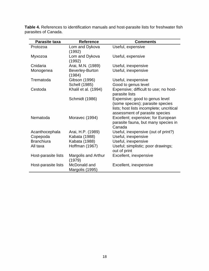
Identification of many parasite species may require consultation of original descriptionsin the primary literature. However, for most common groups, identification to genus andoften to species can be done through the synthetic keys listed in Table 4. Examplespecimens of parasites and hosts should always be retained to confirm identifications.Voucher specimens should be deposited in the permanent collection of a recognizedmuseum, for future reference and use.
Data Analysis
The most common measurements of parasite population levels in hosts are prevalence,mean abundance, and mean intensity (Bush et al. 1997). Prevalence refers to thepercentages of organisms infected by a particular species of parasite. Mean abundancerefers to the number of parasites of a given species per host examined, infected anduninfected. Mean intensity is the mean number of parasites of a given species perinfected host.
The scale of observation in parasitology is also important. A parasite population in anindividual host is an infrapopulation, whereas that in a host population is a componentpopulation. All the parasites of a given species in an ecosystem compose thesuprapopulation. Within an individual host, all the parasites found compose aninfracommunity, and within a host population, a component community. All theparasites found in an ecosystem form the compound community (see Esch et al. 1990).
For microparasites, data are usually presented as prevalence. Quantitative data oninfections can be presented as numbers per unit microscopic counts.
There are numerous measures of diversity used in parasitology, including species

6
richness, the Shannon-Wiener Index, Simpson’s Index, Berger-Parker Index, andBrillouin Index (see Magurran 1988). Species richness is the simplest measure ofdiversity, and is recommended at all scales. The Brillouin Index is recommended formeasurements of infracommunity diversity because it is an appropriate index for fullycensused communities (as in an individual host). For component communities, theShannon-Wiener Index
(H’ = - pi ln pi , where p is the proportion of species i in the community)
is recommended because it is less biased toward dominant species than other indices.However, all indices are fraught with problems of bias and interpretation. The usershould become familiar with these problems, and choose the index most appropriate forthe goals of the study.
It is often important to statistically determine whether parasite populations orcommunities differ from one site to another, or from one time period to another. To doso requires quantitative, replicated measurements. In the case of parasites, each fishhost within a species can be considered a replicate, just as a quadrat on the forest flooror a grab sample of the benthos is a replicate. Replication is especially important ifparasites are to be used as indicators of host population structure, environmentalquality, biodiversity, or for monitoring disease and other problems resulting fromparasitic infections.
Parasite distributions are almost always aggregated or overdispersed, meaning thatmost of the parasites in a population will be found in a small number of hosts, and mostpotential hosts may be lightly infected or uninfected (there are exceptions!). Thus, priorto any statistical analyses, data must be tested for normality. If the distributions are notnormal, an appropriate transformation must be used to normalize the data, beforeparametric statistics can be applied. If the data cannot be normalized, nonparametricstatistics should be used. The investigator should become familiar with the limitations ofthe analysis used and the consequences of violating assumptions inherent in variousstatistical procedures (Underwood 1981).
Quality Assessment/Quality Control (QA/QC)
A QA/QC plan is required for any successful monitoring program. It gives resultscredibility and helps structure the monitoring program. Ways to aid in structuring theprogram include field notes, sample collection forms, sample processing forms,procedures for verifying taxonomic identifications, data screening, and databasemanagement. A sample data sheet is shown in Appendix I. Questionable or uncertainidentifications should be verified by an expert. For establishing sampling guidelines toensure that results are meaningful, see Green (1979) or des Clers (1994).Approximately 20% of program resources should be dedicated to QA/QC.

7
Volunteer and Non-Specialist Involvement
People do not require a great deal of training to do adequate dissections to recoverparasites. University undergraduate classes can acquire a lot of valuable data.However, people with some expertise or experience are required for many, but not all,identifications.
Volunteers and volunteer networks can contribute cursory observations of largeparasites. In fresh water, these include some arthropods on gills and skin of fish. Copepods on gills are sometimes visible with the naked eye; they are white or cream-coloured and often have two egg sacs. The fish louse, Argulus, is quite large, and canbe seen crawling on the body surface. Leeches also can be easily seen on the externalsurface. Certain digenean metacercariae form blackspot and can be seen on the skinor in the flesh of fish. As adults, these parasites infect birds or mammals. The largeyellow grub Clinostomum is found in pustule-like capsules just under the skin of fish,often in the head or tail region. The huge, ribbon-like plerocercoids of the cestodesLigula and Schistocephalus can be found in the body cavities of cyprinids andsticklebacks, respectively. These are tapeworms that use birds as definitive hosts. Alarge redish nematode, Eustrongylides, also can be seen in the body cavity of fish. This nematode is a larval stage, and its adults infect birds. Other tapewormplerocercoids (e.g. Diphyllobothrium, Triaenophorus) are clearly visible as cysts on theviscera or in the musculature of fish. Adult cestodes such as Proteocephalus andacanthocephalans can be seen easily if the intestine is cut open.
Non-specialist volunteers should categorize the common macroparasite groups. Flatworms include digeneans (or flukes) and cestodes (or tapeworms). These are flat,and the cestodes are long and segmented, the digeneans short and rounded. Longnarrow worm-like organisms are nematodes (or roundworms). Tubular animals with aspiny “head”-like portion are acanthocephalans (or thorny-headed worms). Crustaceansare external parasites with hard body parts. Databases collected by volunteer groupsshould be maintained independently from those of experts until verification of species isassured.
Materials and Suppliers
See sections on other organisms for field sampling and collecting equipment. Standardbiological supply houses can provide most of the laboratory materials required. A list ofsuppliers is provided in Table 2.
Acknowledgements
I am grateful to Dan McLaughlin, Al Shostak, and David Rosenberg, whose commentsgreatly improved this document.

8
Experts to Contact for More Information
Dr. Martin Adamson Tel: 604-822-3374Department of Zoology Fax: 604-822-2416University of British Columbia Email: [email protected], British Columbia V6T 1Z4
Dr. David K. Cone Tel: 902-420-5644Department of Biology Fax: 902-420-5261St. Mary’s University Email: [email protected], Nova Scotia B3H 3C3
Dr. Terry Dick Tel: 204-474-9896Department of Zoology Fax: 204-269-7431University of Manitoba Email: [email protected], Manitoba R3T 2N2
Dr. Cam Goater Tel: 780-329-2752Department of Biological Sciences Fax: 780-329-2082University of Lethbridge Email: [email protected], Alberta T1K 3M4
Dr. Tim Goater Tel; 604-753-3245 ext. 2325Department of Biology Fax: 604-755-8749Malaspina University-College Email: [email protected], British Columbia V9R 5S5
Dr. Murray Lankester Tel: 807-343-8528Department of Biology Fax: 807-346-7796Lakehead University Email: [email protected] Bay, Ontario P7B 5E1
Dr. David J. Marcogliese Tel: 514-283-6499Environment Canada Fax: 614-496-7398St. Lawrence Centre Email: [email protected] McGill St., 7th FloorMontreal, Quebec H2Y 2E7
Dr. Dan McLaughlin Tel: 514-848-3409Biology Department Fax: 514-848-2881Concordia University Email: [email protected] de Maisonneuve Blvd. W.Montreal, Quebec H3G 1M8

9
Dr. Lena Measures Tel: 418-775-0571Department of Fisheries and Oceans Fax: 418-775-0740Maurice Lamontagne Institute Email: [email protected] Box 1000Mont-Joli, Quebec G5H 3K6
Dr. Allen W. Shostak Tel: 780-492-1293Department of Biological Sciences Fax: 780-492-9234University of Alberta Email: [email protected], Alberta T6G 2E9
N.B.: The Directory of Parasitologists in Canada may be consulted at:
http://www.biology.ualberta.ca/parasites/indexen/directoryi.htm

10
References
Anderson, R.M. and R. M. May. 1979. Population biology of infectious diseases: Part I.Nature 280: 361-367.
Arai, H.P. 1989. Acanthocephala, p. 1-90. In: L. Margolis and Z. Kabata (eds.) Guide tothe parasites of fishes of Canada. Part III. Canadian Special Publication of Fisheriesand Aquatic Sciences 107.
Arai, M.N. 1989. Cnidaria, p. 91-95. In: L. Margolis and Z. Kabata (eds.) Guide to theparasites of fishes of Canada. Part III. Canadian Special Publication of Fisheries andAquatic Sciences 107.
Arthur, J.R. and E. Albert. 1994. A survey of the parasites of Greenland halibut(Reinhardtius hippoglossoides) caught off Atlantic Canada, with notes on theirzoogeography in this fish. Canadian Journal of Zoology 72: 765-778.
Ash, L.R. and T.C. Orihel. 1991. Parasites: a guide to laboratory procedures andidentification. ASCP Press, Chicago.
Berland, B. 1982. Basic techniques involved in helminth preservation, p. 757. In:Parasites - their world and ours. Abstracts of the 5th International Congress ofParasitology, 7-14 August 1982. Toronto, Ontario.
Beverley-Burton, M. 1984. Monogenea and Turbellaria, p. 5-209. In: L. Margolis and Z.Kabata (eds.) Guide to the parasites of fishes of Canada. Part I. Canadian Special Publication of Fisheries and Aquatic Sciences 74.
Bush, A.O., K.D. Lafferty, J.M. Lotz, and A.W. Shostak. 1997. Parasitology meetsecology on its own terms: Margolis et al. revisited. Journal of Parasitology 83: 575-583.
des Clers, S. 1994. Sampling to detect infections and estimate prevalences inaquaculture. Pisces Press, Stirling, UK.
Esch, G.W., A.W. Shostak, D.J. Marcogliese, and T.M. Goater. 1990. Patterns andprocesses in helminth parasite communities: an overview, p. 1-20. In: G.W. Esch, A. O.Bush, and J.M. Aho (eds.) Parasite communities: patterns and processes. Chapmanand Hall, London, UK.
Gibson, D.I. 1996. Trematoda. p. 1-373. In: L. Margolis and Z. Kabata (eds.) Guide tothe parasites of fishes of Canada. Part IV. Canadian Special Publication of Fisheriesand Aquatic Sciences 124.
Green, R. H. 1979. Sampling design and statisitcal methods for environmentalbiologists. John Wiley and Sons, New York.

11
Hoffman, G.L. 1967. Parasites of North American freshwater fishes. University ofCalifornia Press, Berkeley, California.
Kabata, Z. 1988. Copepoda and Branchiura, p. 3-127. In: L. Margolis and Z. Kabata(eds.) Guide to the parasites of fishes of Canada. Part II - Crustacea. Canadian Special Publication of Fisheries and Aquatic Sciences 101.
Khalil, L.F., A. Jones, and R.A. Bray. 1994. Keys to the cestode parasites ofvertebrates. CAB International, Wallingford, UK.
Lom, J. and I. Dykova. 1992. Protozoan parasites of fishes. Developments inaquaculture and Fisheries Science, Vol. 26. Elsevier Science Publishers B. V.,Amsterdam, The Netherlands.
MacKenzie, K., H.H. Williams, B. Williams, A.H. McVicar, and R. Siddall. 1995.Parasites as indicators of water quality and the potential use of helminth transmission inmarine pollution studies. Advances in Parasitology 35: 85-144.
Magurran, A.E. 1988. Ecological diversity and its measurement. Princeton UniversityPress, Princeton, New Jersey.
Marcogliese, D.J. and D.K. Cone. 1996. On the distribution and abundance of eelparasites in Nova Scotia: influence of pH. Journal of Parasitology 82: 389-399.
Marcogliese, D.J. and D.K. Cone. 1997. Food webs: a plea for parasites. Trends in Ecology and Evolution 12: 320-325.
Margolis, L. and J.R. Arthur. 1979. Synopsis of the parasites of fishes of Canada.Bulletin of the Fisheries Research Board of Canada 179: 1-269.
May, R.M. and R.M. Anderson. 1979. Population biology of infectious diseases: Part II.Nature 280: 455-461.
McDonald, T.E. and L. Margolis. 1995. Synopsis of the parasites of fishes of Canada:Supplement (1978-1993). Canadian Special Publication of Fisheries and AquaticSciences 122: 1-265.
Moravec, F. 1994. Parasitic nematodes of freshwater fishes of Europe. KluwerAcademic Publishers, Dordrecht, The Netherlands.
Price, P.W. 1980. Evolutionary biology of parasites. Princeton University Press,Princeton, New Jersey.
Roberts, L.S. and J. Janovy, Jr. 1996. Foundations of parasitology. 5th edition. W. C.Brown Publishers, Dubuque, Iowa.
12
Schell, S.C. 1985. Handbook of trematodes of North America north of Mexico.University of Idaho Press, Moscow, Idaho.
Schmidt, G. D. 1896. Handbook of tapeworm identification. CRC Press, Boca Raton,Florida.
Sures, B., H. Taraschewski, and E. Jackwerth. 1994. Lead accumulation inPomphorhynchus laevis and its host. Journal of Parasitology 80: 355-357.
Underwood, A.J. 1981. Techniques of analysis of variance in experimental marinebiology and ecology. Oceanography and Marine Biology Annual Reviews 19: 513-605.
Williams, H.H., K. MacKenzie, and A.M. McCarthy. 1992. Parasites as biologicalindicators of the population biology, migrations, diet, and phylogenetics of fish. Reviewsin Fish Biology and Fisheries 2: 144-176.

13
Table 1. Numbers and general characteristics of known parasite species infectingCanadian freshwater fish (Margolis and Arthur 1979; McDonald and Margolis 1995).Note that examination of the Canadian fish fauna for parasites is incomplete, and newCanadian host records and new parasite species continue to be found on a regularbasis. The taxonomy of protists follows that of Roberts and Janovy (1996), and that ofmetazoans follows McDonald and Margolis (1995), with the exception of the Myxozoa,which are now regarded as a metazoan phylum.
Parasite taxa Number ofspecies
General characteristics
Kingdom Protista Subkingdom Protozoa
Unicellular
Phylum Sarcomastigophora 15 Single type of nucleus; flagella and/orpseudopodia
Phylum Apicomplexa 35 Exclusively parasitic; apical complex;micropore(s)
Phylum Microspora 8 Intracellular parasites; unicellular spores;polar filament
Phylum Ciliophora 23 Simple or compound cilia; subpellicularinfraciliature; 2 types of nuclei
Uncertain status 1
Kingdom Animalia Subkingdom Eumetazoa
Multicellular
Phylum Myxozoa 113 Polar capsules; valves; found in internalorgans
Phylum Cnidaria 1 Hydrozoans; parasitic forms rare Phylum Platyhelminthes Flatworms Class Trematoda Exclusively parasitic; short to oblong, rounded Subclass Aspidogastrea 1 Huge septate ventral sucker; in gut of fish Subclass Digenea 113 Usually 2 cuplike muscular suckers;
incomplete digestive system; adults primarilyinternal parasites, larvae internal or underskin; flukes
Class Monogenea 180 Exclusively parasitic; modified posteriorholdfast with hooks; mainly ectoparasites ongills, fins, skin
Class Cestoda 70 Exclusively parasitic; long, usually segmentedworms with suckers and/or hooks on holdfast;adults intestinal, larvae on viscera or in flesh;no digestive system; tapeworms
Phylum Nemathelminthes Class Nematoda 56 Slender worms, pointed or blunt at each end;
complete digestive system; found in internalorgans and viscera; roundworms
Phylum Acanthocephala 23 Exclusively parasitic; cylindrical with hookedprobscis; no digestive system; adultsintestinal, larvae on viscera; thorny-headedworms
Phylum Annelida 19 Suckers at each end; segmentedectoparasites; leeches
Phylum Mollusca 7 Clam-like; on gills; glochidia Phylum Arthropoda Exoskeleton with jointed limbs

14
Class Crustacea Subclass Branchiura 6 Flattened with dorsal shield; unsegmented,
bilobed abdomen; 4 pairs of appendages;ectoparasites; fish louse
Subclass Entomostraca 29 Often highly modified; ectoparasites;copepods
Class Arachnoidea 3 Ectoparasites; mitesTotal 703

15
Table 2. Suppliers for sampling and laboratory equipment.
FIELD SAMPLING EQUIPMENT
Wildlife Supply Company (WILDCO) Tel: 517-799-8100301 Cass Street Fax: 517-799-8115Saginaw, Michigan, USA 48602 Email: [email protected]: http://www.wildco.com
Kahlsico International Corporation Tel: 619-444-2158P.O. Box 947 Tel: 619-444-5944El Cajon, California, USA 92022-0947 Fax: 619-444-0207
Canadian distributor for Kahlsico:Geneq Inc.8047 Jarry E. Tel: 514-354-2511Montreal, Quebec H1J 1H6 Fax: 514-354-6948
223 Signet Drive Tel: 416-747-9889 Toronto, Ontario M9L 1V1 Fax: 416-747-7570

16
LABORATORY EQUIPMENT AND CHEMICALS/REAGENTS/STAINS
Laboratory equipment may be purchased at most scientific supply houses. Someexamples are listed below.
Fisher Scientific Tel: 800-234-7437112 Colonnade Road Fax: 800-463-2996Nepean, Ontario K2E 7L6 Website - http://www.fisher1.com
VWR CanlabAll provinces (except Quebec)2360 Argentia Road Tel: 905-821-9410Mississauga, Ontario L5N 5Z7 Fax: 905-821-3460orQuebec, Ottawa, Kingston8567 Dalton Tel: 514-344-3525Town of Mount Royal, Quebec H4T 1V5 Tel: 514-344-0133Website - http://www.vwrsp.com
Canadawide Scientific Tel: 800-2676-23622300 Walkley Road Unit 414 Fax: 800-814-5162Ottawa, Ontario K1G 6B1
Dissection instruments can be purchased at:
Fine Tools Scientific Tel: 800-665-5355202-277 Mountain Highway Fax: 800-665-4544 North Vancouver, British Columbia V7J 3P2 Email: [email protected] - http://www.finescience.com
Reagents, chemicals and stains can be purchased at:
Sigma-Aldrich Canada Ltd. Tel: 800-565-1400 2149 Winston Park Drive Fax: 800-265-3858 Oakville, Ontario L6H 6J8 Email: [email protected] - http://www.sigald.sial.com/canada
MS 222 can be purchased at:
Syndel Laboratories Ltd.9211 Shaughnessy St. Tel: 604-321-7131Vancouver, British Columbia V6P 6R5

17
Table 3. Recipes for standard fixatives, preservatives, and stains commonly used inparasitology. After Ash and Orihel (1991).
Material Recipe70% ethanol 740 mL 95% ethanol + 260 mL water5% glycerol in 70 % ethanol 50 mL glycerol + 740 mL 95% ethanol + 210 mL
water10% buffered formalin Dissolve 6.10 g dibasic sodium phophate (Na2HPO4)
and 0.15 g monobasic sodium phosphate (NaH2PO4)in 800 mL 37.5% formaldehyde (=100% formalin) +7200 mL water (Available commercially)
AFA (alcohol-formalin-acetic acid)
100 mL 100% formalin + 500 mL 95% ethanol + 50mL glacial acetic acid + 450 mL water
Schneider’s acetocarminestain
45 mL glacial acetic acid + 55 mL water (slowly!).Add 5 g carmine powder, boil 15 min. Cool and filter.For use, add a few drops to small dish of 70%ethanol to make solution moderate to dark pink.(Available commercially)
Glycerol-alcohol mixturesfor clearing largenematodes, to be usedsequentially
1. 50 mL glycerol + 700 mL 95% ethanol + 250 mLwater
2. 100 mL glycerol + 700 mL 95% ethanol + 200 mLwater
3. 200 mL glycerol + 500 mL 95% ethanol + 300 mLwater
4. 500 mL glycerol + 300 mL 95% ethanol + 200 mLwater
5. 700 mL glycerol + 100 mL 95% ethanol + 100 mLwater
6. 1000 mL glycerolLactophenol clearing agent 20 mL glycerol + 10 mL lactic acid + 10 mL melted
phenol crystals + 10 mL water. Mix well.Berland’s fluid 5 mL 100% formalin + 95 mL glacial acetic acid
18
Table 4. References to identification manuals and host-parasite lists for freshwater fishparasites of Canada.
Parasite taxa Reference CommentsProtozoa Lom and Dykova
(1992)Useful, expensive
Myxozoa Lom and Dykova(1992)
Useful, expensive
Cnidaria Arai, M.N. (1989) Useful, inexpensiveMonogenea Beverley-Burton
(1984)Useful, inexpensive
Trematoda Gibson (1996)Schell (1985)
Useful, inexpensiveGood to genus level
Cestoda Khalil et al. (1994)
Schmidt (1986)
Expensive; difficult to use; no host-parasite listsExpensive; good to genus level(some species); parasite specieslists; host lists incomplete; uncriticalassessment of parasite species
Nematoda Moravec (1994) Excellent; expensive; for Europeanparasite fauna, but many species inCanada
Acanthocephala Arai, H.P. (1989) Useful, inexpensive (out of print?)Copepoda Kabata (1988) Useful, inexpensiveBranchiura Kabata (1988) Useful, inexpensiveAll taxa Hoffman (1967) Useful; simplistic; poor drawings;
out of printHost-parasite lists Margolis and Arthur
(1979)Excellent, inexpensive
Host-parasite lists McDonald andMargolis (1995)
Excellent, inexpensive

19
Appendix I. Sample data sheet
Fish parasite data sheet
Locality: _______________ Date: _________
Host: __________________ No: ___________
Fork length: ____________ Weight: _____________ Sex: _______
Maturity: ______________ Condition: Fresh ( ) Held ( ) Refrigerated ( ) Frozen ( ) Fixed ( )
Gonad weight: __________ Stomach weight: (full)_______ (empty) _______
Date of examination: ____________ Examiner: _____________
Blood smear ( ): Esophagus:
External surface:
Smear ( ):
Body cavity:
Fins: Stomach:
Eyes L:
R:
Mesenteries:
Gills L Arc 1 2 3 4
Gills R Arc 1 2 3 4
Operculum L: R:
Intestine:
Smear ( ):Nasal cavities: Spleen:
Smear ( ):Mouth: Liver:
Smear ( ):Brain:Smear ( ):
Gall bladder:Smear ( ):
Heart: Urinary bladder:Smear ( ):
Swim bladder: Kidney:Smear ( ):
Gonads:Smear ( ):
Muscle L:
R:

20