echocardiographic evaluation of mitral valve prostheses · which of the following statements...
TRANSCRIPT
Echocardiographic
Evaluation of Mitral Valve
Prostheses
Dennis A. Tighe, M.D., FACC, FACP, FASE
Cardiovascular Medicine
University of Massachusetts Medical School
Worcester, MA
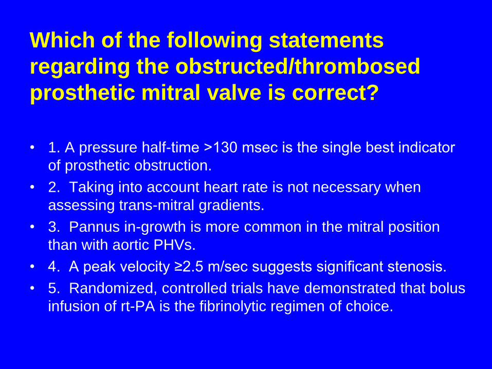
Which of the following statements
regarding the obstructed/thrombosed
prosthetic mitral valve is correct?
• 1. A pressure half-time ˃130 msec is the single best indicator
of prosthetic obstruction.
• 2. Taking into account heart rate is not necessary when
assessing trans-mitral gradients.
• 3. Pannus in-growth is more common in the mitral position
than with aortic PHVs.
• 4. A peak velocity ≥2.5 m/sec suggests significant stenosis.
• 5. Randomized, controlled trials have demonstrated that bolus
infusion of rt-PA is the fibrinolytic regimen of choice.
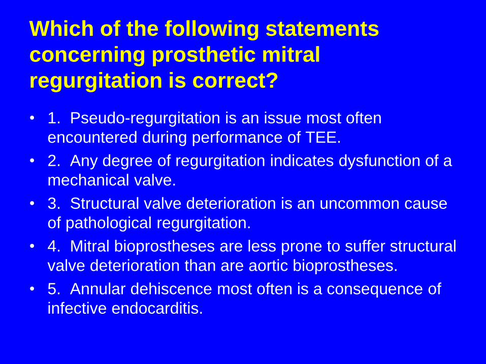
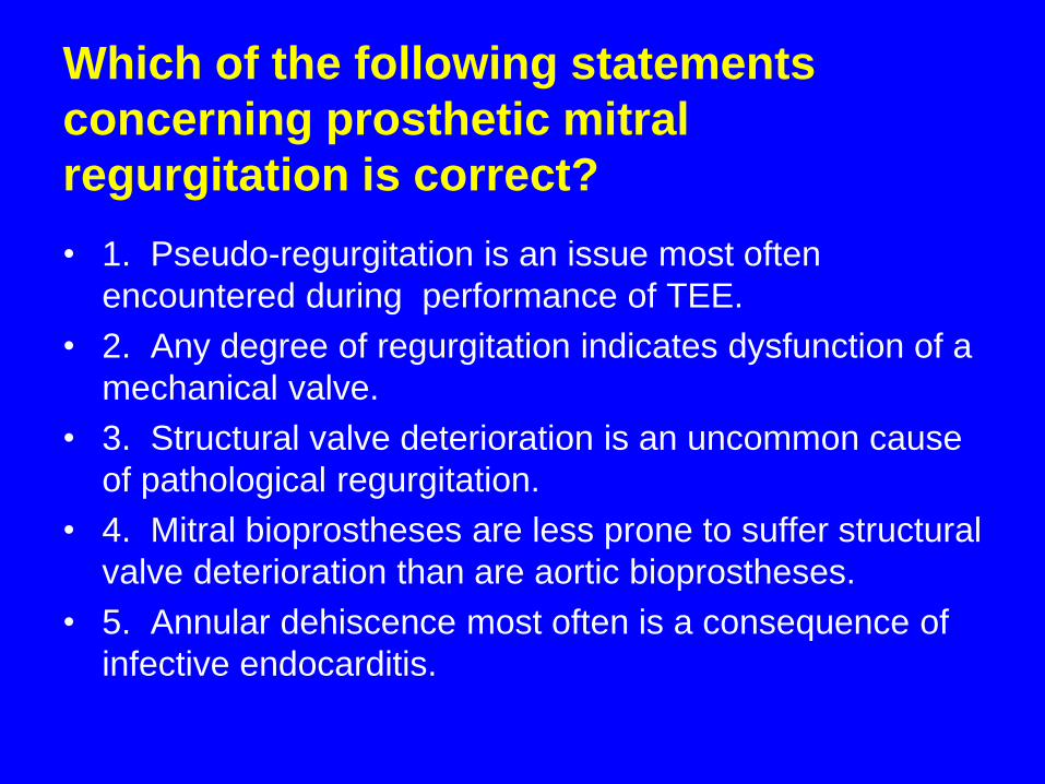
Which of the following statements
concerning prosthetic mitral
regurgitation is correct?
• 1. Pseudo-regurgitation is an issue most often
encountered during performance of TEE.
• 2. Any degree of regurgitation indicates dysfunction of a
mechanical valve.
• 3. Structural valve deterioration is an uncommon cause
of pathological regurgitation.
• 4. Mitral bioprostheses are less prone to suffer structural
valve deterioration than are aortic bioprostheses.
• 5. Annular dehiscence most often is a consequence of
infective endocarditis.

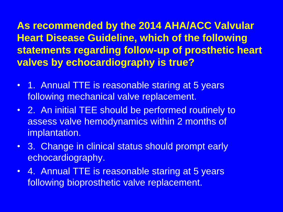
As recommended by the 2014 AHA/ACC Valvular
Heart Disease Guideline, which of the following
statements regarding follow-up of prosthetic heart
valves by echocardiography is true?
• 1. Annual TTE is reasonable staring at 5 years
following mechanical valve replacement.
• 2. An initial TEE should be performed routinely to
assess valve hemodynamics within 2 months of
implantation.
• 3. Change in clinical status should prompt early
echocardiography.
• 4. Annual TTE is reasonable staring at 5 years
following bioprosthetic valve replacement.
www.asecho.org

Nishimura RA et al. Circulation 2014;129:e521-e643.

Overview
• Description of the various types of
prosthetic heart valves
• Echocardiographic evaluation of
normally-functioning prosthetic heart
valves
• Evaluation of prosthetic heart valve
dysfunction
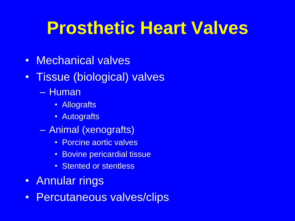
Prosthetic Heart Valves
• Mechanical valves
• Tissue (biological) valves
– Human
• Allografts
• Autografts
– Animal (xenografts)
• Porcine aortic valves
• Bovine pericardial tissue
• Stented or stentless
• Annular rings
• Percutaneous valves/clips
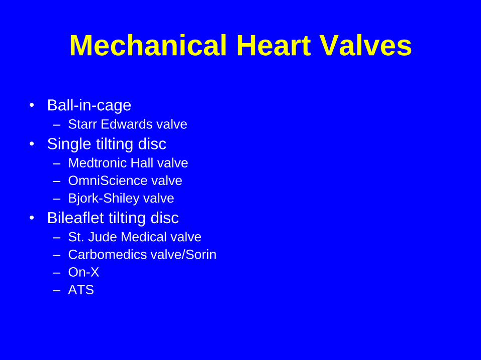
Mechanical Heart Valves
• Ball-in-cage– Starr Edwards valve
• Single tilting disc– Medtronic Hall valve
– OmniScience valve
– Bjork-Shiley valve
• Bileaflet tilting disc– St. Jude Medical valve
– Carbomedics valve/Sorin
– On-X
– ATS
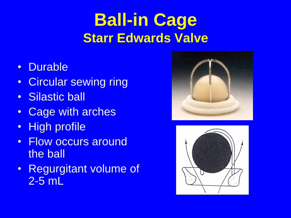
Ball-in CageStarr Edwards Valve
• Durable
• Circular sewing ring
• Silastic ball
• Cage with arches
• High profile
• Flow occurs around the ball
• Regurgitant volume of 2-5 mL
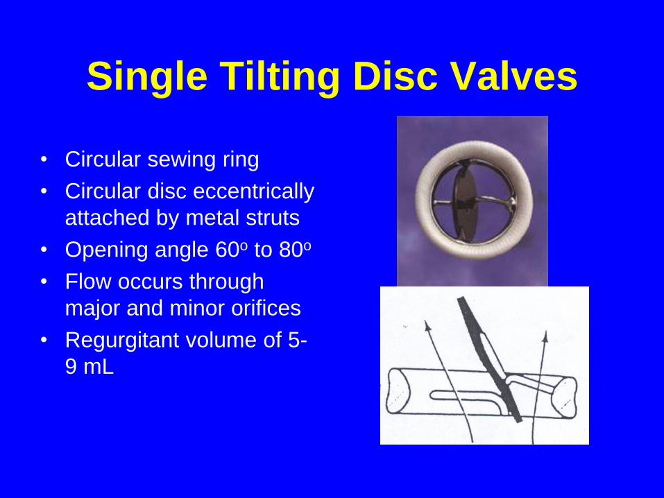
Single Tilting Disc Valves
• Circular sewing ring
• Circular disc eccentrically
attached by metal struts
• Opening angle 60o to 80o
• Flow occurs through
major and minor orifices
• Regurgitant volume of 5-
9 mL
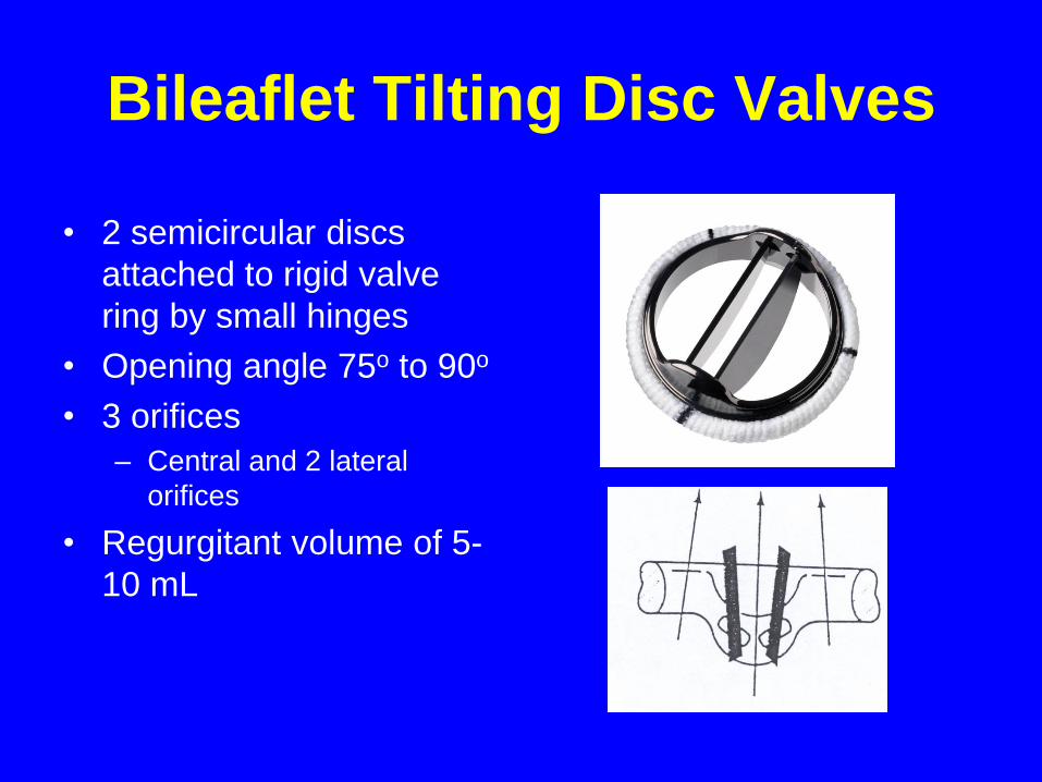
Bileaflet Tilting Disc Valves
• 2 semicircular discs
attached to rigid valve
ring by small hinges
• Opening angle 75o to 90o
• 3 orifices
– Central and 2 lateral
orifices
• Regurgitant volume of 5-
10 mL
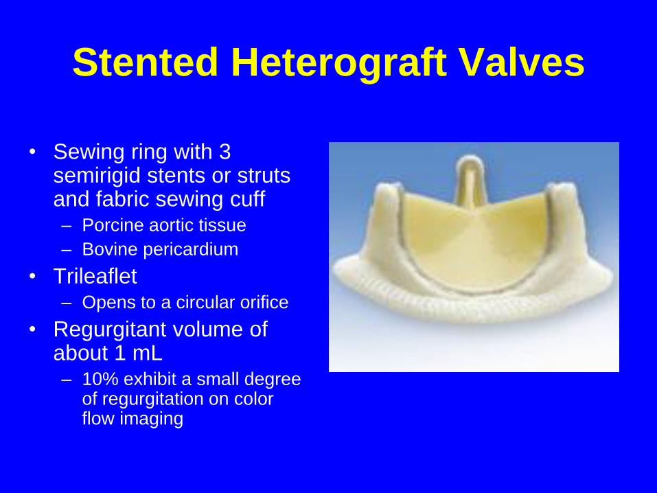
Stented Heterograft Valves
• Sewing ring with 3 semirigid stents or struts and fabric sewing cuff– Porcine aortic tissue
– Bovine pericardium
• Trileaflet– Opens to a circular orifice
• Regurgitant volume of about 1 mL– 10% exhibit a small degree
of regurgitation on color flow imaging
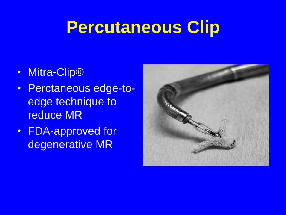
Percutaneous Clip
• Mitra-Clip®
• Perctaneous edge-to-
edge technique to
reduce MR
• FDA-approved for
degenerative MR
Echocardiographic Approach to
Assessment of Prosthetic Heart Valves
• Evaluation similar to that of native
valves
• Reverberations and shadowing play a
significant role
• Fluid dynamics of each specific valve
prosthesis influences the Doppler
findings
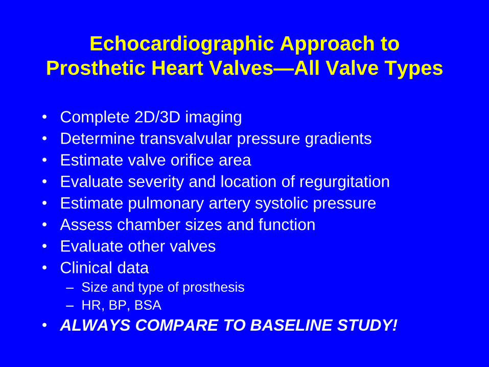
Echocardiographic Approach to
Prosthetic Heart Valves—All Valve Types
• Complete 2D/3D imaging
• Determine transvalvular pressure gradients
• Estimate valve orifice area
• Evaluate severity and location of regurgitation
• Estimate pulmonary artery systolic pressure
• Assess chamber sizes and function
• Evaluate other valves
• Clinical data– Size and type of prosthesis
– HR, BP, BSA
• ALWAYS COMPARE TO BASELINE STUDY!
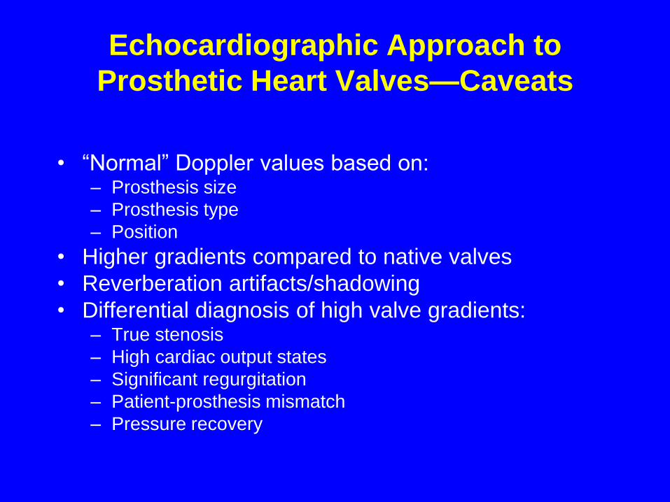
Echocardiographic Approach to
Prosthetic Heart Valves—Caveats
• “Normal” Doppler values based on: – Prosthesis size
– Prosthesis type
– Position
• Higher gradients compared to native valves
• Reverberation artifacts/shadowing
• Differential diagnosis of high valve gradients:– True stenosis
– High cardiac output states
– Significant regurgitation
– Patient-prosthesis mismatch
– Pressure recovery
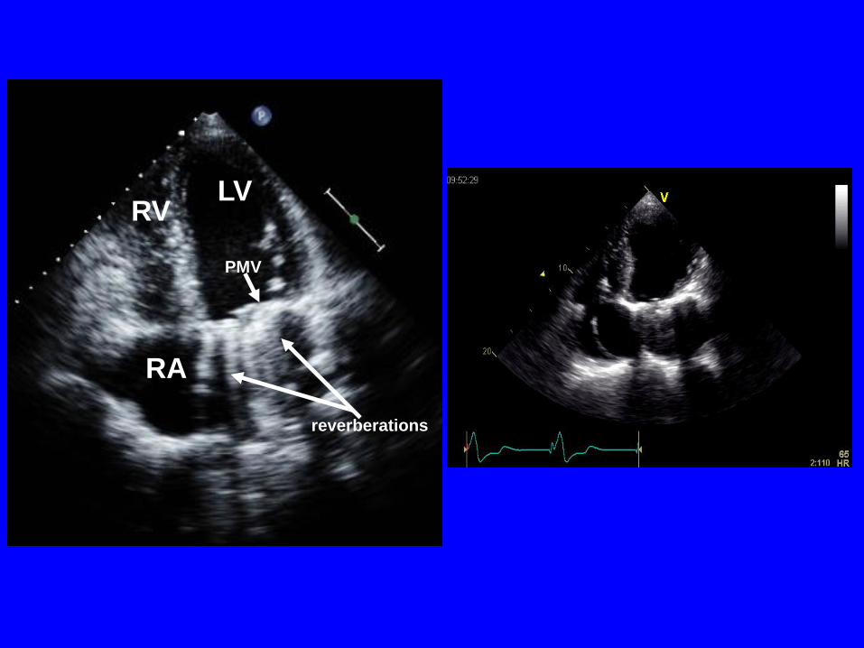
LV
RA
RV
PMV
reverberations
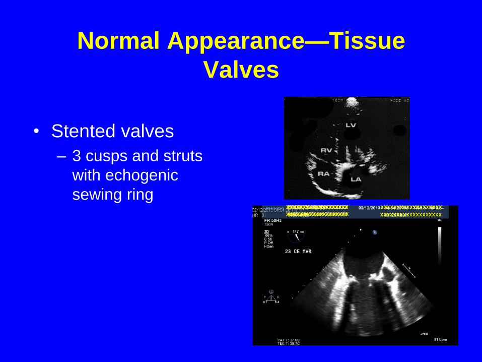
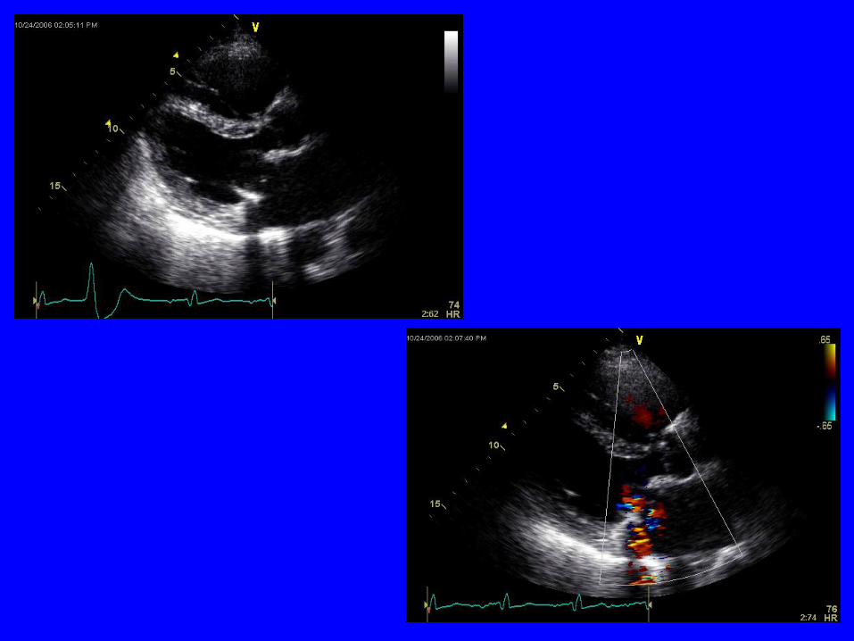
Normal Appearance—Tissue
Valves
• Stented valves
– 3 cusps and struts
with echogenic
sewing ring
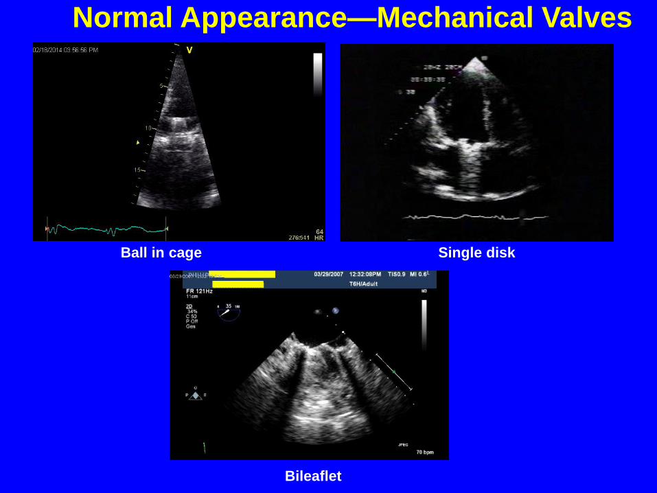
Normal Appearance—Mechanical Valves
Ball in cage Single disk
Bileaflet

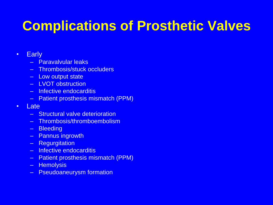
Complications of Prosthetic Valves
• Early– Paravalvular leaks
– Thrombosis/stuck occluders
– Low output state
– LVOT obstruction
– Infective endocarditis
– Patient prosthesis mismatch (PPM)
• Late– Structural valve deterioration
– Thrombosis/thromboembolism
– Bleeding
– Pannus ingrowth
– Regurgitation
– Infective endocarditis
– Patient prosthesis mismatch (PPM)
– Hemolysis
– Pseudoaneurysm formation
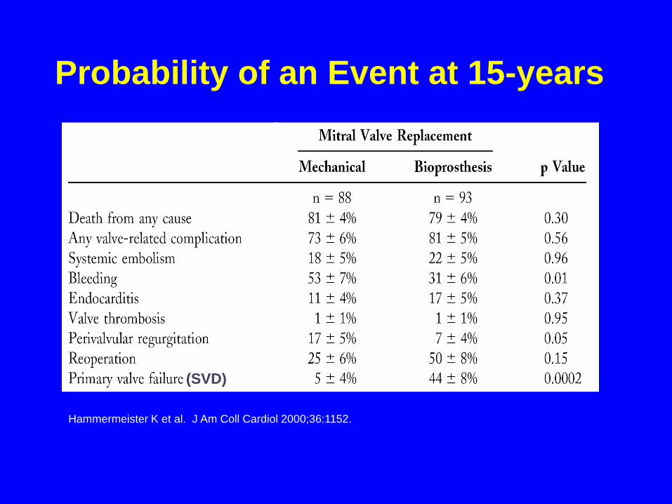
Hammermeister K et al. J Am Coll Cardiol 2000;36:1152.
Probability of an Event at 15-years
(SVD)
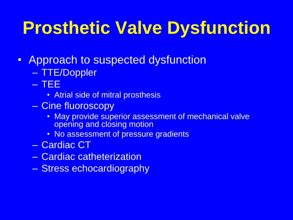
Prosthetic Valve Dysfunction
• Approach to suspected dysfunction– TTE/Doppler
– TEE• Atrial side of mitral prosthesis
– Cine fluoroscopy• May provide superior assessment of mechanical valve
opening and closing motion
• No assessment of pressure gradients
– Cardiac CT
– Cardiac catheterization
– Stress echocardiography
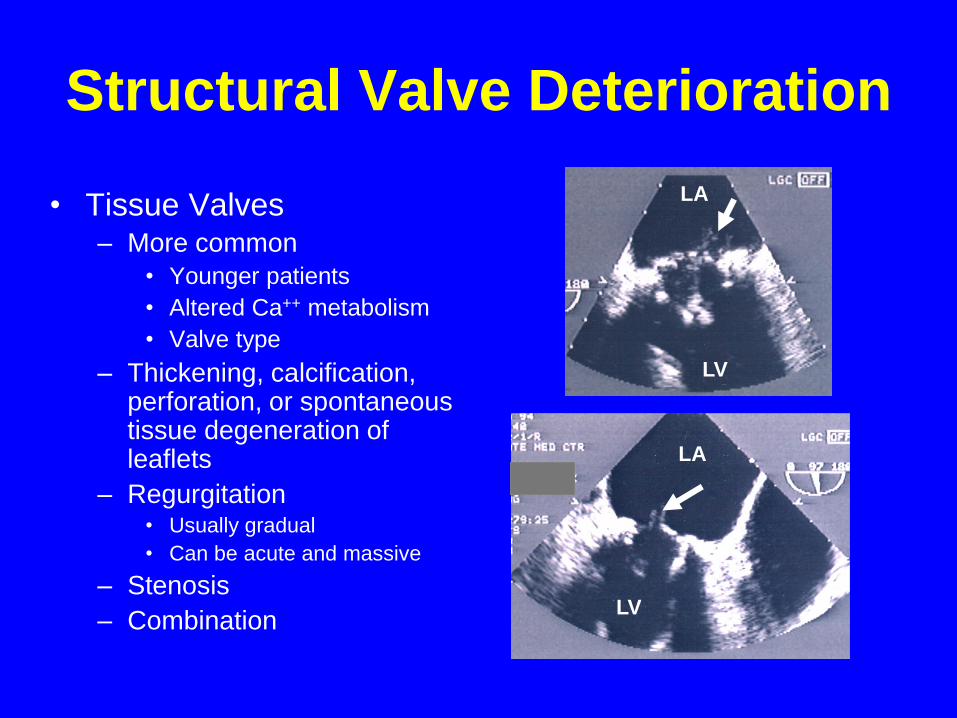
Structural Valve Deterioration
• Tissue Valves– More common
• Younger patients
• Altered Ca++ metabolism
• Valve type
– Thickening, calcification, perforation, or spontaneous tissue degeneration of leaflets
– Regurgitation• Usually gradual
• Can be acute and massive
– Stenosis
– Combination
LA
LV
LA
LV
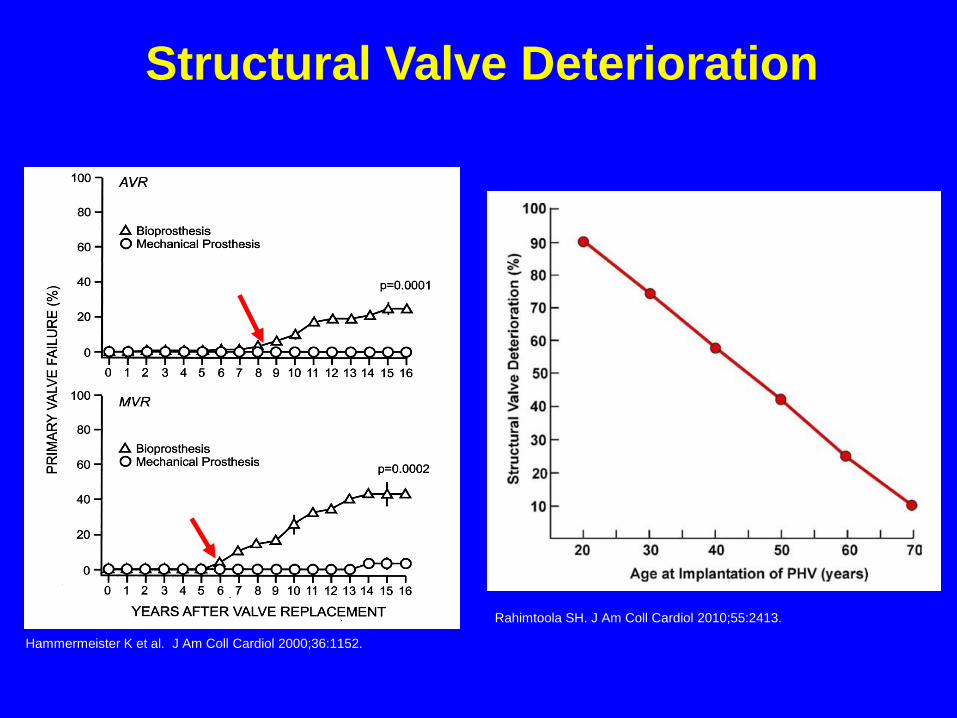
Hammermeister K et al. J Am Coll Cardiol 2000;36:1152.
Structural Valve Deterioration
Rahimtoola SH. J Am Coll Cardiol 2010;55:2413.
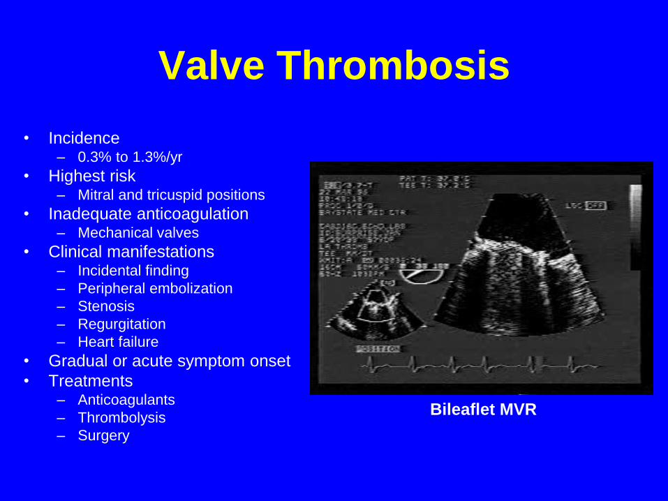
Valve Thrombosis
• Incidence – 0.3% to 1.3%/yr
• Highest risk– Mitral and tricuspid positions
• Inadequate anticoagulation– Mechanical valves
• Clinical manifestations– Incidental finding
– Peripheral embolization
– Stenosis
– Regurgitation
– Heart failure
• Gradual or acute symptom onset
• Treatments– Anticoagulants
– Thrombolysis
– Surgery
Bileaflet MVR
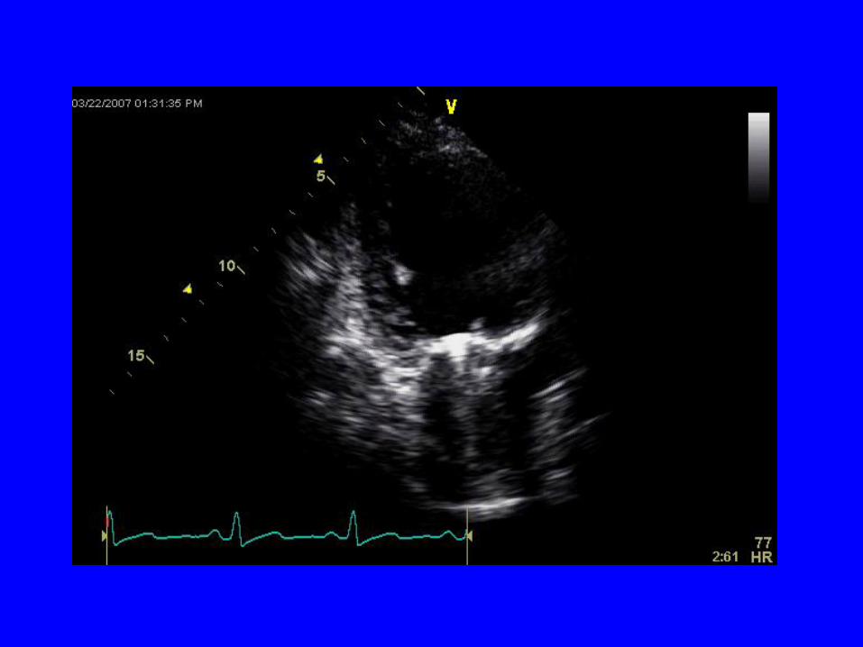
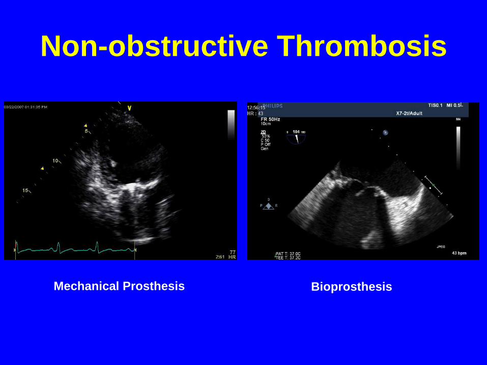
Non-obstructive Thrombosis
Mechanical Prosthesis Bioprosthesis
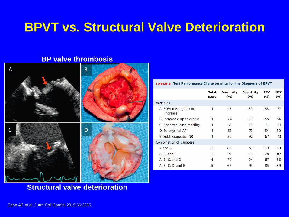
BPVT vs. Structural Valve Deterioration
BP valve thrombosis
Structural valve deterioration
Egbe AC et al. J Am Coll Cardiol 2015;66:2285.
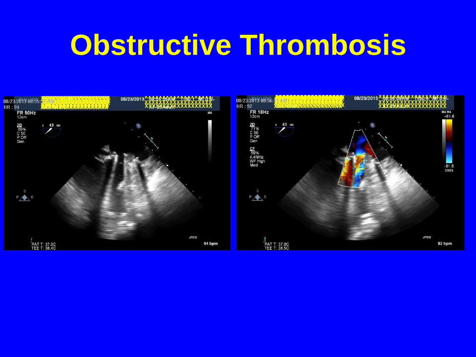
Obstructive Thrombosis
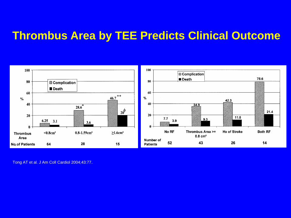
Tong AT et al. J Am Coll Cardiol 2004;43:77.
Thrombus Area by TEE Predicts Clinical Outcome
Nishimura RA et al. Circulation 2014 ;129:e586.
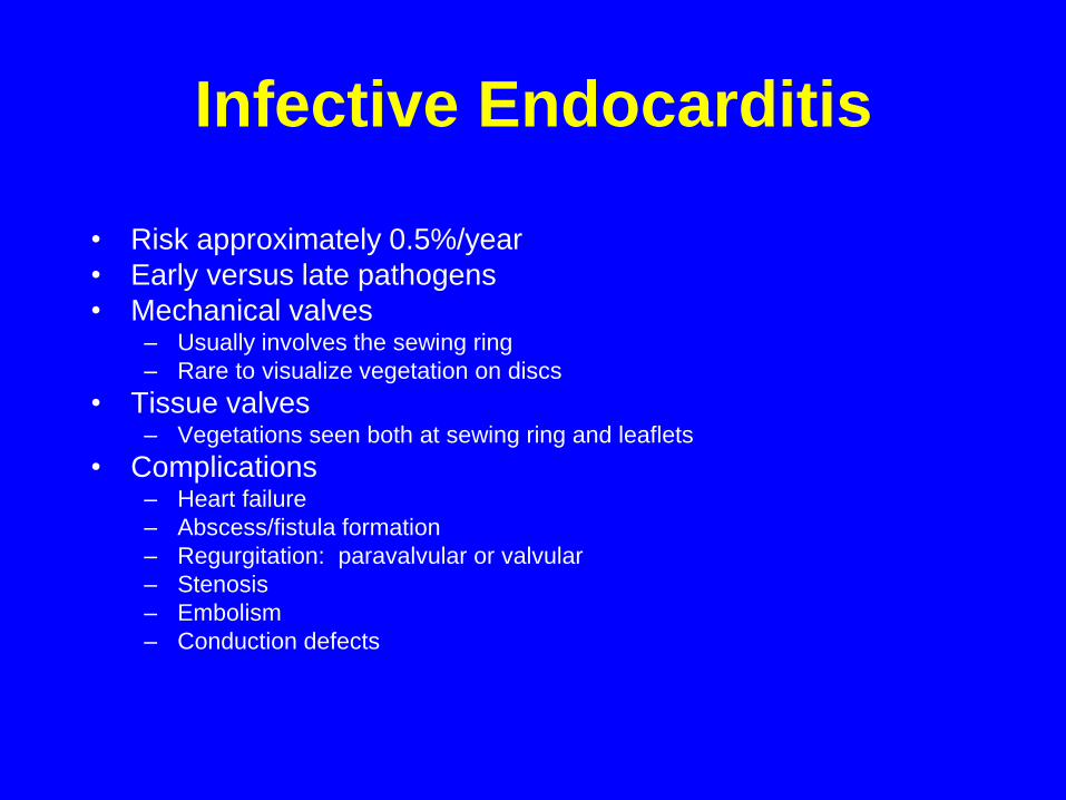
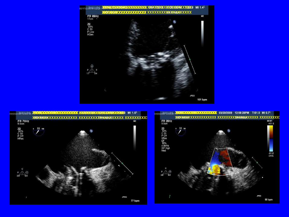
Infective Endocarditis
• Risk approximately 0.5%/year
• Early versus late pathogens
• Mechanical valves– Usually involves the sewing ring
– Rare to visualize vegetation on discs
• Tissue valves– Vegetations seen both at sewing ring and leaflets
• Complications– Heart failure
– Abscess/fistula formation
– Regurgitation: paravalvular or valvular
– Stenosis
– Embolism
– Conduction defects
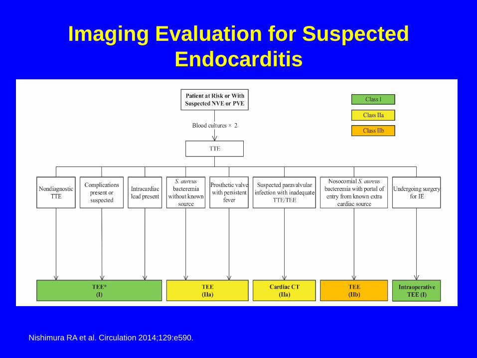
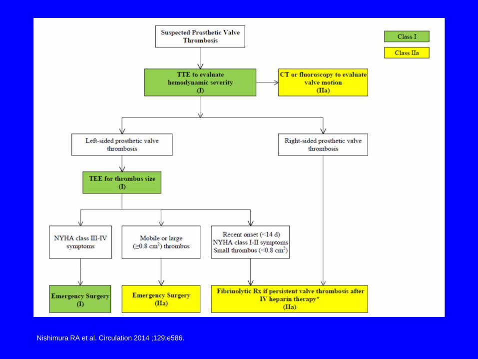
Imaging Evaluation for Suspected
Endocarditis
Nishimura RA et al. Circulation 2014;129:e590.
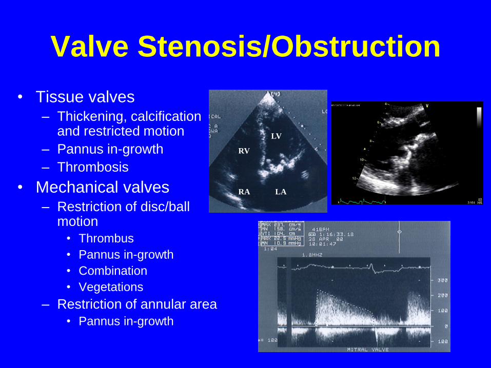
Valve Stenosis/Obstruction
• Tissue valves– Thickening, calcification
and restricted motion
– Pannus in-growth
– Thrombosis
• Mechanical valves– Restriction of disc/ball
motion
• Thrombus
• Pannus in-growth
• Combination
• Vegetations
– Restriction of annular area
• Pannus in-growth
LV
LA
RV
RA
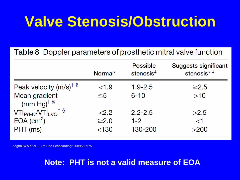
Valve Stenosis/Obstruction
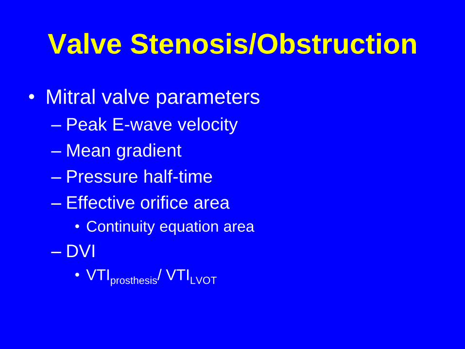
• Mitral valve parameters
– Peak E-wave velocity
– Mean gradient
– Pressure half-time
– Effective orifice area
• Continuity equation area
– DVI
• VTIprosthesis/ VTILVOT
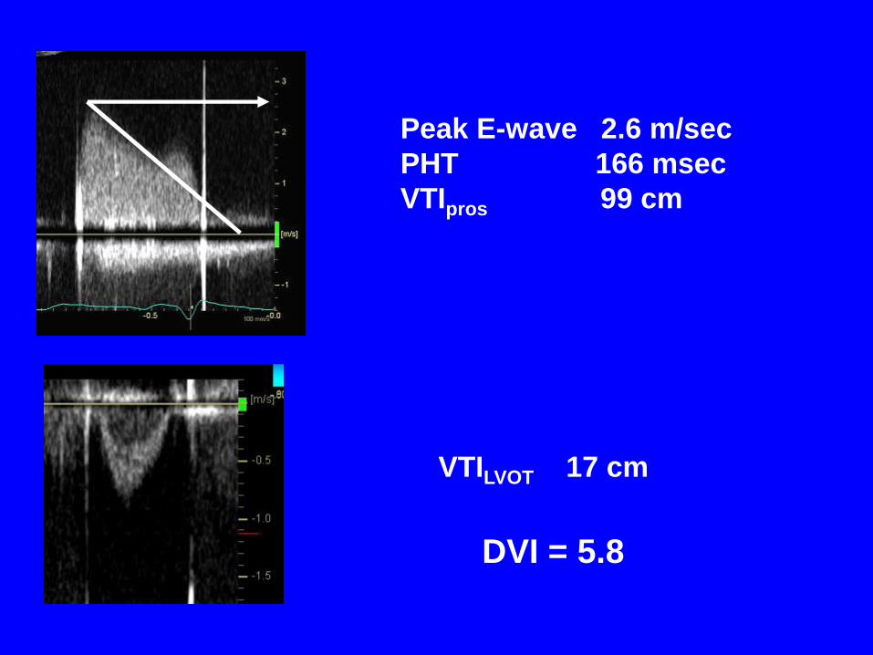
Peak E-wave 2.6 m/sec
PHT 166 msec
VTIpros 99 cm
VTILVOT 17 cm
DVI = 5.8
Fernandes V et al. Am J Cardiol 2002;89:704.
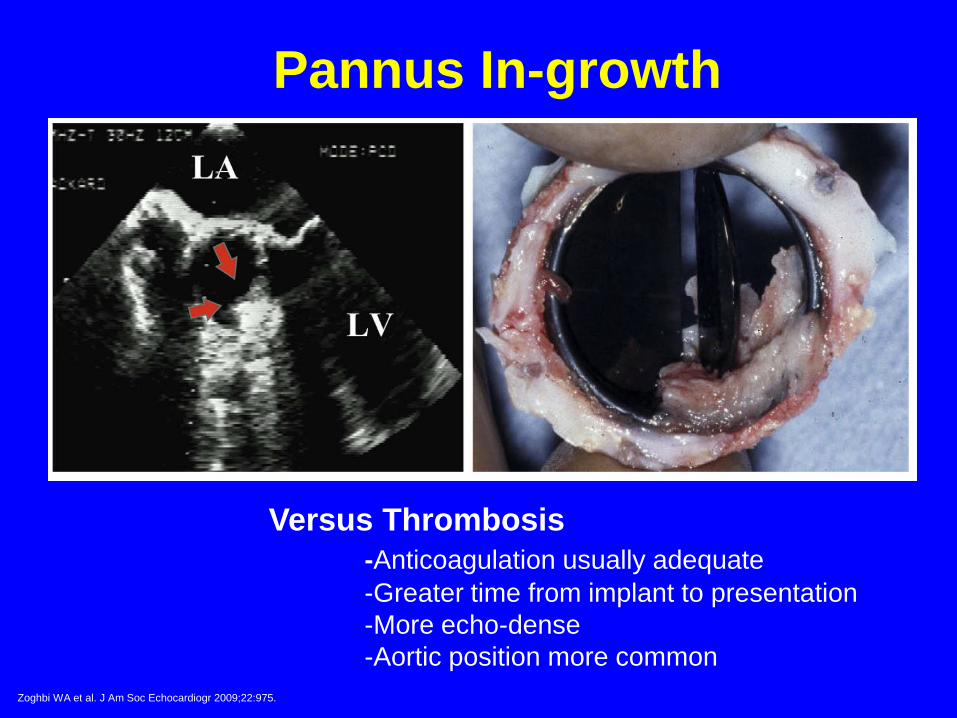
Pannus In-growth
Versus Thrombosis
-Anticoagulation usually adequate
-Greater time from implant to presentation
-More echo-dense
-Aortic position more common
Zoghbi WA et al. J Am Soc Echocardiogr 2009;22:975.
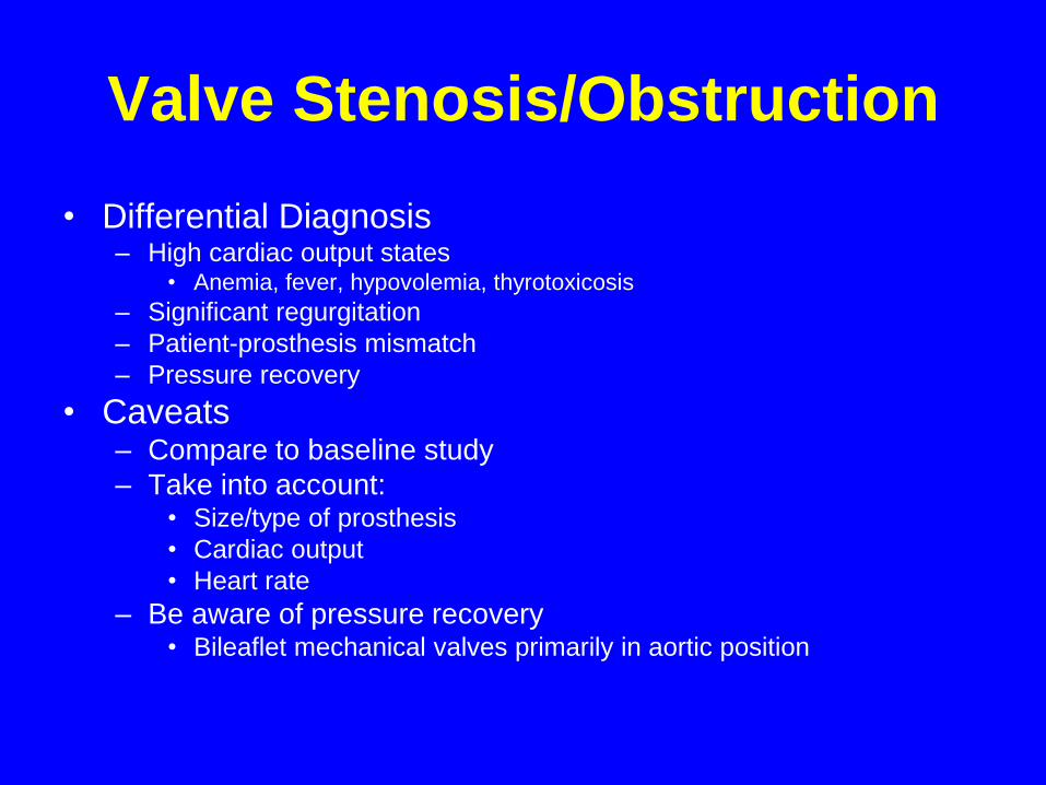
Valve Stenosis/Obstruction
• Differential Diagnosis– High cardiac output states
• Anemia, fever, hypovolemia, thyrotoxicosis
– Significant regurgitation
– Patient-prosthesis mismatch
– Pressure recovery
• Caveats– Compare to baseline study
– Take into account: • Size/type of prosthesis
• Cardiac output
• Heart rate
– Be aware of pressure recovery • Bileaflet mechanical valves primarily in aortic position
Valve Stenosis/Obstruction
Note: PHT is not a valid measure of EOA
Zoghbi WA et al. J Am Soc Echocardiogr 2009;22:975.
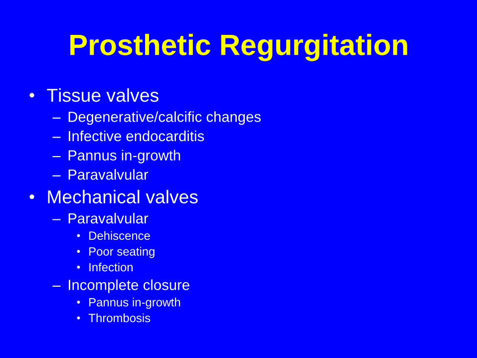
Prosthetic Regurgitation
• Tissue valves– Degenerative/calcific changes
– Infective endocarditis
– Pannus in-growth
– Paravalvular
• Mechanical valves– Paravalvular
• Dehiscence
• Poor seating
• Infection
– Incomplete closure• Pannus in-growth
• Thrombosis
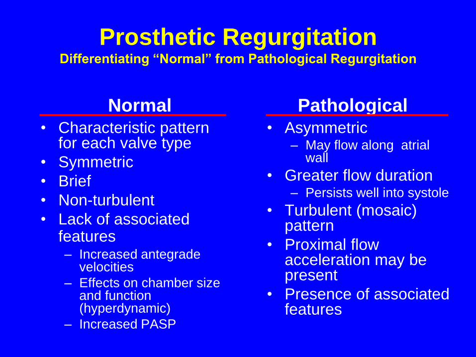
Prosthetic RegurgitationDifferentiating “Normal” from Pathological Regurgitation
• Characteristic pattern for each valve type
• Symmetric
• Brief
• Non-turbulent
• Lack of associated features– Increased antegrade
velocities
– Effects on chamber size and function (hyperdynamic)
– Increased PASP
• Asymmetric– May flow along atrial
wall
• Greater flow duration– Persists well into systole
• Turbulent (mosaic) pattern
• Proximal flow acceleration may be present
• Presence of associated features
Normal Pathological
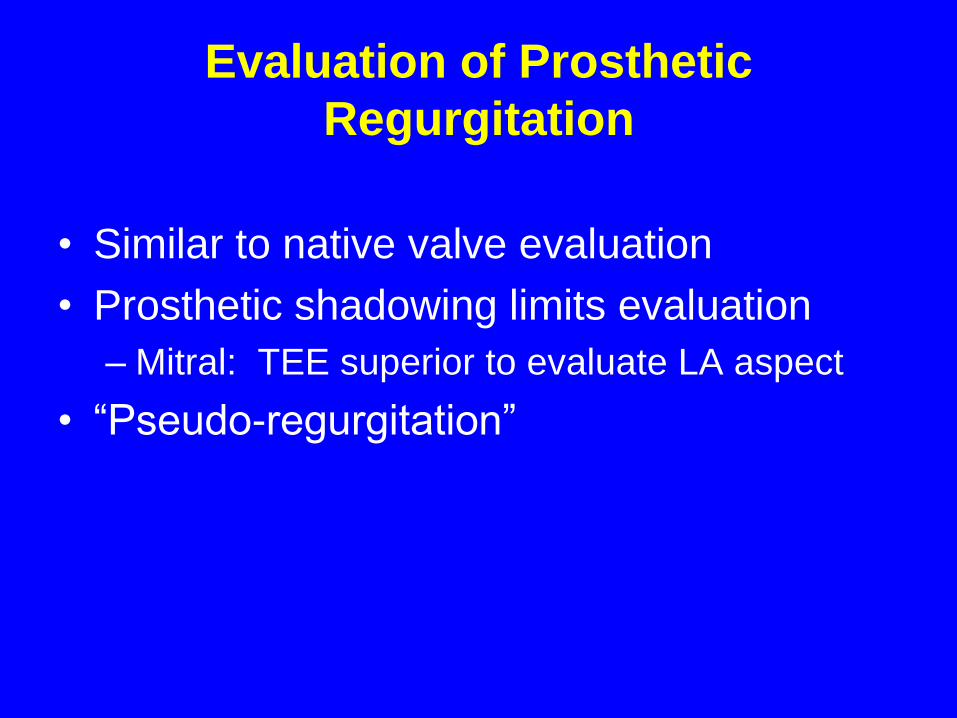
Evaluation of Prosthetic
Regurgitation
• Similar to native valve evaluation
• Prosthetic shadowing limits evaluation
– Mitral: TEE superior to evaluate LA aspect
• “Pseudo-regurgitation”
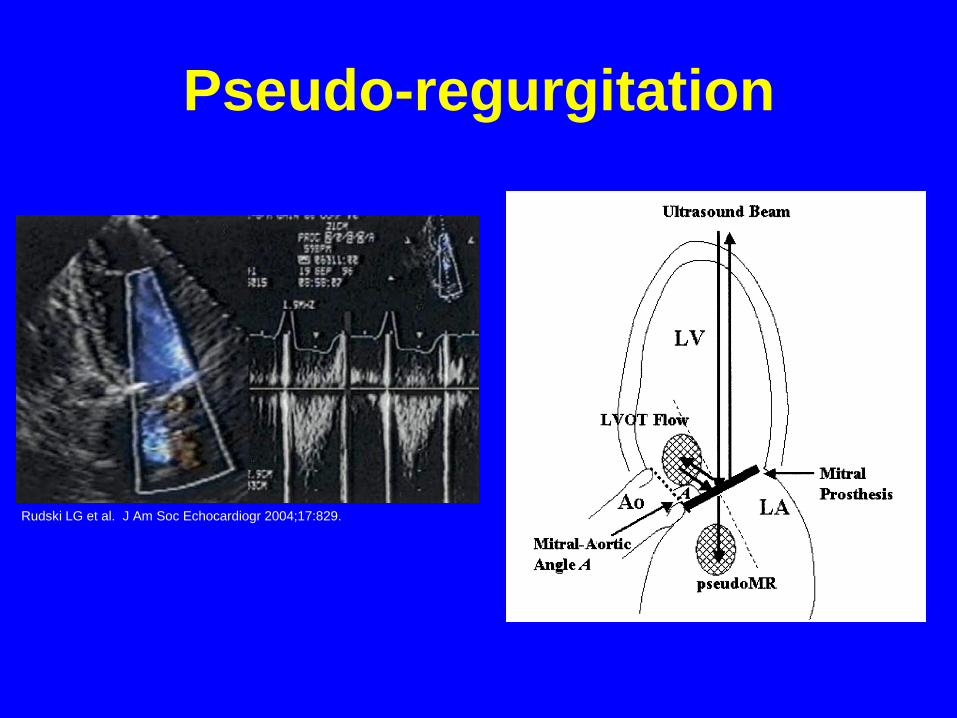
Pseudo-regurgitation
Rudski LG et al. J Am Soc Echocardiogr 2004;17:829.
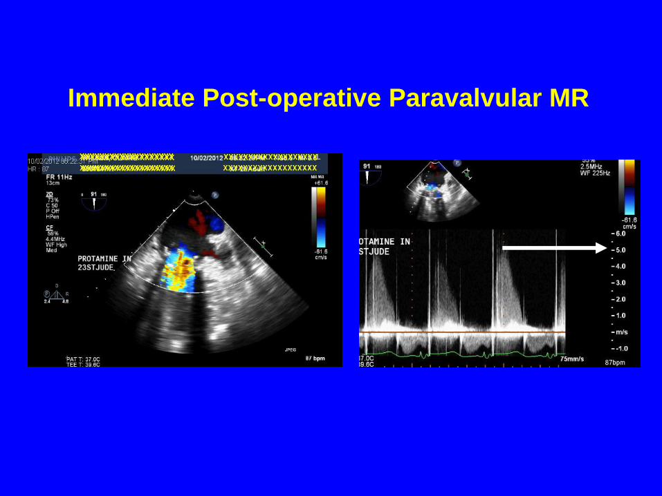
Immediate Post-operative Paravalvular MR
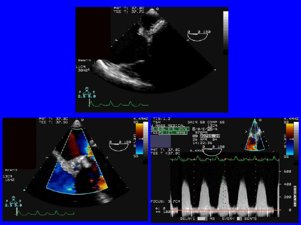
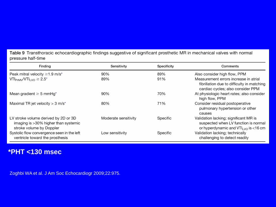
*PHT <130 msec
Zoghbi WA et al. J Am Soc Echocardiogr 2009;22:975.
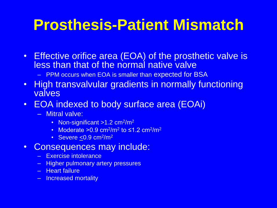
Prosthesis-Patient Mismatch
• Effective orifice area (EOA) of the prosthetic valve is less than that of the normal native valve
– PPM occurs when EOA is smaller than expected for BSA
• High transvalvular gradients in normally functioning valves
• EOA indexed to body surface area (EOAi)– Mitral valve:
• Non-significant >1.2 cm2/m2
• Moderate ˃0.9 cm2/m2 to ≤1.2 cm2/m2
• Severe <0.9 cm2/m2
• Consequences may include:– Exercise intolerance
– Higher pulmonary artery pressures
– Heart failure
– Increased mortality
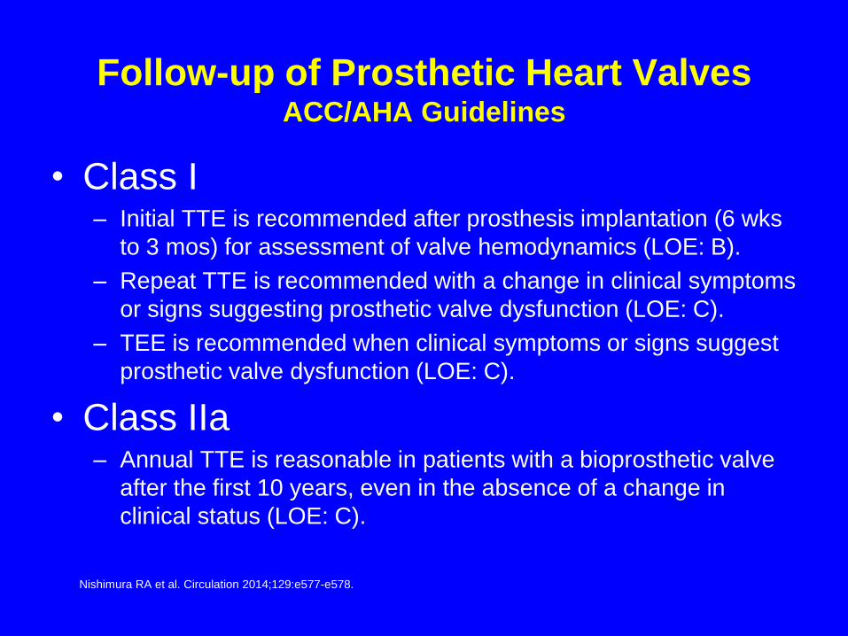
Follow-up of Prosthetic Heart ValvesACC/AHA Guidelines
Nishimura RA et al. Circulation 2014;129:e577-e578.
• Class I– Initial TTE is recommended after prosthesis implantation (6 wks
to 3 mos) for assessment of valve hemodynamics (LOE: B).
– Repeat TTE is recommended with a change in clinical symptoms
or signs suggesting prosthetic valve dysfunction (LOE: C).
– TEE is recommended when clinical symptoms or signs suggest
prosthetic valve dysfunction (LOE: C).
• Class IIa– Annual TTE is reasonable in patients with a bioprosthetic valve
after the first 10 years, even in the absence of a change in
clinical status (LOE: C).

Which of the following statements
regarding the obstructed/thrombosed
prosthetic mitral valve is correct?
• 1. A pressure half-time ˃130 msec is the single best indicator
of prosthetic obstruction.
• 2. Taking into account heart rate is not necessary when
assessing trans-mitral gradients.
• 3. Pannus in-growth is more common in the mitral position
than with aortic PHVs.
• 4. A peak velocity ≥2.5 m/sec suggests significant stenosis.
• 5. Randomized, controlled trials have demonstrated that bolus
infusion of rt-PA is the fibrinolytic regimen of choice.
Which of the following statements
concerning prosthetic mitral
regurgitation is correct?
• 1. Pseudo-regurgitation is an issue most often
encountered during performance of TEE.
• 2. Any degree of regurgitation indicates dysfunction of a
mechanical valve.
• 3. Structural valve deterioration is an uncommon cause
of pathological regurgitation.
• 4. Mitral bioprostheses are less prone to suffer structural
valve deterioration than are aortic bioprostheses.
• 5. Annular dehiscence most often is a consequence of
infective endocarditis.
As recommended by the 2014 AHA/ACC Valvular
Heart Disease Guideline, which of the following
statements regarding follow-up of prosthetic heart
valves by echocardiography is true?
• 1. Annual TTE is reasonable staring at 5 years
following mechanical valve replacement.
• 2. An initial TEE should be performed routinely to
assess valve hemodynamics within 2 months of
implantation.
• 3. Change in clinical status should prompt early
echocardiography.
• 4. Annual TTE is reasonable staring at 5 years
following bioprosthetic valve replacement.

Thank you for your attention