echo in emergency and critical care settings for … · echo in emergency and critical care...
TRANSCRIPT
Echo in Emergency and Critical Care Settings
Teerapat Yingchoncharoen MD.Ramathibodi Hospital
Mahidol University
Complimentary Educational Session for Cardiology Fellows
Sep 30, 2015
Common pitfalls and role of echo in decision making

Mahidol University Echo in emergency and critical care
Outline
• Basic standard echo view • Cases of echo in critical and
emergency settings

IVC size
J Am Soc Echocardiogr 2010;23:685Mahidol University

IVC size and RA pressure
J Am Soc Echocardiogr 2010;23:685
Estimated RAP 3 mmHg 8 mmHg 15 mmHg
Mahidol University
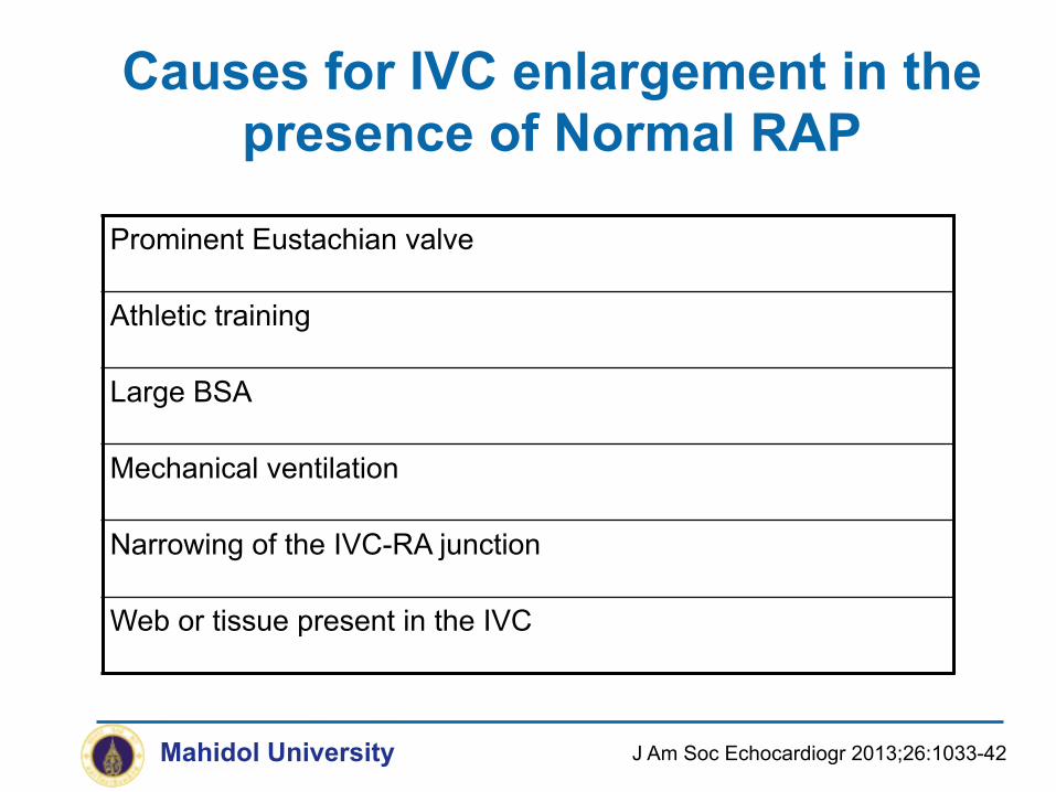
Causes for IVC enlargement in the presence of Normal RAP
J Am Soc Echocardiogr 2013;26:1033-42
Prominent Eustachian valve
Athletic training
Large BSA
Mechanical ventilation
Narrowing of the IVC-RA junction
Web or tissue present in the IVC
Mahidol University
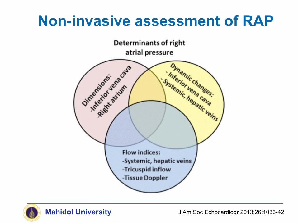
Non-invasive assessment of RAP
J Am Soc Echocardiogr 2013;26:1033-42Mahidol University

Mahidol University
RAMATHIBODI HOSPITAL
Function -Global systolic function -Regional wall motion
Structure -Chamber size -Wall thickness -Valve structure, morphology integrity -Mass (tumor, clot, vegetation) -Pericardial effusion -Congenital heart disease
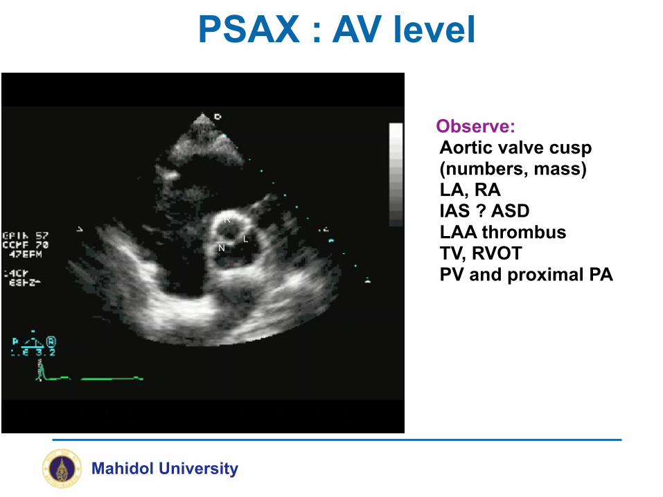
PSAX : AV level
Mahidol University
Observe: -Aortic valve cusp -(numbers, mass) -LA, RA -IAS ? ASD -LAA thrombus -TV, RVOT -PV and proximal PA
R
NL

PSAX Pulmonary trunk bifurcation
Mahidol University Basic Echo
RVOT
MPA
RPALPA
-Look for : PDA, PV disease (PS/PR), RVOT obstruction, PE
Mahidol University Basic Echo
RAMATHIBODI HOSPITAL
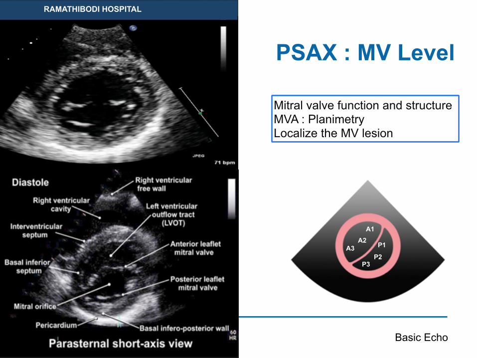
Mitral valve function and structure MVA : Planimetry Localize the MV lesion
PSAX : MV Level
A
P2
P1
P3
A1A2
A3

Mahidol University Basic Echo
RAMATHIBODI HOSPITAL
LV function Septum thickness and motion RV size Pericardial effusion
PSAX : Mid LV Level
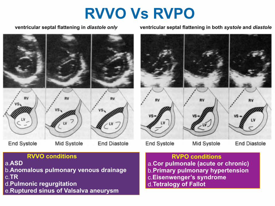
RVVO Vs RVPO• ventricular septal flattening in diastole only • ventricular septal flattening in both systole and diastole
RVVO conditions a.ASD b.Anomalous pulmonary venous drainage c.TR d.Pulmonic regurgitation e.Ruptured sinus of Valsalva aneurysm
RVPO conditions a.Cor pulmonale (acute or chronic) b.Primary pulmonary hypertension c.Eisenwenger’s syndrome d.Tetralogy of Fallot

PSAX : Apex
Mahidol University Basic Echo
RAMATHIBODI HOSPITAL
LV apical motion LV thumbs

Apical 4 chamber
Mahidol University Basic Echo
RAMATHIBODI HOSPITAL
- Global function view - Function of MV and AV - LV size, LA / RA size

Apical 5 chamber
Mahidol University Basic Echo
RAMATHIBODI HOSPITAL
- Tilt the probe anteriorly - Good Doppler alignment for LVOT and MV - Color Doppler assessment for AR and MR - Subvalvular vs. valvular aortic stenosis
Mahidol University
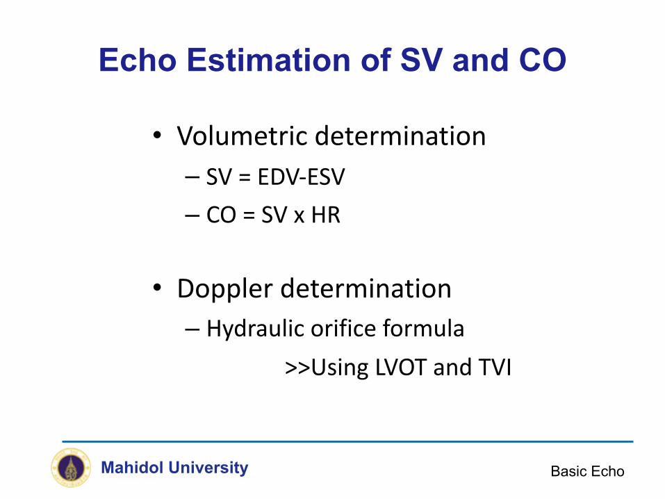
Echo Estimation of SV and CO
Basic Echo
• Volumetricdetermination– SV=EDV-ESV– CO=SVxHR
• Dopplerdetermination– Hydraulicorificeformula >>UsingLVOTandTVI

Mahidol University Tricks and Tips in hemodynamic, mechanic and congenital echocardiography
Volumetric Flow

Mahidol University Tricks and Tips in hemodynamic, mechanic and congenital echocardiography
2

Mahidol University Tricks and Tips in hemodynamic, mechanic and congenital echocardiography

Mahidol University Tricks and Tips in hemodynamic, mechanic and congenital echocardiography

Mahidol University Tricks and Tips in hemodynamic, mechanic and congenital echocardiography
π r2
π (D/2)2
3.14 (D)2 4
= 0.785 (D)2
=
=
=
r

Mahidol University Tricks and Tips in hemodynamic, mechanic and congenital echocardiography
Mahidol University Tricks and Tips in hemodynamic, mechanic and congenital echocardiography
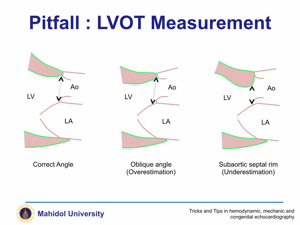
Pitfall : LVOT Measurement
Ao
LA
LV
Correct Angle
Ao
LA
LVAo
LA
LV
Oblique angle (Overestimation)
Subaortic septal rim (Underestimation)

Mahidol University Tricks and Tips in hemodynamic, mechanic and congenital echocardiography
Pitfall : LVOT VTI Measurement

Mahidol University Tricks and Tips in hemodynamic, mechanic and congenital echocardiography
Conservation of Mass Principle

Foreshortening
Mahidol University Basic Echo
RAMATHIBODI HOSPITAL
Foreshortening - Inward motion of the apex - True apex not visualized - Volume underestimation
RAMATHIBODI HOSPITAL
RAMATHIBODI HOSPITAL
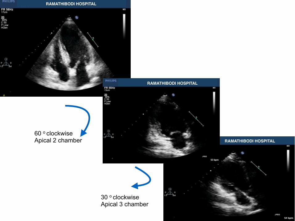
60 o clockwise Apical 2 chamber
30 o clockwise Apical 3 chamber
RAMATHIBODI HOSPITAL
RAMATHIBODI HOSPITAL
RAMATHIBODI HOSPITAL

Apical 2 chamber
Mahidol University Basic Echo
- Only LA & LV (+MV) - Occasionally LAA - Correspond to RAO
RAMATHIBODI HOSPITAL
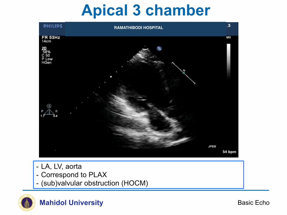
Apical 3 chamber
Mahidol University Basic Echo
- LA, LV, aorta - Correspond to PLAX - (sub)valvular obstruction (HOCM)
RAMATHIBODI HOSPITAL

Subcostal View
Mahidol University Basic Echo
- ASD/VSD visualization - RV wall thickness - Pericardium - Used in patients with limited echo windows
Mahidol University Echo in emergency and critical care
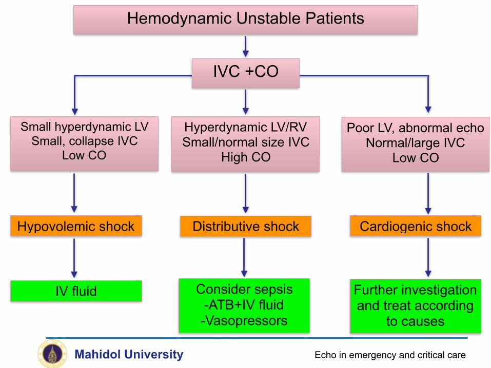
Hemodynamic Unstable Patients
IVC +CO
Small hyperdynamic LV Small, collapse IVC
Low CO
Hypovolemic shock
Hyperdynamic LV/RV Small/normal size IVC
High CO
Poor LV, abnormal echo Normal/large IVC
Low CO
Distributive shock
IV fluid Consider sepsis -ATB+IV fluid -Vasopressors
Cardiogenic shock
Further investigation and treat according
to causes

Mahidol University Echo in emergency and critical care
Hypotension
Hx, PE, Investigations suggestive of cardiogenic shock
Echo
Pericardium
Tamponade
Myocardium
RV LV
PE Pump failure MI, Myocarditis, Drugs
Endocardium
Acute valvular dysfunction
Acute AR/MR
Others
Hypovolemia Don’t forget distal
dissection
Sepsis LA myxoma Severe MS
LVOTO

Mahidol University
Size Location Consolidation or associated mass Loculation Hemodynamics Clearance for tap
Echo Evaluation of pericardial effusion
Echo in emergency and critical care

Mahidol University
Differentiating Pericardial Vs Pleural effusion
Echo in emergency and critical care

Trivial Small Moderate Large
S A X
L A X
Quantification of pericardial effusion
Seen only during systole < 1 cm 1-2 cm >2 cmMeasurement preformed at end diastole
Klein AL et al. J Am Soc Echocardiogr 2013; 26:965-1012
Mahidol University 8th Cardiac Network Forum 2016
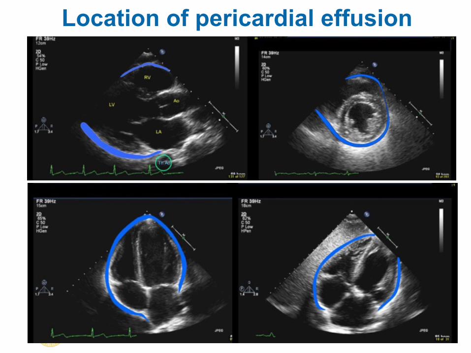
Location of pericardial effusion

Any RA collapse
100% sensitivity 88% specificity
RA Inversion Time Index (RAITI) RA collapse >1/3 cardiac cycle
94% sensitivity 100% specificity
Gillam LD et al. Circulation 1983; 68:294-301
RA collapse begins in end diastole and continues into systole. Considered an “earlier” sign of tamponade.
RA Collapse
Total # frames with inversionTotal # frames in the cardiac cycle
Mahidol University
The most sensitive sign of tamponade is “cyclic compression”

RV Collapse
•Most commonly involves the RV outflow tract (more compressible area of RV)
•When collapse extends form outflow tract to the body of the right ventricle, this is evidence that intrapericardial pressure is elevated more substantially Considered a “later” sign of tamponade RV collapse occurs in early diastole
48-93% sensitivity 50-100% specificity
Mahidol University
The most sensitive sign of tamponade is “cyclic compression”
Echo in emergency and critical care

Doppler Respiratory Variation
• Normal –MV inflow variation <10% –TV inflow variation <25%
• In tamponade –MV inflow variation usually
>30% –TV inflow variation usually
>60%
Hutchison S. Pericardial Diseases, 2009.Mahidol University

• RA collapse
• RV collapse
• Dilated IVC with lack of inspiratory collapse
• Abnormal respiratory variation in tricuspid and mitral flow velocities
• Abnormal hepatic vein flow (expiratory diastolic reversal)
• LA compression (severe)
• LV diastolic compression (severe)
• Swinging heart
Tamponade : Echo and Doppler features
Mahidol University Echo in emergency and critical care

Axillary Access
Medclavicular/Apical Access
SubxiphoidAccess
Right Parasternal
Access
Pericardial Puncture
Site
Skin Puncture
Site
Common access for pericardiocentesis
Mahidol University Echo in emergency and critical care

Clearance for the pericardiocentesis
Subcostal view
At least 1 cm fluid between visceral and
parietal pericardium
No significant adhesions
Effusions not consolidated
Path to pericardium not THROUGH the liver
Mahidol University Echo in emergency and critical care

Mahidol University I2E 2015 Echo in CAD
Complications of MI
Right Ventricular Infarction
Dynamic LVOT Obstruction
- Tachyarrhythmias - Bradyarrrhythmia
Thrombus
Electrical
Mechanical

Mahidol University European Heart Journal-Cardiovascular Imaging 2015;16:119-146
Echocardiographic Contraindications to ECMO

THANK YOU FOR YOUR ATTENTIONEMAIL : [email protected]