echo in cad: wall motion assessment - doctor · pdf fileecho in cad: wall motion assessment...
TRANSCRIPT
Echo in CAD: Wall Motion
Assessment
Joe M. Moody, Jr, MD
UTHSCSA and STVHCS
October 2007
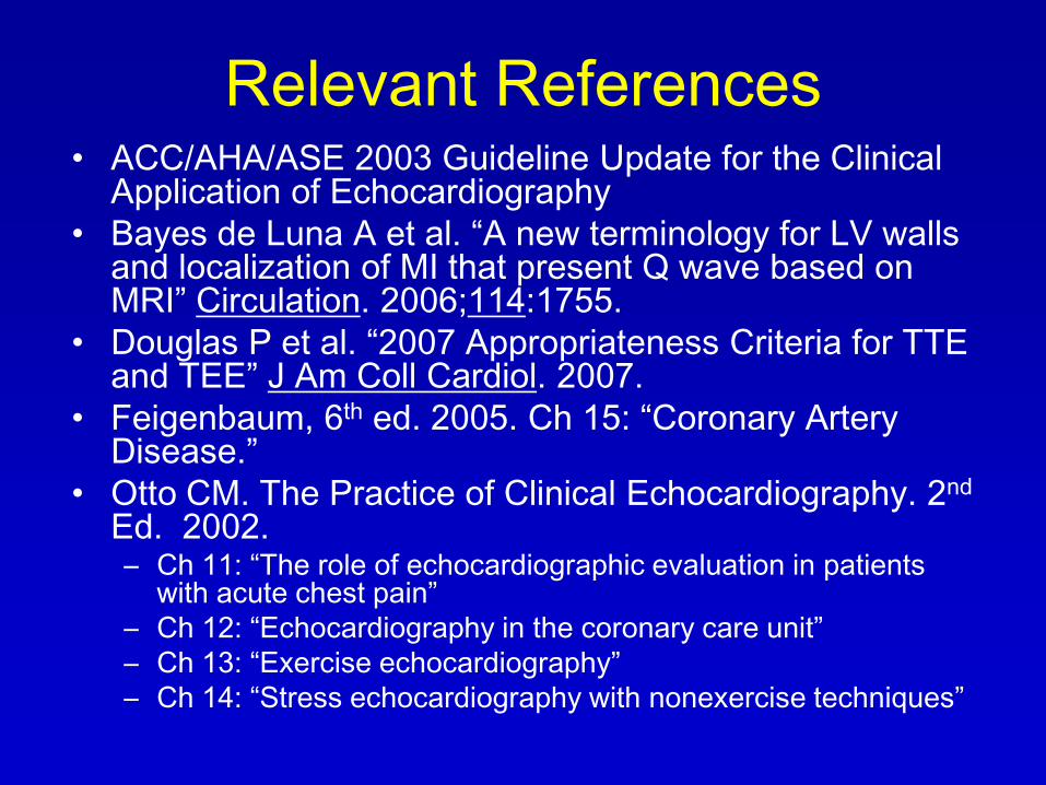
Relevant References• ACC/AHA/ASE 2003 Guideline Update for the Clinical
Application of Echocardiography
• Bayes de Luna A et al. “A new terminology for LV walls and localization of MI that present Q wave based on MRI” Circulation. 2006;114:1755.
• Douglas P et al. “2007 Appropriateness Criteria for TTE and TEE” J Am Coll Cardiol. 2007.
• Feigenbaum, 6th ed. 2005. Ch 15: “Coronary Artery Disease.”
• Otto CM. The Practice of Clinical Echocardiography. 2nd
Ed. 2002.– Ch 11: “The role of echocardiographic evaluation in patients
with acute chest pain”
– Ch 12: “Echocardiography in the coronary care unit”
– Ch 13: “Exercise echocardiography”
– Ch 14: “Stress echocardiography with nonexercise techniques”
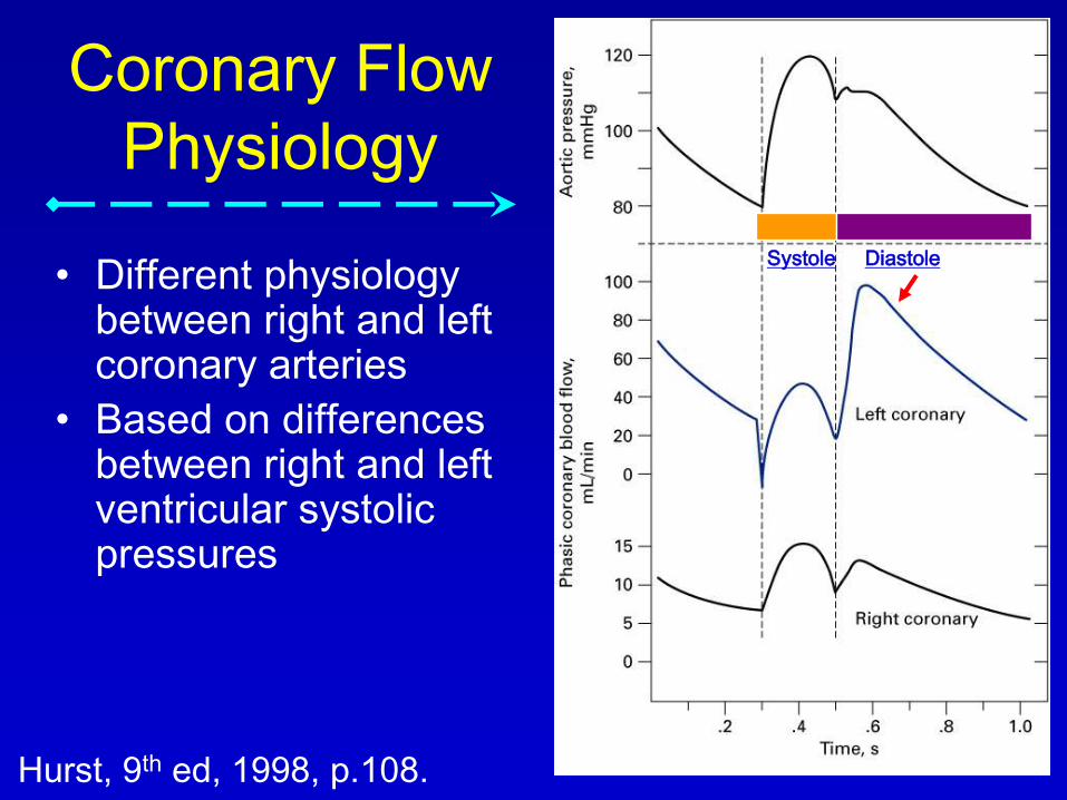
Coronary Flow
Physiology
• Different physiology between right and left coronary arteries
• Based on differences between right and left ventricular systolic pressures
Hurst, 9th ed, 1998, p.108.
Systole Diastole
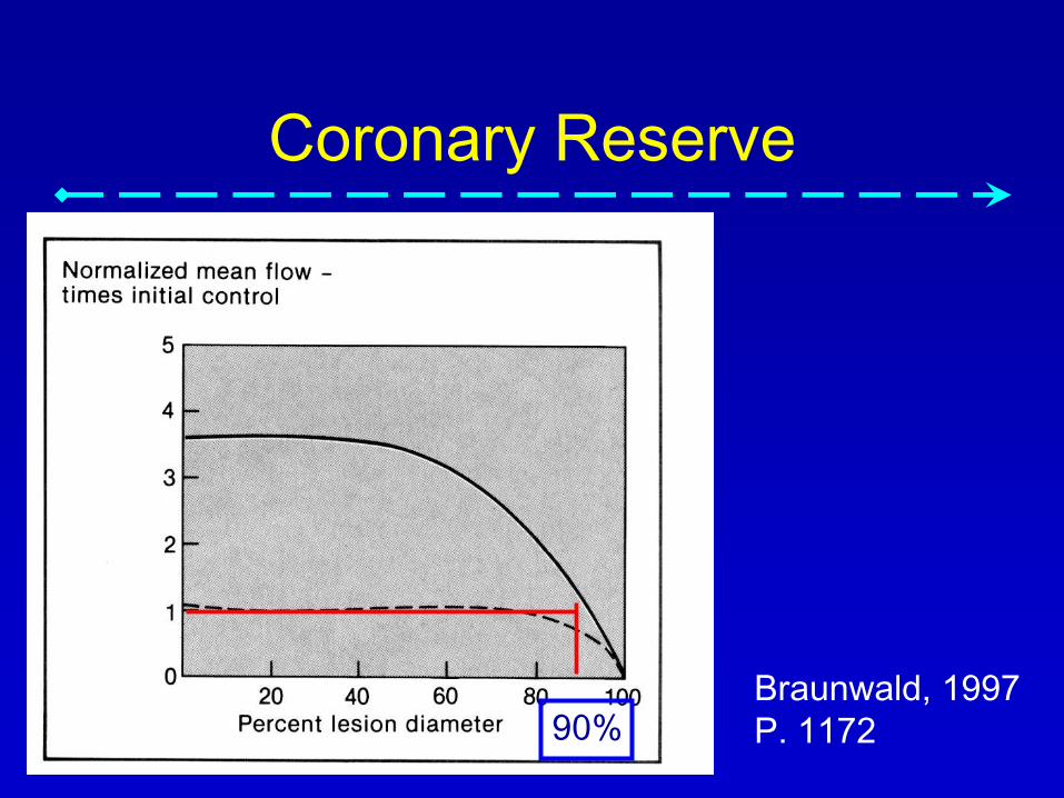
Coronary Reserve
Braunwald, 1997
P. 117290%

Myocardial
Ischemia
• Imbalance between myocardial oxygen demand and supply
DEMANDSUPPLY
Braunwald, 2000
Supply
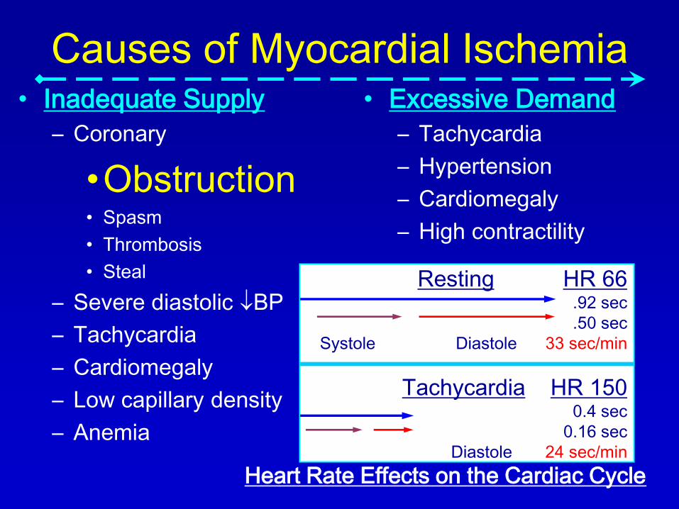
Causes of Myocardial Ischemia• Inadequate Supply
– Coronary
•Obstruction• Spasm
• Thrombosis
• Steal
– Severe diastolic BP
– Tachycardia
– Cardiomegaly
– Low capillary density
– Anemia
• Excessive Demand
– Tachycardia
– Hypertension
– Cardiomegaly
– High contractility
Resting HR 66.92 sec
.50 sec
Systole Diastole 33 sec/min
Tachycardia HR 1500.4 sec
0.16 sec
Diastole 24 sec/min
Heart Rate Effects on the Cardiac Cycle
Ischemic time and outcome• <5 min – recovery in 1-2 min
• 30-120 min – recovery in 48-72 hours
• 4-6 hours – no recovery; scar in 6 weeks
• In clinical practice, may take weeks to months to recover
• Repetitive ischemia may mimic hibernation
• Infarct expansion occurs in about 48 hours, acute thinning (often pain and ECG changes but no biomarker elevation) – risk (substrate) for mechanical complication
• Wall motion abnormality overestimates MI size due to tethering (overestimation is about 15%)
• Vertical (endo-to-epi) tethering gives akinesis if 20% of endocardial thickness is affected.
Feigenbaum, 6th ed. 2005. Ch 15: “Coronary Artery Disease.”
Indications for TTE and TEE
• Concerning test results (CXR, BNP, ECG)
• LV function post MI, first evaluation
• LV function in MI recovery when results will guide therapy
• Hypotension or hemodynamic instability of uncertain or suspected cardiac origin
• TTE during chest pain with suspected but not confirmed ischemia (indeterminate biomarkers or ECG)
• Suspected complication of AMI
• Respiratory failure with suspected cardiac cause
• Known or suspected heart failure, first evaluation
Douglas P et al. “2007 Appropriateness Criteria for TTE and TEE” J Am Coll
Cardiol. 2007.
Technical Points in Wall Motion
• Good endocardial border definition is
key (add contrast if 2-4 segments are
completely or partially obscured)
• Regional wall motion interpretation
requires an experienced
echocardiographer
Otto CM. 2nd Ed. 2002. p. 239.
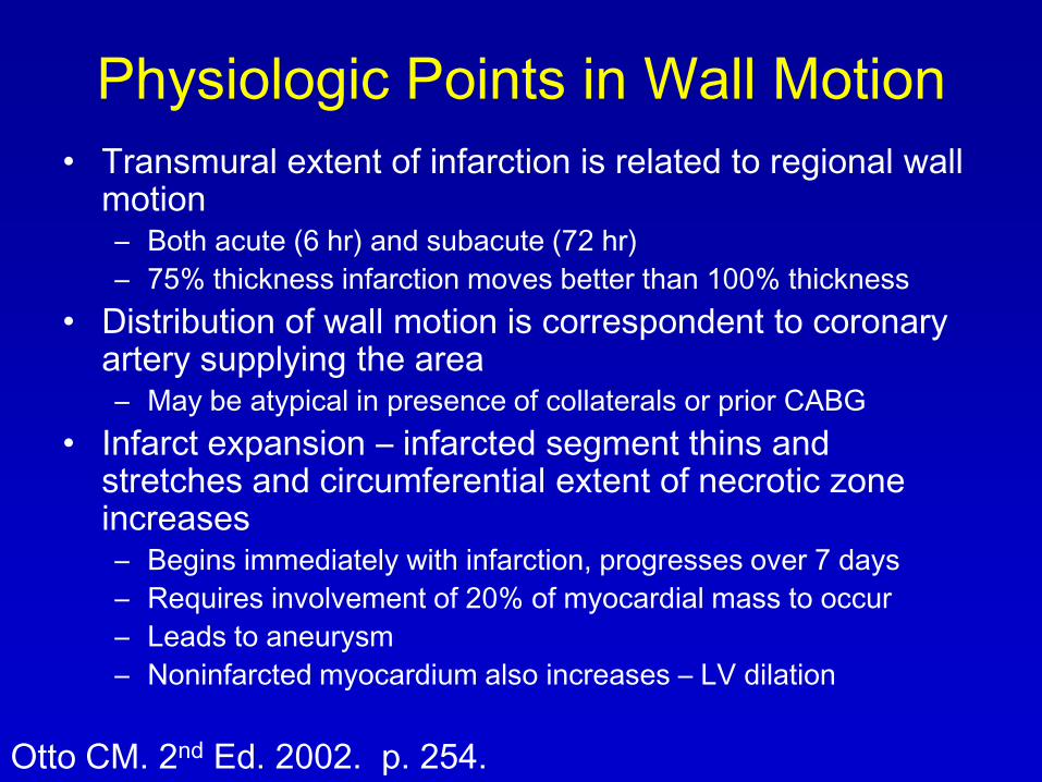
Physiologic Points in Wall Motion
• Transmural extent of infarction is related to regional wall motion– Both acute (6 hr) and subacute (72 hr)
– 75% thickness infarction moves better than 100% thickness
• Distribution of wall motion is correspondent to coronary artery supplying the area– May be atypical in presence of collaterals or prior CABG
• Infarct expansion – infarcted segment thins and stretches and circumferential extent of necrotic zone increases– Begins immediately with infarction, progresses over 7 days
– Requires involvement of 20% of myocardial mass to occur
– Leads to aneurysm
– Noninfarcted myocardium also increases – LV dilation
Otto CM. 2nd Ed. 2002. p. 254.

Wall
Motion
• Definitions– Normal: at least 5 mm endocardial excursion
– Hypokinesia: less than 5 mm
– Akinesia: no inward excursion (less than 2 mm)
– Dyskinesia: outward excursion
• Experimental Ischemia– Threshold of 10-20% reduction in blood flow impairs wall
thickening and 80% reduction results in akinesis
– Decreased wall thickening extends beyond reduced flow (“tethering”)
• Clinical Ischemia (complete balloon occlusion)– Regional endocardial dysfunction in 19 sec
– ECG change in 30 sec
– Chest pain 39 sec
• Clinical Ischemia (stress in region of coronary stenosis)– Wall motion abnormality in 30 sec
– ECG change in 90 sec
Otto CM. 2nd Ed. 2002. p. 275, 307
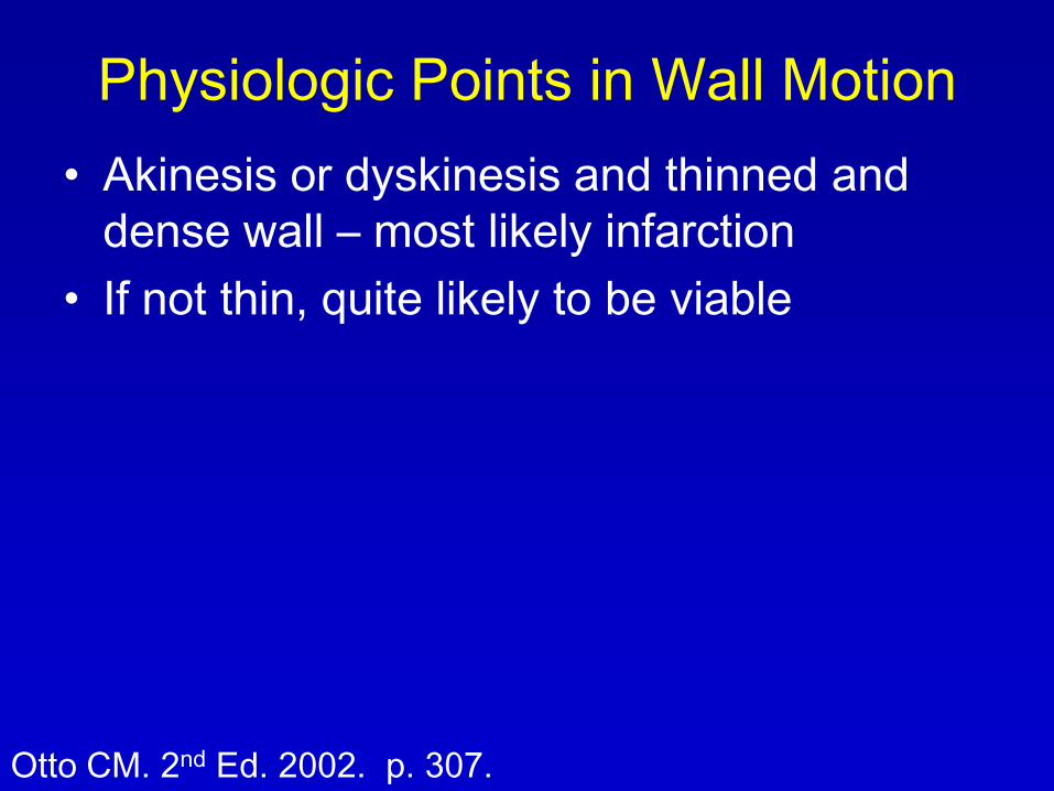
Physiologic Points in Wall Motion
• Akinesis or dyskinesis and thinned and
dense wall – most likely infarction
• If not thin, quite likely to be viable
Otto CM. 2nd Ed. 2002. p. 307.
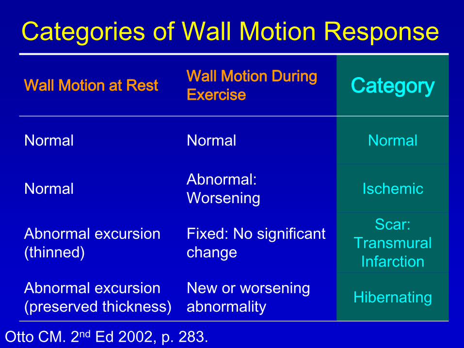
Categories of Wall Motion Response
Wall Motion at RestWall Motion During
ExerciseCategory
Normal Normal Normal
NormalAbnormal:
WorseningIschemic
Abnormal excursion
(thinned)
Fixed: No significant
change
Scar:
Transmural
Infarction
Abnormal excursion
(preserved thickness)
New or worsening
abnormalityHibernating
Otto CM. 2nd Ed 2002, p. 283.

Scoring System for Wall Motion
Score Wall Motion Endocardial Motion Wall thickening
1 Normal Normal Normal (>30%)*
2 Hypokinesis Reduced Reduced (<30%)
3 Akinesis Absent Absent
4 Dyskinesis Outward Thinning
5 Aneurysmal Diastolic deformity Absent or thinning
Otto CM. 2nd Ed 2002, p. 256. *35-40%; from 9-11 to 14-16 mm (Feigenbaum)
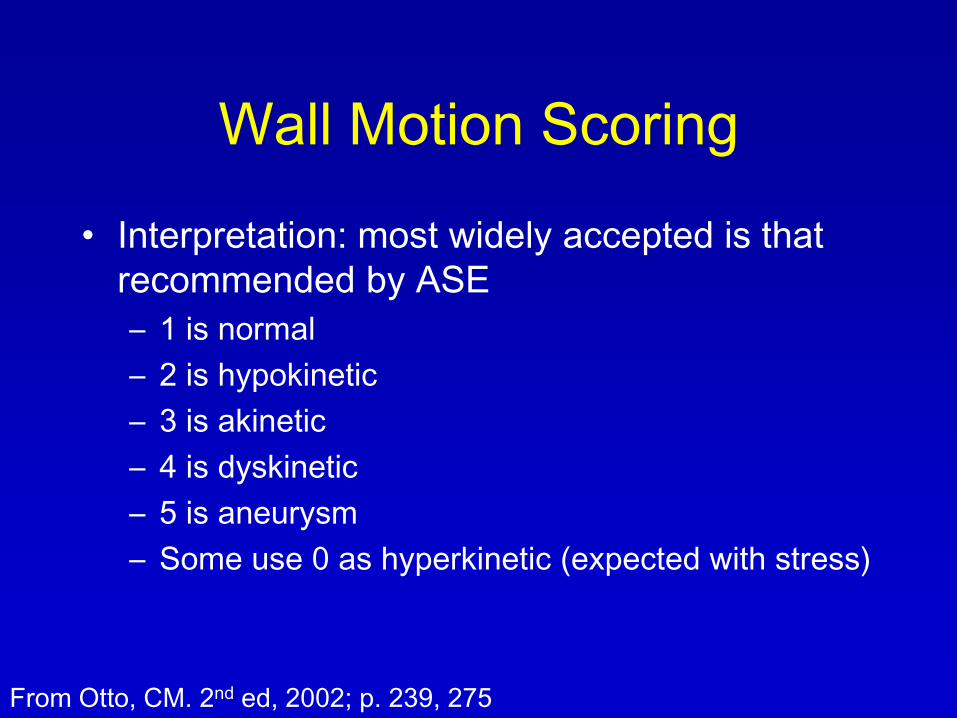
Wall Motion Scoring
• Interpretation: most widely accepted is that
recommended by ASE
– 1 is normal
– 2 is hypokinetic
– 3 is akinetic
– 4 is dyskinetic
– 5 is aneurysm
– Some use 0 as hyperkinetic (expected with stress)
From Otto, CM. 2nd ed, 2002; p. 239, 275

Naming the Views
AHA Scientific
Statement:
Standardized
Myocardial
Segmentation and
Nomenclature for
Tomographic
Imaging of the
Heart. Circulation
2002;105:539
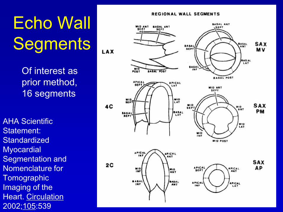
Echo Wall
Segments
AHA Scientific
Statement:
Standardized
Myocardial
Segmentation and
Nomenclature for
Tomographic
Imaging of the
Heart. Circulation
2002;105:539
Of interest as
prior method,
16 segments
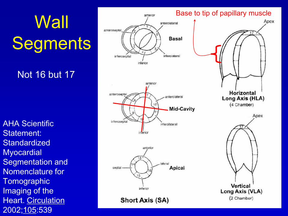
Wall
Segments
AHA Scientific
Statement:
Standardized
Myocardial
Segmentation and
Nomenclature for
Tomographic
Imaging of the
Heart. Circulation
2002;105:539
Not 16 but 17
Base to tip of papillary muscle
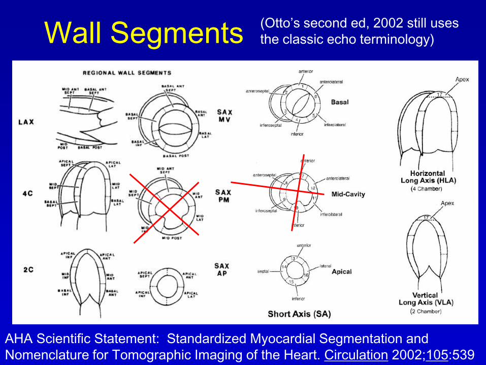
Wall Segments
AHA Scientific Statement: Standardized Myocardial Segmentation and
Nomenclature for Tomographic Imaging of the Heart. Circulation 2002;105:539
(Otto’s second ed, 2002 still uses
the classic echo terminology)
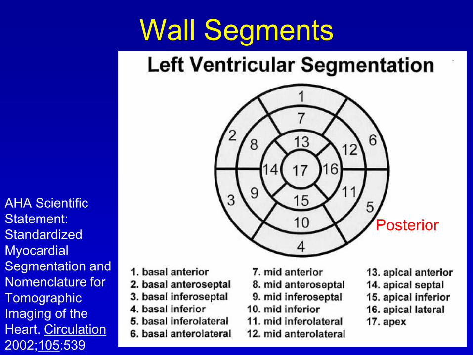
Wall Segments
AHA Scientific
Statement:
Standardized
Myocardial
Segmentation and
Nomenclature for
Tomographic
Imaging of the
Heart. Circulation
2002;105:539
Posterior

Wall Segments
AHA Scientific Statement: Standardized Myocardial Segmentation and
Nomenclature for Tomographic Imaging of the Heart. Circulation 2002;105:539
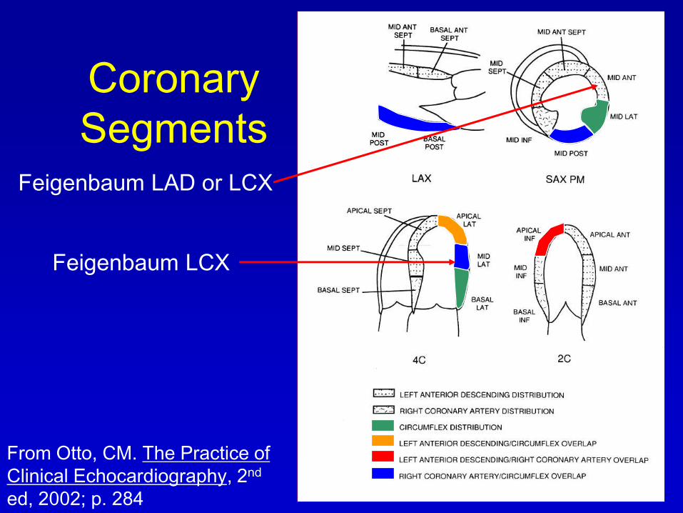
Coronary
Segments
From Otto, CM. The Practice of
Clinical Echocardiography, 2nd
ed, 2002; p. 284
Feigenbaum LCX
Feigenbaum LAD or LCX
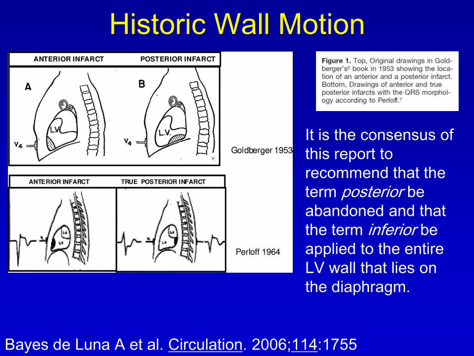
Historic Wall Motion
It is the consensus of
this report to
recommend that the
term posterior be
abandoned and that
the term inferior be
applied to the entire
LV wall that lies on
the diaphragm.
Bayes de Luna A et al. Circulation. 2006;114:1755

Recent Guidelines
Bayes de Luna A et al. Circulation. 2006;114:1755

Correlation with MRI in inferior
base
Bayes de Luna A et al. Circulation. 2006;114:1755
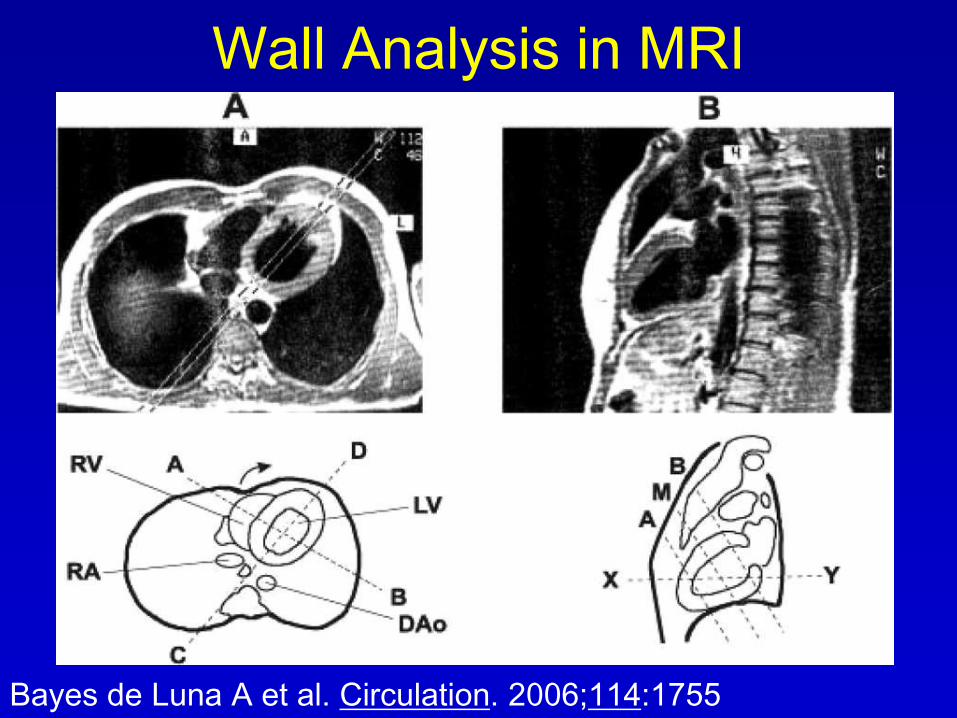
Wall Analysis in MRI
Bayes de Luna A et al. Circulation. 2006;114:1755
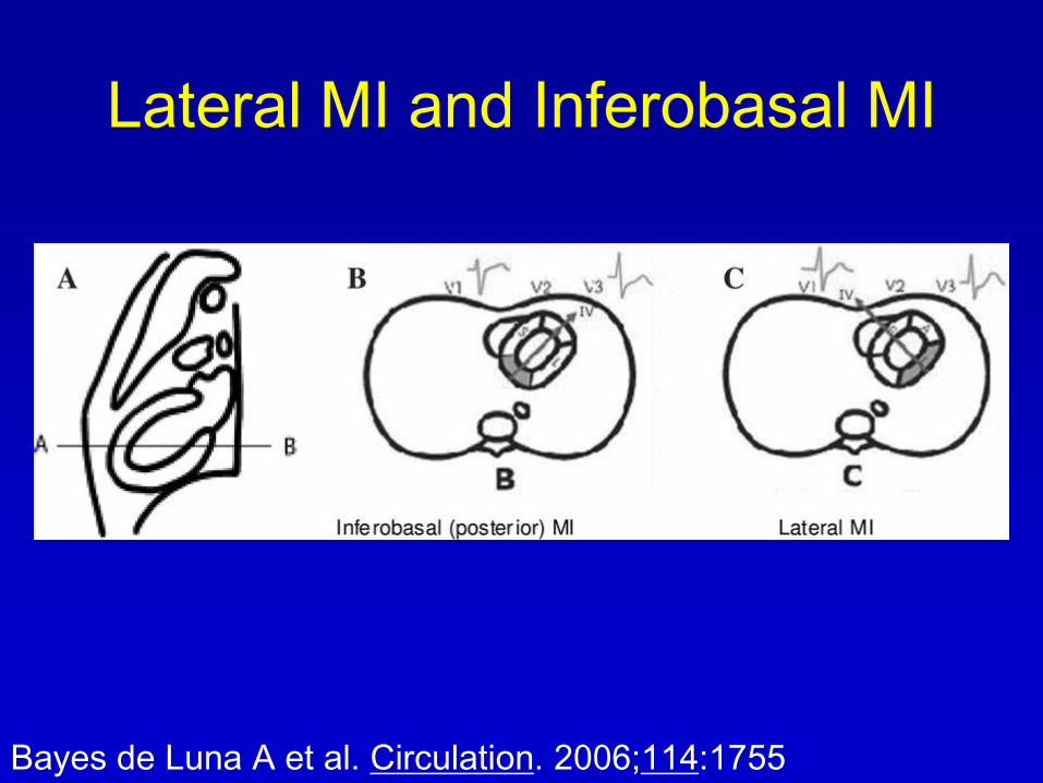
Lateral MI and Inferobasal MI
Bayes de Luna A et al. Circulation. 2006;114:1755

Coronary Distribution
Bayes de Luna A et al. Circulation. 2006;114:1755

ECG
Terminology
Consensus
Bayes de Luna A et al.
Circulation. 2006;114:1755
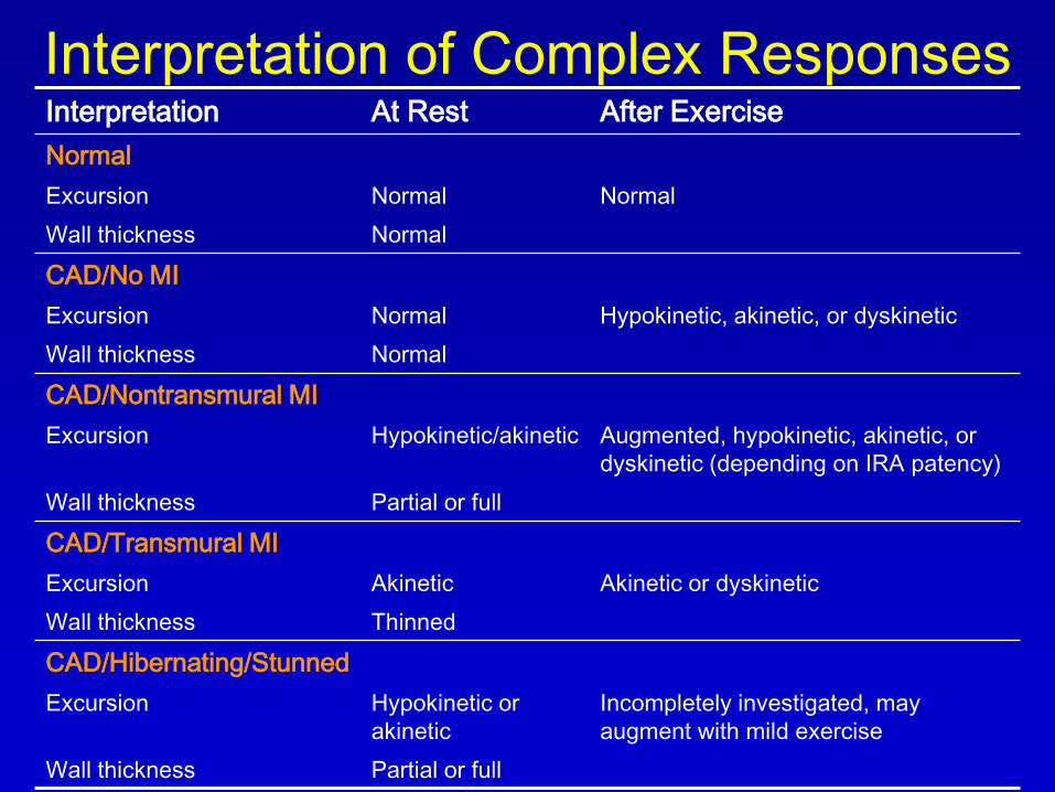
Interpretation of Complex ResponsesInterpretation At Rest After Exercise
Normal
Excursion Normal Normal
Wall thickness Normal
CAD/No MI
Excursion Normal Hypokinetic, akinetic, or dyskinetic
Wall thickness Normal
CAD/Nontransmural MI
Excursion Hypokinetic/akinetic Augmented, hypokinetic, akinetic, or
dyskinetic (depending on IRA patency)
Wall thickness Partial or full
CAD/Transmural MI
Excursion Akinetic Akinetic or dyskinetic
Wall thickness Thinned
CAD/Hibernating/Stunned
Excursion Hypokinetic or
akinetic
Incompletely investigated, may
augment with mild exercise
Wall thickness Partial or full
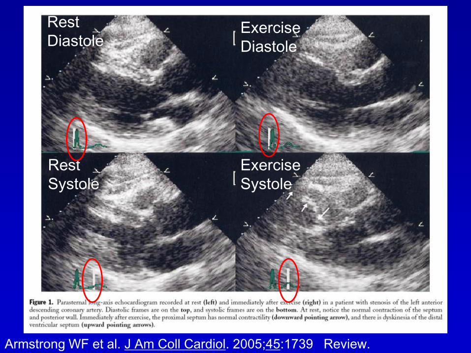
Armstrong WF et al. J Am Coll Cardiol. 2005;45:1739 Review.
Rest
DiastoleExercise
Diastole
Rest
Systole
Exercise
Systole
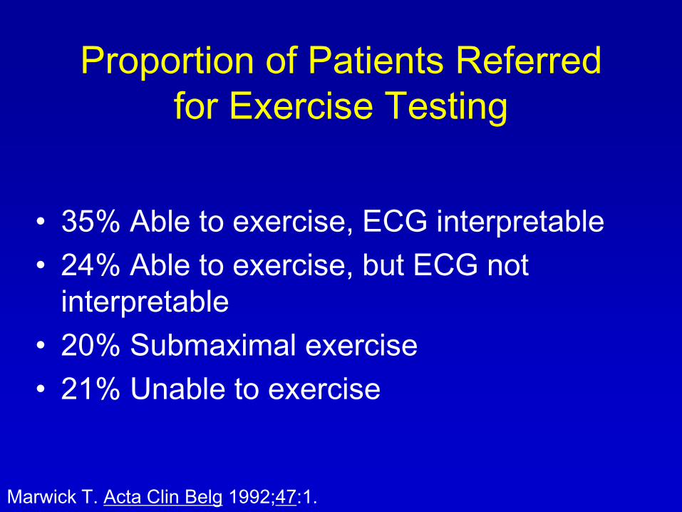
Proportion of Patients Referred
for Exercise Testing
• 35% Able to exercise, ECG interpretable
• 24% Able to exercise, but ECG not
interpretable
• 20% Submaximal exercise
• 21% Unable to exercise
Marwick T. Acta Clin Belg 1992;47:1.

Interpretation of Viability and Ischemia with
Dobutamine EchocardiographyDiagnosis Resting
Function
Low-Dose Peak/Poststress
Function
Normal Normal Normal Hyperkinetic
Ischemic Normal Normal (unless
severe CAD)
Reduction vs. rest
Reduction vs. other
segments
Delayed contrac tion
Viable, patient
IRA
Hypo/akinetic Improvement Sustained
improvement
Viable,
stenosed IRA
Hypo/akinetic Improvement Reduction (c/w low-
dose)
Infarction A/dyskinetic No change No change
From Otto, CM. The Practice of Clinical Echocardiography, 2nd ed, 2002; p. 306
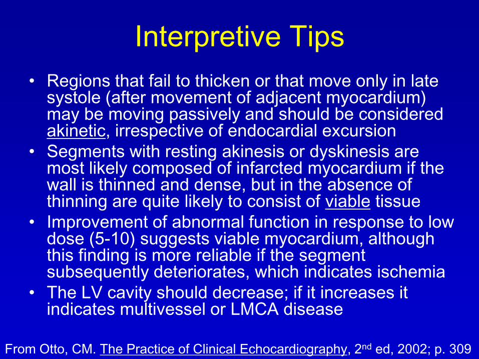
Interpretive Tips
• Regions that fail to thicken or that move only in late systole (after movement of adjacent myocardium) may be moving passively and should be considered akinetic, irrespective of endocardial excursion
• Segments with resting akinesis or dyskinesis are most likely composed of infarcted myocardium if the wall is thinned and dense, but in the absence of thinning are quite likely to consist of viable tissue
• Improvement of abnormal function in response to low dose (5-10) suggests viable myocardium, although this finding is more reliable if the segment subsequently deteriorates, which indicates ischemia
• The LV cavity should decrease; if it increases it indicates multivessel or LMCA disease
From Otto, CM. The Practice of Clinical Echocardiography, 2nd ed, 2002; p. 309
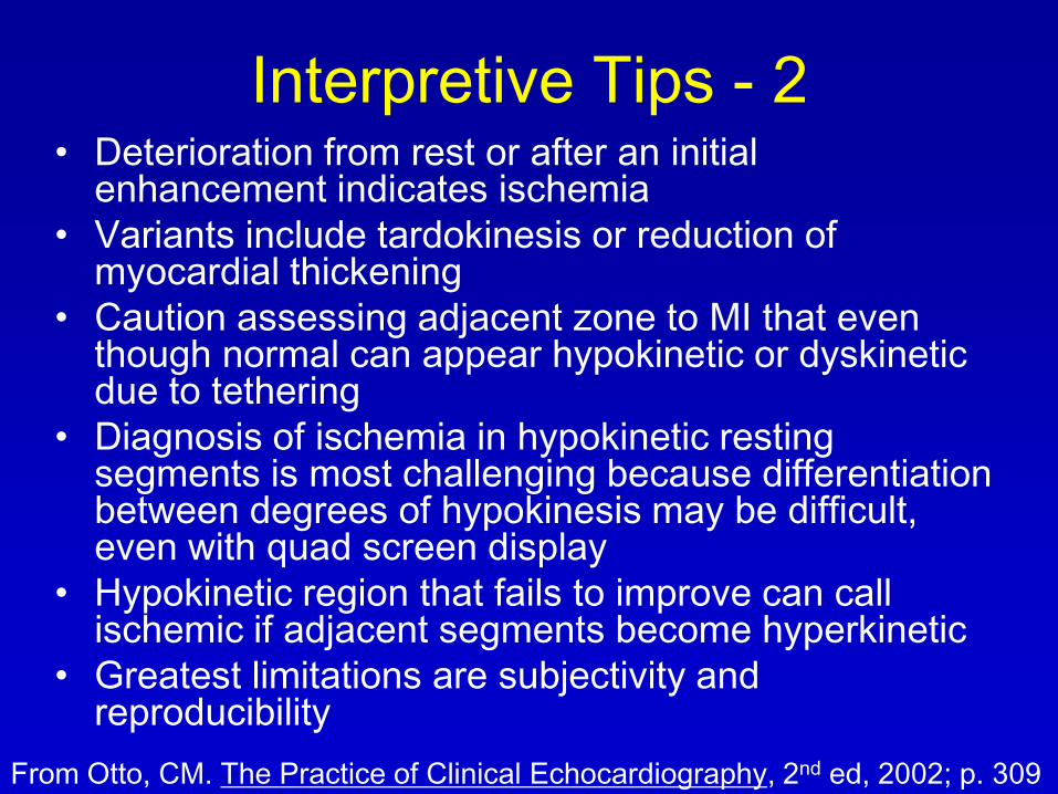
Interpretive Tips - 2• Deterioration from rest or after an initial
enhancement indicates ischemia
• Variants include tardokinesis or reduction of myocardial thickening
• Caution assessing adjacent zone to MI that even though normal can appear hypokinetic or dyskinetic due to tethering
• Diagnosis of ischemia in hypokinetic resting segments is most challenging because differentiation between degrees of hypokinesis may be difficult, even with quad screen display
• Hypokinetic region that fails to improve can call ischemic if adjacent segments become hyperkinetic
• Greatest limitations are subjectivity and reproducibility
From Otto, CM. The Practice of Clinical Echocardiography, 2nd ed, 2002; p. 309

Interpretive Guideline
• Basal inferior or basal septal hypokinesis ignored unless – Adjacent area affected by new dyssynergy
– Clear deterioration to akinesis or dyskinesis
• Induced delayed contraction is ischemic if no BBB
• Identification of ischemia based on expected coronary distribution– A mid septal abnormality was disregarded if the apex was
spared
• Significant resting abnormality– Hypokinesis in at least 3 segments or akinesis in at least 1
segment
– Suggests abnormal test and presence of CAD
Hoffmann R et al. Am J Cardiol. 1998;82:1520-4.
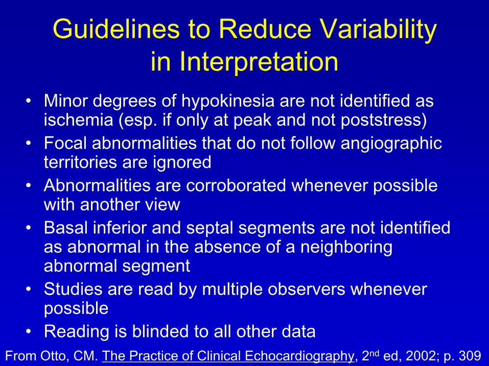
Guidelines to Reduce Variability
in Interpretation
• Minor degrees of hypokinesia are not identified as ischemia (esp. if only at peak and not poststress)
• Focal abnormalities that do not follow angiographic territories are ignored
• Abnormalities are corroborated whenever possible with another view
• Basal inferior and septal segments are not identified as abnormal in the absence of a neighboring abnormal segment
• Studies are read by multiple observers whenever possible
• Reading is blinded to all other data
From Otto, CM. The Practice of Clinical Echocardiography, 2nd ed, 2002; p. 309
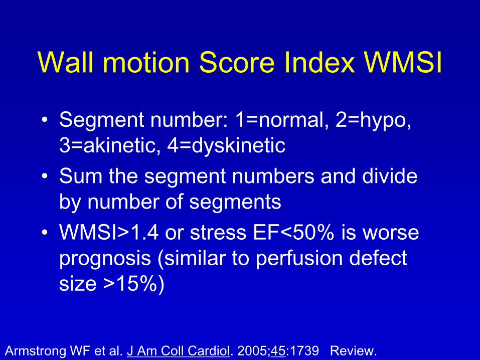
Wall motion Score Index WMSI
• Segment number: 1=normal, 2=hypo,
3=akinetic, 4=dyskinetic
• Sum the segment numbers and divide
by number of segments
• WMSI>1.4 or stress EF<50% is worse
prognosis (similar to perfusion defect
size >15%)
Armstrong WF et al. J Am Coll Cardiol. 2005;45:1739 Review.

Echo in Mechanical
Complications of MI
• VSD
• MR
• Rupture
• True Aneurysm
• Pseudoaneurysm

Thank you.
Questions?