echinococcus multilocularis in wild boar - slu.sestud.epsilon.slu.se/4188/1/vamborg_t_120511.pdf ·...
TRANSCRIPT
Sveriges lantbruksuniversitet Fakulteten för veterinärmedicin och husdjursvetenskap Institutionen för biomedicin och veterinär folkhälsovetenskap
Echinococcus multilocularis in wild boar - aiming at an alternative surveillance method
Tuva Vamborg
Uppsala
2011
Examensarbete inom veterinärprogrammet
ISSN 1652-8697 Examensarbete 2012:44

SLU Sveriges lantbruksuniversitet
Echinococcus multilocularis in wild boar - aiming at an alternative surveillance method
Tuva Vamborg
Handledare: Anna Lundén, Institutionen för biomedicin och veterinär folkhälsovetenskap
Biträdande handledare: Henrik Uhlhorn, Enhet för patologi och viltsjukdomar, SVA
Examinator: Johan Höglund, Institutionen för biomedicin och veterinär folkhälsovetenskap
Examensarbete inom veterinärprogrammet, Uppsala 2011 Fakulteten för Veterinärmedicin och husdjursvetenskap
Institutionen för biomedicin och veterinär folkhälsa Kurskod: EX0239, Nivå AX, 30hp
Nyckelord: Echinococcus multilocularis, wild boar, surveillance
Online publication of this work: http://epsilon.slu.se
ISSN 1652-8697 Examensarbete 2012:44

INDEX
Abstract .................................................................................................................... 1
Sammanfattning ....................................................................................................... 2
Introduction .............................................................................................................. 3
Background .......................................................................................................... 3
Aim ...................................................................................................................... 4
Literature review ...................................................................................................... 4
Wild boar ............................................................................................................. 4
Echinococcus multilocularis ................................................................................ 4
Taxonomy and morphology ............................................................................. 4
Life cycle ......................................................................................................... 5
Human alveolar echinococcosis ....................................................................... 6
Epidemiology ................................................................................................... 7
Diagnostic and surveillance methods .............................................................. 8
Accidental hosts of E. multilocularis ................................................................... 9
Experimentally infected animals ..................................................................... 9
Spontaneously infected species ..................................................................... 10
Infection in pigs and wild boar ...................................................................... 10
Pathological findings in wild boars and pigs ................................................. 10
Materials and methods ........................................................................................... 12
Collection of wild boar samples ........................................................................ 12
Pathological gross and histological examination ............................................... 12
PCR .................................................................................................................... 12
Results .................................................................................................................... 13
Gross and histological examination ................................................................... 13
PCR .................................................................................................................... 15
Discussion .............................................................................................................. 15
Evaluation of this study and the results ............................................................. 15
New surveillance method ................................................................................... 15
Conclusion ............................................................................................................. 16
References .............................................................................................................. 17

1
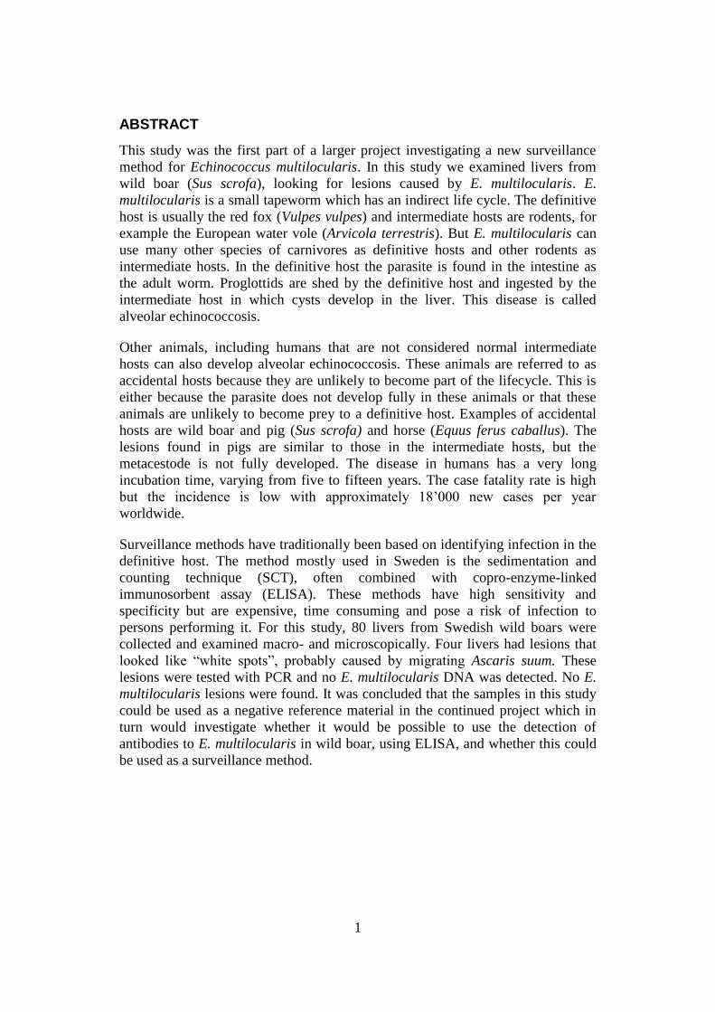
ABSTRACT
This study was the first part of a larger project investigating a new surveillance
method for Echinococcus multilocularis. In this study we examined livers from
wild boar (Sus scrofa), looking for lesions caused by E. multilocularis. E.
multilocularis is a small tapeworm which has an indirect life cycle. The definitive
host is usually the red fox (Vulpes vulpes) and intermediate hosts are rodents, for
example the European water vole (Arvicola terrestris). But E. multilocularis can
use many other species of carnivores as definitive hosts and other rodents as
intermediate hosts. In the definitive host the parasite is found in the intestine as
the adult worm. Proglottids are shed by the definitive host and ingested by the
intermediate host in which cysts develop in the liver. This disease is called
alveolar echinococcosis.
Other animals, including humans that are not considered normal intermediate
hosts can also develop alveolar echinococcosis. These animals are referred to as
accidental hosts because they are unlikely to become part of the lifecycle. This is
either because the parasite does not develop fully in these animals or that these
animals are unlikely to become prey to a definitive host. Examples of accidental
hosts are wild boar and pig (Sus scrofa) and horse (Equus ferus caballus). The
lesions found in pigs are similar to those in the intermediate hosts, but the
metacestode is not fully developed. The disease in humans has a very long
incubation time, varying from five to fifteen years. The case fatality rate is high
but the incidence is low with approximately 18’000 new cases per year
worldwide.
Surveillance methods have traditionally been based on identifying infection in the
definitive host. The method mostly used in Sweden is the sedimentation and
counting technique (SCT), often combined with copro-enzyme-linked
immunosorbent assay (ELISA). These methods have high sensitivity and
specificity but are expensive, time consuming and pose a risk of infection to
persons performing it. For this study, 80 livers from Swedish wild boars were
collected and examined macro- and microscopically. Four livers had lesions that
looked like “white spots”, probably caused by migrating Ascaris suum. These
lesions were tested with PCR and no E. multilocularis DNA was detected. No E.
multilocularis lesions were found. It was concluded that the samples in this study
could be used as a negative reference material in the continued project which in
turn would investigate whether it would be possible to use the detection of
antibodies to E. multilocularis in wild boar, using ELISA, and whether this could
be used as a surveillance method.
2
SAMMANFATTNING
Denna studie var den första delen av ett större projekt med syfte att undersöka en
ny övervakningsmetod för Echinococcus multilocularis. I denna studie
undersöktes levrar från vildsvin (Sus scrofa), med avseende på lesioner orsakade
av E. multilocularis. E. multilocularis är en liten bandmask som har en indirekt
livscykel. Huvudvärden är vanligtvis rödräv (Vulpes vulpes) och mellanvärdar är
gnagare, till exempel vattensork (Arvicola terrestris). Men E. multilocularis kan
använda många andra rovdjursarter som huvudvärd och andra gnagare som
mellanvärdar. I huvudvärden finns de adulta maskarna i tarmen. Proglottider
urskiljs av huvudvärden och äts upp av mellanvärden varvid cystor utvecklas i
levern. Denna sjukdom kallas alveolär ekinokockos.
Andra djur, inklusive människa, som inte anses vara normala mellanvärdar kan
också utveckla alveolär ekinokockos. Dessa djur kallas oavsiktliga värdar
(accidental hosts) eftersom det är osannolikt att de utgör en del av livscykeln. Det
beror antingen på att parasiten inte utvecklas fullständigt i dessa djur eller att det
är osannolikt att de blir uppätna av en huvudvärd. Exempel på oavsiktliga värdar
är vildsvin och svin (Sus scrofa) samt häst (Equus ferus caballus). De
förändringar som finns i grisar liknar dem som ses hos gnagare, men
metacestoderna är inte fullt utvecklade. Sjukdomen hos människa har en mycket
lång inkubationstid, på 5-15 år. Letaliteten är hög, men förekomsten är låg med ca
18'000 nya fall per år i hela världen.
Övervakningsmetoder har traditionellt varit baserade på att identifiera infektionen
hos huvudvärden. Den metod som används mest i Sverige är sedimentation and
counting technique (SCT), ofta i kombination med copro-enzyme-linked
immunosorbent assay (ELISA). Dessa metoder har hög sensitivitet och
specificitet, men är dyra, tidskrävande och innebär en risk för infektion för
personer som utför den. I denna studie samlades 80 levrar från svenska vildsvin in
och undersöktes makroskopiskt och mikroskopiskt. PCR användes på lesioner
som bedömdes kunna vara orsakade av parasiter. Fyra levrar hade "white spots",
troligtvis orsakade av migrerande Ascaris suum. Dessa skador testades med PCR
och var negativa. Inga E. multilocularis lesioner hittades. Slutsatsen var att
proverna i denna studie kunde användas som ett negativt referensmaterial i det
fortsatta projektet vilket i sin tur ska utvärdera huruvida det går att detektera
antikroppar mot E. multilocularis hos vildsvin med ELISA och därmed undersöka
huruvida denna metod kan användas till övervakning.

3
INTRODUCTION
Background
Echinococcus multilocularis, also known as the fox dwarf tapeworm, is a parasite
that can infect humans (Eckert et al., 2001). The disease, known as alveolar
echinococcosis, is rare but the case fatality rate is very high. Most commonly the
parasite has a sylvatic lifecycle with red fox (Vulpes vulpes) as definitive host and
small rodents as intermediate host. But other carnivores like the domestic dog
(Canis lupus familiaris) and the raccoon dog (Nyctereutes procyonoides) can also
act as definitive hosts. In addition to the natural intermediate hosts, other
mammals, such as pigs and wild boars (Sus scrofa), can become infected but
without being a part of the transmission cycle. These animals are often referred to
as accidental hosts.
Because of its properties as a human pathogen, and because it had not been found
in Sweden until February 2011, E. multilocularis is notifiable (SFS, 2004:255)
and prevention and surveillance is being carried out. Prevention has been through
compulsory deworming of cats and dogs before arrival to Sweden (SJVFS
2010:43). Import or travel from a few European countries, considered free from E.
multilocularis, has been excepted from compulsory deworming in these
regulations. However, the requirements of deworming have ceased as of the 1st
January 2012 due to the discovery of E. multilocularis in Sweden in 2011 (SJVFS
2011:49).
Surveillance has been carried out by the National Veterinary Institute through
detection of the parasite in the definitive host, the red fox (Osterman Lind et al.,
2011). The main method used is the sedimentation and counting technique (SCT)
in which the content of the entire intestine is examined, which makes the method
costly, time consuming and poses a risk of infection to personnel performing it.
There are many reasons why surveillance should continue or increase. The
discovery of the parasite in Sweden could lead to the need for intensified
surveillance to be able to establish its epidemiology. As many scientists claim that
climate change and changing environments might have an impact on E.
multilocularis epidemiology it is hard to predict how or if it will spread (Mas
Coma et al., 2008).
In Sweden possible definitive hosts are found throughout the country such as red
fox, arctic fox (Vulpes lagopus), domestic dog and wolf (Canis lupus) (Osterman
Lind et al., 2011). The raccoon dog is migrating to the North of Sweden from
Finland. There is a wealth of intermediate hosts such as voles and mice. There is
also an increasing awareness and fear among the public who wants to learn about
the risks that this parasite entails. Even though the screening method used in
Sweden today has a high sensitivity, it also has many drawbacks, as mentioned
above. There is a need for a more efficient and cheaper method, which could be
used for large scale screening.
4
Aim
This study is the first part of a larger project investigating a new potential
screening method for E. multilocularis. This method would be based on detection
of antibodies to E. multilocularis in wild boar by an enzyme-linked
immunosorbent assay (ELISA). To produce a negative reference material for the
serological analysis the first step involves examining livers from Swedish wild
boar for lesions caused by E. multilocularis.
LITERATURE REVIEW
Wild boar
The wild boar is an omnivore, mostly eating roots, fruits, acorns etc (Markström,
2002). Animal proteins mainly come from worms, larvae and in some cases, small
mammals and eggs. The wild boar was extinguished from Sweden in the 17th
century, but was reintroduced as farmed wild boar in southern Sweden during the
19th
and 20th
centuries. Break-outs and illegal releases during the second half of
the 20th
century resulted in the establishment of small colonies of free-ranging
wild boars. The wild boar is now considered to be a part of the Swedish fauna.
The population was estimated to be at least 150’000 animals in 2008 (Svenska
jägareförbundet, 2009). The estimation is based on the number of wild boars shot
(50’000) and reported by hunters that year. It is estimated that the population can
double in 3 years, and this is likely to result in increased hunting. The population
is distributed from the southernmost part of Sweden to the County of Dalarna, and
a few groups of animals have established even further north. Wild boar of various
subspecies is found in Europe, Northern Africa, and large parts of Asia but it is
not found in very dry areas or in high mountain ranges (Markström, 2002). It has
been introduced to Australia, New Zealand, North and South America.
Echinococcus multilocularis
Taxonomy and morphology
Echinococcus multilocularis is a cestode (eng. tapeworm) of the family Taeniidae
(Taylor et al., 2007; Eckert el al., 2001). Within the Echinococcus genus there are
several other species. They have similar life cycles and morphology. With new
diagnostic tools such as genetic sequencing, the taxonomy of the genus is
changing (Nakao et al., 2007). One example is E. granulosus which previously
was considered to have different strains whereas now, some of these strains are
considered as individual species. Previously, taxonomy was based mainly on
parasite morphology and on which animals that were its natural hosts (Gemmel,
1959; Rausch, 1968). Within the E. multilocularis species there are small strain
differences (Eckert et al., 2001). These differences, however, do not seem related
to the many intermediate or definitive hosts that E. multilocularis uses (Vuitton &
Gottstein, 2010).
E. multilocularis is a small tapeworm, measuring 2-4 mm in its adult form, and
consists of three to five segments (Taylor et al., 2007). The head, which in
tapeworms is called scolex, is followed by a chain of segments, the proglottids.
The scolex has four suckers and small and large hooks in a double row which are
5
used for attachment to the intestinal wall in the definitive host. Like other
tapeworms, it has no alimentary tract and instead absorbs all nutrients through its
outer covering, the tegument. The third segment has genital pores and a uterus and
produces eggs. The eggs are morphologically indistinguishable from eggs of other
tapeworms of the genus Taenia.
Life cycle
Definitive hosts
The life cycle of E. multilocularis is indirect with a carnivore as definitive host
(Taylor et al., 2007). The following canids have been described as definitive
hosts: red fox, arctic fox (Alopex lagopus), wolf, domestic dog, coyote (Canis
latrans), corsac fox (Vulpes corsac) and raccoon dog (Eckert et al., 2001). Felidae
can also be definitive hosts although they usually do not shed as many proglottids.
Felidae that have been identified as definitive hosts are domestic cat (Felis
silvestris f. catus), wildcat (Felis silvestris) and lynx (Lynx lynx).
Intermediate and accidental hosts
There are many animals, rodents and other small mammals that can act as
intermediate hosts. As in humans, the disease is referred to as alveolar
echinococcosis. The common vole (Microtus arvalis), the European water vole
(Arvicola terrestris) and muskrat (Ondatra zibethica) have been described as
important intermediate hosts in central Europe (Eckert, 1997). Of these three
species the last two are found in Sweden (Osterman Lind et al., 2011).
An accidental host is an animal that becomes infected but is unlikely to transmit
the parasite to a definitive host (Eckert, 1997; Eckert et al., 2001). This is either
because the parasite does not develop fully or because the animal is unlikely to
become prey to a definitive host. The wild boar is an example of an accidental
host. E. multilocularis has evolved together with its intermediate hosts (Vuitton &
Gottstein, 2010). Whether the parasite can go through a complete development to
metacestode depends on the balance between the damage the parasite causes and
the host’s immune system.
Life cycle
The definitive host sheds proglottids containing 200-300 eggs, which can be
ingested by an intermediate host (Taylor et al., 2007). The eggs are very resistant
in the environment (Schiller, 1955). They are well suited to cold climates. In
experiments, eggs or entire proglottids have remained viable after 24 hours in
−51°C and after nearly two months in −26°C. In more recent studies eggs have
remained infective after more than a year at + 4°C in water (Veit et al., 1995).
They are less resistant to drier and warmer conditions.
The fully developed egg consists of an oncosphere covered by a shell called the
embryophore, which it loses in the intestine of the intermediate host (Taylor et al.,
2007). The oncosphere then uses its hooks to penetrate the mucosa and access the
circulatory system and subsequently the liver. There it develops into its
metacestode stage in the shape of a multilocular or alveolar cyst. The outer layer
of the cyst is an acellular laminated layer and inside that is a cellular germinal
layer. By asexual budding the germinal layer produces brood capsules within
6
which protoscolexes develops. The protoscolex is a precursor to the scolex. The
cyst is made up of many compartments filled with a gelatinous matrix.
The growth of the cyst is invasive and can metastasize to other organs such as
lungs, brain and muscle (Taylor et al., 2007). The invasive growth can weaken the
intermediate host, increasing the risk of predation. When a predator ingests an
infected intermediate host the protoscolex attaches itself to the intestinal mucosa
and the metacestode develops into the adult tapeworm in five weeks. They survive
in the definitive host’s intestine for up to six months. They then become sterile
which means that the infection in self-limiting in the definitive host (Rausch &
Schiller 1956). The definitive host is usually not affected by the infection.
Human alveolar echinococcosis
Humans can also become infected with E. multilocularis thorugh the same route
as the normal intermediate hosts, and the disease is called the same as in the
intermediate hosts: alveolar echinococcosis (Torgerson et al., 2010). Humans can
become infected either through direct contact with an infected definitive host or
contaminated environment, or through contaminated food which has not been
cooked. Infected humans are considered to be accidental hosts as fully developed
metacestodes are rarely seen. However, in some cases protoscoleces have been
found (Rausch & Yamashita, 1957). Investigations into whether humans can act
as an intermediate host and infect a definitive host have been conducted (Rausch
& Schiller, 1956). In one case, material from alveolar cysts from a human was fed
to a dog. Necropsy was performed on the dog and adult E. multilocularis worms
were identified. A similar method was used in a study in the USSR with the same
results (Lukashenko, 1968).
Human alveolar echinococcosis is a rare disease but has a high case fatality rate
(Kern et al., 2006). Alveolar echinococcosis is most commonly diagnosed in older
people because of its long incubation period of five to 15 years (Eckert et al.,
2001). Primary cysts usually develop in the liver and grow slowly but invasively,
and can be mistaken for a slowly growing hepatic cancer. It can develop without
clinical signs until it has reached a certain size and the liver becomes
compromised. As the disease progresses the cysts may become damaged and
infectious material metastasize to other organs in the body. The most common site
of metastasis is the lungs.
The clinical signs of human alveolar echinococcosis are variable but include
jaundice and epigastric pain (Eckert et al., 2001). The cysts are often discovered
during medical examinations for less specific symptoms such as fatigue, weight
loss etc. Treatment consists of a combination of radical surgery and
chemotherapy. But even with treatment, relapses or other complications are
frequent. Immunosuppressed individuals have a higher risk of developing the
disease. Large scale screenings in human populations have shown high
seroprevalence without signs of disease (Kern el al, 2003; Vuitton & Gottstein,
2010). This indicates that many humans can become infected but their immune
systems fight the parasite and prevent the disease from developing. This new
knowledge has also lead to research into alternative treatment methods involving
vaccinations or immunomodulating chemotherapy.
7
Epidemiology
Echinococcus multilocularis is a parasite that thrives in colder climates and it is
most prevalent in tundra regions such as subarctic regions of Canada, Russia, and
Alaska (Taylor et al., 2007). But its distribution extends over most parts of the
Northern hemisphere.
Europe and Asia
In Europe E. multilocularis was known to be endemic in some parts of France,
Northern Switzerland, southern Germany, and Austria in the 1980’s (Eckert,
1997). But after increased surveillance it was also found further north, east and
south. By the late 1990’s it was found in Belgium, Luxembourg, France,
Switzerland, Liechtenstein, Austria, Germany, Poland, Turkey and the Czech
Republic. However, there can be vast differences in occurrence between different
areas within a country. The red fox is the natural definitive host in Europe but in
areas with a high prevalence a larger proportion of the dog population is found to
carry the parasite compared to areas with lower prevalence.
By 2005 E. multilocularis had been found in several other countries: Denmark,
Lithuania, Slovakia, Hungary and Northern Italy (Romig et al., 2006). It has also
been found in arctic foxes on Svalbard. E. multilocularis was found in Sweden in
the beginning of 2011 (Osterman Lind et al., 2011). During 2011, four foxes were
diagnosed with E. multilocularis, two in the region of Västra Götaland (OIE, 2011
a), one in Södermanland (OIE, 2011 b) and one in the county of Dalarna (OIE,
2011 c). These areas are plotted in figure 1. Today most countries in eastern and
central Europe have endemic areas (Torgerson et al., 2010).
E. multilocularis is found in eight provinces in western, northern and central
China (Zhenguan et al., 2008). In Japan human alveolar echinococcosis was first
diagnosed in the 1930’s on small islands outside Hokkaido (Rausch & Yamashita,
1957). Although definitive and intermediate hosts were examined the parasite
could not be found at that time. Now Hokkaiodo is considered endemic to the
parasite (Goto et al., 2010). Both the European and Asian parts of Russia and
Turkey are endemic (Torgerson et al., 2010). Thus, the distribution of E.
multilocularis in Asia stretches from the borders to Europe all the way to Japan in
the east, and can be found in all Northern and Central Asian countries.
Human alveolar echinococcosis is notifiable in many countries and 2010 an article
summarising these data was published (Torgerson et al., 2010). China (PRC) was
estimated to have 230’000 cases with approximately 16’500 new cases each year.
As the number of new cases each year in the whole world is estimated at
approximately 18´000, more than 90 % of these occur in China. In other areas
which are also considered endemic to the parasite the incidence is very low. In
Europe most human cases are seen in endemic areas, based on data collected
1988-2000 (Kern et al., 2003)
North America
Before the 1960’s E. multilocularis was believed to be confined to a few islands
in the Arctic archipelago but then it was discovered on mainland Canada (Webster
& Cameron, 1967). It is now found along the coast stretching from Alaska,
through the Arctic Archipelago to Newfoundland (Torgerson, et al., 2010) E.

8
multilocularis has also spread south to the central states of USA and Canada.
Even though there are such vast areas which are considered endemic to the
parasite, transmission to man seems to be very rare.
Diagnostic and surveillance methods
Surveillance can be done in different ways, mostly focusing on the definitive
hosts (Eckert, 1997). The prevalence in the human populations has also been
monitored, for example in Japan, USA and Switzerland.
Sedimentation and counting technique
The SCT is the most accurate quantitative method for diagnosis in the definitive
host and is considered the gold standard to which other methods are compared
(Eckert, 2003; Torgerson & Deplazes, 2009). The basic principle for the technique
is as follows: intestine from the definitive host is kept in deep-freeze for a number
of days (Eckert, 2003). The intestine is incised longitudinally and submerged in
physiological saline solution. The mucosa is stripped between two fingers and the
fluid containing the intestinal contents is sedimented several times. The
supernatant is decanted until the sediment is clear. The sediment is examined with
a counting grid in small portions under a microscope with a magnification of
120x. The sensitivity is close to 100% but the method is costly, time consuming
and entails a small risk of infection to the person performing it.
Other counting methods
An adaption of the SCT is the segmental sedimentation and counting technique
(SSCT) (Umhang et al., 2011). The difference to SCT is that only 40% if the
intestine is examined. This method also has a very high sensitivity, 98%, but it is
qualitative rather than quantitative as the SCT. The intestinal scraping technique
(IST) is based on deep mucosal scrapings, usually 15 per intestine which are
examined under microscope (Eckert et al., 2001). This method is not as time
consuming but has a much lower sensitivity of 76-78 % (Eckert, 2003).
Fecal examination
One method used for detection in the definitive host is copro-antigen ELISA
(Eckert, 2003; Torgerson & Deplazes, 2009). This method is designed to detect
antigen in faeces, and has a good sensitivity in cases with moderate to high
numbers of parasites but it is not as sensitive for light infections. The sensitivity
lies between 84 and 95% and the specificity is over 95%. This test can be used on
both live and dead animals. There is a possible cross reactivity with antigens from
other Taenia species. Detection of DNA from E. multilocularis with copro-PCR
has a sensitivity of at least 89% but this is a very expensive method and therefore
not suitable for large scale screening
Diagnosis in the intermediate host
In epidemiological studies intermediate hosts can be collected and different
methods used for diagnosis of E. multilocularis (Torgerson & Deplazes, 2009).
Necropsy is the most commonly used method, and fully developed metacestodes
can easily be identified microscopically. Together with PCR methods the
sensitivity is very high.

9
Surveillance in human populations
Mass screening of the human population has been carried out in a few endemic
areas (Eckert, 1997). This has been done using serological tests and/or imaging
techniques. In Japan it was found that cases of alveolar echinococcosis were
discovered much earlier and prognosis for the patients improved through mass
screening. In Switzerland another approach has been to perform serological tests
on people who are at high risk of coming into contact with the parasite, for
example farmers and veterinarians.
Surveillance in Sweden
In Sweden, SCT and copro-antigen ELISA have been used for surveillance
(Osterman Lind et al., 2011). In a few cases PCR was used on Taeniid eggs.
When the first E. multilocularis parasite was discovered in February 2011,
surveillance was intensified. Instead of SCT, SSCT was used as this is less time
consuming but still has a high sensitivity.
Accidental hosts of E. multilocularis
As mentioned above, there are animals that can become infected with E.
multilocularis in the same manner as an intermediate host but without being a part
in disease transmission (Eckert et al., 2001). These hosts are referred to as dead
end, incidental, aberrant or accidental hosts, by different authors. These terms
have slightly different meanings. In this paper they are referred to as accidental
hosts.
Experimentally infected animals
To investigate the development of E. multilocularis and the tissue reaction in the
intermediate hosts, many different rodents were experimentally infected during
the 1950’s and 1960’s (Ohbayashi et al., 1971). Experimental infections were also
carried out on other animals that had never been found as natural intermediate
hosts. Among these were a horse (Equus ferus caballus) and goats (Capra
aegagrus hircus) that were fed gravid segments of E. multilocularis. They were
found to be unsuitable as intermediate hosts as the metacestode did not develop
fully, but liver lesions could be found. Through histological examination it was
determined that these were caused by E. multilocularis.
Rhesus monkeys (Macaca mulatta) were infected with gravid segments injected
directly into the stomach (Ohbayashi et al., 1971). The monkeys developed
lesions that were similar to those in man. This was consistent with the findings in
a similar study performed earlier in North America (Rausch & Schiller, 1956).
Authors of both articles concluded that in these primates the lesions develop
slowly and that protoscoleces are rarely, or never present. In another study, lambs
(Ovis aries), pigs and calves (Bos primigenius) were fed E. multilocularis
oncospheres (Lukashenko, 1971). Lesions developed which were diagnosed as E.
multilocularis lesions that had been disrupted by the inflammatory response of the
host.
In more recent studies, pigs have been experimentally infected with E.
multilocularis (Deplazes et al., 2005). The pigs, which were ten weeks old and
parasite-free before the study started, were fed large amounts of E. multilocularis

10
eggs. Seven months later the animals were euthanized and examined. All had
developed liver lesions. The lesions differed greatly in size, number and
morphology. Lesions from all livers tested positive for E. multilocularis by PCR.
During the study, the pigs were tested with serum-ELISA and after one month
they were all seropositive. They remained strongly positive throughout the study
that lasted seven months. The animals that turned out to have several small lesions
had higher antibody titers than the animals with just a few large lesions.
Spontaneously infected species
A few animal species have been found to be spontaneously infected with E.
multilocularis but without developing infective protoscoleces and therefore not
able to infect a definitive host (Eckert et al., 2001). These animals are infected in
the same way as a normal intermediate host, i.e. by ingestion of proglottids.
Species described are domestic pigs (Sus scrofa domesticus) (Sakui et al., 1984),
horse (Equus ferus caballus) (Miyauchi, 1984), wild boar (Sus scrofa scrofa)
(Pfister et al., 1993), domestic dog (Canis lupus familiaris) (Eckert et al., 2001),
and a few different species of primates.
Infection in pigs and wild boar
In 1984, in Japan, lesions were found for the first time in the livers of pigs and
diagnosed as E. multilocularis by histology (Sakui et al., 1984). At Japanese
slaughterhouse inspections such lesions were continuously diagnosed (Kamiya et
al., 1987). The first finding of E. multilocularis lesions in wild ungulates was in
wild boars in southern Germany, in an area endemic to the parasite (Pfister et al.,
1993). Infections in domestic pigs or wild boars have also been described in
France (Boucher et al., 2005) Switzerland (Sydler et al., 1998), Lithuania
(Bružinskaitė, 2009) and recently in Poland (Karamon et al., 2011). All these
areas are considered as endemic to the parasite.
Test of viability of the parasitic material and confirmation of diagnosis in pigs has
been done by intraperitoneal injection in Mongolian gerbil (Meriones
unguiculatus), an intermediate host, which developed typical E. multilocularis
cysts containing protoscoleces (Kamiya et al., 1987). For lesions found in wild
boars, the same method was used but only cysts without protoscoleces were found
in the intermediate host (Pfister et al., 1993). However, when material from these
cysts was injected subcutaneously into new gerbils, cysts with protoscoleces did
develop. The first group of gerbils had been euthanized six weeks after
inoculation and the second group 32 weeks after inoculation which might explain
why metacestodes had not developed fully in the first group. Viability has not
been proven in all studies with accidental hosts (Eckert, 1997)
Pathological findings in wild boars and pigs
According to Sydler et al. (1998) E. multilocularis lesions in the liver of pigs or
wild boars are easily detectable and distinguished from common “white spots”
caused by migrating Ascaris suum larvae. The author described lesions found in
spontaneously infected fattening pigs that were up to six months of age and kept
outdoors. The lesions were 0,5 to 1,5 cm in diameter and were distinct, dense and
found close to the surface of the livers. The histological examination revealed
lesions that resembled E. multilocularis cysts in natural intermediate hosts and
11
had a distinct PAS-positive laminated layer. Some lesions had cysts with a lumen
while others were collapsed. There were no protoscoleces but cells of the germinal
layer were found. Calcification and necrosis was often seen next to the laminated
layer. Inside the cysts was a necrotic centre with neutrophils and detritus
surrounded by inflammatory cells. Lymphatic follicles were often found.
The description of E. multilocularis lesions in experimentally infected pigs differs
slightly from that by Sydler et al. (Deplazes el al, 2005). The experimentally
induced lesions were not as prominent and were more variable in size,
morphology and numbers. One pig with large lesions measuring 3-8 mm only had
eight lesions in total, whereas a pig with small lesions, 0,5-1,5 mm had more than
50. The small lesions had blurred borders with obvious fibrous infiltration.
Histological examination showed fibrosis with lymphatic follicles, sometimes
with central necrosis and calcification. Lesions that were between 1,5 and 3 mm
were more distinct. Histology showed a calcified necrotic centre. The largest
lesions were well demarcated and had central necrosis and calcifications.
Surrounding this were epitheloid cells and foreign body giant cells with
connective tissue and lymphatic follicles. Some lesions, both small and large, had
remnants of a laminated layer which was PAS positive. No protoscoleces or
germinative cells were seen.
In the livers of the naturally infected wild boar described by Pfister et al. (1993) a
distinct laminated layer was seen in contrast to experimentally infected pigs where
this was not seen. The authors suggested this may be due to differences in
immunological response or in different strains of E. multilocularis.
12
MATERIALS AND METHODS
Collection of wild boar samples
This study comprised livers from 80 wild boars. The study was conducted at the
National Veterinary Institute in Uppsala. Forty-six of the animals were part of
another project performed by the National Veterinary institute to evaluate wild
boar traps. These animals had been captured and then euthanized by shooting.
Eight wild boars had been shot because of suspected disease or found dead. The
animals of these two categories underwent necropsy. The remaining 26 animals
had been shot by hunters and processed at an approved abattoir where the livers
had been taken out, frozen and sent to the National Veterinary Institute. These
wild boars underwent inspection by the veterinarian on site. All livers were stored
at −20°C before examination. Muscle tissue samples were also collected from all
animals and kept at −20°C for collection of tissue fluid and subsequent detection
of antibodies to E. multilocularis (not included in the present study). The
collection of samples took place between October 2010 and April 2011.
Data about the wild boars were documented. The data included coordinates where
they had been shot or found, estimated age of the animals and post mortal changes
of samples upon arrival. The ages were estimated from their body weight or
appearance and categorized into 0, 1 or ≥2 years. The coordinates were calculated
with a GPS or estimated from the location stated by the hunter or finder.
Pathological gross and histological examination
The pathological examination of the livers was performed by the author with the
help of experienced wildlife pathologists. The post mortal changes were
documented. The surfaces of the livers were examined for pathological lesions.
Then they were sliced in 1 cm thick slices that were examined for lesions in the
parenchyma. All lesions were documented regarding to size, number, location and
appearance. New instruments were used for each liver to prevent any cross-
contamination.
Samples for histology were taken from the parietal surface of all livers and also
from pathological lesions. The samples were fixed in 10% buffered formalin and
embedded in paraffin. They were stained using hematoxylin and eosin. Paraffin
blocks were saved if other (e.g. PAS) staining would to be indicated. All histology
samples were scanned for pathological lesions. This examination was performed
by the author, again with the help of pathologists. Lesions were described with
reference to size, cellular involvement, destruction of tissue, etc.
PCR
All pathological changes that were morphologically indistinguishable from
parasitic lesions were excised and stored at -20°C for PCR analysis. The lesions
that showed these characteristics in histological examination were investigated
with PCR. The material was diluted in TE buffer solution and homogenized.
Nucleic acid was extracted from the homogenate in an extraction robot. The eluate
was then analysed with an in-house PCR method based on SYBR green.
13
RESULTS
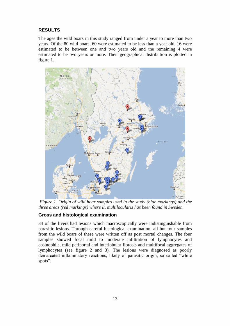
The ages the wild boars in this study ranged from under a year to more than two
years. Of the 80 wild boars, 60 were estimated to be less than a year old, 16 were
estimated to be between one and two years old and the remaining 4 were
estimated to be two years or more. Their geographical distribution is plotted in
figure 1.
Figure 1. Origin of wild boar samples used in the study (blue markings) and the
three areas (red markings) where E. multilocularis has been found in Sweden.
Gross and histological examination
34 of the livers had lesions which macroscopically were indistinguishable from
parasitic lesions. Through careful histological examination, all but four samples
from the wild boars of these were written off as post mortal changes. The four
samples showed focal mild to moderate infiltration of lymphocytes and
eosinophils, mild periportal and interlobular fibrosis and multifocal aggregates of
lymphocytes (see figure 2 and 3). The lesions were diagnosed as poorly
demarcated inflammatory reactions, likely of parasitic origin, so called “white
spots”.
14
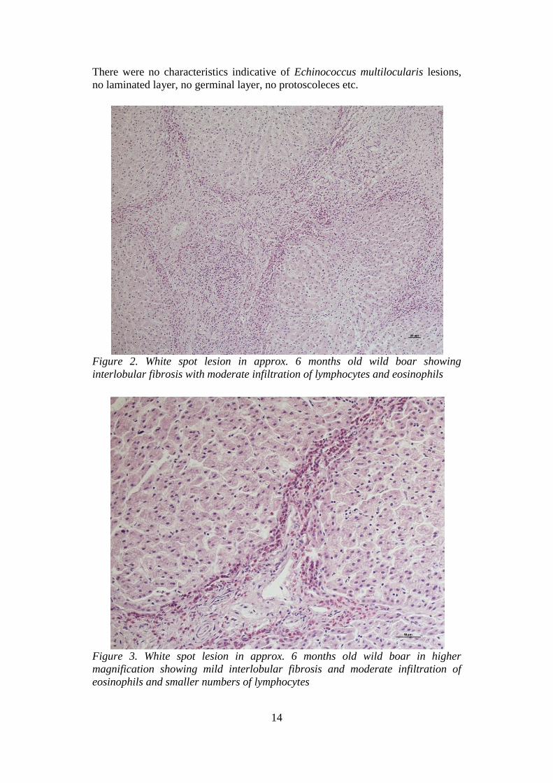
There were no characteristics indicative of Echinococcus multilocularis lesions,
no laminated layer, no germinal layer, no protoscoleces etc.
Figure 2. White spot lesion in approx. 6 months old wild boar showing
interlobular fibrosis with moderate infiltration of lymphocytes and eosinophils
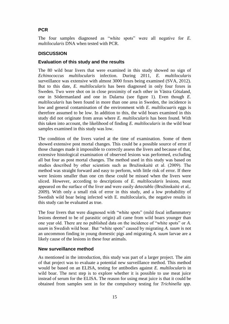
Figure 3. White spot lesion in approx. 6 months old wild boar in higher
magnification showing mild interlobular fibrosis and moderate infiltration of
eosinophils and smaller numbers of lymphocytes
15
PCR
The four samples diagnosed as “white spots” were all negative for E.
multilocularis DNA when tested with PCR.
DISCUSSION
Evaluation of this study and the results
The 80 wild boar livers that were examined in this study showed no sign of
Echinococcus multilocularis infection. During 2011, E. multilocularis
surveillance was extensive with almost 3000 foxes being examined (SVA, 2012).
But to this date, E. multilocularis has been diagnosed in only four foxes in
Sweden. Two were shot on in close proximity of each other in Västra Götaland,
one in Södermanland and one in Dalarna (see figure 1). Even though E.
multilocularis has been found in more than one area in Sweden, the incidence is
low and general contamination of the environment with E. multilocuaris eggs is
therefore assumed to be low. In addition to this, the wild boars examined in this
study did not originate from areas where E. multilocularis has been found. With
this taken into account, the likelihood of finding E. multilocularis in the wild boar
samples examined in this study was low.
The condition of the livers varied at the time of examination. Some of them
showed extensive post mortal changes. This could be a possible source of error if
those changes made it impossible to correctly assess the livers and because of that,
extensive histological examination of observed lesions was performed, excluding
all but four as post mortal changes. The method used in this study was based on
studies described by other scientists such as Bružinskaitė et al. (2009). The
method was straight forward and easy to perform, with little risk of error. If there
were lesions smaller than one cm these could be missed when the livers were
sliced. However, according to descriptions of E. multilocularis lesions, most
appeared on the surface of the liver and were easily detectable (Bružinskaitė et al,.
2009). With only a small risk of error in this study, and a low probability of
Swedish wild boar being infected with E. multilocularis, the negative results in
this study can be evaluated as true.
The four livers that were diagnosed with “white spots” (mild focal inflammatory
lesions deemed to be of parasitic origin) all came from wild boars younger than
one year old. There are no published data on the incidence of “white spots” or A.
suum in Swedish wild boar. But “white spots” caused by migrating A. suum is not
an uncommon finding in young domestic pigs and migrating A. suum larvae are a
likely cause of the lesions in these four animals.
New surveillance method
As mentioned in the introduction, this study was part of a larger project. The aim
of that project was to evaluate a potential new surveillance method. This method
would be based on an ELISA, testing for antibodies against E. multilocularis in
wild boar. The next step is to explore whether it is possible to use meat juice
instead of serum for the ELISA. The reason for using meat juice is that it could be
obtained from samples sent in for the compulsory testing for Trichinella spp.
16
carried out on wild boar muscle tissue. The results from this study means that
samples from the studied wild boars can be used as a negative reference material.
An uncertainty of this method at present is the limited knowledge of how long E.
multilocularis lesions in wild boar livers persist and how long wild boars have a
detectable amount of antibodies against E. multilocularis which increases the
uncertainty in the assessment of the area contaminated. Another factor that is not
well researched is how using meat juice instead of serum for ELISA will affect
the outcome.
The strengths of the method would be: that it is safe for persons performing it,
cheaper and faster than SCT. Collection of samples would be easy as it could be
done with the obligatory trichinosis testing as mentioned above. Advantages of
using samples from wild boar are the fact that they are likely to ingest E.
multilocularis eggs in a contaminated environment and the wild boar population is
increasing fast and spreading fast over the country and is now covering southern
Sweden, which is also the most likely place to find E. multilocularis. The hunting
bag is likely to increase, which would mean increased opportunity to gather blood
or muscle tissue. Another possibility would be to use blood or muscle juice from
out-door domestic pigs but these are not very common in Sweden.
CONCLUSION
There was no evidence that the wild boar used for this study were infected with
Echinococcus multilocularis. Therefore, samples from these animals can be used
as negative reference material when assessing the Echinococcus multilocularis
ELISA in the continued project.
17
REFERENCES
Boucher, J.M., Hanosset, R., Augot, D., Bart, J.M., Morand, M., Piarroux, R., Pozet-
Bouhier, F., Losson, B., Cliquet, F. (2005) Detection of Echinoccous multilocularis in
wild boars using PCR techniques against larval form. Veterinary Parasitology 129,
259-266
Bružinskaitė, R., Šarkūnas, M., Torgerson, P.R., Mathis, A., Deplazes, P. (2009)
Echinococcosis in pigs and intestinal infection with Echinococcus spp. in dogs in
southwestern Lithuania. Veterinary Parasitology 160, 237-241
Deplazes, P., Grimm, F., Sydler, T., Tanner, I., Kapel, C.M.O. (2005) Experimental
alveolar echinococcosis in pigs, lesion development and serological follow up.
Veterinary Parasitology 130, 213-222
Eckert, J. (1997) Epidemiology of Echinococcus multilocularis and E. granulosus in
Central Europe. Parassitologia 39, 337-344
Eckert, J. (2003) Predictive values and quality control of techniques for the diagnosis of
Echinococcus multilocularis in definitive hosts. Acta Tropica 85, 157-163
Eckert, J., Gemmell, M.A., Meslin, F.X., Pawlowski, Z.S. (Eds.) (2001) WHO/OIE
Manual on Echinococcosis in Humans and Animals: A Public Health Problem of
Global Concern. World Organisation for Animal Health (OIE) and World Health
Organization (WHO), Paris, France
Gemmel, M. A. (1959). The fox as a definitive host of Echinococcus and its role in the
spread of hydatid disease. Bulletin of World Health Organisation 20, 87-99
Goto, Y., Sato, K., Yahagi, K., Komatsu, O., Hoshina, H., Abiko, C., Yamasaki, H.,
Kawanaka, M. (2010) Frequent Isolation of Echinococcus multilocularis from the
Livers of Racehorses Slaughetered in Yamagata, Japan. Japanese Journal of
Infectious Diseases 63, 449-451
Kamiya, M., Ooi, H. K., Oku, Y., Okamoto, M., Ohbayashi, M., Seki, N. (1987) Isolation
of Echinococcus multilocularis from the liver of swine in Hokkaido, Japan. Japanese
Journal of Veterinary Research 35 (2), 99-107
Karamon, J., Sroka, J., Cencek, T. The first detection of Echinococcus multilocularis in
slaughtered pigs in Poland. Veterinary Parasitology. In press 2011
Kern, P., Bardonnet, K., Renner, E., Auer, H., Pawlowki, Z., Ammann, R. W., Vuitton,
D. A., Kern, P., European Echinococcosis Registry. (2003) European Echinococcosis
Registry: Human Alveolar Echinococcosis, Europe, 1982–2000. Emerging infectious
diseases 9 (3)
Kern, P., Wen, H., Sato, N., Vuitton, D. A., Gruener, B., Shao, Y., Delabrousse, E.,
Kratzer, W., Bresson-Hadni, S. (2006) WHO classification of alveolar
echinococcosis: Principles and application. Parasitology International 55, 283-287
Lukashenko, N. P. (1968) Comparative Biologic and Pathologic Studies of Alveococcus
multilocularis. Archives of Environmental Health 17, 676-680
Lukashenko, N. P. (1971) Problems of epidemiology and prophylaxis of alveococcosis
(multilocular echinococcosis): a general review – with particular reference to the
U.S.S.R. International Journal of Parasitology 1, 125-134.
Markström, S. (2002) Vildsvin. Stokholm: Jägareförlaget.
Mas Coma, S., Valero, M.A., Bargues, M.D. (2008) Effect of climate change on animal
and zoonotic helminthiases. Scientific and Technical Review - International Office of
Epizootics 27 (2), 443-452
18
Miyauchi, T., Sakui, M., Ishige, M., Fukumoto, S., Ueda, A., Ito, M., Ohbayashi, M.
(1984) A case of Multilocular Echinococcosis in a horse. Japanese journal of
Veterinary Research 32 (3), 171-173
Nakao, M., McManus, D. P., Schantz, P. M., Craig, P. S., Ito, A. (2007) A molecular
phylogeny of the genus Echinococcus inferred from complete mitochondrial
genomes. Parasitology 134, 713-722
Ohbayashi, M., Rausch, R.L., Fay, F.H. (1971) On the ecology and distribution of
Echinococcus spp. (Cestoda: Taeniidae), and characteristics of their development in
the intermediae host. II. Comparative studies on the development of larval E.
multilocularis leuckart, 1863, in the intermediate host. Japanese Journal of Veterinary
Research 1971 Jul;19:Suppl 3:1-53.
OIE – World Organisation for Animal Health [online](2011-02-16a) available from:
http://web.oie.int/wahis/public.php?page=single_report&pop=1&reportid=10263
[2012-04-05]
OIE – World Organisation for Animal Health [online](2011-05-11b) available from:
http://web.oie.int/wahis/public.php?page=single_report&pop=1&reportid=10549
[2012-04-05]
OIE – World Organisation for Animal Health [online](2011-06-15c) available from:
http://web.oie.int/wahis/public.php?page=single_report&pop=1&reportid=10700
[2012-04-05]
Osterman Lind, E., Juremalm, M., Christensson, D., Widgren, S., Hallgren, G., Ågren, E.
O., Ulhorn, H., Lindberg, A., Cedersmyg, M., Wahlström, H. (2011) First detection of
Echinococcus multilocularis in Sweden, February to March 2011. Euro Surveillance
[online] 2011;16(14):pii=19836. Available from:
http://www.eurosurveillance.org/ViewArticle.aspx?ArticleId=19836 [13 January
2012]
Pfister, T., Schad, V., Schelling, U., Lucius, R., Frank, W. (1993) Incomplete
development of larval Echinococcus multilocularis (Cestoda: Taeniida) in
spontaneously infected wild boars. Parasitology Research 79, 617-618
Rausch, R. L (1968) Taxonomic Characters in the Genus Echinococcus (Cestoda:
Taeniidae). Bulletin of the World Health Organization 39, 1-4
Rausch, R. L. & Schiller, E. L. (1956) Studies on the Helminth Fauna of Alaska. XXV.
The Ecology and Public Health Significance of Echinococcus sibiricensis Rausch &
Schiller, 1954 on St Lawrence Island. Parasitology 46 (3/4)
Rausch, R. L., & Yamashita, J. (1957) The Occurrence of Echinococcus multilocularis
Leuckart, 1863, in Japan. Proceedings of the Helminthological Society of Washington
24 (2)
Romig, T., Dinkel, A., Mackenstedt, U. (2006) The present situation of echinococcosis in
Europe. Parasitology International 55, S187-S191
Sakui, M., Ishigie, M., Fukumoto, S., Ueda, A., Ohbayashi, M. (1984) Spontaneous
Echinococcus multilocularis infection in Swine in North-Eastern Hokkaido, Japan.
Japanes Journal of Parasitology 33 (4), 291-296
Schiller, E. L. (1955) Studies on the Helminth Fauna of Alaska. XXVI. Some
observations on the Cold-Resistance of Eggs of Echinococcus sibiricensis Rausch and
Schiller, 1954. Journal of Parasitology 41, 578-582
SFS 2004:255, Smittskyddsförordningen (2004). Stockholm.

19
SJVFS 2010:43, Föreskrifter om ändring i Statens jordbruksverks föreskrifter (SJVFS
2004:51) om införsel av sällskapsdjur och hund- och kattsperma samt hundar, katter
och illrar avsedda för handel (2010). Jönköping.
SJVFS 2011:49, Statens jordbruksverks föreskrifter om införsel av sällskapsdjur och
hund- och kattsperma samt hundar, katter och illrar avsedda för handel (2011).
Jönköping
SVA. SVA:s undersökningar av rävens dvärgbandmask 2011- slutrapport [online] (2012-
01-02) Available from: http://sva.se/sv/Djurhalsa1/Zoonoser/Ravens-
dvargbandmask/Slutrapport_dvargbandmask/ (2012-04-21)
Svenska jägareförbundet (2009) Vildsvinsförvaltning i samverkan. [online]. Available
from:
http://www.jagareforbundet.se/Global/Policys/vildsvinsf%C3%B6rvaltning%20i%20
samverkan.pdf [2011-12-11]
Sydler, T., Mathis, A., Deplazes, P. (1998) Echinococcus multilocularis in the livers of
pigs kept outdoors in Switzerland. European Journal of Veterinary Pathology 4 (1),
43-46
Taylor, M. A., Coop, R. L., Wall, R. L. (2007) Veterinary parasitology. 3 ed. Oxford.
Blackwell Publishing
Torgerson, P.R., & Deplazes, P. (2009) Echinococcosis: diagnosis and diagnostic
interpretation in population studies. Trends in Parasitology 25 (4), 164-170
Torgerson, P. R., Keller, K., Magnotta, M., Ragland, N. (2010) The Global Burden of
Alveolar Echinococcosis. PLoS neglected tropical diseases 4(6): e722.
doi:10.1371/journal.pntd.0000722
Umhang, G., Woronoff-Rhen, N., Combes, B., Boué, F. (2011) Segmental sedimentation
and counting technique (SSCT): An adaptable method for qualitative diagnosis of
Echinococcus multilocularis in fox intestines. Experimental Parasitology 128, 57-60
Veit, P., Bilger, B., Schad, V., Schäfer, J., Frank, W., Lucius, R. (1995) Influence of
environmental factors on the infectivity of Echinococcus multilocularis eggs.
Parasitology 110, 79-86
Vuitton, D. A. & Gottstein, B. (2010) Echinococcus multilocularis and Its Intermediate
Host: A Model of Parasite-Host Interplay. Journal of Biomedicine and Biotechnology
2010, Article ID 923193
Webster, G. A., & Cameron, T. W. M (1967) Epidemiology and diagnosis of
Echinococcosis in Canada. Canadian Medical Association journal 96, 600-607
Zhenghuan, W., Xiaoming, W., Xiaoqing, L. (2008) Echinococcosis in China, a review of
the Epidemiology of Echinococcus spp. Eco Health 5, 115-126