early post-mortem changes and stages of decomposition · pdf fileearly post-mortem changes and...
TRANSCRIPT

Early post-mortem changes and stages of decompositionin exposed cadavers
M. Lee Goff
Received: 1 June 2009 / Accepted: 4 June 2009 / Published online: 25 June 2009� Springer Science+Business Media B.V. 2009
Abstract Decomposition of an exposed cadaver is a continuous process, beginning at the
moment of death and ending when the body is reduced to a dried skeleton. Traditional
estimates of the period of time since death or post-mortem interval have been based on a
series of grossly observable changes to the body, including livor mortis, algor mortis, rigor
mortis and similar phenomena. These changes will be described briefly and their relative
significance discussed. More recently, insects, mites and other arthropods have been
increasingly used by law enforcement to provide an estimate of the post-mortem interval.
Although the process of decomposition is continuous, it is useful to divide this into a series
of five stages: Fresh, Bloated, Decay, Postdecay and Skeletal. Here these stages are
characterized by physical parameters and related assemblages of arthropods, to provide a
framework for consideration of the decomposition process and acarine relationships to the
body.
Keywords Decomposition � Forensic � Acari � Post-mortem changes �Succession
Introduction
There are typically two known points at the beginning of the task of estimating a period of
time since death: the last time the individual was reliably known to be alive and the time at
which the body was discovered. The death occurred between these two points and the aim
is to estimate when it most probably took place. Keep in mind that this will be an estimate,
since it is generally accepted that there is actually no scientific way to precisely determine
the exact period of time since death. What is done is an estimation and in the case of
entomology and acarology, an estimation of the period of arthropod activity on the body.
This period of activity will reflect the minimum period of time since death or post-mortem
interval (PMI), but it will not precisely determine the time of death. In most cases, the later
M. Lee Goff (&)Chaminade University of Honolulu, Honolulu, HI 96816, USAe-mail: [email protected]
123
Exp Appl Acarol (2009) 49:21–36DOI 10.1007/s10493-009-9284-9

point is more accurately known than the former. Individuals tend to recall when they first
encountered the dead body with considerable precision. This is typically not in their
normal daily routine and it makes an impression, even on those accustomed to dealing with
the dead. Once the body is discovered, those processing the scene make meticulous (at
least we hope meticulous) notes including times of arrival, departure, movement of the
body and, finally, when the body is placed into the morgue. By contrast, the time at which
the individual was last reliably known to be alive is often less precise. This is possibly due
to the fact that those having the last contact most probably did not anticipate that this
would be their last encounter with the individual and nothing of significance took place at
the time. For this reason, the precision of the time of discovery and collection of specimens
become of major significance, as they are the anchor for the estimates. Estimation begins
when the arthropods are collected and preserved, thus stopping the biological clock.
As the process of estimating the period of insect activity takes place, it must be kept in
mind that the parameters of the estimate become progressively wider as the period of time
since death increases. The changes to a body that take place immediately following death
are often more rapid than those occurring later during the decomposition process. The
estimate begins, potentially, with a range of plus or minus minutes, goes to hours, days,
weeks, months and finally ‘its been there a long time.’ The last is not the most popular with
law enforcement agencies as they had already guessed that. It should also be kept in mind
that the estimates presented, by their very nature, are not precise.
Decomposition is a continuous process, beginning at the point of death and ending when
the body has been reduced to a skeleton. Although this process is a continuum, virtually
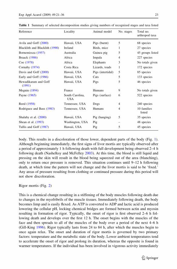
every study presented has divided this process into a series of stages. The number of stages
has varied from one to as many as nine, depending on author and geographic region (Goff
1993) (Table 1). Although the number of stages considered has varied, there does not
appear to be a firm relationship between these and the total number of species observed in
each study. For example, Cornaby (1974) working in Costa Rica using lizards and toads as
animal models noted only one stage for decomposition but recorded 172 species. By
contrast, work in Hawaii by Early and Goff (1986), using domestic cats as the animal
model, recognized five stages of decomposition but recorded 133 species. Other studies
have recognized other numbers but with no real correlation between stages observed and
numbers of taxa reported. To a certain extent, these differences may be related to sampling
methods and taxonomic interests of those involved.
Early post-mortem changes
As death proceeds, there are a series of early changes to the body that result in a definite
change in the physical nature and/or appearance of the body prior to the onset of gross,
recognizable decompositional changes. These changes have traditionally been used in
estimations of the PMI and may be a source of confusion if not recognized. For that reason,
they will be briefly described here.
Livor mortis (Fig. 1)
One of the early changes observable is livor mortis, also referred to as lividity, post-
mortem hypostasis, vibices and suggilations. This is a physical process. While the indi-
vidual is alive, the heart is functioning and circulating the blood. When death occurs,
circulation stops and the blood begins to settle, by gravity, to the lowest portions of the
22 Exp Appl Acarol (2009) 49:21–36
123
body. This results in a discoloration of those lower, dependent parts of the body (Fig. 1).
Although beginning immediately, the first signs of livor mortis are typically observed after
a period of approximately 1 h following death with full development being observed 2–4 h
following death (Nashelksy and McFelley 2003). At this time, the blood is still liquid and
pressing on the skin will result in the blood being squeezed out of the area (blanching),
only to return once pressure is removed. This situation continues until 9–12 h following
death, at which time the pattern will not change and the livor mortis is said to be ‘fixed.’
Any areas of pressure resulting from clothing or continued pressure during this period will
not show discoloration.
Rigor mortis (Fig. 2)
This is a chemical change resulting in a stiffening of the body muscles following death due
to changes in the myofribrils of the muscle tissues. Immediately following death, the body
becomes limp and is easily flexed. As ATP is converted to ADP and lactic acid is produced
lowering the cellular pH, locking chemical bridges are formed between actin and myosin
resulting in formation of rigor. Typically, the onset of rigor is first observed 2–6 h fol-
lowing death and develops over the first 12 h. The onset begins with the muscles of the
face and then spreads to all of the muscles of the body over a period of the next 4–6 h
(Gill-King 1996). Rigor typically lasts from 24 to 84 h, after which the muscles begin to
once again relax. The onset and duration of rigor mortis is governed by two primary
factors: temperature and the metabolic state of the body. Lower ambient temperatures tend
to accelerate the onset of rigor and prolong its duration, whereas the opposite is found in
warmer temperatures. If the individual has been involved in vigorous activity immediately
Table 1 Summary of selected decomposition studies giving numbers of recognized stages and taxa listed
Reference Locality Animal model No. stages Total no.arthropod taxa
Avila and Goff (2000) Hawaii, USA Pigs (burnt) 5 68 species
Blacklith and Blacklith (1990) Ireland Birds, mice 1 27 species
Bornemissza (1957) Australia Guinea pig 5 45 groups listed
Braack (1986) Africa Impala 4 227 species
Coe (1978) Africa Elephants 3 No totals given
Cornaby (1974) Costa Rica Lizards, toads 1 172 species
Davis and Goff (2000) Hawaii, USA Pigs (intertidal) 5 85 species
Early and Goff (1986) Hawaii, USA Cats 5 133 species
Hewadikaram and Goff(1991)
Hawaii, USA Pigs 5 46 species
Megnin (1894) France Humans 9 No totals given
Payne (1965) South Carolina,USA
Pigs (surface) 6 522 species
Reed (1958) Tennessee, USA Dogs 4 240 species
Rodriguez and Bass (1983) Tennessee, USA Humans 4 10 familieslisted
Shalaby et al. (2000) Hawaii, USA Pig (hanging) 5 35 species
Shean et al. (1993) Washington, USA Pig – 48 species
Tullis and Goff (1987) Hawaii, USA Pig 5 45 species
Exp Appl Acarol (2009) 49:21–36 23
123

prior to death, the onset of rigor is more rapid. Body mass and rates of cooling following
death also influence the onset and duration of rigor mortis. As rigor disappears from the
body, the pattern is similar to that seen during the onset, with the muscles of the face
relaxing first.
Algor mortis
Once death has occurred, the body ceases to regulate its internal temperature and the
internal temperature begins to approximate the ambient temperature. In most instances this
involves a cooling of the body until ambient temperature is reached, most often in a period
of 18–20 h (Fisher 2007). Although there are several different approaches, none is com-
pletely satisfactory. The rate of cooling is most often expressed by the so called ‘rule of
thumb’ equation:
Fig. 1 Livor mortis with body lying in supine position showing settling of blood to lower portions. Photocourtesy: Edward McDonough
Fig. 2 Body showing rigor mortis. Photo courtesy: Edward McDonough
24 Exp Appl Acarol (2009) 49:21–36
123

PMIðhÞ ¼ 98:6� Body temperature �Fð Þ½ �=1:5
Any estimate of the post-mortem interval obtained using this technique should be limited
to the very early stages of death (18 h or less) and treated with care. There are several
obvious factors involved in the cooling of the body that may easily influence the rate at
which this occurs. The size of the individual is a major factor. A smaller individual will
cool more rapidly than a larger individual in the same set of conditions. Exposure to
sunlight or heating may also influence the rate of cooling as may clothing and a number of
other factors. The most commonly used temperature in these calculations is from the liver
although rectal temperature may also be employed. As noted by Nashelksy and McFelley
(2003) estimates based on these approaches are to be presented in only very broad terms.
Greenish discoloration
As the body decomposes, gasses are produced in the abdomen and other parts of the body.
Although the exact composition of the gasses may vary from body to body, a significant
component of these gasses is hydrogen sulfide (H2S), a small molecule that readily diffuses
through the body. Hydrogen sulfide will react with the hemoglobin in blood to form
sulfhemoglobin (Clark et al. 1997). This pigment is greenish and may be seen in blood
vessels and in other areas of the body, particularly where livor mortis has formed.
Skin slippage (Fig. 3)
The outer layer of skin, stratum corneum, is dead. It is supposed to be dead and fills a vital
role in water conservation and protection of the underlying (live) skin. This layer is con-
stantly being shed and replaced by underlying epidermis. Upon death, in moist or wet
habitats, epidermis begins to separate from the underlying dermis due to production of
hydrolytic enzymes from cells at the junction between the two layers. The epidermis can then
easily be removed from the body. Slippage may first be observed as the formation of vesicle
formation in dependent portions of the body. In some instances, the skin from the hand may
separate from the underlying dermis as a complete or relatively complete unit. This is termed
Fig. 3 Skin slippage on hand resulting in glove formation
Exp Appl Acarol (2009) 49:21–36 25
123
‘glove formation’ and can be removed from the hand as an intact unit. This skin can be used
for finger printing, often with better results than if the skin remains on the hand.
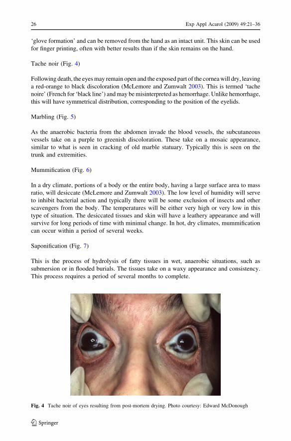
Tache noir (Fig. 4)
Following death, the eyes may remain open and the exposed part of the cornea will dry, leaving
a red-orange to black discoloration (McLemore and Zumwalt 2003). This is termed ‘tache
noire’ (French for ‘black line’) and may be misinterpreted as hemorrhage. Unlike hemorrhage,
this will have symmetrical distribution, corresponding to the position of the eyelids.
Marbling (Fig. 5)
As the anaerobic bacteria from the abdomen invade the blood vessels, the subcutaneous
vessels take on a purple to greenish discoloration. These take on a mosaic appearance,
similar to what is seen in cracking of old marble statuary. Typically this is seen on the
trunk and extremities.
Mummification (Fig. 6)
In a dry climate, portions of a body or the entire body, having a large surface area to mass
ratio, will desiccate (McLemore and Zumwalt 2003). The low level of humidity will serve
to inhibit bacterial action and typically there will be some exclusion of insects and other
scavengers from the body. The temperatures will be either very high or very low in this
type of situation. The desiccated tissues and skin will have a leathery appearance and will
survive for long periods of time with minimal change. In hot, dry climates, mummification
can occur within a period of several weeks.
Saponification (Fig. 7)
This is the process of hydrolysis of fatty tissues in wet, anaerobic situations, such as
submersion or in flooded burials. The tissues take on a waxy appearance and consistency.
This process requires a period of several months to complete.
Fig. 4 Tache noir of eyes resulting from post-mortem drying. Photo courtesy: Edward McDonough
26 Exp Appl Acarol (2009) 49:21–36
123

Putrefaction
This is nature’s recycling process. It is the result of the combined activities of all organisms
involved in decomposition reducing the body to a skeletal state.
Decomposers
In order to consider the process of decomposition and the stages involved, it helps to have
some understanding of what organisms will be involved in the process. There are four
primary categories of organisms involved in decomposition.
Fig. 5 Post-mortem marbling of body. Photo courtesy: William C. Rodriguez
Fig. 6 Mummified body
Exp Appl Acarol (2009) 49:21–36 27
123

Bacteria
There are bacteria associated with both the external and internal aspects of the human
body. While alive, the body defends against these organisms and, in fact, many are actually
beneficial. There is a large component of anaerobic bacteria associated with the human
digestive system. Some of these exist normally in our intestines, such as Escherichia coli,and, as long as they remain in place, do no damage and may assist in breakdown of food
and materials. By contrast, the same organism in the wrong place, such as the kidneys, etc.,
will result in a serious disease condition. Once the individual dies, there are few barriers to
keep them in any particular place and human tissues are excellent growth media. Shortly
after death, these bacteria begin to digest the body from the inside out. This activity is
particularly evident in the areas of the head and abdomen. The metabolic activities of these
bacteria are major components of the decomposition processes.
Fungi/molds
As noted earlier, the outer surface of the human body is comprised of dead material. This
dead outer layer is necessary to assist in the survival of the human body. As a normal
process, as new tissues are produced underneath, the outer stratum corneum is shed as
dander. As it is shed, any attached spores of molds or fungi are also shed from the body.
Following death, the outer layer is no longer shed and the mold and fungi spores will
begin to colonize the external surface of the body, often forming significant mats on the
body.
Insects and acari
Insects and other arthropods are the primary organisms involved in the major decompo-
sition of the body. They arrive at exposed remains shortly after death, often in less than 10
Fig. 7 Saponification insubmerged remains. Photocourtesy: Edward McDonough
28 Exp Appl Acarol (2009) 49:21–36
123

min, and quickly begin their activities. As the rest of this volume is devoted to their
activities, no more needs to be said here.
Vertebrate scavengers
A dead human is a potential food resource for a number of vertebrate scavengers. The
exposed body out of doors is particularly attractive and vulnerable. Direct feeding by
carnivores of all sizes can rapidly alter a dead body. Even small rodents can cause sig-
nificant damage to a body in a relatively short period of time. In the wild it may take less
than a week for scavengers to completely skeletonize a body. In addition to non-domes-
ticated animals, common domestic animals and rodents will also feed on the body in the
absence of their normal food. Pet dogs and particularly cats will feed on their deceased
owners, most often attacking the face and exposed limbs first.
Factors delaying decomposition
Apart from the various organisms involved in the process of decomposition, there are also
several types of factors that serve to stop or retard the rate at which the process continues.
These barriers to decomposition fall into three broad categories.
Physical barriers
Physical barriers to decomposition are those that prevent access of the body by physical
means. A body buried in the soil does not decompose as quickly as one exposed on the
surface. In a similar manner, a body enclosed in a sealed casket or placed into some form
of sealed container will also exhibit a delayed decomposition.
Chemical barriers
The embalming process is specifically designed to prevent the decomposition of the body,
with natural body fluids being drained and replaced with various preservative fluids. As the
body is then typically placed into a casket, the process should, if done properly, delay
decomposition for an extended period of time. The presence of insecticides on, in or near the
body may also serve to delay the onset of insect activity for a period of time. It should be noted
that insecticides will not permanently delay the colonization of the body by insects. In many
cases, immature insects are able to survive on a body with concentrations of an insecticide
that would prove fatal to the adults of the same species (Gunatilake and Goff 1989).
Climatic factors
Temperature can serve as a major factor delaying decomposition. At lower temperatures,
bacterial growth and insect activity can be retarded or even arrested. At temperatures
below 6�C most insect activity ceases but may resume once temperatures rise above this
threshold. In a similar manner, high temperatures will also result in cessation of insect
activity, and, if in a dry habitat, result in mummification of the body. Wind also serves to
inhibit insect flight and thus colonization of the body. Many texts will indicate a wind
speed in excess of 16 km/h will inhibit insect flight. This should not be accepted as a firm
wind speed as in many tropical and island areas, tradewinds typically blow at a speed
Exp Appl Acarol (2009) 49:21–36 29
123
greater than this and there is significant insect activity. Rainfall may also serve as a
temporary barrier but, once the rain ceases, the insects again become quite active.
Relationships of arthropods to a body
There are several distinct relationships between an arthropod and a decomposing body. The
population of arthropods encountered in any given habitat will contain elements unique to
that habitat and components having a wider distribution. Within this population there will
be species having some type of relationship to the decomposing body. This relationship
will vary with taxon and not all relationships will be of equal value to the investigation. All
must be considered as, under different circumstances, there will be different values for the
relationship. There have been four basic relationships between a decomposing body and
arthropods (after Goff 1993).
Necrophagous species
Those taxa actually feeding on the corpse. This group includes many of the true flies
(Diptera) particularly the blow flies (Calliphoridae) and flesh flies (Sarcophagidae), who
are early invaders, and beetles (Coleoptera: Silphidae, Dermestidae). This group includes
species that may be the most significant isolatable taxa for use in estimating a minimum
period of insect activity on the body during the early stages of decomposition (days 1–14).
The Acari, primarily Acaridae and similar taxa, are typically not grossly evident during this
stage, although present.
Predators and parasites of necrophagous species
This is the second most significant group of carrion-frequenting taxa. Many of the beetles
(Coleoptera: Silphidae, Staphylinidae, Histeridae), true flies (Diptera: Calliphoridae,
Stratiomyidae), and wasps (Hymenoptera) parasitic on fly larvae and pupae are included.
In some species, fly larvae (maggots) that are necrophages during the early portions of their
development become predators on other larvae during the later stages of their develop-
ment. Also evident during the later stages of decomposition are species in the Macroc-
helidae, Parasitidae, Uropodidae and Parasitidae, preying on various organisms
encountered in the soil under the decomposing remains.
Omnivorous species
Included in this category are the taxa such as wasps, ants and some beetles, that feed on
both the corpse and associated arthropods. Early and Goff (1986) observed that large
populations of these species may actually retard the rate of carcass removal by depleting
the populations of necrophagous species.
Adventive species
This category includes those taxa that simply use the corpse as an extension of their own
normal habitat, as is the case for the springtails (Collembola), spiders, centipedes and
millipedes.
30 Exp Appl Acarol (2009) 49:21–36
123

Accidentals
Another category that is not always recognized but may still be of significance is what
might be termed ‘accidentals.’ These are species that have no real relationship to the corpse
but still are found on the body. These insects may have fallen onto the body from sur-
rounding vegetation, thus possibly supplying some information on post-mortem movement
of a body. On the other hand, when an insect stops flying, it has to land on something and
that ‘something’ might happen to be the body. This is a fact all too often ignored, even by
entomologists.
Stages of decomposition
Number of stages
As noted earlier, several stages have been proposed for the decomposition process
(Table 1). Although the process is a continuum and discrete stages, characterized by
physical features and distinctive assemblages of insects, do not exist in nature. Regardless,
virtually every study conducted has attempted to divide the process into stages. While
artificial, these stages have definite utility. First, they allow for easy organization of
research reports and discussion. There is also a utility in court proceedings. Typically in the
USA, juries are composed of individuals with little if any background in the biological
sciences. They are often confused and repulsed by the process of decomposition they are
being asked to consider. Under these circumstances, use of stages gives them something to
use for reference and makes their task somewhat easier, if not more pleasant.
In studies conducted in Hawaii, five stages have been recognized and these appear to be
easily applied to studies conducted in temperate areas (Lord and Goff 2003). These stages
are: Fresh, Bloated, Decay, Postdecay and Skeletal or Remains. The most common
modification of this set is to subdivide the Decay Stage into Active Decay and Advanced
Decay stages. Given the subjective nature of these stages, the Decay Stage is treated here
as a single stage. Because detailed discussions of insect and arthropod succession are
presented elsewhere, the treatment here will be primarily an overview.
Fresh stage (Figs. 8, 9)
The Fresh Stage begins at the moment of death and continues until bloating of the body
becomes evident. There are few distinctive, gross decompositional changes associated with
the body during this stage although greenish discoloration of the abdomen, livor, skin
cracking, and tache noir may be observed. The insect invasion of the body generally begins
with the natural body openings of the head (eyes, nose, mouth and ears), anus and genitals,
and wounds present on the body. The first insects to arrive under most circumstances are
the Calliphoridae (blow flies) and Sarcophagidae (flesh flies). Female flies will arrive and
begin to explore the potential sites for oviposition or larviposition. These flies will often go
deep into the openings and either deposit eggs or 1st instar larvae or maggots (Fig. 10).
Whereas the openings associated with the head are uniformly attractive to flies, the
attractiveness of the anus and genital areas may depend on their being exposed or clothed.
Wounds inflicted prior to death have been observed to be more attractive to flies for
colonization if inflicted prior to death, when blood is flowing, than wounds inflicted post-
mortem and lacking a blood flow. During this stage, the eggs laid in the body begin to
Exp Appl Acarol (2009) 49:21–36 31
123

hatch and there is internal feeding activity, although there may be little evidence of this on
the surface.
Bloated stage (Fig. 10)
The principal component of decomposition, putrefaction, begins during the Bloated Stage.
The anaerobic bacteria present in the gut and other parts of the body begin to digest the
tissues. Their metabolic processes result in the production of gasses that first cause a slight
inflation of the abdomen. When this is noted, the Bloated Stage is considered to begin. As
this progresses, the body may assume a fully inflated, balloon-like appearance. The
combined processes of putrefaction and the metabolic activities of the maggots begin to
Fig. 8 Body in Fresh Stage of decomposition. Photo courtesy: William C. Rodriguez
Fig. 9 Adult females Calliphoridae entering nasal openings to oviposit during Fresh Stage of decompo-sition. Photo courtesy: William C. Rodriguez
32 Exp Appl Acarol (2009) 49:21–36
123

cause an increase in the internal temperatures of the body. These temperatures can be
significantly above ambient temperature ([50�C) and the body becomes a distinct habitat,
in many ways independent of the surrounding environment. The adult Calliphoridae are
strongly attracted to the body during this stage in decomposition and significant masses of
maggots are observed associated with the head and other primary invasion sites. While
these populations are visible externally, there are larger populations present internally.
Internal pressures caused by production of gasses result in the seeping of fluids from the
natural body openings during this stage and the strong smell of ammonia is noted. These
fluids seep into the substrate beneath the body and this becomes alkaline. The normal soil
fauna will leave the area under the body as a result of this change in the pH and the
invasion of a set of organisms more closely associated with decomposition begins.
Decay stage (Fig. 11)
The start and termination points for the stages of decomposition are largely subjective, but
there is a definite physical event marking the start of the Decay Stage. This is when the
combined activities of the maggot feeding and bacterial putrefaction result in the breaking
of the outer layer of the skin and the escape of the gasses from the abdomen. At this point,
the body deflates and the Decay Stage is considered to begin. During this stage, strong
odors of decomposition are present. The predominant feature of this stage is the presence
of large feeding masses of Diptera larvae. These are present internally, externally and often
spilling onto the ground beside the body. Some Coleoptera that have been arriving during
earlier stages of decomposition, increase in numbers during the Decay Stage and are often
quite evident. Some predators, such as the Staphylinidae, are seen during the Bloated Stage
and they become more evident now, along with others, such as the Histeridae. In addition
to the predators, necrophages are also evident, increasing in numbers as the process
continues. By the end of this stage, most of the Calliphoridae and Sarcophagidae will have
completed their development and left the remains to pupariate in the surrounding soil. By
the end of the Decay Stage, Diptera larvae will have removed most of the flesh from the
body, leaving only skin and cartilage.
Fig. 10 Body in Bloated stage of decomposition. Photo courtesy: William C. Rodriguez
Exp Appl Acarol (2009) 49:21–36 33
123
Postdecay stage (Fig. 12)
As the body is reduced to skin, cartilage and bone, the Diptera cease to be the predominant
feature. In xerophytic and mesophytic habitats, various groups of Coleoptera will replace
them, with the most commonly seen being the species in the family Dermestidae. These
arrive as adults during the later stages of the Decay Stage but become predominant as
adults and larvae during the Postdecay Stage. Their feeding removes the remaining dried
Fig. 11 Body in Decay stage of decomposition. Photo courtesy: William C. Rodriguez
Fig. 12 Body in Postdecay stageof decomposition. Photocourtesy: William C. Rodriguez
34 Exp Appl Acarol (2009) 49:21–36
123

flesh and cartilage from the bones and the scraping of their mandibles leaves the bones with
a cleaned, polished appearance. In wet habitats (swamps, rainforests, etc.), the Coleoptera
typically are not successful. They are replaced in the process by other groups of arthropods.
These include several Diptera families, such as the Psychodidae, along with their
respective predator/parasite complexes. Associated with this stage in both types of habitat
is an increase in the numbers and diversity of the predators and parasites. The soil-dwelling
taxa increase in number and diversity during this stage.
Skeletal/remains stage (Fig. 13)
This stage is reached when only bones and hair remain. Typically, there are no obviously
carrion-frequenting taxa seen during this stage. During the earlier portions of the Skeletal
Stage, there are a number of soil-dwelling taxa, including mites and Collembola, that can
be used in estimating the period of time since death. As time passes, the pH of the soil
begins to return to the original level and there is a gradual return of components of the
normal soil fauna during this stage. There is no definite end point to this stage and there
may be differences in the soil fauna detectable for a period of months or sometimes years,
indicating that a body was there at some point in time.
Acknowledgments Thanks are extended to Drs. William C. Rodriguez III, Office of the Armed ForcesMedical Examiner, AFIP and Edward T. McDonough, Office of the Chief Medical Examiner, Connecticut,for illustrations of post-mortem artifacts and stages of decomposition.
References
Avila FW, Goff ML (2000) Arthropod succession patterns onto burnt carrion in two contrasting habitats inthe Hawaiian Islands. J Forensic Sci 43:581–586
Blacklith RE, Blacklith RM (1990) Insect infestations of small corpses. J Nat Hist 24:699–709Bornemissza GF (1957) An analysis of arthropod succession in carrion and the effect of its decomposition
on the soil fauna. Aust J Zool 5:1–12Braack LEO (1986) Arthropods associated with carcasses in the northern Kruger National Park. S Afr J
Wildl Res 16:91–98
Fig. 13 Skull in Skeletal/Remains stage of decomposition
Exp Appl Acarol (2009) 49:21–36 35
123

Clark MA, Worrell MB, Pless JE (1997) Post-mortem changes in soft tissue. In: Froede RC (ed) Handbookof forensic pathology, 2nd edn. CAP, Illinois
Coe M (1978) The decomposition of elephant carcasses in the Tsavo (East) National Park, Kenya. J AridEnviron 1:71–86
Cornaby B (1974) Carrion reduction by animals in contrasting tropical habitats. Biotropica 6:51–63Davis JB, Goff ML (2000) Decomposition patterns in terrestrial and intertidal habitats on O’ahu Island and
Coconut Island, Hawai’i. J Forensic Sci 45:824–830Early M, Goff ML (1986) Arthropod succession patterns in exposed carrion on the island of O’ahu,
Hawaiian Islands, USA. J Med Entomol 23:520–531Fisher BAJ (2007) Techniques of crime scene investigation, 7th edn. CRC Press, New YorkGill-King H (1996) Chemical and ultrastructural aspects of decomposition. In: Haglund WD, Sorg MH (eds)
Forensic taphonomy: the post-mortem fate of human remains. CRC Press, New YorkGoff ML (1993) Estimation of post-mortem interval using arthropod development and successional patterns.
Forensic Sci Rev 5:81–94Gunatilake K, Goff ML (1989) Detection of organophosphate poisoning in a putrefying body by analyzing
arthropod larvae. J Forensic Sci 34:714–716Hewadikaram KA, Goff ML (1991) Effect of carcass size on rate of decomposition and arthropod succession
patterns. Am J Forensic Med Pathol 12:235–240Lord WD, Goff ML (2003) Forensic entomology: application of entomological methods to the investigation
of death. In: Froede RC (ed) Handbook of forensic pathology, 2nd edn. CAP, IllinoisMcLemore J, Zumwalt RE (2003) Post-mortem changes. In: Froede RC (ed) Handbook of forensic
pathology, 2nd edn. CAP, IllinoisMegnin P (1894) La faune des cadavers: application de l’entomologie a la medecine legale. Encyclopedia
Scientifique des Aide-Memoire, Masson et Gauthier-Villars, Paris, FranceNashelksy M, McFelley P (2003) Time of death. In: Froede RC (ed) Handbook of forensic taphonomy, 2nd
edn. CAP, IllinoisPayne JA (1965) A summer carrion study of the baby pig Sus scrofa Linnaeus. Ecology 46:592–602Reed HB (1958) A study of dog carcass communities in Tennessee with special reference to the insects. Am
Midl Nat 59:213–245Rodriguez WC, Bass WM (1983) Insect activity and its relationship to decay rates of human cadavers in
East Tennessee. J Forensic Sci 30:836–852Shalaby OA, de Carvalho LML, Goff ML (2000) Comparison of patterns of decomposition in a hanging
carcass and a carcass in contact with the soil in a xerophytic habitat on the island of O’ahu, Hawai’i. JForensic Sci 45:1267–1273
Shean BS, Messinger L, Papworth M (1993) Observations of differential decomposition on sun exposed vs.shaded pig carrion in costal Washington State. J Forensic Sci 38:938–949
Tullis K, Goff ML (1987) Arthropod succession in exposed carrion in a tropical rainforest on O’ahu Island,Hawai’i. J Med Entomol 24:332–339
36 Exp Appl Acarol (2009) 49:21–36
123