e2f3 activity is regulated during the cell cycle and is...
TRANSCRIPT

10.1101/gad.12.14.2120Access the most recent version at doi: 1998 12: 2120-2130Genes Dev.
Gustavo Leone, James DeGregori, Zhen Yan, et al. the induction of S?phaseE2F3 activity is regulated during the cell cycle and is required for
References
http://genesdev.cshlp.org/content/12/14/2120.full.html#related-urlsArticle cited in:
http://genesdev.cshlp.org/content/12/14/2120.full.html#ref-list-1This article cites 36 articles, 23 of which can be accessed free at:
serviceEmail alerting
click heretop right corner of the article orReceive free email alerts when new articles cite this article - sign up in the box at the
http://genesdev.cshlp.org/subscriptions go to: Genes & DevelopmentTo subscribe to
Cold Spring Harbor Laboratory Press
Cold Spring Harbor Laboratory Press on May 19, 2009 - Published by genesdev.cshlp.orgDownloaded from
E2F3 activity is regulated during thecell cycle and is required for theinduction of S phaseGustavo Leone,1,3 James DeGregori,1,3,4 Zhen Yan,2 Laszlo Jakoi,1 Seiichi Ishida,1
R. Sanders Williams,2 and Joseph R. Nevins1,5
1Department of Genetics, Howard Hughes Medical Institute, Duke University Medical Center, Durham, North Carolina27710 USA; 2Departments of Internal Medicine and Molecular Biology/Oncology, University of Texas SouthwesternMedical Center, Dallas, Texas 75235 USA
Previous work has demonstrated the important role of E2F transcription activity in the induction of S phaseduring the transition from quiescence to proliferation. In addition to the E2F-dependent activation of anumber of genes encoding DNA replication activities such as DNA Pol a, we now show that the majority ofgenes encoding initiation proteins, including Cdc6 and the Mcm proteins, are activated following thestimulation of cell growth and are regulated by E2F. The transcription of a subset of these genes, whichincludes Cdc6, cyclin E, and cdk2, is also regulated during the cell cycle. Moreover, whereas overall E2FDNA-binding activity accumulates during the initial G1 following a growth stimulus, only E2F3-bindingactivity reaccumulates at subsequent G1/S transitions, coincident with the expression of thecell-cycle-regulated subset of E2F-target genes. Finally, we show that immunodepletion of E2F3 activityinhibits the induction of S phase in proliferating cells. We propose that E2F3 activity plays an important roleduring the cell cycle of proliferating cells, controlling the expression of genes whose products are rate limitingfor initiation of DNA replication, thereby imparting a more dramatic control of entry into S phase than wouldotherwise be achieved by post-transcriptional control alone.
[Key Words: E2F3-binding activity; E2F transcription control; S-phase induction; cell cycle]
Received April 20, 1998; accepted in revised form May 26, 1998.
Numerous genes have been identified that control thetransition of cells through the cell cycle, including thoseencoding proteins that are critical for the initiation ofDNA replication. The elucidation of the events associ-ated with the control of DNA replication is largely theresult of the combination of genetic and biochemicalanalyses in budding and fission yeast. These studies haveidentified a series of proteins that assemble a functionalorigin of replication (for review, see Stillman 1996; Duttaand Bell 1997; Newlon 1997). At the core of the func-tional origin is the six-component origin recognitioncomplex (ORC) that interacts with origin sequencesthroughout the cell cycle and is critical for DNA repli-cation. Other work has defined a second complex of sixproteins known as the Mcm proteins, that associate withthe DNA-bound ORC and are also essential for replica-tion. Finally, the cdc6 gene, initially identified in geneticscreens as a critical cell cycle regulatory gene governing
the G1/S transition, encodes a protein that appears tofacilitate the association of the Mcm complex with ORC(Donovan et al. 1997; Tanaka et al. 1997). The additionalobservation that the level of the Cdc6 product varies inthe cell cycle suggests that the G1/S accumulation ofCdc6 may contribute to the regulation of origin function.
Intensive efforts devoted to the analysis of mamma-lian cell growth control has complemented and extendedthe studies in yeast. Although basic aspects of the G1
regulatory events are conserved in yeast and higher eu-karyotes, it is clear that the requirements of cell growthcontrol that couple proliferation with cell differentiationhave added additional complexity. This work has re-vealed a pathway controlling the progression of cells outof quiescence, through G1, and into S phase that involvesthe action of G1 cyclin-dependent kinases (cdks) to inac-tivate the Rb tumor suppressor and related proteins,which then leads to the accumulation of E2F transcrip-tion factor activity (for review, see Nevins 1992; Helinand Harlow 1993; Hunter and Pines 1994; Weinberg1995; Sherr 1996). The importance of this pathway formammalian cell growth control is indicated by the factthat disruption of the pathway, either the activation ofpositive-acting components such as the G1 cyclins or the
3These authors contributed equally to this work.4Present address: Department of Biochemistry and Molecular Genetics,University of Colorado Health Sciences Center, Denver, Colorado 80262USA.5Corresponding author.E-MAIL [email protected]; FAX (919) 681-8973.
2120 GENES & DEVELOPMENT 12:2120–2130 © 1998 by Cold Spring Harbor Laboratory Press ISSN 0890-9369/98 $5.00; www.genesdev.org
Cold Spring Harbor Laboratory Press on May 19, 2009 - Published by genesdev.cshlp.orgDownloaded from

inactivation of negative components such as p53, Rb,and the cdk inhibitors, can lead to the loss of cell growthcontrol that underlies the development of virtually allforms of human cancer (Weinberg 1995; Hunter 1997).
In some respects, the role of the E2F transcription fac-tors appears similar to the Saccharomyces cerevisiaetranscriptional regulatory proteins SWI4/6/MBF and theSchizosaccharomyces pombe Cdc10 protein. In particu-lar, E2F regulates transcription of a large number ofgenes that encode DNA replication activities includingDNA polymerase a (Pol a) PCNA, ribonucleotide reduc-tase, and others. Many of these genes are controlled bythe SWI/MBF/Cdc10 proteins in yeast. This similaritynow extends to the control of cdc6 gene transcriptionbased on our recent work (Yan et al. 1998). Consistentwith the role for the E2F transcription factors in theregulation of genes encoding DNA replication activities(Nevins 1992; Helin and Harlow 1993), E2F can induceDNA replication in otherwise quiescent cells (Johnson etal. 1993; Qin et al. 1994; Shan and Lee 1994; DeGregoriet al. 1995b).
Although many of the molecular events involved inthe control of mammalian cell growth have been wellcharacterized, particularly the various signal transduc-tion pathways that are activated when quiescent cellsare stimulated to grow, the role of these activities in thecontrol of cell cycle transitions in proliferating cell popu-lations is much less clear. Because proliferating cellsmaintain a defined G1 phase, the timing of initiation ofDNA replication must be subject to tight control duringeach cell cycle. Although much of the understanding oforigin function has derived from yeast, it is clear thathomologs of many, if not all, of these activities can befound in mammalian cells. Each of the ORC componentsis highly conserved as are the Mcm proteins. In addition,recent work leading to the isolation of clones of mam-
malian (Williams et al. 1997) and Xenopus Cdc6 (Cole-man et al. 1996) has revealed a significant conservationin the structure of this protein.
Recent work that has revealed E2F control of several ofthe genes encoding initiation proteins such as Orc1(Ohtani et al. 1996) and Cdc6 (Yan et al. 1998) suggests acentral role for E2F in the control of DNA replication. Inlight of this, and given the fact that control of initiationof DNA replication is critical in proliferating cells aswell as during the transition out of quiescence, we haveinvestigated the role of E2F in transcription control dur-ing the cell cycle of proliferating cells.
Results
The transcription of a large number of genes encodingDNA replication activities, including Cdc6and the Mcm proteins, is regulated by cell growthand dependent on E2F
Previous work has documented the role of E2F in thegrowth-regulated expression of genes encoding proteinssuch as DHFR, DNA Pol a, thymidine kinase, and vari-ous other genes that encode activities important forDNA replication (Nevins 1992). These genes are ex-pressed at low or undetectable levels in quiescent, non-dividing fibroblasts and are induced following growthstimulation. Recent experiments have extended thisgroup of growth-regulated genes to those encoding pro-teins that mediate the initiation of replication, includingOrc1 (Ohtani et al. 1996), one of six components of theORC, and Cdc6 (Williams et al. 1997; Yan et al. 1998), aprotein that is essential for the formation of a functionalinitiation complex. In addition to the role of Cdc6 andORC in facilitating origin function, a number of experi-ments now point to the role of a second complex, involv-
Figure 1. Growth regulated expression ofgenes encoding DNA replication proteins.(A) Quiescent Ref 52 cells (Q) were stimu-lated with media containing 10% serum.Cells were harvested at the indicated times,processed for Northern analysis as describedin Materials and Methods, and hybridizedwith the indicated probes. The position ofthe cell with respect to cell cycle is indicatedbelow based on FACS analysis of similarlytreated samples. (B) Quiescent cells were in-fected with recombinant adenoviruses ex-pressing either E2F1, E2F2, or E2F3 proteins,or with a control virus containing an emptyexpression cassette (m.o.i. of 100, 100, 200,and 100, respectively). Cells were harvested18 hr postinfection and processed and ana-lyzed as in A. A portion of the cells that wereinfected with the control virus were stimu-lated with media containing 10% serum foran additional 18 hr (Con +).
Cell cycle control of E2F3 activity
GENES & DEVELOPMENT 2121
Cold Spring Harbor Laboratory Press on May 19, 2009 - Published by genesdev.cshlp.orgDownloaded from
ing the 6 Mcm proteins (Mcm2–Mcm7), as critical forinitiation of DNA replication. In light of the fact thatOrc1 and Cdc6 have been found to be tightly regulatedby cell proliferation and dependent on E2F, and takingadvantage of the cloning of each of the genes encodingmammalian Mcm proteins (Holthoff et al. 1996; Kiyonoet al. 1996; Tsuruga et al. 1997), we have investigated thepotential cell growth-dependent control of expression ofthese genes.
As shown in Figure 1A, analysis of RNA from quies-cent cells and from growth-stimulated cells revealed alarge induction of each of the mcm genes. The pattern ofaccumulation of the Mcm transcripts paralleled that ofgenes encoding E2F1 as well as PCNA, cyclin E, andCdc6, which were shown previously to be regulated bycell growth. Only very low levels of the RNAs were de-tected in the quiescent cells which then increased 10- to20-fold following growth stimulation. Clearly, a largenumber of genes encoding DNA replication activities aretightly regulated by cell growth. We also note distinc-tions in the pattern of accumulation of these transcriptsas cells pass through S phase. For instance, whereas E2F1and Mcm7 RNAs clearly remained constant followingthe initial accumulation, cyclin E, PCNA, and Cdc6were consistently observed to decline as cells movedthrough S phase.
Given the role of E2F in the control of transcription ofmany growth-regulated genes including cdc6, and theobservation that the mcm6 gene promoter contains se-quences that match E2F consensus sites (Tsuruga et al.1997), we assayed the effect of E2F overproduction on theexpression of each of the mcm genes. Quiescent REF52fibroblasts were infected with recombinant adenovirusesthat express the E2F1, E2F2, or E2F3 products. RNA wasprepared and then assayed for expression of each of theMcms. As shown in Figure 1B, expression of E2F1, E2F2,or E2F3 resulted in a large induction of each of the mcmgenes, similar to the induction of the cdc6 and orc1genes, and equivalent to that achieved following serumstimulation. We thus conclude that the mcm genes areindeed regulated as a function of cell growth and thatthey are also subject to control by E2F, coincident withthe control of many other genes encoding DNA replica-tion activities.
A subset of E2F targets, including Cdc6, cyclin E,and Cdk2 are cell cycle regulated in proliferating cells
Mechanisms that control the initiation of DNA replica-tion are important not only at the initial G1/S transitionas cells re-enter a cell cycle from a quiescent state, butalso at each subsequent G1/S as cells proliferate in thepresence of growth factors. Although numerous experi-ments have documented the role of E2F activity in theinduction of the initial S phase, following exit from qui-escence, coincident with the activation of a variety ofgenes that encode DNA replication proteins, includingCdc6 (Williams et al. 1997), little is known of the role ofE2F in transcription regulation following this initialG1/S transition, when cells continue to proliferate.
Given the observation that many of the proteins thatdetermine initiation of replication are subject to E2Fcontrol, together with the fact that initiation must beregulated during each cell cycle, we have explored thepossible role of E2F-dependent transcription regulationduring a cell cycle in proliferating cells.
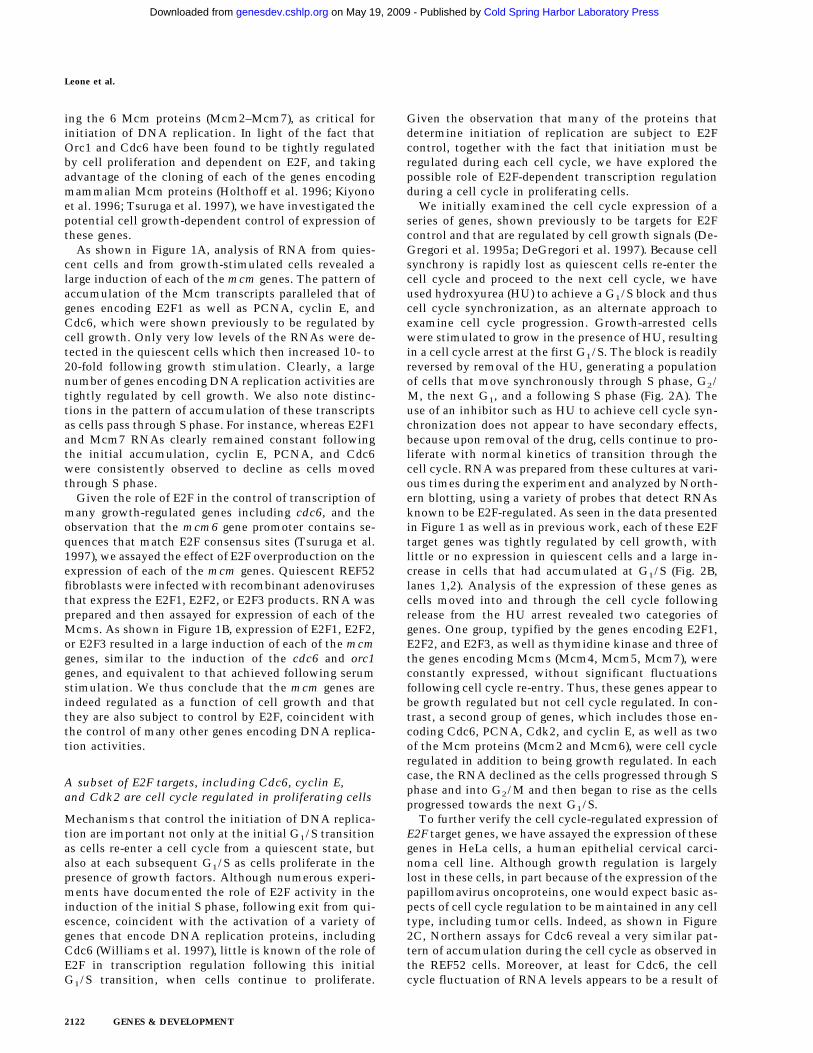
We initially examined the cell cycle expression of aseries of genes, shown previously to be targets for E2Fcontrol and that are regulated by cell growth signals (De-Gregori et al. 1995a; DeGregori et al. 1997). Because cellsynchrony is rapidly lost as quiescent cells re-enter thecell cycle and proceed to the next cell cycle, we haveused hydroxyurea (HU) to achieve a G1/S block and thuscell cycle synchronization, as an alternate approach toexamine cell cycle progression. Growth-arrested cellswere stimulated to grow in the presence of HU, resultingin a cell cycle arrest at the first G1/S. The block is readilyreversed by removal of the HU, generating a populationof cells that move synchronously through S phase, G2/M, the next G1, and a following S phase (Fig. 2A). Theuse of an inhibitor such as HU to achieve cell cycle syn-chronization does not appear to have secondary effects,because upon removal of the drug, cells continue to pro-liferate with normal kinetics of transition through thecell cycle. RNA was prepared from these cultures at vari-ous times during the experiment and analyzed by North-ern blotting, using a variety of probes that detect RNAsknown to be E2F-regulated. As seen in the data presentedin Figure 1 as well as in previous work, each of these E2Ftarget genes was tightly regulated by cell growth, withlittle or no expression in quiescent cells and a large in-crease in cells that had accumulated at G1/S (Fig. 2B,lanes 1,2). Analysis of the expression of these genes ascells moved into and through the cell cycle followingrelease from the HU arrest revealed two categories ofgenes. One group, typified by the genes encoding E2F1,E2F2, and E2F3, as well as thymidine kinase and three ofthe genes encoding Mcms (Mcm4, Mcm5, Mcm7), wereconstantly expressed, without significant fluctuationsfollowing cell cycle re-entry. Thus, these genes appear tobe growth regulated but not cell cycle regulated. In con-trast, a second group of genes, which includes those en-coding Cdc6, PCNA, Cdk2, and cyclin E, as well as twoof the Mcm proteins (Mcm2 and Mcm6), were cell cycleregulated in addition to being growth regulated. In eachcase, the RNA declined as the cells progressed through Sphase and into G2/M and then began to rise as the cellsprogressed towards the next G1/S.
To further verify the cell cycle-regulated expression ofE2F target genes, we have assayed the expression of thesegenes in HeLa cells, a human epithelial cervical carci-noma cell line. Although growth regulation is largelylost in these cells, in part because of the expression of thepapillomavirus oncoproteins, one would expect basic as-pects of cell cycle regulation to be maintained in any celltype, including tumor cells. Indeed, as shown in Figure2C, Northern assays for Cdc6 reveal a very similar pat-tern of accumulation during the cell cycle as observed inthe REF52 cells. Moreover, at least for Cdc6, the cellcycle fluctuation of RNA levels appears to be a result of
Leone et al.
2122 GENES & DEVELOPMENT
Cold Spring Harbor Laboratory Press on May 19, 2009 - Published by genesdev.cshlp.orgDownloaded from
transcriptional control as seen by nuclear run-on mea-surements in synchronized HeLa cell populations (Fig.2D).
The control of cyclin E and cdk2 expression was alsoreflected in the accumulation of each of the proteins dur-ing the cell cycle, as measured by Western blot assays(Fig. 3A). In each case, the protein declined as cells leftthe G1/S arrest and passed through S phase and thenreaccumulated as cells entered the next G1. We have alsomeasured Cdc6 protein accumulation during the cellcycle although because of antibody specificities, we havenot been able to assay for Cdc6 protein in Ref52 cells.
However, assays of HeLa cell fractions yielded a resultthat paralleled that of cyclin E and cdk2 and closelymatched the accumulation of Cdc6 RNA during theHeLa cell cycle (Fig. 3B).
Cell cycle control of E2F activity
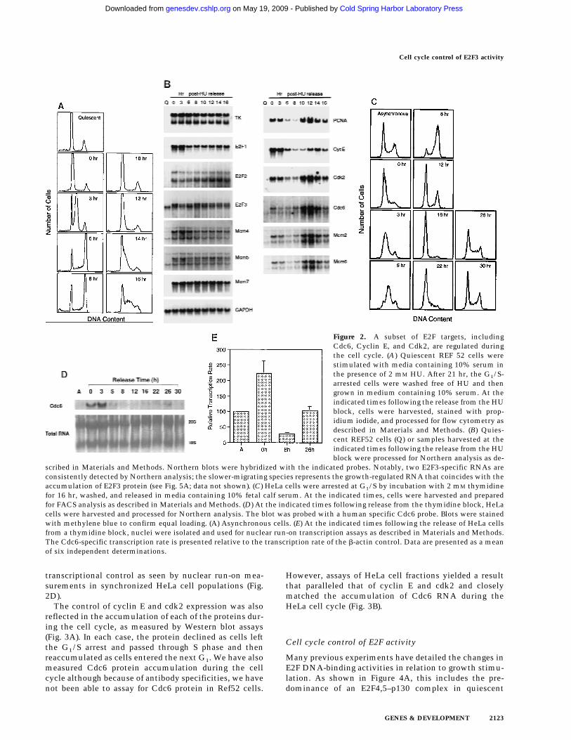
Many previous experiments have detailed the changes inE2F DNA-binding activities in relation to growth stimu-lation. As shown in Figure 4A, this includes the pre-dominance of an E2F4,5–p130 complex in quiescent
Figure 2. A subset of E2F targets, includingCdc6, Cyclin E, and Cdk2, are regulated duringthe cell cycle. (A) Quiescent REF 52 cells werestimulated with media containing 10% serum inthe presence of 2 mM HU. After 21 hr, the G1/S-arrested cells were washed free of HU and thengrown in medium containing 10% serum. At theindicated times following the release from the HUblock, cells were harvested, stained with prop-idium iodide, and processed for flow cytometry asdescribed in Materials and Methods. (B) Quies-cent REF52 cells (Q) or samples harvested at theindicated times following the release from the HUblock were processed for Northern analysis as de-
scribed in Materials and Methods. Northern blots were hybridized with the indicated probes. Notably, two E2F3-specific RNAs areconsistently detected by Northern analysis; the slower-migrating species represents the growth-regulated RNA that coincides with theaccumulation of E2F3 protein (see Fig. 5A; data not shown). (C) HeLa cells were arrested at G1/S by incubation with 2 mM thymidinefor 16 hr, washed, and released in media containing 10% fetal calf serum. At the indicated times, cells were harvested and preparedfor FACS analysis as described in Materials and Methods. (D) At the indicated times following release from the thymidine block, HeLacells were harvested and processed for Northern analysis. The blot was probed with a human specific Cdc6 probe. Blots were stainedwith methylene blue to confirm equal loading. (A) Asynchronous cells. (E) At the indicated times following the release of HeLa cellsfrom a thymidine block, nuclei were isolated and used for nuclear run-on transcription assays as described in Materials and Methods.The Cdc6-specific transcription rate is presented relative to the transcription rate of the b-actin control. Data are presented as a meanof six independent determinations.
Cell cycle control of E2F3 activity
GENES & DEVELOPMENT 2123
Cold Spring Harbor Laboratory Press on May 19, 2009 - Published by genesdev.cshlp.orgDownloaded from

cells, the disappearance of this complex as cells arestimulated to grow, and the appearance of free E2F4 andE2F5 activities as well as an E2F4,5–p107 complex,which also contains cyclin A and cdk2, as cells progressinto S phase. During G1, the accumulation of the regu-lated E2F activities (E2F1, and E2F3) was also observed inparallel with an increase in Rb–E2F complexes thatlikely reflects the overall accumulation of E2F activityduring this time period. We were aided in these assays bythe use of gel electrophoretic conditions that allowed theseparation of individual E2F–DNA complexes (Ikeda etal. 1996). As such, it was possible to identify gel-shiftbands representing the accumulating E2F1 and E2F3 ac-tivities, confirmed by antibody supershift (Fig. 4A, right),as well as by overexpression of individual E2F proteins(data not shown) as cells progressed through G1/S.
The observation that several genes that have beenshown to be growth regulated by E2F are also regulatedduring the cell cycle raises the possibility that E2F ac-tivity might be regulated in the cell cycle. To addressthis possibility, we analyzed nuclear extracts from thesame HU-block experiment utilized for the Northernanalyses (Fig. 2) and measured E2F DNA-binding activityas a function of cell-cycle progression. Assay of the E2Factivity in nuclear extracts from the G1/S cells resultingfrom a HU arrest yielded a pattern similar to that seen inthe serum-stimulated cell extracts (Fig. 4A and 4B, lane20 hr and lane 2, respectively). As the G1/S-arrested cellswere released from the block, allowing the cells to prog-ress through the cell cycle, the accumulated E2F1 andE2F3 activities were seen to decline and then disappearwithin 6 hr of release, a time in which the cells hadpassed through S and accumulated in G2 (Fig. 2A). Thespecific disappearance of E2F1 and E2F3 DNA-bindingactivity was similarly observed in cells that have beenstimulated with serum for 24 hr (Fig. 4A, 24 hr lane), atime in which most cells acquired a G2 DNA content(data not shown). In contrast, there was no change in theabundance of the E2F4 or E2F5 activities in the nuclearextracts during this period of time (Fig. 4B). In addition,whereas E2F1 and E2F3 activities were exclusivelynuclear, >80% of the E2F4 and E2F5 activity was cyto-plasmic, and the level of these activities did not varyduring the cell cycle (Fig. 4B, right). As cells passedthrough G2/M and entered the next G1, there was a re-accumulation of E2F3 activity that peaked as cells en-
tered the next S phase, coincident with the expression ofthe various cell cycle-regulated E2F targets. In contrast,there was little or no reaccumulation of the E2F1 activ-ity. We have not detected E2F2 DNA-binding activity inthese assays.
To ensure that the fluctuations in E2F activity werenot related to the HU-induced cell synchronization, wealso assayed E2F accumulation through two cell cyclesfollowing the stimulation of cell growth by serum addi-tion. REF52 cells were brought to quiescence by serumstarvation and then stimulated to re-enter the cell cycleby serum addition. Samples were taken at various timesand assayed for DNA content by FACS analysis and E2FDNA-binding activity. As shown in Figure 4C, the cellsmaintained good synchrony as they passed from the ini-tial cell cycle and into the second G1. Assays for E2FDNA-binding activity (Fig. 4D) revealed a pattern of ac-cumulation that closely reflected that seen in the HU-synchronized cells. In particular, E2F1 and E2F3 activityaccumulated during the initial G1/S, declined, and thenE2F3 but not E2F1 reappeared in the second G1 phase.Based on these results, we conclude that the control ofE2F3 accumulation is indeed linked to cell-cycle regula-tion and not the method of synchronization.
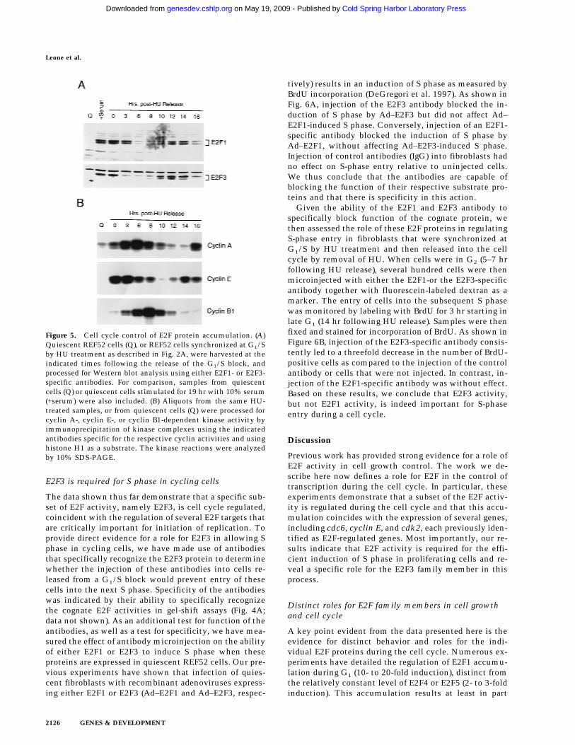
The decline in E2F1 and E2F3 DNA-binding activitiesreflects post-transcriptional regulation (Fig. 2B) and, atleast for E2F1 activity, is consistent with previous workthat has demonstrated an ability of cyclin A/cdk2 tobind to the amino-terminus of the E2F1 protein, phos-phorylate the associated DP1 protein specifically, andresult in the inactivation of the E2F1 DNA-binding ac-tivity (Krek et al. 1994; Xu et al. 1994; Krek et al. 1995;Dynlacht et al. 1997). The fact that the E2F3 proteinshares the cyclin A/cdk2-binding motif suggests thatthis E2F activity may be regulated similarly. Indeed, thekinetics of the decline of E2F1 and E2F3 DNA-bindingactivity as cells pass through S phase coincides with theaccumulation of cyclin A-dependent kinase activity, andthe reappearance of E2F3 activity in the next G1 followsthe decline in cyclin A-dependent kinase activity aftercells pass through G2/M (Fig. 5B).
Additional work has shown that the E2F1 protein issubject to ubiquitin-dependent degradation (Hateboer etal. 1996; Hofmann et al. 1996). Analysis of endogenousE2F1 and E2F3 protein levels by Western blot assays re-vealed a cyclic accumulation of these proteins (Fig. 5A)
Figure 3. Cell cycle regulation of Cyclin E,Cdk2, and Cdc6 protein accumulation. (A)Protein lysates (10 µg per lane) derived fromREF52 cells treated as in the HU arrest/re-lease experiments decribed in Fig. 2A weresubjected to 10% SDS-PAGE, Western blot-ted, and probed with either cyclin E- orcdk2-specific antibodies as indicated. (B)Protein samples (60 µg per lane) from HeLacells treated as in the thymidine arrest/re-lease experiment described in Fig. 2C weresubjected to 10% SDS-PAGE, transferred tonitrocellulose, and probed with antibodiesspecific to human Cdc6 protein.
Leone et al.
2124 GENES & DEVELOPMENT
Cold Spring Harbor Laboratory Press on May 19, 2009 - Published by genesdev.cshlp.orgDownloaded from
that, at least for E2F3, coincides with the pattern of ac-cumulation of DNA-binding activity. Interestingly, al-though there was no reaccumulation of E2F1 DNA-bind-ing activity in the second cell cycle, the E2F1 protein didreaccumulate. Based on these results and previous ex-periments, we conclude that the accumulation of E2F1and E2F3 DNA binding activity during a cell cycle may
be governed by at least two events—the cyclic accumu-lation of cyclin A/cdk2 that affects DNA-binding activ-ity and the control of protein stability by the ubiquitin-dependent proteasome pathway. Moreover, additionalcontrol must provide specificity in preventing the reac-cumulation of E2F1 DNA-binding activity during G1/Sof proliferating cells.
Figure 4. Cell cycle control of E2F activity. (A)Nuclear extracts prepared at various times followingthe stimulation of quiescent REF52 cells (Q) wereassayed for E2F DNA-binding activity by electro-phoretic mobility-shift assays (EMSA) using an E2F-specific 32P-labeled DNA probe (left). Cells similarlystimulated were incubated with BrdU (10 µM), fixedat the indicated times, subsequently immuno-stained with BrdU-specific antibodies, and visual-ized by immunofluorescent microscopy. The per-centage of BrdU-positive cells at each time point isindicated below the DNA-shift gel. The nuclear ex-tract sample from the 20-hr time point presented atleft (G1/S sample) was incubated with either IgG-,E2F1-, E2F2-, or E2F3-specific antibodies prior to be-ing subjected similarly to EMSA (right). The E2F4-and E2F5-specific bands indicated on the DNA gel-shift have been identified similarly using specificantibodies against the respective proteins (data notshown). We have been unable to identify an E2F2-specific DNA-binding activity in these assays. (B)Nuclear extracts prepared at various times followingthe release of cells from an HU block as described inFig. 2A, as well as from quiescent (Q) REF52 cells,were assayed for E2F DNA-binding activity byEMSA using an E2F-specific 32P-labeled DNA probe(left). The position of the cells with respect to cellcycle is indicated below based on the FACS analysisshown in Fig. 2A. Cytoplasmic extracts preparedfrom the same time point samples were assayed for
E2F-binding activity by EMSA using the same E2F-specific probe (right). (C) Ref52 cells were brought to quiescence by serum starvationand then stimulated to grow by addition of fresh medium with serum. Samples were taken at the indicated times and processed forFACS analysis as described in Materials and Methods. (D) Nuclear extracts prepared at various times following serum stimulation andassayed for E2F DNA-binding activity by EMSA using an E2F-specific 32P-labeled DNA probe.
Cell cycle control of E2F3 activity
GENES & DEVELOPMENT 2125
Cold Spring Harbor Laboratory Press on May 19, 2009 - Published by genesdev.cshlp.orgDownloaded from
E2F3 is required for S phase in cycling cells
The data shown thus far demonstrate that a specific sub-set of E2F activity, namely E2F3, is cell cycle regulated,coincident with the regulation of several E2F targets thatare critically important for initiation of replication. Toprovide direct evidence for a role for E2F3 in allowing Sphase in cycling cells, we have made use of antibodiesthat specifically recognize the E2F3 protein to determinewhether the injection of these antibodies into cells re-leased from a G1/S block would prevent entry of thesecells into the next S phase. Specificity of the antibodieswas indicated by their ability to specifically recognizethe cognate E2F activities in gel-shift assays (Fig. 4A;data not shown). As an additional test for function of theantibodies, as well as a test for specificity, we have mea-sured the effect of antibody microinjection on the abilityof either E2F1 or E2F3 to induce S phase when theseproteins are expressed in quiescent REF52 cells. Our pre-vious experiments have shown that infection of quies-cent fibroblasts with recombinant adenoviruses express-ing either E2F1 or E2F3 (Ad–E2F1 and Ad–E2F3, respec-
tively) results in an induction of S phase as measured byBrdU incorporation (DeGregori et al. 1997). As shown inFig. 6A, injection of the E2F3 antibody blocked the in-duction of S phase by Ad–E2F3 but did not affect Ad–E2F1-induced S phase. Conversely, injection of an E2F1-specific antibody blocked the induction of S phase byAd–E2F1, without affecting Ad–E2F3-induced S phase.Injection of control antibodies (IgG) into fibroblasts hadno effect on S-phase entry relative to uninjected cells.We thus conclude that the antibodies are capable ofblocking the function of their respective substrate pro-teins and that there is specificity in this action.
Given the ability of the E2F1 and E2F3 antibody tospecifically block function of the cognate protein, wethen assessed the role of these E2F proteins in regulatingS-phase entry in fibroblasts that were synchronized atG1/S by HU treatment and then released into the cellcycle by removal of HU. When cells were in G2 (5–7 hrfollowing HU release), several hundred cells were thenmicroinjected with either the E2F1-or the E2F3-specificantibody together with fluorescein-labeled dextran as amarker. The entry of cells into the subsequent S phasewas monitored by labeling with BrdU for 3 hr starting inlate G1 (14 hr following HU release). Samples were thenfixed and stained for incorporation of BrdU. As shown inFigure 6B, injection of the E2F3-specific antibody consis-tently led to a threefold decrease in the number of BrdU-positive cells as compared to the injection of the controlantibody or cells that were not injected. In contrast, in-jection of the E2F1-specific antibody was without effect.Based on these results, we conclude that E2F3 activity,but not E2F1 activity, is indeed important for S-phaseentry during a cell cycle.
Discussion
Previous work has provided strong evidence for a role ofE2F activity in cell growth control. The work we de-scribe here now defines a role for E2F in the control oftranscription during the cell cycle. In particular, theseexperiments demonstrate that a subset of the E2F activ-ity is regulated during the cell cycle and that this accu-mulation coincides with the expression of several genes,including cdc6, cyclin E, and cdk2, each previously iden-tified as E2F-regulated genes. Most importantly, our re-sults indicate that E2F activity is required for the effi-cient induction of S phase in proliferating cells and re-veal a specific role for the E2F3 family member in thisprocess.
Distinct roles for E2F family members in cell growthand cell cycle
A key point evident from the data presented here is theevidence for distinct behavior and roles for the indi-vidual E2F proteins during the cell cycle. Numerous ex-periments have detailed the regulation of E2F1 accumu-lation during G1 (10- to 20-fold induction), distinct fromthe relatively constant level of E2F4 or E2F5 (2- to 3-foldinduction). This accumulation results at least in part
Figure 5. Cell cycle control of E2F protein accumulation. (A)Quiescent REF52 cells (Q), or REF52 cells synchronized at G1/Sby HU treatment as described in Fig. 2A, were harvested at theindicated times following the release of the G1/S block, andprocessed for Western blot analysis using either E2F1- or E2F3-specific antibodies. For comparison, samples from quiescentcells (Q) or quiescent cells stimulated for 19 hr with 10% serum(+serum) were also included. (B) Aliquots from the same HU-treated samples, or from quiescent cells (Q) were processed forcyclin A-, cyclin E-, or cyclin B1-dependent kinase activity byimmunoprecipitation of kinase complexes using the indicatedantibodies specific for the respective cyclin activities and usinghistone H1 as a substrate. The kinase reactions were analyzedby 10% SDS-PAGE.
Leone et al.
2126 GENES & DEVELOPMENT
Cold Spring Harbor Laboratory Press on May 19, 2009 - Published by genesdev.cshlp.orgDownloaded from

from the transcriptional derepression of the E2F1 gene ascells leave a quiescent state (Johnson et al. 1994). It isalso evident from the studies presented here that E2F3behaves in a similar manner during the transitionthrough the initial G1. Both E2F1 and E2F3 activitiesthen decline as cells pass through S phase, likely result-ing from cyclin A/cdk2-mediated phosphorylation. Thedichotomy of behavior of E2F1 and E2F3 as cells con-tinue to cycle and enter the next G1 is particularly strik-ing since E2F3 DNA-binding activity reaccumulates inthe succeeding G1, whereas E2F1 activity does not reap-pear after the initial G1. The underlying basis for thisdifferential control is unclear, although it does not ap-pear to involve differences in translational or stabilitycontrol since the steady-state accumulation of E2F1 andE2F3 proteins during the cell cycle is quite similar. Onepossibility might relate to control by Rb, as our prelimi-nary experiments suggest that Rb associates preferen-tially with E2F1. Possibly, there is sufficient Rb availableonce cells have entered a cell cycle to titrate the avail-able E2F1 but, because of differential affinities, this levelof Rb is insufficient to prevent the accumulation of E2F3activity.
The differential regulation of E2F1 and E2F3 duringthe cell cycle, whereby E2F3 continues to cycle but E2F1does not reappear after the accumulation during the ini-tial G1/S, is also interesting in light of experiments thatdefine a role for E2F1 as a signal for apoptosis. Previousexperiments have shown that E2F1-mediated inductionof S phase is frequently followed by apoptosis, largelydependent on p53 (Qin et al. 1994; Shan and Lee 1994;Wu and Levine 1994; Kowalik et al. 1995). Intriguingly,our recent experiments have shown that this activity isunique to the E2F1 protein, despite the fact that other
E2F family members also induce S phase (DeGregori etal. 1997). Thus, the fact that E2F1 DNA-binding activityaccumulates as cells are stimulated to grow and re-entera cell cycle, but does not accumulate once cells are in acell cycle, suggests the possibility that E2F1 plays a roleas a growth checkpoint, ensuring that cell cycle re-entryhas properly occurred. Once the cells are then growing inthe presence of growth factors, this checkpoint might nolonger be critical and only the cyclic accumulation ofE2F3 activity represents the E2F requirement.
A central role for E2F in control of DNA replicationactivities
Based on a large volume of work directed at understand-ing E2F function, it is now clear that this transcriptionalactivity plays a key and central role in the activation ofgenes that encode DNA replication activities. Indeed,considering the fact that the group of E2F-regulatedgenes now includes those encoding deoxynucleotide en-zymes (DHFR, RR, TK, TS), DNA synthetic enzymes(DNA Pol a, PCNA), and proteins that mediate the rec-ognition and utilization of replication origins (Orc1,Cdc6, Mcms), it appears that E2F transcriptional activitymay coordinate the accumulation of most if not all of theessential activities necessary for DNA replication. Theresults presented here also suggest that the role for E2Fgoes beyond the coordination of production of the activi-ties as cells re-enter the cell cycle from a quiescent statebut also involves the coordination of control of severalrate-limiting activities that dictate G1/S control duringthe cell cycle of proliferating cells. In particular, the factthat various studies point to an essential role for Cdc6 aswell as cyclin E/cdk2 in S-phase induction, together
Figure 6. Inhibition of E2F3 activity inhibitsthe cell cycle induction of S phase. (A) Quies-cent REF52 cells were infected with eitherAd–E2F1, Ad–E2F3, or Ad–Con (m.o.i. of 100,200, and 100, respectively). Cells were micro-injected 4 hr postinfection with IgG, or withE2F1- or E2F3-specific antibodies (at an anti-body concentration of 1 µg/ml, containingfluorescein-conjugated dextran as a markerfor detecting injected cells). BrdU was added12 hr later (10 µM) and cells were incubated anadditional 4 hr prior to fixation and immuno-staining with BrdU-specific antibodies. Mi-croinjected cells were visualized by fluores-cent microscopy and the percentage of cellsstaining positively for BrdU is presentedabove. Approximately 150–250 cells were mi-croinjected in two separate experiments, arepresentative experiment is shown. (−) Thequantitation of uninjected cells in the sametissue-culture plate. (B) REF52 cells were syn-
chronized by HU treatment as described in Fig. 2A, and 5–7 hr following the release from the G1/S block, a time when cells arepredominantly in G2, cells were microinjected with IgG, or either with E2F1- or E2F3-specific antibodies. BrdU was then added to thecells 14 hr after the HU release and incubated for a further 3 hr, after which cells were fixed, immunostained, and quantitated for BrdUincorporation as in A. Approximately 150–250 cells were microinjected for each condition, and a representative experiment (similarresults were obtained in five independent experiments) is presented. (−) The quantitation of uninjected cells in the same tissue cultureplate.
Cell cycle control of E2F3 activity
GENES & DEVELOPMENT 2127
Cold Spring Harbor Laboratory Press on May 19, 2009 - Published by genesdev.cshlp.orgDownloaded from

with the finding that the synthesis of these proteins doesindeed oscillate during the cell cycle coincident withE2F3 activity, strongly suggests that E2F control is criti-cal for this event.
Various lines of evidence point to roles for Cdc6, aswell as cyclin E/cdk2, as rate-limiting activities for ini-tiation of DNA replication. Likewise, our previous workhas shown that the accumulation of E2F activity in oth-erwise quiescent cells can lead to an induction of Sphase. The fact that Cdc6, cyclin E, and cdk2 are E2Ftargets, and that their expression is regulated during thecell cycle in parallel with E2F3 accumulation, suggeststhat the E2F-dependent control of Cdc6, together withcyclin E and cdk2, may well represent a rate-determiningevent for initiation of DNA replication. Our preliminaryexperiments have shown that whereas expression ofCdc6 alone is not sufficient to induce S phase, expressionof Cdc6 together with that of cyclin E/cdk2 does induceS phase in otherwise quiescent cells (G. Leone, J. DeGre-gori, R.S. Williams, Z. Yan, and J. Nevins, unpubl.).
Importance of transcriptional and post-transcriptionalcontrol in the cell cycle
In principle, cell cycle control of Cdc6 accumulation, aswell as cyclin E/cdk2 accumulation, could be achievedwithout transcription control. A constant level of themRNAs, and thus constant synthesis of the proteins,coupled with protein degradation during mitosis, wouldresult in oscillation of the activities during the cell cycle.Nevertheless, our observations suggest additional com-plexity whereby a tight interrelationship in the controlof E2F3, cyclin E/cdk2, and cyclin A/cdk2 is evident.Based on the experiments presented here, we suggestthat the expression of Cdc6, cyclin E, and cdk2, which isseen to fluctuate during the cell cycle, is regulated, atleast in part, by the cyclic accumulation of E2F3 tran-scriptional activity. The accumulation of cyclin A/cdk2during S phase would lead to the elimination of E2F3(and E2F1) DNA-binding activity. This, together withthe subsequent degradation of these proteins, would leadto a decline in the expression of a subset of E2F targetgenes. As cells progress through G2 and mitosis, degra-dation of cyclin E and cyclin A, leading to the decline inthe associated kinase activities, would then reset theclock. Continued synthesis of E2F3 results in a reaccu-mulation of E2F activity in the following G1, inductionof cyclin E and Cdc6 synthesis, and induction of S phase.This process continues as long as the cell is growing inthe presence of growth factors. We suggest that tran-scriptional regulation by E2F of the key initiation activi-ties such as Cdc6 and cyclin E/cdk2 provides an addi-tional level of control of the accumulation of these ac-tivities, enhancing the magnitude that would result frompost-transcriptional control alone.
Materials and methods
Cells and viruses
Viral stocks were created as described previously (Schwarz et al.
1995), and the virus was purified by CsCl density-gradient cen-trifugation as described (Nevins et al. 1997). Viral titers weredetermined by an indirect immunofluorescent assay specific forthe viral 72-kd E2 gene product as described (DeGregori et al.1995a) and defined as focus forming units (FFU) per ml. Theconstruction of the recombinant viruses Ad–E2F1, Ad–E2F2,Ad–E2F3, and Ad–Con (a control virus, previously termedAdMb or Ad–CMV, lacking a cDNA insert) have been described(DeGregori et al. 1997).
REF52 cells were grown in DMEM containing 10% serum(5% fetal bovine serum and 5% calf serum). To bring cells toquiescence, cells were plated at ∼6500 cells/cm2, or at 3000cells/cm2 for the HU-block/release experiments, and incubatedovernight. The next day, the cells were washed once withDMEM and the culture medium replaced with DMEM contain-ing 0.25% serum. Cells were incubated further for 36 hr prior tovirus infection or serum stimulation.
Where indicated, cells were subsequently serum stimulatedby replacement with media containing 10% serum. For HU-block/release experiments, quiescent cells were stimulated for21 hr with 10% serum containing 2 mM HU, washed twice withDMEM, and refed with media containing 10% serum (t = 0).
For infections with recombinant adenoviruses, quiescentREF52 cells on plates were infected in DMEM with 20 mM
HEPES at pH 7.2 for 75 min at 37°C at a cell-to-volume ratio of0.5 × 106 cells/ml (0.5 ml for a 35-mm plate, 2 ml for a 100-mmplate, or 5 ml for a 150-mm plate). Following infection, fourvolumes of media containing 0.25% or 10% serum (indicated as+serum) was added to each plate, and the cells were incubated at37°C (DeGregori et al. 1995a).
HeLa cells were plated at a density of 1 × 10⁶ cells/100-mmplate (Northern and Western blotting analysis) or 3 × 10⁶ cells/150-mm plate (nuclear run-on assays) in DMEM supplementedwith 10% fetal bovine serum (complete growth medium) andgrown at 37°C, 5% CO2, for 24 hr. To obtain synchronized cellpopulations, cells were blocked by adding 2 mM thymidine tocomplete growth medium for 16 hr. To release the cells fromG1/S arrest, cultures were washed three times and then incu-bated in prewarmed complete growth medium.
Flow cytometry and BrdU incorporation assays
Cell synchrony was assessed by flow cytometry (Smith et al.1996). BrdU incorporation was determined as described previ-ously (DeGregori et al. 1995b).
Nuclear and cytoplasmic extract preparation
REF52 cells on tissue-culture plates were washed twice withPBS and scraped (in PBS) into microcentrifuge tubes placed inice. Cells were pelleted and resuspended in 10 volumes of hy-potonic lysis buffer (10 mM HEPES at pH 7.5, 10 mM KCl, 3 mM
MgCl2, 0.05% NP-40, 1 mM EDTA, 10 mM NaF, 0.1 mM NaVO4,1 mM PMSF, 1 mM DTT, 1 µg/ml aprotinin, 1 µg/ml leupeptin,10 mM b-glycerophosphate) by pipetting up and down 10 times,and incubated on ice for 30 min. Nuclei were pelleted at 500gfor 5 min at 4°C, and an equal volume of 2× gel-shift lysis buffer(90 mM HEPES at pH 7.9, 0.5 M KCl, 0.15% NP-40, 0.2 mM
EGTA, 20% glycerol, 10 mM NaF, 0.1 mM NaVO4, 1 mM PMSF,1 mM DTT, 1 µg/ml aprotinin, 1 µg/ml leupeptin, 10 mM b-glycerophosphate) was added, mixed, and stored at −70°C (cy-toplasmic fraction). The nuclei were washed once with 10 vol-umes of hypotonic lysis buffer and repelleted. Nuclei were thenlysed in 10 volumes of gel shift buffer (50 mM HEPES at pH 7.9,250 mM KCl, 0.1 mM EDTA, 0.1 mM EGTA, 0.1% NP-40, 10%glycerol, 10 mM NaF, 0.1 mM NaVO4, 1 mM PMSF, 1 mM DTT,
Leone et al.
2128 GENES & DEVELOPMENT
Cold Spring Harbor Laboratory Press on May 19, 2009 - Published by genesdev.cshlp.orgDownloaded from
1 µg/ml aprotinin, 1 µg/ml leupeptin, 10 mM b-glycerophos-phate) on ice for 30 min. Lysates were then spun at full speed ina microcentrifuge for 10 min at 4°C. The supernatants werethen stored at −70°C (nuclear fraction).
E2F DNA-binding assays
E2F assays were performed as described previously (Ikeda et al.1996). Supershift analysis was carried out as previously de-scribed (Ikeda et al. 1996) using antibodies specific against E2F1(SC-251x), E2F2 (SC-633x), E2F3 (SC-878x), or IgG as control(Santa Cruz Biotechnologies).
Kinase assays
Kinase assays were performed as described previously (DeGre-gori et al. 1995) using histone H1 as a substrate.
Northern analysis
Northern analysis of RNA from REF52 cells was performed asdescribed (DeGregori et al. 1995a). For HeLa cell experiments,total RNA was purified using Tri-Pure reagent (Boehringer Mann-heim) according to the manufacturer’s protocol. Thirty micro-grams of total RNA was separated by agarose gel electrophoresisunder denaturing conditions, transferred to nylon membranes,and probed under condition of high stringency with Cdc6 cDNAradiolabeled with 32P as described previously.
Western analysis
REF52 cell lysates (nuclear or cytoplasmic extracts) containingequal amounts of protein were boiled for 5 min in proteinsample buffer and subjected to SDS-PAGE on 10% polyacryl-amide gels. Proteins were transferred onto PVDF membrane asdescribed previously (Ikeda et al. 1996), and the PVDF mem-brane was blocked in TBS (25 mM Tris at pH 7.4, 137 mM NaCl,2.7 mM KCl) containing 10% skim milk for 2 hr. Blots were thenincubated with primary antibodies in TBS containing 5% skimmilk overnight at 4°C, and washed subsequently in TBS con-taining 0.1% Tween 20 for 30 min. Blots were then incubated inTBS containing 5% skim milk and secondary antibodies for 1 hrat room temperature, and then washed for 30 min. Blots wereprocessed with Amersham’s ECL system as described by themanufacturer. Antibodies against E2F1 (SC-251), E2F3 (SC-879),cyclin E (SC-481), and cdk2 (SC-163) were from Santa Cruz Bio-technologies.
HeLa cells were lysed in buffer containing 50 mM HEPES atpH 7.6, 50 mM NaCl, 0.1% SDS, 2 mM EDTA, 2 mM EGTA, and1% NP-40. Whole-cell lysates were mixed immediately withprotein gel-loading buffer and boiled for 3 min. Sixty micro-grams of total protein was separated on 10% SDS-polyacryl-amide gels, transferred to nitrocellulose membranes, and probedwith a polyclonal anti-Cdc6 antibody (Williams et al. 1997) oran anti-actin antibody (Boehringer Mannheim).
Nuclear run-on transcription assays
Nuclei were prepared from HeLa cells by lysis in buffer contain-ing 10 mM Tris at pH 7.4, 10 mM NaCl, 5 mM MgCl2, and 0.5%NP-40, as described (Ferrell 1993). RNA transcripts from 1 × 10⁷
nuclei were extended in the presence of [32P]UTP (Amersham),purified with Tri-Pure reagent (Boehringer Mannheim), and hy-bridized to cDNA (Cdc6 or bactin) immobilized on nylon mem-branes. Cdc6 transcription rate was calculated as the ratio ofradioactivity bound to Cdc6 cDNA relative to that bound to
b-actin cDNA, and values from cells synchronized at specificstages of the cell cycle were compared to values from asynchro-nously growing HeLa cells.
Microinjection
REF52 cells were plated on 35-mm2 plates, treated as indicatedin the legend to Fig. 6, and HEPES (pH 7.9) was added to 30 mM
prior to microinjection. Antibodies at a concentration of 1 µg/ml, containing 1% fluorescein-conjugated dextran (PierceChemical) were microcentrifuged for 15 min (to remove aggre-gates) and injected into the cytoplasm of ∼150–250 cells perplate using an Eppendorf microinjection system with femtotipsneedles (Eppendorf). Antibodies specific for E2F1 (SC-251L) andE2F3 (SC-879L) were from Santa Cruz Biotechnologies, and rab-bit anti-mouse IgG (as control) was from Cappel Labs. Cellswere then incubated at 37°C and BrdU was added as indicated inthe legend to Fig. 6. Cells were washed once with PBS and fixedfirst with 4% paraformaldehyde (in PBS) for 15 min at roomtemperature and subsequently with methanol/acetone (1:1) for10 min. Fixed cells were washed once with PBS and incubatedwith 2 N HCl for 20 min, washed three times with PBS, andfurther incubated in 1% BSA/PBS (in 1% BSA/PBS) for 10 min.Cells injected with E2F1-specific antibodies were preblockedwith 100 µg/ml rabbit anti-mouse IgG in 1% BSA/PBS for 45min, washed extensively, and then incubated with1% BSA/PBSfor an additional 5 min. Cells were then incubated for 1 hr atroom temperature with anti-BrdU Solution (Amersham, cat. no.RPN-202), washed three times with PBS and once with 1%BSA/PBS, and then incubated with rhodamine-conjugated goatanti-mouse IgG (1:75, Boehringer-Mannheim), washed exten-sively, and visualized by immunofluorescence microscopy.
Acknowledgments
We thank Mike Cook and Lynn Martinek of the Duke Compre-hensive Cancer Center Flow Cytometry Facility for flow cyto-metric analysis, Hiroshi Kimura for the generous gift of plas-mids encoding the Mcm proteins, and Kaye Culler for assistancein the preparation of the manuscript. G.L. is supported by afellowship from the Medical Research Council of Canada andthe Alberta Heritage Foundation for Medical Research. Thiswork was supported by a grant from the National Institutes ofHeralth (HL06296) to R.S.W.
The publication costs of this article were defrayed in part bypayment of page charges. This article must therefore be herebymarked ‘‘advertisement’’ in accordance with 18 USC section1734 solely to indicate this fact.
References
Coleman, T.R., P.B. Carpenter, and W.G. Dunphy. 1996. TheXenopus Cdc6 protein is essential for the initiation of asingle round of DNA replication in cell-free extracts. Cell87: 53–63.
DeGregori, J., T. Kowalik, and J.R. Nevins. 1995a. Cellular tar-gets for activation by the E2F1 transcription factor includeDNA synthesis and G1/S regulatory genes. Mol. Cell. Biol.15: 4215–4224.
DeGregori, J., G. Leone, K. Ohtani, A. Miron, and J.R. Nevins.1995b. E2F1 accumulation bypasses a G1 arrest resultingfrom the inhibition of G1 cyclin-dependent kinase activity.Genes & Dev. 9: 2873–2887.
DeGregori, J., G. Leone, A. Miron, L. Jakoi, and J.R. Nevins.1997. Distinct roles for E2F proteins in cell growth control
Cell cycle control of E2F3 activity
GENES & DEVELOPMENT 2129
Cold Spring Harbor Laboratory Press on May 19, 2009 - Published by genesdev.cshlp.orgDownloaded from
and apoptosis. Proc. Natl. Acad. Sci. 94: 7245–7250.Donovan, S., J. Harwood, L.S. Drury, and J.F.X. Diffley. 1997.
Cdc6p-dependent loading of Mcm proteins onto pre-replica-tive chromatin in budding yeast. Proc. Natl. Acad. Sci.94: 5611–5616.
Dutta, A. and S.P. Bell. 1997. Initiation of DNA replication ineukaryotic cells. Annu. Rev. Cell. Dev. Biol. 13: 293–332.
Dynlacht, B.D., K. Moberg, J.A. Lees, E. Harlow, and L. Zhu.1997. Specific regulation of E2F family members by cyclin-dependent kinases. Mol. Cell. Biol. 17: 3867–3875.
Ferrell, R.E. 1993. Analysis of nuclear RNA. In RNA method-ologies, pp. 235–253. Academic Press, San Diego, CA.
Hateboer, G., R.M. Kerkhoven, A. Shvarts, R. Bernards, and R.L.Beijersbergen. 1996. Degradation of E2F by the ubiquitin-proteasome pathway: Regulation by retinoblastoma familyproteins and adenovirus transforming proteins. Genes &Dev. 10: 2960–2970.
Helin, K. and E. Harlow. 1993. The retinoblastoma protein as atranscriptional repressor. Trends Cell Biol. 3: 43–46.
Hofmann, F., F. Martelli, D.M. Livingston, and Z. Wang. 1996.The retinoblastoma gene product protects E2F-1 from degra-dation by the ubiquitin-proteasome pathway. Genes & Dev.10: 2949–2959.
Holthoff, H.P., H. Hameister, and R. Knippers. 1996. A novelhuman Mcm protein: Homology to the yeast replication pro-tein Mis5 and chromosomal location. Genomics 37: 131–134.
Hunter, T. 1997. Oncoprotein networks. Cell 88: 333–346.Hunter, T. and J. Pines. 1994. Cyclins and cancer II: Cyclin D
and CDK inhibitors come of age. Cell 79: 573–582.Ikeda, M.-A., L. Jakoi, and J.R. Nevins. 1996. A unique role for
the Rb protein in controlling E2F accumulation during cellgrowth and differentiation. Proc. Natl. Acad. Sci. 93: 3215–3220.
Johnson, D.G., J.K. Schwarz, W.D. Cress, and J.R. Nevins. 1993.Expression of transcription factor E2F1 induces quiescentcells to enter S phase. Nature 365: 349–352.
Johnson, D.G., K. Ohtani, and J.R. Nevins. 1994. Autoregula-tory control of E2F1 expression in response to positive andnegative regulators of cell cycle progression. Genes & Dev.8: 1514–1525.
Kiyono, T., M. Fujita, Y. Hayashhi, and M. Ishibashi. 1996.Cloning of a cDNA encoding a human homologue ofCDC47, a member of the MCM family. Biochim. Biophys.Acta. 1307: 31–34.
Kowalik, T.F., J. DeGregori, J.K. Schwarz, and J.R. Nevins. 1995.E2F1 overexpression in quiescent fibroblasts leads to induc-tion of cellular DNA synthesis and apoptosis. J. Virol.69: 2491–2500.
Krek, W., M.E. Ewen, S. Shirodkar, Z. Arany, W.G. Kaelin, andD.M. Livingston. 1994. Negative regulation of the growth-promoting transcription factor E2F-1 by a stably bound cyc-lin A-dependent protein kinase. Cell 78: 161–172.
Krek, W., G. Xu, and D.M. Livingston. 1995. Cyclin A-kinaseregulation of E2F-1 DNA binding function underlies sup-pression of an S phase checkpoint. Cell 83: 1149–1158.
Nevins, J.R. 1992. E2F: A link between the Rb tumor suppressorprotein and viral oncoproteins. Science 258: 424–429.
Nevins, J.R., J. DeGregori, L. Jakoi, and G. Leone. 1997. Func-tional analysis of E2F. Methods Enzymol. 283: 205–219.
Newlon, C.S. 1997. Putting it all together: Building a prerepli-cative complex. Cell 91: 717–720.
Ohtani, K., J. DeGregori, G. Leone, D.R. Herendeen, T.J. Kelly,and J.R. Nevins. 1996. Expression of the HsOrc1 gene, a hu-man ORC homolog, is regulated by cell proliferation via theE2F transcription factor. Mol. Cell. Biol. 16: 6977–6984.
Qin, X.-Q., D.M. Livingston, W.G. Kaelin, and P.D. Adams.1994. Deregulated transcription factor E2F-1 expressionleads to S-phase entry and p53-mediated apoptosis. Proc.Natl. Acad. Sci. 91: 10918–10922.
Schwarz, J.K., C.H. Bassing, I. Kovesdi, M.B. Datto, M. Blazing,S. George, X.-F. Wang, and J.R. Nevins. 1995. Expression ofthe E2F1 transcription factor overcomes type b-transforminggrowth factor-mediated growth suppression. Proc. Natl.Acad. Sci. 92: 483–487.
Shan, B. and W.-H. Lee. 1994. Deregulated expression of E2F-1induces S-phase entry and leads to apoptosis. Mol. Cell. Biol.14: 8166–8173.
Sherr, C.J. 1996. Cancer cell cycles. Science 274: 1672–1677.Smith, E.J., G. Leone, J. DeGregori, L. Jakoi, and J.R. Nevins.
1996. The accumulation of an E2F-p130 transcriptional re-pressor distinguishes a G0 from a G1 cell state. Mol. Cell.Biol. 16: 6965–6976.
Stillman, B. 1996. Cell cycle control of DNA replication. Sci-ence 274: 1659–1664.
Tanaka, T., D. Knapp, and K. Nasmyth. 1997. Loading of anMcm protein onto DNA replication origins is regulated byCdc6p and CDKs. Cell 90: 649–660.
Tsuruga, H., N. Yabuta, S. Hosoya, K. Tamura, Y. Endo, and H.Nojima. 1997. HSMCM6: A new member of the humanMCM/P1 family encodes a protein homologous to fissionyeast Mis5. Genes Cells 2: 381–399.
Weinberg, R.A. 1995. The retinoblastoma protein and cell cyclecontrol. Cell 81: 323–330.
Williams, R.S., R.V. Shohet, and B. Stillman. 1997. A humanprotein related to yeast Cdc6p. Proc. Natl. Acad. Sci.94: 142–147.
Wu, X. and A.J. Levine. 1994. p53 and E2F-1 cooperate to me-diate apoptosis. Proc. Natl. Acad. Sci. 91: 3602–3606.
Xu, M., K.-A. Sheppard, C.-Y. Peng, A.S. Yee, and H. Piwnica-Worms. 1994. Cyclin A/CDK2 binds directly to E2F-1 andinhibits the DNA-binding activity of E2F-1/DP-1 by phos-phorylation. Mol. Cell. Biol. 14: 8420–8431.
Yan, Z., J. DeGregori, R.V. Shohet, G. Leone, B. Stillman, J.R.Nevins, and R.S. Williams. 1998. Cdc6 is regulated by E2Fand is essential for DNA replication in mammalian cells.Proc. Natl. Acad. Sci. 95: 3603–3608.
Leone et al.
2130 GENES & DEVELOPMENT
Cold Spring Harbor Laboratory Press on May 19, 2009 - Published by genesdev.cshlp.orgDownloaded from