e erimental brain researche.guigon.free.fr/rsc/article/lacquanitisoechting86b.pdf1 istituto di...
TRANSCRIPT
Exp Brain Res (1986)61:482--496 E erimental Brain Research �9 Springer-Verlag 1986
EMG responses to load perturbations of the upper limb: effect of dynamic coupling between shoulder and elbow motion
F. Lacquaniti t and J.F. Soechting 2
1 Istituto di Fisiologia dei Centri Nervosi, CNR, Milan, Italy 2 Laboratory of Neurophysiology, University of Minnesota, Minneapolis, MN 55455, USA
Summary. Load perturbations were applied to the arm of human subjects under conditions where both limb segments (upper arm and forearm) were free to move. The perturbations consisted of pulses of torque 50 ms in duration and of pseudo-random sequences of such pulses. They were applied to either the forearm or the upper arm. Under all conditions, the perturbations resulted in angular motion at the shoulder and elbow joints and evoked consistent responses in muscles acting about these joints (biceps, triceps, anterior and posterior deltoid). Activity in biceps and triceps was not related simply to angular motion at the elbow joint. For example, activation of biceps could be evoked during elbow flexion (by applying a torque perturbation at the shoulder) as well as during elbow extension (by applying a torque perturbation at the elbow)i The effect of varying degrees of dynamic coupling between upper arm and forearm on EMG responses was investigated by applying torque perturbations to the upper arm over a wide range of elbow angles. When the forearm is extended, such a perturbation induces a greater amount of elbow flexion than when the forearm is in a flexed position. The results of these experiments showed that the larger was the amount of flexion of the forearm induced by the perturbation, the larger was the activation of biceps. The results are incompatible with the notion of a negative feedback of total muscle length as being responsible for the EMG activity following the load perturbations. It is suggested that the EMG responses can best be interpreted functionally in terms of parameters more global than muscle length. Among such global parameters, changes in net torque at a joint resulting from the perturbation gave
Offprint requests to: J.F. Soechting, Department of Physiology, University of Minnesota, Minneapolis, MN 554.55, USA
the best correlation with the pattern of EMG activities observed.
Key words: Load perturbations - Multijointed limb - Feedback control
Introduction
According to the original description by Liddell and Sherrington (1924), the stretch reflex represents the reflex contraction of a functionally isolated muscle which is passively extended. They extrapolated from their observations to suggest that the CNS has at its disposal a mechanism to regulate the length of a muscle in the face of external perturbations. Among the subsequent refinements of the original idea, Merton's proposal (1951) that the reflex loop com- prising muscle spindles and spinal motoneurons func- tions as a length-servo, the spindles providing nega- tive feedback of muscle length, was one of the most notable. Modifications to the length-servo hypothesis have been introduced by a number of authors (Phil- lips 1969; Evarts 1973; Matthews 1981; Terzuolo et al. 1981; Stein 1982). A different hypothesis has also been put forward according to which muscle stiffness is the variable regulated by the reflex loop (Nichols and Houk 1976).
In general, emphasis has been placed mostly on physical parameters which pertain to a single muscle (i.e. muscle length, its rate of change, force, stiff- ness) as both the input and the controlled parameters of the system (Stein 1982). Furthermore, the stretch reflex has been more or less tacitly subsumed as the basic element of control even for whole limb motion (Houk and Rymer 1981). However, several observa- tions concerning relatively unconstrained behavior have begun to accumulate which cannot be readily

483
reconciled with the opera t ions of the classical stretch reflex. Examples are p rov ided by studies on the responses to pe r tu rba t ions dur ing postura l stabiliza- t ion (Nashner 1976; 1977), l ocomot ion (Wand et al. 1980) and speech p roduc t ion (Abbs and Gracco 1984). In n o n e of these studies is the response of a given muscle re la ted simply to changes in its length. Fu r the rmore , in some of these ins tances a stretch reflex as described classically would in fact be de- stabilizing (Nashner 1981). I npu t f rom receptors beside muscle spindles is d e e m e d to be involved in the genera t ion of such responses ( W a n d et al. 1980; Nashner 1981). F r o m a func t iona l po in t of view, these studies indicate that the responses are corre- lated with var iables which are more global than the length of the pe r tu rbed muscle. Perhaps the clearest indicat ion comes f rom the s tudy of A b b s and Gracco (1984), whose data suggest that in response to load per tu rba t ions of the lower lip, bo th uppe r and lower lip muscles are act ivated to control the in ter labia l distance, which is a critical pa r ame te r for speech
product ion. Uppe r l imb stabi l izat ion appears also to involve
responses to load pe r tu rba t ions which are no t simply related to changes in jo in t angular posi t ion. In a previous no te (Lacquani t i and Soecht ing 1984) we showed that E M G responses in e lbow muscles to applied forces depended on both e lbow and shoulder mot ion when the whole l imb was free to move. W e suggested that such behav ior might in fact represen t a response to the ne t change in to rque abou t the elbow, this la t ter pa r ame te r be ing re la ted to the sum of all the forces, external and in terna l , acting at the joint and represent ing a global pa rame te r in the sense ou t l ined above.
In this paper we shall describe in more detail the responses of e lbow and shoulder muscles to load per turba t ions unde r condi t ions in which the whole l imb is free to move.
Methods
Experimental setup
Subjects were seated with theirright upper arm approximately vertical, their forearm semipronated and at different inclinations according to the task. Force perturbations in a sagittal plane were delivered to either the forearm or the upper arm by means of a DC torque motor as shown schematically in Fig. 1. The limb segment was coupled to the motor through a flexible low-compliance steel cable running on a pulley and wound on a flywheel rotating with the motor shaft. The cable was connected to a molded brace which was fitted to the subject's arm or forearm. The direction of the force could be varied by adjusting the position of the pulley. Two exemplifying situations are depicted in Fig. 1. In one case (solid line), clockwise rotation of the torque motor shaft produces a force
Shoulder
Tor oe Motor \ / and Flywheel
Fig. 1. Schematic of the experimental setup. Force perturbations were applied to the arm by means of cable, passing over a pulley and wound around the hub of a flywheel which was attached to the shaft of a torque motor. By adjusting the height of the pulley, forces perpendicular to the upper arm (solid Line) or to the forearm (dashed line) could be exerted. Elbow angle (q5) was measured goniometrically. Shoulder angle ((9) was computed from two points (S1 and $2) on the upper arm
in the backward direction on the upper arm; in the second case (dashed lines) a downward force on the forearm is produced. In both cases the pulleys are placed so that the force is orthogonal to the limb segment. A small constant preload was applied to maintain tension in the cable.
The angle of elbow flexion-extension (q~) was measured electrogoniometrically. Shoulder translation in the sagittal (XZ) plane and its rotation in that same plane ((3 angle) were derived from the positions of two points ($1 and $2) on the upper arm. They were recorded by means of ultrasound emitters and a system of three orthogonal linear microphones (Soechting 1984).
EMG activity of biceps, triceps (long head), and anterior and posterior deltoid was recorded by means of surface electrodes.
Experimental protocol
In a first series of 12 experiments (involving 5 subjects) we evaluated EMG responses to different combinations of angular motions at the shoulder and elbow. To this end, perturbing forces with different points of application and directions were delivered to the subjects at a consta~r~t initial position of their arm: upper arm flexed forward slightly (O ~ 10 ~ and the forearm approximately horizontal (q~ ~ 100~ The perturbations consisted of 50 ms duration torque pulses delivered by the motor at random times to the subjects. (The actual force applied to the arm did not change instantaneously, given the electrical time constant of the torque motor (5.5 ms) and the ,compliance between the motor and the arm.) Subjects were instlmcted to maintain the position of their limb approximately constant, i.e. to resist the perturbation. Data were sampled by a digital computer at a rate of 125 per second for kinematic data and 500/s for electromyographic activity.
A force applied to the upper arm can produce local changes in the pressure on the skin and muscles underlying the cuff. In order to test if such changes in pressure contributed to EMG responses (Wand et al. 1980), in two subjects the location of the cuff was changed, placing it on the forearm, distal to the points of insertion

484
Angle Shoulder
Torque
Ant. Delt.
A ;-/ Flexion
Extension
Angle l Elbow Flexion
Torque Extension
T r i c e p s ~ ,
0 2 0 0 4 0 0 ms 0 2 0 0 4 0 0 m s
Fig. 2A and B. Response to load perturbations on the forearm. Changes in shoulder angle (O), torque (23s), elbow angle (~) and torque (T~) in response to a force perturbation to the forearm in the downward.(A) and upward (B) directions, as indicated schematically above each panel, are shown. The perturbation consisted of a pulse 50 ms in duration, beginning at time 0. Averaged Inll-wave rectified EMG activity of anterior deltoid, biceps and triceps is also shown. The data are from two different subjects. Initial values of the parameters are: A - O = 25 ~ �9 = 110 ~ Ts = 8.3 N - m , T~ = 4.1 N - m , and B - O = 25 ~ �9 = 115 ~ Ts = 10.6 N - m , Te = 5.6 N- re . Scales (per divison) are: 10 ~ (O and r 20 N - m (T~), 5 N - m (Tr 15 ~tV (biceps in 2B), 30 ~tV (triceps), 50 ~tV (anterior deltoid and biceps in A)
of the biceps and triceps. The line of action of the force was then directed backwards, just below the elbow center of rotation. It thus resulted in limb motion equivalent to that produced by a force in the posterior direction applied directly to the upper arm (solid line, Fig. 1).
In two experiments a different type of perturbation was also used to evoke forward flexion at the shoulder. The subject was asked to oppose a constant, backward force applied to the upper arm. The load was then suddenly released in a stepwise fashion, resulting in forward flexion at the shoulder.
In a second series of 7 experiments (involving 3 subjects) we assessed the dependence of EMG responses on the initial elbow angle o f extension, which ranged from 60 ~ to 150 ~ . Backward forces were applied to the upper arm. In four experiments, these consisted of a pulse 50 ms in duration, while in three others, pseudo-random trains of pulses were applied. A 7 th order m-sequence (Davies 1970; O'Leary and Honrubia 1975; Agarwal and Gottlieb 1977; Dufresne et al. 1978; Kearney and Hunter 1983) with 127 binary elements, each of 24 ms duration, was used.
Data analysis
Data from all trials (10 to 20) for each experimental condition were averaged (i.e. elbow and shoulder angles, center of rotation of the shoulder and EMG activities, the latter being full-wave rectified for this purpose). In the case of pseudo-random perturbations, the average response to a single pulse of torque was computed for all
the parameters by cross-correlating the pulse sequence with each of them (Davies 1970; Dufresne et al. 1978). Such responses represent the first-order kernel, i.e. the linear part of the overall impulse response of the system. As a test of the adequacy of such responses to reproduce the overall changes in the output variables, the responses were convolved with the input pulse sequence. This linear convolution could account for about 60% to 70% of the variance of the measured output (Kearney and Hunter 1983; 1984).
The amplitude of the EMG responses to the perturbations was quantified in a standard fashion (cf. Gottlieb and Agarwal 1979; Soechting et al. 1981) by computing the mean amplitude over the first 100 ms after pulse onset. The baseline activity, defined as the mean level of EMG activity over the 100 ms preceding the pulse, was subtracted from the calculated amplitude.
Kinematic data were numerically differentiated (after double- sided exponential smoothing) to provide the angular velocity and acceleration of both shoulder and elbow. From these data, the torques acting, at the shoulder (Ts) and at the elbow (T~) were computed according to the following equations:
Ts = (L + I~-2Acos~)O - (IeAcosdg)~ - Asinag+ 2 + 2 A s i n ~ O + Bs in | Csin(O-~) (1)
Te = (I~-Acos~)O - I ~ + Asin@O z + Csin(q~-O) (2)
Is and I~ are the moments of inertia of the upper arm and forearm about their respective axes of rotation. These coefficients as well as A, B and C are constants which were computed on the basis of

485
-0
0 e-
GO
..El LU
Ang le
Acce l .
T o r q u e
A --/"
Ant.
Delt .
Ang le
Acce l .
T o r q u e
B i ceps
T r i c e p s ..... ~ ~ . . . . . . . . . . . ~ . . . .
0 2 0 0 4 0 0 ms
F lex ion
Extension
F lex ion
Extension
I B
~ x
N 0 200 4 0 0 ms
Fig. 3A and B. Response to external torque perturbation at the shoulder in the posterior direction. Force was applied to the upper arm (A) or to the forearm along a line passing close to the center of rotation of the elbow (B) as indicated schematically above each pan.el. External
o o 2 torque at shoulder and elbow is thus about the same in both instances. Scaling per division is: 10 (O and cP), 4000/s (O and O), 20 N - m (T~), 3 N - m (T~), 30 ~V (biceps and triceps) and 40 I~V (anterior deltoid)
anthropometric data (Evans 1961; Soechting and Lacquaniti 1981). Typical values for the coefficients are: Is = 0.40, I~ = 0.15 and A = 0.18 kg-m 2, B = 12 and C = 5 kg-m2/s 2. Equations (1) and (2) have been derived according to Newtonian mechanics (see Soechting and Lacquaniti 1981; Hollerbach and Flash 1982; Hoy and Zerulcke 1985 for details). As computed, T~ and T~ corre- spond to the net nonconservative torques in the flexor direction at each joint. They result from external forces applied to the arm distally to each joint as well as from forces generated by the active contraction and the passive visco-elastic forces of muscles acting about each joint. (Under static conditions, the torques Ts and Te are equal to the moments due to gravitational forces, which are given by the terms with coefficients B and C.)
Equations (1) and (2) make explicit the dynamic coupling which exists between shoulder and elbow motion. Thus for example, from equation (2) one can see that motion at the elbow can occur even when there is no net torque at the elbow (Te = 0). Furthermore, the amount by which the elbow accelerates as a result of a force applied to the upper arm is a function of the mean elbow angle cP. Neglecting all terms in equations (1).and (2).excePt the inertial terms (i.e. all terms except those in �9 and | one obtains for the situation when there is a torque T~ at the shoulder and no torque at the elbow:
fb ~ Ts(I~ - Acos~)/(IsI~ - A2cos2~ b) (3)
6 ~ T~Id(I~I~ - A2cos2~). (4)
(This assumption is reasonable under our experimental conditions
at the time of application of the pulse, since the initial variations in position and velocity are neg..ligible.) According to Eq. (3), at a mean elbow angle q~ of 135 ~ �9 is predicted to be ten times as large as when q5 is 45 ~ while ~J varies by much less.
Equations (1) and (2) are valid only if there is no translation at the shoulder joint. In some instances, however, an appreciable translation at the shoulder did occur (e.g. in Fig. 4). In such cases, torque at the shoulder and elbow is given by the following equations:
Y's= T& ~s(BcosO- Ccos(O-r ~(BsinO - Csin(O-ff~))/g (5)
T'e= Te + ~tsBcos(O - c#)/g-~sCsin(O- ~)/g (6)
where ~ and zs denote the X and Z components of the linear acceleration of the shoulder joint, g is the gravitational constant (9.8 m/s 2) and the coefficients B and C are the same as those in Eqs. (1) and (2).
Results
Perturbations on the forearm
W h e n a f o r c e is a p p l i e d to t h e f o r e a r m , a t o r q u e is
e x e r t e d a b o u t b o t h t h e e l b o w a n d t h e s h o u l d e r j o i n t ,
s i nce t h e r e is a n a p p r e c i a b l e l e v e r a r m r e l a t i v e t o

486
Angle Shoulder
Torque
Ant. Delt.
Post. Delt.
Angle Elbow
Torque
Biceps
Triceps
A f
1 Fie!ion
Extension
l Flexion
Extension
-B
0 200 400 ms 0 200 400 ms
Fig. 4.4. and B. External torque perturbations at the shoulder in the anterior direction. Data from 2 subjects are shown. Torque was computed including the effects of translation at the shoulder. Scales (per division) are: 10 ~ ((~ and q~), 20 N-m (T0), 5 N-m (T~), 15 ~V (triceps and posterior deltoid in A, biceps, triceps and anterior deltoid in B), 30 ~V (biceps in A, posterior deltoid in B) and 50 ~V (anterior deltoid in B)
both joints. The action of the torque as well as the resulting angular motion is in the same direction (i.e. flexion or extension) at both joints. Under these conditions, there is an activation of muscles which are stretched.
Figure 2 illustrates the two symmetrical cases when the force was directed downward (Fig. 2A) and upward (Fig. 2B), as indicated schematically at the top of the figure. Each panel shows the temporal changes in the indicated variables. The pulse pertur- bation (50 ms duration) was applied at time 0 and resulted in an initial change of both shoulder and elbow torques in the extensor direction in Fig. 2A and in the flexor direction in Fig. 2B. Torques were computed according to Eqs. (1) and (2) of Methods. They then represent the resultant of both external and internal (muscles and tendons) nonconservative forces. In this experiment, the predominant contribu- tion to the net initial changes of the torques was from the externally applied force.
The applied torques resulted in changes in shoul- der and elbow angular position which were initially in
the same direction (extension in Fig. 2A, flexion in Fig. 2B). Maximum angular displacement at the elbow was about 1.5 ~ in each instance and about 3 ~ at the shoulder. The maximum angular velocities were about 20~ at the elbow and 35~ at the shoulder. The E M G responses to the perturbations correlated well with the direction of the initial changes of both torque and angular motion at each joint. Thus, in Fig. 2A, where the perturbation is towards exten- sion, both anterior deltoid and biceps were activated at a latency of about 35 ms and peaked at 95 ms. Their activity subsequently decreased at about the same time that triceps activity began to increase. In Fig. 2B, where the perturbation tended to flex the arm, E M G changes were roughly reciprocal to those just described. Anterior deltoid and biceps activity was depressed starting at about 50 ms, with a minimum at 90 ms, while triceps was activated 75 ms after the onset of the perturbation with a peak at 135 ms. These latencies were estimated by eye from averaged data such as those presented in Fig. 2. Latencies were also estimated by calculating the t ime
487
Angle
Shoulder
Torque
Ant. Deltoid
Angle
Elbow
Torque
Biceps
Triceps
0 200 400 ms
F
E
F
E
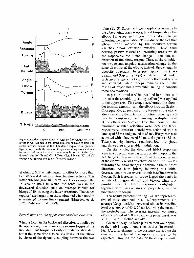
Fig. 5. Unloading step response. A constant force in the backward direction was applied to the upper arm and released at time 0 to evoke forward flexion at the shoulder. Torque, as in previous figures, represents the sum of torques resulting from external forces as well as active and passive muscle forces. Scales (per divison) are: 10 ~ (O and q)), 5 N-m (Ts), 1 N-m (T~), 30 gV (biceps and triceps) and 60 ~tV (anterior deltoid)
at which EMG activity began to differ by more than two standard deviations from baseline activity. This latter criterion gave similar values. (For example, the 15 sets of trials in which the force was in the downward direction gave an average latency for biceps of 40 ms using the latter criterion). The values reported are larger than those obtained when motion is restricted to one limb segment (Marsden et al. 1976; Dufresne et al. 1979).
Perturbations on the upper arm: shoulder extension
When a force in the backward direction is applied t o the upper arm, there results an extensor torque at the shoulder. This torque not only extends the shoulder, but at the same time also causes flexion at the elbow by virtue of the dynamic coupling between the two
joints (Eq. 3). Since the force is applied proximally to the elbow joint, there is no external torque about the elbow. However, net elbow torque does change following the perturbation. This is due to the fact that elbow flexion indnced by the shoulder torque stretches elbow extensor muscles. These then develop passive viscoelastic restoring forces which are responsible for a net change in the extensor direction of the elbow torque. Thus, at the shoulder net torque and angular acceleration change in the same direction; at the elbow, instead, they change in opposite directions. In a preliminary note (Lac- quaniti and Soechting 1984) we showed that, under such circumstances, both anterior deltoid and biceps are activated, while triceps remains silent. The results of experiments presented in Fig. 3 confirm these observations.
In part A, a pulse which resulted in an extensor torque at the shoulder (peaking at 60 ms) was applied to the upper arm. This torque accelerated the shoul- der towards extension and the elbow towards flexion. Consequently, as predicted, the torque at the elbow also changed in the extensor direction (peaking at 65 ms). In this instance, maximum angular displacement at the elbow was 5.7 ~ and 4 ~ at the shoulder. The maximum angular velocities were 95~ and 80%, respectively. Anterior deltoid was activated with a latency of 35 ms and peaked at 85 ms. Biceps was also activated with a latency of 30 ms and a peak at 55 ms. Triceps activity, instead, remained low throughout and showed no appreciable modulation.
On the whole, the described EMG responses were in a direction which is appropriate to oppose the net changes in torque. Thus both at the shoulder and at the elbow there was an activation of flexor muscles following the initial changes in torque in the extensor direction. At both joints, following this initial decrease, net torques overshot their baseline towards flexion. Such increases in torque lagged the peaks in activity of anterior deltoid and biceps. Thus it is possible that the E M G responses contributed, together with passive muscle properties, to this modulation in torque.
The results presented in Fig. 3A are representa- tive of those obtained in all 12 experiments. On average biceps activity increased above its baseline level at a latency of 40 + 10 ms following the onset of the perturbation. The average increase, computed over the period of 300 ms following pulse onset, was 33 + 13 % of baseline activity.
Given the way the force perturbation was applied to the limb in experiments such as that illustrated in Fig. 3A, local changes in the pressure exerted on the skin and muscles of the upper arm are to be expected. Thus, on the basis of these experiments,

488
Angle Shoulder Torque
Ant. Deltoid
Angle Elbow
Torque
Biceps
Triceps
A Flexion
Extension
, ~ 100~ Extensi~ l
Flexion Extension
. / /
0 200 400 ms O 200 400 ms
Fig. 6A and B. External torque perturbation at the shoulder in the posterior direction. In A, the initial elbow angle (q)) was 100~ the arm was more extended (150 ~ in Fig. 6B. Note the greater amount of forearm flexion in B resulting from the perturbation. Scales (per division) are: 10 ~ (O and qb), 20 N-m (Ts), 3 N-m (T~) and 30 ~V (anterior deltoid, biceps and triceps)
one can not exclude that the described E M G responses reflect such local mechanical events. To test for this possibility, in two experiments the force was applied on the forearm distally to the insertions of biceps and triceps. The line of action of the force passed just below the elbow's center of rotation and thus resulted in extensor torque at the shoulder and only a negligible amount of external torque at the elbow. Part B of Fig. 3 shows the results obtained in one such experiment. As anticipated, the overall changes induced by the perturbation in the kinema- tics and torques at the shoulder and elbow were similar to those described for Fig. 3A. (However, due to the different mechanical arrangements, their time courses are not superimposable). The qualita- tive behavior of the E M G responses at both the shoulder and elbow resembles that described previ- ously (Fig. 3A). Both anterior deltoid and biceps were activated at latencies comparable to those in Fig. 3A and both peaked at about 100 ms. In this instance, there was a small increase also in triceps activity, but this was not a consistent finding. Thus,
the pattern of biceps activity during elbow flexion induced by an external torque at the shoulder cannot be attributed to local changes in pressure on the upper arm.
Perturbations on the upper arm: shoulder f lexion
The response to perturbations resulting in forward flexion at the shoulder was also studied by applying a force to the upper arm in the anterior direction. In contrast with the previous experimental situation, there was a significant translation of the shoulder in this case. This asymmetry of behavior is presumably due to the biomechanics of the shoulder joint, forward flexion of the upper limb usually involving also some degree of forward translation due to motion in the scapulo-clavicular complex (Dempster 1965; Dvir and Berme 1978).
Figure 4 presents the results of two experiments in which a force in the anterior direction was applied to the upper arm. Since shoulder translation was not

489
neglible in this case, shoulder and elbow torques were estimated according to Eqs. (5) and (6). The changes in shoulder angular position (O) induced by this perturbation were not as pronounced as those which resulted from a force directed in the posterior direction (Fig. 3). In both subjects, anterior deltoid activity was depressed with a latency of 50 ms and a minimum at 95 ms in response to the initial changes towards flexion in angular position and torque at the shoulder resulting from the perturbation. A roughly reciprocal behavior was exhibited by posterior deltoid.
Flexor torque applied to the upper limb also resulted in elbow extension. The stretch of the elbow flexors in turn led to a net change in elbow torque towards flexion, again due to muscle visco-elastic properties. (On physical grounds, one would expect the initial change in elbow torque to be in the flexor direction; the initial small transient in the extensor direction in the calculated torque is probably due to uncertainties in estimating shoulder motion.) The initial changes in biceps activity in this experimental condition were not as clear-cut as those described in Fig. 3. In Fig. 4A, biceps exhibited a small increase above resting level over the first 80 ms following the onset of the perturbation, while in Fig. 4B there is an initial decrease followed by an increase. However, in all experiments the mean deviation of biceps activity from baseline over the first 100 ms after the onset of the perturbation was not significant statistically. To the contrary, the later changes in activity, namely a decrease in biceps activity with a minimum at 130 ms and activation of triceps with a latency of 100 ms and a maximum at 140 ms, were found consistently.
The lack of a clear-cut reciprocity of biceps responses in Figs. 3 and 4 might stem from the fact that the two experiments were also not reciprocal from a kinematic point of view, since shoulder translation was appreciable in the latter case but not in the former. Such translation can be reduced when there is a tonic contraction of shoulder muscles.
Subjects were asked to oppose an appreciable, constant load applied to the upper arm and acting in the posterior direction. The load was suddenly released in a step-wise-fashion. Consequently, the now unopposed flexor torque produced by the sub- ject resulted in forward flexion at the shoulder. Presumably due to the fixation of the shoulder joint by the active contraction of shoulder muscles, there was no appreciable translation at the shoulder. Thus torques were computed according to Eqs. (1) and (2).
Figure 5 shows the results of one such experi- ment. After the unloading of the arm, shoulder torque rapidly increased towards flexion due to the
unbalanced muscular contraction of shoulder flexors. Consequently, there was flexion at the shoulder and extension at the elbow, as in Fig. 4. Biceps and anterior deltoid activity decreased at the same time (latency of 30 ms) and reached a minimum at 75 ms. Their subsequent time course differed, however. Anterior deltoid activity remained well below its baseline level, consistent with the fact that there was no more loading at the shoulder. Biceps activity instead quickly returned to its resting level, Note that a tonic activation of biceps is required to oppose gravitational torque which remains approximately constant. Triceps activity remained low throughout.
In conclusion, when there was no translation at the shoulder, the biceps response associated with shoulder flexion and elbow extension was roughly reciprocal to that associated with shoulder extension and elbow flexion. This was not the case when there was translation at the; shoulder.
Relationship between EMG responses and initial elbow angle
So far, we have demonstrated that the EMG responses of biceps and triceps to a load perturbation which involves motion at the shoulder and elbow are poorly related to the changes in elbow angular position, per se. T]hus, for instance biceps was activated both when lflae elbow was extended due to an external torque applied to the forearm (Fig. 2) as well as when the elbow flexed as a result of torque applied at the shoul[der (Fig. 3). However, both biceps and triceps are biarticular muscles, spanning the shoulder joint as well as the elbow. Thus, shoulder and elbow ,extension will both result in a stretch of biceps. It is therefore possible that the responses in biceps described in Figs. 3-5 were dominated by the angular motion at the shoulder. One way to test this possibility is to vary the relative amounts of angular motion at the shoulder and elbow resulting from the perturbation. This can be done experimentally by taking advantage of the fact that the amount by which the elbow flexes by virtue of the dynamic coupling between forearm and upper arm is not constant, but depends on the elbow angle ~ . The more extended the elbow, the greater is its angular acceleration towards flexion following an extensor torque at the shoulder (see Eqs. 3 and 4 of Methods).
Representative results of an experiment where the same force was applied to the upper arm at two different angles q5 of elbow extension are shown in Fig. 6. The elbow was more extended in Fig. 6B (150 ~ vs. 100~ while shoulder angle O was the same in both cases (10~ The: same force perturbation was

490
- 1 5 0
A
~-1oo
- 5 0
oi
, , . . . . . . --"
0
-5(3
"O
- 1 0 0
I I I I - 1 5 ( 3
I | l
~ ' ~ .
A E i
z
1 5
1 0
5
I I l . . -- i
'~ 2 ! Z v
so l / / s
�9 #s I I~ 11
/ / l l
l / l / ] �9 / / / ' ~ e
I I I I
" O
.i..,
c~
C
6 0 0
4 0 0
2 0 0
I I I I
6 0 ~ 9 0 ~ 1 2 0 ~ 1 5 0 ~
M e a n E l b o w A n g l e
3 0 0
m 200 CL
O
i5 1 0 0
I I " \~e /e 1 J
t �9 �9
o L _ . _ ~ � 9
I i I I
6 0 ~ 9 0 ~ 1 2 0 ~ 1 5 0 ~
M e a n E l b o w A n g l e
Fig. 7. Dependence of response parame- ters on initial elbow angle. The data are from experiments in which a force in the backward direction was applied to the upper arm and elbow angle of extension (@) was varied. The plots show the maximum angular velocity at the shoul- der (6) in extension and at the elbow (6) in flexion, maximum change of torque at the shoulder (T+) and at the elbow (T~) in extension, and the integral (over 100 ms) of the amplitude of the response in anterior deltoid and biceps in arbitrary units. Data from four experi- ments are shown
applied in both instances, resulting in changes of torque and angular position of the shoulder which were very similar. For instance, extensor velocity at the shoulder peaked at the same time in both cases, with a value of 56~ in Fig. 6A and 61~ in Fig. 6B. The amplitude of the responses of anterior deltoid was also similar in the two cases.
As anticipated, the changes in elbow position and in elbow torque depended strongly on mean elbow angle. The perturbation resulted in a much larger amount of elbow flexion in Fig. 6B (maximum velocity of 86~ compared to Fig. 6A (49~ In Fig. 6A, the maximum angular displacement at the elbow was 2.6 ~ (85% of that at the shoulder), while it was 4.8 ~ (145% of shoulder angular displacement) when
the forearm was extended (Fig. 6B). Corresponding- ly, the initial changes in elbow torque in the extensor direction, due to the viscoelastic properties of elbow muscles, were about twice as large in Fig. 6B as in Fig. 6A. Biceps responses to the perturbation were also very different in the two cases. In both, there was an initial activation of biceps but its amplitude in Fig. 6B was about twice as much as in Fig. 6A.
The results obtained in this and three other experiments are summarized in a quantitative form in Fig, 7. Each experiment involved force perturbations applied to the upper arm at 3 or 4 different values of initial elbow angle, in the range of 70 ~ to 150 ~ . The values of three different parameters for the shoulder (left column) and elbow (right column) are plotted as

491
1 4 5 ~
r
1 1 5 ~
o 90 ~ e ~
m c 7 5 ~
6 0 ~
S h o u l d e r Angle
i = 1 = i i i i i t , r
A n t e r i o r De l to id
i i i t i i i i
1 4 5 ~
1 1 5 ~ <
O s~ 90 ~ m
C
7 5 ~
6 0 ~
E l b o w Angle E lbow Torque
F
. . . . . . . 40'0 0 200 4 0 0 ms 0 200 ms
B i c e p s
n i i i r i i i
0 200 4 0 0 ms
Fig. 8. Impulse response to torque perturbations at the shoulder. Pseudo-random perturbations were applied to the upper arm at varying amounts of mean extension of the forearm. Mean elbow angle (qo) is indicated next to each trace. The traces depict the impulse responses (average responses to a 24 ms duration pulse of force tending to produce backward extension at the shoulder and beginning at t = 24 ms) of O, ~ , Ts, Te and anterior deltoid and biceps. Flexion corresponds to an upward deflection of the traces of the angles and torques. Scales (per division) are: 1 ~ ( 0 and q)), 2 N - m (Ts), 0.5 N - m (Te), 10 ~V (anterior deltoid) and 5 lxV (biceps)
a function of the mean elbow angle. From top to bottom, these parameters are: peak angular velocity (~ and ~), peak torque (Ts and Te) and the mean amplitude (calculated over the 100 ms interval fol- lowing pulse onset) of anterior deltoid and biceps responses. Variability in the data points is clearly present. However, as might be expected, the para- meters for shoulder motion ((3 and Ts) showed no significant trend with changes in mean elbow angle. Instead, due to the different amount of dynamic coupling of the limb segments, both elbow velocity (in the flexor direction) and elbow torque (in the extensor direction) increased monotonically with increasing amounts of elbow extension (qs). As for the biceps response, despite the variability among different experiments, on average it increased as
mean elbow angle increased. Thus, on average, the greater the amount of elbow flexion induced by the perturbation, the larger the activation of biceps over the 100 ms following the onset of the perturbation.
The amplitude of biceps activation was positively correlated with elbow torque in the extensor direc- tion (correlation coefficient r greater than 0.8 in 3 of the 4 experiments). It should be noted, however, that the baseline level of biceps activity also increased significantly with increasing mean elbow angle �9 (see Fig. 6). The correlation coefficient between biceps amplitude and its mean, baseline level was better than 0.8 in these experiments. Thus, the amplitude of the response in biceps was positively correlated with both mean elbow angle and the mean level of biceps activity in these experiments, the coefficient of

492
- 3 0
,g
~, - 2 0 "O
�9 ~ - 1 0
�9 �9 �9 �9
J ! !
0
A
-~o
"o
-20
-30
.,,\ . .--...~
~�9
E I Z v
3
0 I I I !
0 . 6
= 0 . 4 Z v
I-| 0 . 2
I I I
1 5 0
"0 , I
o 1 0 0 ,+.*
a
.d ~" 5 0
0
" ~ ~ 1 7 6 1 7 6 �9 �9 �9 o - �9
I I I !
6 0 ~ 9 0 ~ 1 2 0 ~ 1 5 0 ~
M e a n E l b o w A n g l e
Q.
tO
i5
75 ,
5 0
2 5
: / I t i l
6 0 ~ 9 0 ~ 1 2 0 ~ 1 6 0 ~
M e a n E l b o w A n g l e
Fig. 9. Dependence of response parame- ters on mean elbow angle. The data were obtained using pseudo-random perturbations, as in Fig. 8. Variations in peak angular velocities, maximum changes in torques and integrals of the impulse responses of anterior deltoid and biceps with mean elbow angle �9 are shown
correlation being slightly larger for the latter para- meter.
In all the experiments we have reported so far, the perturbations were delivered at random times but their direction was known to the subject in advance. It is then possible that triggered reactions directed to oppose the perturbat ion contributed to the described E M G responses. Such triggered reactions may occur at moderately short latencies in response to single- choice paradigms when subjects are instructed to resist the perturbation. They are deemed to have neural substrates distinct from the stretch reflex (Houk 1977). In order to avoid the complications of such reaction-time situations, we also performed experiments using pseudo-random perturbations. Such perturbations have proved adequate to describe
myotatic responses of biceps and triceps when motion was restricted to the elbow (Dufresne et al. 1978; Soechting et al. 1981).
Figure 8 shows the results of one experiment in which pseudo-random perturbations were applied to the upper arm. The experiment includes 5 sets of trials in which the mean elbow angle differed (rang- ing from 60 ~ to 145~ The average responses to a pulse of torque lasting 24 ms and tending to extend the upper arm backwards are plotted for shoulder and elbow angles, torque and anterior deltoid and biceps activity. The time courses of the changes in both shoulder angle and torque were approximately independent of mean elbow angle and appeared qualitatively similar to those which have been described in Fig. 6. Also anterior deltoid responses

493
are approximately independent of mean elbow angle and are comparable to those obtained with single pulses of torque (Fig. 6). Their latencies are shorter however (30 ms on average) and maximum amplitude occurs at about 70 ms. These values are comparable to those obtained for biceps and triceps using pseudo-random perturbations when motion was restricted to the elbow joint (Dufresne et al. 1978).
Elbow flexion resulting from the dynamic coupl- ing with shoulder extension increases with increasing mean value of elbow angle ~ , as do the intial changes of elbow torque in the extensor direction. Thus both kinematic and dynamic changes at the elbow are consistent with those described in Fig. 6. Biceps activity also tends to be more strongly modulated at increasing levels of mean elbow extension. The maximum amplitude of the impulse response of biceps, over the first 100 ms after pulse onset increases progressively, in agreement with the results obtained using single pulses of torque. However, there are also appreciable changes in the shape of the response, with a single peak when mean elbow angle was 60 ~ and two distinct peaks in the response at larger angles of elbow extension.
Figure 9 summarizes quantitatively the results obtained in three experiments using pseudo-random perturbations. The same format as in Fig. 7 is used. As in the case of single pulse perturbations, neither angular velocity at the shoulder (0) , shoulder torque (T~) nor the amplitude of the response in anterior deltoid varied with the mean elbow angle ~P. Elbow angular velocity (6) and torque (Te) were instead clearly correlated with this parameter, as was the mean amplitude of the response of biceps in all three experiments. As in the experiments in which single pulses of torque were applied, biceps response amplitude increased significantly as the mean angle of elbow extension increased (correlation coefficient r between biceps amplitude and elbow torque was always greater than 0.8). However, in only one of the three experiments was the amplitude of the biceps responses correlated with the mean level of biceps activity (r > 0.6). In the other two experiments, the correlation coefficients were -0.15 and 0.31.
Thus, the amplitude of the biceps response to single pulses of torque and to pseudo-random pertur- bations increased as the forearm was more extended, i.e. as the perturbation resulted in a larger transient flexion at the elbow. The amplitude of the biceps responses was well correlated with mean biceps acitivty when single pulses of torque were used; this was not the case in the experiments utilizing pseudo- random perturbations. In both instances, the amplitude of the responses was highly correlated with
the amplitude of the changes in torque at the elbow resulting from the perturbation.
Discussion
We have described EMG responses in shoulder and elbow muscles following load perturbations applied to either the upper arm or to the forearm when both limb segments were free to move. Qualitatively, the responses to unpredictable perturbations consisting of pseudo-random pulses of torque were similar to those obtained using a predictable perturbation, namely single pulses of torque. In neither experiment was the direction of the response in elbow muscles uniquely related to the direction of angular motion at the elbow induced by the perturbation, but depended also on the direction of motion at the shoulder. Thus biceps was activated both during elbow extension in conjunction with shoulder extension (as when a force in the downward direction was applied to the fore- arm) as well as during elbow flexion together with shoulder extension (resulting from a force applied to the upper arm in the posterior direction).
Oppositely directed force perturbations resulted in oppositely directed EMG responses. Thus it is unlikely that the responses we have described repre- sent "shortening reactions", since such reactions would predict muscle activation irrespective of the direction of angular motion at a given joint (West- phal 1880; Sherrington 1909; Somojloff and Kisseleff 1927; Andrews et al. 1972). Furthermore, the responses in elbow rauscles cannot be explained on the basis of local changes in pressure on the limb segment resulting from the applied perturbation (Wand et al. 1980), since similar results were obtained when extensor torque at the shoulder was produced by applying a force to the upper arm (Fig. 3A) or to the forearm, aligned with the elbow center of rotation (Fig. 3]3). While in some cases there was a clear coactivation of biceps and anterior deltoid, this was not a general finding (Figs. 4 and 5). Also, the amplitudes of the responses in the two muscles were not well correlated (lSigs. 7 and 9). Thus, it appears difficult to ascribe the observed pattern of responses to fixed synergies between shoulder and elbow mus- cles (Nashner 1977; Soechting and Lacquaniti 1983).
Since both biceps and triceps span the glenohum- eral joint as well as the elbow joint, the dependence of the response of these two muscles on angular motion at both joints could reflect this anatomical arrangement. In fact, their muscle lengths will change as a result of angular motion at each of the two joints. Thus one has to deal with the possibility that the described responses in biceps and triceps, while not uniquely related to changes in elbow joint

494
angle, are nevertheless related to changes in overall muscle length. Biomechanical studies have shown that, given the anatomical arrangement of biceps, a 1 ~ change in elbow angle results in a change in biceps length about twice as large as that produced by a 1 ~ change in shoulder angle (Fick 1911). The amount of elbow flexion relative to shoulder extension resulting from a perturbation depends on the initial angle of elbow flexion (Figs. 7 and 9). In those experiments, the ratio of elbow flexion to shoulder extension ranged from 0.35 (~ = 60 ~ to 1.85 (q~ = 150~ Thus, at elbow angles of less than 90 ~ it is quite possible that biceps was stretched by a posteriorly directed force applied to the upper arm. Even admitting a large degree of uncertainty in Fick's data, such perturba- tions did however lead to an overall decrease in the length of biceps when the forearm was more extended (d# > 120~ The responses in biceps thus cannot be attributed to afferent input related to the overall length of biceps.
However, it is possible that even when the overall length of the muscle does shorten, some muscle fibers and more importantly, muscle spindle recep- tors in parallel with them, may lengthen. This can occur when motor units do not extend from tendon to tendon or when there is a compartmentalization of muscle fibers (cf. Botterman et al. 1983; Richmond et al. 1985). Given such an inhomogeneity in the structure of biceps, together with muscle spindle nonlinearities such as their increased sensitivity to stretch at longer lengths (Poppele et al. 1979), muscle spindle activity originating from biceps can poten- tially account for the observed EMG responses in biceps, provided one makes appropriate assumptions concerning the amount of reciprocal inhibition from elbow extensors. Furthermore, the possibility of muscle spindle feedback from shoulder muscles onto biceps cannot be excluded. For example, in the cat hindlimb there is a convergence of spindle afferents from knee flexors and hip extensors (Eccles et al. 1962). Also, given the latency of the responses, polysynaptic segmental and supraspinal loops involv- ing spindles and tendon organs cannot be excluded. In short, given present lack of knowledge concerning the ultrastructure of biceps in man, the distribution of motor units within that muscle and the organiza- tion of its afferent input, little can be said concerning the neural substrates for the observed behavior.
It may be appropriate, however, to consider what the functional utility of the observed responses may be. The question can be posed: which parameter or parameters are controlled by the EMG responses to a load perturbation under our experimental condition (Stein 1982) or more precisely, to which variables are the responses best related? Obviously, the changes in
biceps activity evoked by the perturbations are not well related to changes in elbow angle. For the reasons stated above, they also do not appear to be related to the change in overall length of biceps.
The possibility still remains that the responses in elbow muscles are related to a weighted sum of changes in shoulder and elbow angular motion (Aqb-KAO, or their derivatives). The results obtained by changing the mean elbow angle tend to argue against this interpretation, since increasing amounts of elbow flexion following the perturbation were associated with increasing amounts of activation of biceps (Figs. 6-9). In some of these experiments, the amplitude of the response of biceps was also correlated with the mean level of biceps activity. In this regard, it is known that when motion is restricted to a single joint, the amplitude of the response to muscle stretch is an increasing function of the mean level of activity in that muscle (Marsden et al. 1976; Gottlieb and Agarwal 1979; Dufresne et al. 1979), Thus, given this nonlinearity, the results obtained using single pulses of torque could be compatible with an input proportional to a weighted sum of shoulder and elbow angular motion, as suggested above. However, the same dependence of the response of biceps on mean elbow angle was observed using pseudo-random perturbations (Fig. 8), where response amplitude was not consistently correlated with mean EMG levels.
In view of all these considerations, the load perturbation responses described in this paper do not appear to be simply related to variables (such as length or its derivatives) representing the state of a single muscle. Rather, in analogy with the responses to perturbations during postural stabilization and speech production (Nashner 1981; Abbs and Gracco 1984), these responses seem to reflect the dynamical state 'of the limb in some global sense. One such global variable is the sum of all the torques acting about a joint. Such a net torque results from the action at a given joint of external forces as well internal forces due to the active contraction of muscles and their viscoelastic restoring forces.
Among the simple physical variables we have considered, changes in net torque gave the best correlation with the patterns of EMG responses. Thus, when net torque at the elbow initially changed towards extension biceps was activated at short latencies. Similarly there was a depression of biceps and activation of triceps when torque changed towards flexion as a consequence of the perturbation. Furthermore, in the experiments where mean elbow angle was changed, the amplitude of the biceps response was correlated positively with the amplitude of the initial changes in net torque at the elbow.

495
One needs to address the question whether or not responses related to changes in torque might be appropriate to oppose the perturbations in the tasks examined. This question cannot be answered conclu- sively at this time, since the theoretical foundations for the control of multiarticulate limb motion have not been developed fully (even in the field of robotics, Paul 1981; Johnson 1982). However, work- ers in this field have recognized the need for taking into account the effects of dynamic coupling between limb segments in implementing control schemes. Physiologically, note that activation of even monoar- ticular muscles at either the elbow or shoulder will produce angular motion at both joints. Similarly, an external perturbation applied to either limb segment will result in angular motion of both. Given this dynamic coupling, there are instances when, at the initiation of an intentional movement involving shoulder and elbow angular motion, there is elbow extension and activation of elbow flexors (Soechting and Lacquaniti 1981; Lacquaniti and Soechting 1982). Furthermore, also in tasks requiring modifica- tion of arm trajectories, the reaction time activation of elbow muscles was not related to the direction of changes in angular motion at the elbow required to alter the trajectory. On the contrary, the pattern of activation of muscles was congruent with the direc- tion of the changes in elbow torque required to correct the movement. Thus, dynamic coupling between shoulder and elbow motion is taken into account also in a reaction time situation (Soechting and Lacquaniti 1983).
Acknowledgment. This work was supported by USPHS grant NS-15018 and by NSF grant BNS-8117625.
References
Abbs J H, Gracco V L (1984) Control of complex motor gestures: orofacial muscle responses to load perturbations of lip during speech. J Neurophysiol 51:705-723
Agarwal G C, Gottlieb G L (1977) Compliance of the human ankle joint. J Biomech Eng 99:166--170
Andrews C J, Burke D, Lance J W (1972) The response to muscle stretch and shortening in Parkinsonian rigidity. Brain 95: 795-812
Bottermann B R, Harem T M, Reinking R M, Stuart D G (1983) Localization of monosynaptic Ia excitatory post-synaptic potentials in the motor nucleus of the cat biceps femoris muscle. J Physiol (London) 338:355-377
Davies W D T (1970) System identification for self-adaptive control. Wiley, London
Dempster W T (1965) Mechanisms of shoulder movement. Arch Plays Med Rehab 46:49-70
Dufresne J R, Soechting J F, Terzuolo C A (1978) Electromyo- graphic response to pseudo-random torque disturbances of human forearm position. Neuroscience 3:1213-1226
Dufresne J R, Soechting J F, Terzuolo C A (1979) Reflex motor output to torque pulses in man: identification of short- and long-latency loops with individual feedback parameters. Neuroscience 4:1493-1500
Dvir Z, Berme N (1978) The shoulder complex in elevation of the arm: a mechanism approach. J Biomech 11:219-225
Eccles J C, Eccles R M, Shealy C N (1962) An investigation into the effect of degenerating primary afferent fibers on the monosynaptic innervation of motoneurons. J Neurophysiol 25:544-558
Evans F G (1961) Biomechanical studies of the museulo-skeletal system. C C Thomas, Springfield, IL
Evarts E V (1973) Motor cortex reflexes associated with learned movement. Science 1'73:501-503
Fick R (1911) Anatomie und Mechanik der Gelenke, Vol 3. Gustav Fischer, Jena
Gottlieb G L, Agarwal G C (1979) Response to sudden torques about ankle in man: myotatic reflex. J Neurophysiol 42: 91-106
Hollerbach J M, Flash T (1982) Dynamic interactions between limb segments during planar arm movement. Biol Cybern 44: 67-77
Honk J C (1977) Participation of reflex mechanisms and reaction time processes in the compensatory adjustments to mechani- cal disturbances. In: Desmedt J E (ed) Cerebral motor control in man: long loop mechanisms. Prog Clin Neurophysiol 4: 193-215
Houk J C, Rymer W Z (1981) Neural control of muscle length and tension. In: Brookart J M, Mountcastle V B (eds) Handbook of physiology, Sect 1, Vol 2, Part 1. Bethesda, Am Physiol Soc, pp 257-324
Hoy M G, Zernicke R F (1985) Modulation of limb dynamics in the swing phase of locomotion. J Biomech 18:49-60
Johnson T L (1982) Feedback control. In: Brady M L e t al. (eds) Robot motion: planning and control. MIT Press, Cambridge; pp 127-146
Kearney R E, Hunter I W (1983) System identification of human triceps surae stretch reflex dynamics. Exp Brain Res 51: 117-127
Kearney R E, Hunter I W (1984) System identification of human stretch reflex dynamics: tibialis anterior. Exp Brain Res 56: 154-161
Lacquanifi F, Soechting J F (1982) Coordination of arm and wrist motion during a reac]hing task. J Neurosci 2:399-408
Lacquaniti F, Soechting J F (1984) Behavior of the stretch reflex in a multi-jointed limb. Brain Res 311:161-166
Liddell E G T, Sherrington C (1924) Reflexes in response to stretch (myotatic reflexes). Proc R Soc B 96:212-242
Marsden C D, Merton P A, Morton H B (1976) Stretch reflex and servo action in a variety of human muscles. J Physiol (Lond) 259:531-560
Matthews P B C (1981) Evolving views on the internal operation and functional role of the muscle spindle. J Physiol (Lond) 320:1-30
Merton P A (1951) The silent period in a muscle of the human hand. J Physiol (Lond) 114:183-198
Nashner L M (1976) Adapting reflexes controlling the human posture. Exp Brain R, es 26:59-72
Nashner L M (1977) Fixed patterns of rapid postural responses among muscles during stance. Exp Brain Res 30:13-24
Nashner L M (1981) Analysis of stance posture in humans. In: Towe A L, Luschei E S (eds) Handbook of behavioral neurobiology, Vol 5. Plenum, New York p 527-565

496
Nichols T R, Houk J C (1976) Improvement of linearity and regulation of stiffness that results from actions of stretch reflex. J Neurophysiol 39:119-142
O'Leary D P, Honrubia V (1975) On-line identification of sensory systems using pseudorandom binary noise inputs. Biophys J 15:505-532
Paul R P (1981) Robot manipulators: mathematics, programming, and control. MIT Press, Cambridge
Philips C G (1969) Motor apparatus of the baboon's hand. Proc R Soc London B 173:141-174
Poppele R E, Kennedy W R, Quick D C (1979) A determination of static mechanical properties of intrafusal muscle in isolated cat muscle spindles. Neuroscience 4:401-411
Richmond F J R, Mac GiUis D R R, Scott D A (1985) Muscle fiber compartmentalization in cat splenius muscles. J Neurophysiol 53:868-885
Samojloff A, Kisseleff M (1927) Die Verkfirzungs- und Ver- lgngerungsreaktionen des Knie-Extensors der decerebrierten Katze. Plfigers Arch 218:267-284
Sherrington C S (1909) On plastic tonus and proprioceptive reflexes. Q J Exp. Physiol 2:109-156
Soechting J F (1984) 1Effect of target size on spatial and temporal . characteristics of.,a pointing mowment4n man. Exp Brain Res
54:121-132
Soechting J F, Dufresene J R, Lacquaniti F (1981) Time-varying properties of myotatic response in man during some simple motor tasks. J Neurophysiol 46:1226--1243
Soechting J F, Lacquaniti F (1981) Invariant characteristics of a pointing movement in man. J Neurosci 1:710-720
Soechting J F, Lacquaniti F (1983) Modification of trajectory of a pointing movement in response to a change in target location. J Neurophysiol 49:548-564
Stein R B (1982) What muscle variable(s) does the nervous system control in limb movements? Behav Brain Sci 5:535-540
Terzuolo C, Soechting J F, Dufresne J R (1981) Operational characteristics of reflex response to changes in muscle length during different motor tasks and their functional utility. In: Pompeiano O, Ajmone Marsan C (eds) Brain mechanisms and perceptual awareness. New York, Raven Press pp 183-210
Wand P, Prochazka A, Sontag K.-H (1980) Neuromuscular responses to gait perturbations in freely moving cats. Exp Brain Res 38:109-114
Westphal C (1880) Ueber eine Art paradoxer Muskel-Komrak- tion. Arch. Psychiat Nervenkrankh 10:243-248
Received March 29, 1985 / Accepted October 14, 1985