dynamics of nucleosomes assessed with time-lapse high
TRANSCRIPT

Dynamics of Nucleosomes Assessed with Time-Lapse High-SpeedAtomic Force MicroscopyAtsushi Miyagi,† Toshio Ando,‡ and Yuri L. Lyubchenko*,†
†Department of Pharmaceutical Sciences, College of Pharmacy, University of Nebraska Medical Center, Omaha, Nebraska68198-6025, United States‡Department of Physics, Kanazawa University, Kanazawa 920-1192, Japan
*S Supporting Information
ABSTRACT: A fundamental challenge of gene regulation is the accessibilityof DNA within nucleosomes. Recent studies performed by various techniques,including single-molecule approaches, led to the realization that nucleosomesare quite dynamic rather than static systems, as they were once considered.Direct data are needed to characterize the dynamics of nucleosomes.Specifically, if nucleosomes are dynamic, the following questions need to be answered. What is the range of nucleosomedynamics? Is a non-ATP-dependent unwrapping of nucleosomes possible? What are the factors facilitating the large-scaleopening and unwrapping of nucleosomes? In previous studies using time-lapse atomic force microscopy (AFM) imaging, wewere able, for the first time, to observe spontaneous, ATP-independent unwrapping of nucleosomes. However, low temporalresolution did not allow visualization of various pathways of nucleosome dynamics. In the studies described here, we appliedhigh-speed time-lapse AFM (HS-AFM) capable of visualizing molecular dynamics on the millisecond time scale to study thenucleosome dynamics. The mononucleosomes were assembled on a 353 bp DNA substrate containing nucleosome-specific 601sequence. With HS-AFM, we were able to observe the dynamics of nucleosome on a subsecond time scale and visualize variouspathways of nucleosome dynamics, such as sliding and unwrapping to various extents, including complete dissociation. Thesestudies highlight an important role of electrostatic interactions in chromatin dynamics. Overall, our findings shed new light onnucleosome dynamics and provide a novel hypothesis for the mechanisms controlling the spontaneous dynamics of chromatin.
The dynamics of the nucleosome core particle (NCP), afundamental unit of chromatin, is a key property of
chromatin, allowing proteins involved in chromatin tran-scription or replication accessibility to DNA wrapped aroundthe histone core.1 Remodeling systems provide the accessibilityof DNA within nucleosomes,2 but this process can be facilitatedby the inherent dynamics of nucleosomes.3 Single-moleculeapproaches of various types were instrumental in character-ization of nucleosome dynamics. These methods includefluorescence resonance energy transfer,4−10 single-moleculeprobing,11,12 and atomic force microscopy (AFM) imag-ing.13−16 Single-molecule fluorescence and time-resolvedtechniques revealed that nucleosomes undergo local dissocia-tion of DNA in the absence of remodeling proteins,8,17−20 andthis process occurs on the subsecond time scale.19 Earlier, AFMimaging was used to characterize chromatin structure at thenanoscale level.21−23 The AFM sample preparation is so gentlethat AFM allows the study of chromatin structure omitting therather traditional glutaraldehyde fixation procedure of thesample.24 Note in this regard, recent studies using time-lapseobservations allowed direct observation of the dynamics andunwrapping of nucleosomes.25,26 The unwrapping of nucleo-somes proceeds from the ends of the particle, resulting inunwrapping of DNA regions as large as dozens of base pairs.This process may lead to a complete unfolding of nucleosomesand dissociation of the histone core from the complex.Nucleosome dissociation occurs in the absence of ATP-dependent protein systems involved in chromatin remodeling,
suggesting that the inherent dynamics of nucleosomes cancontribute to the chromatin unwrapping process. However,because of the slow data acquisition rate of time-lapse AFMtechniques, many dynamic events are missing. The advent ofhigh-speed AFM (HS-AFM)27 and recent advances in thistechnique28 made it possible to directly visualize dynamicprocesses, on the millisecond time scale, of site-specific DNA−protein complexes29,30 and nucleosomes,31 allowing theidentification of novel mechanisms of protein−DNA dynamics.Here we applied HS-AFM to image nucleosome dynamics.
The data demonstrate that nucleosomes undergo spontaneousunwrapping, occurring on the subsecond time scale. Althoughsite exposure is the predominant mode of nucleosomedynamics and unwrapping, transient sliding of the nucleosomealso takes place. The sliding process is facilitated in thepresence of the zwitterion detergent, CHAPS, allowing large-range nucleosome translocation and unwrapping. The unwrap-ping dynamics depends on the DNA sequence facilitating thesliding pathway for sequences with low specificity for bindinghistone cores. The role of intermolecular electrostaticinteractions in chromatin dynamics, dissociation, and unwrap-ping and the interaction with remodeling systems are discussed.
Received: June 20, 2011Revised: August 12, 2011
Article
pubs.acs.org/biochemistry
© XXXX American Chemical Society A dx.doi.org/10.1021/bi200946z |Biochemistry XXXX, XXX, XXX−XXX
■ MATERIALS AND METHODS
Preparation of Nucleosomal DNA. DNA used for thenucleosome assembly is similar to that used in previousstudies.26 DNA substrate was generated by polymerase chainreaction using plasmid pGEM3Z-601 as a template, whichencodes a high-affinity nucleosome positioning sequence 601.32
The 601 sequence (147 bp) was located inside the 353 bpDNA fragment with 127 and 79 bp arm DNA, so that themononucleosome reconstituted with the DNA substrate hastwo DNA arms that differ in length by 1.6-fold. A schematic forthe DNA substrate is shown in Figure S1 of the SupportingInformation.Histone Octamer Assembly and Purification. Histone
octamers were assembled as described in refs 26 and 33.Octamers were separated from tetramer and dimer fractions viasize-exclusion chromatography (SEC) with Superdex 200 PC3.2/30 columns (GE Healthcare) at 4 °C. SEC fractions wereanalyzed for purity and histone stoichiometry using sodiumdodecyl sulfate−polyacrylamide gel electrophoresis. Fractionscontaining histones H2A, H2B, H3, and H4 in approximatelyequal ratios were pooled and concentrated by centrifugation at10000g.Nucleosome Refolding. Nucleosomes were prepared as
described previously.26,34 Briefly, histone octamers and DNAcontaining the nucleosome positioning sequence were mixed inequimolar concentrations in 2 M NaCl and kept for 30 min atroom temperature. A series of dilutions was prepared by using10 mM Tris-HCl to produce final NaCl concentrations of 1,0.67, and 0.5 M. Diluted samples were kept at 4 °C for 1 hbefore being dialyzed against one change of volume of 0.2 MNaCl overnight. Nucleosomes were concentrated using aMicrocon centrifugal filter device (molecular weight cutoff of10000) at 7000g for 10 min at 4 °C and dialyzed against onechange of 200 mL of buffer containing 10 mM HEPES-NaCl(pH 7.5) and 1 mM EDTA for 3 h at 4 °C.Atomic Force Microscopy. The sample preparation
procedure for high-speed AFM was modified to accommodatethe instrument design requirements. Freshly cleaved mica wasmodified with a 167 μM solution of 1-(3-aminopropyl)silatrane(APS) to make APS-mica.26,35,36 APS-mica is a positivelycharged surface that binds DNA primarily electrostaticallyallowing AFM observation of segmental mobility of DNA in thefree state and in protein−DNA complexes;26,37,38 1.5 μL of anAPS solution (167 μM) was placed on the freshly cleaved mica(a disk ∼1.5 mm in diameter glued to the sample stage) for 30min. The surface was rinsed with 3 μL of milli-Q water 10 timesand with 3 μL of “imaging” buffer containing 10 mM HEPES(pH 7.5) and 4 mM MgCl2 five times. A droplet of 1.5 μL ofsolution containing the nucleosome sample was applied to theAPS-mica surface for 5 min and then rinsed with the imagingbuffer. The nucleosome sample was prepared from the stocksolution by dilution into 10 mM Tris-HCl (pH 7.5) and 4 mMMgCl2 buffer.The high-speed AFM instrument used in this work was
developed by the Ando group27,28 and manufactured by RIBM(Tsukuba, Japan). The nominal spring constant of thecantilevers was 0.1−0.2 N/m, and the resonance frequency inwater was between 400 and 1000 kHz. The scanning rate invarious experiments varied between 1 and 5.5 frames persecond.
We used BL-AC10DS-A2 cantilevers (Olympus) modified bythe electron beam deposition method and sharpened by using aplasma etcher.27,28,39
The processing of individual frames of HS-AFM files andconventional AFM images was performed using FemtoscanOnline (Advanced Technologies Center, Moscow, Russia).
■ RESULTS
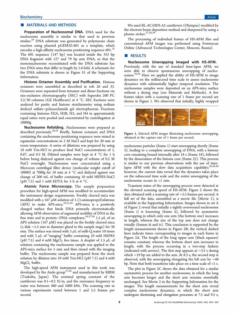
Nucleosome Unwrapping Imaged with HS-AFM.Previously, with the use of standard time-lapse AFM, wewere able to observe spontaneous unwrapping of nucleo-somes.26,36 Here we applied the ability of HS-AFM to imagedynamics on the millisecond time scale to assess nucleosomedynamics with substantially higher temporal resolution. Thenucleosome samples were deposited on an APS-mica surfacewithout a drying step (see Materials and Methods). A fewframes taken with a scanning rate of 1 frame per second areshown in Figure 1. We observed that initially, highly wrapped
nucleosome particles (frame 1) start unwrapping shortly (frame3), leading to a complete unwrapping of DNA, with a histonecore remaining bound internally after 18 s (frame 18), followedby the dissociation of the histone core (frame 25). This processis similar to our previous observations with the use of time-lapse AFM with the slow data acquisition capability;26,36,40
however, the current data reveal that the dynamics takes placeon the subsecond time scale and the entire unwrapping of thenucleosome occurs in <1 min.Transient states of the unwrapping process were detected at
the elevated scanning speed of HS-AFM. Figure 2 shows thedata obtained with a scanning rate of ∼5.5 frames per second. Afull set of the data, assembled as a movie file (Movie 1), isavailable in the Supporting Information. Images shown in set Aof Figure 2 reveal that initially a tightly overwound nucleosome(frame i) is loosening (frame ii), followed by asymmetricunwrapping in which only one arm (the bottom one) increasesin length, whereas the size of the top arm does not changevisually (frames iii and iv). This conclusion is supported by thelength measurements shown in Figure 2B; the vertical dashedlines indicate times corresponding to images in each frame inFigure 2A. The length of the long upper arm (black squares)remains constant, whereas the bottom short arm increases inlength, with the process occurring in a two-step fashion(indicated with arrows). The first step appears at ∼3.5 s duringwhich ∼19 bp are added to the arm. At 8.5 s, the second step isobserved, with the unwrapping elongating the left arm by ∼40bp. Note that both transitions take place on a time scale of <1 s.The plot in Figure 2C shows the data obtained for a similar
asymmetric process for another nucleosome, in which the longarm becomes longer and the short arm remains essentiallyunchanged. See Movie 2 in the Supporting Information for theimages. The length measurements for the short arm revealcomplex nucleosome dynamics, in which the short armundergoes shortening and elongation processes at 7.5 and 9.5 s,
Figure 1. Selected AFM images illustrating nucleosome unwrapping,obtained at the capture rate of 1 frame per second.
Biochemistry Article
dx.doi.org/10.1021/bi200946z |Biochemistry XXXX, XXX, XXX−XXXB

respectively. As in the previous graph, the unwrapping processinvolving dozens of DNA base pairs occurs fast, on thesubsecond time scale.Images in Figure 3A show details for the unwrapping process
(imaging rate of 3.3 frames per second). Initially, the wrappednucleosome (frame i) unfolds generating a loop (frame ii),followed by the loop unfolding (frame iii). The quantitativeanalysis of these data is shown in Figure 3B. The vertical dashedlines in the figure indicate the times of the images shown inFigure 3A. The long arm (squares) remains unchanged,whereas the short arm increases in length sharply at ∼12 s,followed by a gradual increase, reaching a length close to that ofthe left arm. The large set of images assembled as a movie file(Movie 3) is provided in the Supporting Information.Reversible Octamer Sliding in Specific Nucleosomes.
Although unwrapping is the primary process by whichnucleosomes can spontaneously dissociate, HS-AFM allowed
us to observe sliding. Three successive frames of a nucleosomeundergoing sliding are shown in Figure 4A, and a movie file(Movie 4) is available in the Supporting Information. Initially(frame 60s), the nucleosome has a short left arm and a longright arm. In the next image (frame 62s), the left arm is longerthan the right arm, but the situation reverses to the originalposition after 3 s (frame 65s). This data set shows that transientsliding can occur. The length measurement data are assembledin Figure 4B, demonstrating that this rather stable nucleosomeundergoes a fast transient translocation at 62 s. At this time,one arm increases in length accompanied by a decrease inlength of another arm by the same value. The process is thenfully reversed, resulting in the arms adopting their initiallengths. Note that the size of the nucleosome does not change.These data suggest that the nucleosome can undergo slidingthat is transient and reversible. The sliding of the histone corealong the DNA molecule occurs at a distance not exceeding 30nm and takes 3 s.Dynamics of the Nucleosome in the Presence of
CHAPS. We have shown recently that incubation of assemblednucleosomes in the presence of the detergent CHAPS resultedin the formation of nucleosomes with a histone core positionnot corresponding to the location of the specific 601sequence.40 Such nonspecific nucleosomes do not differ fromthe specific ones by the size of the nucleosome particles or thelength of DNA wrapped around the histone core. Wehypothesized that the nonspecific nucleosomes were formedfrom the specific ones by a sliding mechanism.40 We tested thishypothesis in this analysis by performing time-lapse imaging ofnucleosome dynamics in the presence of CHAPS.AFM images of a set of time-lapse frames are shown in
Figure 5A−E. Panel A shows the unprocessed raw image in
Figure 2. Time-lapse HS-AFM visualization of nucleosome unwrap-ping in which only one end of the DNA unwraps. (A) Set of AFMimages for the nucleosome in which the bottom arm increases in size;the initial image (i) and three frames with the same nucleosomecaptured after 4.2 (ii), 7.1 (iii), and 17 s (iv) are shown. (B) Timedependence of the lengths of the upper arm (arm 1, black squares) andbottom arm (arm 2, red diamonds) along with the overall DNA length(green triangles). The dashed lines correspond to the times the imagesshown in A were acquired, and the red arrows correspond to thestepwise unwrapping process. (C) Time dependence for anothernucleosome in which only the length of arm 1 increases. See panel Bfor the code. The data were obtained with a scan rate of 1 frame per353 ms.
Figure 3. Loop formation and unfolding. (A) Set of imagescorresponding to 8.7 (i), 14.7 (ii), and 17.1 s (iii). In panel ii, thepositions of the DNA dissociation and unlooping events are indicatedwith a white arrow. (B) Length measurements for the looping andloop unfolding process. The lengths of the left (arm 1) and right (arm2) arms and the total DNA length are shown with black squares, reddiamonds, and green triangles, respectively. In panel B, the dashedlines correspond to the image acquisition times shown in panel A. Thescan rate is 1 frame per 301 ms.
Biochemistry Article
dx.doi.org/10.1021/bi200946z |Biochemistry XXXX, XXX, XXX−XXXC

which the selected nucleosome is indicated with black dottedlines. The nucleosome was traced and placed on a blackbackground to produce panel B. Panels C−E that correspondto images at later times indicated in the figures were obtainedthe same way. According to the length measurements, theimage in panel B corresponds to the position of thenucleosome core at 601 sequences; we termed this a specificcomplex. Shortening of the small arm is clearly seen in panel C(20 s), and the nucleosome moves to the end of the DNAsubstrate in panel D (frame 32s). Note that visually, the size ofthe nucleosome remains unchanged during this repositioningevent. The size of the nucleosome decreases during furtherobservation, and the nucleosome dissociates completely in
panel E (frame 56s). Length measurements of the DNA armswere performed, and the corresponding graphs are shown inFigure 5F. The changes in length are detected starting at ∼15 s,in which a gradual decrease in the length of arm 2 isaccompanied with a corresponding increase in the length ofarm 1. The first sharp change in the lengths was observed at∼17 s. During the period of one frame, arm 1 increased inlength by ∼15 nm and arm 2 decreased in length by the samevalue. There were no substantial changes in lengths until 26 s,when the second stepwise change of arms occurred. At thisstep, the long arm 1 increases in length by ∼20 nm (blacksquares), accompanied by a decrease in the length of the shortarm 2 by half of this value, ∼10 nm (red circles). Given the factthat one DNA turn in the nucleosome corresponds to 84 bp or∼25 nm, this stepwise translocation of the nucleosome to theend is accompanied by the nucleosome losing approximatelythree-fourths of a turn due to unwrapping of long arm 1. Thisunwrapping process is partially compensated by rewrapping ofthe short arm, bringing back approximately one-third of thenucleosome turn. After this period, the length of the long arm 1increases gradually, leading to a complete unwrapping of thenucleosome and the dissociation of the core at 55 s.Figure 6 illustrates the unwrapping process of the nonspecific
nucleosome. See Movie 5 in the Supporting Information for thefull set of images. In the subset of images (Figure 6A), the firstframe (60 s after start) corresponds to the nucleosome withapproximately two nucleosomal turns. The next image (103 s)shows the same nucleosome after unwrapping of approximatelyone turn. Note that the short arm remains unchanged;therefore, unwrapping is associated with only elongation ofthe long arm. The unwrapping is almost complete at 167 s, andthe core dissociates at 177 s. The length measurement plot isshown in Figure 6B, and a plot of time versus the number ofDNA turns calculated from the arms’ lengths is shown in Figure6C. The vertical dashed lines in panels B and C correspond totimes at which images shown in Figure 6A were taken. Agradual initial unwrapping process occurs during the first 50 s,followed by the first step at ∼70 s during which ∼0.4nucleosome turn is lost. The largest unwrapping step occurs at∼100 s, accompanied by unwrapping of almost one nucleosometurn. The third step (∼140 s) leads to a complete unwrappingof the nucleosome.
Figure 4. Reversible sliding of the nucleosome. (A) Selected AFM images illustrating a reversible sliding. The vertical dashed line corresponds to thecenter of nucleosomal particle at 60 s. The numerals (60s, 62s, and 65s) correspond to the times the images were captured. (B) Dependence of thearms’ lengths (arm 1, black squares, and arm 2, red diamonds) and the total arms’ lengths (green triangles) on time. The scan rate is 1 frame persecond. The scale bar in panel A is 50 nm.
Figure 5. Irreversible sliding of the nucleosome in the presence ofCHAPS. (A−E) Set of HS-AFM images. In panel A, the black dottedlines indicate the DNA arms of interest. Panels B−E show the tracedimages of the molecule on a black background. (F) DNA arm lengthmeasurements. Black squares and red diamonds correspond to thelengths of the longer and shorter arms, respectively, in the first image.The green triangles show the time dependence of the total DNAlength. The data were acquired with thea scan rate of 1.7 frames persecond. The dashed lines in panel F correspond to the times at whichpanels B−D were acquired (from left to right, respectively).
Biochemistry Article
dx.doi.org/10.1021/bi200946z |Biochemistry XXXX, XXX, XXX−XXXD

■ DISCUSSION
Nucleosome Unwrapping at High Speed. Previously,we published the finding that nucleosomes are capable ofunwrapping spontaneously, as observed by AFM.26,36 In thetime-lapse AFM experiments, nucleosomes underwent astepwise unwrapping process in which each step could beobserved between two consecutive frames. The time resolutionunder those conditions was ∼1 min, implying that either theobserved dynamics was that slow or the kinetics on the 1 mintime scale were due to the low temporal resolution of theinstrument. High-speed AFM revealed that the unwrappingprocess is fast and complex; it is accompanied by a number ofsmall fast steps spanning ≤1 s. The experiments withsubsecond temporal resolution demonstrated that unwrappingof a segment as large as 10 nm (∼30 bp) can occur in one∼300 ms step (Figure 2B). The time-resolved fluorescence dataof Li et al.19 showed that the breathing dynamics ofnucleosomes occurs on the 50−250 ms time scale, but thesestudies were not able to measure the spatial range of thesefluctuations. Our HS-AFM data show that DNA segments aslarge as 30 bp dissociate on the 300 ms time scale. Importantly,
the images in Figure 3 are of high spatial resolution (the DNAwidth on the images is ∼3 nm). Furthermore, they reveal thatthe dissociation of an ∼30 bp segment (12 s) led to theformation of a loop with a small curvature followed by therelaxation of the loop and repositioning of the entire arm.Reversible Sliding. The dynamics of nucleosomes is
typically described by the site exposure model,1 according towhich DNA dissociates from the histone core by theunwrapping process. Our previous time-lapse AFM data26
were in line with this model. However, the high-temporalresolution capability of HS-AFM showed that in addition to thesite exposure pathway, the sliding pathway is possible. The datain Figure 4 graphically illustrate this capability. The fullywrapped nucleosome (approximately two nucleosomal turns)rolls quickly (∼1 s) in one direction over ∼38 bp (11 nm), afterwhich it returns to the original position in the same amount oftime, without any change in the number of nucleosomal turns,the size of the nucleosome or the angle between the arms.Thus, nucleosomes do slide, although nucleosome dynamics viathe exposure model is the predominant pathway.Dynamics of Nonspecific Nucleosomes and Sequence
Specificity. The majority of physical, chemical, and structuralstudies of nucleosomes were performed with the DNAsubstrate containing 601 sequence characterized by the highestspecificity for nucleosome formation.32 Indeed, using the vastmajority of our current and previous data,26,36 the nucleosomesare assembled at the 601 sequence leading to nucleosomalparticles with lengths of arms corresponding to the arm lengthsin Figure S1 of the Supporting Information. We have shownrecently that such high sequence specificity can be changed bythe incubation of already assembled nucleosomes in a solutioncontaining the zwitterionic detergent, CHAPS.40 As a result,nucleosome particles with alternative positions relative to the601 sequence are formed; we termed them nonspecificnucleosomes. Such nucleosomal particles differ from specificones only by the position of the nucleosome but have the samemean number of nucleosomal turns as specific particles. Wesuggested previously40 that nonspecific nucleosomes areformed from specific ones by sliding, and the data in thispaper confirm this hypothesis. According to Figure 5, thenucleosome slides by rolling over the short arm. The processprimarily occurs in a stepwise fashion, in which unwrapping ofthe long arm is accompanied by overwrapping of the short arm.The translocation occurs in the range of a second. In additionto the two-step process illustrated in Figure 5, sliding can occurin one step, demonstrated also by the set of data in Figure S2 ofthe Supporting Information.Interestingly, sliding is a one-directional process that
primarily occurs by the elongation of the long arm andshortening of the short arm. Note that for the AFMexperiments, we used positively charged surface interactions,which are a driving force for unfolding of nucleosomes.26,36
Therefore, the long arms bind more strongly to the surface thanthe short arms. Fluctuational unwrapping of the long armfurther increases the stability of its interactions with the surface,and wrapping of the short arm compensates for the loss of thenucleosome’s free energy. As the short arm becomes smaller,the nucleosome becomes capable of directional rolling (sliding)more efficiently. We did not observe this directionality for thespecific nucleosome for which the reversible roll was observed(Figure 4). We explain this observation by the effect of thespecific 601 sequence that provides the most thermodynami-
Figure 6. Unwrapping of the nonspecific nucleosome. (A) Selectedframes corresponding to a few stages of the unwrapping process.Times are shown in each frame. (B) Time dependence of the lengthsof the arms (squares and diamonds) and the total DNA length(triangles) measured for each frame. (C) Time dependence of thenumber of DNA turns in the nucleosome calculated from the arms’lengths. The data were obtained with a scan rate of 1.7 frames persecond. In panels B and C, the dashed lines correspond to the times offirst three images shown in A (from left to right, respectively).
Biochemistry Article
dx.doi.org/10.1021/bi200946z |Biochemistry XXXX, XXX, XXX−XXXE
cally favorable location for the nucleosome. Moreover, in ourprevious experiment, we demonstrated that unwrapping ofnucleosomes with the 601 sequence inside the DNA substrateoccurs symmetrically, suggesting that the central part of the 601sequence plays an important role in the sequence-specificpositioning of the nucleosome. Therefore, we hypothesize thatthe low probability of the sliding pathway observed here, as wellas in other studies with the use of the 601 sequence, is due toproperties of this sequence favoring one pathway over another.The directionality of sliding further supports our model of
the role of electrostatics in the stability and dynamics ofnucleosomes.26,36,40 According to this model, the strengthenedinteraction of the DNA arms with a positively chargedenvironment (the positively charged surface in our AFMexperiments) decreases the stability of the nucleosome. Wehypothesized, therefore, that the surface of chromatinremodeling factors accommodating the nucleosome could bepositively charged to facilitate nucleosome unfolding,26 and arecent study from the Kornberg lab41 supports this hypothesis.In addition to remodeling factors, chromatin interacts with thenuclear membrane that can also modulate chromatin structureand function. Characterization of these interactions isimportant for elucidating mechanisms controlling chromatinfunction. AFM as a topographic technique would be a methodof choice for such studies. Indeed, although AFM primarily usesmica as a substrate for the imaging and the properties of micaare different from the properties of intracellular surfaces,appropriate modification of the surface may mimic the surfacefeatures in vivo. Our studies use a positively charged APS-micasurface, but methods for preparing surfaces with othercharacteristics are being developed.Sequence specificity of nucleosome formation is widely
discussed in the literature (e.g., refs 42 and 43 and referencescited therein). We have shown recently40 and confirmed in thispaper that the stringency for the sequence specificity ofnucleosome positioning can be decreased by adding CHAPS tothe solution. Importantly, this detergent does not dissociate thenucleosome. On the contrary, it stabilizes the nucleosomesagainst dissociation at very low nucleosomal concentrations,facilitating the study of nucleosomes by single-moleculetechniques. This suggests that other compounds, in additionto CHAPS, can modulate the sequence-specific pattern ofnucleosome positioning. Therefore, the analysis of thesequence specificity of nucleosome positioning should takeinto account the possibility of changing this interaction viaenvironmental conditions. Although CHAPS is not aphysiological compound, its structure is rather similar to thestructure of cholesterol. We have shown that water-solublemodification of CHAPS stabilizes nucleosomes, although to alesser extent compared to unmodified CHAPS.40
Given the fact that AFM is a technique that directly probesthe sample, the effect of the scanning tip on nucleosomedynamics needs to be taken into consideration. The HS-AFMinstrument operates in tapping mode; therefore, we need toconsider the effect of two major factors: hitting the sample withan oscillating tip and displacement of the sample duringscanning. The energy transferred by the tip to the sample isproportional to the square of the oscillation amplitude. HS-AFM operates with an amplitude of ∼1 nm, so the estimatesprovided in the Supporting Information demonstrate that theoverall energy induced by the tip is equivalent to an increase intemperature of only ∼9.6 K. Given the fact that this energy is
distributed over the entire system, including water molecules inthe vicinity of the sample, this effect is weak. Another effect ofthe tip is the displacement of the sample. Because of the highoscillation frequency, the time during which the tip contacts thesample is ∼100 ns; therefore, during this period of time, thesample stage moves in the x-direction by only 0.16 nm. Thisvalue is comparable to the size of an atom, so the lateraldisplacement of the sample by the scanning tip is negligible.
■ ASSOCIATED CONTENT
*S Supporting InformationPotential effects of the instrument characteristics on the sampledynamics, schematics explaining the design of the DNAsubstrate, graph of the time-dependent change in the lengthof the short arm during nucleosome sliding, and movies ofanimated data sets of time-lapse images. This material isavailable free of charge via the Internet at http://pubs.acs.org.
■ AUTHOR INFORMATION
Corresponding Author*Department of Pharmaceutical Sciences, University ofNebraska Medical Center, 986025 Nebraska Medical Center,Omaha, NE 68198-6025. Phone: (402) 559-1971. Fax: (402)559-9543. E-mail: [email protected].
FundingThe work is supported by grants from the U.S. Department ofEnergy (DE-FG02-08ER64579), the North Atlantic TreatyOrganization (SfP 983204), the National Science Foundation(EPS-0701892), and the Nebraska Research Initiative (NRI, allto Y.L.L.).
■ ACKNOWLEDGMENTS
We thank Evgeny Menshikov, who participated in the HS-AFMexperiments, Irina Menshikova for the preparation of thenucleosome samples with CHAPS, L. Shlyakhtenko for valuablediscussions and insightful suggestions, and J. Widom and G.Narlikar for useful comments about the results.
■ ABBREVIATIONS
AFM, atomic force microscopy; NCP, nucleosome coreparticle; APS, 1-(3-aminopropyl)silatrane; CHAPS, 3-[(3-cholamidopropyl)dimethylammonio]-1-propanesulfonate.
■ REFERENCES
(1) Widom, J. (1997) Chromatin: The nucleosome unwrapped. Curr.Biol. 7, R653−R655.(2) Saha, A., Wittmeyer, J., and Cairns, B. R. (2006) Chromatin
remodelling: The industrial revolution of DNA around histones. Nat.Rev. Mol. Cell Biol. 7, 437−447.(3) Li, G., and Widom, J. (2004) Nucleosomes facilitate their own
invasion. Nat. Struct. Mol. Biol. 11, 763−769.(4) Lovullo, D., Daniel, D., Yodh, J., Lohr, D., and Woodbury, N. W.
(2005) A fluorescence resonance energy transfer-based probe tomonitor nucleosome structure. Anal. Biochem. 341, 165−172.(5) Leuba, S. H., Anand, S. P., Harp, J. M., and Khan, S. A. (2008)
Expedient placement of two fluorescent dyes for investigating dynamicDNA protein interactions in real time. Chromosome Res. 16, 451−467.(6) Blosser, T. R., Yang, J. G., Stone, M. D., Narlikar, G. J., and
Zhuang, X. (2009) Dynamics of nucleosome remodelling by individualACF complexes. Nature 462, 1022−1027.
Biochemistry Article
dx.doi.org/10.1021/bi200946z |Biochemistry XXXX, XXX, XXX−XXXF
(7) Gurunathan, K., and Levitus, M. (2009) Single-moleculefluorescence studies of nucleosome dynamics. Curr. Pharm. Biotechnol.10, 559−568.(8) Koopmans, W. J., Brehm, A., Logie, C., Schmidt, T., and van
Noort, J. (2007) Single-pair FRET microscopy reveals mononucleo-some dynamics. J. Fluoresc. 17, 785−795.(9) Kelbauskas, L., Chan, N., Bash, R., DeBartolo, P., Sun, J.,
Woodbury, N., and Lohr, D. (2008) Sequence-dependent variationsassociated with H2A/H2B depletion of nucleosomes. Biophys. J. 94,147−158.(10) Koopmans, W. J., Buning, R., Schmidt, T., and van Noort, J.
(2009) spFRET using alternating excitation and FCS revealsprogressive DNA unwrapping in nucleosomes. Biophys. J. 97, 195−204.(11) Zlatanova, J., and Leuba, S. H. (2003) Chromatin fibers, one-at-
a-time. J. Mol. Biol. 331, 1−19.(12) Brower-Toland, B. D., Smith, C. L., Yeh, R. C., Lis, J. T.,
Peterson, C. L., and Wang, M. D. (2002) Mechanical disruption ofindividual nucleosomes reveals a reversible multistage release of DNA.Proc. Natl. Acad. Sci. U.S.A. 99, 1960−1965.(13) Bussiek, M., Toth, K., Brun, N., and Langowski, J. (2005) DNA-
loop formation on nucleosomes shown by in situ scanning forcemicroscopy of supercoiled DNA. J. Mol. Biol. 345, 695−706.(14) Poirier, M. G., Bussiek, M., Langowski, J., and Widom, J. (2008)
Spontaneous access to DNA target sites in folded chromatin fibers. J.Mol. Biol. 379, 772−786.(15) Bussiek, M., Muller, G., Waldeck, W., Diekmann, S., and
Langowski, J. (2007) Organisation of nucleosomal arrays reconstitutedwith repetitive African green monkey α-satellite DNA as analysed byatomic force microscopy. Eur. Biophys. J. 37, 81−93.(16) Montel, F., Menoni, H., Castelnovo, M., Bednar, J., Dimitrov, S.,
Angelov, D., and Faivre-Moskalenko, C. (2009) The dynamics ofindividual nucleosomes controls the chromatin condensation pathway:Direct atomic force microscopy visualization of variant chromatin.Biophys. J. 97, 544−553.(17) Anderson, J. D., Thastrom, A., and Widom, J. (2002)
Spontaneous access of proteins to buried nucleosomal DNA targetsites occurs via a mechanism that is distinct from nucleosometranslocation. Mol. Cell. Biol. 22, 7147−7157.(18) Ahmad, K., and Henikoff, S. (2002) Epigenetic consequences of
nucleosome dynamics. Cell 111, 281−284.(19) Li, G., Levitus, M., Bustamante, C., and Widom, J. (2005) Rapid
spontaneous accessibility of nucleosomal DNA. Nat. Struct. Mol. Biol.12, 46−53.(20) Bucceri, A., Kapitza, K., and Thoma, F. (2006) Rapid
accessibility of nucleosomal DNA in yeast on a second time scale.EMBO J. 25, 3123−3132.(21) Leuba, S. H., Bustamante, C., Zlatanova, J., and van Holde, K.
(1998) Contributions of linker histones and histone H3 to chromatinstructure: Scanning force microscopy studies on trypsinized fibers.Biophys. J. 74, 2823−2829.(22) Yodh, J. G., Lyubchenko, Y. L., Shlyakhtenko, L. S., Woodbury,
N., and Lohr, D. (1999) Evidence for nonrandom behavior in 208-12subsaturated nucleosomal array populations analyzed by AFM.Biochemistry 38, 15756−15763.(23) Yodh, J. G., Woodbury, N., Shlyakhtenko, L. S., Lyubchenko, Y.
L., and Lohr, D. (2002) Mapping nucleosome locations on the 208-12by AFM provides clear evidence for cooperativity in array occupation.Biochemistry 41, 3565−3574.(24) Nikova, D. N., Pope, L. H., Bennink, M. L., van Leijenhorst-
Groener, K. A., van der Werf, K., and Greve, J. (2004) Unexpectedbinding motifs for subnucleosomal particles revealed by atomic forcemicroscopy. Biophys. J. 87, 4135−4145.(25) Lyubchenko, Y. L., Shlyakhtenko, L. S., and Gall, A. A. (2009)
Atomic force microscopy imaging and probing of DNA, proteins, and
protein DNA complexes: Silatrane surface chemistry. Methods Mol.Biol. 543, 337−351.(26) Shlyakhtenko, L. S., Lushnikov, A. Y., and Lyubchenko, Y. L.
(2009) Dynamics of nucleosomes revealed by time-lapse atomic forcemicroscopy. Biochemistry 48, 7842−7848.(27) Ando, T., Kodera, N., Takai, E., Maruyama, D., Saito, K., and
Toda, A. (2001) A high-speed atomic force microscope for studyingbiological macromolecules. Proc. Natl. Acad. Sci. U.S.A. 98, 12468−12472.(28) Ando, T., Uchihashi, T., Kodera, N., Yamamoto, D., Taniguchi,
M., Miyagi, A., and Yamashita, H. (2007) High-speed atomic forcemicroscopy for observing dynamic biomolecular processes. J. Mol.Recognit. 20, 448−458.(29) Crampton, N., Yokokawa, M., Dryden, D. T., Edwardson, J. M.,
Rao, D. N., Takeyasu, K., Yoshimura, S. H., and Henderson, R. M.(2007) Fast-scan atomic force microscopy reveals that the type IIIrestriction enzyme EcoP15I is capable of DNA translocation andlooping. Proc. Natl. Acad. Sci. U.S.A. 104, 12755−12760.(30) Gilmore, J. L., Suzuki, Y., Tamulaitis, G., Siksnys, V., Takeyasu,
K., and Lyubchenko, Y. L. (2009) Single-molecule dynamics of theDNA-EcoRII protein complexes revealed with high-speed atomic forcemicroscopy. Biochemistry 48, 10492−10498.(31) Suzuki, Y., Higuchi, Y., Hizume, K., Yokokawa, M., Yoshimura,
S. H., Yoshikawa, K., and Takeyasu, K. (2010) Molecular dynamics ofDNA and nucleosomes in solution studied by fast-scanning atomicforce microscopy. Ultramicroscopy 110, 682−688.(32) Lowary, P. T., and Widom, J. (1998) New DNA sequence rules
for high affinity binding to histone octamer and sequence-directednucleosome positioning. J. Mol. Biol. 276, 19−42.(33) Filenko, N. A., Kolar, C., West, J. T., Smith, S. A., Hassan, Y. I.,
Borgstahl, G. E. O., Zempleni, J., and Lyubchenko, Y. L. (2011) TheRole of Histone H4 Biotinylation in the Structure of Nucleosomes.PLoS One 6, e16299.(34) Luger, K., Rechsteiner, T. J., Richmond, T. J., Wassarman, Paul
M., and A., P. W. (1999) Preparation of nucleosome core particle fromrecombinant histones. Methods Enzymol. 304, 3−19.(35) Shlyakhtenko, L. S., Gall, A. A., Filonov, A., Cerovac, Z.,
Lushnikov, A., and Lyubchenko, Y. L. (2003) Silatrane-based surfacechemistry for immobilization of DNA, protein-DNA complexes andother biological materials. Ultramicroscopy 97, 279−287.(36) Lyubchenko, Y. L., and Shlyakhtenko, L. S. (2009) AFM for
analysis of structure and dynamics of DNA and protein-DNAcomplexes. Methods 47, 206−213.(37) Lyubchenko, Y. L. (2011) Preparation of DNA and
nucleoprotein samples for AFM imaging. Micron 42, 196−206.(38) Lyubchenko, Y. L., Shlyakhtenko, L. S., and Ando, T. (2011)
Imaging of nucleic acids with atomic force microscopy. Methods 54,274−283.(39) Uchihashi, T., and Ando, T. (2011) High-speed atomic force
microscopy and biomolecular processes. Methods Mol. Biol. 736, 285−300.(40) Menshikova, I., Menshikov, E., Filenko, N., and Lyubchenko, Y.
L. (2011) Nucleosomes structure and dynamics: Effect of CHAPS. Int.J. Biochem. Mol. Biol. 2, 129−137.(41) Lorch, Y., Maier-Davis, B., and Kornberg, R. D. (2010)
Mechanism of chromatin remodeling. Proc. Natl. Acad. Sci. U.S.A. 107,3458−3462.(42) Kaplan, N., Moore, I., Fondufe-Mittendorf, Y., Gossett, A. J.,
Tillo, D., Field, Y., Hughes, T. R., Lieb, J. D., Widom, J., and Segal, E.(2010) Nucleosome sequence preferences influence in vivonucleosome organization. Nat. Struct. Mol. Biol. 17, 918−920, , authorreply 920.(43) Tillo, D., Kaplan, N., Moore, I. K., Fondufe-Mittendorf, Y.,
Gossett, A. J., Field, Y., Lieb, J. D., Widom, J., Segal, E., and Hughes, T.
Biochemistry Article
dx.doi.org/10.1021/bi200946z |Biochemistry XXXX, XXX, XXX−XXXG

R. (2010) High nucleosome occupancy is encoded at humanregulatory sequences. PLoS One 5, e9129.
Biochemistry Article
dx.doi.org/10.1021/bi200946z |Biochemistry XXXX, XXX, XXX−XXXH