dynamical principles in neuroscience -...
TRANSCRIPT

Dynamical principles in neuroscience
Mikhail I. Rabinovich*
Institute for Nonlinear Science, University of California, San Diego,9500 Gilman Drive 0402, La Jolla, California 92093-0402, USA
Pablo Varona
GNB, Departamento de Ingeniería Informática, Universidad Autónoma de Madrid,28049 Madrid, Spain and Institute for Nonlinear Science, University of California,San Diego, 9500 Gilman Drive 0402, La Jolla, California 92093-0402, USA
Allen I. Selverston
Institute for Nonlinear Science, University of California, San Diego,9500 Gilman Drive 0402, La Jolla, California 92093-0402, USA
Henry D. I. Abarbanel
Department of Physics and Marine Physical Laboratory (Scripps Institution ofOceanography) and Institute for Nonlinear Science, University of California,San Diego, 9500 Gilman Drive 0402, La Jolla, California 92093-0402, USA
�Published 14 November 2006�
Dynamical modeling of neural systems and brain functions has a history of success over the last halfcentury. This includes, for example, the explanation and prediction of some features of neuralrhythmic behaviors. Many interesting dynamical models of learning and memory based onphysiological experiments have been suggested over the last two decades. Dynamical models even ofconsciousness now exist. Usually these models and results are based on traditional approaches andparadigms of nonlinear dynamics including dynamical chaos. Neural systems are, however, an unusualsubject for nonlinear dynamics for several reasons: �i� Even the simplest neural network, with only afew neurons and synaptic connections, has an enormous number of variables and control parameters.These make neural systems adaptive and flexible, and are critical to their biological function. �ii� Incontrast to traditional physical systems described by well-known basic principles, first principlesgoverning the dynamics of neural systems are unknown. �iii� Many different neural systems exhibitsimilar dynamics despite having different architectures and different levels of complexity. �iv� Thenetwork architecture and connection strengths are usually not known in detail and therefore thedynamical analysis must, in some sense, be probabilistic. �v� Since nervous systems are able toorganize behavior based on sensory inputs, the dynamical modeling of these systems has to explain thetransformation of temporal information into combinatorial or combinatorial-temporal codes, and viceversa, for memory and recognition. In this review these problems are discussed in the context ofaddressing the stimulating questions: What can neuroscience learn from nonlinear dynamics, and whatcan nonlinear dynamics learn from neuroscience?
DOI: 10.1103/RevModPhys.78.1213 PACS number�s�: 87.19.La, 05.45.�a, 84.35.�i, 87.18.Sn
CONTENTS
I. What are the Principles? 1214
A. Introduction 1214
B. Classical nonlinear dynamics approach for neural
systems 1215
C. New paradigms for contradictory issues 1217
II. Dynamical Features of Microcircuits: Adaptability and
Robustness 1218
A. Dynamical properties of individual neurons and
synapses 1218
1. Neuron models 1218
2. Neuron adaptability and multistability 1219
3. Synaptic plasticity 1222
4. Examples of the cooperative dynamics of
individual neurons and synapses 1223
B. Robustness and adaptability in small microcircuits 1224
C. Intercircuit coordination 1228
D. Chaos and adaptability 1229
III. Informational Neurodynamics 1231
A. Time and neural codes 1231
1. Temporal codes 1231
2. Spatiotemporal codes 1232
3. Coexistence of codes 1233
4. Temporal-to-temporal information
transformation: Working memory 1234
B. Information production and chaos 1237
1. Stimulus-dependent motor dynamics 1237
2. Chaos and information transmission 1239
C. Synaptic dynamics and information processing 1240*Electronic address: [email protected]
REVIEWS OF MODERN PHYSICS, VOLUME 78, OCTOBER–DECEMBER 2006
0034-6861/2006/78�4�/1213�53� ©2006 The American Physical Society1213

D. Binding and synchronization 1242IV. Transient Dynamics: Generation
and Processing of Sequences 1244A. Why sequences? 1244B. Spatially ordered networks 1244
1. Stimulus-dependent modes 12442. Localized synfire waves 1247
C. Winnerless competition principle 12481. Stimulus-dependent competition 12482. Self-organized WLC networks 12493. Stable heteroclinic sequence 12504. Relation to experiments 1251
D. Sequence learning 1252E. Sequences in complex systems with random
connections 1254F. Coordination of sequential activity 1256
V. Conclusion 1258Acknowledgments 1259Glossary 1259References 1260
“Will it ever happen that mathematicians will knowenough about the physiology of the brain, and neuro-physiologists enough of mathematical discovery, for effi-cient cooperation to be possible?”
—Jacques Hadamard
I. WHAT ARE THE PRINCIPLES?
A. Introduction
Building dynamical models to study the neural basisof behavior has a long tradition �Ashby, 1960; Block,1962; Rosenblatt, 1962; Freeman, 1972, 2000�. The un-derlying idea governing neural control of behavior is thethree-step structure of nervous systems that haveevolved over billions of years, which can be stated in itssimplest form as follows: Specialized neurons transformenvironmental stimuli into a neural code. This encodedinformation travels along specific pathways to the brainor central nervous system composed of billions of nervecells, where it is combined with other information. Adecision to act on the incoming information then re-quires the generation of a different motor instruction setto produce the properly timed muscle activity we recog-nize as behavior. Success in these steps is the essence ofsurvival.
Given the present state of knowledge about the brain,it is impossible to apply a rigorous mathematical analysisto its functions such as one can apply to other physicalsystems like electronic circuits, for example. We can,however, construct mathematical models of the phenom-ena in which we are interested, taking account of what isknown about the nervous system and using this informa-tion to inform and constrain the model. Current knowl-edge allows us to make many assumptions and put theminto a mathematical form. A large part of this reviewwill discuss nonlinear dynamical modeling as a particu-larly appropriate and useful mathematical frameworkthat can be applied to these assumptions in order to
simulate the functioning of the different components ofthe nervous system, to compare simulations with experi-mental results, and to show how they can be used forpredictive purposes.
Generally there are two main modeling approachestaken in neuroscience: bottom-up and top-down models.
• Bottom-up dynamical models start from a descrip-tion of individual neurons and their synaptic connec-tions, that is, from acknowledged facts about the de-tails resulting from experimental data that areessentially reductionistic �Fig. 1�. Using these ana-tomical and physiological data, the particular patternof connectivity in a circuit is reconstructed, takinginto account the strength and polarity �excitatory orinhibitory� of the synaptic action. Using the wiringdiagram thus obtained along with the dynamical fea-tures of the neurons and synapses, bottom-up modelshave been able to predict functional properties of
FIG. 1. �Color online� Illustration of the functional parts andelectrical properties of neurons. �a� The neuron receives inputsthrough synapses on its dendritic tree. These inputs may ormay not lead to the generation of a spike at the spike genera-tion zone of the cell body that travels down the axon and trig-gers chemical transmitter release in the synapses of the axonaltree. If there is a spike, it leads to transmitter release andactivates the synapses of a postsynaptic neuron and the processis repeated. �b� Simplified electrical circuit for a membranepatch of a neuron. The nonlinear ionic conductances are volt-age dependent and correspond to different ion channels. Thistype of electrical circuit can be used to model isopotentialsingle neurons. Detailed models that describe the morphologyof the cells use several isopotential compartments imple-mented by these circuits coupled by a longitudinal resistance;these are called compartmental models. �c� A typical spikeevent is of the order of 100 mV in amplitude and 1–2 ms induration, and is followed by a longer after-hyperpolarizationperiod during which the neuron is less likely to generate an-other spike; this is called a refractory period.
1214 Rabinovich et al.: Dynamical principles in neuroscience
Rev. Mod. Phys., Vol. 78, No. 4, October–December 2006

neural circuits and their role in animal behavior.
• Top-down dynamical models start with the analysisof those aspects of an animal’s behavior that are ro-bust, reproducible, and important for survival. Thetop-down approach is a more speculative big-pictureview that has historically led to different levels ofanalysis in brain research. While this hierarchical di-vision has put the different levels on an equal foot-ing, the uncertainty implicit in the top-down ap-proach should not be minimized. The first step inbuilding such large-scale models is to determine thetype of stimuli that elicit specific behaviors; thisknowledge is then used to construct hypothesesabout the dynamical principles that might be respon-sible for their organization. The model should pre-dict how the behavior evolves with a changing envi-ronment represented by changing stimuli.
It is possible to build a sufficiently realistic neural cir-cuit model that expresses dynamical principles evenwithout knowledge of the details of the neuroanatomyand neurophysiology of the corresponding neural sys-tem. The success of such models depends on the univer-sality of the underlying dynamical principles. Fortu-nately, there is a surprisingly large amount of similarityin the basic dynamical mechanisms used by neural sys-tems, from sensory to central and motor processing.
Neural systems utilize phenomena such as synchroni-zation, competition, intermittency, and resonance inquite nontraditional ways with regard to classical nonlin-ear dynamics theory. One reason is that the nonlineardynamics of neural modules or microcircuits is usuallynot autonomous. These circuits are continuously or spo-radically forced by different kinds of signals, such as sen-sory inputs from the changing environment or signalsfrom other parts of the brain. This means that when wedeal with neural systems we have to consider stimulus-dependent synchronization, stimulus-dependent compe-tition, etc. This is a departure from the considerations ofclassical nonlinear dynamics. Another very importantfeature of neuronal dynamics is the coordination of neu-ral activities with very different time scales, for example,theta rhythms �4–8 Hz� and gamma rhythms�40–80 Hz� in the brain.
One of our goals in this review is to understand whyneural systems are very specific from the nonlinear dy-namics point of view and to discuss the importance ofsuch specificities for the functionality of neural circuits.We will talk about the relationship between neuro-science and nonlinear dynamics using specific subjects asexamples. We do not intend to review here the methodsor the nonlinear dynamical tools that are important forthe analysis of neural systems as they have been dis-cussed extensively in many reviews and books �e.g.,Guckenheimer and Holmes, 1986; Crawford, 1991;Abarbanel et al., 1993; Ott, 1993; Kaplan and Glass,1995; Abarbanel, 1997; Kuznetsov, 1998; Arnold et al.,1999; Strogatz, 2001; Izhikevich, 2006�.
B. Classical nonlinear dynamics approach for neural systems
Let us say a few words about the role of classical dy-namical theory. It might seem at first sight that the ap-parently infinite diversity of neural activity makes its dy-namical description a hopeless, even meaningless, task.However, here one can exploit the knowledge accumu-lated in classical dynamical theory, in particular, theideas put forth by Andronov in 1931 concerning thestructural stability of dynamical models and the investi-gation of their bifurcations �Andronov, 1933; Andronovand Pontryagin, 1937; Andronov et al., 1949�. The essen-tial points of these ideas can be traced back to Poincaré�Poincaré, 1892; Goroff, 1992�. In his book La Valeur dela Science, Poincaré �1905� wrote that “the main thingfor us to do with the equations of mathematical physicsis to investigate what may and should be changed inthem.” Andronov’s remarkable approach toward under-standing dynamical systems contained three key points:
• Only models exhibiting activity that does not varywith small changes of parameters can be regarded asreally suitable to describe experiments. He referredto them as models or dynamical systems that arestructurally stable.
• To obtain insight into the dynamics of a system it isnecessary to characterize all its principal types of be-havior under all possible initial conditions. This ledto Andronov’s fondness for the methods of phase-space �state-space� analysis.
• Considering the behavior of the system as a wholeallows one to introduce the concept of topologicalequivalence of dynamical systems and requires anunderstanding of local and global changes of the dy-namics, for example, bifurcations, as control param-eters are varied.
Conserving the topology of a phase portrait for a dy-namical system corresponds to a stable motion of thesystem with small variation of the governing parameters.Partitioning parameter space for the dynamical systeminto regions with different phase-space behavior, i.e.,finding the bifurcation boundaries, then furnishes a com-plete picture of the potential behaviors of a dynamicalmodel. Is it possible to apply such a beautiful approachto biological neural network analysis? The answer is yes,at least for small, autonomous neural systems. However,even in these simple cases we face some important re-strictions.
Neural dynamics is strongly dissipative. Energy de-rived from biochemical sources is used to drive neuralactivity with substantial energy loss in action-potentialgeneration and propagation. Nearly all trajectories inthe phase space of a dissipative system are attracted bysome trajectories or sets of trajectories called attractors.These can be fixed points �corresponding to steady-stateactivity�, limit cycles �periodic activity�, or strange at-tractors �chaotic dynamics�. The behavior of dynamicalsystems with attractors is usually structurally stable.Strictly speaking a strange attractor is itself structurally
1215Rabinovich et al.: Dynamical principles in neuroscience
Rev. Mod. Phys., Vol. 78, No. 4, October–December 2006

unstable, but its existence in the system state space is astructurally stable phenomenon. This is a very importantpoint for the implementation of Andronov’s ideas.
The study of bifurcations in neural models and in invitro experiments is a keystone for understanding thedynamical origin of many single-neuron and circuit phe-nomena involved in neural information processing andthe organization of behavior. Figure 2 illustrates sometypical local bifurcations �their support consists of anequilibrium point or a periodic trajectory—see the de-tailed definition by Arnold et al. �1999�� and some globalbifurcations �their support contains an infinite set of or-bits� of periodic regimes observed in neural systems.Many of these bifurcations are observed both in experi-ments and in models, in particular in the conductance-based Hodgkin-Huxley–type equations �Hodgkin andHuxley, 1952�, considered the traditional framework formodeling neurons, and in the analysis of network stabil-ity and plasticity.
The most striking results in neuroscience based onclassical dynamical system theory have come frombottom-up models. These results include the description
of the diversity of dynamics in single neurons andsynapses �Koch, 1999; Vogels et al., 2005�, the spatiotem-poral cooperative dynamics of small groups of neuronswith different types of connections �Selverston et al.,2000; Selverston, 2005�, and the principles of synchroni-zation in networks with dynamical synapses �Loebel andTsodyks, 2002; Elhilali et al., 2004; Persi et al., 2004�.
Some top-down models also have attempted a classi-cal nonlinear dynamics approach. Many of these modelsare related to the understanding and description of cog-nitive functions. Nearly half a century ago, Ashby hy-pothesized that cognition could be modeled as a dy-namical process �Ashby, 1960�. Neuroscientists havespent considerable effort implementing the dynamicalapproach in a practical way. The most widely studiedexamples of cognitive-type dynamical models are multi-attractor networks: models of associative memory thatare based on the concept of an energy function orLyapunov function for a dynamical system with manyattractors �Hopfield, 1982� �see also Cohen and Gross-berg �1983�; Waugh et al. �1990�; Doboli et al. �2000��.The dynamical process in such networks is often called
FIG. 2. Six examples of limitcycle bifurcations observed inliving and model neural systems�see Chay �1985�; Canavier et al.�1990�; Guckenheimer et al.�1993�; Huerta et al. �1997�; Cre-vier and Meister �1998�; Maedaet al. �1998�; Coombes and Os-baldestin �2000�; Feudel et al.�2000�; Gavrilov and Shilnikov�2000�; Maeda and Makino�2000�; Mandelblat et al. �2001�;Bondarenko et al. �2003�; Gu etal. �2003�; Shilnikov and Cym-balyuk �2005�; Soto-Trevino etal. �2005��.
1216 Rabinovich et al.: Dynamical principles in neuroscience
Rev. Mod. Phys., Vol. 78, No. 4, October–December 2006

“computation with attractors.” The idea is to design dur-ing the learning stage, in a memory network phasespace, a set of attractors, each of which corresponds to aspecific output. Neural computation with attractors in-volves the transformation of a given input stimulus,which defines an initial state inside the basin of attrac-tion of one attractor, leading to a fixed desired output.
The idea that computation or information processingin neural systems is a dynamical process is broadlyaccepted today. Many dynamical models of bothbottom-up and top-down type that address the encodingand decoding of neural information as the input-dependent dynamics of a nonautonomous network havebeen published in the last few years. However, there arestill huge gaps in our knowledge of the actual biologicalprocesses underlying learning and memory, making ac-curate modeling of these mechanisms a distant goal. Forreviews see Arbib et al. �1997� and Wilson �1999�.
Classical nonlinear dynamics has provided some basisfor the analysis of neural ensembles even with largenumbers of neurons in networks organized as layers ofnearly identical neurons. One of the elements of thisformulation is the discovery of stable low-dimensionalmanifolds in a very high-dimensional phase space. Thesemanifolds are mathematical images of cooperativemodes of activity, for example, propagating waves innonequilibrium media �Rinzel et al., 1998�. Models ofthis sort are also interesting for the analysis of spiralwaves in cortical activity as experimentally observed invivo and in vitro �Huang et al., 2004�. Many interestingquestions have been approached by using the phase por-trait and bifurcation analysis of models and by consider-ing attractors and other asymptotic solutions. Neverthe-less, new directions may be required to address theimportant complexity of nervous system functions.
C. New paradigms for contradictory issues
The human brain contains approximately 1011 neuronsand a typical neuron connects with �104 other neurons.Neurons show a wide diversity in terms of their mor-phology and physiology �see Fig. 3�. A wide variety ofintracellular and network mechanisms influence the ac-tivity of living neural circuits. If we take into accountthat even a single neuron often behaves chaotically, wemight argue that such a complex system most likely be-haves as if it were a turbulent hydrodynamic flow. How-ever, this is not what is observed. Brain dynamics aremore or less regular and stable despite the presence ofintrinsic and external noise. What principles does natureuse to organize such behavior, and what mathematicalapproaches can be utilized for their description? Theseare the very difficult questions we need to address.
Several important features differentiate the nervoussystem from traditional dynamical systems:
• The architecture of the system, the individual neuralunits, the details of the dynamics of specific neurons,as well as the connections among neurons are not
usually known in detail, so we can describe themonly in a probabilistic manner.
• Despite the fact that many units within a complexneural system work in parallel, many of them havedifferent time scales and react differently to the samenonstationary events from outside. However, for thewhole system, time is unified and coherent. Thismeans that the neural system is organized hierarchi-cally, not only in space �architecture� but also in time:each behavioral event is the initial condition for thenext window of time. The most interesting phenom-enon for a neural system is the presence not of at-
FIG. 3. Examples of �a� the anatomical diversity of neurons,and �b� the single-neuron membrane voltage activity associ-ated with them. �1� Lobster pyloric neuron; �2� neuron in ratmidbrain; �3� cat thalamocortical relay neuron; �4� guinea piginferior olivary neuron; �5� aplysia R15 neuron; �6� cat tha-lamic reticular neuron; �7� sepia giant axon; �8� rat thalamicreticular neuron; �9� mouse neocortical pyramidal neuron; �10�rat pituitary gonadotropin-releasing cell. In many cases, thebehavior depends on the level of current injected into the cellas shown in �b�. Modified from Wang and Rinzel, 1995.
1217Rabinovich et al.: Dynamical principles in neuroscience
Rev. Mod. Phys., Vol. 78, No. 4, October–December 2006

tractor dynamics but of nonstationary behavior. At-tractor dynamics assumes long-time evolution frominitial conditions; we must consider transient re-sponses instead.
• The structure of neural circuits is—in principle—genetically determined; however, it is neverthelessnot fixed and can change with experience �learning�and through neuromodulation.
We could expand this list, but the facts mentioned al-ready make the point that the nervous system is a veryspecial field for the application of classical nonlinear dy-namics, and it is clear now why neurodynamics needsnew approaches and a fresh view.
We use the following arguments to support an opti-mistic view about finding dynamical principles in neuro-science:
• Complex neural systems are the result of evolution,and thus their complexity is not arbitrary but followssome universal rules. One such rule is that the orga-nization of the central nervous system �CNS� is hier-archical and based on neural modules.
• It is important to note that many modules are orga-nized in a very similar way across different species.Such units can be small, like central pattern genera-tors �CPGs�, or much more complex, like sensorysystems. In particular, the structure of one of the old-est sensory systems, the olfactory system, is more orless the same in invertebrates and vertebrates andcan be described by similar dynamical models.
• The possibility of considering the nervous system asan ensemble of interconnected units is a result of thehigh level of autonomy of its subsystems. The levelof autonomy depends on the degree of self-regulation. Self-regulation of neural units on eachlevel of the nervous system, including individual neu-rons, is a key principle determining hierarchical neu-ral network dynamics.
• The following conjecture seems reasonable: Eachspecific dynamical behavior of the network �e.g.,traveling waves� is controlled by only a few of themany parameters of a system �like neuromodulators,for example�, and these relevant parameters influ-ence the specific cell or network dynamicsindependently—at least in a first approximation. Thisidea can be useful for the mathematical analysis ofnetwork dynamics and can help to build an approxi-mate bifurcation theory. The goal of this theory is topredict the transformation of specific dynamics basedon bifurcation analysis in a low-dimensional controlsubspace of parameters.
• For the understanding of the main principles of neu-rodynamics, phenomenological top-down models arevery useful because even different neural systemswith different architectures and different levels ofcomplexity demonstrate similar dynamics if they ex-ecute similar functions.
In the main part of this review we discuss two criticalfunctional properties of neural systems that at firstglance appear incompatible: robustness and sensitivity.Finding solutions to such apparent contradictions willhelp us formulate some general dynamical principles ofbiological neural network organization. We note two ex-amples.
Many neural systems, especially sensory systems, mustbe robust against noise and at the same time must bevery sensitive to incoming inputs. A new paradigm thatcan deal with the existence of this fundamental contra-diction is the winnerless competition �WLC� principle�Rabinovich et al., 2001�. According to this principle, aneural network with nonsymmetric inhibitory connec-tions is able to exhibit structurally stable dynamics if thestimulus is fixed, and qualitatively change its dynamics ifthe stimulus is changed. This ability is based on differentfeatures of the signal and the noise, and the differentways they influence the dynamics of the system.
Another example is the remarkable reproducibility oftransient behavior. Because transient behavior, in con-trast to the long-term stable stationary activity of attrac-tors, depends on initial conditions, it is difficult to imag-ine how such behavior can be reproducible fromexperiment to experiment. The solution to this paradoxis related to the special role of global and local inhibi-tion, which sets up the initial conditions.
The logic of this review is related to the specificity ofneural systems from the dynamical point of view. In Sec.II we consider the possible dynamical origin of robust-ness and sensitivity in neural microcircuits. The dynam-ics of information processing in neural systems is consid-ered in Sec. III. In Sec. IV, together with otherdynamical concepts, we focus on a new paradigm of neu-rodynamics: the winnerless competition principle in thecontext of sequence generation, sensory coding, andlearning.
II. DYNAMICAL FEATURES OF MICROCIRCUITS:ADAPTABILITY AND ROBUSTNESS
A. Dynamical properties of individual neurons and synapses
1. Neuron models
Neurons receive patterned synaptic input and com-pute and communicate by transforming these synapticinput patterns into an output sequence of spikes. Whyspikes? As spike wave forms are similar, information en-coded in spike trains mainly relies on the interspike in-tervals. Relying on timing rather than on the details ofaction-potential wave forms increases the reliability andreproducibility in interneural communication. Disper-sion and attenuation in transmission of neural signalsfrom one neuron to others changes the wave form of theaction potentials but preserves their timing information,again allowing for reliability when depending on inter-spike intervals.
The nature of spike train generation and transforma-tion depends crucially on the properties of manyvoltage-gated ionic channels in neuron cell membranes.
1218 Rabinovich et al.: Dynamical principles in neuroscience
Rev. Mod. Phys., Vol. 78, No. 4, October–December 2006

The cell body �or soma� of the neuron gives rise to twokinds of processes: short dendrites and one or morelong, tubular axons. Dendrites branch out like trees andreceive incoming signals from other neurons. In somecases the synaptic input sites are on dendritic spines,thousands of which can cover the dendritic arbor. Theoutput process, the axon, transmits the signals generatedby the neuron to other neurons in the network or to aneffector organ. The spikes are rapid, transient, all-or-none �binary� impulses, with a duration of about 1 ms�see Fig. 1�. In most cases, they are initiated at a special-ized region at the origin of the axon and propagatealong the axon without distortion. Near its end, the tu-bular axon divides into branches that connect to otherneurons through synapses.
When the spike emitted by a presynaptic neuronreaches the terminal of its axon, it triggers the emissionof chemical transmitters in the synaptic cleft �the smallgap, of order a few tens of nanometers, separating thetwo neurons at a synapse�. These transmitters bind toreceptors in the postsynaptic neuron, causing a depolar-ization or hyperpolarization in its membrane, exciting orinhibiting the postsynaptic neuron, respectively. Thesechanges in the polarization of the membrane relative tothe extracellular space spread passively from the syn-apses on the dendrites across the cell body. Their effectsare integrated, and, when there is a large enough depo-larization, a new action potential is generated �Kandel etal., 2000�. Other types of synapses called gap junctionsfunction as Ohmic electrical connections between themembranes of two cells. A spike is typically followed bya brief refractory period, during which no further spikescan be fired by the same neuron.
Neurons are quite complex biophysical and biochemi-cal entities. In order to understand the dynamics of neu-rons and neural networks, phenomenological modelshave to be developed. The Hodgkin-Huxley model isforemost among such phenomenological descriptions ofneural activity. There are several classes of neural mod-els possessing various degrees of sophistication. We sum-marize the neural models most often considered in bio-logical network development in Table I. For a moredetailed description of these models see, for example,Koch �1999�, Gerstner and Kistler �2002�, and Izhikevich�2004�.
Detailed conductance-based neuron models take intoaccount ionic currents flowing across the membrane�Koch, 1994�. The neural membrane may contain severaltypes of voltage-dependent sodium, potassium, and cal-cium channels. The dynamics of these channels can alsodepend on the concentration of specific ions. In addi-tion, there is a leakage current of chloride ions. The flowof these currents results in changes in the voltage acrossthe membrane. The probability that a type of ionic chan-nel is open depends nonlinearly on the membrane volt-age and the current state of the channel. These depen-dencies result in a set of several coupled nonlineardifferential equations describing the electrical activity ofthe cell. The intrinsic membrane conductances can en-able neurons to generate different spike patterns, in-
cluding high-frequency bursts of different durationswhich are commonly observed in a variety of motor neu-ral circuits and brain regions �see Fig. 3�b2��. The bio-physical mechanisms of spike generation enable indi-vidual neurons to encode different stimulus features intodistinct spike patterns. Spikes, and bursts of spikes ofdifferent durations, code for different stimulus features,which can be quantified without a priori assumptionsabout those features �Kepecs and Lisman, 2003�.
How detailed does the description of neurons or syn-apses have to be to make a model of neural dynamicsbiologically realistic while still remaining computation-ally tractable? It is reasonable to separate neuron mod-els into two classes depending on the general goal of themodeling. If we wish to understand, for example, howthe ratio of inhibitory to excitatory synapses in a neuralensemble with random connections influences the activ-ity of the whole network, it is reasonable to use a simplemodel that keeps only the main features of neuron be-havior. The existence of a spike threshold and the in-crease of the output spike rate with an increase in theinput may be sufficient. On the other hand, if our goal isto explain the flexibility and adaptability of a small net-work like a CPG to a changing environment, the detailsof the ionic channel dynamics can be of critical impor-tance �Prinz et al., 2004b�. In many cases neural modelsbuilt on simplified paradigms lead to more detailedconductance-based models based on the same dynamicalprinciples but implemented with more biophysically re-alistic mechanisms. A good indication that the level ofthe description was chosen wisely comes if the modelcan reproduce with the same parameters the main bifur-cations observed in the experiments.
2. Neuron adaptability and multistability
Multistability in a dynamical system means the coex-istence of multiple attractors separated in phase space atthe same value of the system’s parameters. In such asystem qualitative changes in dynamics can result fromchanges in the initial conditions. A well-studied case isthe bistability associated with a subcritical Andronov-Hopf bifurcation �Kuznetsov, 1998�. Multistable modesof oscillation can arise in delayed-feedback systemswhen the delay is larger than the response time of thesystem. In neural systems multistability could be amechanism for memory storage and temporal patternrecognition in both artificial �Sompolinsky and Kanter,1986� and living �Canavier et al., 1993� neural circuits. Ina biological nervous system recurrent loops involvingtwo or more neurons are found quite often and are par-ticularly prevalent in cortical regions important formemory �Traub and Miles, 1991�. Multistability emergeseasily in these loops. For example, the conditions underwhich time-delayed recurrent loops of spiking neuronsexhibit multistability were derived by Foss et al. �1996�.The study used both a simple integrate-and-fire neuronand a Hodgkin-Huxley �HH� neuron whose recurrentinputs are delayed versions of their output spike trains.The authors showed that two kinds of multistability with
1219Rabinovich et al.: Dynamical principles in neuroscience
Rev. Mod. Phys., Vol. 78, No. 4, October–December 2006
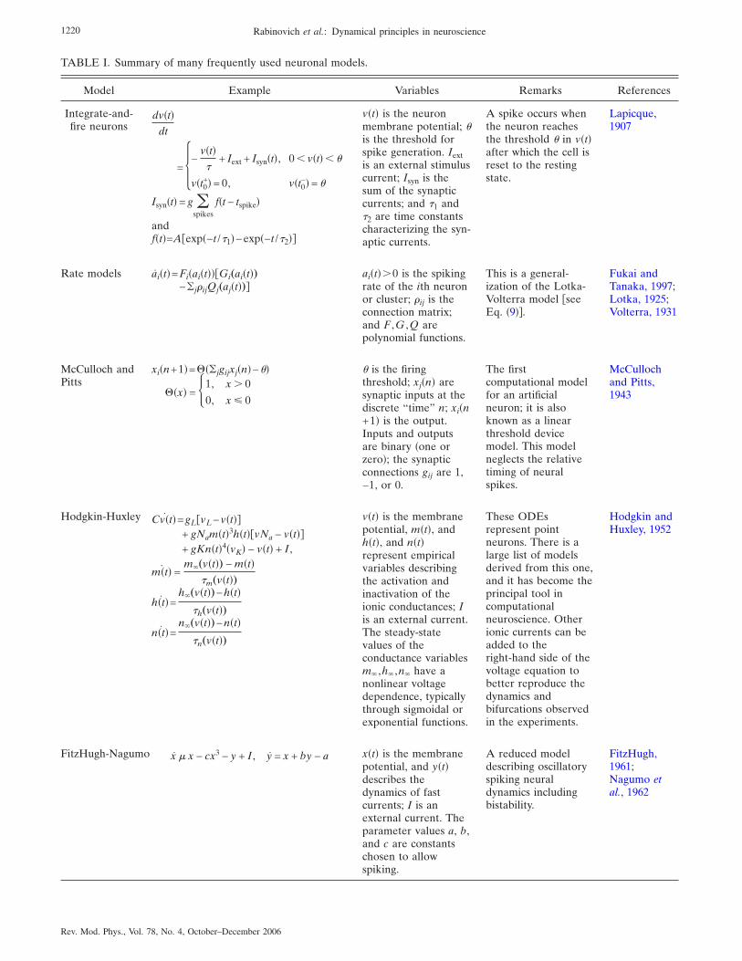
TABLE I. Summary of many frequently used neuronal models.
Model Example Variables Remarks References
Integrate-and-fire neurons
dv�t�dt
= �−v�t��
+ Iext + Isyn�t�, 0 � v�t� � �
v�t0+� = 0, v�t0
−� = ��
Isyn�t� = g �spikes
f�t − tspike�
andf�t�=A�exp�−t /�1�−exp�−t /�2��
v�t� is the neuronmembrane potential; �is the threshold forspike generation. Iextis an external stimuluscurrent; Isyn is thesum of the synapticcurrents; and �1 and�2 are time constantscharacterizing the syn-aptic currents.
A spike occurs whenthe neuron reachesthe threshold � in v�t�after which the cell isreset to the restingstate.
Lapicque,1907
Rate models ai�t�=Fi�ai�t���Gi„ai�t�…−�j�ijQj„aj�t�…�
ai�t��0 is the spikingrate of the ith neuronor cluster; �ij is theconnection matrix;and F ,G ,Q arepolynomial functions.
This is a general-ization of the Lotka-Volterra model �seeEq. �9��.
Fukai andTanaka, 1997;Lotka, 1925;Volterra, 1931
McCulloch andPitts
xi�n+1�=���jgijxj�n�−��
��x� = 1, x � 0
0, x 0� is the firingthreshold; xj�n� aresynaptic inputs at thediscrete “time” n; xi�n+1� is the output.Inputs and outputsare binary �one orzero�; the synapticconnections gij are 1,−1, or 0.
The firstcomputational modelfor an artificialneuron; it is alsoknown as a linearthreshold devicemodel. This modelneglects the relativetiming of neuralspikes.
McCullochand Pitts,1943
Hodgkin-Huxley Cv�t�˙ =gL�vL−v�t��+ gNam�t�3h�t��vNa − v�t��+ gKn�t�4�vK� − v�t� + I ,
m�t�˙ =m„v�t�… − m�t�
�m„v�t�…
h�t�˙ =h„v�t�…−h�t�
�h„v�t�…
n�t�˙ =n„v�t�…−n�t�
�n„v�t�…
v�t� is the membranepotential, m�t�, andh�t�, and n�t�represent empiricalvariables describingthe activation andinactivation of theionic conductances; Iis an external current.The steady-statevalues of theconductance variablesm ,h ,n have anonlinear voltagedependence, typicallythrough sigmoidal orexponential functions.
These ODEsrepresent pointneurons. There is alarge list of modelsderived from this one,and it has become theprincipal tool incomputationalneuroscience. Otherionic currents can beadded to theright-hand side of thevoltage equation tobetter reproduce thedynamics andbifurcations observedin the experiments.
Hodgkin andHuxley, 1952
FitzHugh-Nagumo x � x − cx3 − y + I , y = x + by − a x�t� is the membranepotential, and y�t�describes thedynamics of fastcurrents; I is anexternal current. Theparameter values a, b,and c are constantschosen to allowspiking.
A reduced modeldescribing oscillatoryspiking neuraldynamics includingbistability.
FitzHugh,1961;Nagumo etal., 1962
1220 Rabinovich et al.: Dynamical principles in neuroscience
Rev. Mod. Phys., Vol. 78, No. 4, October–December 2006

TABLE I. �Continued.�
Model Example Variables Remarks References
Wilson-Cowan�
�E�x , t�
�t=−E�x , t�+ �1−rE�x , t��
�Le�E�x , t� � wee�x�−I�x , t� � wei�x�+Ie�x , t��
��I�x , t�
�t=−I�x , t�+ �1−rI�x , t��
�Li�E�x , t� � wie�x�−I�x , t� � wii�x�+Ii�x , t��
�E�x , t� ,I�x , t�� are thenumber density ofactive excitatory andinhibitory neurons atlocation x of thecontinuous neuralmedia. „wee�x� ,wie�x� ,wei�x� ,wii�x�… areconnectivity distribu-tions among the popu-lations of cells. �Le ,Li� are nonlinear re-sponses reflecting dif-ferent populations ofthresholds. The oper-ator � is a convolu-tion involving the con-nectivity distributions.
The first “mean-field”model. It is anattempt to describe acluster of neurons, toavoid the inherentnoisy dynamicalbehavior of individualneurons; by averagingto a distribution noiseis reduced.
Wilson andCowan, 1973
Morris-Lecar v�t�˙ =gL�vL−v�t��+n�t�gn� �vn−v�t��+ gmm„v�t�…�vm − v�t�� + I,
n�t�˙ = „v�t�…�n„v�t�…−n�t��
m�v�=12 �1+tanh
v−vm
vm0 �
n�v�=12 �1+tanh
v−vn
vn0 �
�v�=�n coshv−vn
2vn0
v�t� is the membranepotential; n�t�describes the recoveryactivity of a calciumcurrent; I is anexternal current.
Simplified model thatreduces the number ofdynamical variables ofthe HH model. Itdisplays actionpotential generationwhen changing I leadsto a saddle-nodebifurcation to a limitcycle.
Morris andLecar, 1981
Hindmarsh-Rose x�t�˙ =y�t�+ax�t�2−bx�t�3−z�t�+I
y�t�˙ =C−xx�t�2−y�t�
z�t�˙ = rˆs�x�t� − x0� − z�t�‰
x�t� is the membranepotential; y�t�describes fastcurrents; z�t� describesslow currents; and I isan external current.
Simplified model thatuses a polynomialapproximation to theright-hand side of aHodgkin-Huxleymodel. This modelfails to describe thehyperpolarizedperiods after spikingof biological neurons.
Hindmarshand Rose,1984
Phase oscillatormodels
d�i�t�dt
= � + �j
Hij��i�t� − �j�t����t� is the phase ofthe ith neuron withapproximatelyperiodic behavior; andHij is the connectivityfunction determininghow neuron i and jinteract.
First introduced forchemical oscillators;good for describingstrongly dissipativeoscillating systems inwhich the neurons areintrinsic periodicoscillators.
Cohen et al.,1982;Ermentroutand Kopell,1984;Kuramoto,1984
Map modelsxt+1�i�=
�
1+xt�i�2 +yt�i�
+�
N�j
xt�j�
yt+1�i� = yt�i� − �xt�i� − �
xt represents thespiking activity and ytrepresents a slowvariable. A discretetime map.
One of a class ofsimplephenomenologi-cal models for spiking,bursting neurons. Thiskind of model can becomputationally veryfast, but has little bio-physical foundation.
Cazelles et al.,2001; Rulkov,2002
1221Rabinovich et al.: Dynamical principles in neuroscience
Rev. Mod. Phys., Vol. 78, No. 4, October–December 2006

respect to initial spiking functions exist, depending onwhether the neuron is excitable or repetitively firing inthe absence of feedback.
Following Hebb’s �1949� ideas most studies of themechanisms underlying learning and memory focus onchanging synaptic efficacy. Learning is associated withchanging connectivity in a network. However, the net-work dynamics also depends on complex interactionsamong intrinsic membrane properties, synapticstrengths, and membrane-voltage time variation. Fur-thermore, neuronal activity itself modifies not only syn-aptic efficacy but also the intrinsic membrane propertiesof neurons. Papers by Marder et al. �1996� and Turri-giano et al. �1996� present examples showing thatbistable neurons can provide short-term memorymechanisms that rely solely on intrinsic neuronal prop-erties. While not replacing synaptic plasticity as a pow-erful learning mechanism, these examples suggest thatmemory in networks could result from an ongoing inter-play between changes in synaptic efficacy and intrinsicneuron properties.
To understand the biological basis for such computa-tional properties we must examine both the dynamics ofthe ionic currents and the geometry of neuronal mor-phology.
3. Synaptic plasticity
Synapses as well as neurons are dynamical nonlineardevices. Although synapses throughout the CNS sharemany features, they also have distinct properties. Theyoperate with the following sequences of events: A spikeis initiated in the axon near the cell body, it propagatesdown the axon, and arrives at the presynaptic terminal,where voltage-gated calcium channels admit calcium,which triggers vesicle fusion and neurotransmitter re-lease. The released neurotransmitter then binds to re-ceptors on the postsynaptic neuron and changes theirconductance �Nicholls et al., 1992; Kandel et al., 2000�.This series of events is regulated in many ways, makingsynapses adaptive and plastic.
In particular, the strength of synaptic conductivitychanges in real time depending on their activity, as Katzobserved many years ago �Fatt and Katz, 1952; Katz,1969�. A description of such plasticity was made in 1949by Hebb �1949�. He proposed that “When an axon ofcell A is near enough to excite a cell B and repeatedly orpersistently takes part in firing it, some growth processor metabolic change takes place in one or both cells suchthat A’s efficiency, as one of the cells firing B, is in-creased.” This neurophysiological postulate has sincebecome a central concept in neuroscience through a se-ries of classic experiments demonstrating Hebbian-likesynaptic plasticity. These experiments show that the ef-ficacy of synaptic transmission in the nervous system isactivity dependent and continuously modified. Examplesof such modification are long-term potentiation and de-pression �LTP and LTD�, which involve increased or de-creased conductivity, respectively, of synaptic connec-tions between two neurons, leading to increased or
decreased activity over time. Long-term potentiationand depression are presumed to produce learning by dif-ferentially facilitating the association between stimulusand response. The role of LTP and LTD, if any, in pro-ducing more complex behaviors is less closely tied tospecific stimuli and more indicative of cognition, and isnot well understood.
Long-term potentiation was first reported in the hip-pocampal formation �Bliss and Lomo, 1973�. Changesinduced by LTP can last for many days. Long-term po-tentiation has long been regarded, along with its coun-terpart LTD, as a potential mechanism for short-term-memory formation and learning. In fact, the hypothesisis widely accepted in learning and memory research thatactivity-dependent synaptic plasticity is induced at ap-propriate synapses during memory formation and isboth necessary and sufficient for the information storageunderlying the type of memory mediated by the brainarea in which that plasticity is observed �see for a reviewMartin et al. �2000��. Hebb did not anticipate LTD in1949, but along with LTP it is thought to play a criticalrole in “rewiring” biological networks.
The notion of a coincidence requirement for Hebbianplasticity has been supported by classic studies of LTPand LTD using presynaptic stimulation coupled withprolonged postsynaptic depolarization �see, for example,Malenka and Nicoll �1999��. However, coincidence therewas loosely defined with a temporal resolution of hun-dreds of milliseconds to tens of seconds, much largerthan the time scale of typical neuronal activity charac-terized by spikes that last for a couple of milliseconds. Ina natural setting, presynaptic and postsynaptic neuronsfire spikes as their functional outputs. How preciselymust such spiking activities coincide in order to inducesynaptic modifications? Experiments addressing thiscritical issue led to the discovery of spike-timing-dependent synaptic plasticity �STDP�. Spikes initiate asequence of complex biochemical processes in thepostsynaptic neuron during the short time window fol-lowing synaptic activation. Identifying detailed molecu-lar processes underlying LTP and LTD remains a com-plex and challenging problem. There is good evidencethat it consists of a competition between processes re-moving �LTD� and processes placing �LTP� phosphategroups from on postsynaptic receptors, or increasing�LTP� or decreasing �LTD� the number of such receptorsin a dendritic spine. It is also widely accepted thatN-methyl-D-aspartate �NMDA� receptors are crucial forthe development of LTP or LTD and that it is calciuminflux onto the postsynaptic cell that is critical for bothLTP and LTD.
Experiments on synaptic modifications of excitatorysynapses between hippocampal glutamatergic neurons inculture �Bi and Poo, 1998, 2001� �see Fig. 4� indicate thatif a presynaptic spike arrives at time tpre and a postsyn-aptic spike is observed or induced at tpost, then when�= tpost− tpre is positive the incremental percentage in-crease in synaptic strength behaves as
1222 Rabinovich et al.: Dynamical principles in neuroscience
Rev. Mod. Phys., Vol. 78, No. 4, October–December 2006

�g
g� aPe−�P�, �1�
with �P�1/16.8 ms. When ��0, the percentage de-crease in synaptic strength behaves as
�g
g� − aDe�D�, �2�
with �D�1/33.7 ms. aP and aD are constants. This isillustrated in Fig. 4.
Many biochemical factors contribute differently toLTP and LTD in different synapses. Here we discuss aphenomenological dynamical model of synaptic plastic-ity �Abarbanel et al., 2002� which is very useful for mod-eling neural plasticity; its predictions agree with severalexperimental results. The model introduces two dynami-cal variables P�t� and D�t� that do not have a direct re-lationship with the concentration of any biochemicalcomponents. Nonlinear competition between these vari-ables imitates the known competition in the postsynapticcell. These variables satisfy the following simple first-order kinetic equations:
dP�t�dt
= f„Vpre�t�…�1 − P�t�� − �PP�t� ,
dD�t�dt
= g„Vpost�t�…�1 − D�t�� − �DD�t� , �3�
where the functions f�V� and g�V� are typical logistic orsigmoidal functions which rise from zero to the order ofunity when their argument exceeds some threshold.These driving or input functions are a simplification of
the detailed way in which each dynamical process isforced. The P�t� process is associated with a particulartime constant 1/�P while the D�t� process is associatedwith a different time constant 1/�D. Experiments showthat �P��D, and this is the primary embodiment of thetwo different time scales seen in many observations. Thetwo time constants are a coarse-grained representationof the diffusion and leakage processes which dampenand terminate activities. Presynaptic voltage activityserves to release neurotransmitters in the usual mannerand this in turn induces the postsynaptic action of P�t�,which has a time course determined by the time constant�P
−1. Similarly, the postsynaptic voltage, constant or timevarying, can be associated with the induction of the D�t�process.
P�t� and D�t� compete to produce a change in synapticstrength �g�t� as
d�g�t�dt
= ��P�t�D��t� − D�t�P��t�� , �4�
where ��1 and ��0. This dynamical model reproducessome of the key STDP experimental results like, for ex-ample, those shown in Fig. 4. It also accounts for thecase where the postsynaptic cell is depolarized while apresynaptic spike train is presented to it.
4. Examples of the cooperative dynamics of individual neuronsand synapses
To illustrate the dynamical significance of plastic syn-apses we consider the synchronization of two neurons: aliving neuron and an electronic model neuron coupledthrough a STDP or inverse STDP electronic synapse.Using hybrid circuits of model electronic neurons andbiological neurons is a powerful method for analyzingneural dynamics �Pinto et al., 2000; Szücs et al., 2000;LeMasson et al., 2002; Prinz et al., 2004a�. The represen-tation of synaptic input to a cell using a computer tocalculate the response of the synapse to specifiedpresynaptic input goes under the name “dynamic clamp”�Robinson and Kawai, 1993; Sharp et al., 1993�. It hasbeen shown in modeling and in experiments �Nowotny,Zhigulin, et al., 2003; Zhigulin et al., 2003� that couplingthrough plastic electronic synapses leads to neural syn-chronization or, more correctly, entrainment that is morerapid, more flexible, and much more robust againstnoise than synchronization mediated by connections ofconstant strength. In these experiments the neural cir-cuit consists of a specified presynaptic signal, a simulatedsynapse �via the dynamic clamp�, and a postsynaptic bio-logical neuron from the Aplysia abdominal ganglion.The presynaptic neuron is a spike generator producingspikes of predetermined form at predetermined times.The synapse and its plasticity are simulated by dynamicclamp software �Nowotny, 2003�. In each update cycle of 100 �s the presynaptic voltage is acquired, the spikegenerator voltage is updated, the synaptic strength is de-termined according to the learning rule, and the result-ing synaptic current is calculated and injected into theliving neuron through a current injection electrode. As
FIG. 4. Spike-timing-dependent synaptic plasticity observed inhippocampal neurons. Each data point represents the relativechange in the amplitude of evoked postsynaptic current afterrepetitive application of presynaptic and postsynaptic spikingpairs �1 Hz for 60 s� with fixed spike timing �t, which is de-fined as the time interval between postsynaptic and presynap-tic spiking within each pair. Long-term potentiation �LTP� anddepression �LTD� windows are each fitted with an exponentialfunction. Modified from Bi, 2002.
1223Rabinovich et al.: Dynamical principles in neuroscience
Rev. Mod. Phys., Vol. 78, No. 4, October–December 2006

one presents the presynaptic signal many times, the syn-aptic conductance changes from one fixed value to an-other depending on the properties of the presynapticsignal.
The calculated synaptic current is a function of thepresynaptic and postsynaptic potentials of the spike gen-erator Vpre�t� and the biological neuron Vpost�t�, respec-tively. It is calculated according to the following model.The synaptic current depends linearly on the differencebetween the postsynaptic potential Vpost and its reversalpotential Vrev, on an activation variable S�t�, and on itsmaximal conductance g�t�:
Isyn�t� = g�t�S�t��Vpost�t� − Vrev� . �5�
The activation variable S�t� is a nonlinear function of thepresynaptic membrane potential Vpre and represents thepercentage of neurotransmitter docked on the postsyn-aptic cell relative to the maximum that can dock. It hastwo time scales: a docking time and an undocking time.We take it to satisfy the dynamical equation
dS�t�dt
=S„Vpre�t�… − S�t�
�syn�S1 − S„V1�t�…�. �6�
S�V� is a sigmoid function which we take to be
S�V� = tanh��V − Vth�/Vslope� for V � Vth
0 otherwise. �7�
The time scale is �syn�S1−1� for neurotransmitter dock-ing and �synS1 for undocking. For AMPA excitatory re-ceptors, the docking time is about 0.5 ms, and the un-docking time is about 1.5 ms. The maximal conductanceg�t� is determined by the learning rule discussed below.In the experiments, the synaptic current is updated at 10 kHz.
To determine the maximal synaptic conductance g�t�of the simulated STDP synapse, an additive STDP learn-ing rule was used. This is accurate if the time betweenpresented spike pairs is long compared to the time be-tween spikes in the pair. To avoid runaway behavior, theadditive rule was applied to an intermediate graw thatwas then filtered through a sigmoid function. In particu-lar, the change �graw in synaptic strength is given by
�graw��t� = �A+�t − �0
�+e−��t−�0�/�+ for �t � �0
A−�t − �0
�−e��t−�0�/�− for �t � �0,� �8�
where �t= tpost− tpre is the difference between postsynap-tic and presynaptic spike times. The parameters �+ and�− determine the widths of the learning windows for po-tentiation and depression, respectively, and the ampli-tudes A+ and A− determine the magnitude of synapticchange per spike pair. The shift �0 reflects the finite timeof information transport through the synapse.
As one can see in Fig. 5, the postsynaptic neuronquickly synchronizes to the presynaptic spike generatorwhich presents spikes with an interspike interval �ISI� of255 ms �top panel�. The synaptic strength continuously
adapts to the state of the postsynaptic neuron, effec-tively counteracting adaptation and other modulationsof the system �bottom panel�. This leads to a very pre-cise and robust synchronization at a nonzero phase lag.The precision of the synchronization manifests itself insmall fluctuations of the postsynaptic ISIs in the syn-chronized state. Robustness and phase lag cannot beseen directly in Fig. 5. Spike-timing-dependent plasticityis a mechanism that enables synchronization of neuronswith significantly different intrinsic frequencies as onecan see in Fig. 6. The significant increase in the regimeof synchronization associated with synaptic plasticity is awelcome, perhaps surprising, result and addresses theissue raised above about robustness of synchronizationin neural circuits.
B. Robustness and adaptability in small microcircuits
The precise relationship between the dynamics of in-dividual neurons and the mammalian brain as a wholeremains extremely complex and obscure. An importantreason for this is a lack of knowledge on the detailedcell-to-cell connectivity patterns as well as a lack ofknowledge on the properties of the individual cells. Al-though large-scale modeling of this situation is at-tempted frequently, parameters such as the number andkind of synaptic connections can only be estimated. By
FIG. 5. Example of a synchronization experiment. Top: Theinterspike intervals �ISIs� of the postsynaptic biological neu-ron. Bottom: The synaptic strength g�t�. Presynaptic spikeswith ISI of 255 ms were presented to a postsynaptic neuronwith periodic oscillations at an ISI of 330 ms. Before couplingwith the presynaptic spike generator, the biological neuronspikes tonically at its intrinsic ISI of 330 ms. Coupling wasswitched on with g�t=0�=15 nS at time 6100 s. As one can seethe postsynaptic neuron quickly synchronizes to the presynap-tic spike generator �top panel, dashed line�. The synapticstrength continuously adapts to the state of the postsynapticneuron, effectively counteracting adaptation and other modu-lations of the system. This leads to a very precise and robustsynchronization at a nonzero phase lag. The precision of thesynchronization manifests itself in small fluctuations of thepostsynaptic ISIs in the synchronized state. Robustness andphase lag cannot be seen directly. Modified from Nowotny,Zhigulin, et al., 2003.
1224 Rabinovich et al.: Dynamical principles in neuroscience
Rev. Mod. Phys., Vol. 78, No. 4, October–December 2006

using the less complex microcircuits �MCs� of inverte-brates, a more detailed understanding of neural circuitdynamics is possible.
Central pattern generators are small MCs that canproduce stereotyped cyclic outputs without rhythmicsensory or central input �Marder and Calabrese, 1996;
Stein et al., 1997�. Thus CPGs are oscillators, and theimage of their activity in the corresponding system statespace is a limit cycle when oscillations are periodic and astrange attractor in more complex cases. Central patterngenerators underlie the production of most motor com-mands for muscles that execute rhythmic animal activitysuch as locomotion, breathing, heartbeat, etc. The CPGoutput is a spatiotemporal pattern with specific phaselags between the temporal sequences corresponding tothe different motor units �see below�.
The network architecture and the main features ofCPG neurons and synapses are known much better thanany other brain circuits. Examples of typical inverte-brate CPG networks are shown in Fig. 7. Common tomany CPG circuits are electrical and inhibitory connec-tions and the spiking-bursting activity of their neurons.The characteristics of the spatiotemporal patterns gener-ated by the CPG, such as burst frequency, phase, length,etc., are determined by the intrinsic properties of eachindividual neuron, the properties of the synapses, andthe architecture of the circuit.
The motor patterns produced by CPGs fall into twocategories: those that operate continuously such as res-piration �Ramirez et al., 2004� or heartbeat �Cymbalyuket al., 2002�, and those that are produced intermittentlysuch as locomotion �Getting, 1989� or chewing �Selver-ston, 2005�. Although CPGs autonomously establish cor-rect rhythmic firing patterns, they are under constantsupervision by descending fibers from higher centers andby local reflex pathways. These inputs allow the animalto constantly adapt its behavior to the immediate envi-ronment, which suggests that there is considerable flex-ibility in the dynamics of motor systems. In additionthere is now a considerable body of information showingthat anatomically defined small neural circuits can bereconfigured in a more general way by neuromodulatorysubstances in the blood, or released synaptically so thatthey are functionally altered to produce different stablespatiotemporal patterns, which must also be flexible inresponse to sensory inputs on a cycle-by-cycle basis; see
FIG. 6. �Color online� The presynaptic signal generator pre-sents a periodic spike train with ISI of T1 to a postsynapticneuron with ISI of T2
0, before coupling. When neurons arecoupled, T2
0→T2. We plot the ratio of these periods after cou-pling as a function of the ratio before coupling �a�, for a syn-apse with constant g and �b� for a synaptic connection g�t�following the rule in the text. The enlarged domain of one-to-one synchronization in the latter case is quite clear and, asshown by the change in the error bar sizes, the synchronizationis much better. This result persists when noise is added to thepresynaptic signal and to the synaptic action �not shown�.Modified from Nowotny, Zhigulin, et al., 2003.
FIG. 7. Examples of invertebrate CPG micro-circuits from arthropod, mollusk, and annelidpreparations. All produce rhythmic spa-tiotemporal motor patterns when activated bynonpatterned input. The black dots representchemical inhibitory synapses. Resistors repre-sent electrical connections. Triangles arechemical excitatory synapses, and diodes arerectifying synapses �electrical synapses inwhich the current flows only in one direction�.Individual neurons are identifiable from onepreparation to another.
1225Rabinovich et al.: Dynamical principles in neuroscience
Rev. Mod. Phys., Vol. 78, No. 4, October–December 2006

Simmers and Moulins �1988�, for example.Central pattern generators have similarity with neural
MCs in the brain �Silberberg et al., 2005; Solis and Per-kel, 2005; Yuste et al., 2005� and are often studied asmodels of neural network function. In particular, thereare important similarities between vertebrate spinalcord CPGs and neocortical microcircuits which havebeen emphasized by Yuste et al. �2005�: �i� CPG interac-tions, which are fundamentally inhibitory, dynamicallyregulate the oscillations. Furthermore, subthreshold-activated voltage-dependent cellular conductances thatpromote bistability and oscillations also promote syn-chronization with specific phase lags. The same cellularproperties are also present in neocortical neurons, andunderlie the observed oscillatory synchronization in thecortex. �ii� Neurons in spinal cord CPGs show bistablemembrane dynamics, which are commonly referred to asplateau potentials. A correlate of bistable membrane be-havior, in this case termed “up” and “down” states, hasalso been described in the striatum and neocortex bothin vivo and in vitro �Sanchez-Vives and McCormick,2000; Cossart et al., 2003�. It is still unclear whether thisbistability arises from intrinsic or circuit mechanisms ora combination of the two �Egorov et al., 2002; Shu et al.,2003�. �iii� Both CPGs and cortical microcircuits demon-strate attractor dynamics and transient dynamics �see,for example, Abeles et al. �1993�; Ikegaya et al. �2004��.�iv� Modulations by sensory inputs and neuromodulatorsare also a common characteristic that is shared betweenCPGs and cortical circuits. Examples in CPGs includethe modulation of oscillatory frequency, of temporal co-ordination among different populations of neurons, ofthe amplitude of network activity, and of the gating ofCPG input and output �Grillner, 2003�. �v� Switching be-tween different states of CPG operation �for example,switching coordinated motor patterns for differentmodes of locomotion� is under sensory afferent and neu-rochemical modulatory control. This makes CPGs mul-tifunctional and dynamically plastic. Switching betweencortical activity states is also under modulatory control,as shown, for example, by the role of the neurotransmit-ter dopamine in working memory in monkeys�Goldman-Rakic, 1995�. Thus modulation reconfiguresmicrocircuit dynamics and transforms activity states tomodify behavior.
The CPG concept was built around the idea that be-haviorally relevant spatiotemporal cyclic patterns aregenerated by groups of nerve cells without the need forrhythmic inputs from higher centers or feedback fromstructures that are moving. If activated, isolated inverte-brate preparations can generate such rhythms for manyhours and as a result have been extremely important intrying to understand how simultaneous cooperative in-teractions between many cellular and synaptic param-eters can produce robust and stable spatiotemporal pat-terns �see Fig. 8�d��. An example of a three-neuron CPGphase portrait is shown in Figs. 8�a�–8�c�. The effect of ahyperpolarizing current leads to changes in the patternas reflected by the phase portrait in Figs. 8�b� and 8�c�.
Neural oscillations arise either through interactionsamong neurons �network-based mechanism� or throughinteractions among currents in individual neurons �pace-maker mechanism�. Some CPGs use both mechanisms.In the simplest case, one or more neurons with intrinsicbursting activity acts as the pacemaker for the entireCPG circuit. The intrinsic currents may be constitutivelyactive or they may require activation by neuromodula-tors, so-called conditional bursters. Synaptic connectionsact to determine the pattern by exciting or inhibitingother neurons at the appropriate time. Such networksare extremely robust and have generally been thought tobe present in systems in which the rhythmic activity isactive all or most of the time. In the second case, it is thesynaptic interactions between nonbursty neurons thatgenerate the rhythmic activity and many schemes for thetypes of connections necessary to do this have been pro-posed. Usually reciprocal inhibition serves as the basisfor generating bursts in antagonistic neurons and thereare many examples of cells in pattern-generating micro-circuits connected in this way �see Fig. 7�. Circuits of thistype are usually found for behaviors that are intermit-tent in nature and require a greater degree of flexibilitythan those based on pacemaker cells.
Physiologists know that reciprocal inhibitory connec-tions between oscillatory neurons can produce, as a re-sult of the competition, sequential activity of neuronsand rhythmic spatiotemporal patterns �Szekely, 1965;Stent and Friesen, 1977; Rubin and Terman, 2004�. How-ever, even for a rather simple MC, consisting of justthree neurons, there is no quantitative description. If theconnections are symmetric, the MC can reach an attrac-
FIG. 8. �Color� Phase portrait of typical CPG output. The datawere recorded in the pyloric CPG of the lobster stomatogastricganglion. Each axis represents the firing rate of one of threepyloric neurons: LP, VD, and PD �see Fig. 7�. �a� The orbit ofthe oscillating pyloric system is shown in blue and the averageorbit is shown in red; �b� the same but with a hyperpolarizingdc current injected into the PD; �c� the difference between theaveraged orbits; �d� time series of the membrane potentials ofthe three neurons. Figure provided by T. Nowotny, R. Levi,and A. Szücs.
1226 Rabinovich et al.: Dynamical principles in neuroscience
Rev. Mod. Phys., Vol. 78, No. 4, October–December 2006

tor. It is reasonable to hypothesize that asymmetric in-hibitory connections are necessary to preserve the orderof patterns with more than two phases per cycle. Thecontradiction, noted earlier, between robustness andflexibility can then be resolved because external signalscan modify the effective topology of connections so onecan have functionally different networks for differentstimuli.
Theoretical analysis and computer experiments withMCs based on the winnerless competition principle �dis-cussed in Sec. IV.C� show that sufficient conditions forthe generation of sequential activity do exist and therange of allowed nonsymmetric inhibitory connections isquite wide �Rabinovich et al., 2001; Varona, Rabinovich,et al., 2002; Afraimovich, Rabinovich, et al., 2004� Weillustrate this using a Lotka-Volterra rate description ofneuron activity:
dai�t�dt
= ai�t��1 − �j=1
N
�ij�Si�aj�t�� + Si, i = 1, . . . ,N ,
�9�
where the rate of each N neuron is ai�t�, the connectionmatrix is �ij, and the stimuli Si are constants here. Thismodel can be justified as a rate neural model as follows�Fukai and Tanaka, 1997�. The firing rate ai�t� and mem-brane potential vi�t� of the ith neuron can be describedby
ai�t� = G�vi − �� , �10�
dvi�t�dt
= − vi�t� + Ii�t� , �11�
where G�vi−�� is a gain function, � and are constants,and the input current Ii�t� to neuron i is generated by therates aj�t� of the other neurons:
Ii�t� = Si − �j
N
�ijaj�t� . �12�
Here Si is the input and �ij is the strength of the inhibi-tory synapse from neuron j to neuron i. We suppose thatG�x� is a sigmoidal function:
G�x� = G0/�1 + exp�− �x�� . �13�
Let us then make two assumptions: �i� the firing rate isalways much smaller than its maximum value G0; and �ii�the system is strongly dissipative �this is reasonable be-cause we are considering inhibitory networks�. Based onthese assumptions, after combining and rescaling Eqs.�10�–�13�, we obtain the Lotka-Volterra rate description�9� with an additional positive term on the right side thatcan be replaced by a constant �see Fukai and Tanaka�1997� for details�.
The tests of whether WLC is operating in a reducedpyloric CPG circuit are shown in Fig. 9. This study usedestimates of the synaptic strengths shown in Fig. 9�a�.Some of the key questions here are these. �i� What is theminimal strength for the inhibitory synapse from the py-
loric dilator �PD� neuron or AB group to the VD neu-ron such that WLC exists? �ii� Does the connectivityobtained from the competition without winner conditionproduce the order of activation observed in the pyloricCPG? �iii� Is this dynamics robust against noise, in thesense that strong perturbations of the system do not al-ter the sequence? If the strengths of �ij are taken as
�ij = � 1 1.25 0
0.875 1 1.25
X/80 0.625 1� ,
the WLC formulas imply that the sufficient conditionsfor a reliable and robust cyclic sequence are satisfied ifX�160. The activation sequence of the rate model withnoise shown in Fig. 9�c� is similar to that observed ex-perimentally in the pyloric CPG. When additive Gauss-ian noise is introduced into the rate equations, the acti-vation order of neurons is not broken, but the period ofthe limit cycle depends on the level of perturbation.Therefore the cyclic competitive sequence is robust andcan be related to the synaptic connectivity seen in realMCs. If individual neurons in a MC are not oscillating,one can consider small subgroups of neurons that mayform oscillatory units and apply the WLC principle tothese units.
An important question about modeling the rhythmicactivity of small inhibitory circuits is how the specificdynamics of individual neurons influences the networkrhythm generation. Figure 10 represents the three-dimensional �3D� projection of the many-dimensionalphase portrait of a circuit with the same architecture as
FIG. 9. �Color online� Competition without winner in a modelof the pyloric CPG. �a� Schematic diagram of the three-neuronnetwork used for rate modeling. Black dots represent chemicalinhibitory synapses with strengths given in nanoseconds �X�160�. �b� Phase portrait of the model: The limit cycle corre-sponding to the rhythmic activity is in the 2D simplex �Zeemanand Zeeman, 2002�. �c� Robustness in the presence of noise:Noise introduced into the model shows no effect on the orderof activation for each type of neuron. Figure provided by R.Huerta.
1227Rabinovich et al.: Dynamical principles in neuroscience
Rev. Mod. Phys., Vol. 78, No. 4, October–December 2006

shown in Fig. 9�a� but using Hodgkin-Huxley spiking-bursting neuron models. The switching dynamics seen inthe rate model is shown in Fig. 9�c�, and this circuit isrobust when noise is added to it.
Pairs of neurons can interact via inhibitory, excitatory,or electrical �gap junction� synapses to produce basicforms of neural activity which can serve as the founda-tion for MC dynamics. Perhaps the most common �andwell-studied� CPG interaction consists of reciprocal in-hibition, an arrangement that generates a rhythmicbursting pattern in which neurons fire approximately outof phase with each other �Wang and Rinzel, 1995�. Thisis called a half-center oscillator. It occurs when there issome form of excitation to the two neurons sufficient tocause their firing and some form of decay mechanism toslow high firing frequencies. The dynamical range of thebursting activity varies with the synapse strength and insome instances can actually produce in-phase bursting.Usually reciprocal excitatory connections �unstable iftoo large� or reciprocal excitatory-inhibitory connectionsare able to reduce the intrinsic irregularity of neurons�Varona, Torres, Abarbanel, et al., 2001�.
Modeling studies with electrically coupled neuronshave also produced nonintuitive results �Abarbanel etal., 1996�. While electrical coupling is generally thoughtto provide synchrony between neurons, under certainconditions the two neurons can burst out of phase witheach other �Sherman and Rinzel, 1992; Elson et al., 1998,2002�; see Fig. 11 and also Chow and Kopell �2000� andLewis and Rinzel �2003�. An interesting modeling studyof three neurons �Soto-Trevino et al., 2001� with syn-apses that are activity dependent found that the synapticstrengths self-adjusted in different combinations to pro-duce the same three-phase rhythm. There are many ex-amples of vertebrate MCs in which a collection of neu-rons can be conceptually isolated to perform a particularfunction or to represent the canonical or modular circuitfor a particular brain region �see Shepherd �1998��.
C. Intercircuit coordination
It is often the case that more or less independent MCsmust synchronize in order to perform some coordinatedfunction. There is a growing literature suggesting thatlarge groups of neurons in the brain synchronize oscilla-tory activity in order to achieve coherence. This may bea mechanism for binding disparate aspects of cognitivefunction into a whole �Singer, 2001�, as we will discuss inSec. III.D. However, it is more persuasive to examineintercircuit coordination in motor circuits where thephases of different segments or limbs actually controlmovements. For example, the pyloric and gastric circuitscan be coordinated in the crustacean stomatogastric sys-tem by a higher-level modulatory neuron that channelsthe faster pyloric rhythm to a key cell in the gastric millrhythm �Bartos and Nushbaum, 1997; Bartos et al., 1999��Fig. 12�. In crab stomatogastric MCs, the gastric millcycle has a period of approximately 10 s while the py-loric period is approximately 1 s. When an identifiedmodulatory projection neuron �MCN1� �Fig. 12�a�� is ac-tivated, the gastric mill pattern is largely controlled by
FIG. 10. �Color online� Three-dimensional projection of themany-dimensional phase portrait of a circuit with the samearchitecture as the one shown in Fig. 9, using Hodgkin-Huxleyspiking-bursting neuron models.
FIG. 11. Artificial electrical coupling between two living cha-otic PD cells of the stomatogastric ganglion of a crustacean candemonstrate both synchronous and asynchronous regimes ofactivity. In this case the artificial electrical synapse was built ontop of the existing natural coupling between two PD cells. Thisshows different synchronization levels �a�–�d� as a function ofthe artificial coupling ga and a dc current I injected in one ofthe cells. �a� With their natural coupling ga=0 the two cells aresynchronized and display irregular spiking-bursting activity. �b�With an artificial electrical coupling that changes the sign ofthe current ga=−200 nS, and thus compensates the naturalcoupling, the two neurons behave independently. �c� Increasingthe negative conductance leads to a regularized antiphase spik-ing activity �by mimicking mutual inhibitory synapses�. �d�With no artificial coupling but adding a dc current two neuronsare synchronized, displaying tonic spiking activity. Modifiedfrom Elson et al., 1988.
1228 Rabinovich et al.: Dynamical principles in neuroscience
Rev. Mod. Phys., Vol. 78, No. 4, October–December 2006

interactions between MCN1 and gastric neurons LG andInt 1 �Bartos et al., 1999�. When Int 1 is stimulated, theAB to LG synapse �see Fig. 12�b�� plays a major role indetermining the gastric cycle period and coordinationbetween the two rhythms. The two rhythms become co-ordinated because LG burst onset occurs with a constantlatency after the onset of the triggering pyloric input.These results suggest that intercircuit synapses can en-able an oscillatory circuit to control the speed of aslower oscillatory circuit as well as provide a mechanismfor intercircuit coordination �Bartos et al., 1999�.
Another type of intercircuit coupling occurs amongsegmental CPGs. In the crayfish, abdominal appendages
called swimmerets beat in a metachronal rhythm fromposterior to anterior with a frequency-independentphase lag of about 90°. Like most rhythms of this kind,the phase lag must remain constant over different fre-quencies. In theoretical and experimental studies byJones et al. �2003�, it was shown that such phase con-stancy could be achieved by ascending and descendingexcitatory and inhibitory synapses, if the right connec-tions were made. It appears realistic to look at rhythmicMCs as recurrent networks with many intrinsic feedbackconnections so that the information on a complete spa-tiotemporal pattern is contained in the long-term activ-ity of just a few neurons in the circuit. The number ofintercircuit connections necessary for coordination ofthe rhythms is therefore much smaller than the totalnumber of neurons in the MC.
To investigate coordinating two elements of a popula-tion of neurons, one may investigate how various cou-plings, implemented in a dynamical clamp, might oper-ate in the cooperative behavior of two pyloric CPGs.This is a hybrid and simplified model of the more com-plex interplay between brain areas whose coordinatedactivity might be used to achieve various functions. Wenow describe such a set of experiments.
Artificially connecting neurons from the pyloric CPGof two different animals using a dynamic clamp couldlead to different kinds of coordination depending onwhich neurons are connected and what kind of synapsesare used �Szücs et al., 2004�. Connecting the pacemakergroup with electrical synapses could achieve same-phasesynchrony; connecting them with inhibitory synapsesprovided much better coordination but out of phase.The two pyloric circuits �Fig. 13� are representative ofcircuits driven by coupled pacemaker neurons that com-municate with each other via both graded and conven-tional chemical interactions. But while the unit CPGpattern is formed in this way, coordinating fibers mustuse spike-mediated postsynaptic potentials only. Ittherefore becomes important to know where in the cir-cuit to input these connections in order to achieve maxi-mum effectiveness in terms of coordinating the entirecircuit and ensuring phase constancy at different fre-quencies. Simply coupling the PDs together electricallyis rather ineffective although the bursts �not spikes� dosynchronize completely even at high coupling strengths.The fact that the two PDs are usually running at slightlydifferent frequencies leads to bouts of chaos in the twoneurons, i.e., a reduction in regularity. More effectivesynchronization occurs when the pacemaker groups arelinked together with moderately strong reciprocal in-hibitory synapses in the classic half-center configuration.Bursts in two CPGs are of course 180° out of phase, butthe frequencies are virtually identical. The best in-phasesynchronization is obtained when the LPs are coupled tothe contralateral PDs with inhibitory synapses �Fig. 13�.
D. Chaos and adaptability
Over the past decades there have been many reportsof the observation of chaos in the analysis of various
FIG. 12. �a� Schematic circuit diagram underlying MCN1 acti-vation of the gastric mill rhythm of a crustacean. The circuitrepresents two phases of the rhythm, retraction �left� and pro-traction �right�. Lighter lines represent inactive connections.LG, Int1, and DG are members of the gastric CPG and ABand PD are members of the pyloric CPG. Arrows representfunctional transmission pathways from the MCN1 neuron.Bars are excitatory and dots are inhibitory. �b� The gastric millcycle period; the timing of each cycle is a function of the py-loric rhythm frequency. With the pyloric rhythm turned off, thegastric rhythm cycles slowly �LG�. Replacing the AB inhibitionof Int1 with current into LG using a dynamic clamp reducesthe gastric mill cycle period. Modified from Barots et al., 1999.
1229Rabinovich et al.: Dynamical principles in neuroscience
Rev. Mod. Phys., Vol. 78, No. 4, October–December 2006

time courses of data from a variety of neural systemsranging from the simple to the complex �Glass, 1995;Korn and Faure, 2003�. Perhaps the outstanding featureof these observations is not the presence of chaos butthe appearance of low-dimensional dynamical systemsas the origin of spectrally broadband, nonperiodic sig-nals observed in many instances �Rabinovich and Abar-banel, 1998�. All chaotic oscillations occur in a boundedstate-space region of the system. This state space is cap-tured by the multivariate time course of the vector ofdynamical degrees of freedom associated with neuralspike generation. These degrees of freedom are com-prised of the membrane voltage and the characteristicsof the various ion currents in the cell. Using nonlineardynamical tools one can reconstruct a mathematicallyfaithful proxy state space for the neuron by using themembrane voltage and its time-delayed values as coor-dinates for the state space �see Fig. 14�.
Chaos seems to be almost unavoidable in natural sys-tems comprised of numerous simple or slightly complexsubsystems. As long as there are three or more dimen-sions, chaotic motions are generic in the broad math-ematical sense. So neurons are dealt a chaotic hand bynature and may have little choice but to work with it.Accepting that chaos is more or less the only choice, wecan ask what benefits accrue to the robustness andadaptability of neural activity.
Chaos itself may not be essential for living systems.However, the multitude of regular regimes of operationthat can be accomplished in dynamical systems com-posed of elements which themselves can be chaotic givesrise to a basic principle that nature may use for the or-
ganization of neural assemblies. In other words, chaos isnot responsible for the work of various neural struc-tures, but rather for the fact that those structures func-tion at the edge of instability, and often beyond it. Byrecognizing chaotic motions in a system state space asunstable, but bounded, this geometric notion gives cre-dence to the otherwise unappealing idea of system insta-bility. The instability inherent in chaotic motions, ormore precisely in nonlinear dynamics of systems withchaos, facilitates the extraordinary ability of neural sys-tems to adapt, make transitions from one pattern of be-havior to another when the environment is altered, andconsequently create a rich variety of patterns. Thuschaos gives a means to explore the opportunities avail-able to the system when the environment changes, andacts as a precursor to adaptive, reliable, and robust be-havior for living systems.
Throughout evolution neural systems have developeddifferent methods of self-control or self-organization.On the one hand, such methods preserve all advantagesof the complex behavior of individual neurons, such asallowing regulation of the time period of transitions be-tween operating regimes, as well as regulation of theoperation frequency in any given regime. They also pre-serve the possibility of a rich variety of periodic andnonperiodic regimes of behavior; see Fig. 11 and Elsonet al. �1988� and Varona, Torres, Huerta, et al. �2001�. Onthe other hand, these control or organizational tech-niques provide the needed predictability of behavioralpatterns in neural assemblies.
Organizing chaotic neurons through appropriate wir-ing associated with electrical, inhibitory, and excitatoryconnections appears to allow for essentially regular op-eration of such an assembly �Huerta et al., 2001�. As anexample we mention the dynamics of an artificial micro-
FIG. 13. Coupling of two biological pyloric CPGs Pyl 1 andPyl 2 by means of dynamic clamp artificial inhibitory synapses.The dynamic clamp is indicated by DCL. Reciprocal inhibitorycoupling between the pacemaker groups AB and PD leads toantiphase synchronization while nonreciprocal coupling fromthe LPs produces in-phase synchronization. Figure provided byA. Szücs.
FIG. 14. Chaotic spiking-bursting activity of isolated CPGneutrons. Top panel: Chaotic membrane potential time seriesof a synaptically isolated LP neuron from the pyloric CPG.Bottom panel: State-space attractor reconstructed from thevoltage measurements of the LP neuron shown in the toppanel using delayed coordinates �x�t� ,y�t� ,z�t��= �V�t� ,V�t−T� ,V�t−2T��. This attractor is characterized by two positiveLyapunov exponents. Modified from Rabinovich and Abar-banel, 1998.
1230 Rabinovich et al.: Dynamical principles in neuroscience
Rev. Mod. Phys., Vol. 78, No. 4, October–December 2006

circuit that mimics the leech heartbeat CPG �Calabreseet al., 1995�. This CPG model consists of six chaotic neu-rons implemented with Hindmarsh-Rose equations re-ciprocally coupled to their neighbors through inhibitorysynapses. The modeling showed that in spite of chaoticoscillations of individual neurons the cooperative dy-namics is regular and, most importantly, the period ofbursting of the cooperative dynamics sensitively de-pends on the values of the inhibitory connections�Malkov et al., 1996� �see Fig. 15�. This example showsthe high level of adaptability of a network consisting ofchaotic elements.
Chaotic signals have many of the traditional charac-teristics attributed to noise. In the present context werecognize that both chaos and noise are able to organizethe irregular behavior of individual neurons or neuralassemblies, but the principal difference is that dynamicalchaos is a controllable irregularity, possessing structurein state space, while noise is an uncontrollable action ofdynamical systems. This distinction is extremely impor-tant for information processing as discussed below �seeSec. III.B.2 and its final remarks�.
There are several possible functions for noise �Lind-ner et al., 2004�, even seen as high-dimensional essen-tially unpredictable chaotic motion, in neural networkstudies. In high-dimensional systems composed here ofmany coupled nonlinear oscillators, there may be smallbasins of attraction where, in principle, the system couldbecome trapped. Noise will blur the basin boundariesand remove the possibility that the main attractors couldaccidentally be missed and highly functional synchro-nized states lost to neuronal activity. Some noise maypersist in the dynamics of neurons to smooth out theactions of the chaotic dynamics active in creating robust,adaptable networks.
Chaos should not be mistaken for noise, as the formerhas phase-space structure which can be utilized for syn-chronization, transmission of information, and regular-ization of the network for performance of critical func-tions. In the next section we discuss the role of chaos ininformation processing and information creation.
III. INFORMATIONAL NEURODYNAMICS
The flow of information in the brain goes from sen-sory systems, where it is captured and encoded, to cen-tral nervous systems, where it is further processed togenerate response signals. In the central nervous systemcommand signals are generated and transported to themuscles to produce motor behavior. At all these stageslearning and memory processes that need specific repre-sentations take place. Thus it is not surprising that thenervous system has to use different coding strategies atdifferent levels of the transport, storage, and use of in-formation. Different transformations of codes have beenproposed for the analysis of spiking activity in the brain.The details depend on the particular system under studybut some generalization is possible in the framework ofanalyzing the spatial, temporal, and spatiotemporalcodes. There are many unknown factors related to thecooperation between these different forms of informa-tion coding. Some key questions are as follows: �i� Howcan neural signals be transformed from one coding spaceto another without loss of information? �ii� What dy-namical mechanisms are responsible for storing time inmemory? �iii� Can neural systems generate new informa-tion based on their sensory inputs? In this section, wediscuss some important experimental results and newparadigms that can help to address these questions.
A. Time and neural codes
Information from sensory systems arrives at sensoryneurons as analog changes in light intensity or tempera-ture, or chemical concentration of an odorant, or skinpressure, etc. These analog data are represented in in-ternal neural circuit dynamics and computations byaction-potential sequences passed from sensory receiv-ers to higher-order brain processes. Neural codes guar-antee the efficiency, reliability, and robustness of the re-quired neural computations �Machens, Gollisch, et al.,2005�.
1. Temporal codes
Two of the central questions in understanding the dy-namics of information processing in the nervous systemare how information is encoded and how the codingspace depends on time-dependent stimuli.
A code in the biophysical context of the nervous sys-tem is a specific representation of the information oper-ated on or created by neurons. A code requires a unit ofinformation. However, this is already a controversial is-sue since, as we have previously discussed, informationis conveyed through chemical and electrical synapses,neuromodulators, hormones, etc., which makes it diffi-cult to point out a single universal unit of information. Aclassical assumption at the cellular level, valid for manyneural systems, is that a spike is an all-or-nothing eventand thus a good candidate for a unit of information, atleast in a computational sense. This is not the only sim-
FIG. 15. Average bursting period of the model heartbeat CPGactivity as a function of the inhibitory coupling �. Modifiedfrom Malkov et al., 1996.
1231Rabinovich et al.: Dynamical principles in neuroscience
Rev. Mod. Phys., Vol. 78, No. 4, October–December 2006

plification needed to analyze neural codes for a first ap-proach. A coding scheme needs to determine a codingspace and take into account time.
A common hypothesis is to consider a universal timefor all neural elements. Although this is the approach wediscuss here, we remind the reader that this is also anarguable issue, since neurons can sense time in manydifferent ways: by their intrinsic activity �subcellular dy-namics� or by external input �synaptic and network dy-namics�. Internal and external �network� clocks are notnecessarily synchronized and can have different degreesof precision, time scales, and absolute references. Somedynamical mechanisms can contribute to make neuraltime unified and coherent.
On the one hand, when we consider just a single neu-ron, a spike as the unit of information, and a universaltime, we can talk about two different types of encoding:the frequency of firing can encode information about thestimulus in a rate code; on the other hand, the exacttemporal occurrence of spikes can encode the stimulusand its response in a precise timing code. The two cod-ing alternatives are schematically represented in Fig. 16.In this context, a precise timing or temporal code is acode in which relative spike timings �rather than spikecounts� are essential for information processing. Severalexperimental recordings have shown the presence ofboth types of single-cell coding in the nervous system�Adrian and Zotterman, 1926; Barlow, 1972; Abeles,1991; McClurkin et al., 1991; Softky, 1995; Shadlen andNewsome, 1998�. In particular, fine temporal precisionand reliability of spike dynamics are reported in manycell types �Segundo and Perkel, 1969; Mainen andSejnowski, 1995; deCharms and Merzenich, 1996; deRyter van Steveninck et al., 1997; Segundo et al., 1998;Mehta et al., 2002; Reinagel and Reid, 2002�. Single neu-rons can display these two codes in different situations.
2. Spatiotemporal codes
A population of coupled neurons can have a codingscheme different from the sum of the individual codingmechanisms. Interactions among neurons through theirsynaptic connections, i.e., their cooperative dynamics, al-low for more complex coding paradigms. There is muchexperimental evidence which shows the existence of so-called population codes that collectively express a com-plex stimulus better than the individual neurons �see,
e.g., Georgopoulus et al. �1986�; Wilson and McNaugh-ton �1993�; Fitzpatrick et al. �1997�; Pouget et al. �2000��.The efficacy of population coding has been assessedmainly using measures of mutual information in model-ing efforts �Seung and Sompolinsky, 1993; Panzeri et al.,1999; Sompolinsky et al., 2001�.
Two elements can be used to build population codes:neuronal identity �i.e., neuronal space� and the time oc-currence of neural events �i.e., the spikes�. Accordingly,information about the physical world can be encoded intemporal or spatial �combinatorial� codes, or combina-tions of these two: spike time can represent physicaltime �a pure temporal code�, spike time can representphysical space, neuronal space can represent physicaltime �a pure spatial code�, and neuronal space can rep-resent physical space �Nádasdy, 2000�. When we con-sider a population of neurons, information codes can bespatial, temporal, or spatiotemporal.
Population coding can also be characterized as inde-pendent or correlated �deCharms and Christopher,1998�. In an independent code, each neuron represents aseparate signal: all information that is obtainable from asingle neuron can be obtained from that neuron alone,without reference to the activities of other neurons. Fora correlated or coordinated coding messages are carriedat least in part by the relative timing of the signals froma population of neurons.
The presence of network coding, i.e., a spatiotemporaldynamical representation of incoming messages, hasbeen confirmed in several experiments. As an example,we discuss here the spatiotemporal representation ofepisodic experiences in the hippocampus �Lin et al.,2005�. Individual hippocampal neurons respond to awide variety of external stimuli �Wilson and McNaugh-ton, 1994; Dragoi et al., 2003�. The response variabilityat the level of individual neurons poses an obstacle tothe understanding of how the brain achieves its robustreal-time neural coding of the stimulus �Lestienne,2001�. Reliable encoding of sensory or other networkinputs by spatiotemporal patterns resulting from the dy-namical interaction of many neurons under the action ofthe stimulus can solve this problem �Hamilton andKauer, 1985; Laurent, 1996; Vaadia et al., 1999�.
Lin et al. �2005� showed that mnemonic short-timeepisodes �a form of one-trial learning� can trigger firingchanges in a set of CA1 hippocampal neurons with spe-cific spatiotemporal relationships. To find such represen-tations in the central nervous system of an animal is anextremely difficult experimental and computationalproblem. Because the individual neurons that partici-pate in the representation of a specific stimulus and forma temporal neural cluster in different trials can be differ-ent, it is necessary to measure simultaneously the activ-ity of a large number of neurons. In addition, because ofthe variability in the individual neuron responses, thespatiotemporal patterns of different trials may also lookdifferent. Thus, to show the functional importance of thespatiotemporal representation of the stimulus, thereader has to use sophisticated methods of data analysis.Lin et al. �2005� developed a 96-channel array to record
FIG. 16. �Color online� Two possible codes for the activity of asingle neuron. In a rate code, different inputs �A–D� are trans-formed into different output spiking rates. In a timing code,different inputs are transformed into different spiking se-quences with precise timing.
1232 Rabinovich et al.: Dynamical principles in neuroscience
Rev. Mod. Phys., Vol. 78, No. 4, October–December 2006
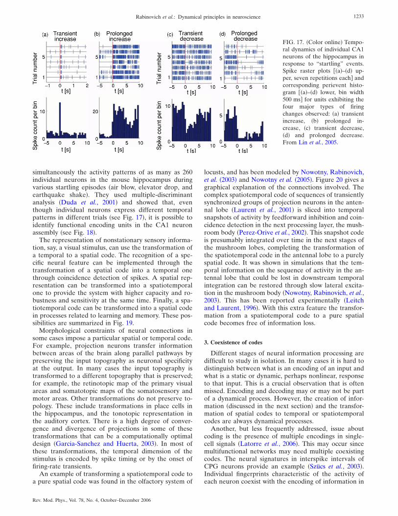
simultaneously the activity patterns of as many as 260individual neurons in the mouse hippocampus duringvarious startling episodes �air blow, elevator drop, andearthquake shake�. They used multiple-discriminantanalysis �Duda et al., 2001� and showed that, eventhough individual neurons express different temporalpatterns in different trials �see Fig. 17�, it is possible toidentify functional encoding units in the CA1 neuronassembly �see Fig. 18�.
The representation of nonstationary sensory informa-tion, say, a visual stimulus, can use the transformation ofa temporal to a spatial code. The recognition of a spe-cific neural feature can be implemented through thetransformation of a spatial code into a temporal onethrough coincidence detection of spikes. A spatial rep-resentation can be transformed into a spatiotemporalone to provide the system with higher capacity and ro-bustness and sensitivity at the same time. Finally, a spa-tiotemporal code can be transformed into a spatial codein processes related to learning and memory. These pos-sibilities are summarized in Fig. 19.
Morphological constraints of neural connections insome cases impose a particular spatial or temporal code.For example, projection neurons transfer informationbetween areas of the brain along parallel pathways bypreserving the input topography as neuronal specificityat the output. In many cases the input topography istransformed to a different topography that is preserved;for example, the retinotopic map of the primary visualareas and somatotopic maps of the somatosensory andmotor areas. Other transformations do not preserve to-pology. These include transformations in place cells inthe hippocampus, and the tonotopic representation inthe auditory cortex. There is a high degree of conver-gence and divergence of projections in some of thesetransformations that can be a computationally optimaldesign �Garcia-Sanchez and Huerta, 2003�. In most ofthese transformations, the temporal dimension of thestimulus is encoded by spike timing or by the onset offiring-rate transients.
An example of transforming a spatiotemporal code toa pure spatial code was found in the olfactory system of
locusts, and has been modeled by Nowotny, Rabinovich,et al. �2003� and Nowotny et al. �2005�. Figure 20 gives agraphical explanation of the connections involved. Thecomplex spatiotemporal code of sequences of transientlysynchronized groups of projection neurons in the anten-nal lobe �Laurent et al., 2001� is sliced into temporalsnapshots of activity by feedforward inhibition and coin-cidence detection in the next processing layer, the mush-room body �Perez-Orive et al., 2002�. This snapshot codeis presumably integrated over time in the next stages ofthe mushroom lobes, completing the transformation ofthe spatiotemporal code in the antennal lobe to a purelyspatial code. It was shown in simulations that the tem-poral information on the sequence of activity in the an-tennal lobe that could be lost in downstream temporalintegration can be restored through slow lateral excita-tion in the mushroom body �Nowotny, Rabinovich, et al.,2003�. This has been reported experimentally �Leitchand Laurent, 1996�. With this extra feature the transfor-mation from a spatiotemporal code to a pure spatialcode becomes free of information loss.
3. Coexistence of codes
Different stages of neural information processing aredifficult to study in isolation. In many cases it is hard todistinguish between what is an encoding of an input andwhat is a static or dynamic, perhaps nonlinear, responseto that input. This is a crucial observation that is oftenmissed. Encoding and decoding may or may not be partof a dynamical process. However, the creation of infor-mation �discussed in the next section� and the transfor-mation of spatial codes to temporal or spatiotemporalcodes are always dynamical processes.
Another, but less frequently addressed, issue aboutcoding is the presence of multiple encodings in single-cell signals �Latorre et al., 2006�. This may occur sincemultifunctional networks may need multiple coexistingcodes. The neural signatures in interspike intervals ofCPG neurons provide an example �Szücs et al., 2003�.Individual fingerprints characteristic of the activity ofeach neuron coexist with the encoding of information in
FIG. 17. �Color online� Tempo-ral dynamics of individual CA1neurons of the hippocampus inresponse to “startling” events.Spike raster plots ��a�–�d� up-per, seven repetitions each� andcorresponding perievent histo-gram ��a�–�d� lower, bin width500 ms� for units exhibiting thefour major types of firingchanges observed: �a� transientincrease, �b� prolonged in-crease, �c� transient decrease,�d� and prolonged decrease.From Lin et al., 2005.
1233Rabinovich et al.: Dynamical principles in neuroscience
Rev. Mod. Phys., Vol. 78, No. 4, October–December 2006
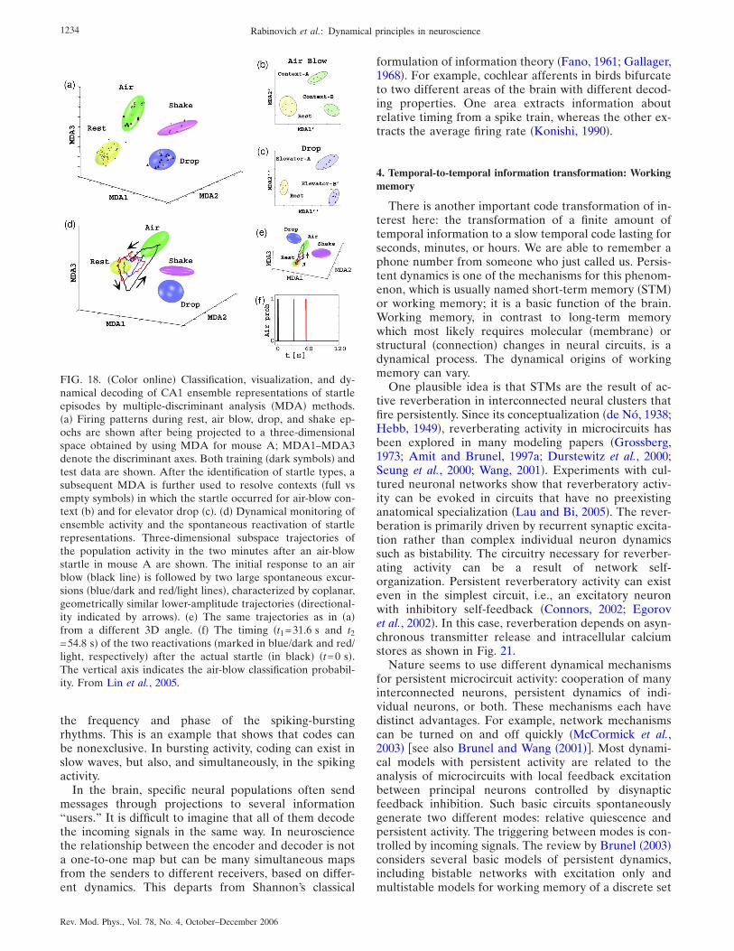
the frequency and phase of the spiking-burstingrhythms. This is an example that shows that codes canbe nonexclusive. In bursting activity, coding can exist inslow waves, but also, and simultaneously, in the spikingactivity.
In the brain, specific neural populations often sendmessages through projections to several information“users.” It is difficult to imagine that all of them decodethe incoming signals in the same way. In neurosciencethe relationship between the encoder and decoder is nota one-to-one map but can be many simultaneous mapsfrom the senders to different receivers, based on differ-ent dynamics. This departs from Shannon’s classical
formulation of information theory �Fano, 1961; Gallager,1968�. For example, cochlear afferents in birds bifurcateto two different areas of the brain with different decod-ing properties. One area extracts information aboutrelative timing from a spike train, whereas the other ex-tracts the average firing rate �Konishi, 1990�.
4. Temporal-to-temporal information transformation: Workingmemory
There is another important code transformation of in-terest here: the transformation of a finite amount oftemporal information to a slow temporal code lasting forseconds, minutes, or hours. We are able to remember aphone number from someone who just called us. Persis-tent dynamics is one of the mechanisms for this phenom-enon, which is usually named short-term memory �STM�or working memory; it is a basic function of the brain.Working memory, in contrast to long-term memorywhich most likely requires molecular �membrane� orstructural �connection� changes in neural circuits, is adynamical process. The dynamical origins of workingmemory can vary.
One plausible idea is that STMs are the result of ac-tive reverberation in interconnected neural clusters thatfire persistently. Since its conceptualization �de Nó, 1938;Hebb, 1949�, reverberating activity in microcircuits hasbeen explored in many modeling papers �Grossberg,1973; Amit and Brunel, 1997a; Durstewitz et al., 2000;Seung et al., 2000; Wang, 2001�. Experiments with cul-tured neuronal networks show that reverberatory activ-ity can be evoked in circuits that have no preexistinganatomical specialization �Lau and Bi, 2005�. The rever-beration is primarily driven by recurrent synaptic excita-tion rather than complex individual neuron dynamicssuch as bistability. The circuitry necessary for reverber-ating activity can be a result of network self-organization. Persistent reverberatory activity can existeven in the simplest circuit, i.e., an excitatory neuronwith inhibitory self-feedback �Connors, 2002; Egorovet al., 2002�. In this case, reverberation depends on asyn-chronous transmitter release and intracellular calciumstores as shown in Fig. 21.
Nature seems to use different dynamical mechanismsfor persistent microcircuit activity: cooperation of manyinterconnected neurons, persistent dynamics of indi-vidual neurons, or both. These mechanisms each havedistinct advantages. For example, network mechanismscan be turned on and off quickly �McCormick et al.,2003� �see also Brunel and Wang �2001��. Most dynami-cal models with persistent activity are related to theanalysis of microcircuits with local feedback excitationbetween principal neurons controlled by disynapticfeedback inhibition. Such basic circuits spontaneouslygenerate two different modes: relative quiescence andpersistent activity. The triggering between modes is con-trolled by incoming signals. The review by Brunel �2003�considers several basic models of persistent dynamics,including bistable networks with excitation only andmultistable models for working memory of a discrete set
FIG. 18. �Color online� Classification, visualization, and dy-namical decoding of CA1 ensemble representations of startleepisodes by multiple-discriminant analysis �MDA� methods.�a� Firing patterns during rest, air blow, drop, and shake ep-ochs are shown after being projected to a three-dimensionalspace obtained by using MDA for mouse A; MDA1–MDA3denote the discriminant axes. Both training �dark symbols� andtest data are shown. After the identification of startle types, asubsequent MDA is further used to resolve contexts �full vsempty symbols� in which the startle occurred for air-blow con-text �b� and for elevator drop �c�. �d� Dynamical monitoring ofensemble activity and the spontaneous reactivation of startlerepresentations. Three-dimensional subspace trajectories ofthe population activity in the two minutes after an air-blowstartle in mouse A are shown. The initial response to an airblow �black line� is followed by two large spontaneous excur-sions �blue/dark and red/light lines�, characterized by coplanar,geometrically similar lower-amplitude trajectories �directional-ity indicated by arrows�. �e� The same trajectories as in �a�from a different 3D angle. �f� The timing �t1=31.6 s and t2=54.8 s� of the two reactivations �marked in blue/dark and red/light, respectively� after the actual startle �in black� �t=0 s�.The vertical axis indicates the air-blow classification probabil-ity. From Lin et al., 2005.
1234 Rabinovich et al.: Dynamical principles in neuroscience
Rev. Mod. Phys., Vol. 78, No. 4, October–December 2006

of pictures with structured excitation and global inhibi-tion.
Working memory is used for tasks such as planning,organizing, rehearsing, and movement preparation. Ex-periments with functional magnetic resonance imagingreveal some aspects of the dynamics of working memory�see, for example, Diwadkar et al. �2000� and Nystrom etal. �2000��. It is important to note that working memoryhas a limited capacity of around four to seven items�Cowan, 2001; Vogel and Michizawa, 2004�. An essentialfeature attributed to working memory is the labile andtransient nature of its representations. Because suchrepresentations involve many coupled neurons from cor-tical areas �Curts and D’Esposito, 2003�, it is natural tomodel working memory as the spatiotemporal dynamicsof large neural networks.
A popular idea is to model working memory with at-tractors. Representation of items in working memory byattractors may guarantee its robustness. Although ro-bustness is an important requisite for a working-memorysystem, its transient properties are also important. Con-sider a foraging task in which an animal uses visual inputto catch prey �Nakahara and Doya, 1998�. It is helpful tostore the location of the prey in the animal’s workingmemory if the prey goes behind a bush and the sensorycue becomes temporarily unavailable. However, thememory should not be retained forever because the preymay have actually gone away or may have been eaten byanother animal. Furthermore, if more prey appears nearthe animal, the animal should quickly load the locationof the new prey into its working memory without beingdisturbed by the old memory.
This example illustrates that there are more require-ments for a working-memory system than solely robust
maintenance. First, the activity should be maintainedbut not for too long. Second, the activity should be resetquickly when there is a novel sensory cue that needs tobe stored. In other words, the neural dynamics involvedin working memory for goal-directed behaviors shouldhave the properties of long-term maintenance and quickswitching. A corresponding model based on “near-saddle-node” bifurcation dynamics has been suggestedby Nakahara and Doya �1998�. The authors have ana-lyzed the dynamics of a network of model neural unitsthat are described by the following map �see Fig. 22�:
yi�tn+1� = F�ayi�tn� + b + �j�i
�ijyi�tn� + �iIi�tn�� , �14�
where yi�tn� is the firing rate of the ith unit at time tn,F�z�=1/ �1+exp�−z�� is a sigmoid function, a is the self-connection weight, �ij are the lateral connection weights,Ii�t� are external inputs, b is the bias, and �i are con-stants used to scale the inputs Ii�t�. As the bias b is in-creased, the number of fixed points changes sequentiallyfrom one to two, three, two, and then back to one. Asaddle-node bifurcation occurs when the stable transi-tion curve y�tn+1�=F�z� is tangent to the fixed pointy�tn+1�=y�tn� �see Fig. 22�. Just near the saddle-node bi-furcation the system shows persistent activity. Thismeans that it spends a long time in the narrow channelbetween the bisectrix and the sigmoid activation curveand then goes to the fixed point quickly. Such dynamicalbehavior reminds one of the well-known intermittencyphenomenon in physics �Landau and Lifshitz, 1987�. Be-cause the effect of the sum of the lateral and externalinputs in Eq. �14� is equivalent to a change in the bias,the mechanism may satisfy the requirements of the dy-
FIG. 19. �Color online� Sum-mary of possible scenarios forthe transformation of codes,their functional implications,and the dynamical mechanisminvolved.
1235Rabinovich et al.: Dynamical principles in neuroscience
Rev. Mod. Phys., Vol. 78, No. 4, October–December 2006

namics of working memory for goal-directed behavior:long-term maintenance and quick switching.
Another reasonable model for working memory con-sists of competitive networks with stimulus-dependentinhibitory connections �as in Eq. �9��. One of the advan-tages of such a model is the ability to have both workingmemory and stimulus discrimination. This idea was pro-posed by Machens, Romo, et al. �2005� in relation to thefrontal-lobe neural architecture. The network first per-ceives the stimulus, then holds it in the working memory,and finally makes a decision by comparing that stimuluswith another one. The model integrates both workingmemory and decision making since the number of stablefixed points and the size of the basins of attractors arecontrolled by the connection matrix �ij�S� which de-pends on the stimuli S. The working-memory phase cor-responds to the bifurcation boundary, i.e., �ij=�ji=�ii. Inthe state space of the dynamical model, this phase isrepresented by a stable manifold called a “continuous
attractor.” This is an attractor that consists of continuoussets of fixed points �see Amari �1977� and Seung �1998��.Thus the stimulus creates a specific fixed point and, atthe next stage, the working memory �a continuous at-tractor� maintains it. During the comparison and deci-sion phase, the second stimulus is mapped onto the samestate space as another attractor. The criterion of the de-cision maker is reflected in the positions of the separa-trices that separate the basins of attraction of different
FIG. 20. �Color� Illustration of the transformation of temporalinto spatial information. If a coincidence detection occurs, thelocal excitatory connections activate the neighbors of the ac-tive neuron �yellow neurons�. Coincidence detection of input isnow more probable in these activated neighborhoods than inother Kenyon cells �KCs�. Which of the neighbors might fire aspike, however, depends on the activity of the projection neu-rons �PNs� in the next cycle. It might be a different neuron foractive group B of PNs �upper branch� than for active group C�lower branch�. In this way local sequences of active KCs form.These depend on the identity of active PNs �coincidence de-tection� as well as on the temporal order of their activity �ac-tivated neighborhoods�. Modified from Nowotny, Rabinovich,et al., 2003.
FIG. 21. �Color online� Reverberation can be the dynamicalorigin for working memory in minimal circuits. �a� Most neu-rons respond to excitatory stimuli �upward steps in the linebelow �c�� by spiking only as long as each stimulus lasts. �b�Very rare neurons are bistable: brief excitation leads to persis-tent spiking, always at the same rate; brief inhibition �down-ward steps in the line below �c�� can turn it off. �c� Multistableneurons persistently increase or decrease their spiking across arange of rates in response to repeated brief stimuli. �d� In thereverberatory network model of short-term memory discussedin the text, an excitatory stimulus �left arrow� leads to recur-sive activity in interconnected neurons. Inhibitory stimuli �baron the right� can halt the activity. �e� Egorov et al. �2002� sug-gest that graded persistent activity in single neurons �as in �c��might be triggered by a pulse of internal Ca2+ ions that enterthrough voltage-gated channels; Ca2+ then activates calcium-dependent nonspecific cation �CAN� channels, through whichan inward current �largely comprising Na+ ions� enters, persis-tently exciting the neuron. The positive feedback loop �brokenarrows� may include the activity of many ionic channels. Modi-fied from Connors, 2002.
FIG. 22. Temporal responses of self-recurrent units: Near-saddle-node bifurcation with a=11.11, b=−7.9 �center panels�.Increased bias, b=−3.0 �left panels�. Decreased biasb=−9.0 �right panels�. Modified from Nakahara and Doya,1998.
1236 Rabinovich et al.: Dynamical principles in neuroscience
Rev. Mod. Phys., Vol. 78, No. 4, October–December 2006

stimuli, i.e., fixed points �see an alternative approach inRabinovich and Huerta, 2006�.
We think that the intersection of the mechanisms re-sponsible for persistent activity of single neurons withthe activity of a network with local or nonlocal recur-rence provides robustness against noise and perturba-tions, and at the same time makes working memorymore flexible.
B. Information production and chaos
Information processing in the nervous system involvesmore than the encoding, transduction, and transforma-tion of incoming information to generate a correspond-ing response. In many cases, neural information is cre-ated by the joint action of the stimulus and theindividual neuron and network dynamics. A creative ac-tivity like improvisation on the piano or writing a newpoem results in part from the production of new infor-mation. This information is generated by neural circuitsin the brain and does not directly depend on the envi-ronment.
Time-dependent visual hallucinations are one ex-ample of information produced by neural systems, inthis case the visual cortex, themselves. Such hallucina-tions consist in seeing something that is not in the visualfield. There are interesting models, beginning from thepioneering paper of Ermentrout and Cowan �1979�, thatexplain how the intrinsic circuitry of the brain’s visualcortex can generate the patterns of activity that underliehallucinations. These hallucination patterns usually takethe form of checkerboards, honeycombs, tunnels, spi-rals, and cobwebs �see two examples in Fig. 23�. Becausethe visual cortex is an excitable medium it is possible touse spatiotemporal amplitude equations to describe thedynamics of these patterns �see the next section�. Thesemodels are based on advances in brain anatomy andphysiology that have revealed strong short-range con-nections and weaker long-range connections betweenneurons in the visual cortex. Hallucination patterns canbe quasistatic, periodically repeatable, or chaotically re-peatable as in low-dimensional convective turbulence;see for a review Rabinovich et al. �2000�. Unpredictabil-ity of the specific pattern in the hallucination sequences
�movie� means the generation of information that inprinciple can be characterized by the value of theKolmogorov-Sinai entropy �Scott, 2004�.
The creation or production of new information is atheme that has been neglected in theoretical neuro-science, but it is a provocative and challenging point thatwe discuss in this section. As mentioned before, infor-mation production or creation must be a dynamical pro-cess. Below we discuss an example that emphasizes theability of neural systems to produce information-richoutput from information-poor input.
1. Stimulus-dependent motor dynamics
A simple network with which we can discuss the cre-ation of new information is the gravity-sensing neuralnetwork of the marine mollusk Clione limacina. Clioneis a blind planktonic animal, negatively buoyant, thathas to maintain continuous motor activity in order tokeep its preferred head-up orientation. Its motor activityis controlled by wing CPGs and tail motor neurons thatuse signals from its gravity-sensing organs, the statocysts�Panchin et al., 1995�. A six-receptor neural networkmodel with synaptic inhibition has been built to describea single statocyst �Varona, Rabinovich, et al., 2002� �seeFig. 24�. This is a small sphere in which a statolith, astonelike structure, moves according to the gravitational
FIG. 23. Hallucinations generated by LSD are an example of adynamical representation of the internal activity of the visualcortex without an input stimulus. Figure shows examples of �a�funnel and �b� spiral hallucinations. Modified from Bressloff, etal. 2001.
FIG. 24. �Color online� Dual sensory network dynamics. Toppanels: Schematic representation of the dual role of a singlestatocyst, the gravity sensory organ of the mollusk Clione. Dur-ing normal swimming, a stonelike structure, the statolith, hitsthe mechanoreceptor neurons that react to this excitation. InClione’s hunting behavior, the statocyst receptors receive addi-tional excitation from the cerebral hunting neuron �H� whichgenerates a winnerless competition among them. Bottom pan-els: Chaotic sequential switching displayed by the activity ofthe statocyst during hunting mode in a model of a six-receptornetwork. This panel displays the time intervals in which eachneuron is active �ai�0.03�. Each neuron is represented by adifferent color. The dotted rectangles indicate the activation-sequence locks among units that are active at a given timeinterval within each network for time windows in which all sixneurons are active.
1237Rabinovich et al.: Dynamical principles in neuroscience
Rev. Mod. Phys., Vol. 78, No. 4, October–December 2006

field. The statolith excites the neuroreceptors by press-ing down on them. When excited, the receptors sendsignals to the neural systems responsible for wing beat-ing and tail orientation.
The statocysts have a dual role �Levi et al., 2004,2005�. During normal swimming only neurons that areexcited by the statolith are active, and this leads to awinner-take-all dynamical mode as a result of inhibitoryconnections in the network. �Winner-take-all dynamics isessentially the same as the attractor-based computa-tional ideas discussed earlier.� However, when Clione issearching for its food, a cerebral hunting neuron exciteseach neuron of the statocyst �see Fig. 24�. This triggers acompetition between all statocyst neurons whose signalsparticipate in the generation of a complex motion thatthe animal uses to scan the immediate space until it findsits prey �Levi et al., 2004, 2005� �see Fig. 25�. The follow-ing Lotka-Volterra-type dynamics can be used to de-scribe the activity of this network:
dai�t�dt
= ai�t����H,S� − �j=1
N
�ijaj�t� + Hi�t�� + Si�t� ,
�15�
where ai�t��0 represents the instantaneous spiking rateof the statocyst neurons, Hi�t� represents the excitatorystimulus from the cerebral hunting interneuron to neu-
ron i, Si�t� represents the action of the statolith on thereceptor that it is pressing, and �ij is the nonsymmetricstatocyst connection matrix. When there is no stimulusfrom the hunting neuron, Hi=0, or the statolith, Si=0,then ��H ,S�=−1 and all neurons are silent. When thehunting neuron is active Hi�0 and/or the statolith ispressing one of the receptors, Si�0, ��H ,S�= +1.
During hunting Hi�0, and we assume that the actionof the hunting neuron overrides the effect of the sta-tolith and thus Si�0. As a result of the competition, thereceptors display a highly irregular, in fact chaotic,switching activity. The phase-space image of the chaoticdynamics of the statocyst model in this behavioral modeis a strange attractor �the heteroclinic loops in the phasespace of Eq. �15� become unstable; see Sec. IV.C�. Forsix receptors we have shown �Varona, Rabinovich, et al.,2002� that the observed dynamical chaos is characterizedby two positive Lyapunov exponents.
The bottom panel in Fig. 24 is an illustration of thenonsteady switching activity of the receptors. An inter-esting phenomenon can be seen in this figure and is alsopointed out in Fig. 26. Although the timing of each ac-tivity is irregular, the sequence of switching among thestatocyst receptors is the same for those neurons that areactive at a given time window. Dotted rectangles in Fig.24 point out this fact. The activation-sequence lock
FIG. 25. �Color online� Clione swimming trajectories in differ-ent situations. �a� Three-dimensional trajectory of routineswimming. Here and in the following figures, different colors�gray tones� are used to emphasize the three-dimensional per-ception of the trajectories and change according to the x axis.The indicated time t is the duration of the trajectory. �b� Swim-ming trajectory of Clione with the statocysts surgically re-moved. �c� Trajectory of swimming during hunting behaviorevoked by the contact with the prey. �d� Trajectory of swim-ming after immersion of Clione in a solution that pharmaco-logically evokes hunting. Modified from Levi et al., 2004.
FIG. 26. �Color online� Irregular switching in a network of sixstatocyst receptors. Traces represent the instantaneous spikingrate of each neuron ai �neurons 1,2,3 are shown in �a�, neurons4,5,6 in �b��. Note that after a neuron is silent for a while, itsactivity reappears with the same sequence relative to the oth-ers �see arrows, and Fig. 24�. �c� A projection of the phaseportrait of the strange attractor in 3D space; see model �15�.
1238 Rabinovich et al.: Dynamical principles in neuroscience
Rev. Mod. Phys., Vol. 78, No. 4, October–December 2006

among the statocyst receptor neurons emerges in spiteof the highly irregular timing of the switching dynamicsand is a feature that can be used for motor coordination�Venaille et al., 2005�.
In this example the winnerless competition is trig-gered by a constant excitation to all statocyst receptors�Hi=ci; see details by Varona, Rabinovich, et al. �2002��.Thus the stimulus has low information content. None-theless, the network of statocyst receptors can use thisactivity to generate an information-rich signal with posi-tive Kolmogorov-Sinai entropy. This entropy is equal tothe value of the new information encoded in the dy-namical motion. The statocyst sensory network is thusmultifunctional and can generate a complex spatiotem-poral pattern useful for motor coordination even whenits dynamics are not evoked by gravity, as during hunt-ing.
2. Chaos and information transmission
To illustrate the role of chaos in information transmis-sion, we use as an example the inferior olive �IO�, whichis an input system to the cerebellum. Neurons of the IOmay chaotically recode the high-frequency informationcarried by its inputs into chaotic, low-rate output�Schweighofer et al., 2004�. The IO has been proposed asa system that controls and coordinates different rhythmsthrough the intrinsic oscillatory properties of its neuronsand the nature of their electrical interconnections�Llinás and Welsh, 1993; de Zeeuw et al., 1998�. It hasalso been implicated in motor learning �Ito, 1982� and incomparing tasks of intended and achieved movements asa generator of error signals �Oscarsson, 1980�.
Experimental recordings show that IO cells are elec-trically coupled and display subthreshold oscillationsand spiking activity. Subthreshold oscillations have a rel-evant role for information processing in the context of asystem with extensive electrical coupling. In such sys-tems the spiking activity can be propagated through thenetwork, and, in addition, small differences in hyperpo-larized membrane potentials propagate among neigh-boring cells.
A modeling study suggests that electrical coupling inIO neurons may induce chaos, which would allowinformation-rich, but low-firing-rate, error signals toreach individual Purkinje cells in the cerebellar cortex.This would provide the cerebellar cortex with essentialinformation for efficient learning without disturbing on-going motor control. The chaotic firing leads to the gen-eration of IO spikes with different timing. Because theIO has a low firing rate, an accurate error signal will beavailable for individual Purkinje cells only after re-peated trials. Electrical coupling can provide the sourceof disorder that induces a chaotic resonance in the IOnetwork �Schweighofer et al., 2004�. This resonanceleads to an increase in information transmission by dis-tributing the high-frequency components of the error in-puts over the sporadic, irregular, and non-phase-lockedspikes.
The IO single-neuron model consists of two compart-ments that include a low-threshold calcium current�ICal
�, an anomalous inward rectifier current �Ih�, aHodgkin-Huxley-type sodium current �INa�, and a de-layed rectifier potassium current �IKd� in the somaticcompartment �see Table I�. The dendritic compartmentcontains a calcium-activated potassium current �IKCa�and a high-threshold calcium current �ICah
�. This com-partment also receives electrical connections fromneighboring neurons. Fast ionic channels are located inthe soma, and slow channels are located in the dendriticcompartment. Some of the channel conductances de-pend on the calcium concentration. The equations foreach compartment of a single neuron can be summa-rized as
CMdV�t�
dt= − �Iion + Il + Iinj + Icomp� , �16�
where CM is the membrane capacitance, Il is a leak cur-rent, Iinj is the injected stimulus current, Icomp connectsthe compartments, and Iion is the sum of the currentsabove for each compartment. In addition, the dendriticcompartment has the electrical coupling current Iec=gc�i�V�t�−Vi�t��, where the index i runs over the neigh-bors of each neuron, and gc is the electrical couplingconductance.
Each IO neuron is represented by a system of ordi-nary differential equations �ODEs�, and the network is aset of these systems coupled through the electrical cou-pling currents Iec. The networks examined consisted of2�2, 3�3, and 9�3 neurons, where cells are connectedto their two, three, or four neighbors depending on theirpositions in the grid.
This is a complex network, even when it is only 2�2,and one must select an important feature of the dynam-ics to characterize its behavior. The largest Lyapunovexponent of the network is a good choice as it is inde-pendent of initial conditions and tells us about informa-tion flow in the network. Figure 27 displays the largestLyapunov exponent for each network as a function ofthe electric coupling conductance gc. We also see in Fig.27 that the gc producing the largest Lyapunov exponentyields the largest information transfer through the net-work, evaluated as the average mutual information perspike.
In a more general framework than the IO, it is re-markable that the chaotic activity of individual neuronsunexpectedly underlies higher flexibility and, at thesame time, greater accuracy and precision in their neuraldynamics. The origin of this phenomenon is the poten-tial ability of coupled neurons with chaotic behavior tosynchronize their activities and generate rhythms whoseperiod depends on the strength of the coupling or othernetwork parameters �for a review see Rabinovich andAbarbanel �1998� and Aihara �2002��. Networks withmany chaotic neurons can generate interesting transientdynamics, i.e., chaotic itinerancy �CI� �Tsuda, 1991;Rowe, 2002�. Chaotic itinerancy results from weak insta-bilities in the attractors, i.e., attractor sets in whose
1239Rabinovich et al.: Dynamical principles in neuroscience
Rev. Mod. Phys., Vol. 78, No. 4, October–December 2006

neighborhood there are trajectories that do not go to theattractors �Milnor-type attractors�. A developed CI mo-tion needs both many neurons and a very high level ofinterconnections. This is in contrast to the traditionalconcept of computation with attractors �Hopfield, 1982�.Chaotic itinerancy yields computations with transienttrajectories; in particular, there can be motion alongseparatrices as in winnerless competition dynamics �Sec.IV.C�. Although CI is an interesting phenomenon, ap-plying it to explain and predict the activity of sensorysystems �Kay, 2003�, and to any nonautonomous neuralcircuit dynamics, poses a question that has not been an-swered yet: How can CI be reproducible and robustagainst noise and at the same time sensitive to a stimu-lus?
To conclude this section it is necessary to emphasizethat the answer to the question of the functional role ofchaos in real neural systems is still unclear. In spite ofthe attractiveness of such ideas as �i� chaos makes neuralcircuits more flexible and adaptive, �ii� chaotic dynamicscreate information and can help to store it �see above�,and �iii� the nonlinear dynamical analyses of physiologi-cal data �e.g., electroencephalogram time series� can beimportant for the prediction or control of pathologicalneural states, it is extremely difficult to confirm theseideas directly in in vivo or even in vitro experiments. Inparticular, there are three obstacles that can fundamen-
tally hinder the power of data analyses: �i� finite statisti-cal fluctuations, �ii� external noise, and �iii� nonstation-arity of the neural circuit activity �see, for example, Laiet al. �2003��.
C. Synaptic dynamics and information processing
Synaptic transmission in many networks of the ner-vous system is dynamical, meaning that the magnitudeof postsynaptic responses depends on the history ofpresynaptic activity �Thompson and Deuchars, 1994;Fuhrmann et al., 2002�. This phenomenon is indepen-dent of �or in addition to� the plasticity mechanisms ofthe synapses �discussed in Sec. II.A.3�. The role of syn-apses is often considered to be the simple notification tothe postsynaptic neuron of presynaptic cell activity.However, electrophysiological recordings show that syn-aptic transmission can imply activity-dependent changesin response to presynaptic spike trains. The magnitudeof postsynaptic potentials can change rapidly from onespike to another, depending on the particular temporaldistribution of the presynaptic signals. Thus each singlepostsynaptic response can encode information about thetemporal properties of the presynaptic signals.
The magnitude of the postsynaptic response is deter-mined by the interspike intervals of the presynaptic ac-tivity and by the probabilistic nature of neurotransmitterrelease. In depressing synapses a short interval betweenpresynaptic spikes is followed by small postsynaptic re-sponses, while long presynaptic interspike intervals arefollowed by a large postsynaptic response. Facilitatingsynapses tend to generate responses that grow with suc-cessive presynaptic spikes. In this context, several theo-retical efforts have tried to explore the capacity of singleresponses of dynamical synapses to encode temporal in-formation about the timing of presynaptic events.
Theoretical models for dynamical synapses are basedon the time variation of the fraction of neurotransmitterreleased from the presynaptic terminal R�t�, 0R�t�1. When a presynaptic spike occurs at time tsp, thefraction U of available neurotransmitters and the recov-ery time constant �rec determine the rate of return ofresources R�t� to the available presynaptic pool. In adepressing synapse, U and �rec are constant. A simplemodel describes the fraction of synaptic resources avail-able for transmission as �Fuhrmann et al., 2002�
dR�t�dt
=1 − R�t�
�rec− UR�t���t − tsp� , �17�
and the amplitude of the postsynaptic response at timetsp is proportional to R�tsp�.
For a facilitating synapse, U becomes a function oftime U�t� increasing at each presynaptic spike and de-caying to the baseline level when there is no presynapticactivity:
FIG. 27. �Color online� Chaotic dynamics increases informa-tion transmission in IO models. Top panel: Largest Lyapunovexponent as a function of the electrical coupling strength gc fordifferent IO networks of nonidentical cells. Bottom panel: Net-work average mutual information per spike as a function of gc.Modified from Schweighofer et al., 2004.
1240 Rabinovich et al.: Dynamical principles in neuroscience
Rev. Mod. Phys., Vol. 78, No. 4, October–December 2006

dU�t�dt
= −U�t��facil
+ U1�1 − U�t����t − tsp� , �18�
where U1 is a constant determining the step increase inU�t� and �facil is the relaxation time constant of the fa-cilitation.
Other approaches to modeling dynamical synapses in-clude probabilistic models to account for fluctuations inpresynaptic release of neurotransmitters. At a synapticconnection with N release sites we can assume that ateach site there can be, at most, one vesicle available forrelease, and that the release at each site is an indepen-dent event. When a presynaptic spike is produced attime tsp, each site containing a vesicle will release it withthe same probability U�t�. Once a release occurs, the sitecan be refilled during a time interval dt with probabilitydt /�rec. The probabilistic release and recovery can bedescribed by the probability Pv�t� for a vesicle to beavailable for release at any time t:
dPv�t�dt
=1 − Pv�t�
�rec− U�t�Pv�t���t − tsp� . �19�
Figure 28 shows how this formulation permits an accu-rate description of a depressing synapse in response to aspecified presynaptic spike train.
The transmission of sensory information from the en-vironment to decision centers through neural communi-cation channels requires a high degree of reliability and
sensitivity from networks of heterogeneous, inaccurate,and sometimes unreliable components. The propertiesof the channel itself, assuming the sensor is accurate,must be richer than conventional channels studied in en-gineering applications. Those channels are passive and,when of high quality, can relay inputs accurately to areceiver. Neural communication channels are composedof dynamically active elements capable of complex au-tonomous oscillations. Individually, chaotic neurons cancreate information in a way similar to the study of non-linear systems with unstable trajectories: Two states ofthe system, indistinguishable because only finite-resolution observations can occur, may through the ac-tion of the instabilities of the nonlinear dynamics findthemselves in the future widely separated in state space,and thus distinguishable. Information about differentstates that was unavailable at one time may becomeavailable at a later time.
Biological neural communication pathways are able torecover information from a hidden coding space and totransfer information from one time scale to another be-cause of the intrinsic nonlinear dynamics of synapses. Asan example, we discuss a very simple neural informationchannel composed of sensory input in the form of aspike train that arrives at a model neuron and thenmoves through a realistic dynamical synapse to a secondneuron where the information in the initial sensory sig-nal is read �Eguia et al., 2000�. The model neurons arefour-dimensional generalizations of the Hindmarsh-Rose neuron, and a model of chemical synapse derivedfrom first-order kinetics is used. The four-dimensionalmodel neuron has a rich variety of dynamical behaviors,including periodic bursting, chaotic bursting, continuousspiking, and multistability. For many of these regimes,the parameters of the chemical synapse can be tuned sothat the information about the stimulus, which is un-readable to the first neuron in the path, can be recov-ered by the dynamical activity of the synapse, and thesecond neuron can read it �see Fig. 29�.
The quantitative description of this unexpected phe-nomenon was done by calculating the average mutualinformation I�S ,N1� between the stimulus S and the re-sponse of the first neuron N1, and I�S ,N2� between thestimulus and the response of the second neuron N2. Theresult in the example shown in Fig. 29 is I�S ,N2��I�S ,N1�. This result indicates how nonlinear synapsesand neurons acting as input and output systems along acommunication channel can recover information appar-ently hidden in earlier synaptic connections in the path-way. Here the measure of information transmission usedis the average mutual information between elements,and because the channel is active and nonlinear, the av-erage mutual information between the sensory sourceand the final neuron may be greater than the averagemutual information found in an intermediate neuron inthe channel �but not greater than the original informa-tion�.
Another form of synaptic dynamics involved in infor-mation processing and especially in learning is STDP�already discussed in Sec. II.A.3�. Information transduc-
FIG. 28. Dynamical synapses imply that synaptic transmissiondepends on previous presynaptic activity. This shows the aver-age postsynaptic activity generated in response to a presynap-tic spike train �bottom trace� in a pyramidal neuron �top trace�and in a model of a depressing synapse �middle trace�. Postsyn-aptic potential in the model is computed using a passive mem-brane mechanism �m�dV /dt�=−V+RiIsyn�t�, where Ri is the in-put resistance. Modified from Tsodyks and Markram, 1997.
1241Rabinovich et al.: Dynamical principles in neuroscience
Rev. Mod. Phys., Vol. 78, No. 4, October–December 2006

tion is influenced by STDP �Chechik, 2003; Hopfield andBrody, 2004�, which also plays an important role in bind-ing and synchronization.
D. Binding and synchronization
We have discussed the diversity of neuron types andthe variability of neural activity. Neural processing re-quires the fast interaction of many neurons in differentneural subsystems. There are several dynamical mecha-nisms contributing to the complex integration of infor-mation that neural systems perform. Among them, thesynchronization of neural activity is the one that hascaptured the most attention. Synchronization of neuralactivity is also one of the proposed solutions to a widelydiscussed question in neuroscience: the binding prob-lem, which we describe briefly in this section.
The binding problem was originally formulated as atheoretical problem by von der Malsburg in 1981 �see areview by von der Malsburg �1999�, and Roskies �1999�;Singer �1999��. However, examples of binding had al-ready been proposed by Rosenblatt �1962� for the visualsystem �for a review of the binding problem in vision seeSinger �1999�, and Wolfe and Cave �1999��. The bindingproblem is formulated as the need for a coherent repre-sentation of an object provided by the association of all
its features �shape, color, location, speed, etc.�. The as-sociation of all features or binding allows a unified per-ception of the object. The binding problem is a general-ized task of the nervous system as it seeks to reconstructany total perception from its components. There are alsocognitive binding problems related to cognitive identifi-cation and memory. No doubt the binding problem, likemany other problems in biology, has multiple solutions.These solutions are most likely implemented throughthe use of dynamical mechanisms for the control of neu-ral activity.
The most widely studied mechanism proposed tosolve the binding problem is temporal synchrony �ortemporal correlation� �Singer and Gray, 1995�. It hasbeen suggested by von der Malsburg and Schneider�1986� that synchronization is the basis for perceptualbinding. However, there is still criticism of the temporalbinding hypothesis �Ghose and Maunsell, 1999; Riesen-huber and Poggio, 1999�. Obviously, neural oscillationsand synchronous signals are ubiquitous in the brain, andneural systems can make use of these phenomena toencode, learn, and create effective outputs. There areseveral lines of experimental evidence that reveal theuse of synchronization and activity correlation for bind-ing tasks. Figure 30 shows an example of how neuralsynchronization correlates with the perceptual segmen-tation of a complex visual pattern into distinct, spatiallyoverlapping surfaces �Castelo-Branco et al., 2000� �seethe figure caption for details�. Indeed, modeling studiesshow that involving time in these processes can lead tothe binding of different features. The idea is to use thecoincidence of certain events in the dynamics of differ-ent neural units for binding. Usually such dynamicalbinding is represented by synchronous neurons or neu-rons that are in phase with an external field. However,dynamical events such as phase or frequency variationsusually are not very reproducible and robust. As dis-cussed in the next section, it is reasonable to hypothesizethat brain circuits displaying sequential switching of neu-ral activity use the coincidence of this switching toimplement dynamical binding of different WLC net-works.
Any spatiotemporal coding needs the temporal coor-dination of neural activity among different populationsof neurons to provide �i� better recognition of specificfeatures, �ii� faster processing, �iii� higher informationcapacity, and �iv� feature binding. Neural synchroniza-tion has been observed throughout the nervous system,particularly in sensory systems, for example, in the olfac-tory system �Laurent and Davidowitz, 1994� and the vi-sual system �Gray et al., 1989�. From the point of view ofdynamical system theory, transient synchronization is anideal mechanism for binding neurons into assemblies forseveral reasons: �i� the synchronized neurons do not nec-essarily have to be neighbors; �ii� a synchronizationevent depends on the state of the neuron and the stimu-lus and can be very selective, that is, neurons from thesame network can be temporal members of different cellassemblies at different instants of time; �iii� basic brainrhythms are able to synchronize neurons responsible for
FIG. 29. Example of recovery of hidden information in neuralchannels. A presynaptic cell receives specified input and con-nects to a postsynaptic cell through a dynamical synapse. Toppanel: the time series of synaptic input to the presynaptic cellJ1�t�; middle panel: the membrane potential of the first burst-ing neuron X1�t�; bottom panel: the membrane potential of thesecond bursting neuron X2�t�. Note that features of the inputhidden in the response X1�t� are recovered in the responsefollowing a dynamical synapse X2�t� �note hyperpolarizationregions for X2�. Modified from Eguia et al., 2000.
1242 Rabinovich et al.: Dynamical principles in neuroscience
Rev. Mod. Phys., Vol. 78, No. 4, October–December 2006

the processing of information from different sensory in-puts; and �iv� the synchronization is possible even be-tween neural oscillators with strongly different frequen-cies �Rabinovich et al., 2006�.
In early visual processing neurons that encode fea-tures of a complex visual percept are associated in func-
tional assemblies through gamma-frequency synchroni-zation �Engel et al., 2001�. When sensory stimuli areperceptually or attentionally selected, and the respectiveneurons are bound together to raise their saliency, thengamma-frequency synchronization among these neuronsis also enhanced. Gamma-mediated coupling and itsmodulation by attention are not limited to the visualsystem: they are also found in the auditory �Tiitinen etal., 1993� and somatosensory domains �Desmedt andTomberg, 1994�. Gamma oscillations allow visiomotorbinding between posterior and central brain regions�Rodriguez et al., 1999� and are involved in short-termmemory. As a means for dynamically binding neuronsinto assemblies, gamma-frequency synchronization ap-pears to be the prime mechanism for stabilizing corticalconnections among members of a neural assembly overtime. On the other hand, neurons can increase or de-crease the strength of their synaptic connections de-pending on the precise coincidence of their activation�STDP�, and gamma-frequency synchronization pro-vides the required temporal precision.
Hatsopoulos et al. �2003� and Jackson et al. �2003� re-vealed the functional significance of neural synchroniza-tion and correlations within the motor system. Preemi-nent among brain actions must be the aggregation ofdisparate spiking patterns to form spatially and tempo-rally coherent neural codes that then drive alpha motorneurons and their associated muscles. Essentially, motorbinding seems to describe exactly what motor structuresof the mammalian brain do: provide high-level coordi-nation of simple and complex voluntary movements.Neurons with similar functional output have an in-creased likelihood of exhibiting neural synchronization.
In contrast to classical synchronization �Pikovskyet al., 2001�, synchronization in the CNS is always tran-sient. The phase-space image of transient synchroniza-tion can be a saddle limit cycle in the vicinity of whichthe system spends finite time. Alternatively, it can be alimit cycle whose basin of attraction decreases in time.In both cases the system is able to leave the synchroni-zation region after a specific stage of processing is com-pleted and proceed with the next task. This is a broadarea where the issues and approaches are not settled,and thus it provides an opportunity for innovative ideasto explain the phenomenon.
To conclude this section, we note that the functionalrole of synchronization in the CNS and the importanceof spike-timing coding in general are still a subject ofdebate. On the one hand, it is possible to build modelsthat use dynamical patterns of spikes for neural compu-tations, e.g., representation, recognition, and decisionmaking. Examples of such spike-timing-based computa-tional models have been discussed by Hopfield andBrody �Hopfield and Brody, 2001; Brody and Hopfield,2003�. In this work the authors showed, in particular,that spike synchronization across many neurons can beachieved in the absence of direct synaptic interactionsbetween neurons through phase locking to a commonunderlying oscillatory potential �like gamma oscillation;see above�. On the other hand, the real connections of
FIG. 30. An example of binding showing dependence of syn-chrony on transparency conditions and receptive field �RF�configuration in the cat visual cortex. �a� Stimulus configura-tion. �b� Synchronization between neurons with nonoverlap-ping RFs and similar directional preferences recorded fromareas A18 and PMLS of the cat visual cortex. Left, RF constel-lation and tuning curves; right, cross correlograms for re-sponses to a nontransparent �left� and transparent plaid �right�moving in the cells’ preferred direction. Grating luminancewas asymmetric to enhance perceptual transparency. Smalldark correlograms are shift predictors. �c� Synchronization be-tween neurons with different direction preferences recordedfrom A18 �polar and RF plots, left�. Top, correlograms of re-sponses evoked by a nontransparent �left� and a transparent�right� plaid moving in a direction intermediate to the cells’preferences. Bottom, correlograms of responses evoked by anontransparent plaid with reversed contrast conditions �left�,and by a surface defined by coherent motion of intersections�right�. Scale on polar plots: discharge rate in spikes per sec-ond. Scale on correlograms: abscissa, shift interval in ms, binwidth 1 ms; ordinate, number of coincidences per trial, nor-malized. Modified from Castelo-Branco et al., 2000.
1243Rabinovich et al.: Dynamical principles in neuroscience
Rev. Mod. Phys., Vol. 78, No. 4, October–December 2006

such theoretical models with experiments in vivo are notestablished �see also Fell et al. �2003� and O’Reilly et al.�2003��.
IV. TRANSIENT DYNAMICS: GENERATIONAND PROCESSING OF SEQUENCES
A. Why sequences?
The generation and control of sequences is of crucialimportance in many aspects of animal life. Workingmemory, bird songs, finding food in a labyrinth, jumpingfrom one stone to another on the shore—all these arethe results of sequential activity generated by the ner-vous system. Lashley called the problem of coordinationof constituent actions into organized sequential spa-tiotemporal patterns the action syntax problem �Lashley,1960�. The generation of sequences is also important forintermediate information processing as we discuss be-low.
The sequences can be cyclic, like many brain rhythmsand spatiotemporal patterns generated by CPGs. Theycan also be irregular, like neocortical theta oscillations�4–10 Hz� generated spontaneously in cortical networks�Bao and Wu, 2003� �see Fig. 31�. The sequences can befinite in time like those generated by a neural circuitunder the action of external input as in sensory systems.From a physicist’s point of view, any reproducible finitesequence that is functionally meaningful results from thecooperative transient dynamics of the corresponding
neural ensemble or individual neurons. Even brainrhythms demonstrate transient dynamics because thecircuit’s periodic activity is modulated by nonstationarysensory inputs or signals from the periphery. It is impor-tant to emphasize the fundamental role of inhibition inthe generation and control of sequences in the nervoussystem.
In this section we concentrate on the origin of se-quence generation and the mechanisms of reproducibil-ity, sensitivity, and functional reorganization of MCs. Inthe standard study of nonlinear dynamical systems, at-tention is focused on the long-time behavior of a system.This is typically not the relevant question in neuro-science. Here we must address the transient responses toa stimulus external to the neural system and must con-sider the short-term binding of a collection of responses,perhaps from different sensory inputs, to facilitate ac-tion commands directed to the motor system. If you at-tempt to swat a fly, it cannot ask you to perform thisaction many times so that it can average over your ac-tions, allowing it to perform some standard optimal re-sponse. Few flies wanting this repetition would survive.
B. Spatially ordered networks
1. Stimulus-dependent modes
Many neural ensembles are anatomically organized asslightly inhomogeneous excitable media. Examples ofsuch media are retina �Tohya et al., 2003�, IO network�Leznik and Llinas, 2002�, cortex �Ichinohe et al., 2003�,and thalamocortical layers �Contreras et al., 1996�. Allthese are neuronal lattices with chemical or electricalconnections occurring primarily between neighbors.There are some general dynamical mechanisms of se-quence generation in such spatially ordered networks.These mechanisms are usually related to the existence ofwave modes such as those shown in Fig. 31 that aremodulated by external inputs or stimuli.
Many significant observational and modeling resultsfor this subject are found in the visual system. Visualsystems are organized differently for different classes ofanimals. For example, the mammalian visual cortex hasseveral topographically organized representations of thevisual field and neurons at adjacent points in the cortexare excited by stimuli presented at adjacent regions ofthe visual field. This indicates there is a continuous map-ping of the coordinates of the visual field to the coordi-nates of the cortex �van Essen, 1979�. In contrast to sucha mapping connections from the visual field to the visualcortex in the turtle, for example, are more complex: Alocal spot in the visual field activates many neurons inthe cortex but in an ordered way. As a result the excita-tion of the turtle visual cortex is distributed and not lo-calized, and this suggests the temporal dynamics of sev-eral interacting membrane modes �see Fig. 32�. In themammalian cortex a moving stimulus evokes a localizedwave or wave front, while in the turtle visual cortex adifferentially moving stimulus modulates temporal inter-actions of the cortical modes differently and is repre-sented by different sequential switchings between them.
FIG. 31. �Color online� Spontaneous spatiotemporal patternsobserved in the neocortex in vitro under the action of carba-chol. Images composed of optical signals recorded by eightdetectors arranged horizontally. The optical signal from eachdetector was normalized to the maximum on that detector dur-ing that period and normalized values were assigned colorsaccording to a linear color scale �at the top right�. The tracesabove images 2 and 5 are optical signals from two optical de-tectors labeled with this color scale. The x direction of theimages represents time �12 s� and the y direction of each imagerepresents 2.6 mm of space in cortical tissue. Note also that thefirst spike had a high amplitude but propagated more slowly inthe tissue. Modified from Bao and Wu, 2003.
1244 Rabinovich et al.: Dynamical principles in neuroscience
Rev. Mod. Phys., Vol. 78, No. 4, October–December 2006

To understand the dynamics of the wave modes, i.e.,stability, sensitivity to stimuli, dependence on neuro-modulators, etc., one has to build a model that is basedon the experimental information about the possibility ofthese modes maintaining the topological space structureobserved in experiments. In many similar situations onecan introduce cooperative or population variables thatcan be interpreted as the amplitude of such modes de-pending on time. The corresponding amplitude equa-tions are essentially the widely studied evolution equa-tions of the dynamical theory of pattern formation�Cross and Hohenberg, 1993; Rabinovich et al., 2000�.
For an analysis of the wave mode dynamics of theturtle visual cortex Senseman and Robbins �1999� usedthe Karhunen-Loeve decomposition and a snapshot of aspatiotemporal pattern at time t= t0 could be repre-sented as a weighted sum of basic modes Mi�x ,y� withcoordinates �x ,y� on the image:
u�x,y,t0� = �i
N
ai�t0�Mi�x,y� , �20�
where u�x ,y , t� represents the cooperative dynamics ofthese modes. The presentation of different visualstimuli, such as spots of light at different points in thevisual field, produced spatiotemporal patterns repre-sented by different trajectories in the phase spacea1�t� ,a2�t� , . . . ,an�t�. Du et al. �2005� showed that it ispossible to make a reduction in the dimensionality of thewave modes by a second Karhunen-Loeve decomposi-tion, which maps in some time window the trajectory in�ai� space into a point in a low-dimensional space �seeFig. 33�. The observed transient dynamics is similar tothe experimental results on the representation of differ-ent odors in the invertebrate olfactory system �see Fig.46 and Galan et al. �2004��. Nenadic et al. �2002� used alarge-scale computer model of turtle visual cortex to re-produce qualitatively the features of the cortical modedynamics seen in these experiments.
It is remarkable that not only do spatiotemporal pat-terns evoked by a direct stimulus look like wave modes,but even spontaneous activity in the sensory cortex iswell organized and very different from turbulent flow�Arieli et al., 1996�. This means that the common as-sumption about the stochastic and uncorrelated sponta-
neous activity of neighboring neurons in neuralnetworks �see, for example, van Vreeswijk and Sompo-linsky �1996�; Amit and Brunel �1997b�� is not alwayscorrect. Local field potentials and recordings from singleneurons indicate the presence of highly synchronous on-going activity patterns or wave modes �see Fig. 34�. Thespontaneous activity of a single neuron connected withothers, in principle, can be reconstructed using theevoked patterns of network activity �Tsodyks et al.,1999�.
There are some illustrative models of wave modesthat we note here. In 1977 Amari �1977� found spatiallylocalized regions of high neural activity �“bumps”� innetwork models consisting of a single layer of coupledexcitatory and inhibitory rate neurons. Laing et al.�2002� extended Amari’s results to a nonmonotonic con-nection function �“Mexican hat” with oscillating tails��shown in Fig. 35� and a neural layer in two spatial di-mensions:
�u�x,y,t��t
= − u�x,y,t�
+ ��� ��x − q,y − p�f„u�q,p,t�…dq dp ,
�21�
f�u� = 2e−�/�u − th�2��u − th� , �22�
��x,y� = e−b�x2+y2�b sin��x2 + y2� + cos��x2 + y2�� .
�23�
An example of a typical localized mode in such neuralmedia with local excitation and long-range inhibition isrepresented in Fig. 36. Different modes �with differentnumbers of bumps� can be switched from one to anotherby transient external stimuli. Multiple items can bestored in this model because of the oscillating tails of the
FIG. 32. �Color online� Sequential changing of cortical modesin the turtle visual cortex. Comparison between the spatial or-ganization of the cortical activity in the turtle visual system andthe normal modes of a rectangular membrane �drum�. FromSenseman and Robbins, 1999.
FIG. 33. �Color� Space representation of cortical responses inthe turtle visual cortex to left, center, and right stimuli. FromDu et al., 2005.
1245Rabinovich et al.: Dynamical principles in neuroscience
Rev. Mod. Phys., Vol. 78, No. 4, October–December 2006

effective connection strength. This is the result of thecommon activity of the excitatory and inhibitory connec-tions between neurons. Inhibition plays a crucial role forthe stability of localized modes �Laing et al., 2002�.
Localized modes with different numbers of bumps re-mind one of complex localized patterns in a dissipativenonequilibrium media �Rabinovich et al., 2000�. Based
on this analogy, it is reasonable to hypothesize that dif-ferent modes may coexist in a neural layer and theirinteraction and annihilation can explain the sequentialeffectiveness of the different events. This suggests theycould be a model of sequential working memory �seebelow�.
Many rhythms of the brain can take the form ofwaves: spindle waves �7–14 Hz� seen at the onset ofsleep �Kim et al., 1995�, slower delta rhythms of deepersleep, the synchronous discharge during an epileptic sei-
FIG. 35. Connection function ��x ,y�, centered at the center ofthe domain. Modified from Laing et al., 2002.
FIG. 34. �Color online� Relation between the spiking activity of a single neuron and the population state of cortical networks. �a�From bottom to top: stimulus time course; correlation coefficient of the instantaneous snapshot of population activity with thespatial pattern obtained by averaging over all patterns observed at the times corresponding to spikes evoked by the optimalorientation of the stimulus called the neuron’s preferred cortical state �PCS� pattern; observed spike train of evoked activity withthe optimal orientation for that neuron; reconstructed spike train. The similarity between the reconstructed and observed spiketrains is evident. Also, strong upswings in the values of correlation coefficients are evident each time the neuron emits bursts ofaction potentials. Every strong burst is followed by a marked downswing in the values of the correlation coefficients. �b� The sameas �a�, but for a spontaneous activity recording session from the same neuron �eyes closed�. �c� The neuron’s PCS, calculated duringevoked activity and used to obtain both �a� and �b�. �d� The cortical state corresponding to spontaneous action potentials. The twopatterns are nearly identical �correlation coefficient 0.81�. �e� and �f� Another example of the similarity between the neuron’s PCS�e� and the cortical state corresponding to spontaneous activity �f� from a different cat obtained with the high-resolution imagingsystem �correlation coefficient 0.74�. Modified from Tsodyks et al., 1999.
FIG. 36. Six-bump stable solution of the model �21�–�23�: b=0.45, �=0.1, th=1.5. Modified from Laing et al., 2002.
1246 Rabinovich et al.: Dynamical principles in neuroscience
Rev. Mod. Phys., Vol. 78, No. 4, October–December 2006

zure �Connors and Amitai, 1997�, waves of excitationassociated with sensory processing, 40-Hz oscillations,and others. In thalamocortical networks the same clus-ters of neurons are responsible for different modes ofrhythmic activity. What is the dynamical origin of suchmultifunctionality? There is no unique answer to thisquestion, and there are several different mechanismsthat can be responsible for it �we have already discussedthis for small invertebrate networks; see Sec. II.B�. Ter-man et al. �1996� studied the transition between spin-dling and delta sleep rhythms. The authors showed thatthese two rhythms make different uses of the fast inhi-bition and slow inhibition generated by thalamic reticu-laris cells. These two types of inhibition are mediated inthe cortex by GABA�A� and GABA�B� receptors, re-spectively �Schutter, 2002; Tams et al., 2003�.
The wave mode equation discussed above is familiarto physicists and can be written both when interactionsbetween neuron populations are homogeneous and iso-tropic �Ermentrout, 1998� and when the neural layer ispartitioned into domains or hypercolumns like the pri-mary visual cortex �V1� of cats and primates, which has acrystallinelike structure at the millimeter length scale�Bressloff, 2002; Bressloff and Cowan, 2002�.
In the next section we discuss the propagation of pat-terns of synchronous activity along spatially orderedneural networks.
2. Localized synfire waves
Auditory and visual sensory systems have a very hightemporal resolution. For example, the retina is able toresolve sequential temporal patterns with a precision inthe millisecond range. Does the transmission of sensoryinformation from the periphery to the cortex maintainsuch high resolution? If the answer is yes, what are thedynamical mechanisms responsible for this? These ques-tions are still open.
There are several neurophysiological experiments thatshow the ability of neural systems to transmit tempo-rarily modulated responses of sensory networks withhigh precision over several processing levels. For ex-ample, cross correlations between simultaneously re-corded responses of retinal cells relay neurons withinthe thalamus, and cortical neurons show that the oscilla-tory patterning is reliably transmitted to the cortex witha resolution in the millisecond range �see for reviewsSinger �1999� and Nase et al. �2003��. A similar phenom-enon was observed by Kimpo et al. �2003� who showedevidence for the preserved timing of spiking activitythrough multiple steps of a neural control loop in thebird brain. The dynamical origin of such precise messagepropagation, independent of the rate fluctuation, is oftenattributed to synchronization of the many neurons in theoverall circuit �Abeles, 1991; Diemann et al., 1999�.
We now discuss briefly the dynamics of waves of syn-chronous neural firing, i.e., synfire waves. One modelingstudy �Diesmann et al., 1999� has shown that the stablepropagation of localized synfire waves, short-lasting syn-chronous spiking activity, is possible along a sequence of
layers or pools of neurons in a feedforward cortical net-work such as the one shown in Fig. 37, a synfire chain�Abeles, 1991�. The degree of temporal accuracy ofspike times among the pools’ members determineswhether subsequent pools can reproduce �or even im-prove� this accuracy �Fig. 38�a��, or whether synchronousexcitation disperses and eventually dies out as in Fig.38�b� for a smaller number of spikes in the volley. Thusin the context of synfire network function the quality oftiming is judged on whether synchronous spiking is sus-tained or whether it dies out.
Diesmann et al. �1999�, Cateau and Fukai �2001�, Kis-tler and de Zeeuw �2002�, and Nowotny and Huerta�2003� have shown that if the pool size is more than acritical value determined by the connectivity betweenlayers, the wave activity initiated at the first pool propa-gates from one pool to the next, forming a synfire wave.Nowotny and Huerta �2003� have theoretically proventhat no other states exist beyond synchronized or unsyn-chronized volleys as shown in the experiments by Reyes�2003�.
The synfire feedforward chain �Fig. 37� is an oversim-plified model for analyzing synfire waves because in re-ality any network with synfire chains is embedded in alarger cortical network that also has inhibitory neurons
FIG. 37. Sequence of pools of excitatory neurons, connected ina feedforward way by so-called divergent and convergent con-nections. The network is called a synfire chain if it supports thepropagation of synchronous spike patterns. Modified from Ge-waltig et al., 2001.
FIG. 38. Propagation of firing activity in synfire chains. �a�Stable and �b� unstable propagation of synchronous spiking ina model of cortical networks. Raster displays of propagatingspike volley along fully connected synfire chain. Panels showthe spikes in ten successive groups of 100 neurons each �syn-aptic delays arbitrarily set to 5 ms�. Initial spike volley �notshown� was fully synchronized, containing �a� 50 or �b� 48spikes. Modified from Diesmann et al., 1999.
1247Rabinovich et al.: Dynamical principles in neuroscience
Rev. Mod. Phys., Vol. 78, No. 4, October–December 2006

and many recurrent connections. This problem is dis-cussed in detail by Aviel et al. �2003�.
C. Winnerless competition principle
1. Stimulus-dependent competition
Here we consider a paradigm of sequence generationthat does not depend on the geometrical structure of theneural ensemble in physical space. It can, for example,be a two-dimensional layer with connections betweenneighbors or a three-dimensional network with sparserandom connections. This paradigm can be helpful forthe explanation and prediction of many dynamical phe-nomena in neural networks with excitatory and inhibi-tory synaptic connections. The paradigm is called thewinnerless competition principle. We have touched onaspects of WLC networks earlier, and here we expandon their properties and their possible use in neuro-science.
“Survival of the fittest” is a cliché that is often associ-ated with the term competition. However, competition isnot merely a means of determining the winner, as in awinner-take-all network. It is also a multifunctional in-strument that nature uses at all levels of the neuronalhierarchy. Competition is also a mechanism that main-tains the highest level of variability and stability of neu-ral dynamics, even if it is a transient behavior.
Over two hundred years ago the mathematiciansBorda and de Condorcet were interested in the processof plurality elections at the French Royal Academy ofSciences. They considered voting dynamics in a case ofthree candidates A, B, and C. If A beats B and B beatsC in a head-to-head competition, we might reasonablyexpect A to beat C. Thus predicting the results of theelection is easy. However, this is not always the case. Itmay happen that C beats A, resulting in a so-called Con-dorcet triangle, and there is no real winner in such acompetitive process �Borda, 1781; Saari, 1995�. This ex-ample is also called a “voting paradox.” The dynamicalimage of this phenomenon is a robust heteroclinic cycle�see Fig. 39�. In some specific cases the heteroclinic cycleis even structurally stable �Guckenheimer and Holmes,1988; Krupa, 1997; Stone and Armbruster, 1999; Ashwinet al., 2003; Postlethwaite and Dawes, 2005�.
The competition without a winner is also known inhydrodynamics: Busse and Heikes discovered that con-vective roll patterns in a rotating plane layer exhibit se-quential changes of the roll’s direction as a result of thecompetition between patterns with different roll orien-tations. No pattern becomes a winner and the systemexhibits periodic or chaotic switching dynamics �Busseand Heikes, 1980�. For review see Rabinovich et al.�2000�. The same phenomenon has been discovered in agenetic system, i.e., in experiments with a synthetic net-work of three transcriptional regulators �Elowitz andLeibler, 2000�. Specifically, these authors described threerepressor genes A, B, and C organized in a closed chainwith unidirectional inhibitory connections such that A,B, and C beat each other. This network behaves like a
clock: it periodically induces synthesis of green fluores-cent proteins as an indicator of the state of individualcells on a time scale of hours.
In neural systems such clock competitive dynamicscan result from the inhibitory connections among neu-rons. For example, Jefferys et al. �1996� showed that hip-pocampal and neocortical networks of mutually inhibi-tory interneurons generate collective 40-Hz rhythms�gamma oscillations� when excited tonically. Another ex-ample of neural competition without a winner was dis-cussed by Ermentrout �1992�. The author studied thedynamics of a single inhibitory neuron connected to asmall cluster of loosely coupled excitatory cells and ob-served the emergence of a limit cycle through a hetero-clinic cycle. For autonomous dynamical systems compe-tition without a winner is a well-known phenomenon.
We use the term WLC principle for the nonautono-mous transient dynamics of neural systems receiving ex-ternal stimuli and exhibiting sequential switching amongtemporal winners. The main point of the WLC principleis the transformation of incoming inputs into spatiotem-poral outputs based on the intrinsic switching dynamicsof the neuronal ensemble �see Fig. 40�. In the phasespace of the network, such switching dynamics are rep-resented by a heteroclinic sequence whose architecturedepends on the stimulus. Such a sequence consists of
FIG. 39. �Color online� Illustration of WLC dynamics. Toppanel: Phase portrait corresponding to the autonomous WLCdynamics of a three-dimensional case. Bottom panel: Projec-tion of a nine-dimensional heteroclinic orbit of three inhibitorycoupled FitzHugh-Nagumo spiking neurons in a three-dimensional space �the variables �1, �3, �3 are linear combina-tions of the actual phase variables of the system�. FromRabinovich et al., 2001.
1248 Rabinovich et al.: Dynamical principles in neuroscience
Rev. Mod. Phys., Vol. 78, No. 4, October–December 2006

many saddle equilibria or saddle cycles and many het-eroclinic orbits connecting them, i.e., many separatrices.The sequence can serve as an attracting set if everysemistable set has only one unstable direction �see alsoAshwin and Timme �2005��.
The key points on which WLC networks are based arethe following: �i� the stimulus-dependent heteroclinic se-quence corresponding to a specific order of switchinghas a large basin of attraction, i.e., the sequence is ro-bust; and �ii� the topology of the heteroclinic sequencesensitively depends on the incoming signals, i.e., WLCdynamics have a high resolution.
In this manner stimulus-dependent sequential switch-ing of neurons or groups of neurons �clusters� is able toresolve the fundamental contradiction between sensitiv-ity and robustness in sensory recognition. Any kind ofsequential activity can be programmed, in principle, by anetwork with stimulus-dependent nonsymmetric inhibi-tory connections. It can be the creation of spatiotempo-ral patterns of motor activity, the transformation of thespatial information into spatiotemporal information forsuccessful recognition �see Fig. 40�, and many othercomputations.
The generation of sequences in inhibitory networkshas already been discussed when we analyzed the dy-namics of CPGs �see Sec. II.B� focusing on rhythmicactivity. The mathematical image in phase space of therhythmic sequential switching shown in Figs. 8 and 9 is alimit cycle in the vicinity of the heteroclinic contour �cf.Fig. 39�a��.
WLC dynamics can be described in the framework ofneural models at different levels. These could be ratemodels, Hodgkin-Huxley-type models, or even simplemap models �see Table I�. For spiking neurons or groupsof synchronized spiking neurons in a network with non-symmetrical lateral inhibition WLC may lead to switch-ing between active and inactive states. The mathemati-cal image of such switching activity is also a heteroclinicloop, but in this case the separatrices do not connectsaddle equilibrium points �Fig. 39�a�� but saddle limitcycles as shown in Fig. 39�b�. The WLC dynamics in amodel network of nine spiking neurons with inhibitoryconnections is shown in Fig. 41. Similar results based on
a map model of neurons have been reported by Casado�2003�.
An important advantage of WLC networks is thatthey can produce different spatiotemporal patterns inresponse to different stimuli, and, remarkably, neuronsspontaneously form synchronized clusters despite theabsence of excitatory synaptic connections. For a discus-sion of synchronization with inhibition see also vanVreeswijk et al. �1994� and Elson et al. �2002�.
Finally WLC networks also possess a strikingly differ-ent capacity or ability to represent in a distinguishablemanner a number of different patterns. In an attractorcomputation network of the Hopfield variety, a networkwith N attractors has been shown to have a capacity ofapproximately N /7. In a simple WLC network with Nnodes, this capacity has been shown �Rabinovich et al.,2001� to be of order e�N−1�!, which is a remarkable gainin capacity.
2. Self-organized WLC networks
It is generally accepted that there is insufficient ge-netic information available to account for all the synap-tic connectivity in the brain. How then can the func-tional architecture of WLC circuits be generated in theprocess of development?
One possible answer has been found by Huerta andRabinovich. Starting with a model circuit consisting of100 rate model neurons connected randomly with weakinhibitory synapses, new synaptic strengths are com-puted for the connections using Hebb learning rules inthe presence of weak noise. The neuron rates ai�t� satisfya Lotka-Volterra model familiar from our earlier discus-sion. In this case the matrix �ij�t� is a dynamical variable:
dai�t�dt
= ai�t����S� − �j
�ij�t�aj�t�� + �i�t� . �24�
��S� is a function dependent on the stimulus S, �ij�t� arethe strengths of the inhibitory connections determined
FIG. 40. Transformation of the identity spatial input into spa-tiotemporal output based on the intrinsic sequential dynamicsof a neural ensemble with WLC.
FIG. 41. Spatiotemporal patterns generated by a network ofnine FitzHugh-Nagumo neurons with inhibitory connections.The left and right panels correspond to two different stimuli.From Rabinovich et al., 2001.
1249Rabinovich et al.: Dynamical principles in neuroscience
Rev. Mod. Phys., Vol. 78, No. 4, October–December 2006

by some learning rules, and �i�t� is Gaussian noise with��i�t��j�t���=��ij��t− t��. The learning is described by theequations
d�ij�t�dt
= �ij�t�g„ai�t�,aj�t�,S… − ��ij�t� − �� , �25�
where g�ai ,aj ,S� represents the strengthening of interac-tions from neuron i to neuron j as a function of theexternal stimulus S. The parameter � represents thelower bound of the coupling strengths among neurons.Figure 42 shows the activity of representative neurons ina network built with this model. After the self-organization phase, this network displays WLC switch-ing dynamics.
Winnerless competition dynamics can also be the re-sult of local self-organization in networks of HH modelneurons that display STDP with inhibitory synaptic con-nections as shown in Fig. 43. Such mechanisms of self-organization, as shown by Nowotny and Rabinovich, canbe appropriate for networks that generate not onlyrhythmic activity but also transient heteroclinic se-quences.
3. Stable heteroclinic sequence
The phase-space image of nonrhythmic WLC dynam-ics is a trajectory in the vicinity of a stable heteroclinicsequence �SHS� in the state space of the system. Such asequence �see Fig. 44� is an open chain of saddle fixedpoints connected by one-dimensional separatrices whichretain nearby trajectories in its vicinity. The flexibility ofWLC dynamics is provided by their dependence on theidentity of participating neural clusters of stimuli. Se-quence generation in chainlike or layerlike networks ofneurons may result from a feedforward wavelike propa-gation of spikes like waves in synfire chains �see above�.In contrast, WLC dynamics does not need a specific spa-tial organization of the network. However, the image ofa wave is a useful one, because in the case of WLC awave of neural activity propagates in state space alongthe SHS. Such a wave is initiated by a stimulus. The
speed of the sequential switching depends on the noiselevel �. Noise controls the distance between trajectoriesrealized by the system and the SHS. For trajectories thatget closer to the SHS the time that the system spendsnear semistable states �saddles�, i.e., the interval be-tween switching, becomes longer �see Fig. 44�.
The mechanism of reproducing transient sequentialneural activity has been analyzed by Aframovich, Zhigu-lin, et al. �2004� �see Fig. 44�. It is quite general and doesnot depend on the details of the neuronal model. Saddlepoints in the phase space of the neural network can bereplaced by saddle limit cycles or even chaotic sets thatdescribe neural activity in more detail, as in typical spik-ing or spiking-bursting models. This feature is importantfor neural modeling because it may help to build abridge between the concepts of neural competition andsynchronization of spikes.
We can formulate the necessary conditions for theconnectivity of a WLC network that must be satisfied inorder for the network to exhibit reproducible sequentialdynamics along the heteroclinic chain. As before, webase our discussion on the rate model
FIG. 42. �Color� Result of simulating a network of 100 neuronssubject to the learning rule g�ai ,aj�=aiaj�10 tanh�aj−ai�+1�.The activity of representative neurons in this network is shownin different colors. The system starts from random initial con-ditions for the connections. The noise level is �=0.01. For sim-plicity, the switching activity of only four of the 100 neurons isshown.
FIG. 43. �Color online� Example of WLC dynamics entrainedin a network by a local learning rule. In isolation, the four HHneurons in the network are rebound bursters, i.e., they fire abrief burst of spikes after being strongly inhibited. The all-to-all inhibitory synapses in the small network are governed by aSTDP learning rule which strengthens the synapse for positivetime delays between postsynaptic and presynaptic activity andweakens it otherwise. Such STDP of inhibitory synapses hasbeen observed in the entorhinal cortex of rats �Haas et al.,2006�. �a� Before entrainment the neurons just follow the inputsignal of periodic current pulses. �b� The resulting burstsstrengthen the forward synapses corresponding to the inputsequence making them eventually strong enough to cause re-bound bursts. �c� After entrainment activating any one of theneurons leads to an infinite repetition of the trained sequencecarried by the successive rebound bursts of the neurons.
1250 Rabinovich et al.: Dynamical principles in neuroscience
Rev. Mod. Phys., Vol. 78, No. 4, October–December 2006

ai�t�dt
= ai�t���i�S� l� − �j
Nl
�ij�S� l�aj�t�� + �i�t� , �26�
where �i�t� is an external Gaussian noise. In this model it
is assumed that the stimulus S� l influences the matrix �ijand increments �i only in the subnetwork Nl. Each in-crement �i controls the time constant of an initial expo-nential growth from the resting state ai�t�=0. As shownby Aframovich, Zhigulin, et al. �2004� to assure that theSHS is in the phase space of the system �26� the follow-ing inequalities must be satisfied:
�ik−1
�ik
� �ik−1ik�
�ik−1
�ik
+ 1, �27�
�ik+1
�ik
− 1 � �ik+1ik�
�ik+1
�ik
, �28�
�iik� �ik−1ik
+�i − �ik−1
�ik
. �29�
�imis the increment of the mth saddle whose unstable
manifold is one dimensional; �ik±1ikis the strength of the
inhibitory connection between neighboring saddles inthe heteroclinic chain. The computer modeling result ofa network with parameters that satisfy �27�–�29� isshown in Fig. 45.
In the next section we discuss some experiments thatsupport the SHS paradigm.
4. Relation to experiments
The olfactory system may serve as one example of aneural system that generates transient, but trial-to-trial
reproducible, sequences of neuronal activity which canbe explained with the WLC principle. The complex in-trinsic dynamics in the antennal lobe �AL� of insectstransform static sensory stimuli into spatiotemporal pat-terns of neural activity �Laurent et al., 2001�. Severalexperimental results about the reproducibility of thetransient spatiotemporal AL dynamics have been pub-lished �Stopfer et al., 2003; Galan et al., 2004; Mazor andLaurent, 2005� �see Fig. 46�. In experiments described byGalan et al. �2004� bees were presented with differentodors, and neural activity in the AL was recorded usingcalcium imaging. The authors analyzed the transient tra-jectories in the projection neuron activity space andfound that trajectories representing different trials ofstimulation with the same odor were very similar. It wasshown that after a time interval of about 800 ms differ-ent odors are represented in phase space by differentstatic attractors, i.e., the transient spatiotemporal pat-terns converge to different spatial patterns of activity.However, the authors emphasize that due to the repro-ducibility of the transient dynamics some odors wererecognized in the early transient stage as soon as 300 msafter the onset of the odor presentation. It is highlylikely that the transient trajectories observed in theseexperiments represent realizations of a SHS.
The generation of reproducible sequences plays also akey role in the high vocal center �HVC� of the songbirdsystem �Hahnloser et al., 2002�. Like a CPG, this neuralsystem is able to generate sparse spatiotemporal pat-terns without any rhythmic stimuli in vitro �Solis andPerkel, 2005�. In its projections to the premotor nucleusRA, HVC in an awake singing bird sends sparse burstsof high-frequency signals once for each syllable of thesong. These bursts have an interspike interval about2 ms and last about 8 ms within a syllable time scale of100–200 ms. The bursts are shown for several HVC→RA projection neurons in Fig. 47. The HVC also con-tains many inhibitory interneurons �Mooney andPrather, 2005�. The interneurons burst densely through-out the vocalizations, in contrast to the bursting of theRA-projecting HVC neurons at single precise timings. A
FIG. 44. A stable open heteroclinic sequence in a neural cir-cuit with WLC. Wsi
is a stable manifold of the ith saddle fixedpoint �heavy dots�. The trajectories in the vicinity of the SHSrepresent sequences with different timings. The time intervalsbetween switches is proportional to T �ln �� / u, where u is apositive Lyapunov exponent that characterizes the one-dimensional unstable separatrices of the saddle points �Stoneand Holmes, 1990�. Modified from Afraimovich, Zhigulin,et al., 2004.
FIG. 45. �Color online� Time series of the activity of a WLCnetwork during ten trials �only 20 neurons are shown�: simula-tions of each trial were started from a different random initialcondition. In this plot each neuron is represented by a differ-ent color and its level of activity by the saturation of the color.From Afraimovich, Zhigulin, et al., 2004.
1251Rabinovich et al.: Dynamical principles in neuroscience
Rev. Mod. Phys., Vol. 78, No. 4, October–December 2006

plausible hypothesis is that HVC’s synaptic connectionsare nonsymmetric and WLC can be a mechanism of theneural spatiotemporal pattern generation of the song.This would provide a basis for the reproducible pat-terned output from the HVC when it receives a songcommand stimulus.
D. Sequence learning
Sequence learning and memory as sequence genera-tion require temporal asymmetry in the system. Suchasymmetry can result from specific properties of the net-work connections, in particular, asymmetry of the con-nections, or can result from temporal asymmetry in thedynamical features of individual neurons and synapses,or both. The specific dynamical mechanisms of sequencelearning depend on the time scale of the sequence that
this neural system needs to learn. Learning of fast se-quences, 20–30 ms and faster, needs precise synchroni-zation of the spikes or phases of neural waves. One pos-sible mechanism for this can be the learning of synfirewaves. For slow sequences, like autonomous repetitivebehavior, it would be preferable to learn relevant behav-ioral events that typically occur on the time scale of hun-dreds of milliseconds or slower and the switching �tran-sitions� between them. Networks whose dynamics arebased on WLC are able to do such a job. We considerhere slow sequence learning and spatial sequentialmemory �SSM�.
The idea is that sequential memory is encoded in amultidimensional dynamical system with a SHS. Each ofthe saddle points represents an event in a sequence to beremembered. Once the state of the system approachesone fixed point representing a certain event, it is drawnalong an unstable separatrix toward the next fixed point,and the mechanism repeats itself. The necessary connec-tions are formed in the learning phases by different sen-sory inputs originated by sequential events.
Seliger et al. �2003� have discussed a model of the SSMin the hippocampus. It is well accepted that the hippo-campus plays the central role in acquisition and process-ing information related to representing motion in physi-cal space. The most spectacular manifestation of thisrole is the existence of so-called place cells which repeat-edly fire when an animal is in a certain spatial location�O’Keefe and Dostrovsky, 1971�. Experimental researchalso favors an alternative concept of spatial memorybased on a linked collection of stored episodes �Wilsonand McNaughton, 1993�. Each episode comprises a se-quence of events, which, besides spatial locations, mayinclude other features of the environment �orientation,odor, sound, etc.�. It is plausible to describe the corre-sponding learning with a population model that repre-sents neural activity by rate coding. Seliger et al. �2003�have proposed a two-layer dynamical model of SSM thatcan answer the following key questions: �i� How is acertain event, e.g., an image of the environment, re-corded in the structure of the synaptic connections be-
FIG. 46. �Color� Transient AL dynamics. Top panel: Trajecto-ries of the antennal lobe activity during poststimulus relaxationin one bee. Modified from Galan et al., 2004. Bottom panel:Visualization of trajectories representing the response of a PNpopulation in a locust AL over time. Time-slice points werecalculated from 110 PN responses to four concentrations �0.01,0.05, 0.1, 1� of three odors, projected onto three dimensionsusing locally linear embedding, an algorithm that computeslow-dimensional, neighborhood-preserving embeddings ofhigh-dimensional inputs �Roweis and Saul, 2000�. Modifiedfrom Stopfer et al., 2003.
FIG. 47. �Color online� HVC songbird patterns. Spike rasterplot of ten HVC�RA� neurons recorded in one bird duringsinging. Each row of tick marks shows spikes generated duringone rendition of the song or call; roughly ten renditions areshown for each neuron. Modified from Hahnloser et al., 2002.
1252 Rabinovich et al.: Dynamical principles in neuroscience
Rev. Mod. Phys., Vol. 78, No. 4, October–December 2006

tween multiple sensory neurons �SNs� and a single prin-cipal neuron �PN� during learning? �ii� What kind ofcooperative dynamics forces individual PNs to fire se-quentially, in a way that would correspond to a specificsequence of snapshots of the environment? �iii� Howcomplex should this network be in order to store a cer-tain number of different episodes without mixing differ-ent events or storing spurious episodes?
The two-layer structure of the SSM model is reminis-cent of the projection network implementation of thenormal form projection algorithm �NFPA�; see Bairdand Eeckman �1993�. In the NFPA model, the dynamicsof the network is cast in terms of normal form equationswhich are written for amplitudes of certain normalforms corresponding to different patterns stored in thesystem. The normal form dynamics can be chosen tofollow certain dynamical rules. Baird and Eeckman�1993� have shown that a Hopfield-type network withimproved capacity can be built using this approach. Fur-thermore, it has been suggested �Baird and Eeckman,1993� that specific choices of the coupling matrix for thenormal form dynamics can lead to multistability amongmore complex attracting sets than simple fixed points,such as limit cycles or even chaotic attractors. For ex-ample, quasiperiodic oscillations can be described by anormal form that corresponds to a multiple Hopf bifur-cation �Guckenheimer and Holmes, 1986�. As shown be-low, a model of SSM after learning is completed can beviewed as a variant of the NFPA with a specific choice ofnormal form dynamics corresponding to winnerlesscompetition among different patterns.
To illustrate these ideas consider a two-level networkof Ns SNs �xi�t�� and Np principal neurons �ai�t��. Onecan reasonably assume that sensory neurons do not havetheir own complex dynamics and are slaved either toexternal stimuli in the learning or storing regime or tothe PNs in the retrieval regime. In the learning regime,xi�t� is a binary input pattern consisting of 0’s and 1’s.During the retrieval phase, xi�t�=�j=1
Np Pijaj�t�, where Pij isthe Ns�Np projection matrix of connections among SNsand PNs.
The PNs are driven by SNs during the learning phase,but they also have their own dynamics controlled by in-hibitory interconnections. When learning is complete,the direct driving from SNs is disconnected. The equa-tions for the PN rates ai�t� read
dai�t�dt
= ai�t� − ai�t��j=1
Np
Vijaj�t� + �ai�j=1
Ns
PijTxj�t� + ��t� ,
�30�
where ��0 in the learning phase and �=0 in the re-trieval phase, and Pij
T is the projection matrix. The cou-pling between SNs and PNs is bidirectional. The lastterm on the right-hand side of Eq. �30� represents smallpositive external perturbations which can input signalsfrom other parts of the brain that control learning andretrieval dynamics.
After a certain pattern is presented to the model, thesensory stimuli reset the state of the PN layer accordingto the projection rule ai�t�=�j=1
NS PijTxj�t�, but ai�t� change
according to Eq. �30�.The dynamics of SNs and PNs during the learning and
retrieval phases have two learning processes: �i� formingthe projection matrix Pij which is responsible for con-necting a group of sensory neurons of the first layer cor-responding to a certain stored pattern to a single PNwhich represents this pattern at the PN level; and �ii�learning of the competition matrix Vij which is respon-sible for the temporal �logical� ordering of the sequentialmemory.
The slow learning dynamics of the projection matrix iscontrolled by the following equation:
Pij = �ai��xj − Pij� �31�
with ��1. We assume that initially all Pij connectionsare nearly identical Pij=1+�ij, where �ij are small ran-dom perturbations, �j�ij=0, ��ij
2�=�02�1. Additionally,
we assume that initially the matrix Vij is purely competi-tive: Vii=1 and Vij=V0�1 for i� j.
Suppose we want to memorize a certain pattern A inour projection matrix. We apply a set of inputs Ai corre-sponding to the pattern A of the SNs. As before, weassume that external stimuli render the SNs in one oftwo states: excited, Ai=1, and quiescent, Ai=0. The ini-tial state of the PN layer is fully excited: ai�0�=�jPijAj.According to the competitive nature of interactions be-tween PNs after a short transient, only one of them, theneuron A which corresponds to the maximum ai�0�, re-mains excited and the others become quiescent. Whichneuron becomes responsible for the pattern A is actuallyrandom, as it depends on the initial projection matrixPij. It follows from Eq. �31� that for small � synapses ofsuppressed PNs do not change, whereas synapses of thesingle excited neuron evolve such that connections be-tween excited SNs and PNs neurons amplify toward ��1, and connections between excited PNs and quiescentSNs decay to zero. As a result, the first input pattern willbe recorded in one of the matrix Pij rows, while otherrows will remain almost unchanged. Now suppose thatwe want to record a second pattern different from thefirst one. We can repeat the procedure described above,namely, apply external stimuli associated with pattern Bto the SNs, project them to the initial state of the PNlayer, ai�0�=�jPijBj, and let the system evolve. Since syn-aptic connections from SNs suppressed by the first pat-tern to neuron A have been eliminated, a new set ofstimuli corresponding to pattern B will excite neuron Amore weakly than most of the others, and competitionwill lead to selection of one PN B different from neuronA. In this way we can record as many patterns as thereare PNs.
The sequential order of the patterns recorded in theprojection network is determined by the competitionmatrix Vij, Eq. �30�. Initially it is set to Vij=V0�1 for i� j and Vii=1 which corresponds to winner-take-all com-petition. The goal of sequential spatial learning is to
1253Rabinovich et al.: Dynamical principles in neuroscience
Rev. Mod. Phys., Vol. 78, No. 4, October–December 2006

record the transition of pattern A to pattern B in theform of suppressing the competition matrix elementVBA. We suppose that the slow dynamics of the nondi-agonal elements of the competition matrix are con-trolled by the delay-differential equation
Vij = �ai�t�aj�t − ���V1 − Vij� , �32�
where � is constant. Equation �32� shows that only thematrix elements corresponding to ai�t��0 and aj�t−���0 are changing toward the asymptotic value V1�1 cor-responding to the desired transition. Since most of thetime, except for short transients, only one PN is excited,only one of the connections Vij is changing at any time.As a result, an arbitrary, nonrepeating, sequence of pat-terns can be recorded.
When a test pattern T is presented to the sensorylayer, xi�0�=T�i� ai�0�=�iPij
TTj, and T resembles one ofthe recorded patterns, this will initiate a periodic se-quence of patterns corresponding to the previously re-corded sequence in the network. Figure 48 shows thebehavior of principal neurons after different initial pat-terns resembling different digits have been presented. Inboth cases, the system quickly settles onto a cyclic gen-eration of patterns associated with a given test pattern.At any given time, except for a short transient time be-tween patterns, only a single PN is on, corresponding toa particular pattern.
E. Sequences in complex systems with random connections
The level of cellular and network complexity in thenervous system leads one to ask: How do evolution andgenetics build a complex brain? Comparative studies ofthe neocortex indicate that early mammalian neocorti-ces were composed of only a few cortical fields and inprimates the neocortex expanded dramatically; the num-ber of cortical fields increased and the connectivity be-tween them became very complex. The architecture ofthe microcircuitry of the mammalian neocortex remainslargely unknown in terms of cell-to-cell connections;however, the connections of groups of neurons withother groups are becoming better understood thanks tonew anatomical techniques and the use of slice tech-niques. Many parts of the neocortex developed understrict genetic control as precise networks with connec-tions that appear similar from animal to animal. Ko-zloski et al. �2001� discussed visual networks in this con-text. However, the local connectivity can be probabilisticor random as a consequence of experience-dependentplasticity and self-organization �Chklovskii et al., 2004�.In particular, the imaging of individual pyramidal neu-rons in the mouse barrel cortex over a period of weeks�Maravall et al., 2004� showed that sensory experiencedrives the formation and elimination of synapses andthat these changes might underlie adaptive remodelingof neural circuits.
Thus the brain appears as a compromise between ex-isting genetic constraints and the need to adapt, i.e., net-works are formed by both genetics and activity-dependent or self-organizing mechanisms. This makes itvery difficult to determine the principles of network ar-chitecture and to build reasonable dynamical modelsthat are able to predict the reactions of a complex neuralsystem to changes in the environment; we have to takeinto account that even self-organized networks are un-der genetic control but in a different sense. For example,genetics can control the average balance between exci-tatory and inhibitory synaptic connections, sparseness ofthe connections, etc. The point of view that the infantcortex is not a completely organized machine is based onthe supposition that there is insufficient storage capacityin the DNA to control every neuron and every synapse.This idea was formulated first by Alan Turing in 1948�Ince, 1992�.
A simple calculation reveals that the total size of thehuman genome can specify the connectivity of about 105
neurons. The human brain actually contains around 1011
neurons. Let us say that we have N neurons. Each neu-ron requires Np log2N bits to completely specify its con-nections, where p is the average number of connections.Therefore we need at least N2p log2N bits to specify theentire on-off connectivity matrix of N neurons. If theconnectivity degree p is not very sparse then we justneed N2 bits. So, if we solve min�N2 ,N2p log2N�=3.3�109 base pairs in the human genome using a connec-tivity degree of 1%, we obtain a maximum of 105 neu-rons that can be completely specified. Since we do notknow how much of the genome is used for brain connec-
FIG. 48. Amplitudes of principal neurons during the memoryretrieval phase in a two-layer dynamical model of sequentialspatial memory. �a� Periodic retrieval, two different test pat-terns presented; �b� aperiodic retrieval with modulated inhibi-tion �see text�. Modified from Seliger et al., 2003.
1254 Rabinovich et al.: Dynamical principles in neuroscience
Rev. Mod. Phys., Vol. 78, No. 4, October–December 2006
tivity, it is not possible to narrow down the estimation.Nevertheless, it does not make sense to expect thewhole genome to specify all connections in the brain.This simple estimate makes clear that learning and syn-aptic plasticity have a very important role in determin-ing the connectivity of brain circuits.
The dynamics of complex network models are difficultto dissect. The mapping of the corresponding local andglobal bifurcations in a low-dimensional system has beenextensively studied. To perform such analysis in high-dimensional systems is very demanding if not impos-sible. Average measures, such as mean firing rates, aver-age membrane potential, correlations, etc., can help usto understand the dynamics of the network as a functionof a few variables. One of the first models to use a mean-field approach was the Wilson-Cowan model �Wilsonand Cowan, 1973�. Individual neurons in the model re-semble integrate-and-fire neurons with a membrane in-tegration time � and a refractory period r. Wilson andCowan’s main hypothesis is that the unreliable indi-vidual responses, when grouped together, can lead tomore reliable operations. The Wilson-Cowan formalismcan be reduced to the following equations:
��E�x,t�
�t= − E�x,t� + �1 − rE�x,t��
� Le�� E�y,t�wee�y,x�dy
−� I�y,t�wei�y,x�dy + Se�x,t�� , �33�
��I�x,t�
�t= − I�x,t� + �1 − rI�x,t��
� Li�� E�y,t�wie�y,x�dy
−� I�y,t�wii�y,x�dy + Si�x,t�� , �34�
where E�x , t� and I�x , t� are the proportions of firingneurons in the excitatory and inhibitory population, thecoordinate x is a continuous variable that represents theposition in the cortical surface, wee, wei, wie, and wii arethe connectivity weights, and Se and Si are external in-puts to the excitatory and inhibitory populations, respec-tively. The gain functions Le and Li basically reflect theexpected proportions of excitatory and inhibitory neu-rons receiving at least threshold excitation per unit oftime. One subtle trick used in the derivation of thismodel is that the membrane integration time is intro-duced through synaptic connections. The model ex-pressed in this form attempts to eliminate the uncer-tainty of single neurons by grouping them according tothose with reliable common responses. We are still leftwith the problem of what to expect in a network of clus-ters connected randomly to each other. Here we willdiscuss it in more detail.
In a random network of excitatory and inhibitory neu-rons, it is not uncommon to find oscillatory activity �Jin,2002; Huerta and Rabinovich, 2004�. However, it is moreinteresting to study the transient behavior of neural re-current networks. These are fast behaviors and impor-tant for sensory processing and for the control of motorcommands. In studying this one needs to address twomain issues: �i� whether it is possible to consistently findnetworks with random connections, described by equa-tions similar to Eqs. �33� and �34�, behaving regularly,and �ii� whether transient behavior in these networks isreproducible.
Huerta and Rabinovich �2004� showed, using theWilson-Cowan formalism, periodic sequential activity�limit cycles� is more likely to be found in regions of thecontrol parameter space where inhibitory and excitatorysynapses are slightly out of balance. However, reproduc-ible transient dynamics is more likely found in the re-gion of parameter space far from balanced excitationand inhibition. In particular, the authors investigated themodel
�dxi�t�
dt= ���
j=1
NE
wijEExj�t� − �
j=1
NI
wijEIyj�t� + Si
E� − xi�t� ,
�35�
�dyi�t�
dt= ���
j=1
NE
wijIExj�t� − �
j=1
NI
wijIIyj�t� + Si
I� − yi�t� ,
�36�
where x�t� and yi�t� represent the fractions of active neu-rons in cluster i of the excitatory and inhibitory popula-tions, respectively. The numbers of excitatory and inhibi-tory clusters are NE and NI. The labels E and I are usedto denote quantities associated with the excitatory orinhibitory populations, respectively. The external inputsSE,I are instantaneous kicks applied to a fraction of thetotal population at time zero. The gain function is ��z�= �tanh��z−b� /��+1� /2, with a threshold b=0.1 belowthe excitatory and inhibitory synaptic strength of asingle connection. Clusters are taken to have very sharpthresholds of excitability by choosing �=0.01. There is awide range of values that generates similar results. Thetime scale is set as done by Wilson and Cowan �1973�,�=10 ms. The connectivity matrices wij
XY have entriesdrawn from a Bernoulli process �Huerta and Rabino-vich, 2004�. The main control parameters in this problemare the probabilities of connections from population topopulation.
Now we can answer the following question: What kindof activity can a network with many neurons and ran-dom connections produce? Intuition suggests that theanswer has to be a complex multidimensional dynamics.However, this is not the case �Fig. 49�: most observablestimulus-dependent dynamics are more simple and re-producible; periodic, transient, or chaotic �also low di-mensional�.
1255Rabinovich et al.: Dynamical principles in neuroscience
Rev. Mod. Phys., Vol. 78, No. 4, October–December 2006

This is a very important point for understanding cor-tex dynamics that involves the cooperative activity ofmany complex networks �units or microcircuits�. Fromthe functional point of view, the stimulus-dependent dy-namics of the cortex can be considered as a coordinatedbehavior of many units with low-dimensional transientdynamics. This is the basis of a new approach to cortexmodeling named the “liquid-state machine” �Maass etal., 2002�.
F. Coordination of sequential activity
Coordination of different sequential behaviors is cru-cially important for survival. From the modeling point ofview it is a very complex problem. The IO �a network
already discussed in Sec. III.B.2� has been suggested as asystem that coordinates motor voluntary movements in-volving several simultaneous rhythms �Llinás and Welsh,1993�. Here an example of how subthreshold oscillationscoordinate different incommensurate rhythms in a com-mensurate fashion is shown. In the IO, neurons are elec-trically coupled to their close neighbors. Their activity ischaracterized by subthreshold oscillations and spikingactivity �see Fig. 50�. The cooperative dynamics of theIO under the action of several incommensurate inputshas been modeled by Varona, Aguirre, et al. �2002�. Theresults of these large-network simulations show that theelectrical coupling of IO neurons produces quasisyn-chronized subthreshold oscillations. Because spiking ac-tivity can happen only on top of these oscillations, in-commensurate inputs can produce regions with differentcommensurate spiking frequencies. Several spiking fre-quencies are able to coexist in these networks. The co-existence of different rhythms is related to the differentclusterization of the spatiotemporal patterns.
Another important question related to coordinationof several sequential behaviors concerns the dynamicalprinciples that can be a basis for fast neuronal planningand reaction to a changing environment. One might
FIG. 49. �Color online� Three-dimensional projections ofsimulations of random networks of 200 neurons. For illustra-tive purposes we show three types of dynamics that can begenerated by a random network: �top� chaos, �middle� limitcycle �both in the areas of parameter space that are close tobalanced�, and �bottom� transient dynamics �far from bal-anced�.
FIG. 50. �Color� Spatiotemporal patterns of coordinatedrhythms induced by stimuli in a model of the inferior olive.Several structures with different frequencies can coexist simul-taneously in a commensurate representation of the spiking fre-quencies when several stimuli are present. Incommensuratestimuli are introduced in the form of current injections in dif-ferent clusters of the network. �Panels on the right show thepositions of the input clusters.� These current injections inducedifferent spiking frequencies in the neurons. Colors in thesepanels represent different current injections, and thus differentspiking frequencies in the input clusters. Top row shows theactivity of a network with two different input clusters. Bottomrow shows the activity of a network with 25 different inputclusters. Sequences develop in time from left to right. Regionswith the same color have synchronous behavior. Color barmaps the membrane potential. Red corresponds to spikingneurons �−45 mV is above the firing threshold in the model�.Dark blue means hyperpolarized activity. Bottom panel showsthe activity of a single neuron with subthreshold oscillationsand spiking activity. Modified from Varona, Aguirre, et al.,2002.
1256 Rabinovich et al.: Dynamical principles in neuroscience
Rev. Mod. Phys., Vol. 78, No. 4, October–December 2006

think that the WLC principle can be generalized in or-der to organize the sequential switching according to �i�the learned skill and �ii� the dynamical sensory inputs.The corresponding mathematical model might be similarto Eqs. �26�–�29� together with a learning rule similar toEq. �25�. Stimuli Sl change sequentially and the timing ofeach step �the time that the system spends moving fromthe vicinity of one saddle to the vicinity of the next one;see Fig. 51� should be coordinated with the time ofchange in the environment. In recurrent networks, as aresult of learning, the stimulus can go sequentially to thespecific goal of an optimal heteroclinic sequence amongmany such sequences that exist in the phase space of themodel. What is important is that at the same time, i.e., inparallel with the choosing of the rest of the motor plan,the already existing part of the motor activity plan isexecuted.
The two ideas just discussed can be applied to thecerebellar circuit, which is an example of a complex re-current network �see Fig. 52�. To give an impression ofthe complexity of the cerebellar cortex we note that it isorganized into three layers: the molecular layer, thePurkinje cell layer, and the granule cell layer. Only twosignificant inputs reach the cerebellar cortex: mossy fi-bers and climbing fibers. Mossy fibers are in the majority�4:1� and carry a wealth of sensory and contextual infor-mation of multiple modalities. They make specializedexcitatory synapses in structures called “glomeruli” withthe dendrites of numerous granule cells. Granule cellaxons form parallel fibers that run transversely in themolecular layer, making excitatory synapses withPurkinje cells. Each Purkinje cell receives �150 000 syn-apses. These synapses are thought to be major storagesites for the information acquired during motor learning.The Purkinje cell axon provides the only output fromthe cerebellar cortex. This is via the deep cerebellar nu-clei. Each Purkinje cell receives just one climbing fiberinput from the inferior olive, but this input is very pow-erful because it involves several hundreds of synapticcontacts. The climbing fiber is thought to have a role inteaching in the cerebellum. The Golgi cell is excited bymossy fibers and granule cells and exercises an inhibi-tory feedback control upon granule cell activity. Stellateand basket cells are excited by parallel fibers in order toprovide feedforward inhibition to Purkinje cells.
The huge number of inhibitory neurons and the archi-tecture of the cerebellar networks �de Zeeuw et al.,1998� support the generalized WLC mechanism for co-ordination. A widely discussed hypothesis is that thespecific circuitry of the IO, cerebellar cortex, and deepcerebellar nuclei called the slow loop �see Fig. 52� canserve as a dynamical working memory or as a neuronalclock with �100-ms cycle time which would make it easyto connect it to behavioral time scales �Kistler and deZeeuw, 2002; Melamed et al., 2004�.
Temporal coordination and, in particular, synchroniza-tion of neural activity is a robust phenomenon, fre-quently observed across populations of neurons with di-verse membrane properties and intrinsic frequencies. Inthe light of such diversity the question of how precise
synchronization can be achieved in heterogeneous net-works is critical. Several mechanisms have been sug-gested and many of them require an unreasonably highdegree of network homogeneity or very strong connec-tivity to achieve coherent neural activity. As discussedabove �Sec. II.A.4�, in a network of two synapticallycoupled neurons STDP at the synapse leads to the dy-namical self-adaptation of the synaptic conductance to avalue that is optimal for the entrainment of the postsyn-aptic neuron. It is interesting to note that just a fewSTDP synapses are able to make the entrainment of a
FIG. 51. Illustration of the learned sequential switching in arecurrent network with WLC dynamics: Thin lines, possiblelearned sequences; thick line, sequential switching chosen on-line by the dynamical stimulus.
FIG. 52. A schematic representation of the mammalian cer-ebellar circuit. Arrows indicate the direction of transmissionacross each synapse. Sources of mossy fibers: Ba, basket cell;BR, brush cell; cf, climbing fiber; CN, cerebellar nuclei; Go,Golgi cell; IO, inferior olive; mf, mossy fiber; pf, parallel fiber;PN, pontine nuclei; sb and smb, spiny and smooth branches ofP cell dendrites, respectively; PC, Purkinje cell; bat, basket cellterminal; pcc, P cell collateral; no, nucleo-olivary pathway; nc,collateral of nuclear relay cell. Modified from Voogd andGlickstein, 1998.
1257Rabinovich et al.: Dynamical principles in neuroscience
Rev. Mod. Phys., Vol. 78, No. 4, October–December 2006

heterogeneous network of electrically coupled neuronsmore effective �Zhigulin and Rabinovich, 2004�. It hasbeen shown that such a network oscillates with a muchhigher degree of coherence than when it is subject tostimulation that is mediated by STDP synapses as com-pared with stimulation through static synapses. The ob-served phenomenon depends on the number of stimu-lated neurons, the strength of electrical coupling, andthe degree of heterogeneity. In reality, long-term plastic-ity depends not only on spike timing �STDP� but also onthe firing rate and the cooperativity among differentneuronal inputs �Sjöström et al., 2001�. This makes mod-eling self-organization and learning more challenging.
Real behavior in nonstationary or complex environ-ments, as already discussed, requires switching betweendifferent sequential activities. Jancke et al. �2000� haveidentified distributed regions in different parts of thecortex that are involved in the switching among sequen-tial movements. It is important for dynamical modelingthat this differential pattern of activation is not seen forsimple repetitive movements. Thus such movements aretoo simple to evoke additional activation. This meansthat a dynamical model that aims to describe the se-quential behavior in general has to correctly describethe switching from a low-dimensional subspace to ahigh-dimensional state space, and vice versa. There areno general methods for describing multidimensional dis-sipative nonlinear systems with such transient but repro-ducible dynamics. We think that the WLC principlemight be the first step in this direction.
V. CONCLUSION
Physicists, mathematicians, and physiologists all agreethat an important attribute of any dynamical model ofCNS activity is that not only should it be able to fit theavailable anatomical and physiological data, but itshould also be capable of explaining function and pre-dicting behavior. However, the ways in which physicistsand mathematicians, on one hand, and physiologists, onthe other hand, use modeling are based on their ownexperience and views and thus are different. In this re-view we tried to bring these different viewpoints closertogether and, using many examples from the sensory,motor, and central nervous systems, discussed just a fewprinciples like reproducibility, adaptability, robustness,and sensitivity.
Let us return to the questions formulated at the be-ginning of the review:
• What can nonlinear dynamics learn from neuro-science?
• What can neuroscience learn from nonlineardynamics?
After reading this review, we hope the reader can joinus in integrating the key messages in our presentation.Perhaps we may offer our compact formulation.
Addressing the first question of what nonlinear dy-namics can learn from neuroscience:
• The most important activities of neuronal systemsare transient and cannot be understood by analyzingattractor dynamics alone. These need to be aug-mented by reliable descriptions of stimulus-dependent transient motions in state space as thiscomprises the heart of most neurobiological activity.Nonetheless, because the dynamics of realistic neu-ronal models are strongly dissipative, their stimulus-dependent transient behavior is strongly attracted tosome low-dimensional manifolds embedded in thehigh-dimensional state space of the neural network.It is a strong stimulus to nonlinear dynamics to de-velop a theory of reasonably low-dimensional tran-sient activity and, in particular, to consider the localand global bifurcations of such objects as homoclinicand heteroclinic trajectories.
• For many dynamical problems of neuroscience, incontrast to traditional dynamical approaches, the ini-tial conditions do matter crucially. Persistent neu-ronal activity �i.e., dynamical memory�, stimulus-dependent transient competition, stimulus-dependent transient synchronization, and stimulus-dependent synaptic plasticity are all aspects of this.Clearly, addressing these important phenomena willrequire an expansion in our approaches to dynamicalsystems.
Addressing the second question of what neurosciencecan learn from nonlinear dynamics:
• Dynamical models confirm the key role of inhibitionin neuronal systems. The function of inhibition is notjust to organize a balance with excitation in order tostabilize a network but much more: �a� inhibitorynetworks can generate rhythms, such as reproducibleand adaptive motor rhythms in CPGs, or gammarhythms in the brain; �b� they are responsible for thetransformation of an identity sensory code to a spa-tiotemporal code important for better recognition inan acoustically cluttered environment; and �c� thanksto inhibition, neural systems can be at the same timevery sensitive to their input and robust against noise.
• Dynamical chaos is not just a fundamental phenom-enon but also important for the survival of living or-ganisms. Neuronal systems may use chaos for the or-ganization of nontrivial behavior such as theirregular hunting-swimming of Clione and for the or-ganization of higher brain functions.
• The improvement in yield, stability, and longevity ofmultielectrode recordings, new imaging techniques,combined with new data processing methods, haveallowed neurophysiologists to describe brain activi-ties as the dynamics of spatiotemporal patterns insome virtual space. We think this is a basis for build-ing a bridge between transient large-scale brain ac-tivity and animal behavior.
And finally as we pursue the investigation of dynami-cal principles in neuroscience, we hope that eventuallynot to see these two questions apart from one another
1258 Rabinovich et al.: Dynamical principles in neuroscience
Rev. Mod. Phys., Vol. 78, No. 4, October–December 2006
but as an integrated approach to deep and complex sci-entific problems.
ACKNOWLEDGMENTS
We thank Ramon Huerta and Thomas Nowotny fortheir help, and Rafael Levi and Valentin Zhigulin foruseful comments. This work was supported by NSFGrant No. NSF/EIA-0130708, and Grant No. PHY0414174; NIH Grant No. 1 R01 NS50945 and Grant No.NS40110; MEC BFI2003-07276, and Fundación BBVA.
GLOSSARYAL antennal lobe, the first site of
sensory integration from the ol-factory receptors of insects.
AMPA receptors transmembrane receptor forthe neurotransmitter glutamatethat mediates fast synaptictransmission.
bumps spatially localized regions ofhigh neural activity.
CA1 subsystem of the hippocampuswith a very active role in gen-eral memory.
carbachol chemical that induces oscilla-tions in in vitro preparations.
Clione marine mollusk whose nervoussystem is frequently used inneurophysiology studies.
CNS central nervous system.CPG central pattern generator, a
small neural circuit that canproduce stereotyped rhythmicoutputs without rhythmic sen-sory or central input.
depolarization any change in the neuron mem-brane potential that makes itmore positive than when thecell is in its resting state.
dynamic clamp a computer setup to insert vir-tual conductances into a neuralmembrane typically used toadd synaptic input to a cell bycalculating the response cur-rent to a specific presynaptic in-put.
GABA neurotransmitter of typicallyinhibitory synapses; they can bemediated by fast GABA�A� orslow GABA�B� receptors.
heteroclinic loop a closed chain of heteroclinictrajectories.
heteroclinic trajectory trajectory that lies simulta-neously on the stable manifoldof one saddle point �or limitcycle� and the unstable mani-fold of another saddle �or limitcycle� connecting them.
HH Hodgkin-Huxley neuronmodel.
HVC high vocal center in the brain ofsongbirds.
hyperpolarization any change in the neuron mem-brane potential that makes itmore negative than when thecell is in its resting state.
interneurons neurons whose axons remainwithin a particular brain regionas contrasted with projectionneurons, which have axons pro-jecting to other brain regions,or with motoneurons, which in-nervate muscles.
IO inferior olive, a neural systemthat is an input to the cerebel-lar cortex presumably involvedin motor coordination.
KCs Kenyon cells, interneurons ofthe mushroom body of insects.
Kolmogorov-Sinai a measure of the degree of pre-entropy dictability of further states vis-
ited by a chaotic trajectorystarted within a small region ina state space.
LP lateral pyloric neuron of thecrustacean stomatogastricCPG.
LTD long-term depression, activity-dependent decrease of synapticefficacy transmission.
LTM long-term memory.LTP long-term potentiation,
activity-dependent reinforce-ment of synaptic efficacy trans-mission.
Lyapunov exponents j the rate of exponential diver-gence from perturbed initialconditions in the jth directionof the state space. For trajecto-ries belonging to a strange at-tractor the spectrum j is inde-pendent of initial conditionsand characterizes the stablechaotic behavior.
MCs microcircuits; circuits com-posed of a small number ofneurons that perform specificoperational tasks.
mushroom body lobed subsystem of the insectbrain involved in classification,learning, and memory of odors.
mutual information a measure of the independenceof two signals X and Y, i.e., theinformation of X that is sharedby Y. In the discrete case, if thejoint probability density func-tion of X and Y is p�x ,y�=P�X=x ,Y=y�, the probability
1259Rabinovich et al.: Dynamical principles in neuroscience
Rev. Mod. Phys., Vol. 78, No. 4, October–December 2006

density function of X alone isf�x�=P�X=x�, and the prob-ability density function of Yalone is g�y�=P�Y=y�, thenthe mutual information of Xand Y is given by I�X ,Y�=�x,yp�x ,y�log2�p�x ,y� / f�x�g�y��.
neuromodulators a substance other than a neu-rotransmitter, released by neu-rons that can affect the intrinsicand synaptic dynamics of otherneurons.
neurotransmitters chemicals that are used to relayat the synapses the signals be-tween neurons.
pacemaker neuron or circuit that has en-dogenous rhythmic activity.
PD pyloric dilator neuron of thecrustacean CPG.
phase synchronization the onset of a certain relation-�locking� ship between the phases of
coupled self-sustained oscilla-tors.
place cell a type of neuron found in thehippocampus that fires stronglywhen an animal is in a specificlocation in an environment.
plasticity changes that occur in the orga-nization of synaptic connec-tions or intracellular dynamics.
PN projection or principal neurons.Purkinje cell main cell type of the cerebellar
cortex.RA premotor nucleus of the song-
bird brain.receptor a protein on the cell membrane
that binds to a neurotransmit-ter, neuromodulator, or othersubstance, and initiates the cel-lular response to the ligand.
receptor neuron sensory neuron.SHS stable heteroclinic sequence.SN sensory neuron.SSM sequential spatial memory.statocyst balance organ in some inverte-
brates that consists of a sphere-like structure containing a min-eralized mass �statolith� andseveral sensory neurons alsocalled statocyst receptors.
STDP spike-timing-dependent plastic-ity.
STM short-term memory.structural stability condition in which small
changes in the parameters donot change the topology of thephase portrait in the statespace.
synapse specialized junction throughwhich neurons signal to one an-
other. There are at least threedifferent types of synapses: ex-citatory and inhibitory chemi-cal synapses and electrical syn-apses or gap junctions.
synfire chain propagation of synchronousspiking activity in a sequence oflayers of neurons belonging toa feedforward network.
WLC winnerless-competition prin-ciple for the nonautonomoustransient dynamics of neuralsystems receiving externalstimuli and exhibiting sequen-tial switching among temporal“winners.”
REFERENCES
Abarbanel, H., R. Huerta, and M. I. Rabinovich, 2002, Proc.Natl. Acad. Sci. U.S.A. 99, 10132.
Abarbanel, H. D. I., 1997, Analysis of Observed Chaotic Data�Springer, New York�.
Abarbanel, H. D. I., R. Huerta, M. I. Rabinovich, N. F.Rulkov, P. F. Rowat, and A. I. Selverston, 1996, Neural Com-put. 8, 1567.
Abarbanel, H. D. I., M. Rabinovich, and M. Sushchik, 1993,Introduction to Nonlinear Dynamics for Physicists �World Sci-entific, Singapore�.
Abeles, M., 1991, Corticonics: Neural Circuits of the CerebralCortex �Cambridge University Press, Cambridge, England�.
Abeles, M., H. Bergman, E. Margalit, and E. Vaadia, 1993, J.Neurophysiol. 79, 1629.
Adrian, E. D., and Y. Zotterman, 1926, J. Physiol. �London�61, 151.
Afraimovich, V., V. Zhigulin, and M. Rabinovich, 2004, Chaos14, 1123.
Afraimovich, V. S., M. I. Rabinovich, and P. Varona, 2004, Int.J. Bifurcation Chaos Appl. Sci. Eng. 14, 1195.
Aihara, K., 2002, Proc. IEEE 90, 919.Amari, S., 1977, Biol. Cybern. 27, 77.Amit, D., and N. Brunel, 1997a, Cereb. Cortex 7, 237.Amit, D. J., and N. Brunel, 1997b, Cereb. Cortex 7, 237.Andronov, A., 1933, in The First All-Union Conference on
Auto-oscillations �GTTI, Moscow Leningrad�, pp. 32–71.Andronov, A., A. Vitt, and S. Khaikin, 1949, Theory of Oscil-
lations �Princeton University Press, Princeton, NJ�.Andronov, A. A., and L. Pontryagin, 1937, Dokl. Akad. Nauk
SSSR 14, 247.Arbib, M. A., P. Érdi, and J. Szentágothai, 1997, Neural Orga-
nization: Structure, Function, and Dynamics �Bradford Book/MIT, Cambridge, MA�.
Arieli, A., A. Sterkin, A. Grinvald, and A. Aertsen, 1996, Sci-ence 273, 1868.
Arnold, V., V. Afrajmovich, Y. Ilyashenko, L. Shilnikov, and N.Kazarinoff, 1999, Bifurcation Theory and Catastrophe Theory�Springer, New York�.
Ashby, W. R., 1960, Design for a Brain, 2nd ed. �Wiley, NewYork�.
Ashwin, P., M. Field, A. Rucklidge, and R. Sturman, 2003,Chaos 13, 973.
Aswin, P., and M. Timme, 2005, Nature �London� 436, 36.
1260 Rabinovich et al.: Dynamical principles in neuroscience
Rev. Mod. Phys., Vol. 78, No. 4, October–December 2006

Aviel, Y., C. Mehring, M. Abeles, and D. Horn, 2003, NeuralComput. 15, 1321.
Baird, B., and F. Eeckman, 1993, in Associative Neural Memo-ries: Theory and Implementation, edited by M. H. Hassoun�Oxford University Press, New York�, p. 135.
Bao, W., and J.-Y. Wu, 2003, J. Neurophysiol. 90, 333.Barlow, H. B., 1972, Perception 1, 371.Bartos, M., Y. Manor, F. Nadim, E. Marder, and M. P. Nus-
baum, 1999, J. Neurosci. 19, 6650.Bartos, M., and M. P. Nusbaum, 1997, J. Neurosci. 17, 2247.Bi, G., 2002, Biol. Cybern. 87, 319.Bi, G., and M. Poo, 1998, J. Neurosci. 18, 10464.Bi, G., and M. Poo, 2001, Annu. Rev. Neurosci. 24, 139.Bliss, T. V., and T. Lomo, 1973, J. Physiol. �London� 232, 331.Block, H. D., 1962, Rev. Mod. Phys. 34, 123.Bondarenko, V., G. S. Cymbalyuk, G. Patel, S. Deweerth, and
R. Calabrese, 2003, Neurocomputing 52-54, 691.Borda, J. C., 1781, Memoire sur les elections au scrutin �His-
toire de l’Academie Royale des Sciences, Paris�.Bressloff, P., 2002, Phys. Rev. Lett. 89, 088101.Bressloff, P., and J. D. Cowan, 2002, Neural Comput. 14, 493.Bressloff, P., J. Cowan, M. Golubitsky, P. Thomas, and M.
Weiner, 2001, Philos. Trans. R. Soc. London, Ser. B 356, 299.Brody, C., and J. Hopfield, 2003, Neuron 37, 843.Brunel, N., 2003, Cereb. Cortex 13, 1151.Brunel, N., and X. Wang, 2001, J. Comput. Neurosci. 11, 63.Busse, F., and K. Heikes, 1980, Science 208, 173.Calabrese, R., F. Nadim, and O. Olsen, 1995, J. Neurobiol. 27,
390.Canavier, C., D. Baxter, J. Clark, and J. Byrne, 1993, J. Neu-
rophysiol. 69, 2252.Canavier, C., J. Clark, and J. Byrne, 1990, Biophys. J. 57, 1245.Casado, J., 2003, Phys. Rev. Lett. 91, 208102.Castelo-Branco, M., R. Goebel, S. Neuenschwander, and W.
Singer, 2000, Nature �London� 405, 685.Cateau, H., and T. Fukai, 2001, Neural Networks 14, 675.Cazelles, B., M. Courbage, and M. I. Rabinovich, 2001, Euro-
phys. Lett. 56, 504.Chay, T., 1985, Physica D 16, 223.Chechik, G., 2003, Neural Comput. 15, 1481.Chklovskii, D. B., B. W. Mel, and K. Svoboda, 2004, Nature
�London� 431, 78.Chow, C., and N. Kopell, 2000, Neural Comput. 12, 1643.Cohen, A., P. Holmes, and R. Rand, 1982, J. Math. Biol. 13,
345.Cohen, M. A., and S. Grossberg, 1983, IEEE Trans. Syst. Man
Cybern. 13, 815.Connors, B., 2002, Nature �London� 420, 133.Connors, B. W., and Y. Amitai, 1997, Neuron 18, 347.Contreras, D., A. Destexhe, T. Sejnowski, and M. Steriade,
1996, Science 274, 771.Coombes, S., and A. Osbaldestin, 2000, Phys. Rev. E 62, 4057.Cossart, R., D. Aronov, and R. Yuste, 2003, Nature �London�
423, 283.Cowan, N., 2001, Behav. Brain Sci. 24, 87.Crawford, J., 1991, Rev. Mod. Phys. 63, 991.Crevier, D., and M. Meister, 1998, J. Neurophysiol. 79, 1869.Cross, M., and P. Hohenberg, 1993, Rev. Mod. Phys. 65, 851.Curts, C. E., and M. D’Esposito, 2003, Trends Cogn. Sci. 7,
415.Cymbalyuk, G., O. Gaudry, M. Masino, and R. Calabrese,
2002, J. Neurosci. 22, 10580.deCharms, R., and M. Merzenich, 1996, Nature �London� 361,
610.deCharms, R. C., 1998, Proc. Natl. Acad. Sci. U.S.A. 95, 15166.de Nó, R. L., 1938, J. Neurophysiol. 1, 207.de Ryter van Steveninck, R., G. Lewen, S. Strong, R. Koberle,
and W. Bialek, 1997, Science 275, 1805.Desmedt, J., and C. Tomberg, 1994, Neurosci. Lett. 168, 126.de Zeeuw, C. I., J. I. Simpson, C. C. Hoogenaraad, N. Galjart,
S. K. E. Koekkoek, and T. J. H. Ruigrok, 1998, Trends Neu-rosci. 21, 391.
Diesmann, M., M.-O. Gewaltig, and A. Aertsen, 1999, Nature�London� 402, 529.
Diwadkar, V. A., P. A. Carpenter, and M. Just, 2000, Neuroim-age 11, 85.
Doboli, S., A. A. Minai, and P. Best, 2000, Neural Comput. 12,1009.
Dragoi, G., K. Harris, and G. Buzsaki, 2003, Neuron 39, 843.Du, X., B. K. Ghosh, and P. Ulinski, 2005, IEEE Trans.
Biomed. Eng. 52, 566.Duda, R., E. Hart, and D. Stork, 2001, Pattern Classification
�Wiley, New York�.Durstewitz, D., J. Seamans, and T. Sejnowski, 2000, Nat. Neu-
rosci. 3, 1184.Egorov, A., B. Hamann, E. Fransen, M. Hasselmo, and A.
Alonso, 2002, Nature �London� 420, 173.Eguia, M. C., M. I. Rabinovich, and H. D. I. Abarbanel, 2000,
Phys. Rev. E 62, 7111.Elhilali, M., J. Fritz, D. Klein, J. Z. Simon, and S. Shamma,
2004, J. Neurosci. 24, 1159.Elowitz, M., and S. Leibler, 2000, Nature �London� 403, 335.Elson, R. C., A. I. Selverston, H. D. I. Abarbanel, and M. I.
Rabinovich, 2002, J. Neurophysiol. 88, 1166.Elson, R. C., A. I. Selverston, R. Huerta, N. F. Rulkov, M. I.
Rabinovich, and H. D. I. Abarbanel, 1998, Phys. Rev. Lett.81, 5692.
Engel, A. K., P. Fries, and W. Singer, 2001, Nat. Rev. Neurosci.2, 704.
Ermentrout, B., 1992, Neural Networks 5, 415.Ermentrout, G., and J. Cowan, 1979, Biol. Cybern. 34, 137.Ermentrout, G. B., 1998, Rep. Prog. Phys. 61, 353.Ermentrout, G. B., and N. Kopell, 1984, SIAM J. Math. Anal.
15, 215.Fano, R. M., 1961, Ed., Transmission of Information: A Statis-
tical Theory of Communications �MIT, New York�.Fatt, P., and B. Katz, 1952, J. Physiol. �London� 117, 109.Fell, J., G. Fernandez, P. Klaver, C. Elger, and P. Fries, 2003,
Brain Res. Rev. 42, 265.Feudel, U., A. Neiman, X. Pei, W. Wojtenek, H. Braun, M.
Huber, and F. Moss, 2000, Chaos 10, 231.FitzHugh, R., 1961, Biophys. J. 1, 445.Fitzpatrick, D. C., R. Batra, T. R. Sanford, and S. Kuwada,
1997, Nature �London� 388, 871.Foss, J., A. Longtin, B. Mensour, and J. Milton, 1996, Phys.
Rev. Lett. 76, 708.Freeman, W., 1972, Progress in Theoretical Biology �Academic,
New York�, Vol. 2.Freeman, W., 2000, Neurodynamics: An Exploration in Meso-
scopic Brain Dynamics �Springer, New York�.Fuhrmann, G., I. Segev, H. Markram, and M. Tsodyks, 2002, J.
Neurophysiol. 87, 140.Fukai, T., and S. Tanaka, 1997, Neural Comput. 9, 77.Galan, R. F., S. Sachse, C. G. Galizia, and A. V. M. Herz, 2004,
Neural Comput. 16, 999.
1261Rabinovich et al.: Dynamical principles in neuroscience
Rev. Mod. Phys., Vol. 78, No. 4, October–December 2006

Gallager, R. G., 1968, Ed., Information Theory and ReliableCommunication �Wiley, New York�.
Garcia-Sanchez, M., and R. Huerta, 2003, J. Comput. Neuro-sci. 15, 5.
Gavrilov, N., and A. Shilnikov, 2000, Am. Math. Soc. Transl.200, 99.
Georgopoulos, A. P., A. B. Schwartz, and R. E. Kettner, 1986,Science 233, 1416.
Gerstner, W., and W. Kistler, 2002, Spiking Neuron Models�Cambridge University Press, Cambridge, England�.
Getting, P., 1989, Annu. Rev. Neurosci. 12, 185.Gewaltig, M.-O., M. Diesmann, and A. Aertsen, 2001, Neural
Networks 14, 657.Ghose, G., and J. Maunsell, 1999, Neuron 24, 79.Glass, L., 1995, The Handbook of Brain Theory and Neural
Networks �MIT, Cambridge�, pp. 186–189.Goldman-Rakic, P., 1995, Neuron 14, 477.Goroff, D., 1992, Ed., New Methods of Celestial Mechanics
�AIP, New York�.Gray, C., P. Konig, A. Engel, and W. Singer, 1989, Nature
�London� 338, 334.Grillner, S., 2003, Nat. Rev. Neurosci. 4, 573.Grossberg, S., 1973, Stud. Appl. Math. 52, 213.Gu, H., M. Yang, L. Li, Z. Liu, and W. Ren, 2003, Phys. Lett. A
319, 89.Guckenheimer, J., S. Gueron, and R. Harris-Warrick, 1993,
Philos. Trans. R. Soc. London, Ser. B 341, 345.Guckenheimer, J., and P. Holmes, 1986, Nonlinear Oscillations,
Dynamical Systems, and Bifurcations of Vector Fields�Springer, New York�.
Guckenheimer, J., and P. Holmes, 1988, Math. Proc. Cam-bridge Philos. Soc. 103, 189.
Hass, J., T. Nowotny, and H. D. I. Abarbanel, 2006, J. Neuro-physiol., doi:10.1152/jn.00551.2006.
Hahnloser, R. H., A. A. Kozhevnikov, and M. S. Fee, 2002,Nature �London� 419, 65.
Hamilton, K., and J. Kauer, 1985, Brain Res. 338, 181.Hatsopoulos, N., L. Paninski, and J. Donoghue, 2003, Exp.
Brain Res. 149, 478.Hebb, R., 1949, The Organization of Behavior �Wiley, New
York�.Hindmarsh, J. L., and R. M. Rose, 1984, Proc. R. Soc. London,
Ser. B 221, 87.Hodgkin, A. L., and A. F. Huxley, 1952, J. Physiol. �London�
117, 500.Hopfield, J., and C. Brody, 2001, Proc. Natl. Acad. Sci. U.S.A.
98, 1282.Hopfield, J., and C. Brody, 2004, Proc. Natl. Acad. Sci. U.S.A.
101, 337.Hopfield, J. J., 1982, Proc. Natl. Acad. Sci. U.S.A. 79, 2554.Huang, X., W. C. Troy, Q. Yang, H. Ma, C. R. Laing, S. J.
Schiff, and J.-Y. Wu, 2004, J. Neurosci. 24, 9897.Huerta, R., and M. I. Rabinovich, 2004, Phys. Rev. Lett. 93,
238104.Huerta, R., M. Rabinovich, H. H. D. I. Abarbanel, and M.
Bazhenov, 1997, Phys. Rev. E 55, R2108.Huerta, R., P. Varona, M. Rabinovich, and H. Abarbanel, 2001,
Biol. Cybern. 84, L1.Ichinohe, N., F. Fujiyama, T. Kaneko, and K. S. Rockland,
2003, J. Neurosci. 23, 1372.Ikegaya, Y., G. Aaron, R. Cossart, D. Aronov, I. Lampl, D.
Ferster, and R. Yuste, 2004, Science 304, 559.
Ince, D., 1992, Ed., Mechanical Intelligence: Collected Works ofA. M. Turing �North-Holland, Amsterdam�.
Ito, M., 1982, Annu. Rev. Neurosci. 5, 275.Izhikevich, E., 2004, IEEE Trans. Neural Netw. 15, 1063.Izhikevich, E., 2006, Dynamical Systems in Neuroscience: The
Geometry of Excitability and Bursting �MIT, Cambridge,MA�.
Jackson, A., V. Gee, S. Baker, and R. Lemon, 2003, Neuron 38,115.
Jancke, L., M. Himmelbach, N. Shah, and K. Zilles, 2000, Neu-roimage 12, 528.
Jefferys, J., R. Traub, and M. Whittington, 1996, Trends Neu-rosci. 19, 202.
Jin, D. Z., 2002, Phys. Rev. Lett. 89, 208102.Jones, S. R., B. Mulloney, T. J. Kaper, and N. Kopell, 2003, J.
Neurosci. 60, 3457.Kandel, E. R., J. H. Schwartz, and T. M. Jessell, 2000, Prin-
ciples of Neural Science �McGraw-Hill, New York�.Kaplan, D., and L. Glass, 1995, Understanding Nonlinear Dy-
namics �Springer, New York�.Katz, B., 1969, The Release of Neurotransmitter Substances
�Thomas, Springfield, IL�.Kay, L., 2003, Chaos 13, 1057.Kepecs, A., and J. Lisman, 2003, Network Comput. Neural
Syst. 14, 103.Kim, U., T. Bal, and D. A. McCormick, 1995, J. Neurophysiol.
74, 1301.Kimpo, R. R., F. E. Theunissen, and A. J. Doupe, 2003, J.
Neurosci. 23, 5730.Kistler, W. M., and C. I. de Zeeuw, 2002, Neural Comput. 14,
2597.Koch, C., 1999, Biophysics of Computation �Oxford University
Press, New York�.Konishi, M., 1990, Cold Spring Harbor Symp. Quant. Biol. 55,
575.Korn, H., and P. Faure, 2003, C R. Seances Soc. Biol. Fil 326,
787.Kozloski, J., F. Hamzei-Sichani, and R. Yuste, 2001, Science
293, 868.Krupa, P., 1997, J. Nonlinear Sci. 7, 129.Kuramoto, Y., 1984, Chemical Oscillations, Waves, and Turbu-
lence �Springer, New York�.Kuznetsov, Y., 1998, Elements of Applied Bifurcation Theory,
2nd ed. �Springer, New York�.Lai, Y. C., M. A. F. Harrison, M. G. Frei, and I. Osorio, 2003,
Phys. Rev. Lett. 91, 068102.Laing, C. R., W. C. Troy, B. Gutkin, and G. B. Ermentrout,
2002, SIAM J. Appl. Math. 63, 62.Landau, L. D., and E. M. Lifshitz, 1987, Fluid Mechanics �Per-
gamon, New York�.Lapicque, L., 1907, J. Physiol. Pathol. Gen. 9, 620.Lashley, K., 1960, in The Neuropsychology of Lashley, edited
by F. A. Beach, D. O. Hebb, C. T. Morgan, and H. W. Nissen�McGraw-Hill, New York�, pp. 506–521.
Latorre, R., F. B. Rodriquez, and P. Varona, 2006, Biol. Cy-bern. 95, 169.
Lau, P., and G. Bi, 2005, Proc. Natl. Acad. Sci. U.S.A. 102,10333.
Laurent, G., 1996, Trends Neurosci. 19, 489.Laurent, G., and H. Davidowitz, 1994, Science 265, 1872.Laurent, G., M. Stopfer, R. W. Friedrich, M. I. Rabinovich,
and H. D. I. Abarbanel, 2001, Annu. Rev. Neurosci. 24, 263.Leitch, B., and G. Laurent, 1996, J. Comp. Neurol. 372, 487.
1262 Rabinovich et al.: Dynamical principles in neuroscience
Rev. Mod. Phys., Vol. 78, No. 4, October–December 2006

LeMasson, G., S. R.-L. Masson, D. Debay, and T. Bal, 2002,Nature �London� 417, 854.
Lestienne, R., 2001, Prog. Neurobiol. �Oxford� 65, 545.Levi, R., P. Varona, Y. I. Arshavsky, M. I. Rabinovich, and A.
I. Selverston, 2004, J. Neurophysiol. 91, 336.Levi, R., P. Varona, Y. I. Arshavsky, M. I. Rabinovich, and A.
I. Selverston, 2005, J. Neurosci. 25, 9807.Lewis, T., and J. Rinzel, 2003, J. Comput. Neurosci. 14, 283.Leznik, E., and R. Llinas, 2002, Ann. N.Y. Acad. Sci. 978, 529.Lin, L., R. Osan, S. Shoham, W. Jin, W. Zuo, and J. Tsien,
2005, Proc. Natl. Acad. Sci. U.S.A. 102, 6125.Lindner, B., J. Garca-Ojalvo, A. Neiman, and L. Schimansky-
Geier, 2004, Phys. Rep. 392, 2004.Llinás, R., and J. P. Welsh., 1993, Curr. Opin. Neurobiol. 3, 958.Loebel, A., and M. Tsodyks, 2002, J. Comput. Neurosci. 13,
111.Lotka, A. J., 1925, Elements of Physical Biology �Williams &
Wilkins Co., Baltimore�.Maass, W., T. Natschläger, and H. Markram, 2002, Neural
Comput. 14, 2531.Machens, C., T. Gollisch, O. Kolesnikova, and A. Herz, 2005,
Neuron 47, 447.Machens, C., R. Romo, and C. Brody, 2005, Science 307, 1121.Maeda, Y., and H. Makino, 2000, BioSystems 58, 93.Maeda, Y., K. Pakdaman, T. Nomura, S. Doi, and S. Sato, 1998,
Biol. Cybern. 78, 265.Mainen, Z., and T. Sejnowski, 1995, Science 268, 1503.Malenka, R. C., and R. A. Nicoll, 1999, Science 285, 1870.Malkov, V., M. Rabinovich, and M. M. Sushchik, 1996, Pro-
ceedings of Nizhny Novgorod University, edited by V. D.Shalfeev �Nizhny Novgorov Univerity, Nizhny Novgorod�, p.72.
Mandelblat, Y., Y. Etzion, Y. Grossman, and D. Golomb, 2001,J. Comput. Neurosci. 11, 43.
Maravall, M., I. Y. Y. Koh, W. Lindquist, and K. Svoboda,2004, Cereb. Cortex 10, 1093.
Marder, E., and R. L. Calabrese, 1996, Physiol. Rev. 76, 687.Marder, E., L. A. G. Turrigiano, Z. Liu, and J. Golowasch,
1996, Proc. Natl. Acad. Sci. U.S.A. 93, 13481.Martin, S. J., P. D. Grimwood, and R. G. M. Morris, 2000,
Annu. Rev. Neurosci. 23, 649.Mazor, O., and G. Laurent, 2005, Neuron 48, 661.McClurkin, J. W., L. M. Optican, B. J. Richmond, and T. J.
Gawne, 1991, Science 253, 675.McCormick, D., Y. Shu, A. Hasenstaub, M. Sanches-Vives, M.
Badoual, and T. Bal, 2003, Cereb. Cortex 13, 1219.McCulloch, W. S., and W. Pitts, 1943, Bull. Math. Biophys. 5,
115.Mehta, M. R., A. K. Lee, and M. A. Wilson, 2002, Nature
�London� 417, 741.Melamed, O., W. Gerstner, W. Maas, M. Tsodyks, and H.
Markram, 2004, Trends Neurosci. 27, 11.Mooney, R., and J. Prather, 2005, J. Neurosci. 25, 1952.Morris, C., and H. Lecar, 1981, Biophys. J. 35, 193.Nádasdy, Z., 2000, J. Physiol. �Paris� 94, 505.Nagumo, J., S. Arimoto, and S. Yoshizawa, 1962, Proc. IRE 50,
2061.Nakahara, H., and K. Doya, 1998, Neural Comput. 10, 113.Nase, G., W. Singer, H. Monyer, and A. K. Engel, 2003, J.
Neurophysiol. 90, 1115.Nenadic, Z., B. K. Ghosh, and P. Ulinski, 2002, IEEE Trans.
Biomed. Eng. 49, 753.Nicholls, J. G., A. R. Martin, and B. G. Wallace, 1992, From
Neuron to Brain: A Cellular and Molecular Approach to theFunction of the Nervous System �Sinauer Associates, Sunder-land, MA�.
Nowotny, T., 2003, URL http://inls.ucsd.edu/$\sim$nowotny/dynclamp.html
Nowotny, T., and R. Huerta, 2003, Biol. Cybern. 89, 237.Nowotny, T., R. Huerta, H. Abarbanel, and M. Rabinovich,
2005, Biol. Cybern. 93, 436.Nowotny, T., M. I. Rabinovich, R. Huerta, and H. D. I. Abar-
banel, 2003, J. Comput. Neurosci. 15, 271.Nowotny, T., V. P. Zhigulin, A. I. Selverston, H. D. I. Abar-
banel, and M. I. Rabinovich, 2003, J. Neurosci. 23, 9776.Nystrom, L. E., T. S. Braver, F. W. Sabb, M. R. Delgado, D. C.
Noll, and J. D. Cohen, 2000, Neuroimage 11, 424.O’Keefe, J., and J. Dostrovsky, 1971, Brain Res. 34, 171.O’Reilly, R., R. Busby, and R. Soto, 2003, The Unity of
Consciousness—Binding, Integration and Dissociation �Ox-ford University Press, Oxford�, Chap. 3, p. 2.5.
Oscarsson, O., 1980, in The Inferior Olivary Nucleus, edited byJ. Courville, C. de Montigny, and Y. Lamarre �Raven, NewYork�, pp. 279–289.
Ott, E., 1993, Chaos in Dynamical Systems �Cambridge Uni-versity Press, Cambridge, England�.
Panchin, Y., Y. Arshavsky, T. Deliagina, L. Popova, and G.Orlovsky, 1995, J. Neurophysiol. 73, 1924.
Panzeri, S., S. Schultz, A. Treves, and E. Rolls, 1999, Proc. R.Soc. London, Ser. B 266, 1001.
Perez-Orive, J., O. Mazor, G. C. Turner, S. Cassenaer, R. I.Wilson, and G. Laurent, 2002, Science 297, 359.
Persi, E., D. Horn, V. Volman, R. Segev, and E. Ben-Jacob,2004, Neural Comput. 16, 2577.
Pikovsky, A., M. Rosenblum, and J. Kurths, 2001, Synchroni-zation: A Universal Concept in Nonlinear Sciences �Cam-bridge University Press, Cambridge, England�.
Pinto, R., P. Varona, A. Volkovskii, A. Szücs, H. Abarbanel,and M. Rabinovich, 2000, Phys. Rev. E 62, 2644.
Poincaré, A., 1905, Le Valeur de la Science �Flammarion,Paris�.
Poincaré, H., 1892, Méthodes Nouvells de la Mécanique Céleste�Gauthier-Villars, Paris�.
Postlethwaite, C. M., and J. H. P. Dawes, 2005, Nonlinearity 18,1477.
Pouget, A., P. Dayan, and R. Zemel, 2000, Nat. Rev. Neurosci.1, 125.
Prinz, A., L. F. Abbott, and E. Marder, 2004a, Trends Neuro-sci. 27, 218.
Prinz, A. A., D. Bucher, and E. Marder, 2004b, Nat. Neurosci.7, 1345.
Rabinovich, M., A. Ezersky, and P. Weidman, 2000, The Dy-namics of Patterns �World Scientific, Singapore�.
Rabinovich, M., R. Huerta, and P. Varona, 2006, Phys. Rev.Lett. 96, 014101.
Rabinovich, M., A. Volkovskii, P. Lecanda, R. Huerta, H. D. I.Abarbanel, and G. Laurent, 2001, Phys. Rev. Lett. 87, 068102.
Rabinovich, M. I., and H. D. I. Abarbanel, 1998, Neuroscience87, 5.
Rabinovich, M. I., and R. Huerta, 2006, Phys. Rev. Lett. 97,188103.
Ramirez, J., A. Tryba, and F. Pena, 2004, Curr. Opin. Neuro-biol. 6, 665.
Reinagel, P., and R. C. Reid, 2002, J. Neurosci. 22, 6837.Reyes, A. D., 2003, Nat. Neurosci. 6, 593.Riesenhuber, M., and T. Poggio, 1999, Neuron 24, 87.
1263Rabinovich et al.: Dynamical principles in neuroscience
Rev. Mod. Phys., Vol. 78, No. 4, October–December 2006

Rinzel, J., D. Terman, X. Wang, and B. Ermentrout, 1998, Sci-ence 279, 1351.
Robinson, H., and N. Kawai, 1993, J. Neurosci. Methods 49,157.
Rodriguez, E., N. George, J. Lachaux, J. Martinerie, B.Renault, and F. Varela, 1999, Nature �London� 397, 430.
Rosenblatt, F., 1962, Principles of Neurodynamics: Perceptionsand the Theory of Brain Mechanisms �Spartan Books, NewYork�.
Roskies, A., 1999, Neuron 24, 7.Rowe, D., 2002, Behav. Brain Sci. 24, 5.Roweis, S., and L. Saul, 2000, Science 290, 2323.Rubin, J. E., and D. Terman, 2004, J. Comput. Neurosci. 16,
211.Rulkov, N. F., 2002, Phys. Rev. E 65, 041922.Saari, G., 1995, Basic Geometry of Voting �Springer-Verlag,
Berlin�.Sanchez-Vives, M., and D. McCormick, 2000, Nat. Neurosci. 3,
1027.Schutter E. D., 2002, Curr. Biol. 12, R363.Schweighofer, N., K. Doya, H. Fukai, J. V. Chiron, T. Fu-
rukawa, and M. Kawato, 2004, Proc. Natl. Acad. Sci. U.S.A.101, 4655.
Scott, A., 2004, Ed., Encylopedia of Nonlinear Science �Rout-ledge, New York�.
Segundo, J. P., and D. H. Perkel, 1969, UCLA Forum Med. Sci.11, 349.
Segundo, J. P., G. Sugihara, P. Dixon, M. Stiber, and L. F.Bersier, 1998, Neuroscience 87, 741.
Seliger, P., L. S. Tsimring, and M. I. Rabinovich, 2003, Phys.Rev. E 67, 011905.
Selverston, A., 2005, Cell Mol. Neurobiol. 25, 223.Selverston, A., M. Rabinovich, H. Abarbanel, R. Elson, A.
Szücs, R. Pinto, R. Huerta, and P. Varona, 2000, J. Physiol.�Paris� 94, 357.
Senseman, D. M., and K. A. Robbins, 1999, J. Neurosci. 19,RC3 1.
Seung, H., D. Lee, B. Reis, and D. Tank, 2000, J. Comput.Neurosci. 9, 171.
Seung, H. S., 1998, in Advances in Neural Information Process-ing Systems, edited by M. I. Jordan, M. J. Kearns, and S. A.Solla �MIT, Cambridge, MA�, Vol. 10.
Seung, H. S., and H. Sompolinsky, 1993, Proc. Natl. Acad. Sci.U.S.A. 90, 10749.
Shadlen, M. N., and W. T. Newsome, 1998, J. Neurosci. 18,3870.
Sharp, A., M. O’Neil, L. Abbott, and E. Marder, 1993, TrendsNeurosci. 16, 389.
Shepherd, G., 1998, The Synaptic Organization of the Brain�Oxford University Press, New York�.
Sherman, A., and J. Rinzel, 1992, Proc. Natl. Acad. Sci. U.S.A.89, 2471.
Shilnikov, A., and G. Cymbalyuk, 2005, Phys. Rev. Lett. 94,048101.
Shu, Y., A. Hasenstaub, and D. McCormick, 2003, Nature�London� 423, 288.
Silberberg, G., S. Grillner, F. LeBeau, R. Maex, and H.Markram, 2005, Trends Neurosci. 28, 541.
Simmers A. J., and M. Moulins, 1988, J. Neurophysiol. 59, 740.Singer, W., 1999, Neuron 24, 49.Singer, W., 2001, Ann. N.Y. Acad. Sci. 929, 123.Singer, W., and C. M. Gray, 1995, Annu. Rev. Neurosci. 18,
555.
Sjöström, P. J., G. G. Turrigiano, and S. B. Nelson, 2001, Neu-ron 32, 1149.
Softky, W. R., 1995, Curr. Opin. Neurobiol. 5, 239.Solis, M., and D. Perkel, 2005, J. Neurosci. 25, 2811.Sompolinsky, H., and I. Kanter, 1986, Phys. Rev. Lett. 57, 2861.Sompolinsky, H., H. Yoon, K. Kang, and M. Shamir, 2001,
Phys. Rev. E 64, 051904.Soto-Trevino, C., P. Rabbah, E. Marder, and F. Nadim, 2005, J.
Neurophysiol. 94, 590.Soto-Trevino, C., K. A. Thoroughman, E. Marder, and L. E.
Abbott, 2001, Nat. Neurosci. 4, 297.Stein, S. G., S. Grillner, A. I. Selverston, and G. S. Douglas,
1997, Eds., Neurons, Networks, and Motor Behavior �MIT,Cambridge, MA�.
Stent, G. S., and W. O. Friesen, 1977, Biol. Cybern. 28, 27.Stone, E., and D. Armbruster, 1999, Chaos 9, 499.Stone, E., and P. Holmes, 1990, SIAM J. Appl. Math. 50, 726.Stopfer, M., V. Jayaraman, and G. Laurent, 2003, Neuron 39,
991.Strogatz, S. H., 2001, Nonlinear Dynamics and Chaos: With
Applications to Physics, Biology, Chemistry and Engineering�Perseus Books Group, Cambridge, MA�.
Szekely, G., 1965, Acta Physiol. Acad. Sci. Hung. 27, 285.Szücs, A., R. D. Pinto, M. I. Rabinovich, H. D. I. Abarbanel,
and A. I. Selverston, 2003, J. Neurophysiol. 89, 1363.Szücs, A., P. Varona, A. Volkovskii, H. D. I. Abarbanel, M.
Rabinovich, and A. Selverston, 2000, NeuroReport 11, 563.Szücs, A., A. I. Selverston, M. I. Rabinovich, and H. D. I.
Abarbanel, 2004, Soc. Neurosci Abs. 420, 4.Tams, G., A. Lorincz, A. Simon, and J. Szabadics, 2003, Sci-
ence 299, 1902.Terman, D., A. Bose, and N. Kopell, 1996, Proc. Natl. Acad.
Sci. U.S.A. 93, 15417.Thompson, A. M., and J. Deuchars, 1994, Trends Neurosci. 17,
119.Tiitinen, H., J. Sinkkonen, K. Reinikainen, K. Alho, J. Lavi-
kainen, and R. Naatanen, 1993, Nature �London� 364, 59.Tohya, S., A. Mochizuki, and Y. Iwasa, 2003, J. Theor. Biol.
221, 289.Traub, R. D., and R. Miles, 1991, Ann. N.Y. Acad. Sci. 627,
277.Tsodyks, M., T. Kenet, A. Grinvald, and A. Arieli, 1999, Sci-
ence 286, 1943.Tsodyks, M., and H. Markram, 1997, Proc. Natl. Acad. Sci.
U.S.A. 94, 719.Tsuda, I., 1991, World Futures 32, 167.Turrigiano, G. G., E. Marder, and L. Abbott, 1996, J. Neuro-
physiol. 75, 963.Vaadia, E., I. Haalman, M. Abeles, H. Bergman, Y. Prut, H.
Slovin, and A. Aertsen, 1995, Nature �London� 373, 515.van Essen, D. C., 1979, Annu. Rev. Neurosci. 2, 227.van Vreeswijk, C., L. F. Abbott, and G. B. Ermentrout, 1994, J.
Comput. Neurosci. 1, 313.van Vreeswijk, V., and H. Sompolinsky, 1996, Science 274,
1724.Varona, P., C. Aguirre, J. J. Torres, M. I. Rabinovich, and H.
D. I. Abarbanel, 2002, Neurocomputing 44-46, 685.Varona, P., M. I. Rabinovich, A. I. Selverston, and Y. I. Ar-
shavsky, 2002, Chaos 12, 672.Varona, P., J. J. Torres, H. D. I. Abarbanel, M. I. Rabinovich,
and R. Elson, 2001, Biol. Cybern. 84, 91.Varona, P., J. J. Torres, R. Huerta, H. D. I. Abarbanel, and M.
I. Rabinovich, 2001, Neural Networks 14, 865.
1264 Rabinovich et al.: Dynamical principles in neuroscience
Rev. Mod. Phys., Vol. 78, No. 4, October–December 2006

Venaille, A., P. Varona, and M. I. Rabinovich, 2005, Phys. Rev.E 71, 061909.
Vogel, E. K., and M. G. Machizawa, 2004, Nature �London�428, 748.
Vogels, T., K. Rajan, and L. Abbott, 2005, Annu. Rev. Neuro-sci. 28, 357.
Volterra, V., 1931, in Animal Ecology, edited by R. N. Chap-man �McGraw-Hill, New York�, pp. 409–448.
von der Malsburg, C., 1999, Neuron 24, 95.von der Malsburg, C., and W. Schneider, 1986, Biol. Cybern.
54, 29.Voogd, J., and M. Glickstein, 1998, Trends Neurosci. 21, 370.Wang, X., 2001, Trends Neurosci. 24, 455.Wang, X.-J., and J. Rinzel, 1995, The Handbook of Brain
Theory and Neural Networks �MIT, Cambridge, MA�, p. 686.
Waugh, F., C. Marcus, and R. Westervelt, 1990, Phys. Rev.Lett. 64, 1986.
Wilson, H. R., 1999, Spikes, Decisions, and Actions �OxfordUniversity Press, New York�.
Wilson, H. R., and J. D. Cowan, 1973, Kybernetik 13, 55.Wilson, M. A., and B. L. McNaughton, 1993, Science 261, 1055.Wilson, M. A., and B. McNaughton, 1994, Science 265, 676.Wolfe, J., and K. Cave, 1999, Neuron 24, 11.Yuste, R., J. MacLean, J. Smith, and A. Lansner, 2005, Nat.
Rev. Neurosci. 6, 477.Zeeman, E., and M. Zeeman, 2002, Nonlinearity 15, 2019.Zhigulin, V. P., and M. I. Rabinovich, 2004, Neurocomputing
58-60, 373.Zhigulin, V. P., M. I. Rabinovich, R. Huerta, and H. D. I. Abar-
banel, 2003, Phys. Rev. E 67, 021901.
1265Rabinovich et al.: Dynamical principles in neuroscience
Rev. Mod. Phys., Vol. 78, No. 4, October–December 2006