dynamic regulation at the neuronal plasma membrane: … · 2016-10-08 · part of theamino acids,...
TRANSCRIPT
University of Massachusetts Medical SchooleScholarship@UMMS
GSBS Dissertations and Theses Graduate School of Biomedical Sciences
6-13-2013
Dynamic Regulation at the Neuronal PlasmaMembrane: Novel Endocytic Mechanisms ControlAnesthetic-Activated Potassium Channels andAmphetamine-Sensitive Dopamine Transporters:A DissertationLuke R. GabrielUniversity of Massachusetts Medical School Worcester, [email protected]
Follow this and additional works at: http://escholarship.umassmed.edu/gsbs_diss
Part of the Amino Acids, Peptides, and Proteins Commons, and the Molecular and CellularNeuroscience Commons
This material is brought to you by eScholarship@UMMS. It has been accepted for inclusion in GSBS Dissertations and Theses by an authorizedadministrator of eScholarship@UMMS. For more information, please contact [email protected].
Repository CitationGabriel, Luke R., "Dynamic Regulation at the Neuronal Plasma Membrane: Novel Endocytic Mechanisms Control Anesthetic-Activated Potassium Channels and Amphetamine-Sensitive Dopamine Transporters: A Dissertation" (2013). University ofMassachusetts Medical School. GSBS Dissertations and Theses. Paper 725.http://escholarship.umassmed.edu/gsbs_diss/725
ii
DYNAMIC REGULATION AT THE NEURONAL PLASMA MEMBRANE: NOVEL
ENDOCYTIC MECHANISMS CONTROL ANESTHETIC-ACTIVATED
POTASSIUM CHANNELS AND AMPHETAMINE-SENSITIVE DOPAMINE
TRANSPORTERS
A Dissertation Presented
By
LUKE ROBERT GABRIEL
Submitted to the Faculty of the University of Massachusetts Graduate School of Biomedical Sciences, Worcester
in partial fulfillment of the requirements for the degree of
DOCTOR of PHILOSOPHY
JUNE 13, 2013
NEUROSCIENCE
iii
DYNAMIC REGULATION AT THE NEURONAL PLASMA MEMBRANE: NOVEL ENDOCYTIC MECHANISMS CONTROL ANESTHETIC-ACTIVATED
POTASSIUM CHANNELS AND AMPHETAMINE-SENSITIVE DOPAMINE TRANSPORTERS
A Dissertation Presented
By
LUKE ROBERT GABRIEL
The signatures of the Dissertation Defense Committee signify completion and approval as to style and content of the Dissertation
Haley Melikian, Ph.D.,Thesis Advisor
William Kobertz, Ph.D., Member of Committee
Ann Rittenhouse, Ph.D., Member of Committee
Reid Gilmore, Ph.D., Member of Committee
Michael Robinson, Ph.D., Member of Committee
The signature of the Chair of the Committee signifies that the written dissertation meets the requirements of the Dissertation Committee
Andrew Tapper, Ph.D., Chair of Committee
The signature of the Dean of the Graduate School of Biomedical Sciences signifies that the student has met all graduation requirements of the school.
Anthony Carruthers, Ph.D., Dean of the Graduate School of Biomedical Sciences
Program in Neuroscience
June 13, 2013

iv
Dedication
This dissertation is dedicated to my loving partner Kathryn Devaney without whom
I would not have been able to accomplish this feat.
Moreover, I would like to dedicate this, my academic career’s culmination to my
mother, Janet Gabriel, who read Chicken Little every night for two years, kindling
my love for understanding the unknown.

v
Acknowledgments
I would like to acknowledge my mentor, Haley Melikian, for her patient guidance
during my graduate school matriculation. I would like to thank Bill Kobertz and Ann
Rittenhouse for collaboration and help during my foray into potassium channel
biology.
I would have been unable to complete this work without my co-workers, past and
present, specifically Zachary Stevens, Demetra Orthodoxou, Patrick Kearney, and
Sijia Wu. Special thanks to Anatoli Lvov for designing and executing experiments
for the KCNK3 portion of my dissertation and Liwang Lu for training in mouse brain
sectioning.
I would like to acknowledge my funding agencies during my graduate studies: the
National Institutes of Health and the UMass Medical School Center for AIDS
Research.

vi
Abstract
Endocytic trafficking dynamically regulates neuronal plasma membrane protein
presentation and activity, and plays a central role in excitability and plasticity. Over
the course of my dissertation research I investigated endocytic mechanisms
regulating two neuronal membrane proteins: the anesthetic-activated potassium
leak channel, KCNK3, as well as the psychostimulant-sensitive dopamine
transporter (DAT). My results indicate that KCNK3 internalizes in response to
Protein Kinase C (PKC) activation, using a novel pathway that requires the
phosphoserine binding protein, 14-3-3β, and demonstrates for the first time
regulated KCNK3 channel trafficking in neurons. Additionally, PKC-mediated
KCNK3 trafficking requires a non-canonical endocytic motif, which is shared
exclusively between KCNK3 and sodium-dependent neurotransmitter
transporters, such as DAT. DAT trafficking studies in intact ex vivo adult striatal
slices indicate that DAT endocytic trafficking has both dynamin-dependent and –
independent components. Moreover, DAT segregates into two populations at the
neuronal plasma membrane: trafficking-competent and -incompetent. Taken
together, these results demonstrate that novel, non-classical endocytic
mechanisms dynamically control the plasma membrane presentation of these two
important neuronal proteins.
vii
Table of Contents
SIGNATURE PAGE…………………………………………………………………….iii
DEDICATION……………………………………………………………………………iv
ACKNOWLEDGMENTS………………………………………………………………..v
ABSTRACT……………………………………………………………………………...vi
LIST OF FIGURES…………………………………………………………..………....ix
LIST OF ABBREVIATIONS…………………………………………………………...xiii
PREFACE………….…………………………………………………………………...xv
CHAPTER I: INTRODUCTION……………….………………………………………..1
Neurotransmission and neuronal endocytic trafficking…...………………….1
Mechanisms governing endocytic trafficking………………………….………4
K+ Leak Channels and neuronal excitability…………………………………..8
The dopamine transporter……………..……………………………………...13
Dopaminergic neurotransmission…………………………………………….17
Dopamine transporter trafficking.…………………………………………….18
CHAPTER II: MATERIALS AND METHODS………………………………………..26
CHAPTER III: TRAFFICKING OF THE PH-SENSITIVE POTASSIUM LEAK
CHANNEL KCNK3………………...…………………………………………………..40
Introduction……………………………………………………………………..40
Results......................................................................................................42
Discussion.................................................................................................72

viii
CHAPTER IV: DOPAMINE TRANSPORTER ENDOCYTIC TRAFFICKING:
DIFFERENTIAL DEPENDENCE ON DYNAMIN AND THE ACTIN
CYTOSKELETON................................................................................................79
Introduction...............................................................................................79
Results......................................................................................................81
Discussion...............................................................................................105
CHAPTER V: DISCUSSION………………………………………………………...112
TRAFFICKING OF THE PH-SENSITIVE POTASSIUM LEAK CHANNEL
KCNK3....................................................................................................112
DOPAMINE TRANSPORTER ENDOCYTIC TRAFFICKING:
DIFFERENTIAL DEPENDENCE ON DYNAMIN AND THE ACTIN
CYTOSKELETON...................................................................................119
BIBLIOGRAPHY……………………………………………………………………...125

ix
List of Figures
FIGURE 3.1: KCNK3 Currents are Specifically Downregulated by PKC Activation
in HEK 293T Cells…………………………………………………………..………....43
FIGURE 3.2: KCNK3 Currents are Specifically Downregulated by PKC Activation
in Cerebellar Granule Neurons……………………………………………………….45
FIGURE 3.3: KCNK3 Requires a Specific Solubilization Protocol………………..47
FIGURE 3.4: PKC Activation Reduces KCNK3 Surface Levels in HEK 293T Cells
…………………………………………………………………………………………...50
FIGURE 3.5: Production and Purification of an Antibody that Specifically
Recognizes KCNK3……………………………………………………………………52
FIGURE 3.6: PKC Activation Reduces KCNK3 Surface Levels in Cerebellar
Granule Neurons……………………………………………………………………….54
FIGURE 3.7: KCNK3 Internalization Specifically Requires PKC Activation and
Traffics to Transferrin-positive Endosomes………………………………………….56
FIGURE 3.8: KCNK3 is Not Degraded Following PKC-Mediated Internalization..58

x
FIGURE 3.9: KCNK3 Currents Are Specifically Downregulated by mGluR1/5
Agonists in Cerebellar Granule Neurons…………………………………………….60
FIGURE 3.10: KCNK3 Internalizes in Response to mGluR1/5 Agonists via a PKC-
specific Mechanism……………………………………………………………………62
FIGURE 3.11: The KCNK3 Carboxy Terminus Contains an Endocytic Signal….64
FIGURE 3.12: Residues 335-337 in the KCNK3 Carboxy Terminus are Required
for PKC-mediated Functional and Surface Losses………………………………….66
FIGURE 3.13: The Phosphoserine Binding Protein 14-3-3β is a Saturable Factor
Required for PKC-mediated KCNK3 Internalization………………………………..68
FIGURE 3.14: The Phosphoserine Binding Protein 14-3-3β is Required for PKC-
mediated KCNK3 Functional and Surface Losses………………………………….70
FIGURE 4.1: Dynamin Inhibition Reduces DAT Activity but Does Not Block PKC-
Mediated DAT Downregulation in PC12 Cells……………………………………….82

xi
FIGURE 4.2: PKC-mediated DAT Internalization is Dynamin Independent in DAT-
PC12 Cells……………………………………………………………………………...84
FIGURE 4.3: Dynole Treatment Blocks Transferrin Receptor Internalization……85
FIGURE 4.4: Dynamin Inhibition Reduces DAT Surface Levels in DAT SK-N-MC
Cells.…………………………………………………………………………………….87
FIGURE 4.5: Dynamin Inhibition Reduces DAT Surface Levels and Blocks PKC-
Stimulated DAT Internalization in Acute Mouse Striatal Slice…………………….90
FIGURE 4.6: Monensin Treatment Blocks DAT Recycling………………………...93
FIGURE 4.7: Monensin Treatment Blocks Transferrin Receptor Recycling……..95
FIGURE 4.8: Recycling Blockade with Monensin Prevents PKC-Mediated DAT
Internalization in Acute Mouse Striatal Slices……………………………………….96
FIGURE 4.9: Recycling Blockade by 18°C Incubation Prevents PKC-mediated
DAT Internalization in DAT-SK-N-MC Cells…………………………………………98

xii
FIGURE 4.10: PMA Decreases DAT Surface Expression at 18°C in DAT-PC12
cells..…………………………………………………………………………………….99
FIGURE 4.11: Dynamin is Required for PKC-mediated DAT Internalization but Not
Constitutive Endocytosis…………………………………………………………….101
FIGURE 4.12: DAT Plasma Membrane Recycling Requires Dynamin via an Actin-
Dependent Mechanism………………………………………………………………103
FIGURE 4.13: DAT is Segregated Between Trafficking-Competent and –
Incompetent Pools at the Surface…………………………………………………..106

xiii
List of Abbreviations
14-3-3β Phosphoserine binding protein
ACSF Artificial cerebrospinal fluid
AMPA 2-amino-3-(3-hydroxy-5-methyl-isoxazol-4-yl)propanoic acid
AMPH Amphetamine
BCA Bicinchonoic acid assay
BIM Bis-(indole)maleimide
CCD Charge-coupled device
CGN Cerebellar granule neurons
CV Column volume
CytoD Cytochalasin D
DA Dopamine
DAPI 4',6-diamidino-2-phenylindole
DAT Dopamine transporter
DHPG (S)-3,5-dihydroxyphenylglycine
DMEM Dulbecco’s Modification of Eagle’s Medium
EGFR Epidermal growth factor receptor
ENaC Epithelial sodium channel
GABA γ-Aminobutyric acid
GFP Green flourescent protein
GLUT4 Glucose transporter type 4
KCNK3 Potassium channel subfamily K member 3
KCNK9 Potassium channel subfamily K member 9
mGluR Metabotropic glutamate receptor
NMDA N-methyl-D-aspartate
NHS N-hydroxy-succinimide

xiv
PKA Protein Kinase A
PKC Protein Kinase C
PMA Phorbol 12-myristate 13-acetate
RIPA Radio-Immunoprecipitation Assay
TBS-T Tris-buffered saline, pH 7.4 and 0.1% Tween-20
TCEP Tris (2-carboxyethyl)phosphine
TEA Tetraethylammonium
TfR Transferrin receptor
TH Tyrosine hydroxylase
TMB 3,3’,5,5’-tetramethylbenzidine
Tris 2-Amino-2-hydroxymethyl-propane-1,3-diol
TTX Tetrodotoxin
xv
Preface
Parts of this dissertation have appeared in the following:
Gabriel, L., Lvov, A., Orthodoxou, D., Rittenhouse, A. R., Kobertz, W. R., & Melikian, H. E. (2012). The Acid-sensitive, Anesthetic-activated Potassium Leak Channel, KCNK3, Is Regulated by 14-3-3β-dependent, Protein Kinase C (PKC)-mediated Endocytic Trafficking. Journal of Biological Chemistry, 287(39), 32354-32366. Gabriel, L., Stevens, Z., & Melikian, H. (2009). Measuring plasma membrane protein endocytic rates by reversible biotinylation. Journal of visualized experiments: JoVE, (34)
1
CHAPTER I
INTRODUCTION
Neurotransmission and neuronal endocytic trafficking
The brain is the center of the nervous system and the most complex biological
structure known. This complexity enables the simple ability to process visual
stimuli to the more complicated and subtle social cue interpretation required in
modern human society. The human brain consists of more than 100 billion
neurons, which process and transmit information in the form of electrical action
potentials that are converted to chemical signals, in the form of small molecule
neurotransmitters at the terminal bouton (Albright, Jessell, Kandel, & Posner,
2000). Dysfunctional neuronal communication is the underlying cause of many
psychiatric and neurological diseases, such as schizophrenia (Lisman, 2012),
Parkinson’s disease (Michel, Toulorge, Guerreiro, & Hirsch, 2013), and autism
(Roussignol et al., 2005).
Intrinsic neuronal activity is regulated by the ion flow across the plasma membrane,
which establishes an electrochemical gradient that provides the thermodynamic
energy source for numerous cellular processes, such as action potential
propagation and small molecule transport. Under resting conditions, the
membrane potential is hyperpolarized to -70mV. When the membrane is
depolarized to a threshold value, -40mV, voltage-gated sodium channels activate
2
and allow sodium ions to flow inward, activating an action potential. The action
potential propagates from the cell soma down the axon to the axon terminal.
Following their activation, sodium channels rapidly inactivate and voltage-gated
potassium channels open and repolarize the membrane back to the negatively
polarized membrane resting potential. Once the action potential arrives at and
depolarizes the axon terminal, it drives voltage-gated calcium channel activation,
which initiates synaptic vesicle fusion and, consequently, neurotransmitter release.
The synaptic vesicle contents (neurotransmitters) then act at pre- and post-
synaptic receptors to activate or inhibit ion channels, or activate receptors that
initiate signal cascades on the pre- or post-synaptic neuron. The neurotransmitter
signal is then terminated either by presynaptic reuptake that is facilitated by
neurotransmitter transporters, or by enzymatic degradation.
This multi-step process enables neurons to regulate their excitability and action
potential frequency. The importance of ion channels in normal neuronal activity is
best illustrated by instances where channels dysfunction. For example, a mutation
in the sodium channel SCN1A causes increased action potential frequency leading
to epileptic seizure (Escayg et al., 2000; Lossin, Wang, Rhodes, Vanoye, &
George, 2002). Action potential termination is equally important as a mutation in
the voltage-gated potassium channel Kv1.1 causes a longer polarization
rectification step and leads to episodic ataxia (Browne et al., 1994; D'Adamo, Liu,
Adelman, Maylie, & Pessia, 1998). In addition to the action potential’s importance,
3
dysfunctional neurosecretion can also have a profound effect on homeostasis. For
example, mutations in the vesicle biogenesis protein dysbindin cause decreased
neurotransmitter levels in synaptic vesicles, leading to decreased neurotransmitter
release and is correlated with schizophrenia susceptibility (Murotani et al., 2007;
Straub et al., 2002). Furthermore, mutations in dopamine β-hydroxylase (DBH)
prevents norepinephrine (NE) synthesis and, accordingly, noradrenergic
neurotransmission and results in profound orthostatic hypotension (Robertson et
al., 1991). Consequently, mechanisms that control both neuronal excitability and
chemical transmission have important impact on behavior.
Regulated membrane trafficking (endo– and exocytosis) specifically alters protein
surface expression in response to cellular cues and plays a pivotal role in neuronal
function. For example, the ionotropic glutamate receptor GluR2 in CA1
hippocampal post-synaptic termini, upon phosphorylation by src family kinases,
internalizes into endosomal compartments consequently muting neuronal
response to excitatory glutamate signaling ultimately leading to long-term
depression (Scholz et al., 2010). Conversely, the ionotropic glutamate receptor
GluR1 in CA1 hippocampal post-synaptic termini, upon phosphorylation by protein
kinase A (PKA), re-inserts into the plasma membrane, leading to long-term
potentiation (Ehlers, 2000). Additionally, the GABAA receptor inserts into the
plasma membrane from intracellular compartments following insulin signaling and
thereby enhances inhibitory GABA neurotransmission (Wan et al., 1997).

4
Moreover, mutations in the GABAA receptor intracellular loop result in decreased
endocytosis and leads to deficits in spatial memory because of elevated inhibitory
neurotransmission in CA3 hippocampal neurons (Kittler et al., 2008). This acute
redistribution phenomenon is not restricted to ligand-gated channels as the
voltage–gated Kv1.2 channel internalizes following M1 muscarinic receptor
activation and subsequent phosphorylation in the channel’s C-terminus, which
leads to dissociation from the actin-binding protein, cortactin, and increased
neuronal excitability (Hattan, Nesti, Cachero, & Morielli, 2002). Consequently,
understanding the mechanisms that regulate neuronal membrane trafficking is
likely to significantly enhance our understanding of the mechanisms that impact
neuronal excitability and neurotransmission. This thesis will examine how
endocytosis regulates two neuronal proteins that significantly influence excitability
and neurotransmission: the acid-sensitive potassium leak channel KCNK3 and the
amphetamine- and cocaine-sensitive dopamine transporter (DAT).
Mechanisms governing endocytic trafficking
Cells rely upon multiple trafficking mechanisms to regulate surface protein
expression. These mechanisms are defined by the constituent proteins necessary
to mediate trafficking and can be divided into clathrin-dependent and –independent
mechanisms. Clathrin-mediated endocytosis (CME) is the major means by which
the majority of plasma membrane proteins internalize. Membrane cargo proteins,
such as the low-density lipoprotein receptor (LDLR; (Mello, Brown, Goldstein, &

5
Anderson, 1980)) and the transferrin receptor (TfR; (Draper, Goda, Brodsky, &
Pfeffer, 1990)). These cargo proteins are sequestered to clathrin-coated pits
(CCPs) via interactions between intrinsic endocytic signals encoded on the cargo
and the clathrin adaptor protein, AP-2 (Keyel et al., 2006). After cargo is targeted
to CCPs, the membrane invaginates and pinches off from the plasma membrane,
giving rise to clathrin-coated vesicles. This process requires the GTPase dynamin
for the ultimate scission step, by which the endocytic vesicle is ‘pinched’ off the
plasma membrane citation. The clathrin coat subsequently dissociates from the
vesicle, and the vesicle progresses to endosomal compartments.
Two endocytic signals for clathrin-mediated endocytosis have been extensively
characterized: the dileucine and tyrosine–containing motifs (Bonifacino & Traub,
2003). The dileucine motif, in the degenerate form [DE]XXXL[LI] uses the clathrin
adaptor protein AP–2 to sort into vesicles for endoyctosis. [DE]XXXL[LI] and is
encoded by a variety of divergent proteins, such as the vesicular monoamine
transporter VMAT2 (Tan, Waites, Liu, Krantz, & Edwards, 1998), the glucose
transporter GLUT4 (Verhey, Yeh, & Birnbaum, 1995), and tyrosinase, (Honing,
Sandoval, & von Figura, 1998), and is recognized both at the plasma membrane
and in internal membranes. In these cases, the signal serves to targets and
sequesters these proteins to endosomal compartments specific to their respective
functions, For example, tyrosinase is targeted to the melanosome by its dileucine
endocytic signal. The tyrosine-containing motif consists of the amino acid
6
sequences NPXY or YXXØ. The NPXY signal is found on Type I membrane
proteins, such as the LDL receptor, and is involved in rapid internalization. The
YXXØ signal is found on a wide variety of proteins, like the transferrin receptor
(Jing, Spencer, Miller, Hopkins, & Trowbridge, 1990) and furin, (Schafer et al.,
1995) and is responsible for rapid internalization from the plasma membrane as
well as playing a role in membrane protein steady–state distribution. The
discovery and characterization of signals that are responsible for endocytic sorting
and cellular distribution is ongoing in the field of membrane trafficking.
As work progresses in understanding membrane trafficking determinants, so–
called “non–canonical” signals are being discovered. The neuron-specific
potassium-chloride cotransporter 2 (KCC2) contains a non-canonical di-leucine
motif (LLXXEE) that regulates its rapid internalization through CME (Zhao et al.,
2008). Another example is the purinoreceptor P2X4, which contains a non-
canonical tyrosine-based motif (YXXGΦ) that also internalizes through CME
(Royle, Bobanovic, & Murrell-Lagnado, 2002). Whether all non-canonical
endocytic signals use clathrin is an area of ongoing study. The further discovery
and study of these non-canonical endocytic signals can further expand on the
understanding on the molecules involved in protein surface expression regulation.
In addition to CME there are many clathrin-independent mechanisms that mediate
endocytosis. Lipid-rich domains, termed rafts, are the site of other mechanisms
7
beyond CME that are used to internalize surface proteins. These can be further
subdivided in to dynamin-dependent and –independent mechanisms. Caveolin, a
membrane-associated protein, is required for a form of clathrin-independent,
dynamin-dependent trafficking that occurs at these rafts. Caveolin has been
shown to be mediate internalization for the SV40 virion, GM1 ganglioside, and GPI-
linked proteins (Balasubramanian, Scott, Castle, Casanova, & Schwartz, 2007;
Kirkham et al., 2005; Tagawa et al., 2005). Moreover, caveolin is required to
sequester GPCRs to lipid-rich domains whereupon they undergo internalization
(Burgueno et al., 2003; Veyrat-Durebex, Pomerleau, Langlois, & Gaudreau, 2005).
There is no clearly defined endocytic signal that targets cargo proteins to caveolae,
nor are the molecules required, beyond caveolin, for caveolae formation known.
However, experimentally disrupting the plasma membrane lipid composition has
been demonstrated to block caveolin-dependent endocytosis.
Lipid raft-mediated internalization that is independent of clathrin, dynamin and
caveolin has also been reported for many membrane proteins. Flotillins appear to
play a role in many of these cases. Flotillins are caveolin-related membrane-
associated proteins that are enriched in membrane raft microdomains and have
been shown to mediate internalization for proteoglycans and the GPI-linked CD59
molecule (Ait-Slimane, Galmes, Trugnan, & Maurice, 2009; Payne, Jones, Chen,
& Zhuang, 2007). Flotillin-mediated endocytosis is mediated by an actin-
dependent mechanism (Langhorst, Solis, Hannbeck, Plattner, & Stuermer, 2007).

8
Furthermore, flotillin also regulates endosomal sorting following endocytosis, as
shRNA-mediated flotillin depletion targeted Shiga toxin to lysosomes instead of the
trans-Golgi network (Pust, Dyve, Torgersen, van Deurs, & Sandvig, 2010). Flotillin
also can act as an activator of CME. For example, flotillin can divert Nieman-Pick
C1-like 1 protein (NPC1L1) to target to lipid rafts, which are then internalized via
CME (Zhang et al., 2011). The identification of adaptor molecules, endocytic
signals, and cargoes in flotillin-dependent trafficking is an emerging field.
Other trafficking mechanisms at lipid rafts require the GTPases Arf6 or RhoA,
which traffic MHCI and interleukin 2 receptor (IL2-R), respectively (Lamaze et al.,
2001; Naslavsky, Weigert, & Donaldson, 2003). These endocytic mechanisms do
not require clathrin, caveolin, or dynamin and represent wholly different cellular
processes that can also act to regulate cell surface protein expression. Given the
myriad of mechanisms regulating cellular surface protein trafficking, this process
offers dynamic regulation at the plasma membrane for any number of critical
molecules.
K+ Leak Channels and neuronal excitability
The electrochemical gradient is constantly in flux by tonic ion flow through
membrane-bound K+ ‘leak’ channels. Unlike voltage gated K+ channels, which rely
on membrane potential to open, leak channels are constitutively open and allow

9
potassium to flow down its concentration gradient (Talley, Sirois, Lei, & Bayliss,
2003). Although described in the literature for decades and their activity subtracted
as background (Hodgkin & Huxley, 1952), the proteins responsible for leak current
were largely unknown until the cloning and characterization of the Drosophila
melanogaster K+ channel KCNKØ (Goldstein, Price, Rosenthal, & Pausch, 1996).
Subsequently, the cloning and initial characterization found 15 genes encoding
KCNK channels in humans (Goldstein, Bockenhauer, O'Kelly, & Zilberberg, 2001).
Most potassium channels contain a single pore per channel monomer and require
4 pore-forming domains (tetrameric) to assemble a functional channel. KCNK
channels are marked by containing two potassium-conducting pore helices per
monomeric subunit, and consequently are named the tandem pore family, KCNK
or K2P. Thus, only two channel monomers are required to form a dimeric multimer
with four pore-forming domains.
There is a wide diversity to the general stimuli that K+ leak channels respond to for
their activation. These are mainly voltage-independent with some having weak
voltage dependence (TWIK-1; (Lesage et al., 1996)) whereas others respond to
small molecules (TRESK-1 inhibition in response to arachidonic acid; (Sano et al.,
2003)). Ultimately, the membrane localization and overall activity of leak K+
channels is the steady-state determinant of both excitable and non-excitable
resting membrane potential in cells. The total current elicited by a particular
channel is determined, in large part, by its cellular distribution, specifically its

10
presence on the plasma membrane. Despite their critical importance in setting the
resting membrane potential, studies focused on the mechanisms underlying their
regulation are still in their infancy.
The two–pore K+ leak channel KCNK3 (TASK–1 or K2P3.1) channel is expressed
in a wide variety of tissues including the liver, heart, kidney, and brain (Duprat et
al., 1997; Lopes, Gallagher, Buck, Butler, & Goldstein, 2000). KCNK3 is the target
for volatile anesthetics, such as isofluorane and halothane (Lopes, Zilberberg, &
Goldstein, 2001). These agents increase channel activity and are believed to be
responsible for the respiratory depression found in patients under general
anesthesia, as well as playing a role in somnolence. Conversely, channel activity
is decreased by treatment with local anesthetics, like (Kindler, Yost, & Gray, 1999).
This channel’s alternate name (TASK: TWIK-related acid sensitive K+ channel)
indicates its sensitivity to extracellular pH. as, At physiological pH, the channel is
active with an open probability of p=0.5, whereas at acidic pH the channel does
not open (p<0.001) (Y. Kim, Bang, & Kim, 1999). The channel functions as a
homo-dimer but, interestingly, can hetero-oligomerize with a closely related two-
pore channel, KCNK9. This KCNK3-KCNK9 heterodimer exhibits an increase in
pH sensitivity as compared to KCNK3 and is expressed at high levels in
hypoglossal motor neurons (Berg, Talley, Manger, & Bayliss, 2004). Whether the
KCNK3-KCNK9 heterodimer and the KCNK3 homodimer undergo the same small
molecule regulation has not been fully explored.

11
KCNK3 has a prominent role in a variety of physiological functions, both in the
CNS and periphery. Extracellular hypoxia in the brain results in KCNK3
inactivation (Buckler, Williams, & Honore, 2000), which is believed contribute to
excitotoxicity during oxygen deprivation. In cultured cerebellar granule neurons,
hypoxia inhibits channel activity and depolarizes the neurons, which ultimately
facilitates neuronal death (Plant, Kemp, Peers, Henderson, & Pearson, 2002).
KCNK3 also plays a central role in carotid body control of respiration in response
to changes in blood oxygen levels. This is best illustrated in KCNK3(-/-) mice, which
do not exhibit enhanced respiration rates in response to low blood oxygen (Trapp,
Aller, Wisden, & Gourine, 2008). Additionally, KCNK3 -/- mice display malformed
adrenal cortex and primary hypoaldosteronism, suggesting that KCNK3 plays a
critical role in adrenal gland development (Davies et al., 2008).
KCNK3 has also been implicated in multiple sclerosis and HIV progression.
Pharmacological channel inhibition slows multiple sclerosis progression by
deactivating T-cells responsible for the autoimmune response mediating myelin
destruction (Bittner et al., 2012). Additionally, KCNK3 activity is correlated with
Type I HIV particle budding (Hsu, Seharaseyon, Dong, Bour, & Marban, 2004).
Mechanistically, the HIV protein Vpu associates with the channel and leads to
decreased current. This KCNK3-associated current downregulation is a
requirement for viral maturation and cellular egress. KCNK3 is also as KCNK3 is

12
the molecular target for the Szechuan spicy peppercorn agent, specifically the
sanshool compound, and mediates the numbed tongue sensation induced by
eating this seasoning through direct channel inhibition and subsequent
hypoglossal sensory neuron activation (Bautista et al., 2008). Surface expression
regulation could, in part, be responsible for these functional effects.
At the cellular level, there are several signaling pathways that have been shown to
regulate KCNK3 activity. Specifically, both protein kinase C (PKC) and adenylate
cyclase activity decrease KCNK3-associated currents (Lopes et al., 2000)..
Furthermore, upstream of PKC, phospholipase C activation also decreases
KCNK3 activity (X. Chen et al., 2006; Schiekel et al., 2013). The endothelin-1
receptor, which signals via Gq activation of PKC, also decreases KCNK3-
associated currents (Tang et al., 2009). PKC regulation of KCNK3 activity is likely
to have profound physiological impact, as PKC-mediated KCNK3 downregulation
was recently reported to cause arrythmia following heart surgery (Harleton et al.,
2013). Protein kinase G-mediated phosphorylation also modulates KCNK3
intrinsic channel by decreasing proton sensitivity and thus causing overall greater
activity at physiological pH (Toyoda et al., 2010). To date, it is unclear whether
membrane trafficking is involved in KCNK3 regulation.
Although KCNK3 plasma membrane trafficking has not been studied, molecules
required for biosynthetic KCNK3 trafficking have been reported. The channel is

13
held in the endoplasmic reticulum by COPI and p11 binding (Girard et al., 2002).
Following PKA phosphorylation at the KCNK3 carboxy terminus, the
phosphoprotein binding protein 14-3-3β displaces these binding partners and is
required for efficient endoplasmic reticulum exit (Mant, Elliott, Eyers, & O'Kelly,
2011; O'Kelly, Butler, Zilberberg, & Goldstein, 2002). Due to its wide distribution
of expression and its high activity at physiological pH, KCNK3 is an important, yet
poorly understood, molecule that contributes to maintaining the potassium
gradient. Whether the regulation previously observed depends on plasma
membrane channel trafficking is still an open question.
The dopamine transporter
Work done by Julius Axelrod and colleagues in the early 1960s demonstrated that
radiolabeled norepinephrine, when introduced systemically, would accumulate in
tissue heavily innervated by sympathetic nerves (Whitby, Axelrod, & Weil-
Malherbe, 1961). Further work in dennervated rodents, discovered that
presynaptic boutons are the uptake site (Birmingham & Iversen, 1969). This
observation was the basis for the hypothesis that there is a process that
transported monoamines across the plasma membrane into these tissues. In the
central nervous system, it was discovered that brain regions did not transport
norepinephrine equally, with areas innervated by dopaminergic neurons (i.e.
dorsal striatum) transporting both D- and L-stereoisomers with equal affinity,
whereas noradrenergic areas (i.e. locus coeruleus) showing specificity for the L-
14
isomer (Snyder & Coyle, 1969). Consequently dopamine, thought to only be a
precursor in the norepinephrine synthesis pathway, was observed to preferentially
accumulate in the striatum, leading to the hypothesis that dopamine (DA) was a
neurotransmitter and that a specific reuptake mechanism existed to limited its
extracellular half-life.
Following this discovery, requirements for DA transport were further characterized.
Transport was found to be both sodium- and chloride- dependent in synaptosomes
(Bogdanski, Blaszkowski, & Tissari, 1970; Iversen, 1974; Kuhar & Zarbin, 1978).
DA transport in synaptosomes was potently inhibited by amphetamine (AMPH) and
cocaine treatment (Horn, Coyle, & Snyder, 1971). Later, the DA transporter was
identified as being the ‘cocaine receptor’ described as the site for cocaine’s
reinforcing effects (Calligaro & Eldefrawi, 1987). The anti-Parkinson drug
benztropine blocked DA uptake, indicating the importance for DA in movement
(Coyle & Snyder, 1969). Consequently, the DA transporter is an important protein
to discover fully its identity and function.
The DA transporter (DAT) belongs to the SLC6 gene family of solute carriers,
whose members include the GABA transporter and norepinephrine and serotonin
transporters. The identification of specific gene products in the SLC6 family was
first shown in 1988 when brain-derived RNA pools were expressed in Xenopus
oocytes, which conferred neurotransmitter uptake intracellular neurotransmitter
15
accumulation (Blakely, Robinson, & Amara, 1988). This was followed by the
independent expression cloning of the GABA (Guastella et al., 1990) and NE
(Pacholczyk, Blakely, & Amara, 1991) transporters. Sequence comparison of
these two neurotransmitter transporters revealed ~50% homology, suggesting that
they were members of a larger transporter gene family. Based on sequence
similarities, homology cloning strategies successfully identified cDNA clones for
the serotonin and DA transported were found (Blakely et al., 1991; Kilty, Lorang,
& Amara, 1991). The ability to specifically express these proteins opened new
methods to study the transporters.
While DAT primarily clears DA from the synapse via a sodium- and chloride-
dependent mechanism, other functions have also been attributed to DAT. DAT
mediates a transport-independent chloride current, whose cellular importance is
still being investigated (Ingram, Prasad, & Amara, 2002). Interestingly, DAT can
also operate in a voltage- and calcium-dependent “reverse transport” mode that
effluxes DA through DAT (Khoshbouei, Wang, Lechleiter, Javitch, & Galli, 2003;
Raiteri, Cerrito, Cervoni, & Levi, 1979). However, free cytosolic DA levels are low,
due to efficient packaging dopamine into vesicles, whether this activity is important
has yet to be attributed to any particular neuronal function.
DAT’s importance in movement and as a psychostimulant target is demonstrated
in DAT(-/-) mice, which are hyperlocomotive and do not exhibit psychostimulant-
16
induced hyperlocomotion (Giros, Jaber, Jones, Wightman, & Caron, 1996).
Furthermore, there is a DA dearth in dopaminergic terminals and the DA clearance
from synapses is slowed, demonstrating the critical importance of DAT not only for
re-uptake, but also for maintaining synaptic DA levels. Cocaine has been shown
to competitively bind the transporter although the reinforcing effects of the drug still
occur in DAT(-/-) mice (Giros et al., 1996). Due to confounding developmental
expression alterations in other cocaine-binding transporters, such as the
norepinephrine transporter, an elegant experiment was performed to specifically
assay DAT’s importance in mediating cocaine reinforcement. A DAT knock-in
mouse expressing a cocaine-insensitive DAT point mutant lacks cocaine-mediated
conditioned place preference (R. Chen et al., 2006) and does not self administer
cocaine demonstrating that DAT is required for cocaine reinforcement in a
minimally perturbed neurological system (Thomsen, Han, Gu, & Caine, 2009).
In addition to being the primary target for cocaine, DAT is also the main locus for
AMPH’s reinforcing properties. In contrast with cocaine, which is a competititve
DAT antagonist, AMPH is a competitive DAT substrate that is transported into the
cytoplasm. Following transport into the cytoplasm, AMPH increases intracellular
DA levels by reversing VMAT2 transport and initiates a kinase cascade to begin
the transporter to run in reverse, leading to efflux of this newly released free
intracellular DA (Kahlig et al., 2006). Methylphenidate, a pharmaceutical
prescribed for attention deficit hyperactivity disorder (ADHD) patients, also targets

17
the transporter (Schweri et al., 1985). As a target for addictive and therapeutic
psychostimulants, DAT is a critically important neurological molecule and
understanding its regulation has wide physiological importance.
Dopaminergic Neurotransmission
Dopaminergic neurotransmission is critical for movement initiation, motivation and
reward pathways, as well as mood stabilization. Dopaminergic signaling arises
from a finite subset of brain nuclei: the substantia nigra and the ventral tegmental
area (VTA). Furthermore, dopaminergic neurons have two firing patterns: tonic
and phasic (Floresco, West, Ash, Moore, & Grace, 2003; Grace & Bunney, 1983).
Tonic firing is constant low frequency DA release whereas a short, high frequency
burst of DA release, marks phasic firing. These two firing patterns emit significantly
different synaptic DA levels and post-synaptic effects. Both firing forms are
required for dopaminergic neurotransmission. For example, tonic dopaminergic
transmission in the nigrostriatal pathway is required in the striatum to inhibit spastic
movement (Hauber, 1998). Alteration in the nigrostriatal tonic dopaminergic firing
leads to disinhibition and allows for movement initiation. Phasic firing in the
mesolimbic pathway is induced during behavioral reinforcement (Schultz, 2013).
Furthermore, switching from tonic to phasic firing in mesocorticolimbic
dopaminergic neurotransmission has been shown to be required for depression
resistance, as mice unable to switch their firing patterns are more susceptible to
socially-induced depression (Chaudhury et al., 2013). As dopaminergic signaling
18
is critical for these behaviors, signal latency must be regulated. The plasma
membrane dopamine transporter (DAT) is primarily responsible for synaptic DA
clearance and controlling dopaminergic signal latency and consequently regulation
for this important protein would be important for these physiological activities.
DAT has been implicated in neurological disease. Aberrant dopaminergic
signaling is observed in schizophrenic patients and although DAT levels are not
measurably different in these patients, altered protein-protein interactions have
been reported in the transporter, specifically a reduced association with the D2
receptor (Brunelin, Fecteau, & Suaud-Chagny, 2013). Depressed patients have
greater DAT expression and transporter levels are decreased when chronically
treated with bupropion, a dopamine and norepinephrine transporter inhibitor
(Argyelan et al., 2005). Moreover, dopaminergic signaling is perturbed in mouse
socially-induced depression models (Chaudhury et al., 2013). Recent work has
shown that DAT mutations correlate with attention deficit hyperactivity disorder
(ADHD) and that the ADHD-correlated R615C DAT mutant has an increased
endocytic rate (Sakrikar et al., 2012). Consequently, DAT function and regulation
are important to maintaining neurological function and communication.
Dopamine Transporter Trafficking
Regulated trafficking from the surface is an important aspect to functional control.
In electron micrographs of the mouse striatum, DAT was reported to be localized

19
both at the cell surface in perisynaptic sites as well as in internal membrane-bound
compartments (Nirenberg et al., 1997). Work by Melikian and Buckley
demonstrated, that under basal conditions there is a significant endosomal DAT
pool in PC12 cells. Further, PKC activation drives DAT from the plasma
membrane to recycling endosomes, demonstrating for the first time that DAT
surface expression is acutely regulated by membrane trafficking. Consequently,
DAT internalization and recycling has since been a target for extensive studies for
DAT.
Internalized DAT colocalizes with the small GTPases Rab-5 (early endosome
marker) and -11 (recycling endosome marker) demonstrating that DAT internalizes
into recycling endosomes (Melikian & Buckley, 1999; Sorkina, Doolen, Galperin,
Zahniser, & Sorkin, 2003). Rab11 has been shown to be required for constitutive
DAT recycling (Furman, Lo, Stokes, Esteban, & Gnegy, 2009). Constitutively
active Rab11 mutant co-expression with DAT resulted in increased surface DAT
expression, whereas the dominant-negative Rab11 mutant led to reduced surface
DAT expression. These results are consistent with DAT following an endocytic-
recycling pathway akin to that seen for the transferrin receptor (Ren et al., 1998).
At the cell surface, DAT is in both lipid-raft and non-lipid-raft membrane domains,
suggesting that DAT surface trafficking may be facilitated by at least two distinct
endocytic mechanisms (Foster, Adkins, Lever, & Vaughan, 2008). DAT undergoes

20
constitutive internalization and recycling to the surface, with a surface half-life of
approximately 13 min when measured in heterologous systems. DAT constitutively
internalized into vesicular structures and partially co-localized with transferrin
(Loder & Melikian, 2003). Clathrin mediated endocytosis has been implicated for
constitutive DAT endocytosis in heterologous cell systems using either
overexpression of a dominant-negative dynamin I mutant (K44A) or knockdown of
clathrin heavy chain, which block internalization (Sorkina, Hoover, Zahniser, &
Sorkin, 2005).
DAT trafficking is acutely regulated by PKC activation, resulting in a rapid loss of
surface DAT. PKC activation by phorbol 12-myristate 13-acetate (PMA) in rat
pheochromacytoma PC-12 cells stably expressing hDAT increases the DAT
endocytic rate and decreases the transporter recycling rate, thus reducing DAT
surface levels (Loder & Melikian, 2003). Among all PKC isoforms, PKCβ is
potentially important in constitutive maintenance of the surface DAT. Compared
with wild-type mice, PKCβ(-/-) mice have a reduced surface DAT expression level
and reduced DA uptake with no difference in the total DAT expression in the
striatum (Chen et al., 2009). Recently, work in the Gnegy lab showed that PKCβ
promotes DAT recycling to the plasma membrane in response to D2 receptor
activation (Chen et al., 2013).

21
PKC-dependent regulation of DAT trafficking is readily apparent in various
heterologous cell types, as well as in striatal dopaminergic nerve endings. In
addition to PKC, basal DAT surface expression is also modulated by Akt, a protein
kinase in the insulin pathway immediately downstream of PI3K. Akt activity
correlates with increased DAT surface levels and overexpression of a dominant-
negative mutant of Akt (K179R) or treatment with Akt inhibitors reduces DAT
surface DAT expression (Garcia et al., 2005). PKC-dependent DAT internalization
is also dependent upon ubiquitylation of three lysine groups in the N-terminus of
DAT; mutation of these lysines resulted in a diminished internalization of DAT to
PKC activation (Miranda, Wu, Sorkina, Korstjens, & Sorkin, 2005). The NEDD4–2
(neural precursor cell expressed, developmentally downregulated 4–2) protein
was demonstrated to be a requisite component of PKC-dependent DAT
ubiquitination and internalization (Vina-Vilaseca & Sorkin, 2010).
In addition to PKC-stimulated DAT trafficking, DAT surface expression is regulated
by carboxy terminal palmitoylation at residue C580. DAT palmitoylation is required
to maintain basal DAT surface levels and a DAT C580A mutant lacks
palmitoylation and exhibits cell decreased surface expression (Foster & Vaughan,
2011). The physiological purpose for DAT’s palmitoylation is still being studied, as
the transporter is an integral membrane protein that would not require the
modification to target to the membrane. Whether this plays a role in microdomain
22
targeting at the plasma membrane, or as a locus to recruit DAT binding partners
remains to be seen.
In addition to their actions as competitive DAT inhibitors, both AMPH and cocaine
acutely impact DAT trafficking. AMPH has a biphasic effect on DAT trafficking in
heterologous expression systems and ex vivo striatal synaptosomes where the
psychostimulant increases DAT surface expression over seconds of exposure,
and then decreases DAT surface levels over minutes (Johnson, Furman, Zhang,
Guptaroy, & Gnegy, 2005). It has been shown that AMPH treatment results in DAT
internalization or reduced DA uptake in cultured cell lines stably expressing DAT
or in striatal synaptosomes. Studies using the K44A dynamin mutant
demonstrated that AMPH-induced DAT internalization is dynamin-dependent and
is blocked by DAT inhibitors, such as cocaine and mazindol, indicating that AMPH
must bind to the transporter to elicit internalization (Saunders et al., 2000). Other
DAT substrates, such as DA and methamphetamine, also significantly reduce
surface DAT expression or DA uptake (Chi & Reith, 2003). Cocaine will increase
DAT surface expression in heterologous cells and anesthetized rats (Daws et al.,
2002). Ca2+/calmodulin-dependent protein kinase II (CaMKII) has been implicated
in AMPH-induced DAT internalization (Fog et al., 2006). AMPH increases
intracellular Ca2+ and activates CaMKII activity in striatal synaptosomes and in
heterologous cells. The AMPH-induced internalization mechanism requires Akt
inhibition via activation of CaMKII. However, the role of CaMKII in constitutive DAT

23
internalization is unclear as CaMKII inhibitors blocked AMPH-induced DAT
internalization, but CaMKII inhibitors on do not alter basal DAT levels.
Our lab has uncovered a non–canonical endocytic signal in the dopamine
transporter’s carboxy terminus and is comprised of ten amino acids
(FREKLAYAIA) that regulate both constitutive endocytosis as well as endocytosis
elicited by PKC activation (Holton, Loder, & Melikian, 2005; Navaroli et al., 2011).
In those studies, a gain-of-funcition assay in which the DAT C-terminus was fused
to the non-internalizing membrane protein Tac demonstrated that DAT contains an
endocytic signal. Using alanine–scanning mutagenesis, we determined that the
hydrophobic residues L, Y, and I are responsible for the constitutive endocytosis,
whereas the charged residues R, E, and K are the amino acids that confer
sensitivity to PKC activation. Further mutagenesis studies revealed that mutations
in the F, R, and E residues increase the constitutive DAT endocytic rate
(Boudanova, Navaroli, Stevens, & Melikian, 2008b), N-terminal DAT truncations
also result in an increased constitutive endocytic rate (Sorkina, Richards, Rao,
Zahniser, & Sorkin, 2009). These results support the hypothesis that DAT surface
expression may be regulated by an endocytic braking mechanism mediated by N-
and C-terminal domains.
The endocytic sorting machinery that FREKLAYAIA requires is currently unknown.
However, a yeast two-hybrid screen using FREKLAYAIA as bait revealed that the
24
small neuronal GTPase Rin, interacts with the DAT carboxy terminus and is
required for PKC-stimulated DAT internalization. Rin cellular role is not well
understood but it has been shown to interact with the PAR6/Cdc42/aPKC complex,
which is involved in cell polarization via actin remodeling. Interestingly, a genomic
screen in Caenorhabditis elegans has recently implicated the PAR6 complex in
endocytosis and membrane recycling (Balklava, Pant, Fares, & Grant, 2007;
Georgiou, Marinari, Burden, & Baum, 2008). FREKLAYAIA’s discovery and initial
characterization has elicited a search for other proteins that could potentially use
this signal to regulate its surface presentation.
Many of the studies characterizing factors affecting constitutive or substrate-
induced trafficking of DAT have relied heavily on the use of heterologous cells, and
caution should be used in interpretation of the data. Our lab has found that PKC-
stimulated DAT internalization was sensitive to DAT expression levels, suggesting
that studies in overexpression systems may not accurately reflect relevant DAT
trafficking mechanisms. of DAT was insensitive to PKC in cells expressing high
basal levels of DAT. In addition to the effect of expression levels, the use of cell
types that do not contain the normal contingent of proteins available in the DA
terminal could also affect trafficking results. Therefore, findings from heterologous
cells should be replicated in animal models. Moreover, previous studies describing
molecular requirements for DAT trafficking relied on chronic depletion of trafficking
molecules or shRNA-mediated depletion of trafficking proteins. Molecules such as

25
clathrin and dynamin are required during multiple membrane trafficking steps and
their chronic perturbation may disrupt cellular trafficking globally, overstating their
direct role in DAT trafficking.

26
CHAPTER II
MATERIALS AND METHODS
Materials: Anti-HA antibody (3F10) was from Roche Applied Science. Mouse anti-
transferrin receptor antibody (H68.4) and fluorophore-conjugated secondary
antibodies were from Invitrogen (Grand Island, NY). Mouse anti-actin antibodies
and HRP-conjugated secondary antibodies were from Santa Cruz Biotechnology
(Santa Cruz, CA). Rabbit anti-turboGFP antibodies were from Evrogen (Moscow,
Russia). Rat anti-DAT (MAB369) and mouse anti-tyrosine hyrdroxylase antibodies
were from Millipore (Billerica, MA). Dynasore, dynole, and phorbol 12-myristate
13-acetate (PMA) for KCNK3 experiments were from Tocris (Minneapolis, MN).
PMA for DAT experiments was from Calbiochem. Sulfo-NHS-SS-biotin,
streptavidin agarose, AminoLink Coupling Kit, and 3,3’,5,5’-tetramethylbenzidine
(TMB) substrate were from Pierce Biotechnology (Rockford, Il).
Dihydroxyphenylethylamine, 3, 4-[Ring-2, 5, 6-3H]- (Dopamine) was from Perkin-
Elmer (Waltham, MA). Monensin and reagents used for ACSF were from
SigmaAldrich (St. Louis, MO). All other reagents were from Fisher Scientific
(Waltham, MA) or Sigma Aldrich and were of highest quality possible.
cDNA constructs: Rat HA3-KCNK3 pcDNA3.1(+) cDNA was the generous gift of
Dr. Doug Bayliss (Mass. General Hospital, Boston, MA). Human 14-3-3-GFP-C1
27
cDNA was the gift of Dr. Mitsuo Ikebe (UMass Medical School, Worcester, MA).
KCNK3 point mutants were generated using the QuikChange Mutagenesis kit
(Stratagene), and mutant cDNA regions were subcloned back into the wild type
KCNK3 plasmids at XcmI/XbaI sites. All mutations were confirmed by
dideoxynucleotide cycle sequencing (GeneWiz, New Jersey). shRNA constructs
were purchased from Origene (Rockville, MD).
Cell culture and Transfections:
HEK 293T cells were cultured at 37°C, 5% CO2 in Dulbecco’s Modification of
Eagle’s Medium (DMEM) supplemented with 10% fetal bovine serum, 2 mM L–
glutamine, and penicillin/streptomycin. For transfections, HEK cells were seeded
in 6 well cultureware 24 hours prior to transfection, were transiently transfected
with Lipofectamine 2000 according to the manufacturer’s instructions (Invitrogen),
and were assayed 48 hours post-transfection. For wide-field microscopy, cells
were replated onto poly-D-lysine coated glass coverslips 24 hours post
transfection. For knockdown experiments, KCNK3 cDNA and shRNA plasmids
were co-transfected into HEK cells and assayed 48 or 72 hours post-transfection.
The human neuroblastoma cell line SK-N-MC stably expressing hDAT (DAT-SK-
N-MC) was maintained in MEM supplemented with 10% fetal bovine serum, 2 mM
L-glutamine and 100 Units/mL penicillin/streptomycin at 37°C in 5% CO2. DAT-
SK-N-MC cells were plated on 6-well cultureware (1.6 x 10⁶ cells/well) or on #1.5

28
glass coverslips (2.0 x 10⁵ cells/well) in 24-well cultureware and assayed 24 hours
later for biochemistry and microscopy, respectively.

29
PC12 cells stably expressing hDAT (Loder & Melikian, 2003; Melikian & Buckley,
1999) were cultured at 37°C, 10% CO2 in DMEM supplemented with 5% calf
serum, 5% horse serum, 2 mM L-glutamine, 100 Units/mL penicillin/streptomycin,
0.2 mg/ml geneticin. For biochemical assays, DAT PC12 cells were plated on 6-
well cultureware (1.5 x 10⁶ cells/well) and assayed 24 hours later.
Cerebellar granule neuron preparation: Cerebellar granule neurons were isolated
from P6 Sprague-Dawley rat pups. Animals were sacrificed and their cerebelli
were surgically removed, minced and incubated in 0.05% Trypsin for 10 minutes
under C02-balanced O2 at 37°C. Following trypsinization, tissue was washed three
times in CTPM (α-MEM supplemented with B27, 10% horse serum, 2 mM L-
glutamine, 10 Units/mL penicillin/streptomycin, 100 kunitz/mL DNAse I, 25 mM
KCl, 6 mg/mL glucose) and was dissociated by triturating progressively through
21G-26G needle holes in flame-sealed P1000 pipette tips. Cells were collected by
centrifugation (1000xg, 5 min), re-suspended in CTPM and plated on poly-D-lysine
coated glass coverslips (4 x 10⁴ cells/well) or in 12 well cultureware (7.5 x 10⁵
cells/well) for electrophysiological and biochemical assays, respectively. Cells
were grown at 37°C, 5% CO2 for 5 hours and the media was changed to
Neurobasal media [Invitrogen] supplemented with B27, 10 Units/mL
penicillin/streptomycin, 2 mM L-glutamine, and 25 mM KCl. Neurons were
assayed at 4-13 DIV.
30
Striatal Slice Preparation: P30-38 male C57/B6 mice were sacrificed by cervical
dislocation and decapitated. The brains were immediately chilled in sucrose-
supplemented artificial cerebrospinal fluid (SACSF [2.5 mM KCl, 1.2 mM
NaH2PO4, 1.2 mM MgCl2, 2.4 mM CaCl2, 26 mM NaHCO3, 11 mM glucose, and
250 mM sucrose]) with 95% O2/ 5% CO2. Brains were mounted on a Vibratome
1500 sectioning system and 300 µm coronal sections were made. Sections
corresponding to the striatum were kept. Following sectioning, striatal slices were
incubated in ACSF (125 mM NaCl, 2.5 mM KCl, 1.2 mM NaH2PO4, 1.2 mM MgCl2,
2.4 mM CaCl2, 26 mM NaHCO3, and 11 mM glucose) at 31°C for 40 min with 95%
O2/ 5% CO2, then immediately used for trafficking studies.
Electrophysiological recordings: HEK Cells: Cells were transiently transfected with
recombinant cDNA clones of the KCNK3 or KCNK9 channel (0.2 μg), green
fluorescent protein (pEGFP-C1; 0.25 μg), and empty plasmid (pcDNA3.1(-), 0.55
μg) using 4 μl of Lipofectamine and 6 μl of PLUSTM reagent (Invitrogen). For
shRNA experiments, 2 μg of DNA (1 μg of shRNA, 0.2 μg of channel, 0.8 μg of
pcDNA3.1) and 4 μl of Lipofectamine were used. After terminating the transfections
(4 h), cells were reseeded onto 8-mm round coverglasses (Warner Instruments)
and incubated for an additional 24–48 h. K+ currents were recorded at room
temperature (24 ± 2°C) from voltage-clamped cells. The recording chamber was
continuously perfused with extracellular (bath) solution that contained 160 mM

31
NaCl, 2.5 mM KCl, 2 mM CaCl2, 1 mM MgCl2, 8 mM glucose, and 10 mM HEPES
(pH 7.5 with NaOH). Patch electrode tip resistance was 1–3 megaohms when filled
with intracellular (electrode) solution that contained 126 mM KCl, 20 mM NaCl, 0.5
mM MgCl2, 0.1 mM EGTA, 0.5 mM ATP-Na2, 0.3 mM GTP-Na3, 10 mM HEPES
(pH 7.5 with KOH). Transfected (pEGFP-expressing) cells were identified using a
GFP filter set of the Axiovert 40 CFL inverted light microscope (Zeiss). Currents
were assayed for native-like function using a “family” of traces protocol, in which a
cell held at −80 mV was stepped for 50 ms every 15 s to potentials between −120
and +45 mV in 15-mV increments, followed by a 20-ms command to −120 mV. The
effect of PKC activation on the current was studied using phorbol 12-myristate 13-
acetate, which was dissolved in ethanol and diluted in bath solution to 1 μM
(0.006% final ethanol concentration). Current amplitude versus time was
monitored by holding cells at −80 mV and recording the current during a 50-ms
test depolarization (40 mV) every 30 s.
CGNs: Leak current from 5–14-day in vitro CGNs was recorded at room
temperature (22–24°C) using the whole-cell configuration of an Axopatch 200B
patch clamp amplifier (Axon Instruments, Foster City, CA). Electrodes were pulled
from borosilicate glass capillaries (Drummond Scientific Co., Broomall, PA) and
fire-polished to a tip diameter of ∼1 μm. The total pipette access resistance ranged
from 2.0 to 2.5 megaohms. Cells were held at −20 mV to inactive voltage-activated
Ca2+, Na+, and K+ channels. Currents were elicited every 10 s by stepping from

32
−20 mV to various test potentials for 50 ms using the CED Signal software suite,
version 2.15 (Cambridge Electronic Design, Cambridge, UK). Currents were
filtered at 5 kHz using the amplifier's four-pole, low pass Bessel filter, digitized at
20 kHz with a micro1401 interface (CED), and stored on a personal computer.
Electrodes were filled with internal solution containing 140 mM KCl, 4 mM NaCl,
10 mM HEPES, 10 mM EGTA, 5 mM MgCl2, 4 mM ATP, 0.4 mM GTP, pH 7.5.
Cells were patched in calcium Tyrode's solution containing 145 mM NaCl, 5.4 mM
KCl, 5 mM CaCl2, 10 mM HEPES with the pH adjusted to 7.5. After rupturing the
cell membrane, the bath solution was exchanged by a gravity-fed perfusion
system. The external solution contained 140 mM NaCl, 3 mM KCl, 10 mM HEPES,
10 mM glucose, 2 mM MgCl2, 2 mM CaCl2, 10 mM TEA, 0.0005 mM tetrodotoxin
(TTX), pH 7.5. TEA and TTX were included in the bath solution to inhibit KCa current
and any residual Nav current, respectively. To activate PKC, PMA (1 μM) or (S)-
3,5-dihydroxyphenylglycine (DHPG) (1 μM) was added to the bath after 2 min of
measuring stable currents.
KCNK3 Antibody Preparation and Purification: An antigenic peptide
corresponding to residues 311-327 in the KCNK3 C-terminus was synthesized and
HPLC purified. Antigen was linked to a proprietary immune carrier used to
immunize New Zealand rabbits according to standard 90-day protocols for antisera
production (21st Century Biochemicals; Marlboro, MA). Antigen was cross-linked
to agarose beads with the AminoLink Immobilization Kit according to the

33
manufacturer’s instructions. Antigen (1 mg) was diluted in an equal volume of 100
mM sodium phosphate, pH 7.0 and incubated in activated agarose resin for 20
minutes at room temperature. The cross-linking reaction was quenched in 1 M
Tris-HCl, pH 7.4 for 20 min at room temperature. Antigen-beads were poured into
a 2 mL column and washed in 10 column volumes (CV) of 1 M NaCl. Column was
equlibrated in PBS and chilled to 4°C. Antiserum (bleed 3) was applied to the
antigen column, washed in PBS (10 CV), and eluted in 100 mM glycine, pH 2.5.
Protein content was measured by A280 absorbance.
Enzyme-linked Immunosorbent Assay: Peptides corresponding to residues 311-
327 in the KCNK3 C-terminus were cross-linked to 96 well plates with 1 M Na2CO3,
pH 9.0 at room temperature for 60 minutes. Wells were washed three times in
Tris-buffered saline, pH 7.4 and 0.1% Tween-20 (TBS-T) at room temperature.
Antisera (bleed 3) and purified antibody were diluted at 1:16000 to 1:250 and
incubated in antigen-coated wells for 2 h at room temperature. Wells were washed
three times in TBS-T at room temperature. Anti-rabbit secondary antibodies linked
to alkaline phosphatase (1:5000) were incubated in all wells for 60 minutes at room
temperature. Wells were washed three times in TBS-T at room temperature.
Antibody binding was visualized by TMB addition. The colorimetric reaction was
quenched by equal volume addition of 2 M H2SO4. Antibody reactivity was
measured by A450 absorbance.

34
Cell surface biotinylation for adherent cells: Cells were treated as described and
chilled to 4°C in an ice bath. Cells were washed three times in ice-cold PBS++.
Surface proteins were covalently labeled with 1.0 mg/mL sulfo-NHS-SS-biotin in
ice-cold PBS++ twice for 15 min at 4°C. Following biotinylation, cells were washed
three times in PBS++ with 100 mM glycine (Quench) and incubated in Quench twice
for 15 min at 4°C. Cells were washed three times in ice-cold PBS++, then lysed as
described for each protein and protein concentrations were determined using the
BCA protein assay (Pierce). Biotinylated proteins were isolated by batch
streptavidin chromatography (overnight, 4°C) and bound proteins were eluted in
SDS-PAGE sample buffer. Samples were resolved by SDS-PAGE as described
below.
Striatal slice biotinylation: Striatal slices were prepared as described. Surface
proteins were covalently labeled with 1.0 mg/mL sulfo-NHS-SS-biotin in ice-cold
ACSF for 45 min at 4°C, with bubbling 95% O2/ 5% CO2. Following biotinylation,
slices were washed twice in ice-cold ACSF followed by 3 washes in ice-cold ACSF
supplemented with 100 mM glycine. Slices were incubated twice in ice-cold ACSF
supplemented with glycine for 20 min at 4°C, with bubbling 95% O2/ 5% CO2.
Slices were washed four times with ice-cold ACSF and lysed as described below.
Protein concentrations were determined using the BCA protein assay (Pierce).
Biotinylated proteins were isolated by batch chromatography (overnight, 4°C) and

35
bound proteins were eluted in SDS-PAGE sample buffer. Samples were resolved
by SDS-PAGE as described below.
SDS-PAGE and Immunoblotting: KCNK3: HEK293T cells transfected with HA3-
KCNK3 or rat cerebellar granule neurons were surface biotinylated and lysed in
3.2 mM dodecyl maltoside, 50 mM Tris, pH 7.4, 150 mM NaCl, 2 mM EDTA, 1
µg/mL leupeptin, 1 µg/mL pepstatin, 1 µg/mL aprotinin, and 1 mM phenylmethyl
sulfonyl fluoride for 20 min at 4°C. Biotinylated protein and ¼ total cellular lysate
were resolved on 10% SDS-PAGE, transferred to nitrocellulose and KCNK3 was
detected by immunoblotting using rat anti-HA antibody (Roche). Immunoreactive
bands were detected using a VersaDoc CCD-camera system and non-saturating
bands were quantified using Quantity One software (Biorad).
SDS-PAGE and Immunoblotting: DAT: DAT-PC12, DAT-SK-N-MC and mouse
striatal slices were lysed in RIPA (50 mM Tris pH 7.4, 150 mM NaCl, 2 mM EDTA,
1% Triton X-100, 1% sodium deoxycholate, and 0.1% sodium dodecyl sulfate) with
1 µg/mL leupeptin, 1 µg/mL pepstatin, 1 µg/mL aprotinin, and 1 mM phenylmethyl
sulfonyl fluoride for 20 min at 4°C. For SK-N-MC experiments, surface proteins
and ¼ total cellular lysate were resolved on 10% SDS-PAGE, transferred to
nitrocellulose and DAT was detected by immunoblotting using rat anti-DAT
antibody (Millipore). For striatal slice experiments, surface proteins and 100%
cellular lysate were resolved on 18% SDS-PAGE, transferred to nitrocellulose and

36
DAT was detected by immunoblotting using rat anti-DAT antibody (Millipore).
Immunoreactive bands were detected using a VersaDoc CCD-camera system and
non-saturating bands were quantified using Quantity One software (Biorad).
Immunocytochemistry and Wide-field microscopy: Cells were transfected in 6-well
dishes and 24 h post-transfection were trypsinized and re-plated on poly-D-lysine-
coated plates. 48 hours post-transfection, cells were treated as indicated, rinsed
in PBS and fixed in 4% paraformaldehyde prepared in PBS for 10 min at room
temperature. For transferrin (Tf) co-localization experiments, cells were loaded
with 20 ng/µL Alexa594-Tf during drug treatments. Cells were blocked and
permeabilized in blocking solution (PBS, 1% IgG/Protease-free BSA, 5% goat
serum, 0.2% Triton X-100) for 30 min at room temperature, followed by incubation
with the indicated primary antibodies for 45 min at room temperature. Cells were
washed with PBS and incubated with Alexa594- or Alexa488-conjugated secondary
antibodies (Invitrogen) for 45 minutes at room temperature. Cells were washed
with PBS, dried and mounted on glass slides with ProLong Gold Mounting Medium
with DAPI (Invitrogen). Immunoreactive cells were visualized with a Zeiss Axiovert
200M microscope using a 63X, 1.4 N.A. oil immersion objective and 0.4 µm optical
sections were captured through the z-axis with a Retiga-1300R cooled CCD
camera (Qimaging) using Slidebook 5.0 software (Intelligent Imaging Innovations).
Z-stacks were deconvolved with a constrained iterative algorithm using measured
37
point spread functions for each fluorescent channel. All images shown are single
0.4 µm planes through the center of each cell.
[3H]Dopamine Uptake Assay: Stably transfected DAT SK-N-MC cells were seeded
onto 24-well plates, and [3H]DA uptake was measured 24 h after transfection. Cells
were rinsed and incubated in KRH buffer (120mM NaCl, 4.7 mM KCl, 2.2 mM
CaCl2, 1.2 mM MgSO4, 1.2 mM KH2PO4, 0.18% glucose, and 10 mM HEPES, pH
7.4) at 37°C for indicated times with the indicated drugs. Uptake was initiated by
adding [3H]DA with indicated concentrations containing the monoamine oxidase
inhibitor pargyline and ascorbic acid. Assays proceeded for 10 min (37°C) and
were terminated by rapidly washing cells with ice-cold KRH buffer. Cells were
solubilized in scintillation fluid, and accumulated radioactivity was determined by
liquid scintillation counting in a Wallac Microbeta scintillation plate counter.
Nonspecific uptake was defined in the presence of 10 μM GBR12909 and all
samples included 100 nM desipramine to block uptake contribution by
endogenously expressed norepinephrine transporters.
Internalization Assay: DAT SK-N-MC cells were seeded onto 6-well plates and
internalization rates were measured 24 hours later. Cells were incubated in 10 µM
dynole (an inhibitor for dynamin I and II) for 20 min at 37°C. Cells were rapidly
chilled and biotinylated twice for 15 min at 4°C with 2.0 mg/mL sulfo-NHS-SS-biotin
in PBS++ and were quenched twice for 15 min at 4°C in Quench. Cells were rapidly

38
warmed to 37°C, and incubated in 1 μM PMA for 10 min at 37°C, followed by
rapidly cooling to 4°C to stop internalization. Residual surface biotin was stripped
by reducing twice with 50 mM TCEP in Buffer NT (20 mM Tris, pH 8.6, 150 mM
NaCl, 1 mM EDTA, and 0.2% BSA) for 15 min at 4°C. Cells were lysed in RIPA
with protease inhibitors (1 μM leupeptin, 1 μM pepstatin, 1 μM aprotinin, and 1 μM
phenylmethyl sulfonyl fluoride) and protein concentrations were determined using
the BCA protein assay (Pierce). Equal protein masses were incubated with
streptavidin beads (overnight, 4°C) to separate internalized biotinylated protein
and bound proteins were eluted in 35 µL 2X SDS-PAGE sample buffer for 15 min
at room temperature. Internalized protein was resolved with 10% SDS-PAGE and
immunoblotted for DAT, as described previously.
Transferrin Uptake Assay: DAT-PC12 and DAT-SK-N-MC cells were treated as
described followed by incubation in 1 µg/mL transferrin-Alexa594 (Tf594) for 5 min at
37°C. For monensin experiments (an inhibitor of endosomal recycling), cells were
incubated in 50 µg/mL transferrin for 10 min at 37°C. Cells were immediately
chilled by washing three times in ice-cold PBS++ followed by incubation in stripping
solution (500 mM NaCl and 500 mM acetic acid) for 15 min at 4°C. Cells were
washed three times in room temperature PBS followed by incubation in 4%
paraformaldehyde for 10 minutes while shaking at room temperature. Cells were
washed three times with PBS followed by complete aspiration. Plates were

39
inverted and dried for 30 min at 37°C. Coverslips were mounted in ProLong Gold
Mounting Medium with DAPI (Invitrogen) and imaged with wide-field microscopy.

40

41
CHAPTER III
TRAFFICKING OF THE pH-SENSITIVE K+ LEAK CHANNEL KCNK3
INTRODUCTION
Potassium (K+) leak channels are major determinants of neuronal membrane
potential and excitability ((Dodson & Forsythe, 2004; Johnston, Forsythe, & Kopp-
Scheinpflug, 2010). “2P” K+channels are composed of two monomers, each with
two pore-forming domains (Bayliss, Sirois, & Talley, 2003; Buckingham, Kidd, Law,
Franks, & Sattelle, 2005; Enyedi & Czirjak, 2010; Goldstein, Wang, Ilan, & Pausch,
1998; Mathie, Al-Moubarak, & Veale, 2010), as opposed to tetrameric K+channels,
with each monomer contributing two pores (Ahern & Kobertz, 2009; Gouaux &
Mackinnon, 2005). KCNK3 (TASK-1) channels are widely expressed, with
enriched expression reported in motor neurons (Talley, Solorzano, Lei, Kim, &
Bayliss, 2001), cerebellar granule neurons (Millar et al., 2000), and the carotid
body (Bayliss, Talley, Sirois, & Lei, 2001). KCNK3 assembles as a functional
homo- or heterodimer with its homolog KCNK9. Both KCNK3 homo- and
heterodimers are acid-sensitive (Czirjak & Enyedi, 2002) and are activated by
volatile anesthetics (Gruss et al., 2004), which decrease spontaneous neuronal
firing rates (Putzke et al., 2007; Talley & Bayliss, 2002). KCNK3 is also inhibited
by sanshool (Bautista et al., 2008), the Szechuan peppercorn component that
induces a numbing sensation. Studies with KCNK3(−/−) mice demonstrated that

42
KCNK3 is critical for neuroprotection during stroke (Meuth et al., 2009); for
chemosensory control of breathing (Trapp et al., 2008); and for adrenal cortex
development, aldosterone production, and response to increased dietary sodium
intake (Davies et al., 2008). Taken together, KCNK3 plays a major role in a
number of physiological functions throughout the CNS and periphery, and
mechanisms that alter KCNK3 function and availability are likely to have a
significant systemic impact.
Multiple regulatory proteins control KCNK3 maturation and surface expression. In
vitro studies demonstrate C-terminal KNCK3 phosphorylation by PKA correlated
to enhanced KCNK3 surface expression, presumably by increased forward
trafficking from the ER (Mant et al., 2011). In contrast, βCOP binds to the KCNK3
N terminus and prevents egress from the ER and Golgi to the plasma membrane
(O'Kelly et al., 2002). The KCNK3/βCOP protein-protein interaction can be
mitigated by p11 and 14-3-3β, both of which bind to the KCNK3 C terminus and
increase KCNK3 forward trafficking (Girard et al., 2002; Renigunta et al., 2006).
Mounting evidence demonstrates that KCNK3 activity is acutely regulated, via
either protein kinase C (PKC) (Lopes et al., 2000) or Gq-coupled receptor activation
(X. Chen et al., 2006; Mathie, 2007; Veale et al., 2007). However, the mechanisms
mediating KCNK3 regulation are completely unknown. In the current study, we
show that PKC-regulated endocytic trafficking acutely modulates KCNK3. KCNK3

43
C-terminal residues are critical for KCNK3 endocytosis and define a novel
K+ channel endocytic signal. Moreover, we demonstrate that 14-3-3β is absolutely
required for KCNK3 internalization, a role heretofore not described for a 14-3-3
protein. These results demonstrate that KCNK3 is not static in the plasma
membrane but is dynamically trafficked, enabling potassium leak channels to
acutely modulate neuronal excitability and potentially contribute to synaptic
plasticity.
RESULTS
KCNK3 currents are downregulated by PKC activation
Previous studies reported PKC-dependent losses in KCNK3 currents in
heterologous expression systems (Lopes et al., 2000) and cardiac myocytes
(Besana et al., 2004). Given that the KCNK3 C terminus encodes SREKLQYSIP,
a sequence homologous to the dopamine transporter (DAT) PKC-regulated
endocytic signal, FREKLAYAIA, we hypothesized that KCNK3 may undergo PKC-
mediated endocytic trafficking as a means to acutely regulate KCNK3 function. To
test this possibility, we first asked whether PKC-mediated KCNK3 down-regulation
occurred in neurons, by measuring whole cell leak current in CGNs, in which
KCNK3 is endogenously expressed. Upon blocking Nav currents with TTX and Kca
currents with TEA, we detected acid-sensitive (Fig. 3.1), indicating native KCNK3
and KCNK9 expression as observed previously in CGNs (Kang, Han, Talley,
Bayliss, & Kim, 2004). The individual traces shown in Fig. 3.1 illustrate the

44
FIGURE 3.1: KCNK3 Currents are Specifically Downregulated by PKC Activation in HEK 293T Cells: Whole-cell K+ leak currents from cells expressing either KCNK3 (A) or KCNK9 (B) channels were induced using depolarizing voltage steps from −80 to +40 mV every 30 s. For the each channel, representative traces taken before (black) and at the end of 15 min treatment (red) with either vehicle (ethanol) or 1 μM PMA are shown on the left. Average normalized time courses of currents recorded during this treatment are shown in the middle. Mean ± S.E.M. (error bars) of normalized current inhibition (after a 15-min exposure to PMA) from several identical experiments is shown on the right. *, p < 0.001; Student's t test; n = 3–5

45
decrease in leak current amplitude of CGNs following 15 min of treatment with 1
μM PMA. The average decrease in current amplitude over time can be observed
and contrasted with currents recorded in the presence of vehicle, which remained
stable over 15 min (Fig. 3.1). Consequently, after 15 min of PMA, 63 ± 10% (Fig
3.1) of the leak current remained, and this decrease in amplitude was significantly
different from that in vehicle-treated cells (99 ± 13%) as shown in the summary
“bar graph” in Fig. 3.1 (p < 0.05; two-way Student's t test for two means; n = 8–
9/group).
Because CGNs express both KCNK3 and KCNK9 subunits, we next determined
whether the PKC-mediated inhibition of the acid-sensitive leak current in neurons
was specific for one of the KCNK subunits. Native acid-sensitive leak currents are
generated by homo- and heterodimers composed of KCNK3 and its homolog,
KCNK9. KCNK9 is 82% identical to KCNK3; however, their C termini are highly
divergent, and KCNK9 does not encode a SREKLQYSIP endocytic motif.
Treatment with 1 μM PMA decreased KCNK3 currents to 64.1 ± 3.4% of base-line
levels by 15 min (Fig. 3.2), which was significantly lower than currents measured
in vehicle-treated cells (101.6 ± 5.2% base line; p < 0.001; Student's t test; n = 3–
5). In contrast, PMA had no effect on KCNK9 currents (vehicle = 84.8 ± 7.8% base
line; PMA = 82.9 ± 3.6% base line; p = 0.89; Student's t test; n = 3) (Fig 3.2). These
results suggested that the PMA-induced losses in acid-sensitive leak current in
CGNs were specific for channels containing the KCNK3 subunit.

46
FIGURE 3.2: KCNK3 Currents are Specifically Downregulated by PKC Activation in Cerebellar Granule Neurons: Shown are whole-cell recordings of K+ leak current in the presence of 0.5 μM TTX and 10 mM TEA. (A) average decrease in current amplitude following bath solution exchange from pH 7.5 to 6.5 (n = 7). (B) zinc sensitivity following introduction of 100 μM zinc (Zn2+) (n = 5). (C) representative traces of leak current at time 0 min (black line) and 15 min following 1 μM PMA (red line). (D) average percentage of current remaining after 15 min with 1 μM PMA is significantly different from that with an equal volume of vehicle (Veh) added to the bath solution. *, p < 0.05; Student's t test; n = 8–9/group. (E) average normalized time courses of leak current following introduction of either vehicle (○) or 1 μM PMA (●) into the bath at time 0. Currents were sampled every 10 s (n = 8–9/group). Error bars, S.E.M.

47
KCNK3 requires a special solubilization protocol
Due to their high hydrophobic content, membrane proteins are often resistant to
solubilization or prone to aggregation when dissolved in detergents.
Consequently, detergent selection and optimization is required to maintain
membrane proteins in solution in order to perform biochemical studies. KCNK3
contains 4 transmembrane domains and 2 hydrophobic potassium-conducting
pore helices, which comprise 43% of the protein’s amino acid sequence. Initial
expression and characterization of epitope-tagged HA-KCNK3 in PC12 used the
classical radioimmunoprecipitation assay (RIPA) buffer for lysis, which contains
the detergents 1% Triton X-100, 1% sodium deoxycholate, and 0.1% sodium
dodecyl sulfate. Following immunoblotting, the anti-HA immunoreactivity exhibited
minimal electrophorectic mobility and was concentrated primarily in the stacking
gel, consistent with protein aggregation (Fig 3.3). To circumvent this obstacle, we
optimized the solubilization conditions for KCNK3, testing previous approaches in
which potassium channels were expressed and solubilized. The bacterial
potassium channel KcsA was cloned and purified, ultimately leading to its
crystallization and structural determination, using the amphiphile detergent decyl
maltoside. We tested whether a related detergent, dodecyl maltoside, could more
efficiently solubilize KCNK3 transiently expressed in HEK 293T cells. This
detergent contains a two methyl addition to the aliphatic chain as well as a lower
critical micelle concentration. Using this detergent, we observed show the

48
FIGURE 3.3: KCNK3 Requires a Specific Solubilization Protocol: Mature KCNK3 is insoluble in RIPA lysis buffer and present following solubilization in dodecyl maltoside. Left: Immunoblot of HA-KCNK3 lysed in either RIPA (Lane 1) or DDM (Lane 2). Right: HEK293T cells were transfected with HA-KCNK3, and cell lysates were analyzed 48 h post-transfection. Lysates were treated with 1 unit of endoglycosidase H (EndoH) or peptide:N-glycosidase F (PNGaseF) for 3 h at 30°C and resolved by SDS-PAGE, and immunoblots were probed using anti-HA antibody. Note detection of both KCNK3 mature (endoglycosidase H-insensitive) and immature (endoglycosidase H-sensitive) species.

49
presence of two anti-HA reactive bands, at ~50 and ~100 kD, whereas no
immunoreactivity was detected in non-transfected cells (Fig 3.3).
The presence of two immunoreactive KCNK3 bands could be due to proteolysis,
a mixture of KCNK3 biosynthetic intermediates, or the presence of monomeric and
dimeric (i.e. non-dissociated) KCNK3 subunits. To distinguish between these
possibilities, we differentially deglycosylated HEK lysates with endoglycosidase H
(EndoH) or peptide-N-glycosidase F (PNGaseF) to inspect the core protein and
the channel’s glycosylation state. EndoH selectively cleaves high mannose N-
linked glycans before they receive post-ER processing in the Golgi. Sensitivity to
this enzyme indicates an immature channel and resistance shows post-ER
trafficking. PNGase F cleaves all N-linked glycans and reveals the mobility of the
non-glycosylated (core) protein. As can be seen in Figure 3.3, PNGase treatment
reduced the 50 and 100 kDa bands to 45 and 90 kDa, respectively, demonstrating
the presence of N-linked glycosylation. In contrast, neither the 50 nor 100 kDa
bands were sensitive to Endo H treatment. These data suggest that both 50 and
100kDa KCNK3 species are biosynthetically mature (post-ER/Golgi) protein, and
that they represent KCNK3 monomer and non-dissociated dimmers. KCNK3 is
present in both mature and immature states at its monomeric size. Future
experiments will only use the mature, higher monomeric and dimeric bands for
surface protein quantification.

50
KCNK3 internalizes in response to PKC activation
PKC-mediated losses in KCNK3 activity could be due to changes in channel
conductance, open probability or surface number due to acute endocytic
trafficking. However, the timecourse of KCNK3 downregulation in response to
PKC activation suggested a membrane trafficking event. To test whether PKC-
mediated KCNK3 functional losses were due to surface losses, we first employed
cell-surface biotinylation to measure KCNK3 surface levels in response to PKC
activation in transfected HEK cells. The results are shown in Figure 3.4. Under
vehicle-treated conditions, 65.0±6.9% total KCNK3 was at the cell surface.
Treatment with 1 µM PMA, 30 min, 37°C significantly decreased KCNK3 surface
levels to 47.4 ± 5.7% total KCNK3 (p<0.04, Student’s t test, n = 9). Of note, both
mature and immature KCNK3 species are detectable in total cell lysates, whereas
only mature protein is detected in surface fractions.
We next asked whether PKC-induced losses in KCNK3 activity in CGNs were also
due to rapid endocytosis. To detect native KCNK3, we first attempted to use a
commercially available anti-KCNK3 antibody. While this reagent specifically
detected KCNK3 via immunocytochemistry, it failed to detect a KCNK3-specific
band by immunoblot in transfected HEK293 cells and was deemed unsuitable for
use in biochemical studies. Thus, in order to perform biochemical studies in
primary CGNs, we raised rabbit antisera directed against the KCNK3 carboxy
terminus and, using affinity chromatography, purified the antisera using the

51
FIGURE 3.4: PKC Activation Reduces KCNK3 Surface Levels in HEK 293T Cells: HEK cells transfected with HA-KCNK3 were treated or not treated with 1 μM PMA for 30 min at 37 °C, and KCNK3 surface levels were measured by surface biotinylation as described in Methods. Top: representative immunoblot showing total and surface KCNK3 protein detected with anti-HA antibody. Bottom: average data expressed as KCNK3 surface levels ± S.E.M. (error bars). *p < 0.04, significantly different from vehicle control; Student's t test; n = 9.
52
antigenic peptide (Fig. 3.5). This reagent specifically detects KCNK3 monomers
in transfected HEK cells and CGNs (Fig 3.5). We used this reagent to ask whether
PKC-induced losses in KCNK3 activity in CGNs were due to KCNK3
internalization. CGNs were treated ±1 µM PMA for 20 minutes at 37°C, followed
by cell-surface biotinylation. Following vehicle treatment, we observed 5.0 ± 0.4
% of total KCNK3 on the cell surface (Fig. 3.6). Treatment with 1µM PMA, 30 min,
37°C significantly decreased KCNK3 surface levels to 3.74 ± 0.2% of total KCNK3
(p<0.04, Student’s t test, n = 7) (Fig. 3.6). This translates to a 25.2% loss in surface
KCNK3 protein, which is consistent with PMA-induced reduction of KCNK3
currents in HEK cells and acid-sensitive leak currents in CGNs.
We next used cellular imaging to examine KCNK3 cellular distribution in HEK cells.
Under basal conditions, KCNK3 expressed prominently at the cell perimeter and
co-localized with the IL2 receptor (Tac), co-expressed as a plasma membrane
marker, (Fig. 3.7). Following treatment with 1µM PMA for 30 minutes at 37°C, we
observed a marked loss of KCNK3 from the cell surface, and redistribution into
intracellular puncta (Fig. 3.7), whereas Tac remained on the cell surface. PMA-
induced losses from the cell surface were completely blocked by pre-treating cells
with 1 µM BIM (Fig. 3.7), demonstrating that PMA-induced losses were PKC-
mediated. In order to test whether KCNK3 traffics via the endocytic pathway we
next labeled endosomes with Alexa594-transferrin (Tf) during PMA-induced KCNK3

53
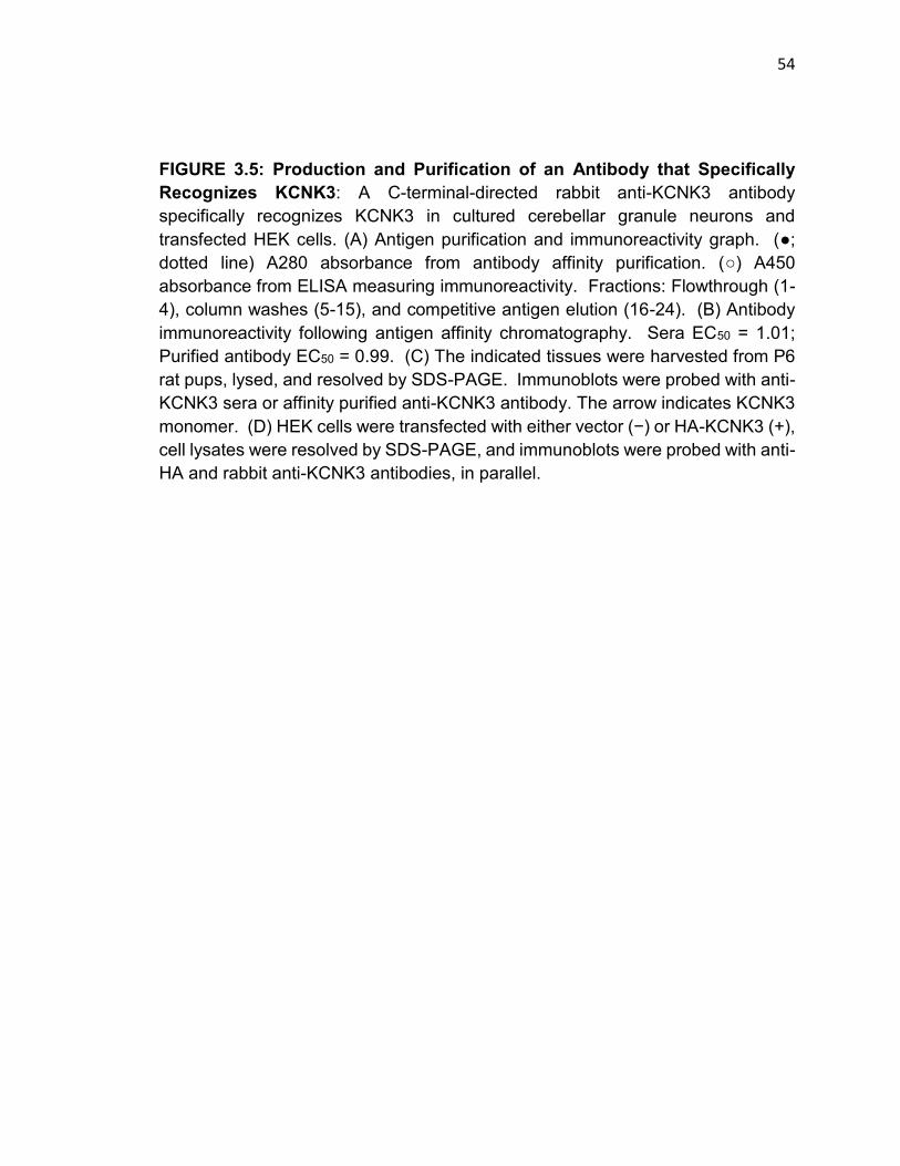
54
FIGURE 3.5: Production and Purification of an Antibody that Specifically Recognizes KCNK3: A C-terminal-directed rabbit anti-KCNK3 antibody specifically recognizes KCNK3 in cultured cerebellar granule neurons and transfected HEK cells. (A) Antigen purification and immunoreactivity graph. (●; dotted line) A280 absorbance from antibody affinity purification. (○) A450 absorbance from ELISA measuring immunoreactivity. Fractions: Flowthrough (1-4), column washes (5-15), and competitive antigen elution (16-24). (B) Antibody immunoreactivity following antigen affinity chromatography. Sera EC50 = 1.01; Purified antibody EC50 = 0.99. (C) The indicated tissues were harvested from P6 rat pups, lysed, and resolved by SDS-PAGE. Immunoblots were probed with anti-KCNK3 sera or affinity purified anti-KCNK3 antibody. The arrow indicates KCNK3 monomer. (D) HEK cells were transfected with either vector (−) or HA-KCNK3 (+), cell lysates were resolved by SDS-PAGE, and immunoblots were probed with anti-HA and rabbit anti-KCNK3 antibodies, in parallel.
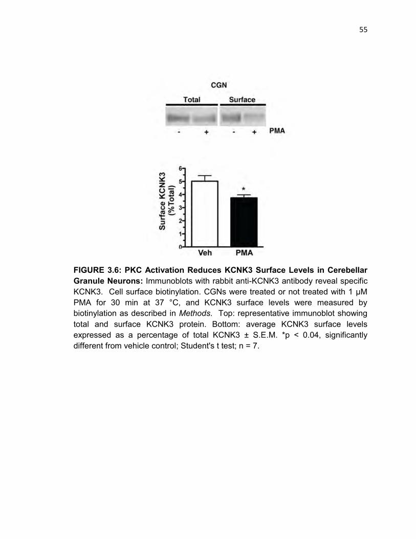
55
FIGURE 3.6: PKC Activation Reduces KCNK3 Surface Levels in Cerebellar Granule Neurons: Immunoblots with rabbit anti-KCNK3 antibody reveal specific KCNK3. Cell surface biotinylation. CGNs were treated or not treated with 1 μM PMA for 30 min at 37 °C, and KCNK3 surface levels were measured by biotinylation as described in Methods. Top: representative immunoblot showing total and surface KCNK3 protein. Bottom: average KCNK3 surface levels expressed as a percentage of total KCNK3 ± S.E.M. *p < 0.04, significantly different from vehicle control; Student's t test; n = 7.

56
internalization. As seen in Figure 3.7, KCNK3 internalized into a subset of Tf-
positive endosomes, suggesting that KCNK3 traffics via the endosomal pathway
and, specifically, through Tf-positive endosomes. Following internalization,
proteins can divert to either degradative or recycling endocytic pathways. To test
whether KCNK3 is subject to degradation following internalization, we monitored
KCNK3 stability over time ± 1 µM PMA, following pretreatment with the
translational inhibitor cyclohexamide. KCNK3 was highly stable over a 60 minute
period, and PMA treatment had no effect on total KCNK3 protein levels (Fig. 3.8),
suggesting that following internalization KCNK3 traffics through a non-degradative
pathway. Taken together, these data indicate that KCNK3 undergoes PKC-
stimulated, non-degradative endocytosis.
Group I mGluR activation induces KCNK3 downregulation and
internalization
To investigate physiologically relevant stimuli that may regulate KCNK3 trafficking,
we tested whether activating Group I mGluRs would alter KCNK3 function in
CGNs. Group I mGluRs couple to Gq activation, which ultimately activates PKC,
and are abundantly expressed in CGNs (Prezeau et al., 1994). Moreover, Group
I mGluR activation has been shown to decrease acid-sensitive currents in motor
neurons (Talley, Lei, Sirois, & Bayliss, 2000). CGNs were treated with the Group
I mGluR agonist dihydroxy-phenyl-glycine (DHPG, 1 µM) during whole cell
recording. We observed a rapid and significant reduction in KCNK3-associated

57

58
FIGURE 3.7: KCNK3 Internalization Specifically Requires PKC Activation and Traffics to Transferrin-positive Endosomes: HEK cells were co-transfected with KCNK3 and the cell surface marker IL2αR (Tac) and were pretreated with or without 1 μM BIM, followed by treatment with or without 1 μM PMA for 30 min at 37°C. Surface Tac was labeled on non-permeabilized cells with αTac antibody (green). Cells were then permeabilized and stained for KCNK3 (red). Images were captured and analyzed as described under Methods. Scale bars, 10 μm. (A) representative images; (B) image quantification. Data are expressed as percentage of vehicle surface levels ± S.E.M. (error bars). *p<0.05, significantly different from vehicle, BIM, and BIM/PMA; one-way analysis of variance with Tukey's multiple comparison test; n = 12. (C) Cells were treated or not treated with 1 μM PMA for 30 min at 37°C in the presence of Alexa594-Tf (green), fixed, permeabilized, and stained for KCNK3 (red). Scale bars, 10 μm. Enlarged view, scale bar, 1 μm.
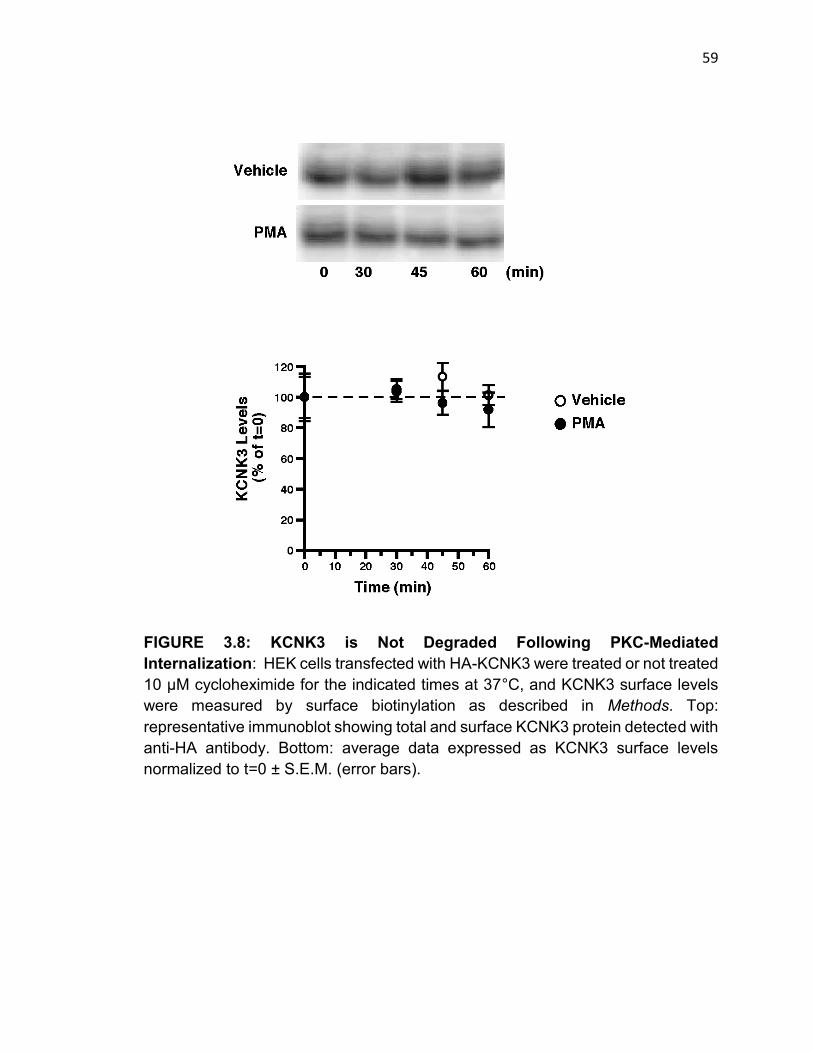
59
FIGURE 3.8: KCNK3 is Not Degraded Following PKC-Mediated Internalization: HEK cells transfected with HA-KCNK3 were treated or not treated 10 μM cycloheximide for the indicated times at 37°C, and KCNK3 surface levels were measured by surface biotinylation as described in Methods. Top: representative immunoblot showing total and surface KCNK3 protein detected with anti-HA antibody. Bottom: average data expressed as KCNK3 surface levels normalized to t=0 ± S.E.M. (error bars).
60
current in response to DHPG treatment, which was comparable in magnitude to
that observed following PMA treatment (56 ± 10% current remaining; n = 8; p<0.01)
(Fig 3.9). To determine whether DHPG-mediated losses in current were PKC-
dependent, CGNs were pretreated with 1µM BIM prior to DHPG application. BIM
alone had no effect on acid-sensitive currents (Fig. 3.9), but completely blocked
DHPG-mediated loss in KCNK3 currents. These results indicate that KCNK3
undergoes PKC-mediated functional downregulation in response to Group I
mGluR activation.
We next asked whether Group I mGluR activation stimulates KCNK3
internalization and, if so, whether PKC activation was required. HEK cells were
co-transfected with KCNK3 and either GFP alone or with mGluR5 in a bicistronic
expression vector. Treatment with 1 µM DHPG for 20 min at 37°C, had no effect
on KCNK3 surface levels in cells expressing KCNK3 and GFP (p= 0.89; Student’s
t test; n=6) (Fig. 3.10). In contrast, when KCNK3 was co-expressed with mGluR5,
1µM DHPG treatment decreased KCNK3 surface levels to 70.7 ± 5.0% of KCNK3
levels in vehicle-treated cells. (Fig. 3.10). mGluR5-mediated KCNK3
internalization required PKC activation, as pretreatment with 1µM BIM completely
blocked mGluR5-mediated KCNK3 surface losses (Fig. 3.10). Taken together, our
results indicate that KCNK3 is subject to acute internalization in response to PKC
activation, and that Group I mGluR activation can trigger KCNK3 endocytosis.
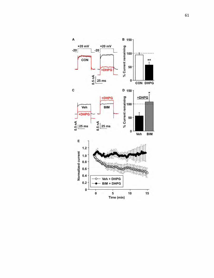
61
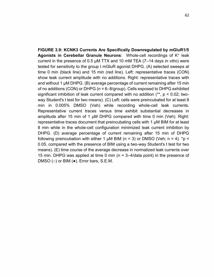
62
FIGURE 3.9: KCNK3 Currents Are Specifically Downregulated by mGluR1/5 Agonists in Cerebellar Granule Neurons: Whole-cell recordings of K+ leak current in the presence of 0.5 μM TTX and 10 mM TEA (7–14 days in vitro) were tested for sensitivity to the group I mGluR agonist DHPG. (A) selected sweeps at time 0 min (black line) and 15 min (red line). Left: representative traces (CON) show leak current amplitude with no additions. Right: representative traces with and without 1 μM DHPG. (B) average percentage of current remaining after 15 min of no additions (CON) or DHPG (n = 6–8/group). Cells exposed to DHPG exhibited significant inhibition of leak current compared with no addition (**, p < 0.02; two-way Student's t test for two means). (C) Left: cells were preincubated for at least 8 min in 0.005% DMSO (Veh) while recording whole-cell leak currents. Representative current traces versus time exhibit substantial decreases in amplitude after 15 min of 1 μM DHPG compared with time 0 min (Veh). Right: representative traces document that preincubating cells with 1 μM BIM for at least 8 min while in the whole-cell configuration minimized leak current inhibition by DHPG. (D) average percentage of current remaining after 15 min of DHPG following preincubation with either 1 μM BIM (n = 3) or DMSO (Veh; n = 4). *p < 0.05, compared with the presence of BIM using a two-way Student's t test for two means). (E) time course of the average decrease in normalized leak currents over 15 min. DHPG was applied at time 0 min (n = 3–4/data point) in the presence of DMSO (○) or BIM (●). Error bars, S.E.M.
63
FIGURE 3.10: KCNK3 Internalizes in Response to mGluR1/5 Agonists via a PKC-specific Mechanism: (A) Cell surface biotinylation. Cells were treated or not treated with 1 μM DHPG for 20 min at 37°C, and KCNK3 surface levels were measured as described under Methods. Top: representative immunoblot showing total and surface KCNK3 protein. Bottom: average KCNK3 surface levels expressed as percentage of vehicle ± S.E.M. (error bars). *p < 0.03, significantly different from vehicle control; Student's t test; n = 6. (B) cells were treated or not treated with 1 μM BIM for 20 min at 37 °C followed by treatment with or without 1 μM DHPG for 20 min at 37°C, and KCNK3 surface levels were measured as described under Methods. Top: representative immunoblot showing KCNK3 total and surface protein. Bottom: average KCNK3 surface levels expressed as percentage of vehicle ± S.E.M. *p < 0.03, significantly different from vehicle control; Student's t test; n = 6.
64
KCNK3 carboxy terminal residues are both necessary and sufficient for PKC-
regulated KCNK3 internalization
We next sought to identify the molecular determinants of PKC-mediated KCNK3
internalization. We previously reported that the dopamine transporter (DAT)
encodes a novel carboxy terminal endocytic signal, FREKLAYAIA, that regulates
both constitutive internalization, as well as trafficking in response to PKC activation
(Boudanova et al., 2008b; Holton et al., 2005). In particular, the REK residues
have been shown by our laboratory to be sensitive to PKC activation. The tandem
pore potassium channel KCNK3 (TASK-1) encodes a homologous sequence,
SREKLQYSIP, in carboxy terminal residues 334-343, which is absent in the
KCNK3 homolog, KCNK9 (Fig. 3.11). We used a gain-of-function assay to ask
whether the KCNK3 carboxy terminus was sufficient to drive internalization of an
endocytic-defective reporter protein, Tac. As seen in Figure 3.10, Tac is highly
expressed at the cell surface under trafficking restrictive conditions (4°C), and
remains at the surface in trafficking permissive conditions (37°C). In contrast, a
Tac fusion protein expressing the KCNK3 carboxy terminus robustly internalized
at 37°C. Internalization was specific to the KCNK3 carboxy terminus, as a Tac-
KCNK9 fusion protein failed to internalize (Fig. 3.11). These results indicate that
the KCNK3 carboxy terminus encodes residues sufficient to drive endocytosis.
Given the homology between the DAT endocytic signal with the SREKLQYSIP
motif in KCNK3, we next tested whether residues involved in PKC-regulated DAT

65
FIGURE 3.11: The KCNK3 Carboxy Terminus Contains an Endocytic Signal: PC12 were transfected with the indicated constructs and assayed 24–48 h post-transfection. (A) Sequence alignment between DAT and the potassium channel KCNK3 and KCNK9 C-terminal subregions. (B) Immunocytochemistry. Intact cells were incubated in anti-Tac antibody at 4°C, shifted to 37°C for 30 min, and then fixed and imaged as described under Methods.

66
internalization were also necessary for KCNK3 downregulation and internalization.
To test this possibility, we mutated KCNK3 residues 335-337 (REK) to alanines
and tested the capacity of this mutant to undergo PKC-mediated downregulation
and internalization. The KCNK3 335-337(3A) mutant expressed and exhibited
whole cell currents comparable to those measured for wildtype channel (Fig. 3.12).
As observed previously, treatment with 1µM PMA resulted in a significant, time-
dependent decrease in wildtype KCNK3 currents (Fig. 3.12). In contrast, KCNK3
335-337(3A) was completely insensitive to PMA treatment, and no current loss
was observed (Fig. 3.12). We next investigated whether abolished PKC-mediated
KCNK3 335-337(3A) current losses were due to perturbation of KCNK3
internalization. Cellular imaging revealed that KCNK3 335-337(3A) expressed at
the cell surface, but failed to internalize following PKC activation (Fig. 3.12). We
next used surface biotinylation to compare wildtype and KCNK3 335-337(3A)
surface levels ±PKC activation. As shown in Figure 3.12, we confirmed that
mutating KCNK3 residues 335–337 significantly blocked PKC-mediated KCNK3
surface losses (wild type: 63.7 ± 7.5% of vehicle levels; REK/AAA: 114.4 ± 12.4%
of vehicle levels, p<0.02, Student's t test, n = 4), demonstrating that these three
residues are key determinants of PKC-mediated KCNK3 functional down-
regulation and internalization.

67
FIGURE 3.12: Residues 335-337 in the KCNK3 Carboxy Terminus are Required for PKC-mediated Functional and Surface Losses: HEK cells were transfected with the indicated constructs and assayed 24–48 h post-transfection. The REK/AAA mutation prevents PMA-induced inhibition of KCNK3 current. (A) Left: representative traces of KCNK3 current recorded from HEK293T cells expressing either WT or REK/AAA mutant channels recorded before (black) and after (red) 1 μM PMA treatment (40-mV test pulse). Middle: average normalized time course of WT (open circles; n= 3) or REK/AAA (filled circles; n= 5) currents recorded during a 15-min exposure to 1 μM PMA. Right: mean ± S.E.M. of current remaining after a 15-min exposure to PMA (p= 0.007). (B) Immunocytochemistry. Cells were treated or not treated with 1 μM PMA for 30 min at 37°C, fixed, stained for KCNK3, and imaged as described under Methods. Scale bars, 10 μm. (D) cell surface biotinylation. Cells were treated or not treated with 1 μM PMA for 15 min at 37°C, and KCNK3 surface levels were measured as described under Methods. Average KCNK3 surface levels following PMA treatment are expressed as percentage of vehicle ± S.E.M. (error bars). *p< 0.02, significantly different from WT; Student's t-test; n= 4.

68
PKC-mediated KCNK3 endocytosis requires 14-3-3
Previous work from our laboratory demonstrated that PKC-induced DAT trafficking
relies on a saturable factor (Loder & Melikian, 2003). In that study, increased DAT
expression levels correlated with losses in PKC-induced transporter
internalization. To test whether PKC-stimulated KCNK3 internalization also relied
upon a saturable factor, we performed overexpression experiments in transiently
transfected HEK cells in which 5-fold more KCNK3 cDNA was used for
transfections, which translated into a 10-fold increase in KCNK3 protein expression
(Fig 3.13). Under vehicle conditions, KCNK3 was readily apparent at the cell
surface (Fig. 3.13). However, overexpression completely abolished PKC-
mediated KCNK3 internalization (Fig 3.13). We next tested whether co-expression
of a co-factor could rescue KCNK3 in an overexpression system. The
phosphoserine binding protein 14-3-3 is reported to facilitate KCNK3 exit from the
ER and to interact with the KCNK3 distal carboxy terminus. Importantly, KCNK3
encodes a canonical (R/K)XX(Y/F)pSXP 14-3-3 binding site in the SREKLQYSIP
region (SREKLQYSIP), prompting us to ask whether 14-3-3 may also play a role
in PKC-mediated KCNK3 endocytosis. We tested this possibility by co-
overexpressing KCNK3 and 14-3-3 and assessing whether PKC-mediated
KCNK3 internalization could be rescued by 14-3-3. Co-expression of 14-3-3
completely rescued the ability of KCNK3 to internalize in response to PKC
activation in the overexpression system (Fig. 3.13). This suggests that 14-3-3

69
FIGURE 3.13: The Phosphoserine Binding Protein 14-3-3β is a Saturable Factor Required for PKC-mediated KCNK3 Internalization: HEK cells were transfected with 5X the normal HA-KCNK3 with or without 14-3-3β-GFP and assayed for trafficking 48 h post-transfection. Channel overexpression blocked PKC-mediated KCNK3 trafficking and 14-3-3 β co-expression rescued the trafficking blockade. Immunocytochemistry: Cells were treated or not treated with 1 μM PMA for 30 min at 37°C, fixed, stained for KCNK3 (red), and imaged as described under Methods. GFP expression indicates co-transfection (green).
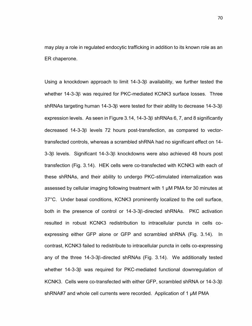
70
may play a role in regulated endocytic trafficking in addition to its known role as an
ER chaperone.
Using a knockdown approach to limit 14-3-3 availability, we further tested the
whether 14-3-3 was required for PKC-mediated KCNK3 surface losses. Three
shRNAs targeting human 14-3-3 were tested for their ability to decrease 14-3-3
expression levels. As seen in Figure 3.14, 14-3-3 shRNAs 6, 7, and 8 significantly
decreased 14-3-3 levels 72 hours post-transfection, as compared to vector-
transfected controls, whereas a scrambled shRNA had no significant effect on 14-
3-3 levels. Significant 14-3-3 knockdowns were also achieved 48 hours post
transfection (Fig. 3.14). HEK cells were co-transfected with KCNK3 with each of
these shRNAs, and their ability to undergo PKC-stimulated internalization was
assessed by cellular imaging following treatment with 1 µM PMA for 30 minutes at
37°C. Under basal conditions, KCNK3 prominently localized to the cell surface,
both in the presence of control or 14-3-3-directed shRNAs. PKC activation
resulted in robust KCNK3 redistribution to intracellular puncta in cells co-
expressing either GFP alone or GFP and scrambled shRNA (Fig. 3.14). In
contrast, KCNK3 failed to redistribute to intracellular puncta in cells co-expressing
any of the three 14-3-3-directed shRNAs (Fig. 3.14). We additionally tested
whether 14-3-3 was required for PKC-mediated functional downregulation of
KCNK3. Cells were co-transfected with either GFP, scrambled shRNA or 14-3-3
shRNA#7 and whole cell currents were recorded. Application of 1 µM PMA
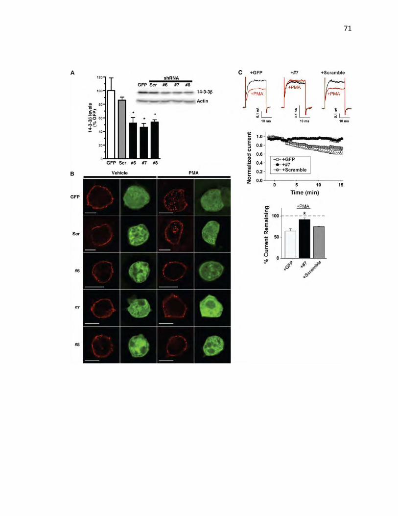
71
72
FIGURE 3.14: The Phosphoserine Binding Protein 14-3-3β is Required for PKC-mediated KCNK3 Functional and Surface Losses: HEK cells were transfected with the indicated constructs and assayed 48 h (A) or 72 h (B and C) post-transfection. (A) shRNA-mediated 14-3-3β knockdown. Average 14-3-3β protein levels are expressed as percentage of control levels ± S.E.M. *p < 0.004, significantly different from GFP-transfected controls; one-way analysis of variance with Dunnett's post hoc analysis; n=4. Inset, representative immunoblot probed for 14-3-3β and actin (loading control). (B) Immunocytochemistry. Cells were treated or not treated with 1 μM PMA for 30 min at 37 °C, fixed, and stained for KCNK3 (red). GFP expression indicates shRNA co-transfection (green). Images were captured and analyzed as described under Methods. Scale bars, 10 μm. (C) PKC-mediated inhibition of KCNK3 current requires 14-3-3β. Top: representative whole-cell current traces from HEK293T cells co-expressing KCNK3 with either GFP, shRNA 7, or scrambled shRNA recorded before (black) and after (red) 1 μM PMA treatment (40-mV test pulse). Average normalized time course (middle) and mean ± S.E.M. (error bars) of current remaining (bottom) after 15 min of PMA treatment are shown; n=9 for GFP control; n=4 for scrambled and #7 shRNAs. *p < .02, significantly different from wildtype, , one-way ANOVA with Bonferroni's multiple comparison test.

73
resulted in a time-dependent decrease in KCNK3 currents in cells co-expressing
either GFP or scrambled shRNA (Fig. 3.14). However, 14-3-3-directed shRNA#7
completely blocked the PMA-induced downregulation of KCNK3 (Fig. 3.14). Taken
together, these results indicate that 14-3-3 plays a requisite role in PKC-mediated
KCNK3 internalization, and implicates 14-3-3 in regulated endocytosis, in addition
to its previously defined role in ER export.
DISCUSSION
Membrane protein trafficking is a central mechanism controlling neuronal
excitability and plasticity in the brain. Regulated internalization of ligand-gated ion
channels, such as AMPA (Bredt & Nicoll, 2003), NMDA (Perez-Otano & Ehlers,
2005) and GABAA receptors (Kittler, McAinsh, & Moss, 2002), is a key factor in
synaptic plasticity. Recent studies indicate that endocytic trafficking acutely
regulates several different types of K+ channels, including KATP (Manna et al.,
2010) and KCa.2.1 (Correa, Muller, Collingridge, & Marrion, 2009), suggesting that
regulated membrane trafficking is a means to rapidly control K+ channel density.
Indeed, a recent study demonstrated that TWIK1 undergoes regulated
internalization via a classic dileucine endocytic signal (Feliciangeli et al., 2010). In
the current study, our data reveal that dynamic endocytic trafficking regulates acid-
sensitive K+ leak channel surface expression. This finding suggests a role for
KCNK3 to uniquely contribute to synaptic plasticity. KCNK3 internalization would
be predicted to depolarize the membrane potential, which would inactivate Nav

74
channels, causing the membrane to reach threshold more slowly, fail in firing,
and/or spike at a slower frequency of bursting. Alternatively, membrane
depolarization mediated by regulated KCNK3 trafficking could relieve Mg2+ block
of NMDA receptors, increasing the probability of NMDA receptor firing and
downstream plasticity events in response to an excitatory postsynaptic potential.
Thus, in combination with established ligand-gated ion channel trafficking, acutely
regulating KCNK3 surface levels would give rise to a multimodal, context-
dependent plasticity of membrane excitability that could last for many minutes.
Prior studies using phorbol esters (Lopes et al., 2000), Group I mGluR agonists
(Talley et al., 2000), and M1/M3 muscarinic agonists (Meuth et al., 2003)
demonstrated a PKC-mediated functional down-regulation of KCNK3 and/or acid-
sensitive currents, respectively. However, the mechanisms underlying this down-
regulation have not been well defined. We observed significant loss of KCNK3
activity in response to PMA treatment over a 15-min time course in both CGNs and
HEK cells, which is consistent with the time course for endocytosis. We used
surface biotinylation and cellular imaging to directly test whether PKC-stimulated
KCNK3 current losses were due to internalization. We observed significant PKC-
dependent losses in surface KCNK3 following PMA treatment both in HEK cells
and CGNs, which were completely blocked by the PKC inhibitor BIM. The
magnitude of KCNK3 surface losses paralleled PKC-mediated KCNK3 currents
losses, consistent with endocytosis as the primary mechanism responsible for

75
PKC-mediated KCNK3 inhibition. Moreover, we observed losses in acid-sensitive
leak currents and KCNK3 internalization via activation of endogenously expressed
Group I mGluRs, consistent with previous reports demonstrating Group I mGluR-
mediated KCNK3 down-regulation Previous studies reported rapid KCNK3 activity
losses in response to direct Gq activation (X. Chen et al., 2006) or via glutamatergic
signaling (Chemin et al., 2003) in heterologous expression systems and CGNs,
whereas we observed a slower time course of KCNK3 inhibition. These differences
may be due to direct Gq activation in a heterologous expression system versus
indirect via Gq coupling in our experiments. This difference may also reflect the
much higher DHPG concentrations used in previous studies, compared with those
used in our studies (10 and 100 μM versus 1 μM in our study). Indeed, decreased
cAMP production in response to high DHPG concentrations has been reported,
which is likely mediated by Group III mGluR activation. It is not known whether
PKC activation results in KCNK3 phosphorylation, either directly or indirectly. A
recent report demonstrated that endothelin-1 down-regulates KCNK3 and leads to
PKC-dependent KCNK3 phosphorylation in pulmonary artery smooth muscle cells
(Tang et al., 2009). However, it should be noted that these phosphorylation studies
relied upon the commercially available anti-KCNK3 antibody that, in our hands,
does not recognize a KNCK3-specific band. It is interesting to note that under basal
conditions, KNCK3 surface expression in primary CGN cultures was <6% total
KCNK3 protein, which differed markedly from observed surface levels in
transfected HEK cells. Nevertheless, these values are consistent with those

76
reported for other channels in both primary cultured neurons (Gross, Yao, Pong,
Jeromin, & Bassell, 2011) and acute brain slices (D. Y. Kim, Gersbacher,
Inquimbert, & Kovacs, 2011). Low channel surface density (as a fraction of the
total available channel) may reflect large intracellular endocytic pools tightly
regulated by neuron-specific mechanisms. Alternatively, they may reflect a high
degree of turnover in neuronal systems, with large, forward trafficking protein pools
to maintain steady state channel levels in the membrane.
Following internalization from the cell surface, proteins can diverge to either
recycling or degradative endocytic pathways (Bonifacino & Traub, 2003). For
example, EGFR (Huang, Kirkpatrick, Jiang, Gygi, & Sorkin, 2006) and δ-opioid
receptors (von Zastrow, 2010) enter late endosomes and are degraded upon
internalization, whereas the TfR is primarily recycled. We observed KCNK3 co-
localization in an early endosome/TfR-positive vesicle population following
internalization and detected no losses in total KCNK3 protein following PKC
stimulation. These results suggest that internalized KCNK3 is likely to enter a
recycling, rather than a degradative, pathway.
Previous studies from our laboratory investigating mechanisms responsible for
PKC-stimulated DAT trafficking revealed a novel endocytic regulatory domain
(FREKLAYAIA) encoded in the DAT C-terminus that is highly conserved across
the SLC6 transporter gene family and is the locus for an endocytic braking

77
mechanism (Boudanova et al., 2008b). Sequence comparison across the
mammalian genome revealed that KCNK3 is the only membrane protein outside
of the SLC6 transporter gene family to encode a homologous endocytic signal.
Gain-of-function assays revealed that the KCNK3 C terminus is sufficient for
endocytosis, and that the REK residues are absolutely required for PKC-mediated
KCNK3 down-regulation and internalization. These results offer further insight into
the previous results of Talley and Bayliss (Talley & Bayliss, 2002), in which KCNK3
C-terminal deletions that encompassed the 335–344 region abolished TRH
receptor-mediated KCNK3 inhibition, which also occurs via Gq activation. It is
currently not clear how these charged residues function to target either DAT or
KCNK3 to the endocytic machinery. Answers to this question await future studies.
PKC-stimulated KCNK3 endocytosis absolutely required the phosphoserine-
binding protein 14-3-3β. 14-3-3β belongs to the family of 14-3-3 phosphoserine-
binding proteins, which are widely expressed in the brain and periphery and exist
as homo- or heterodimers (Obsilova, Silhan, Boura, Teisinger, & Obsil, 2008). 14-
3-3 binds target proteins primarily at either RSXpSXP or (R/K)XφX(pS/pT)XP
motifs (Yaffe et al., 1997), and several proteins encode multiple 14-3-3 binding
sites (Tzivion, Luo, & Avruch, 2000). 14-3-3 binding can 1) induce conformation
changes that facilitate catalytic activity or protein-protein interactions, 2) mask
domains to prevent protein-protein interactions, or 3) facilitate protein co-
localization. Previous work demonstrated that 14-3-3β binds to a non-canonical

78
14-3-3 binding motif in the distal KCNK3 C terminus and is required for KCNK3
egress from the ER (Zuzarte et al., 2009). In addition, other 14-3-3 isoforms can
interact with the KCNK3 C-terminus and promote increased KCNK3 surface
density (Rajan et al., 2002); however, it is not clear whether the increased KCNK3
surface expression is due to ER egress or endocytic trafficking. Our results indicate
that 14-3-3β is also necessary for PKC-mediated KNCK3 internalization. We noted
that a ∼50% 14-3-3β knockdown did not markedly disrupt KCNK3 surface
targeting, whereas PKC-mediated endocytosis was abolished. This may suggest
that forward trafficking from the ER is less sensitive to 14-3-3β levels than KCNK3
surface populations. Alternatively, other accessory proteins working in consort with
14-3-3β at the cell surface may be expressed in limited quantities and are thereby
more sensitive to losses in 14-3-3β. Interestingly, the epithelial sodium channel,
ENaC, constitutively internalizes in a 14-3-3- and Nedd4-2-dependent manner,
and aldosterone increases ENaC surface expression via blocking 14-3-3-
dependent ENaC internalization and degradation (Ichimura et al., 2005). The
mechanism by which 14-3-3β promotes KCNK3 internalization is not known; nor
is it clear whether a mechanism distinct from 14-3-3β-dependent ER exit is at play.
It is interesting to note that the SREKLQYSIP region is similar to the mode II 14-3-
3β binding motif (R/K)XφX(pS/pT)XP. Although we do not currently know whether
14-3-3β controls KCNK3 internalization directly or indirectly, this site is a candidate
locus for potential 14-3-3β/KCNK3 endocytic interactions, distinct from the
identified sequence controlling KCNK3 egress from the ER. Future studies

79
exploring the possibility of this sequence as a bona fide 14-3-3β binding site should
be illuminating.

80
CHAPTER IV
Dopamine Transporter Endocytic Trafficking: Differential Dependence on
Dynamin and the Actin Cytoskeleton
INTRODUCTION
The plasma membrane dopamine transporter (DAT) is expressed exclusively in
dopaminergic neurons in the central nervous system and functions to transport
extracellular dopamine (DA) back into these neurons, thus providing temporal and
spatial regulation of DA neurotransmission. Mutations in DAT have been reported
in neurological diseases, such as attention deficit hyperactivity disorder (ADHD),
Parkinson’s disease and schizophrenia. The psychostimulants amphetamine
(AMPH) (Horn et al., 1971), cocaine (Calligaro & Eldefrawi, 1987), and
methylphenidate (Schweri et al., 1985) all competitively inhibit DAT, and DAT
binding to cocaine is requisite for the rewarding properties of this drug (R. Chen et
al., 2006; Thomsen et al., 2009). DAT surface expression determines the DA
clearance efficiency and is acutely regulated by endocytic trafficking (Buckley,
Melikian, Provoda, & Waring, 2000). DAT undergoes constitutive endocytosis and
recycling (Melikian & Buckley, 1999; Sorkina et al., 2005). Importantly, DAT
endocytic trafficking is acutely modulated by PKC activation and exposure to
AMPH (Saunders et al., 2000), as well as trafficking regulated by cell signaling
81
pathways, as well as by DAT substrates (AMPH) and inhibitors (cocaine) (Daws et
al., 2002).
The molecular mechanism regulating DAT endocytosis and recycling is still under
investigation, and previous studies have reported conflicting results. Previous work
has shown that constitutive and PKC-mediated DAT endocytosis require the
classical clathrin-mediated endocytic molecules clathrin and dynamin (Daniels &
Amara, 1999; Eriksen et al., 2009; Saunders et al., 2000). Conversely, it has been
shown that the lipid-raft associated protein flotillin-1 is required for PKC-mediated
DAT internalization, in a clathrin-independent process (Cremona et al., 2011).
These studies relied upon chronic shRNA-mediated depletion of endocytic
molecules or co-expression of dominant negative mutant molecules and were
primarily carried out in heterologous expression systems. Chronic trafficking
protein depletion over several days may act to perturb all membrane trafficking,
which could mask or overstate the necessity for DAT trafficking. Moreover, DAT
is expressed in a small subset of neurons and heterologous expression systems
may not contain the full complement of proteins required for physiological DAT
regulation. Here, we test DAT’s trafficking dependence on dynamin in acute
mouse striatal slices using pharmacological dynamin inhibitors in order to assess
trafficking requirements in a physiologically relevant and minimally perturbed
milieu. Our results demonstrate a differential dependence on dynamin for
constitutive and PKC-regulated DAT endocytic trafficking.
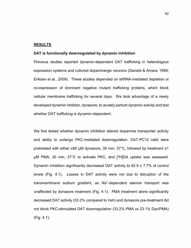
82
RESULTS
DAT is functionally downregulated by dynamin inhibition
Previous studies reported dynamin-dependent DAT trafficking in heterologous
expression systems and cultured dopaminergic neurons (Daniels & Amara, 1999;
Eriksen et al., 2009). These studies depended on shRNA-mediated depletion or
co-expression of dominant negative mutant trafficking proteins, which block
cellular membrane trafficking for several days. We took advantage of a newly
developed dynamin inhibitor, dynasore, to acutely perturb dynamin activity and test
whether DAT trafficking is dynamin-dependent.
We first tested whether dynamin inhibition altered dopamine transporter activity
and ability to undergo PKC-mediated downregulation. DAT-PC12 cells were
pretreated with either ±80 µM dynasore, 30 min, 37°C, followed by treatment ±1
µM PMA, 30 min, 37°C to activate PKC, and [3H]DA uptake was assessed.
Dynamin inhibition significantly decreased DAT activity to 62.9 ± 7.7% of control
levels (Fig. 4.1). Losses in DAT activity were not due to disruption of the
transmembrane sodium gradient, as Na+-dependent alanine transport was
unaffected by dynasore treatment (Fig. 4.1). PMA treatment alone significantly
decreased DAT activity (33.2% compared to Veh) and dynasore pre-treatment did
not block PKC-stimulated DAT downregulation (33.2% PMA vs 23.1% Dyn/PMA)
(Fig. 4.1).
83
FIGURE 4.1: Dynamin Inhibition Reduces DAT Activity but Does Not Block PKC-Mediated DAT Downregulation in PC12 Cells: Uptake assays following dynasore treatment in DAT-PC12 cells. DAT-PC12 cells were treated ± 80 μM dynasore (Dyn), 30 min, 37°C followed by ± 1 μM PMA, 30 min, 37°C and DAT function and surface levels were measured by [3H]dopamine uptake assay as described in Methods. (A) Data are expressed as % of vehicle specific DA uptake ±S.E.M. *p<0.002, (one way ANOVA with Bonferroni’s post-hoc test, n=9 (Veh and Dynasore) or n=6 (PMA and Dynasore/PMA). (B) Data are expressed as % of vehicle specific alanine uptake ±S.E.M, p=0.45 (Student’s t test, n=3).
84
Dynamin inhibition decreases DAT surface levels
To test whether dynasore-mediated losses in DAT function were due to losses in
DAT surface protein, we treated DAT-PC12 cells ±80 µM dynasore, 30 min, 37°C
and measured DAT surface levels using surface biotinylation. As can be seen in
Figure 4.2, at steady state, there was 25.5 ± 1.0% of DAT on the surface.
Dynasore treatment significantly reduced DAT surface levels to 14.5 ± 2.9% (Fig
4.2). To test for dynasore-mediated off-target effects we used dynole 34-2, which
has a higher affinity (10 µM vs 80 µM) and acts allosterically to inhibit dynamin,
whereas dynasore is a competitive inhibitor at dynamin’s GTP binding site. As
dynole 34-2 has not been extensively characterized, we tested if this inhibitor
effectively blocked transferrin receptor (TfR) internalization, a dynamin-dependent
endocytic event. We treated PC12 cells with 10 µM dynole for 30 min at 37°C and
measured fluorescent transferrin internalization (a marker for TfR endocytosis). In
vehicle-treated cells, transferrin was internalized and found in internal puncta,
which is indicative of normal endocytosis. However, in dynole treated cells there
was no visible puncta, consistent with a TfR endocytic blockade (Fig. 4.3). To
measure DAT surface levels, we performed surface biotinylation on DAT-PC12
cells. Following 10 μM dynole treatment, DAT surface levels significantly
decreased to 15.8 ± 0.9%. Additionally, we determined if PKC-mediated surface
losses were dynamin dependent. Following 1 µM PMA treatment for 30 minutes
at 37°C, DAT surface levels were significantly decreased from Veh (10.3 ± 1.3%
compared to 25.5 ± 1.0%), as has been shown previously (Fig. 4.2). Moreover,

85
FIGURE 4.2: PKC-mediated DAT Internalization is Dynamin Independent in DAT-PC12 Cells: Surface biotinylations following dynasore treatment in DAT PC12 cells. DAT-PC12 cells were treated ± 80 μM dynasore or 10 μM dynole for 30 min at 37°C followed by ± 1 μM PMA for 30 minutes at 37°C and DAT and surface levels were measured by and surface biotinylation, as described in Methods. Left: Surface biotinylation - representative immunoblot. Right: Averaged data. Data are expressed as %vehicle surface DAT levels ± S.E.M. *p<0.001, (one way ANOVA with Bonferonni’s post-hoc test, n=3-4).

86
FIGURE 4.3: Dynole treatment blocks transferrin receptor internalization: DAT-PC12 and DAT-SK-N-MC were treated with 10 μM dynole for 30 minutes at 37°C (top and middle rows) or treated with 10 μM dynole for 20 minutes at 37°C (bottom row) followed by incubation at 4°C for 60 minutes and transferrin receptor internalization was measured and visualized as described in Methods.
87
both dynasore and dynole treatment did not block the PKC-mediated DAT
internalization (Dynole: 15.8 ± 0.8% compared to 5.0 ± 1.0%; Dynasore: 14.5 ±
2.9% compared 4.5 ± 0.5%) (Fig 4.2). These data suggest that, in PC12 cells,
PKC-mediated DAT internalization is dynamin–independent.
To test whether DAT surface losses in response to dynamin inhibition were
dependent on cellular context, we also tested the effect of dynasore in the
dopaminergic neuroblastoma cell line SK-N-MC, stably transfected with DAT
(DAT-SK-N-MC). Similar to results observed in DAT-PC12 cells, 80 µM dynasore
treatment, 30 min, 37°C significantly decreased surface DAT to 77.0 ± 5.6% control
levels (Fig. 4.4). To assure that effects on DAT surface levels were not due to
possible off-target dynasore effects, we also tested dynole 34-2, and its inactive
analog dynole 31-2. Similar to our results with dynasore, 10 µM dynole treatment,
30 min, 37°C significantly decreased DAT surface levels to 68.0 ± 6.8% of vehicle-
treated cells, whereas dynole 31-2 had no significant effect on DAT surface levels
(97.6 ± 8.4% of control; (Fig 4.4). To test if PKC-mediated surface losses were
dynamin dependent, DAT-SK-N-MC cells were pretreated ± 10 µM dynole, 30 min,
37°C followed by PKC activation with 1µM PMA, 30 min, 37°C. PKC activation
induced a significant decrease in DAT surface levels following vehicle pretreatment
to 78.6 ± 6.0% of control levels. In contrast, dynole pretreatment completely
blocked any additional PKC-mediate DAT surface losses (68.0 ± 6.8% versus 69.8
± 3.8%; Fig. 4.4). In order to assess cell permeability to the biotinylation reagent,

88

89
FIGURE 4.4: Dynamin Inhibition Reduces DAT Surface Levels in DAT SK-N-MC Cells: Surface biotinylation studies: (A-D) DAT-SK-N-MC cells were treated ± 10 μM dynole for 30 minutes at 37°C followed by treatment ± 1 μM PMA for 30 minutes at 37°C or were treated 10 μM dynole 31-2 or 80 μM dynasore for 30 minutes at 37°C and DAT surface levels were measured by surface biotinylation, as described in Methods. (A) 10 μM dynole, representative blot. (B) 10 μM dynole, averaged data: DAT surface levels: Data are expressed as %vehicle surface DAT levels ±S.E.M. *Significantly different than vehicle, one-way ANOVA with Bonferonni’s post hoc test, p<0.002, n=8. (C) Total DAT protein: Data are expressed as %vehicle total DAT levels ±S.E.M. p=0.51, (one way ANOVA with Bonferonni’s post-hoc test, n=5). (D) Biotinylated actin levels: Data are expressed as %biotinylated actin of total actin ±S.E.M p=0.62, (one way ANOVA with Bonferonni’s post-hoc test, n=5). (E) 80 μM dynasore and 10 μM dynole 31-2, representative immunoblot. (F) 80 μM dynasore and 10 μM dynole 31-2, averaged data: DAT surface levels: Data are expressed as %vehicle surface DAT levels ±S.E.M. *Significantly different than vehicle, one-way ANOVA with Bonferonni’s post hoc test, p<0.05, n=8.

90
we measured the biotinylated actin population. We saw low levels of biotinylated
actin (~1.0%) across all experimental conditions indicating that the SK-N-MC cells
were intact and that trafficking could occur normally (Fig. 4.4). Previous reports
have indicated that internalized DAT is targeted for degradation (Miranda et al.,
2005). To test if this was occurring in SK-N-MC cells, we compared the total DAT
levels across experimental conditions. Total DAT protein was not significantly
different following either dynole (119.6 ± 7.9% vehicle levels) or PMA (115.4 ±
13.6% vehicle levels) treatments over the experimental time-course (Fig. 4.4). The
conflicting results between PC12 and SK-N-MC cells prompted us to ask whether
dynamin was required for basal and PKC-stimulated DAT trafficking in a native
preparation.
The incongruity in the results regarding dynamin’s role in DAT trafficking between
these two cell lines prompted us to develop a new tool to ask these questions about
dynamin’s role in DAT trafficking. Consequently, we performed surface
biotinylation on acute mouse striatal slices, a highly enriched dopaminergic
terminal region. As can be seen in Figure 4.5, we detected robust DAT surface
expression in the striatum (43.7 ± 3.8% total DAT on the surface) under vehicle-
treated conditions. PKC activation with 1 µM PMA, 30 min, 37°C significantly
decreased DAT surface levels to 77.3 ± 3.6% compared to Veh). In slices treated
with 10µM dynole, 30 min, 37°C, we observed a significant reduction in DAT
surface levels (80.7 ± 4.6% compared to Veh). Dynole pretreatment completely
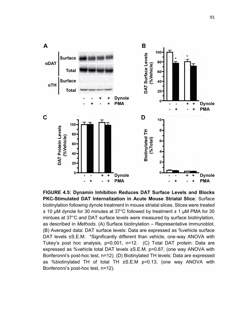
91
FIGURE 4.5: Dynamin Inhibition Reduces DAT Surface Levels and Blocks PKC-Stimulated DAT Internalization in Acute Mouse Striatal Slice: Surface biotinylation following dynole treatment in mouse striatal slices. Slices were treated ± 10 μM dynole for 30 minutes at 37°C followed by treatment ± 1 μM PMA for 30 mintues at 37°C and DAT surface levels were measured by surface biotinylation, as described in Methods. (A) Surface biotinylation – Representative immunoblot. (B) Averaged data: DAT surface levels: Data are expressed as %vehicle surface DAT levels ±S.E.M. *Significantly different than vehicle, one-way ANOVA with Tukey’s post hoc analysis, p<0.001, n=12. (C) Total DAT protein: Data are expressed as %vehicle total DAT levels ±S.E.M. p=0.87, (one way ANOVA with Bonferonni’s post-hoc test, n=12). (D) Biotinylated TH levels: Data are expressed as %biotinylated TH of total TH ±S.E.M p=0.13, (one way ANOVA with Bonferonni’s post-hoc test, n=12).
92
abolished further PMA stimulated losses (80.7 ± 4.6% vs 71.4 ± 4.3%). To test the
slice integrity and assure that the biotinylation reagent did not gain access to
intraceullar proteins in dopaminergic neurons, we immunoblotted for tyrosine
hydroxylase (TH) in parallel. Biotinylated TH levels averaged ~0.4% (Fig. 4.5),
consistent with the striatal slices being intact and the assay reflecting accurate
DAT surface measurements. To test whether dynyamin inhibition or PKC
activation target DAT for degradation in the striatal slices, we compared the total
DAT levels across experimental conditions. Total DAT protein was not significantly
different following either dynole (104 ± 5.3% vehicle levels) or PMA (100.4 ± 4.3%
vehicle levels) treatments over the experimental time-course (Fig. 4.5). Thus, we
conclude that 1) DAT traffics in SK-N-MC cells similarly to that observed in mouse
striatum and 2) the surface loss seen during PKC activation and dynamin inhibition
was the same magnitude.
Recycling blockade reveals DAT is in two surface pools
The observed DAT surface losses in SK-N-MC cells and striatal slices in response
to dynamin inhibition suggest that dynamin is not required for basal DAT
internalization, but may be required for PKC-stimulated DAT surface losses.
Another possible interpretation is that there is a finite pool of endocytic-competent
DAT in SK-N-MC cells and adult dopaminergic neurons that was depleted during
the dynole treatment, and that the remaining DAT surface pool is endocytic-
incompetent and therefore unable to internalize following PKC activation. To
93
distinguish between these possibilities, we used two independent approaches to
deplete surface DAT by blocking endocytic recycling in DAT-SK-N-MC cells:
Monensin treatment, which blocks the vacuolar H+ ATPase, and 18°C temperature
blockade, which we previously demonstrated blocks DAT endocytic recycling
(Loder & Melikian, 2003). To test whether the DAT pool that was resistant to basal
internalization had the capacity to internalize in response to PKC activation, we
initially depleted surface DAT in striatal slices by blocking endosomal recycling by
treatment with the ionophore, monensin. Monensin is a Na+/H+ antiporter inhibitor
whose action has been shown to block DAT recycling back to the plasma
membrane (Sorkina et al., 2005). We reasoned that if all surface DAT is endocytic
competent, we would expect continual surface DAT losses over time, whereas if
there is an endocytic-incompetent pool we would expect initial surface losses that
plateau over time. Treatment with 25 µM monensin, 37°C resulted in an initial
surface DAT loss that plateaued by 60 min (Fig 4.6). To test whether monensin
was effectively blocking endocytic recycling in our hands, we used a pulse-chase
approach to measure TfR recycling in SK-N-MC cells. Cells were treated with 25
µM monensin for 60 minutes at 37°C to block endocytic recycling. A 5 min Tf-
Alexa594 pulse, followed by a 20 min unlabeled Tf chase, was applied to the cells
to monitor endocytic recycling, which would be detectable as an intracellular
fluorescence loss following the Tf chase. In vehicle-treated cells, we observed
robust Tf loading after 5 min that was completely chased from the cells. In contrast,
monensin treatment markedly blocked recycling, indicated by the retention of

94
FIGURE 4.6: Monensin Treatment Blocks DAT Recycling: DAT-SK-N-MC were treated with 25 μM monensin for the indicated duration at 37°C. Surface DAT was measured by biotinylation as described in Methods. Top: Representative immunoblot. Bottom: Averaged data: DAT surface levels: data are expressed as %T=0 surface DAT ± S.E.M.
95
fluorescent transferrin (Fig. 4.7). Using these recycling block conditions, we
depleted the trafficking-competent DAT surface pool and tested whether the
remaining DAT surface population was capable of PKC-stimulated internalization.
Mouse striatal slices were pretreated with either vehicle or 25 µM Monensin, 60
min, 37°C, followed by treatment with either vehicle or 1 µM PMA, 30 min, 37°C
and surface biotinylation to measure DAT surface expression. Monensin treatment
significantly reduced DAT surface levels to 73.1 ± 5.0% as compared to vehicle-
treated slices, consistent with DAT surface depletion due to recycling blockade
(Fig. 4.8). PKC activation significantly decreased DAT surface levels, and
monensin pretreatment completely blocked further PKC-stimulated DAT surface
losses (73.1 ± 5.0% compared to 63.2 ± 4.6%). These data support the hypothesis
that surface DAT is segregated between trafficking-competent and –incompetent
pools. Recycling blockade also did not target DAT to a degradative fate, as total
DAT protein levels were unchanged over the course of the experiment for either
monensin (92.3 ± 7.7% vehicle levels) or PMA (98.5 ± 5.5% vehicle levels) (Fig.
4.8). It should be noted that monensin treatment also increased neuronal
permeability to the biotinylation reagent, as biotinylated TH levels were higher than
those seen in the vehicle-treated slices (vehicle: 1.7 ± 0.3% vs. monensin: 8.5 ±
0.9%; Fig. 4.8).
To further test whether DAT was segregated between trafficking -competent and -
incompetent surface pools, we performed 18°C recycling blockade in DAT-SK-N-

96
FIGURE 4.7: Monensin Treatment Blocks Transferrin Receptor Recycling: DAT-SK-N-MC were treated with 25 μM monensin for 60 minutes at 37°C and transferrin receptor (red) recycling was measured and visualized as described in Methods.

97
FIGURE 4.8: Recycling Blockade with Monensin Prevents PKC-Mediated DAT Internalization in acute mouse striatal slices. Surface biotinylations following recycling blockade with monensin treatment in mouse striatal slices. Slices were treated ± 25 μM monensin for 60 mintues at 37°C followed by treatment ± 1 μM PMA for 30 minutes at 37°C. DAT surface levels were measured by surface biotinylation, as described in Methods. (A) Surface biotinylation – mouse striatal slice - representative immunoblot. (B) Averaged data. Data are expressed as %vehicle surface DAT levels ±S.E.M, *p<0.0001. (one-way ANOVA with Bonferroni post hoc analysis, n=21). (C) Total DAT protein: Data are expressed as %vehicle DAT total levels ±S.E.M., p=0.46 (one-way ANOVA with Bonferroni post hoc analysis, n=21). (D) Biotinylated tyrosine hydroxylase levels: Data are expressed as %biotinylated TH of total TH ±S.E.M *p<0.001 (one-way ANOVA with Bonferroni post hoc analysis, n=21).

98
MC cells. DAT-SK-N-MC cells were incubated at 18°C for 45 minutes followed by
treatment with 1 µM PMA for 45 minutes to activate PKC. As can be seen in Figure
4.9, DAT surface depletion using an 18°C recycling blockade also blocked PKC-
regulated DAT surface loss (98.3 ± 9.4% vehicle levels), consistent with the
existence of a trafficking-incompetent DAT surface pool. It is possible that a lack
of PMA effect at 18°C was due to inability of PMA to activate PKC at 18°C. To
control for this possibility we tested whether PMA could drive DAT internalization
at 18°C in DAT-PC12 cells, which did not exhibit a trafficking incompetent DAT
pool (see Fig. 4.2). As can be seen in Figure 4.10, PKC activation at 18°C
decreased DAT surface levels (47.1 ± 9.4% vehicle levels) in DAT-PC12 cells,
demonstrating PMA activity at 18°C and that lack of PMA-induced DAT
internalization following 18°C recycling blockade was not due to lack of PKC
activation.
DAT internalization does not require dynamin
Since trafficking-competent DAT was fully depleted during the course of our dynole
treatment, we were not able to address whether PKC-mediated DAT internalization
is dynamin-dependent. In order to test whether PKC-mediated DAT internalization
is dynamin dependent, we used reversible biotinylation to directly measure basal
and PKC-stimulated DAT internalization rates during dynole treatment in DAT-SK-
N-MC cells. In order to assure that dynole incubations were sufficiently long to
impact dynamin-dependent endocytosis, but not so long as to deplete the
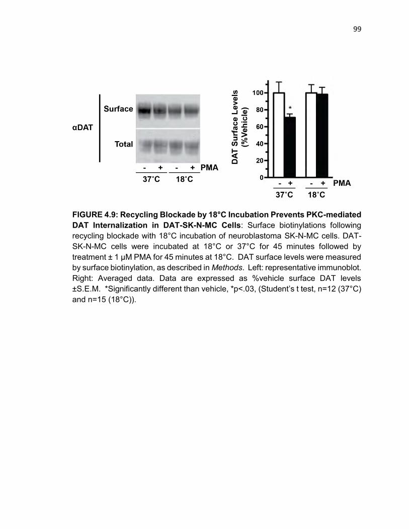
99
FIGURE 4.9: Recycling Blockade by 18°C Incubation Prevents PKC-mediated DAT Internalization in DAT-SK-N-MC Cells: Surface biotinylations following recycling blockade with 18°C incubation of neuroblastoma SK-N-MC cells. DAT-SK-N-MC cells were incubated at 18°C or 37°C for 45 minutes followed by treatment ± 1 μM PMA for 45 minutes at 18°C. DAT surface levels were measured by surface biotinylation, as described in Methods. Left: representative immunoblot. Right: Averaged data. Data are expressed as %vehicle surface DAT levels ±S.E.M. *Significantly different than vehicle, *p<.03, (Student’s t test, n=12 (37°C) and n=15 (18°C)).
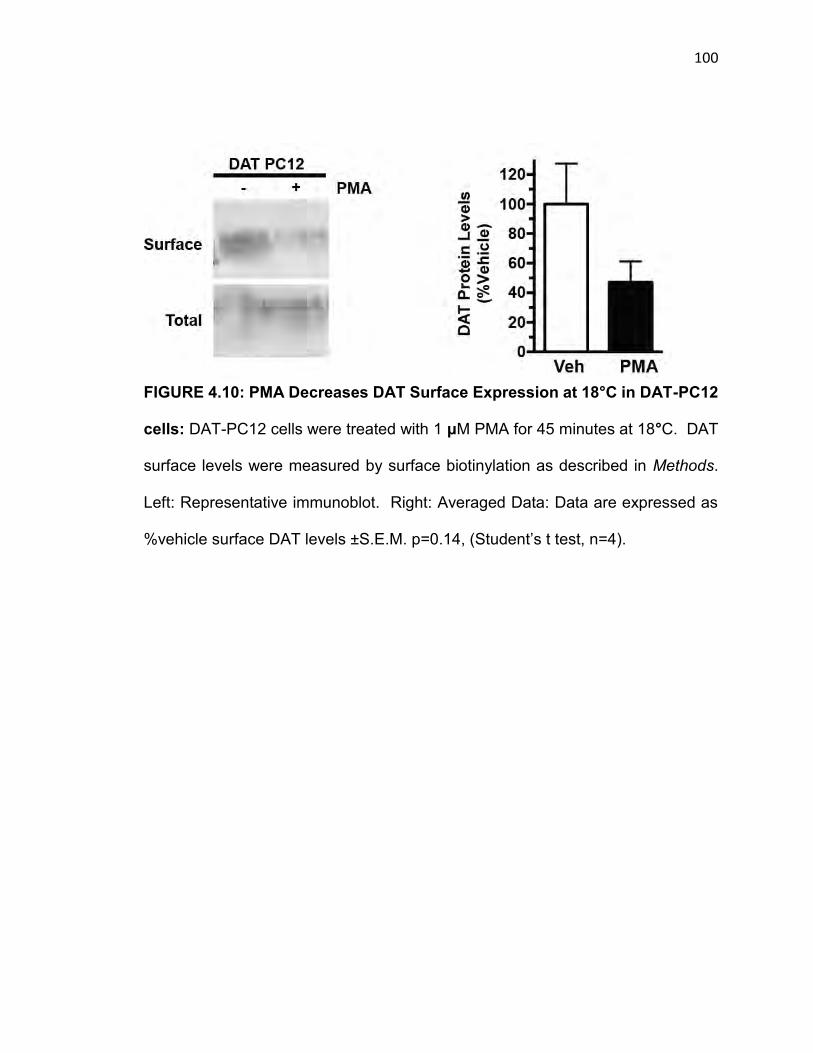
100
FIGURE 4.10: PMA Decreases DAT Surface Expression at 18°C in DAT-PC12
cells: DAT-PC12 cells were treated with 1 μM PMA for 45 minutes at 18°C. DAT
surface levels were measured by surface biotinylation as described in Methods.
Left: Representative immunoblot. Right: Averaged Data: Data are expressed as
%vehicle surface DAT levels ±S.E.M. p=0.14, (Student’s t test, n=4).
101
trafficking competent DAT pool, we developed an altered internalization protocol
in which cells were treated with dynole for 20 min, 37°C prior to biotinylating. This
short dynole pre-incubation was sufficient to block dynamin-dependent TfR
internalization (Fig. 4.3). Using the reversible biotinylation protocol, we directly
measured DAT internalization rates in DAT-SK-N-MC cells following this short
dynole pre-incubation. Under control conditions, dynole had no significant effect
on DAT internalization (68.8 ± 9.3% of Veh; Fig 4.11), consistent with the
hypothesis that constitutive DAT trafficking is dynamin independent. PKC
activation with 1 µM PMA increased DAT internalization rates to 179.1 ± 20.5% of
Veh), but did not increase DAT internalization in dynole-treated cells (68.8 ± 9.3%
compared 71.4 ± 11.6% if Veh). These results demonstrate that constitutive DAT
internalization is dynamin-independent, but that dynamin is required for PKC-
stimulation of DAT internalization rates. These results further suggest that
dynamin is required for DAT endocytic recycling back to the plasma membrane,
and that dynole treatment depleted DAT surface levels by imposing a recycling
blockade.
Dynamin regulates DAT trafficking via an actin-dependent mechanism
Given that DAT recycling appeared dynamin-dependent, we next aimed to test
what mechanisms were facilitating dynamin-dependent DAT recycling. The
cytoskeletal protein actin is known to regulate protein trafficking (Swiatecka-Urban
et al., 2002) and dynamin has been shown to be required for small actin stress

102
FIGURE 4.11: Dynamin is Required for PKC-mediated DAT Internalization but Not Constitutive Endocytosis: Internalization assay following dynole-treated DAT-SK-N-MC cells. Cells were treated ± 10 μM dynole, 20 min, 37°C, then biotinylated at 4°C, followed by ± 1 μM PMA, ± 10 μM dynole for 10 min at 37°C. Left: Internalization assay – representative immunoblot of internalized DAT. Right: Averaged data. Data are expressed as %vehicle surface DAT levels ±S.E.M. * p<0.001 (one-way ANOVA with Bonferroni post hoc analysis, n=4)
103
fibre polymerization (Cao, Deacon, Reczek, Bretscher, & von Zastrow, 1999;
Mooren, Kotova, Moore, & Schafer, 2009), making this protein a likely candidate
in the DAT trafficking mechanism. To test a putative role for actin in dynamin-
dependent DAT endocytic recycling, we disrupted the actin cytoskeleton with
cytochalasin D in mouse striatal slices and tested whether DAT surface levels were
sensitive to dynole. Slices were pretreated with either vehicle or 0.2 µg/mL
cytochalasin D (CytoD), 30 min, 37°C, followed by treatment with either vehicle or
10 µM dynole, 30 min, 37°C. CytoD treatment alone significantly reduced DAT
surface levels (69.9 ± 5.1% compared to Veh; Fig. 4.12), consistent with a role for
actin in DAT endocytic trafficking. As previously described (Fig. 4.5), dynole
treatment significantly reduce DAT surface levels to 67.2 ± 5.0% vehicle levels,
and pretreatment with CytoD completely blocked further dynole-mediated DAT
surface losses (67.2 ± 5.0% compared to 66.6 ± 7.0% of Veh; Fig. 4.12). CytoD
treatment had no effect on slice permeability to the biotinylation reagent, as seen
in immunoblots probed for TH in parallel (Fig. 4.12). CytoD treatment also did not
target DAT to a degradative fate, as total DAT protein levels were unchanged over
the course of the experiment for CytoD (91.7 ± 10.1% vehicle levels) (Fig. 4.12).
To test whether CytoD treatment globally effected endocytic trafficking, we
measured TfR surface levels in the mouse striatal slices. Using surface
biotinylation, we observed no detectable differences in surface TfR levels (118 ±
12.6% compare to Veh; Fig. 4.12) demonstrating that CytoD treatment did not
globally reduce surface protein levels.
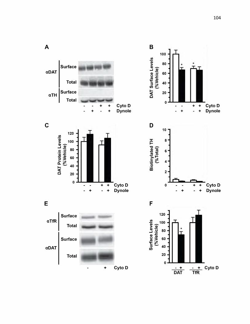
104

105
FIGURE 4.12: DAT Plasma Membrane Recycling Requires Dynamin via an Actin-Dependent Mechanism: Surface biotinylations following cytochalasin D (Cyto D) treatment mouse striatal slices. (A-D) Slices were treated ± 0.2 ug/mL cytochalasin D for 30 minutes at 37°C followed by treatment ± 10 μM dynole for 30 minutes at 37°C and DAT surface levels were measured by surface biotinylation, as described in Methods. (E-F) Slices were treated ± 0.2 ug/mL cytochalasin D for 30 minutes at 37°C and TfR and DAT surface levels were measured by surface biotinylation. (A) Surface biotinylation – Representative immunoblot. (B) Averaged data. Data are expressed as %vehicle surface DAT levels ±S.E.M. *p<0.004 (one-way ANOVA with Bonferroni’s post hoc analysis, p<0.004, n=7). (C) Total DAT protein: Data are expressed as %vehicle total DAT levels ±S.E.M. p=0.35, (one way ANOVA with Bonferonni’s post-hoc test, n=7). (D) Biotinylated TH levels: Data are expressed as %biotinylated TH of total TH ±S.E.M p=0.18, (one way ANOVA with Bonferonni’s post-hoc test, n=7). (E) Surface biotinylation – Representative immunoblot. (F) Averaged data. Data are expressed as %vehicle surface DAT or %vehicle surface TfR levels ±S.E.M. DAT: *p<0.02 (Student’s t test, n=6) and TfR: p=0.34 (Student’s t test, n=6).
106
DISCUSSION
Dopamine transporter activity is the major mechanism mediating synaptic
dopamine clearance and signal latency (Giros et al., 1996). Membrane trafficking
constitutes an important role in regulating transporter function. Recent studies
indicate that DAT trafficking requires the classical endocytic molecules clathrin and
dynamin (Eriksen et al., 2009; Sorkina et al., 2005). These studies depended on
chronic trafficking perturbation through shRNA-mediated knockdown or co-
expression of dominant negative mutant proteins, as well as heterologous cell
expression or cultured dopaminergic neurons. We took advantage of newly
developed pharmacological dynamin inhibitors acutely to block dynamin function
and test whether DAT trafficking is dynamin-dependent in mouse striatal slices.
Our results clearly demonstrate a differential dependence on dynamin for
endocytic recycling and PKC-mediated DAT internalization, but not for constitutive
DAT endocytosis (Fig. 4.5).
Using the pharmacological dynamin inhibitor, we teased out dynamin’s role in DAT
trafficking at presynaptic termini, which is the site for DAT regulation in
neurotransmitter clearance. We show that dynamin is not required for constitutive
DAT internalization (Fig. 4.11), as dynole treatment did not alter basal DAT
internalization rates. In contrast, PKC-mediated DAT internalization is strictly
dynamin-dependent. There have been other reports demonstrating that DAT
endocytosis may not require dynamin. The membrane raft associated protein

107
FIGURE 4.13: DAT is Segregated Between Trafficking-Competent and –Incompetent Pools at the Surface: DAT Trafficking model. Surface DAT is segregated between two surface pools: trafficking-competent and -incompetent. Basal DAT internalization occurs via a dynamin-independent mechanism whereas PKC mediated DAT trafficking occurs via a dynamin-dependent mechanism. DAT recycling back to the plasma membrane is dynamin and actin dependent.

108
flotillin-1 has been shown to be required for PKC-mediated DAT trafficking
(Cremona et al., 2011). The role for dynamin in that process was not specifically
explored, but trafficking from lipid rafts has been shown to be both dynamin
dependent and independent. Furthermore, DAT internalization is regulated by
presynaptic D2 receptors, where receptor activation ultimately elicits decreased
DAT surface levels, as well as D2 trafficking (Lee et al., 2007). D2 receptor
trafficking has been shown to be insensitive to dynamin inhibition (Kotowski, Hopf,
Seif, Bonci, & von Zastrow, 2011; Vickery & von Zastrow, 1999). Since DAT and
D2 interact, this raises the possibility that they are co-trafficking via a dynamin-
independent mechanism into endosomal compartments.
Interestingly, we see that surface resident DAT is not present in a uniform pool,
but rather two pools: one of which undergoes constitutive and PKC-mediated
trafficking and one which does not respond to either of these two regulatory events
(Fig. 4.8). A previous study from Foster and associates reported that DAT is
segregated between lipid raft and non-raft domains at the cell surface (Foster et
al., 2008), raising the possibility that either raft or non-raft microdomains may
facilitate trafficking competent and incompetent DAT populations. Additionally, a
DAT trafficking mutant, R615C, found in an ADHD patient, has been shown to
constitutively internalize at a higher rate and is found in a different distribution
compared to wild-type between raft and non-raft membrane domains at the surface
(Sakrikar et al., 2012). Future work measuring DAT trafficking specifically from

109
these two membrane domains will be informative regarding if this is the
segregation is used to delineate trafficking requirements.
Conflicting results have been reported regarding DAT’s endocytic fate during basal
and PKC-stimulated internalization, both in neurons and heterologous expression
systems. Previous reports show that DAT ubiquitylation is required for PKC-
mediated DAT trafficking (Miranda et al., 2005). This post-translational
modification is then required for DAT degradation, as mutations in N-terminal
lysines prevent ubiquitylation and slow DAT turnover (Vina-Vilaseca & Sorkin,
2010). These studies were done in heterologous cells treated with PMA for 1-2
hours, potentially masking DAT fate as this does not likely reflect physiologically
relevant stimuli. Additionally, work done in the Gether lab in cultured dopaminergic
neurons reports that internalized DAT preferentially sorts to Rab7-positive
endosomes, indicating a predominantly degradative fate (Eriksen et al., 2009).
This study tracked a fluorescent cocaine analog bound to DAT to test
internalization and endosomal sorting. Persistent ligand binding has been reported
to alter DAT endocytic fate, tempering the interpretation for this result (Daws et al.,
2002). Moreover, DAT trafficking was PMA-insensitive and no DAT recycling was
observed, inconsistent with our results in the striatal slice as well as DAT trafficking
studies in heterologous cells. We observe DAT surface losses in dynole-treated
slices, consistent with a role for dynamin in constitutive DAT recycling, and DAT
was not significantly degraded over the experimental time-course (Fig. 4.5).
110
DAT has been described to constitutively recycle to the plasma membrane and co-
localize in Rab11 endosomal compartments in heterologous cells (Furman, Lo, et
al., 2009). We show for the first time that DAT recycles in striatal presynaptic
termini (Fig. 4.8). Recent work has shown that D2 activation will cause a rapidly
observed increase in DAT surface levels in striatal synaptosomes followed by the
aforementioned DAT surface reduction (Chen et al., 2013). This rapid release to
increase surface expression is reminiscent of the regulated recycling seen in the
glucose transporter GLUT4. There, insulin signaling will elicit an Akt-mediated
cascade releasing GLUT4-positive endosomes to fuse at the cell surface, rapidly
increasing glucose uptake in response food intake (Foley, Boguslavsky, & Klip,
2011). Insulin treatment also acutely increases DAT activity and increases
transporter suface expression through PI3K signaling (Carvelli et al., 2002). AMPH
will also cause a rapidly observed increase in DAT surface levels followed by a
slower occurring reduction in surface DAT (Chen et al., 2009). Moreover, AMPH-
induced DAT trafficking is correlated with decreased Akt activity via a CaMKII-
dependent pathway and can be inhibited by insulin treatment (Garcia et al., 2005).
Rapid DAT recycling was also observed following dopamine uptake, with maximal
DAT surface levels observed after 80 seconds (Furman, Chen, et al., 2009). This
could provide a mechanism for neurons to mute consequent neurotransmittory
events by providing greater uptake following dopamine release. Whether the
111
molecules required for the DAT surface reduction are also mediating the increase
in DAT surface levels remain to be seen.
The precise mechanism for this recycling is not fully known, but we show that that
the cytoskeletal protein actin is required for this trafficking destination. Actin
filaments have been shown to mediate recycling endosome transport to the cell.
The cystic fibrosis conductance regulator requires actin association for recycling
back to the plasma membrane via the channel’s C-terminal PDZ domain
(Swiatecka-Urban et al., 2002). Furthermore, actin is involved in tubular
endosome formation that recycle β2-adrenergic receptor back to the cell surface in
a dynamin-dependent process (Cao et al., 1999). This is consistent with our
results in which dynamin and actin work in concert to regulate DAT surface levels
in the striatum (Fig. 4.12). Nevertheless, the full complement of molecular
components necessary for the DAT recycling pathway are still under study.
We found that the dopamine transporter constitutively internalizes via a dynamin-
independent mechanism, whereas PKC-mediated DAT internalization is dynamin-
dependent, as has been previously described (Fig. 4.13). We show that
constitutively internalized DAT does not result in DAT degradation, and that DAT
recycling back to the plasma membrane occurs via a dynamin-dependent, actin-
mediated mechanism. Futhermore, we show that not all DAT surface molecules

112
are able to constitutively internalize and that constitutive and PKC-mediated DAT
trafficking draw from the same trafficking-competent surface pool.
113
CHAPTER V
Discussion
KCNK3 Undergoes PKC-mediated Trafficking via a 14-3-3β-Dependent
Mechanism
Endocytic trafficking is a mechanism to regulate the plasma membrane function of
membrane proteins. Recent studies indicate that endocytic trafficking acutely
regulates several different types of K+ channels, including KATP (Manna et al., 2010)
and KCa.2.1 (Correa et al., 2009), suggesting that regulated membrane trafficking is
a means to rapidly control K+ channel surface density. In the current studies, our
data revealed that dynamic endocytic trafficking regulates acid-sensitive K+ leak
channel surface expression. This finding suggests a role for KCNK3 trafficking
could potentially contribute to neuronal excitability. KCNK3 internalization would
be predicted to depolarize the membrane potential, and prohibit Nav channel
activation, resulting in a slower frequency of action potential initiation.
Alternatively, membrane depolarization mediated by regulated KCNK3 trafficking
could potentially be sufficient to relieve Mg2+ block of NMDA receptors, increasing
the probability of NMDA receptor firing and downstream plasticity events in
response to an excitatory postsynaptic potential. Thus, in combination with
established ligand-gated ion channel trafficking, acutely regulating KCNK3 surface
levels could give rise to a context-dependent plasticity of membrane.
114
Prior studies using phorbol esters (Lopes et al., 2000), Group I mGluR agonists
(Talley et al., 2000), and M1/M3 muscarinic agonists (Meuth et al., 2003)
demonstrated a PKC-mediated functional down-regulation of KCNK3 and/or acid-
sensitive currents, respectively. However, the mechanisms underlying this down-
regulation have not been well defined. We observed significant loss of KCNK3
activity in response to PMA treatment over a 15-min time course in both CGNs and
HEK cells, which is consistent with the time course for endocytosis. We used
surface biotinylation and cellular imaging to directly test whether PKC-stimulated
KCNK3 current losses were due to internalization. We observed significant PKC-
dependent losses in surface KCNK3 following PMA treatment both in HEK cells
and CGNs, which were completely blocked by the PKC inhibitor BIM. The
magnitude of KCNK3 surface losses paralleled PKC-mediated KCNK3 currents
losses, consistent with endocytosis as the primary mechanism responsible for
PKC-mediated KCNK3 inhibition. Moreover, we observed losses in acid-sensitive
leak currents and KCNK3 internalization via activation of endogenously expressed
Group I mGluRs, consistent with previous reports demonstrating Group I mGluR-
mediated KCNK3 down-regulation (Talley et al., 2000). Previous studies reported
rapid KCNK3 activity losses in response to direct Gq activation (X. Chen et al.,
2006) or via glutamatergic signaling (Chemin et al., 2003; Talley et al., 2000) in
heterologous expression systems and CGNs, whereas we observed a slower time
course of KCNK3 inhibition. This difference may also reflect the much higher
DHPG concentrations used in previous studies, compared with those used in our

115
studies (10 and 100 μM versus 1 μM in our study). Decreased cAMP production in
response to high DHPG concentrations has been reported, which is likely mediated
by Group III mGluR activation (Sekiyama et al., 1996). It is not known whether PKC
activation results in KCNK3 phosphorylation, either directly or indirectly. A recent
report demonstrated that endothelin-1 down-regulates KCNK3 and leads to PKC-
dependent KCNK3 phosphorylation in pulmonary artery smooth muscle cells
(Tang et al., 2009). However, it should be noted that these phosphorylation studies
relied upon the commercially available anti-KCNK3 antibody that, in our hands,
does not recognize a KNCK3-specific band. It is interesting to note that under basal
conditions, KNCK3 surface expression in primary CGN cultures was <6% total
KCNK3 protein, which differed markedly from observed surface levels in
transfected HEK cells. Nevertheless, these values are consistent with those
reported for other channels in both primary cultured neurons and acute brain slices
(Gross et al., 2011; D. Y. Kim et al., 2011). Low channel surface density (as a
fraction of the total available channel) may reflect large intracellular endocytic
pools tightly regulated by neuron-specific mechanisms. Alternatively, they may
reflect a high degree of turnover in neuronal systems, with large, forward trafficking
protein pools to maintain steady state channel levels in the membrane.
The large intracellular pool may also reflect a mechanism by which neurons can
initiate KCNK3 trafficking to the membrane leading to neuronal quiescence or in
response to extracellular cues. Indeed, KCNK3 activity is required for mediating
116
neurodegeneration induced by stroke-induced ischemia (Hawkins & Butt, 2013).
The mechanistic understanding for this apoptosis-supporting function could open
new avenues to acutely treat stroke and diminish damage during the
cerebrovascular incident. Additionally, acute regulation of KCNK3 surface
expression has implications in nociception and pharmacological pain
management. KCNK3 is expressed in nociceptive dorsal root ganglia (Cooper,
Johnson, & Rau, 2004). As we observed a large intracellular KCNK3 pool (Fig
3.6), channel recycling to the surface would act to inhibit these neurons. Although
recycling determinants for the channel have not been described, KCNK3 shares a
trafficking motif with DAT (Fig. 3.11) and may share trafficking mechanisms
between the two proteins. Acute release from intracellular pools for KCNK3, and
DAT, is an interesting area of study and should be illuminating regarding its effect
on neuronal function.
KCNK3 activity has been shown to be required for multiple sclerosis progression.
CD4+ T-lymphocytes that target myelin and cause its degradation are activated in
a KCNK3-dependent process (Bittner et al., 2012). A newly developed
pharmacological KCNK3 inhibitor, A293, is able to inhibit T-cell activation, prevent
myelin loss, and extend longevity in a multiple sclerosis mouse model. Although
this is mediated through direct channel inactivation, pharmacological KCNK3
trafficking induction could also be a mechanism to slow disease progression and
have broader application in auto-immune disease treatment.
117
KCNK3 and a closely related leak channel KCNK9 have been show to form
heterodimers in neurons (Berg et al., 2004). Whether PKC-mediated KCNK3
trafficking will occur in the heterodimer is not known, but it offers a possibility that
KCNK9’s insensitivity to PKC will act dominantly and prevent potential
KCNK3/KCNK9 trafficking. Conversely, KCNK3 may act dominantly and induce
KCNK9 surface losses following PKC activation. Differential targeting in response
to hetero-oligomerization and competing endocytic signals has been reported in
the asialoglycoprotein receptor and alters the receptor’s polarized distribution from
apical to basolateral (Fuhrer, Geffen, Huggel, & Spiess, 1994). Whether
KCNK3/KCNK9 dimers display differential regulation is an area of ongoing study.
Following internalization from the cell surface, proteins can diverge to either
recycling or degradative endocytic pathways. For example, EGFR (Huang et al.,
2006) and δ-opioid receptors (von Zastrow, 2010) enter late endosomes and are
degraded upon internalization, whereas the TfR and µ opioid receptors (Jennifer
Whistler lab) are primarily recycled. We observed KCNK3 co-localization in an
early endosome/TfR-positive vesicle population following internalization and
detected no losses in total KCNK3 protein following PKC stimulation. These results
suggest that internalized KCNK3 is likely to enter a recycling, rather than a
degradative, pathway.
118
Previous studies from our laboratory investigating mechanisms responsible for
PKC-stimulated DAT trafficking revealed a novel endocytic regulatory domain
(FREKLAYAIA) encoded in the DAT C-terminus (Holton et al., 2005) that is highly
conserved across the SLC6 transporter gene family and is the locus for an
endocytic braking mechanism (Boudanova, Navaroli, Stevens, & Melikian, 2008a).
These results offer further insight into the previous results of Talley and Bayliss
(Talley & Bayliss, 2002), in which KCNK3 C-terminal deletions that encompassed
the 335–344 region abolished TRH receptor-mediated KCNK3 inhibition, which
also occurs via Gq activation. It is currently not clear how these charged residues
function to target either DAT or KCNK3 to the endocytic machinery. Also,
FREKLAYAIA mediates constitutive DAT internalization and the question remains
if KCNK3 constitituvely internalizes and was not directly addressed in this study.
An interesting area to further characterize the similarity between these
homologous sequences would be to exchange the endocytic signals and test for
sufficiency for the more completely described DAT FREKLAYAIA to drive KCNK3
trafficking in response to physiological cues known to mediate DAT trafficking,
such as insulin stimulation. Previous reports observed that KCNK3 activity is
modulated by metabolic cues, since glucose treatment in orexinergic neurons will
decrease channel activity (Burdakov et al., 2006). Whether DAT and KCNK3
share a cell signaling response to metabolic cues could be addressed, such as Akt
signaling for DAT and insulin, is an interesting area of study.

119
PKC-stimulated KCNK3 endocytosis absolutely required the phosphoserine-
binding protein 14-3-3β. 14-3-3 binds target proteins primarily at either RSXpSXP
or (R/K)XφX(pS/pT)XP motifs, and several proteins encode multiple 14-3-3 binding
sites (Tzivion et al., 2000; Yaffe et al., 1997). 14-3-3 binding can induce
conformation changes that facilitate catalytic activity or protein-protein interactions,
mask domains to prevent protein-protein interactions, or facilitate protein co-
localization. Previous work demonstrated that 14-3-3β binds to a non-canonical
14-3-3 binding motif in the distal KCNK3 C terminus and is required for KCNK3
egress from the ER (O'Kelly et al., 2002). Our results indicate that 14-3-3β is also
necessary for PKC-mediated KNCK3 internalization. We noted that a ∼50% 14-3-
3β knockdown did not markedly disrupt KCNK3 surface targeting, whereas PKC-
mediated endocytosis was abolished. This may suggest that forward trafficking
from the ER is less sensitive to 14-3-3β levels than KCNK3 surface populations.
Alternatively, other accessory proteins working in consort with 14-3-3β at the cell
surface may be expressed in limited quantities and are thereby more sensitive to
losses in 14-3-3β. Interestingly, the epithelial sodium channel, ENaC, constitutively
internalizes in a 14-3-3- and Nedd4-2-dependent manner, and aldosterone
increases ENaC surface expression via blocking 14-3-3-dependent ENaC
internalization and degradation (Ichimura et al., 2005). Furthermore, 14-3-3
interaction with the α2 adrenergic receptor supports receptor surface expression
and dephosphorylation followed by subsequent 14-3-3 dissociation initiates
endocytosis, consistent with a role for 14-3-3 as an endocytic brake (Wang &

120
Limbird, 2007). . Although we do not currently know whether 14-3-3β controls
KCNK3 internalization directly or indirectly, this site is a candidate locus for
potential 14-3-3β/KCNK3 endocytic interactions, distinct from the identified
sequence controlling KCNK3 egress from the ER. Future studies exploring the
possibility of this sequence as a bona fide 14-3-3β binding site and/or endocytic
braking mechanism should be informative.
DOPAMINE TRANSPORTER ENDOCYTIC TRAFFICKING: DIFFERENTIAL
DEPENDENCE ON DYNAMIN AND THE ACTIN CYTOSKELETON
Dopamine transporter activity is the major mechanism mediating synaptic
dopamine clearance and signal latency. Membrane trafficking constitutes an
important role in regulating transporter function. Recent studies indicate that DAT
trafficking requires the classical endocytic molecules clathrin and dynamin
(Eriksen et al., 2009; Sorkina et al., 2005). These studies depended on chronic
trafficking perturbation through shRNA-mediated knockdown or co-expression of
dominant negative mutant proteins, as well as heterologous cell expression or
cultured dopaminergic neurons. We took advantage of newly developed
pharmacological dynamin inhibitors acutely to block dynamin function and test
whether DAT trafficking is dynamin-dependent in mouse striatal slices. Our results
clearly demonstrate a differential dependence on dynamin for endocytic recycling
and PKC-mediated DAT internalization, but not for constitutive DAT endocytosis.

121
Using the pharmacological dynamin inhibitor, we teased out dynamin’s role in DAT
trafficking at presynaptic termini, which is the site for DAT regulation in
neurotransmitter clearance. We show that dynamin is not required for constitutive
DAT internalization, as dynole treatment did not alter basal DAT internalization
rates. In contrast, PKC-mediated DAT internalization is strictly dynamin-
dependent. There have been other reports demonstrating that DAT endocytosis
may not require dynamin. The membrane raft associated protein flotillin-1 has
been shown to be required for PKC-mediated DAT trafficking (Cremona et al.,
2011). The role for dynamin in that process was not specifically explored, but
trafficking from lipid rafts has been shown to be both dynamin-dependent and -
independent. Furthermore, DAT internalization is regulated by presynaptic D2
receptors, where receptor activation ultimately elicits decreased DAT surface
levels, as well as D2 trafficking (Lee et al., 2007). D2 receptor trafficking has been
shown to be insensitive to dynamin inhibition (Kotowski et al., 2011; Vickery & von
Zastrow, 1999). Since DAT and D2 interact, this raises the possibility that they are
co-trafficking via a dynamin-independent mechanism into endosomal
compartments.
Interestingly, we see that surface resident DAT is not present in a uniform pool,
but rather two pools: one of which undergoes constitutive and PKC-mediated
trafficking and one which does not respond to either of these two regulatory events.
A previous study from Foster and associates reported that DAT is segregated
122
between lipid raft and non-raft domains at the cell surface (Foster et al., 2008),
raising the possibility that either raft or non-raft microdomains may facilitate
trafficking competent and incompetent DAT populations. Additionally, a DAT
trafficking mutant, R615C, found in an ADHD patient, has been shown to
constitutively internalize at a higher rate and is found in a different distribution
compared to wild-type between raft and non-raft membrane domains at the surface
(Sakrikar et al., 2012). Future work measuring DAT trafficking specifically from
these two membrane domains will be informative regarding if this is the
segregation is used to delineate trafficking requirements.
The functional implications for segregated DAT populations at the cell surface are
not known. This could provide a mechanism by which dopaminergic neurons can
mute responses to cell signaling pathways, preventing profound DAT surface loss.
As DAT(-/-) mice display behavioural deficits (Giros et al., 1996) and dopaminergic
signal latency has been implicated in mood (Chaudhury et al., 2013) and cognitive
disorders (Brunelin et al., 2013), potential ‘over-trafficking’ would have deleterious
effects. Finite surface pools have been described for the potassium channel
K(V)10.1, where constitutively internalizing channel will plateau after 45 minutes,
consistent with the DAT model ((Kohl, Lorinczi, Pardo, & Stuhmer, 2011).
Moreover, this channel was observed to internalize via two different mechanisms
(clathrin-deoendent and clathrin-independent fluid phase uptake), also consistent
with our observed dynamin-dependent and –independent DAT trafficking
123
pathways. Furthermore, whether related neurotransmitter transporter, like NET or
SERT, are also segregated between trafficking-competent and –incompetent
pools is an interesting question. NET and SERT have been shown to localize to
raft and non-raft membrane domains (Matthies et al., 2009; Muller, Wiborg, &
Haase, 2006). Studies testing whether they contain a finite trafficking-competent
surface pool could have broader implications for the SLC6 protein family.
Conflicting results have been reported regarding DAT’s endocytic fate during basal
and PKC-stimulated internalization, both in neurons and heterologous expression
systems. Previous reports show that DAT ubiquitylation is required for PKC-
mediated DAT trafficking (Miranda et al., 2005). This post-translational
modification is then required for DAT degradation, as mutations in N-terminal
lysines prevent ubiquitylation and slow DAT turnover (Vina-Vilaseca & Sorkin,
2010). These studies were performed in a non-neuronal heterologous expression
system and imposed 1-2 hour PMA treatments, resulting in an extensive PKC
activation that may not reflect a physiologically relevant stimulus. This approach
could have artifactually targeted DAT to a degradative fate. Additionally, work from
Eriksen and associates in cultured dopaminergic neurons demonstrated that
internalized DAT preferentially sorts to Rab7-positive endosomes, indicating a
predominantly degradative fate (Eriksen et al., 2009). This study tracked a
fluorescent cocaine analog bound to DAT to test internalization and endosomal
sorting. Persistent ligand binding has been reported to alter DAT endocytic fate,

124
tempering the interpretation for this result (Daws et al., 2002). Moreover, DAT
trafficking was PMA-insensitive and no DAT recycling was observed, inconsistent
with our results in the striatal slice as well as DAT trafficking studies in
heterologous cells. We observe DAT surface losses in dynole-treated slices,
consistent with a role for dynamin in constitutive DAT recycling, and DAT was not
significantly degraded over the experimental time-course. However, one caveat
to our findings is that we, as yet, do not know the dynamin-dependent endocytic
compartment in which DAT accumulates. If this is an early endosomal
compartment, we may be blocking PKC-stimulated DAT degradation by arresting
progression to degradative endocytic vesicles. Future studies tracking DAT
endocytic trafficking should be illuminating in this regard.
DAT has been described to constitutively recycle to the plasma membrane and co-
localize in Rab11 endosomal compartments in heterologous cells (Furman, Lo, et
al., 2009). Our results suggest that DAT recycles in striatal presynaptic termini.
Recent work has shown that D2 activation will cause a rapidly observed increase
in DAT surface levels in striatal synaptosomes followed by the aforementioned
DAT surface reduction (Chen et al., 2013). This rapid release to increase surface
expression is reminiscent of the regulated recycling seen in the glucose transporter
GLUT4. There, insulin signaling will elicit an Akt-mediated cascade releasing
GLUT4-positive endosomes to fuse at the cell surface, rapidly increasing glucose
uptake in response food intake (Foley et al., 2011). Insulin treatment also acutely

125
increases DAT activity and increases transporter surface expression through PI3K
signaling (Carvelli et al., 2002). AMPH will also cause a rapidly (seconds)
observed increase in DAT surface levels followed by a slower (minutes) occurring
reduction in surface DAT (Chen et al., 2009). Moreover, AMPH-induced DAT
trafficking is correlated with decreased Akt activity via a CaMKII-dependent
pathway and can be inhibited by insulin treatment (Garcia et al., 2005). Rapid DAT
recycling was also observed following dopamine uptake, with maximal DAT
surface levels observed after 80 seconds (Furman, Chen, et al., 2009). This could
provide a mechanism for neurons to mute dopaminergic neurotransmission by
providing greater uptake following dopamine release. Whether the molecules
required for the DAT surface reduction are also mediating the increase in DAT
surface levels remain to be seen.
The precise mechanism for DAT recycling is not fully known, but we show that that
the cytoskeletal protein actin is required at the dynamin-dependent step in the
recycling process. Actin filaments have been shown to mediate recycling
endosome transport to the cell. The cystic fibrosis conductance regulator requires
actin association for recycling back to the plasma membrane via the channel’s C-
terminal PDZ domain (Swiatecka-Urban et al., 2002). Furthermore, actin is
involved in tubular endosome formation that recycle β2-adrenergic receptor back
to the cell surface in a dynamin-dependent process (Cao et al., 1999). This is
consistent with our results in which dynamin and actin work in concert to regulate
126
DAT surface levels in the striatum. Nevertheless, the full complement of molecular
components necessary for the DAT recycling pathway are still under study.

127
Ahern, C. A., & Kobertz, W. R. (2009). Chemical tools for K(+) channel biology. Biochemistry, 48(3), 517-526. doi: 10.1021/bi8018515
Ait-Slimane, T., Galmes, R., Trugnan, G., & Maurice, M. (2009). Basolateral internalization of GPI-anchored proteins occurs via a clathrin-independent flotillin-dependent pathway in polarized hepatic cells. Mol Biol Cell, 20(17), 3792-3800. doi: 10.1091/mbc.E09-04-0275
Albright, T. D., Jessell, T. M., Kandel, E. R., & Posner, M. I. (2000). Neural science: a century of progress and the mysteries that remain. Neuron, 25 Suppl, S1-55.
Argyelan, M., Szabo, Z., Kanyo, B., Tanacs, A., Kovacs, Z., Janka, Z., & Pavics, L. (2005). Dopamine transporter availability in medication free and in bupropion treated depression: a 99mTc-TRODAT-1 SPECT study. J Affect Disord, 89(1-3), 115-123. doi: 10.1016/j.jad.2005.08.016
Balasubramanian, N., Scott, D. W., Castle, J. D., Casanova, J. E., & Schwartz, M. A. (2007). Arf6 and microtubules in adhesion-dependent trafficking of lipid rafts. Nat Cell Biol, 9(12), 1381-1391. doi: 10.1038/ncb1657
Balklava, Z., Pant, S., Fares, H., & Grant, B. D. (2007). Genome-wide analysis identifies a general requirement for polarity proteins in endocytic traffic. Nat Cell Biol, 9(9), 1066-1073. doi: 10.1038/ncb1627
Bautista, D. M., Sigal, Y. M., Milstein, A. D., Garrison, J. L., Zorn, J. A., Tsuruda, P. R., . . . Julius, D. (2008). Pungent agents from Szechuan peppers excite sensory neurons by inhibiting two-pore potassium channels. Nat Neurosci, 11(7), 772-779.
Bayliss, D. A., Sirois, J. E., & Talley, E. M. (2003). The TASK family: two-pore domain background K+ channels. Mol Interv, 3(4), 205-219. doi: 10.1124/mi.3.4.205
Bayliss, D. A., Talley, E. M., Sirois, J. E., & Lei, Q. (2001). TASK-1 is a highly modulated pH-sensitive 'leak' K(+) channel expressed in brainstem respiratory neurons. Respir Physiol, 129(1-2), 159-174.
Berg, A. P., Talley, E. M., Manger, J. P., & Bayliss, D. A. (2004). Motoneurons express heteromeric TWIK-related acid-sensitive K+ (TASK) channels containing TASK-1 (KCNK3) and TASK-3 (KCNK9) subunits. J Neurosci, 24(30), 6693-6702.
Besana, A., Barbuti, A., Tateyama, M. A., Symes, A. J., Robinson, R. B., & Feinmark, S. J. (2004). Activation of protein kinase C epsilon inhibits the two-pore domain K+ channel, TASK-1, inducing repolarization abnormalities in cardiac ventricular myocytes. J Biol Chem, 279(32), 33154-33160. doi: 10.1074/jbc.M403525200
Birmingham, A. T., & Iversen, L. L. (1969). Uptake and metabolism of 3H-noradrenaline by normal and by denervated vasa deferentia of guinea-pigs and rats. Br J Pharmacol, 35(2), P356-358.
Bittner, S., Bauer, M. A., Ehling, P., Bobak, N., Breuer, J., Herrmann, A. M., . . . Meuth, S. G. (2012). The TASK1 channel inhibitor A293 shows efficacy in a mouse model of multiple sclerosis. Exp Neurol, 238(2), 149-155. doi: 10.1016/j.expneurol.2012.08.021

128
Blakely, R. D., Berson, H. E., Fremeau, R. T., Jr., Caron, M. G., Peek, M. M., Prince, H. K., & Bradley, C. C. (1991). Cloning and expression of a functional serotonin transporter from rat brain. Nature, 354(6348), 66-70. doi: 10.1038/354066a0
Blakely, R. D., Robinson, M. B., & Amara, S. G. (1988). Expression of neurotransmitter transport from rat brain mRNA in Xenopus laevis oocytes. Proc Natl Acad Sci U S A, 85(24), 9846-9850.
Bogdanski, D. F., Blaszkowski, T. P., & Tissari, A. H. (1970). Mechanisms of biogenic amine transport and storage. IV. Relationship between K+ and the Na+ requirement for transport and storage of 5-hydroxytryptamine and norepinephrine in synaptosomes. Biochim Biophys Acta, 211(3), 521-532.
Bonifacino, J. S., & Traub, L. M. (2003). Signals for sorting of transmembrane proteins to endosomes and lysosomes. Annu Rev Biochem, 72, 395-447.
Boudanova, E., Navaroli, D. M., Stevens, Z., & Melikian, H. E. (2008a). Dopamine transporter endocytic determinants: Carboxy terminal residues critical for basal and PKC-stimulated internalization. Mol Cell Neurosci.
Boudanova, E., Navaroli, D. M., Stevens, Z., & Melikian, H. E. (2008b). Dopamine transporter endocytic determinants: carboxy terminal residues critical for basal and PKC-stimulated internalization. Mol Cell Neurosci, 39(2), 211-217. doi: 10.1016/j.mcn.2008.06.011
Bredt, D. S., & Nicoll, R. A. (2003). AMPA receptor trafficking at excitatory synapses. Neuron, 40(2), 361-379.
Browne, D. L., Gancher, S. T., Nutt, J. G., Brunt, E. R., Smith, E. A., Kramer, P., & Litt, M. (1994). Episodic ataxia/myokymia syndrome is associated with point mutations in the human potassium channel gene, KCNA1. Nat Genet, 8(2), 136-140. doi: 10.1038/ng1094-136
Brunelin, J., Fecteau, S., & Suaud-Chagny, M. F. (2013). Abnormal striatal dopamine transmission in schizophrenia. Curr Med Chem, 20(3), 397-404.
Buckingham, S. D., Kidd, J. F., Law, R. J., Franks, C. J., & Sattelle, D. B. (2005). Structure and function of two-pore-domain K+ channels: contributions from genetic model organisms. Trends Pharmacol Sci, 26(7), 361-367. doi: 10.1016/j.tips.2005.05.003
Buckler, K. J., Williams, B. A., & Honore, E. (2000). An oxygen-, acid- and anaesthetic-sensitive TASK-like background potassium channel in rat arterial chemoreceptor cells. J Physiol, 525 Pt 1, 135-142.
Buckley, K. M., Melikian, H. E., Provoda, C. J., & Waring, M. T. (2000). Regulation of neuronal function by protein trafficking: a role for the endosomal pathway. J Physiol, 525 Pt 1, 11-19.
Burdakov, D., Jensen, L. T., Alexopoulos, H., Williams, R. H., Fearon, I. M., O'Kelly, I., . . . Verkhratsky, A. (2006). Tandem-pore K+ channels mediate inhibition of orexin neurons by glucose. Neuron, 50(5), 711-722.
129
Burgueno, J., Enrich, C., Canela, E. I., Mallol, J., Lluis, C., Franco, R., & Ciruela, F. (2003). Metabotropic glutamate type 1alpha receptor localizes in low-density caveolin-rich plasma membrane fractions. J Neurochem, 86(4), 785-791.
Calligaro, D. O., & Eldefrawi, M. E. (1987). High affinity stereospecific binding of [3H] cocaine in striatum and its relationship to the dopamine transporter. Membr Biochem, 7(2), 87-106.
Cao, T. T., Deacon, H. W., Reczek, D., Bretscher, A., & von Zastrow, M. (1999). A kinase-regulated PDZ-domain interaction controls endocytic sorting of the beta2-adrenergic receptor. Nature, 401(6750), 286-290. doi: 10.1038/45816
Carvelli, L., Moron, J. A., Kahlig, K. M., Ferrer, J. V., Sen, N., Lechleiter, J. D., . . . Galli, A. (2002). PI 3-kinase regulation of dopamine uptake. J Neurochem, 81(4), 859-869.
Chaudhury, D., Walsh, J. J., Friedman, A. K., Juarez, B., Ku, S. M., Koo, J. W., . . . Han, M. H. (2013). Rapid regulation of depression-related behaviours by control of midbrain dopamine neurons. Nature, 493(7433), 532-536. doi: 10.1038/nature11713
Chemin, J., Girard, C., Duprat, F., Lesage, F., Romey, G., & Lazdunski, M. (2003). Mechanisms underlying excitatory effects of group I metabotropic glutamate receptors via inhibition of 2P domain K+ channels. EMBO J, 22(20), 5403-5411. doi: 10.1093/emboj/cdg528
Chen, R., Daining, C. P., Sun, H., Fraser, R., Stokes, S. L., Leitges, M., & Gnegy, M. E. (2013). Protein kinase Cbeta is a modulator of the dopamine D2 autoreceptor-activated trafficking of the dopamine transporter. J Neurochem, 125(5), 663-672. doi: 10.1111/jnc.12229
Chen, R., Furman, C. A., Zhang, M., Kim, M. N., Gereau, R. W. t., Leitges, M., & Gnegy, M. E. (2009). Protein kinase Cbeta is a critical regulator of dopamine transporter trafficking and regulates the behavioral response to amphetamine in mice. J Pharmacol Exp Ther, 328(3), 912-920. doi: 10.1124/jpet.108.147959
Chen, R., Tilley, M. R., Wei, H., Zhou, F., Zhou, F. M., Ching, S., . . . Gu, H. H. (2006). Abolished cocaine reward in mice with a cocaine-insensitive dopamine transporter. Proc Natl Acad Sci U S A, 103(24), 9333-9338. doi: 10.1073/pnas.0600905103
Chen, X., Talley, E. M., Patel, N., Gomis, A., McIntire, W. E., Dong, B., . . . Bayliss, D. A. (2006). Inhibition of a background potassium channel by Gq protein alpha-subunits. Proc Natl Acad Sci U S A, 103(9), 3422-3427.
Chi, L., & Reith, M. E. (2003). Substrate-induced trafficking of the dopamine transporter in heterologously expressing cells and in rat striatal synaptosomal preparations. J Pharmacol Exp Ther, 307(2), 729-736. doi: 10.1124/jpet.103.055095
Cooper, B. Y., Johnson, R. D., & Rau, K. K. (2004). Characterization and function of TWIK-related acid sensing K+ channels in a rat nociceptive cell. Neuroscience, 129(1), 209-224. doi: 10.1016/j.neuroscience.2004.06.066

130
Correa, S. A., Muller, J., Collingridge, G. L., & Marrion, N. V. (2009). Rapid endocytosis provides restricted somatic expression of a K+ channel in central neurons. J Cell Sci, 122(Pt 22), 4186-4194. doi: 10.1242/jcs.058420
Coyle, J. T., & Snyder, S. H. (1969). Antiparkinsonian drugs: inhibition of dopamine uptake in the corpus striatum as a possible mechanism of action. Science, 166(3907), 899-901.
Cremona, M. L., Matthies, H. J., Pau, K., Bowton, E., Speed, N., Lute, B. J., . . . Yamamoto, A. (2011). Flotillin-1 is essential for PKC-triggered endocytosis and membrane microdomain localization of DAT. Nat Neurosci, 14(4), 469-477. doi: 10.1038/nn.2781
Czirjak, G., & Enyedi, P. (2002). Formation of functional heterodimers between the TASK-1 and TASK-3 two-pore domain potassium channel subunits. J Biol Chem, 277(7), 5426-5432. doi: 10.1074/jbc.M107138200
D'Adamo, M. C., Liu, Z., Adelman, J. P., Maylie, J., & Pessia, M. (1998). Episodic ataxia type-1 mutations in the hKv1.1 cytoplasmic pore region alter the gating properties of the channel. EMBO J, 17(5), 1200-1207. doi: 10.1093/emboj/17.5.1200
Daniels, G. M., & Amara, S. G. (1999). Regulated trafficking of the human dopamine transporter. Clathrin-mediated internalization and lysosomal degradation in response to phorbol esters. J Biol Chem, 274(50), 35794-35801.
Davies, L. A., Hu, C., Guagliardo, N. A., Sen, N., Chen, X., Talley, E. M., . . . Barrett, P. Q. (2008). TASK channel deletion in mice causes primary hyperaldosteronism. Proc Natl Acad Sci U S A, 105(6), 2203-2208. doi: 10.1073/pnas.0712000105
Daws, L. C., Callaghan, P. D., Moron, J. A., Kahlig, K. M., Shippenberg, T. S., Javitch, J. A., & Galli, A. (2002). Cocaine increases dopamine uptake and cell surface expression of dopamine transporters. Biochem Biophys Res Commun, 290(5), 1545-1550. doi: 10.1006/bbrc.2002.6384
Dodson, P. D., & Forsythe, I. D. (2004). Presynaptic K+ channels: electrifying regulators of synaptic terminal excitability. Trends Neurosci, 27(4), 210-217. doi: 10.1016/j.tins.2004.02.012
Draper, R. K., Goda, Y., Brodsky, F. M., & Pfeffer, S. R. (1990). Antibodies to clathrin inhibit endocytosis but not recycling to the trans Golgi network in vitro. Science, 248(4962), 1539-1541.
Duprat, F., Lesage, F., Fink, M., Reyes, R., Heurteaux, C., & Lazdunski, M. (1997). TASK, a human background K+ channel to sense external pH variations near physiological pH. EMBO J, 16(17), 5464-5471.
Ehlers, M. D. (2000). Reinsertion or degradation of AMPA receptors determined by activity-dependent endocytic sorting. Neuron, 28(2), 511-525.
Enyedi, P., & Czirjak, G. (2010). Molecular background of leak K+ currents: two-pore domain potassium channels. Physiol Rev, 90(2), 559-605. doi: 10.1152/physrev.00029.2009

131
Eriksen, J., Rasmussen, S. G., Rasmussen, T. N., Vaegter, C. B., Cha, J. H., Zou, M. F., . . . Gether, U. (2009). Visualization of dopamine transporter trafficking in live neurons by use of fluorescent cocaine analogs. J Neurosci, 29(21), 6794-6808. doi: 10.1523/JNEUROSCI.4177-08.2009
Escayg, A., MacDonald, B. T., Meisler, M. H., Baulac, S., Huberfeld, G., An-Gourfinkel, I., . . . Malafosse, A. (2000). Mutations of SCN1A, encoding a neuronal sodium channel, in two families with GEFS+2. Nat Genet, 24(4), 343-345. doi: 10.1038/74159
Feliciangeli, S., Tardy, M. P., Sandoz, G., Chatelain, F. C., Warth, R., Barhanin, J., . . . Lesage, F. (2010). Potassium channel silencing by constitutive endocytosis and intracellular sequestration. J Biol Chem, 285(7), 4798-4805. doi: 10.1074/jbc.M109.078535
Floresco, S. B., West, A. R., Ash, B., Moore, H., & Grace, A. A. (2003). Afferent modulation of dopamine neuron firing differentially regulates tonic and phasic dopamine transmission. Nat Neurosci, 6(9), 968-973. doi: 10.1038/nn1103
Fog, J. U., Khoshbouei, H., Holy, M., Owens, W. A., Vaegter, C. B., Sen, N., . . . Gether, U. (2006). Calmodulin kinase II interacts with the dopamine transporter C terminus to regulate amphetamine-induced reverse transport. Neuron, 51(4), 417-429. doi: 10.1016/j.neuron.2006.06.028
Foley, K., Boguslavsky, S., & Klip, A. (2011). Endocytosis, recycling, and regulated exocytosis of glucose transporter 4. Biochemistry, 50(15), 3048-3061. doi: 10.1021/bi2000356
Foster, J. D., Adkins, S. D., Lever, J. R., & Vaughan, R. A. (2008). Phorbol ester induced trafficking-independent regulation and enhanced phosphorylation of the dopamine transporter associated with membrane rafts and cholesterol. J Neurochem, 105(5), 1683-1699. doi: 10.1111/j.1471-4159.2008.05262.x
Foster, J. D., & Vaughan, R. A. (2011). Palmitoylation controls dopamine transporter kinetics, degradation, and protein kinase C-dependent regulation. J Biol Chem, 286(7), 5175-5186. doi: 10.1074/jbc.M110.187872
Fuhrer, C., Geffen, I., Huggel, K., & Spiess, M. (1994). The two subunits of the asialoglycoprotein receptor contain different sorting information. J Biol Chem, 269(5), 3277-3282.
Furman, C. A., Chen, R., Guptaroy, B., Zhang, M., Holz, R. W., & Gnegy, M. (2009). Dopamine and amphetamine rapidly increase dopamine transporter trafficking to the surface: live-cell imaging using total internal reflection fluorescence microscopy. J Neurosci, 29(10), 3328-3336.
Furman, C. A., Lo, C. B., Stokes, S., Esteban, J. A., & Gnegy, M. E. (2009). Rab 11 regulates constitutive dopamine transporter trafficking and function in N2A neuroblastoma cells. Neurosci Lett, 463(1), 78-81. doi: 10.1016/j.neulet.2009.07.049

132
Garcia, B. G., Wei, Y., Moron, J. A., Lin, R. Z., Javitch, J. A., & Galli, A. (2005). Akt is essential for insulin modulation of amphetamine-induced human dopamine transporter cell-surface redistribution. Mol Pharmacol, 68(1), 102-109. doi: 10.1124/mol.104.009092
Georgiou, M., Marinari, E., Burden, J., & Baum, B. (2008). Cdc42, Par6, and aPKC regulate Arp2/3-mediated endocytosis to control local adherens junction stability. Curr Biol, 18(21), 1631-1638. doi: 10.1016/j.cub.2008.09.029
Girard, C., Tinel, N., Terrenoire, C., Romey, G., Lazdunski, M., & Borsotto, M. (2002). p11, an annexin II subunit, an auxiliary protein associated with the background K+ channel, TASK-1. EMBO J, 21(17), 4439-4448.
Giros, B., Jaber, M., Jones, S. R., Wightman, R. M., & Caron, M. G. (1996). Hyperlocomotion and indifference to cocaine and amphetamine in mice lacking the dopamine transporter. Nature, 379(6566), 606-612. doi: 10.1038/379606a0
Goldstein, S. A., Bockenhauer, D., O'Kelly, I., & Zilberberg, N. (2001). Potassium leak channels and the KCNK family of two-P-domain subunits. Nat Rev Neurosci, 2(3), 175-184.
Goldstein, S. A., Price, L. A., Rosenthal, D. N., & Pausch, M. H. (1996). ORK1, a potassium-selective leak channel with two pore domains cloned from Drosophila melanogaster by expression in Saccharomyces cerevisiae. Proc Natl Acad Sci U S A, 93(23), 13256-13261.
Goldstein, S. A., Wang, K. W., Ilan, N., & Pausch, M. H. (1998). Sequence and function of the two P domain potassium channels: implications of an emerging superfamily. J Mol Med (Berl), 76(1), 13-20.
Gouaux, E., & Mackinnon, R. (2005). Principles of selective ion transport in channels and pumps. Science, 310(5753), 1461-1465. doi: 10.1126/science.1113666
Grace, A. A., & Bunney, B. S. (1983). Intracellular and extracellular electrophysiology of nigral dopaminergic neurons--1. Identification and characterization. Neuroscience, 10(2), 301-315.
Gross, C., Yao, X., Pong, D. L., Jeromin, A., & Bassell, G. J. (2011). Fragile X mental retardation protein regulates protein expression and mRNA translation of the potassium channel Kv4.2. J Neurosci, 31(15), 5693-5698. doi: 10.1523/JNEUROSCI.6661-10.2011
Gruss, M., Bushell, T. J., Bright, D. P., Lieb, W. R., Mathie, A., & Franks, N. P. (2004). Two-pore-domain K+ channels are a novel target for the anesthetic gases xenon, nitrous oxide, and cyclopropane. Mol Pharmacol, 65(2), 443-452. doi: 10.1124/mol.65.2.443
Guastella, J., Nelson, N., Nelson, H., Czyzyk, L., Keynan, S., Miedel, M. C., . . . Kanner, B. I. (1990). Cloning and expression of a rat brain GABA transporter. Science, 249(4974), 1303-1306.
Harleton, E., Besana, A., Comas, G. M., Danilo, P., Jr., Rosen, T. S., Argenziano, M., . . . Feinmark, S. J. (2013). Ability to induce atrial fibrillation in the peri-operative

133
period is associated with phosphorylation-dependent inhibition of TWIK protein-related acid-sensitive potassium channel 1 (TASK-1). J Biol Chem, 288(4), 2829-2838. doi: 10.1074/jbc.M112.404095
Hattan, D., Nesti, E., Cachero, T. G., & Morielli, A. D. (2002). Tyrosine phosphorylation of Kv1.2 modulates its interaction with the actin-binding protein cortactin. J Biol Chem, 277(41), 38596-38606. doi: 10.1074/jbc.M205005200
Hauber, W. (1998). Involvement of basal ganglia transmitter systems in movement initiation. Prog Neurobiol, 56(5), 507-540.
Hawkins, V., & Butt, A. (2013). TASK-1 channels in oligodendrocytes: A role in ischemia mediated disruption. Neurobiol Dis, 55, 87-94. doi: 10.1016/j.nbd.2013.03.016
Hodgkin, A. L., & Huxley, A. F. (1952). A quantitative description of membrane current and its application to conduction and excitation in nerve. J Physiol, 117(4), 500-544.
Holton, K. L., Loder, M. K., & Melikian, H. E. (2005). Nonclassical, distinct endocytic signals dictate constitutive and PKC-regulated neurotransmitter transporter internalization. Nat Neurosci, 8(7), 881-888. doi: 10.1038/nn1478
Honing, S., Sandoval, I. V., & von Figura, K. (1998). A di-leucine-based motif in the cytoplasmic tail of LIMP-II and tyrosinase mediates selective binding of AP-3. EMBO J, 17(5), 1304-1314. doi: 10.1093/emboj/17.5.1304
Horn, A. S., Coyle, J. T., & Snyder, S. H. (1971). Catecholamine uptake by synaptosomes from rat brain. Structure-activity relationships of drugs with differential effects on dopamine and norepinephrine neurons. Mol Pharmacol, 7(1), 66-80.
Hsu, K., Seharaseyon, J., Dong, P., Bour, S., & Marban, E. (2004). Mutual functional destruction of HIV-1 Vpu and host TASK-1 channel. Mol Cell, 14(2), 259-267.
Huang, F., Kirkpatrick, D., Jiang, X., Gygi, S., & Sorkin, A. (2006). Differential regulation of EGF receptor internalization and degradation by multiubiquitination within the kinase domain. Mol Cell, 21(6), 737-748. doi: 10.1016/j.molcel.2006.02.018
Ichimura, T., Yamamura, H., Sasamoto, K., Tominaga, Y., Taoka, M., Kakiuchi, K., . . . Isobe, T. (2005). 14-3-3 proteins modulate the expression of epithelial Na+ channels by phosphorylation-dependent interaction with Nedd4-2 ubiquitin ligase. J Biol Chem, 280(13), 13187-13194. doi: 10.1074/jbc.M412884200
Ingram, S. L., Prasad, B. M., & Amara, S. G. (2002). Dopamine transporter-mediated conductances increase excitability of midbrain dopamine neurons. Nat Neurosci, 5(10), 971-978. doi: 10.1038/nn920
Iversen, L. L. (1974). Uptake mechanisms for neurotransmitter amines. Biochem Pharmacol, 23(14), 1927-1935.
Jing, S. Q., Spencer, T., Miller, K., Hopkins, C., & Trowbridge, I. S. (1990). Role of the human transferrin receptor cytoplasmic domain in endocytosis: localization of a specific signal sequence for internalization. J Cell Biol, 110(2), 283-294.
Johnson, L. A., Furman, C. A., Zhang, M., Guptaroy, B., & Gnegy, M. E. (2005). Rapid delivery of the dopamine transporter to the plasmalemmal membrane upon
134
amphetamine stimulation. Neuropharmacology, 49(6), 750-758. doi: 10.1016/j.neuropharm.2005.08.018
Johnston, J., Forsythe, I. D., & Kopp-Scheinpflug, C. (2010). Going native: voltage-gated potassium channels controlling neuronal excitability. J Physiol, 588(Pt 17), 3187-3200. doi: 10.1113/jphysiol.2010.191973
Kahlig, K. M., Lute, B. J., Wei, Y., Loland, C. J., Gether, U., Javitch, J. A., & Galli, A. (2006). Regulation of dopamine transporter trafficking by intracellular amphetamine. Mol Pharmacol, 70(2), 542-548. doi: 10.1124/mol.106.023952
Kang, D., Han, J., Talley, E. M., Bayliss, D. A., & Kim, D. (2004). Functional expression of TASK-1/TASK-3 heteromers in cerebellar granule cells. J Physiol, 554(Pt 1), 64-77.
Keyel, P. A., Mishra, S. K., Roth, R., Heuser, J. E., Watkins, S. C., & Traub, L. M. (2006). A single common portal for clathrin-mediated endocytosis of distinct cargo governed by cargo-selective adaptors. Mol Biol Cell, 17(10), 4300-4317. doi: 10.1091/mbc.E06-05-0421
Khoshbouei, H., Wang, H., Lechleiter, J. D., Javitch, J. A., & Galli, A. (2003). Amphetamine-induced dopamine efflux. A voltage-sensitive and intracellular Na+-dependent mechanism. J Biol Chem, 278(14), 12070-12077. doi: 10.1074/jbc.M212815200
Kilty, J. E., Lorang, D., & Amara, S. G. (1991). Cloning and expression of a cocaine-sensitive rat dopamine transporter. Science, 254(5031), 578-579.
Kim, D. Y., Gersbacher, M. T., Inquimbert, P., & Kovacs, D. M. (2011). Reduced sodium channel Na(v)1.1 levels in BACE1-null mice. J Biol Chem, 286(10), 8106-8116. doi: 10.1074/jbc.M110.134692
Kim, Y., Bang, H., & Kim, D. (1999). TBAK-1 and TASK-1, two-pore K(+) channel subunits: kinetic properties and expression in rat heart. Am J Physiol, 277(5 Pt 2), H1669-1678.
Kindler, C. H., Yost, C. S., & Gray, A. T. (1999). Local anesthetic inhibition of baseline potassium channels with two pore domains in tandem. Anesthesiology, 90(4), 1092-1102.
Kirkham, M., Fujita, A., Chadda, R., Nixon, S. J., Kurzchalia, T. V., Sharma, D. K., . . . Parton, R. G. (2005). Ultrastructural identification of uncoated caveolin-independent early endocytic vehicles. J Cell Biol, 168(3), 465-476. doi: 10.1083/jcb.200407078
Kittler, J. T., Chen, G., Kukhtina, V., Vahedi-Faridi, A., Gu, Z., Tretter, V., . . . Moss, S. J. (2008). Regulation of synaptic inhibition by phospho-dependent binding of the AP2 complex to a YECL motif in the GABAA receptor gamma2 subunit. Proc Natl Acad Sci U S A, 105(9), 3616-3621. doi: 10.1073/pnas.0707920105
Kittler, J. T., McAinsh, K., & Moss, S. J. (2002). Mechanisms of GABAA receptor assembly and trafficking: implications for the modulation of inhibitory neurotransmission. Mol Neurobiol, 26(2-3), 251-268. doi: 10.1385/MN:26:2-3:251
135
Kohl, T., Lorinczi, E., Pardo, L. A., & Stuhmer, W. (2011). Rapid internalization of the oncogenic K+ channel K(V)10.1. PLoS One, 6(10), e26329. doi: 10.1371/journal.pone.0026329
Kotowski, S. J., Hopf, F. W., Seif, T., Bonci, A., & von Zastrow, M. (2011). Endocytosis promotes rapid dopaminergic signaling. Neuron, 71(2), 278-290. doi: 10.1016/j.neuron.2011.05.036
Kuhar, M. J., & Zarbin, M. A. (1978). Synaptosomal transport: a chloride dependence for choline, GABA, glycine and several other compounds. J Neurochem, 31(1), 251-256.
Lamaze, C., Dujeancourt, A., Baba, T., Lo, C. G., Benmerah, A., & Dautry-Varsat, A. (2001). Interleukin 2 receptors and detergent-resistant membrane domains define a clathrin-independent endocytic pathway. Mol Cell, 7(3), 661-671.
Langhorst, M. F., Solis, G. P., Hannbeck, S., Plattner, H., & Stuermer, C. A. (2007). Linking membrane microdomains to the cytoskeleton: regulation of the lateral mobility of reggie-1/flotillin-2 by interaction with actin. FEBS Lett, 581(24), 4697-4703. doi: 10.1016/j.febslet.2007.08.074
Lee, F. J., Pei, L., Moszczynska, A., Vukusic, B., Fletcher, P. J., & Liu, F. (2007). Dopamine transporter cell surface localization facilitated by a direct interaction with the dopamine D2 receptor. EMBO J, 26(8), 2127-2136. doi: 10.1038/sj.emboj.7601656
Lesage, F., Guillemare, E., Fink, M., Duprat, F., Lazdunski, M., Romey, G., & Barhanin, J. (1996). TWIK-1, a ubiquitous human weakly inward rectifying K+ channel with a novel structure. EMBO J, 15(5), 1004-1011.
Lisman, J. (2012). Excitation, inhibition, local oscillations, or large-scale loops: what causes the symptoms of schizophrenia? Curr Opin Neurobiol, 22(3), 537-544. doi: 10.1016/j.conb.2011.10.018
Loder, M. K., & Melikian, H. E. (2003). The dopamine transporter constitutively internalizes and recycles in a protein kinase C-regulated manner in stably transfected PC12 cell lines. J Biol Chem, 278(24), 22168-22174. doi: 10.1074/jbc.M301845200
Lopes, C. M., Gallagher, P. G., Buck, M. E., Butler, M. H., & Goldstein, S. A. (2000). Proton block and voltage gating are potassium-dependent in the cardiac leak channel Kcnk3. J Biol Chem, 275(22), 16969-16978.
Lopes, C. M., Zilberberg, N., & Goldstein, S. A. (2001). Block of Kcnk3 by protons. Evidence that 2-P-domain potassium channel subunits function as homodimers. J Biol Chem, 276(27), 24449-24452.
Lossin, C., Wang, D. W., Rhodes, T. H., Vanoye, C. G., & George, A. L., Jr. (2002). Molecular basis of an inherited epilepsy. Neuron, 34(6), 877-884.
Manna, P. T., Smith, A. J., Taneja, T. K., Howell, G. J., Lippiat, J. D., & Sivaprasadarao, A. (2010). Constitutive endocytic recycling and protein kinase C-mediated lysosomal
136
degradation control K(ATP) channel surface density. J Biol Chem, 285(8), 5963-5973. doi: 10.1074/jbc.M109.066902
Mant, A., Elliott, D., Eyers, P. A., & O'Kelly, I. M. (2011). Protein kinase A is central for forward transport of two-pore domain potassium channels K2P3.1 and K2P9.1. J Biol Chem, 286(16), 14110-14119. doi: 10.1074/jbc.M110.190702
Mathie, A. (2007). Neuronal two-pore-domain potassium channels and their regulation by G protein-coupled receptors. J Physiol, 578(Pt 2), 377-385. doi: 10.1113/jphysiol.2006.121582
Mathie, A., Al-Moubarak, E., & Veale, E. L. (2010). Gating of two pore domain potassium channels. J Physiol, 588(Pt 17), 3149-3156. doi: 10.1113/jphysiol.2010.192344
Matthies, H. J., Han, Q., Shields, A., Wright, J., Moore, J. L., Winder, D. G., . . . Blakely, R. D. (2009). Subcellular localization of the antidepressant-sensitive norepinephrine transporter. BMC Neurosci, 10, 65. doi: 10.1186/1471-2202-10-65
Melikian, H. E., & Buckley, K. M. (1999). Membrane trafficking regulates the activity of the human dopamine transporter. J Neurosci, 19(18), 7699-7710.
Mello, R. J., Brown, M. S., Goldstein, J. L., & Anderson, R. G. (1980). LDL receptors in coated vesicles isolated from bovine adrenal cortex: binding sites unmasked by detergent treatment. Cell, 20(3), 829-837.
Meuth, S. G., Budde, T., Kanyshkova, T., Broicher, T., Munsch, T., & Pape, H. C. (2003). Contribution of TWIK-related acid-sensitive K+ channel 1 (TASK1) and TASK3 channels to the control of activity modes in thalamocortical neurons. J Neurosci, 23(16), 6460-6469.
Meuth, S. G., Kleinschnitz, C., Broicher, T., Austinat, M., Braeuninger, S., Bittner, S., . . . Wiendl, H. (2009). The neuroprotective impact of the leak potassium channel TASK1 on stroke development in mice. Neurobiol Dis, 33(1), 1-11. doi: 10.1016/j.nbd.2008.09.006
Michel, P. P., Toulorge, D., Guerreiro, S., & Hirsch, E. C. (2013). Specific needs of dopamine neurons for stimulation in order to survive: implication for Parkinson disease. FASEB J. doi: 10.1096/fj.12-220418
Millar, J. A., Barratt, L., Southan, A. P., Page, K. M., Fyffe, R. E., Robertson, B., & Mathie, A. (2000). A functional role for the two-pore domain potassium channel TASK-1 in cerebellar granule neurons. Proc Natl Acad Sci U S A, 97(7), 3614-3618. doi: 10.1073/pnas.050012597
Miranda, M., Wu, C. C., Sorkina, T., Korstjens, D. R., & Sorkin, A. (2005). Enhanced ubiquitylation and accelerated degradation of the dopamine transporter mediated by protein kinase C. J Biol Chem, 280(42), 35617-35624. doi: 10.1074/jbc.M506618200
Mooren, O. L., Kotova, T. I., Moore, A. J., & Schafer, D. A. (2009). Dynamin2 GTPase and cortactin remodel actin filaments. J Biol Chem, 284(36), 23995-24005. doi: 10.1074/jbc.M109.024398
137
Muller, H. K., Wiborg, O., & Haase, J. (2006). Subcellular redistribution of the serotonin transporter by secretory carrier membrane protein 2. J Biol Chem, 281(39), 28901-28909. doi: 10.1074/jbc.M602848200
Murotani, T., Ishizuka, T., Hattori, S., Hashimoto, R., Matsuzaki, S., & Yamatodani, A. (2007). High dopamine turnover in the brains of Sandy mice. Neurosci Lett, 421(1), 47-51. doi: 10.1016/j.neulet.2007.05.019
Naslavsky, N., Weigert, R., & Donaldson, J. G. (2003). Convergence of non-clathrin- and clathrin-derived endosomes involves Arf6 inactivation and changes in phosphoinositides. Mol Biol Cell, 14(2), 417-431. doi: 10.1091/mbc.02-04-0053
Navaroli, D. M., Stevens, Z. H., Uzelac, Z., Gabriel, L., King, M. J., Lifshitz, L. M., . . . Melikian, H. E. (2011). The plasma membrane-associated GTPase Rin interacts with the dopamine transporter and is required for protein kinase C-regulated dopamine transporter trafficking. J Neurosci, 31(39), 13758-13770. doi: 10.1523/JNEUROSCI.2649-11.2011
Nirenberg, M. J., Chan, J., Pohorille, A., Vaughan, R. A., Uhl, G. R., Kuhar, M. J., & Pickel, V. M. (1997). The dopamine transporter: comparative ultrastructure of dopaminergic axons in limbic and motor compartments of the nucleus accumbens. J Neurosci, 17(18), 6899-6907.
O'Kelly, I., Butler, M. H., Zilberberg, N., & Goldstein, S. A. (2002). Forward transport. 14-3-3 binding overcomes retention in endoplasmic reticulum by dibasic signals. Cell, 111(4), 577-588.
Obsilova, V., Silhan, J., Boura, E., Teisinger, J., & Obsil, T. (2008). 14-3-3 proteins: a family of versatile molecular regulators. Physiol Res, 57 Suppl 3, S11-21.
Pacholczyk, T., Blakely, R. D., & Amara, S. G. (1991). Expression cloning of a cocaine- and antidepressant-sensitive human noradrenaline transporter. Nature, 350(6316), 350-354. doi: 10.1038/350350a0
Payne, C. K., Jones, S. A., Chen, C., & Zhuang, X. (2007). Internalization and trafficking of cell surface proteoglycans and proteoglycan-binding ligands. Traffic, 8(4), 389-401. doi: 10.1111/j.1600-0854.2007.00540.x
Perez-Otano, I., & Ehlers, M. D. (2005). Homeostatic plasticity and NMDA receptor trafficking. Trends Neurosci, 28(5), 229-238. doi: 10.1016/j.tins.2005.03.004
Plant, L. D., Kemp, P. J., Peers, C., Henderson, Z., & Pearson, H. A. (2002). Hypoxic depolarization of cerebellar granule neurons by specific inhibition of TASK-1. Stroke, 33(9), 2324-2328.
Prezeau, L., Carrette, J., Helpap, B., Curry, K., Pin, J. P., & Bockaert, J. (1994). Pharmacological characterization of metabotropic glutamate receptors in several types of brain cells in primary cultures. Mol Pharmacol, 45(4), 570-577.
Pust, S., Dyve, A. B., Torgersen, M. L., van Deurs, B., & Sandvig, K. (2010). Interplay between toxin transport and flotillin localization. PLoS One, 5(1), e8844. doi: 10.1371/journal.pone.0008844
138
Putzke, C., Hanley, P. J., Schlichthorl, G., Preisig-Muller, R., Rinne, S., Anetseder, M., . . . Eberhart, L. (2007). Differential effects of volatile and intravenous anesthetics on the activity of human TASK-1. Am J Physiol Cell Physiol, 293(4), C1319-1326.
Raiteri, M., Cerrito, F., Cervoni, A. M., & Levi, G. (1979). Dopamine can be released by two mechanisms differentially affected by the dopamine transport inhibitor nomifensine. J Pharmacol Exp Ther, 208(2), 195-202.
Rajan, S., Preisig-Muller, R., Wischmeyer, E., Nehring, R., Hanley, P. J., Renigunta, V., . . . Daut, J. (2002). Interaction with 14-3-3 proteins promotes functional expression of the potassium channels TASK-1 and TASK-3. J Physiol, 545(Pt 1), 13-26.
Ren, M., Xu, G., Zeng, J., De Lemos-Chiarandini, C., Adesnik, M., & Sabatini, D. D. (1998). Hydrolysis of GTP on rab11 is required for the direct delivery of transferrin from the pericentriolar recycling compartment to the cell surface but not from sorting endosomes. Proc Natl Acad Sci U S A, 95(11), 6187-6192.
Renigunta, V., Yuan, H., Zuzarte, M., Rinne, S., Koch, A., Wischmeyer, E., . . . Preisig-Muller, R. (2006). The retention factor p11 confers an endoplasmic reticulum-localization signal to the potassium channel TASK-1. Traffic, 7(2), 168-181. doi: 10.1111/j.1600-0854.2005.00375.x
Robertson, D., Haile, V., Perry, S. E., Robertson, R. M., Phillips, J. A., 3rd, & Biaggioni, I. (1991). Dopamine beta-hydroxylase deficiency. A genetic disorder of cardiovascular regulation. Hypertension, 18(1), 1-8.
Roussignol, G., Ango, F., Romorini, S., Tu, J. C., Sala, C., Worley, P. F., . . . Fagni, L. (2005). Shank expression is sufficient to induce functional dendritic spine synapses in aspiny neurons. J Neurosci, 25(14), 3560-3570. doi: 10.1523/JNEUROSCI.4354-04.2005
Royle, S. J., Bobanovic, L. K., & Murrell-Lagnado, R. D. (2002). Identification of a non-canonical tyrosine-based endocytic motif in an ionotropic receptor. J Biol Chem, 277(38), 35378-35385. doi: 10.1074/jbc.M204844200
Sakrikar, D., Mazei-Robison, M. S., Mergy, M. A., Richtand, N. W., Han, Q., Hamilton, P. J., . . . Blakely, R. D. (2012). Attention deficit/hyperactivity disorder-derived coding variation in the dopamine transporter disrupts microdomain targeting and trafficking regulation. J Neurosci, 32(16), 5385-5397. doi: 10.1523/JNEUROSCI.6033-11.2012
Sano, Y., Inamura, K., Miyake, A., Mochizuki, S., Kitada, C., Yokoi, H., . . . Furuichi, K. (2003). A novel two-pore domain K+ channel, TRESK, is localized in the spinal cord. J Biol Chem, 278(30), 27406-27412. doi: 10.1074/jbc.M206810200
Saunders, C., Ferrer, J. V., Shi, L., Chen, J., Merrill, G., Lamb, M. E., . . . Galli, A. (2000). Amphetamine-induced loss of human dopamine transporter activity: an internalization-dependent and cocaine-sensitive mechanism. Proc Natl Acad Sci U S A, 97(12), 6850-6855. doi: 10.1073/pnas.110035297
Schafer, W., Stroh, A., Berghofer, S., Seiler, J., Vey, M., Kruse, M. L., . . . Garten, W. (1995). Two independent targeting signals in the cytoplasmic domain determine
139
trans-Golgi network localization and endosomal trafficking of the proprotein convertase furin. EMBO J, 14(11), 2424-2435.
Schiekel, J., Lindner, M., Hetzel, A., Wemhoner, K., Renigunta, V., Schlichthorl, G., . . . Daut, J. (2013). The inhibition of the potassium channel TASK-1 in rat cardiac muscle by endothelin-1 is mediated by phospholipase C. Cardiovasc Res, 97(1), 97-105. doi: 10.1093/cvr/cvs285
Scholz, R., Berberich, S., Rathgeber, L., Kolleker, A., Kohr, G., & Kornau, H. C. (2010). AMPA receptor signaling through BRAG2 and Arf6 critical for long-term synaptic depression. Neuron, 66(5), 768-780. doi: 10.1016/j.neuron.2010.05.003
Schultz, W. (2013). Updating dopamine reward signals. Curr Opin Neurobiol, 23(2), 229-238. doi: 10.1016/j.conb.2012.11.012
Schweri, M. M., Skolnick, P., Rafferty, M. F., Rice, K. C., Janowsky, A. J., & Paul, S. M. (1985). [3H]Threo-(+/-)-methylphenidate binding to 3,4-dihydroxyphenylethylamine uptake sites in corpus striatum: correlation with the stimulant properties of ritalinic acid esters. J Neurochem, 45(4), 1062-1070.
Sekiyama, N., Hayashi, Y., Nakanishi, S., Jane, D. E., Tse, H. W., Birse, E. F., & Watkins, J. C. (1996). Structure-activity relationships of new agonists and antagonists of different metabotropic glutamate receptor subtypes. Br J Pharmacol, 117(7), 1493-1503.
Snyder, S. H., & Coyle, J. T. (1969). Regional differences in H3-norepinephrine and H3-dopamine uptake into rat brain homogenates. J Pharmacol Exp Ther, 165(1), 78-86.
Sorkina, T., Doolen, S., Galperin, E., Zahniser, N. R., & Sorkin, A. (2003). Oligomerization of dopamine transporters visualized in living cells by fluorescence resonance energy transfer microscopy. J Biol Chem, 278(30), 28274-28283. doi: 10.1074/jbc.M210652200
Sorkina, T., Hoover, B. R., Zahniser, N. R., & Sorkin, A. (2005). Constitutive and protein kinase C-induced internalization of the dopamine transporter is mediated by a clathrin-dependent mechanism. Traffic, 6(2), 157-170. doi: 10.1111/j.1600-0854.2005.00259.x
Sorkina, T., Richards, T. L., Rao, A., Zahniser, N. R., & Sorkin, A. (2009). Negative regulation of dopamine transporter endocytosis by membrane-proximal N-terminal residues. J Neurosci, 29(5), 1361-1374. doi: 10.1523/JNEUROSCI.3250-08.2009
Straub, R. E., Jiang, Y., MacLean, C. J., Ma, Y., Webb, B. T., Myakishev, M. V., . . . Kendler, K. S. (2002). Genetic variation in the 6p22.3 gene DTNBP1, the human ortholog of the mouse dysbindin gene, is associated with schizophrenia. Am J Hum Genet, 71(2), 337-348. doi: 10.1086/341750
Swiatecka-Urban, A., Duhaime, M., Coutermarsh, B., Karlson, K. H., Collawn, J., Milewski, M., . . . Stanton, B. A. (2002). PDZ domain interaction controls the endocytic
140
recycling of the cystic fibrosis transmembrane conductance regulator. J Biol Chem, 277(42), 40099-40105. doi: 10.1074/jbc.M206964200
Tagawa, A., Mezzacasa, A., Hayer, A., Longatti, A., Pelkmans, L., & Helenius, A. (2005). Assembly and trafficking of caveolar domains in the cell: caveolae as stable, cargo-triggered, vesicular transporters. J Cell Biol, 170(5), 769-779. doi: 10.1083/jcb.200506103
Talley, E. M., & Bayliss, D. A. (2002). Modulation of TASK-1 (Kcnk3) and TASK-3 (Kcnk9) potassium channels: volatile anesthetics and neurotransmitters share a molecular site of action. J Biol Chem, 277(20), 17733-17742.
Talley, E. M., Lei, Q., Sirois, J. E., & Bayliss, D. A. (2000). TASK-1, a two-pore domain K+ channel, is modulated by multiple neurotransmitters in motoneurons. Neuron, 25(2), 399-410.
Talley, E. M., Sirois, J. E., Lei, Q., & Bayliss, D. A. (2003). Two-pore-Domain (KCNK) potassium channels: dynamic roles in neuronal function. Neuroscientist, 9(1), 46-56.
Talley, E. M., Solorzano, G., Lei, Q., Kim, D., & Bayliss, D. A. (2001). Cns distribution of members of the two-pore-domain (KCNK) potassium channel family. J Neurosci, 21(19), 7491-7505.
Tan, P. K., Waites, C., Liu, Y., Krantz, D. E., & Edwards, R. H. (1998). A leucine-based motif mediates the endocytosis of vesicular monoamine and acetylcholine transporters. J Biol Chem, 273(28), 17351-17360.
Tang, B., Li, Y., Nagaraj, C., Morty, R. E., Gabor, S., Stacher, E., . . . Olschewski, A. (2009). Endothelin-1 inhibits background two-pore domain channel TASK-1 in primary human pulmonary artery smooth muscle cells. Am J Respir Cell Mol Biol, 41(4), 476-483. doi: 10.1165/rcmb.2008-0412OC
Thomsen, M., Han, D. D., Gu, H. H., & Caine, S. B. (2009). Lack of cocaine self-administration in mice expressing a cocaine-insensitive dopamine transporter. J Pharmacol Exp Ther, 331(1), 204-211. doi: 10.1124/jpet.109.156265
Toyoda, H., Saito, M., Okazawa, M., Hirao, K., Sato, H., Abe, H., . . . Kang, Y. (2010). Protein kinase G dynamically modulates TASK1-mediated leak K+ currents in cholinergic neurons of the basal forebrain. J Neurosci, 30(16), 5677-5689. doi: 10.1523/JNEUROSCI.5407-09.2010
Trapp, S., Aller, M. I., Wisden, W., & Gourine, A. V. (2008). A role for TASK-1 (KCNK3) channels in the chemosensory control of breathing. J Neurosci, 28(35), 8844-8850.
Tzivion, G., Luo, Z. J., & Avruch, J. (2000). Calyculin A-induced vimentin phosphorylation sequesters 14-3-3 and displaces other 14-3-3 partners in vivo. J Biol Chem, 275(38), 29772-29778. doi: 10.1074/jbc.M001207200
Veale, E. L., Kennard, L. E., Sutton, G. L., MacKenzie, G., Sandu, C., & Mathie, A. (2007). G(alpha)q-mediated regulation of TASK3 two-pore domain potassium channels: the role of protein kinase C. Mol Pharmacol, 71(6), 1666-1675.

141
Verhey, K. J., Yeh, J. I., & Birnbaum, M. J. (1995). Distinct signals in the GLUT4 glucose transporter for internalization and for targeting to an insulin-responsive compartment. J Cell Biol, 130(5), 1071-1079.
Veyrat-Durebex, C., Pomerleau, L., Langlois, D., & Gaudreau, P. (2005). Internalization and trafficking of the human and rat growth hormone-releasing hormone receptor. J Cell Physiol, 203(2), 335-344. doi: 10.1002/jcp.20233
Vickery, R. G., & von Zastrow, M. (1999). Distinct dynamin-dependent and -independent mechanisms target structurally homologous dopamine receptors to different endocytic membranes. J Cell Biol, 144(1), 31-43.
Vina-Vilaseca, A., & Sorkin, A. (2010). Lysine 63-linked polyubiquitination of the dopamine transporter requires WW3 and WW4 domains of Nedd4-2 and UBE2D ubiquitin-conjugating enzymes. J Biol Chem, 285(10), 7645-7656. doi: 10.1074/jbc.M109.058990
von Zastrow, M. (2010). Regulation of opioid receptors by endocytic membrane traffic: mechanisms and translational implications. Drug Alcohol Depend, 108(3), 166-171. doi: 10.1016/j.drugalcdep.2010.02.014
Wan, Q., Xiong, Z. G., Man, H. Y., Ackerley, C. A., Braunton, J., Lu, W. Y., . . . Wang, Y. T. (1997). Recruitment of functional GABA(A) receptors to postsynaptic domains by insulin. Nature, 388(6643), 686-690. doi: 10.1038/41792
Wang, Q., & Limbird, L. E. (2007). Regulation of alpha2AR trafficking and signaling by interacting proteins. Biochem Pharmacol, 73(8), 1135-1145. doi: 10.1016/j.bcp.2006.12.024
Whitby, L. G., Axelrod, J., & Weil-Malherbe, H. (1961). The fate of H3-norepinephrine in animals. J Pharmacol Exp Ther, 132, 193-201.
Yaffe, M. B., Rittinger, K., Volinia, S., Caron, P. R., Aitken, A., Leffers, H., . . . Cantley, L. C. (1997). The structural basis for 14-3-3:phosphopeptide binding specificity. Cell, 91(7), 961-971.
Zhang, J. H., Ge, L., Qi, W., Zhang, L., Miao, H. H., Li, B. L., . . . Song, B. L. (2011). The N-terminal domain of NPC1L1 protein binds cholesterol and plays essential roles in cholesterol uptake. J Biol Chem, 286(28), 25088-25097. doi: 10.1074/jbc.M111.244475
Zhao, B., Wong, A. Y., Murshid, A., Bowie, D., Presley, J. F., & Bedford, F. K. (2008). Identification of a novel di-leucine motif mediating K(+)/Cl(-) cotransporter KCC2 constitutive endocytosis. Cell Signal, 20(10), 1769-1779. doi: 10.1016/j.cellsig.2008.06.011
Zuzarte, M., Heusser, K., Renigunta, V., Schlichthorl, G., Rinne, S., Wischmeyer, E., . . . Preisig-Muller, R. (2009). Intracellular traffic of the K+ channels TASK-1 and TASK-3: role of N- and C-terminal sorting signals and interaction with 14-3-3 proteins. J Physiol, 587(Pt 5), 929-952. doi: 10.1113/jphysiol.2008.164756