dynamic obstructions of the equine upper …stud.epsilon.slu.se/4161/1/odelros_e_120430.pdfsveriges...
TRANSCRIPT
Sveriges lantbruksuniversitet
Fakulteten för veterinärmedicin och husdjursvetenskap
Dynamic obstructions of the equine upper
respiratory tract
Emma Odelros
Självständigt arbete i veterinärmedicin, 15 hp
Veterinärprogrammet, examensarbete för kandidatexamen Nr. 2012: 24
Institutionen för biomedicin och veterinär folkhälsovetenskap
Uppsala 2012

Sveriges lantbruksuniversitet
Fakulteten för veterinärmedicin och husdjursvetenskap
Dynamic obstructions of the equine upper respiratory tract
Obstruktiva problem i övre luftvägarna hos häst
Emma Odelros
Handledare:
Clarence Kvart, SLU, Institutionen för anatomi och fysiologi
Examinator:
Mona Fredriksson, SLU, Institutionen för biomedicin och veterinär folkhälsovetenskap
Omfattning: 15 hp
Kurstitel: Självständigt arbete i veterinärmedicin
Kurskod: EX0700
Program: Veterinärprogrammet
Nivå: Grund, G2E
Utgivningsort: SLU Uppsala
Utgivningsår: 2012
Omslagsbild: -
Serienamn, delnr: Veterinärprogrammet, examensarbete för kandidatexamen Nr. 2012: 24
Institutionen för biomedicin och veterinär folkhälsovetenskap, SLU
On-line publicering: http://epsilon.slu.se
Nyckelord: häst, svalg, dysfunktion, obstruktion, struppipning, DDSP, etiologi, orsak
Key words: horse, equine, larynx, dysfunction, obstruction, roaring, DDSP, etiology, cause

TABLE OF CONTENTS
Summary .................................................................................................................................... 1
Sammanfattning ......................................................................................................................... 2
Introduction ................................................................................................................................ 3
Material and methods ................................................................................................................. 3
Literary review ........................................................................................................................... 4
Anatomy and physiology of the upper airways ...................................................................... 4
Dynamic obstructions of the upper respiratory tract .............................................................. 6
Dorsal displacement of the soft palate ............................................................................... 6
Laryngeal hemiplegia ......................................................................................................... 7
Axial deviation of aryepiglottic folds ................................................................................. 8
Epiglottal retro flexion ....................................................................................................... 8
Epiglottal entrapment ......................................................................................................... 8
Discussion .................................................................................................................................. 9
References ................................................................................................................................ 11
1
SUMMARY
Dysfunction of the upper respiratory tract is a common cause of impaired performance and
intolerance to exercise in racehorses and include several upper airway obstructions. The ones
termed dynamic obstructions are primarily seen during physical exertion and affected horses
often appear to be normal during endoscope examination at rest. The correlation between
diagnoses made at rest respectively exercise is low, suggesting that an examination at rest
alone is likely to be insufficient.
The upper respiratory tract is exposed to great differences in pressure throughout the
respiratory cycle; variations that are further altered during strenuous exercise and affect the
rigidity of upper airway structures. In presence of great fluctuations in pressure, stability is
achieved through coordinated and synchronous neuromuscular mechanisms. Due to nerve
damage or anatomical abnormalities, these functions may be disrupted and further cause a
dynamic collapse of the upper respiratory tract when pressure changes become too severe.
The etiology behind the neuromuscular dysfunction is not yet fully understood. Since the
underlying cause of the obstructions is unknown, further studies are required in order to
prevent the dynamic collapses from occurring in the future.
2
SAMMANFATTNING
Obstruktiva problem i de övre luftvägarna är en vanlig orsak till nedsatt prestationsförmåga
hos trav- och galopphästar. Förändringarna uppstår i huvudsak vid fysisk ansträngning och
drabbade hästar uppvisar vanligtvis inga tecken på dysfunktion vid endoskopisk undersökning
i vila. Korrelationen mellan diagnos som ställts vid endoskopi i vila respektive under arbete är
således låg och att enbart göra en undersökning i vila är sannolikt inte tillräckligt för att ställa
en korrekt diagnos.
Hästens övre luftvägar utsätts för stora tryckskillnader under respirationscykelns olika faser,
dessa ökar ytterligare vid hård ansträngning och utgör en stor påfrestning på de anatomiska
strukturerna i svalget. Ett välutvecklat neuromuskulärt samspel ser normalt sett till så att de
övre luftvägarna upprätthåller sin struktur, men nervskador eller anatomiska defekter kan
rubba denna funktion och vidare leda till att en dynamisk kollaps i svalget uppstår när
tryckförändringarna blir för omfattande.
Den egentliga etiologin bakom förändringarna är inte helt klarlagd, varpå ytterligare studier
behövs för att uppkomsten av dessa problem ska kunna förebyggas i framtiden.
3
INTRODUCTION
For centuries, horse breeding has resembled a fine selection of Darwinism. Descendants of
greatness are regarded as equals until one of them reaches the finish line first. In that very
moment, what distinguishes a failure from success?
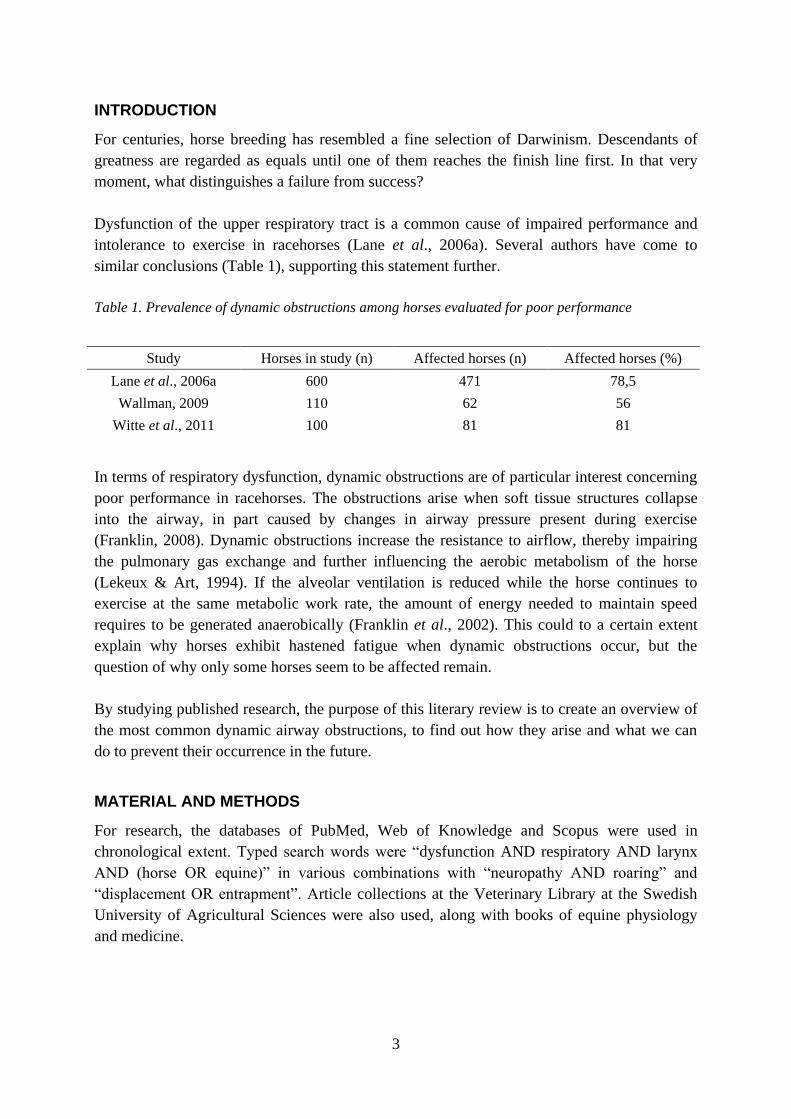
Dysfunction of the upper respiratory tract is a common cause of impaired performance and
intolerance to exercise in racehorses (Lane et al., 2006a). Several authors have come to
similar conclusions (Table 1), supporting this statement further.
Table 1. Prevalence of dynamic obstructions among horses evaluated for poor performance
In terms of respiratory dysfunction, dynamic obstructions are of particular interest concerning
poor performance in racehorses. The obstructions arise when soft tissue structures collapse
into the airway, in part caused by changes in airway pressure present during exercise
(Franklin, 2008). Dynamic obstructions increase the resistance to airflow, thereby impairing
the pulmonary gas exchange and further influencing the aerobic metabolism of the horse
(Lekeux & Art, 1994). If the alveolar ventilation is reduced while the horse continues to
exercise at the same metabolic work rate, the amount of energy needed to maintain speed
requires to be generated anaerobically (Franklin et al., 2002). This could to a certain extent
explain why horses exhibit hastened fatigue when dynamic obstructions occur, but the
question of why only some horses seem to be affected remain.
By studying published research, the purpose of this literary review is to create an overview of
the most common dynamic airway obstructions, to find out how they arise and what we can
do to prevent their occurrence in the future.
MATERIAL AND METHODS
For research, the databases of PubMed, Web of Knowledge and Scopus were used in
chronological extent. Typed search words were “dysfunction AND respiratory AND larynx
AND (horse OR equine)” in various combinations with “neuropathy AND roaring” and
“displacement OR entrapment”. Article collections at the Veterinary Library at the Swedish
University of Agricultural Sciences were also used, along with books of equine physiology
and medicine.
Study Horses in study (n) Affected horses (n) Affected horses (%)
Lane et al., 2006a 600 471 78,5
Wallman, 2009 110 62 56
Witte et al., 2011 100 81 81

4
LITERARY REVIEW
Anatomy and physiology of the upper airways
The horse possesses a complex composition of upper airway structures, creating large
dimensional changes as inhaled air approaches the lungs (Figure 1). The parts of the
respiratory tract that cause airway obstructions are primarily the ones that tends to collapse
during physical exertion. These are particular structures of the pharyngeal cavity and the
larynx.
Figure 1. Overview of the equine upper respiratory tract (Kisonaite, 2012).
Horses are compulsory nasal breathers, an evolutionary advantage that enables them to graze
and still maintain olfaction and the ability to sense predators. The base of the larynx is tightly
apposed to the caudal margin of the soft palate, resulting in a non-existing communication
between the oropharynx and the nasopharynx. This explains why horses in respiratory distress
cannot resort to oral breathing, which other animals do as high flow resistance and turbulence
arises in the airways. (Holcombe & Ducharme, 2008)
Resistance and turbulence are altered as the velocity of airflow increases. During exercise,
increased oxygen demand extends the respiratory minute volume of the horse markedly. At
moderate exercise, minute volume is increased due to likewise increases in tidal volume and
respiratory rate. When exercise becomes strenuous, the increase in minute volume is primarily
caused by an increase in respiratory rate. This results in an increase of airflow velocity,
further affecting resistance, turbulence and pressure in the airways. (Lekeux & Art, 1994)

5
The equine larynx is composed of cartilage and muscle (Figure 2) and constitutes a
connection between the pharynx and trachea. The larynx contains cricoid, thyroid, epiglottal
and paired arytenoid cartilages with synovial articulation between them. Various membranes
and ligaments hold the apparatus together, with a suite of small, paired intrinsic muscles that
connects the cartilages and influences
their mutual relations. Moreover,
extrinsic laryngeal musculature
connects the larynx to the pharynx,
tongue, hyoid bone and sternum.
(Holcombe & Ducharme, 2008)
The structures of the equine upper
airways are exposed to great
variations in pressure during
inhalation. This can be explained by
Bernoulli’s principle, stating that an
increase in flow velocity will reduce
the pressure and further initiate a
collapse of structures towards one
another. Velocity of inhaled air
reaches peak values in the rostral and
ventral end of the nasopharynx,
where the upper airway diameter is
the smallest. This results in high
resistance to airflow and reduced
pressure. Speed is reduced as the
diameter increases in the middle part
of the nasopharynx, but further
constriction in the laryngeal area
increases air velocity again and
generates turbulence in airflow. Regions of reverse airflow arise dorsal to the rostral larynx
due to constrictions and abrupt changes in the geometry of the airways, further contributing to
turbulence. (Rakesh et al., 2008)
The larynx is supported by cartilage, but the pharyngeal region is principally supported by
skeletal muscles and relies on their contraction for stability. Throughout the respiratory cycle,
neuromuscular mechanisms are essential for dilation and stabilization of airway structures
(Holcombe & Ducharme, 2008). Tensor muscle contraction is shown to inhibit collapse of the
soft structures in the upper airways during inspiration (Lekeux & Art, 1994). For example, M.
hyoepiglotticus, the only muscle that attaches to the epiglottis, is likely to be a dilating muscle
whose activity increases with breathing effort. Contraction of the muscle stabilizes the
epiglottis during inspiration, preventing prolapse of it into the rima glottidis (Holcombe &
Ducharme, 2008).
Figure 2. The larynx of the horse (Kisonaite, 2012).
6
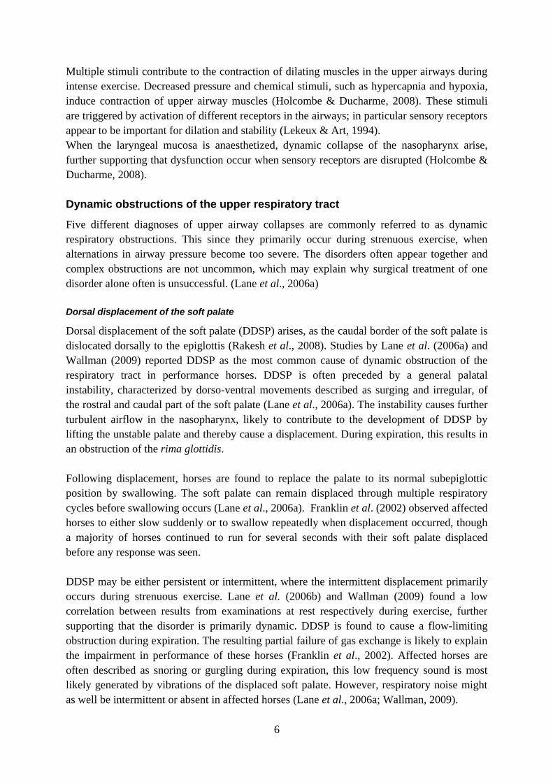
Multiple stimuli contribute to the contraction of dilating muscles in the upper airways during
intense exercise. Decreased pressure and chemical stimuli, such as hypercapnia and hypoxia,
induce contraction of upper airway muscles (Holcombe & Ducharme, 2008). These stimuli
are triggered by activation of different receptors in the airways; in particular sensory receptors
appear to be important for dilation and stability (Lekeux & Art, 1994).
When the laryngeal mucosa is anaesthetized, dynamic collapse of the nasopharynx arise,
further supporting that dysfunction occur when sensory receptors are disrupted (Holcombe &
Ducharme, 2008).
Dynamic obstructions of the upper respiratory tract
Five different diagnoses of upper airway collapses are commonly referred to as dynamic
respiratory obstructions. This since they primarily occur during strenuous exercise, when
alternations in airway pressure become too severe. The disorders often appear together and
complex obstructions are not uncommon, which may explain why surgical treatment of one
disorder alone often is unsuccessful. (Lane et al., 2006a)
Dorsal displacement of the soft palate
Dorsal displacement of the soft palate (DDSP) arises, as the caudal border of the soft palate is
dislocated dorsally to the epiglottis (Rakesh et al., 2008). Studies by Lane et al. (2006a) and
Wallman (2009) reported DDSP as the most common cause of dynamic obstruction of the
respiratory tract in performance horses. DDSP is often preceded by a general palatal
instability, characterized by dorso-ventral movements described as surging and irregular, of
the rostral and caudal part of the soft palate (Lane et al., 2006a). The instability causes further
turbulent airflow in the nasopharynx, likely to contribute to the development of DDSP by
lifting the unstable palate and thereby cause a displacement. During expiration, this results in
an obstruction of the rima glottidis.
Following displacement, horses are found to replace the palate to its normal subepiglottic
position by swallowing. The soft palate can remain displaced through multiple respiratory
cycles before swallowing occurs (Lane et al., 2006a). Franklin et al. (2002) observed affected
horses to either slow suddenly or to swallow repeatedly when displacement occurred, though
a majority of horses continued to run for several seconds with their soft palate displaced
before any response was seen.
DDSP may be either persistent or intermittent, where the intermittent displacement primarily
occurs during strenuous exercise. Lane et al. (2006b) and Wallman (2009) found a low
correlation between results from examinations at rest respectively during exercise, further
supporting that the disorder is primarily dynamic. DDSP is found to cause a flow-limiting
obstruction during expiration. The resulting partial failure of gas exchange is likely to explain
the impairment in performance of these horses (Franklin et al., 2002). Affected horses are
often described as snoring or gurgling during expiration, this low frequency sound is most
likely generated by vibrations of the displaced soft palate. However, respiratory noise might
as well be intermittent or absent in affected horses (Lane et al., 2006a; Wallman, 2009).

7
The etiology of the condition is not clearly understood, but likely to be multifactorial and
there are several theories of the origin. One probable cause is neuromuscular dysfunction,
possibly induced by inflammation or infection of the upper airways (Couroché-Malblanc et
al., 2010). This hypothesis is further supported by the development of DDSP in healthy
horses when the pharyngeal branch of N. vagus is anaesthetized (Holcombe et al., 1997).
Several authors have come to the conclusion that young horses are more likely to develop
DDSP (Lane et al., 2006; Wallman, 2009). A possible cause of this relationship might be the
high prevalence of pharyngeal lymphoid hyperplasia (PLH) in young racehorses, since upper
respiratory inflammation may cause DDSP (Couroché-Malblanc et al., 2010).
Laryngeal hemiplegia
Laryngeal hemiplegia (LH) is commonly referred to as recurrent laryngeal neuropathy (RLN),
since the cause of the dysfunction is confirmed to be neuropathy of one of the recurrent
laryngeal nerves (Derksen et al., 2001). Most commonly, the left nerve is affected. Recurrent
laryngeal neuropathy is considered a distal axonopathy, meaning that larger myelinated nerve
fibers degenerate proximally from the end plate toward the cell body, leading to atrophy of
the muscles that the axon innerves. The condition may be related to the length of the nerve
fibers of the left recurrent laryngeal nerve, which are the longest motor neurons in the horse.
This could possibly also explain why larger horses seem to be predisposed. It is also possible
that the distal axonopathy has an inherited basis. (Rush & Mair, 2004)
Other possible causes of nerve damage initiating the axonopathy are presence of abscesses,
tumours or infections in the upper airway region. There is also evidence of a correlation
between nerve damage and the anatomical proximity to the left jugular vein, suggesting that
damage may be caused by thrombophlebitis or perivascular injections of irritants (Davenport-
Goodall & Parente, 2003).
The neuropathy leads to dysfunction and atrophy of the intrinsic laryngeal muscles, in
particular M. cricoarytenoideus dorsalis (Rush & Mair, 2004). The defect can be either
persistent or intermittent and affect either one or both arytenoid cartilages, though most
commonly the left is affected. In examination, a failure of complete abduction of the cartilage
is seen, resulting in a dynamic movement of the cartilage of the affected side towards the
midline of the rima glottidis during inspiration. However, Lane et al. (2006b) found that 19 %
of horses diagnosed with severe laryngeal hemiplegia at rest were able to maintain full
abduction of the arytenoid cartilages during exercise. The condition severely narrows the
opening to the trachea and laryngeal hemiplegia is often accompanied by ipsilateral vocal fold
collapse. The defect is also known to give rise to a characteristic inspiratory noise, often
referred to as roaring. (Derksen et al., 2001)
8
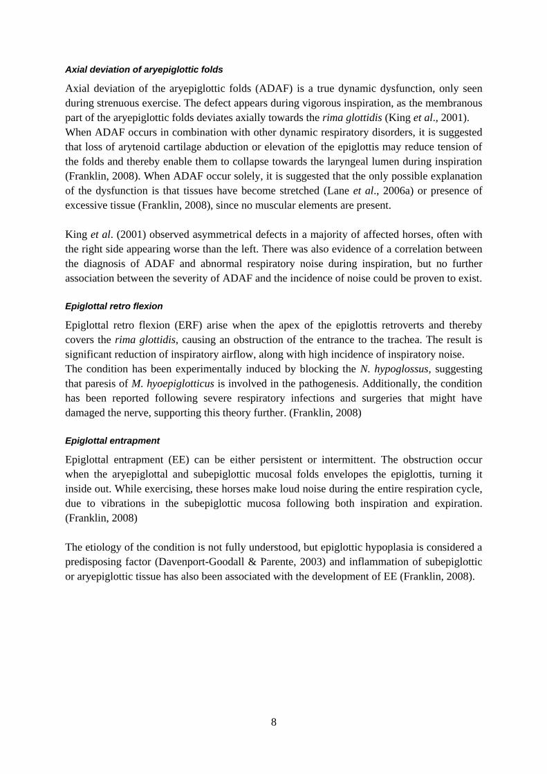
Axial deviation of aryepiglottic folds
Axial deviation of the aryepiglottic folds (ADAF) is a true dynamic dysfunction, only seen
during strenuous exercise. The defect appears during vigorous inspiration, as the membranous
part of the aryepiglottic folds deviates axially towards the rima glottidis (King et al., 2001).
When ADAF occurs in combination with other dynamic respiratory disorders, it is suggested
that loss of arytenoid cartilage abduction or elevation of the epiglottis may reduce tension of
the folds and thereby enable them to collapse towards the laryngeal lumen during inspiration
(Franklin, 2008). When ADAF occur solely, it is suggested that the only possible explanation
of the dysfunction is that tissues have become stretched (Lane et al., 2006a) or presence of
excessive tissue (Franklin, 2008), since no muscular elements are present.
King et al. (2001) observed asymmetrical defects in a majority of affected horses, often with
the right side appearing worse than the left. There was also evidence of a correlation between
the diagnosis of ADAF and abnormal respiratory noise during inspiration, but no further
association between the severity of ADAF and the incidence of noise could be proven to exist.
Epiglottal retro flexion
Epiglottal retro flexion (ERF) arise when the apex of the epiglottis retroverts and thereby
covers the rima glottidis, causing an obstruction of the entrance to the trachea. The result is
significant reduction of inspiratory airflow, along with high incidence of inspiratory noise.
The condition has been experimentally induced by blocking the N. hypoglossus, suggesting
that paresis of M. hyoepiglotticus is involved in the pathogenesis. Additionally, the condition
has been reported following severe respiratory infections and surgeries that might have
damaged the nerve, supporting this theory further. (Franklin, 2008)
Epiglottal entrapment
Epiglottal entrapment (EE) can be either persistent or intermittent. The obstruction occur
when the aryepiglottal and subepiglottic mucosal folds envelopes the epiglottis, turning it
inside out. While exercising, these horses make loud noise during the entire respiration cycle,
due to vibrations in the subepiglottic mucosa following both inspiration and expiration.
(Franklin, 2008)
The etiology of the condition is not fully understood, but epiglottic hypoplasia is considered a
predisposing factor (Davenport-Goodall & Parente, 2003) and inflammation of subepiglottic
or aryepiglottic tissue has also been associated with the development of EE (Franklin, 2008).
9
DISCUSSION
Dysfunction of the upper respiratory tract is a common cause of poor performance and
intolerance to exercise in racehorses (Lane et al., 2006a; Wallman, 2009; Witte et al., 2011).
The reason for this is not fully known, which truly emphasizes the need of further studies. In
order to prevent the obstructions from arising in the future, the originating cause of the
neuromuscular dysfunction needs to be known. Several etiologies of the collapses have been
discussed, some of them proven to be more likely than others. Evolution of laryngeal
anatomy, genetic predisposition and presence of airway infections are some of the suggested
causes of dysfunction. However, further studies are required before any of them may be
confirmed or ruled out.
It is possible that changes of the anatomical constitution of the equine upper airway have
reached beyond individual differences, evolutionary creating a new standard among
racehorses. This would explain why diagnosis of dynamic obstructions is far more common
today than it was a decade ago, but this may just as well be due to improved diagnostic
techniques. Breeding of racehorses is nevertheless a true survival of the fittest and it is not
very likely that the most successful horses have suffered from respiratory defects. It may
further be discussed whether we simply have reached the endpoint of equine strain, if the
pressure changes in general are too severe for soft structures of the airway to handle. This
would again explain why the prevalence of the obstructions has increased, but it contradicts
the fact that the horse making the hardest physical effort reaches the finish line first.
There are suspicions of an inheritance of a genetic predisposition for certain collapses (Rush
& Mair, 2004). If this is found to be true, equine racing federations may have to consider
ruling out affected horses from breeding. Not so much because of the choking horses
themselves, since no one is likely to want an offspring from a horse that always finished last.
Operated horses may, on the other hand, show no sign of earlier disorder and achieve great
results, while possibly passing on native defects to a future offspring. Ruling these horses out
would though require access to veterinary journals and hard detective work in order to map all
individuals with faults corrected. No owner could be expected to report this voluntarily, since
exclusion from breeding could equal great financial losses.
It is generally known that exercising with upper airway infections may result in future
obstructive problems. Many horses undergo pre-race endoscope examinations to ensure that
they do not suffer from a pharyngitis that may influence their performance, but what many
seems to have forgotten is that just as much damage could be done during exercise.
Performing endoscope examinations or blood samplings before every training session is not
possible out of several perspectives, leading to the conclusion that the very best must be to
avoid irritation of the upper airway mucosa from the beginning. The value of good ventilated
stable environments and high quality roughage to can therefore not be emphasized enough.
Lymphoid hyperplasia (PLH) is commonly found in the pharynx of racehorses and considered
a harmless condition when the horses are still young. In older horses, it is interpreted as a sign
10
of upper airway inflammation and a general precaution is always taken considering racing
these horses. Young horses often continue to race and exercise as before, even though
diagnosed with PLH. Dorsal displacement of the soft palate (DDSP) is also found to be more
common among young horses (Couroché-Malblanc et al., 2010) and there is a suggested
connection between PLH and the high prevalence of DDSP in young individuals. This could
imply that these follicles may cause more damage than earlier assumed, stating that this
possible connection needs to be further investigated.
Another suspected cause of dysfunction is perivascular injections meant for the left jugular
vein, possibly damaging the N. laryngeus recurrens and thereby inducing laryngeal
hemiplegia (Davenport-Goodall & Parente, 2003). Damage could potentially be done to the
nerve by perivascular injections of irritants or excesses from trombophlebitis caused by
permanent catheters. Given that this is true, it may be questioned why injections are still
preferably made in the left jugular vein. Injecting in the right jugular vein is seldom a
problem, except for being more technically difficult for right-handed to manage. Some horses
lack a functioning right jugular vein and certain surgical procedures requires the permanent
catheter to be placed in the left jugular due to the head position of the horse. Otherwise, no
disadvantages of right-sided jugular injections are to be found. Presence of laryngeal
hemiplegia may not be of importance to an average riding horse, but the consequences could
be fatal to a racehorse, why right-sided injections truly should be considered when treating
racehorses.
Dynamic obstructions of the upper airways arise due to presence of great fluctuations in
pressure, generated by intense breathing. This implies that examinations at rest are likely to
leave out conditions that solely occur during physical exertion. Further, it is known that
certain conditions, which appear at rest, are corrected during exercise (Lane et al, 2006b). The
relevance of such examination alone may for that reason be questioned.
Today, fine surgical methods are developed to correct the dynamic obstructions and many
surgeons perform the procedures on routine. We should ask ourselves to what limit it is
reasonable to correct the faults, without dedicating a minute to consider why they are so
common. When affected horses are diagnosed, they are likely to already have experienced
choking several times. It is therefore of particular importance to find the cause of the
dysfunctions, so that horses never have to choke from the very beginning in the future. When
we have reached that point, there will be no further need of advanced surgeries.
Post surgery, there is a lack of reported success rate of the procedures. Just as successful they
may be, there is also a constant risk of contributing to further nerve damage. It is thereby
possible to give rise to one particular dysfunction while performing surgery to correct another.
No one knows if horses will be reluctant to make a physical effort again with the memory of
choking present, making the result of a surgical procedure even more insecure. After all, no
one can be blamed for not running in dyspnea. And maybe, that lack of access to oxygen can
be distinguished as the difference between a failure and success.
11
REFERENCES
Courouce Malblanc, A., Deniau, V., Rossignol, F., Corde, R., Leleu, C., & Maillard, K. (2010).
Physiological measurements and prevalence of lower airway diseases in Trotters with dorsal
displacement of the soft palate. Equine Veterinary Journal, 42, 246–255.
Davenport-Goodall, C. L. M., & Parente, E. J. (2003). Disorders of the larynx. The Veterinary Clinics
of North America. Equine Practice, 19(1), 169–187.
Derksen, F. J., Holcombe, S. J., Hartmann, W., Robinson, N. E., & Stick, J. A. (2001). Spectrum
analysis of respiratory sounds in exercising horses with experimentally induced laryngeal
hemiplegia or dorsal displacement of the soft palate. American Journal of Veterinary Research,
62(5), 659–664.
Franklin, S. H. (2008). Dynamic collapse of the upper respiratory tract: A review. Equine veterinary
education, 20(4), 212–224.
Franklin, S. H., Naylor, J. R .J., & Lane, J. G. (2002). Effect of dorsal displacement of the soft palate
on ventilation and airflow during high-intensity exercise. Equine Veterinary Journal, (Supplement
34), 379–383.
Holcombe, S., Derksen, F., Stick, J., & Robinson, N. (1997). Effects of bilateral hypoglossal and
glossopharyngeal nerve blocks on epiglottic and soft palate position in exercising horses.
American Journal of Veterinary Research, 58(9), 1022–1026.
Holcombe, S. J., & Ducharme, N. G. (2008). Upper airway function of normal horses during exercise.
Equine Exercise Physiology (pp. 170–192). UK: Saunders.
King, D. S., Tulleners, E., Martin Jr., B. B., Parente, E. J., & Boston, R. (2001). Clinical Experiences
With Axial Deviation of the Aryepiglottic Folds in 52 Racehorses. Veterinary Surgery, 30(2),
151–160.
Lane, J. G., Bladon, B., & Franklin, S. H. (2006a). Dynamic obstructions of the equine upper
respiratory tract. Part 1: Observations during high-speed treadmill endoscopy of 600
Thoroughbred racehorses. Equine Veterinary Journal, 38(5), 393–399.
Lane, J. G., Bladon, B., & Franklin, S. H. (2006b). Dynamic obstructions of the equine upper
respiratory tract. Part 2: Comparison of endoscopic findings at rest and during high-speed
treadmill exercise of 600 Thoroughbred racehorses. Equine Veterinary Journal, 38(5), 401–407.
Lekeux, P., & Art, T. (1994). The respiratory system: Anatomy, physiology and adaptions to exercise
and training. The Athletic Horse: Principles and Practice of Equine Sports Medicine (pp. 79-127).
Philadelphia: Saunders.
Rakesh, V., Ducharme, N. G., Datta, A. K., Cheetham, J., & Pease, A. P. (2008). Development of
equine upper airway fluid mechanics model for Thoroughbred racehorses. Equine Veterinary
Journal, 40(3), 272–279.
Rush, B., & Mair, T. (2004). Equine respiratory diseases (pp. 108-114). Oxford: Blackwell
publishing.
Wallman, A. (2010). Obstruktiva problem från de övre luftvägarna hos travhästar som undersökts med
videoendoskop under maximal ansträngning, 2010:2.

12
Witte, S. H. P., Witte, T. H., Harriss, F., Kelly, G., & Pollock, P. (2011). Association of owner-
reported noise with findings during dynamic respiratory endoscopy in Thoroughbred racehorses.
Equine Veterinary Journal, 43(1), 9-17.
A great thank you to the very best K for the illustrations (copyright Konstancia Kisonaite, 2012).