dynamic mechanochemical feedback between curved …
TRANSCRIPT

ARTICLE
Dynamic mechanochemical feedback betweencurved membranes and BAR proteinself-organizationAnabel-Lise Le Roux 1,8✉, Caterina Tozzi 2,8, Nikhil Walani2, Xarxa Quiroga 1, Dobryna Zalvidea1,
Xavier Trepat1,3,4,5, Margarita Staykova 6, Marino Arroyo 1,2,7✉ & Pere Roca-Cusachs 1,3✉
In many physiological situations, BAR proteins reshape membranes with pre-existing cur-
vature (templates), contributing to essential cellular processes. However, the mechanism and
the biological implications of this reshaping process remain unclear. Here we show, both
experimentally and through modelling, that BAR proteins reshape low curvature membrane
templates through a mechanochemical phase transition. This phenomenon depends on initial
template shape and involves the co-existence and progressive transition between distinct
local states in terms of molecular organization (protein arrangement and density) and
membrane shape (template size and spherical versus cylindrical curvature). Further, we
demonstrate in cells that this phenomenon enables a mechanotransduction mode, in which
cellular stretch leads to the mechanical formation of membrane templates, which are then
reshaped into tubules by BAR proteins. Our results demonstrate the interplay between
membrane mechanics and BAR protein molecular organization, integrating curvature sensing
and generation in a comprehensive framework with implications for cell mechanical
responses.
https://doi.org/10.1038/s41467-021-26591-3 OPEN
1 Institute for Bioengineering of Catalonia (IBEC), the Barcelona Institute of Technology (BIST), 08028 Barcelona, Spain. 2 Universitat Politècnica de Catalunya(UPC), Campus Nord, Carrer de Jordi Girona, 1, 3, 08034 Barcelona, Spain. 3 Universitat de Barcelona, 08036 Barcelona, Spain. 4 Institució Catalana deRecerca i Estudis Avançats (ICREA), Passeig de Lluís Companys, 23, 08010 Barcelona, Spain. 5 Centro de Investigación Biomédica en Red de Cáncer(CIBERONC), 08028 Barcelona, Spain. 6 Department of Physics, University of Durham, Durham, UK. 7 Centre Internacional de Met̀odes Numer̀ics enEnginyeria (CIMNE), 08034 Barcelona, Spain. 8These authors contributed equally: Anabel-Lise Le Roux, Caterina Tozzi. ✉email: [email protected];[email protected]; [email protected]
NATURE COMMUNICATIONS | (2021) 12:6550 | https://doi.org/10.1038/s41467-021-26591-3 | www.nature.com/naturecommunications 1
1234
5678
90():,;
Due to the curved shape and membrane binding of Bin/Amphiphysin/Rvs (BAR) domains, proteins containingsuch domains have the interesting ability to reshape
membranes. Furthermore, because of their elongated shape, theycan align along a preferred direction, adopting a nematic orga-nization that impinges anisotropic curvature on the membrane.In equilibrium and for high concentrations, this leads to thegeneration of membrane tubes with high curvatures, comparableto BAR intrinsic curvatures (of the order of 101 nm). For instance,incubation of small vesicles with a high concentration of BARproteins leads to highly curved tubes covered by a dense proteinscaffold where the elongated molecules are nematicallyarranged1,2. In a different system, GUVs with sufficiently highbound protein density rapidly expel thin protein-rich tubes in atension-dependent manner3. On thin membrane tubes pulled outof giant unilamellar vesicles (GUVs), BAR proteins can alsochange the radius of the tube and the force required to hold it4.Beyond this well-known paradigm of thin tubes in equilibrium, inmany physiological situations, BAR proteins dynamically interactwith pre-existing curved membrane templates. Such templatescan include for instance invaginations caused by nanoscaletopographical features on the cell substrate5, mechanical folds6,7,or endocytic structures8,9. Due to their affinity for curved mem-branes, BAR proteins are thus bound to sense and reshape suchtemplates in ways that are important in physiological processes10
like endocytosis11, the build-up of caveolar structures12,13, themaintenance of cell polarity14, and the modulation of actinpolymerization15. However, how BAR proteins reshape mem-branes with initial curvatures that can be well below theirintrinsic values, with which dynamics, how this depends on initialmembrane shape, and what are the implications, remainsunknown.
To address this issue, we developed a versatile experimentalsystem combined with theoretical and computational modeling tostudy the dynamic reshaping of cellular-like membrane structuresof a broad range of shapes and sizes. In commonly used systemssuch as tube pulling assays10 or curved substrates16,17, imposedcurvature creates tensed curved structures. In cells, such tensedstructures (created either from nanoscale curved topographies orfrom actin pulling on the membrane) can for instance recruitN-BAR proteins and enhance endocytosis5 or trigger therecruitment of effectors related to actin polymerization15,18.However, both in vitro and in cells tensed structures preventextensive shape remodeling. In contrast, here we use a differentphysiologically relevant signal in the form of stretch and releasecycles. In our system, we create curved membrane features off asupported lipid bilayer (SLB) by applying a successive lateralstretch and compression. As previously shown in SLBs6 and incells7, this leads to the storage of excess membrane area in free-standing, low tension, easily reshaped protrusions of tubular orspherical shape. In contrast with tubes pulled out of GUVs, wherea tip force and tension are required to stabilize their shape, in oursystem tubes are stabilized osmotically without a pulling force. Toassess the effect of tension, we also control osmolarity to generatetensed spherical caps off the SLB6. These protrusions emergingfrom a flat SLB can serve as model system for membrane tem-plates such as endocytic buds, or osmotically/mechanically-induced structures.
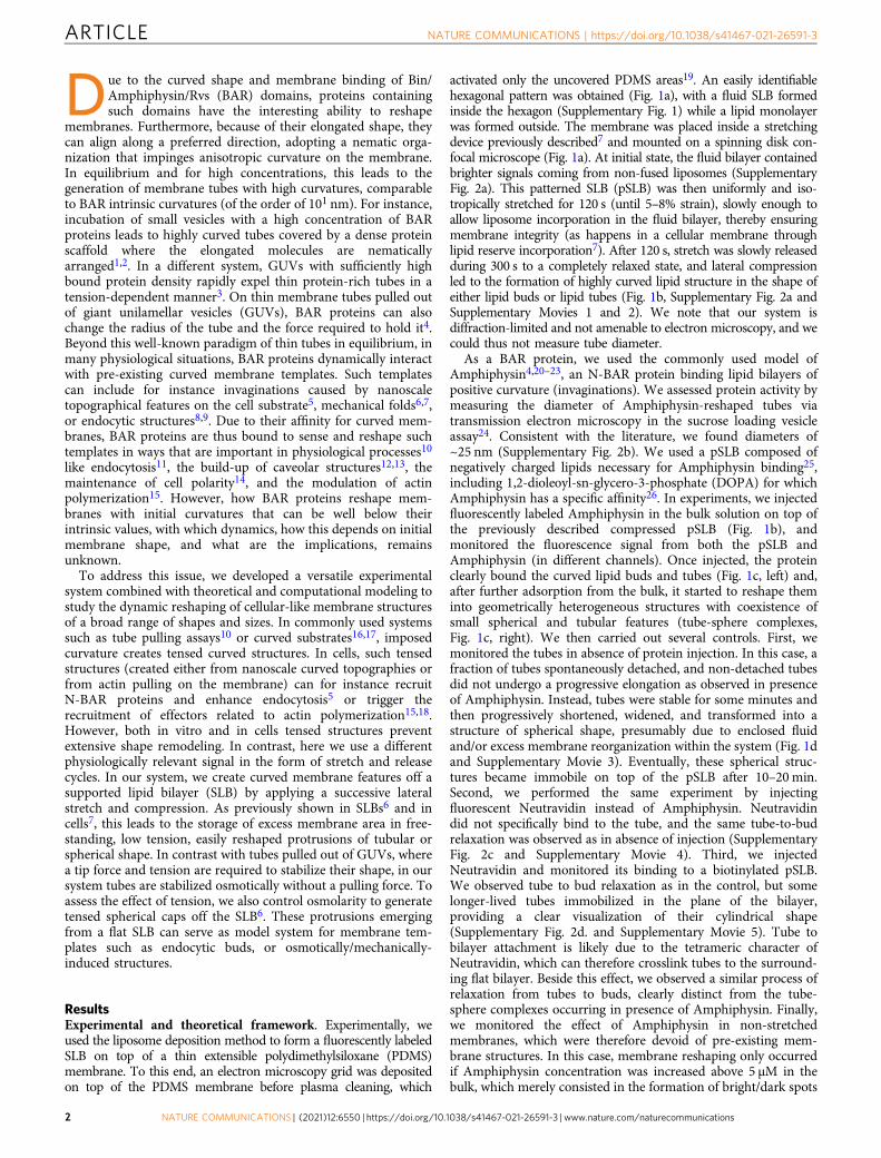
ResultsExperimental and theoretical framework. Experimentally, weused the liposome deposition method to form a fluorescently labeledSLB on top of a thin extensible polydimethylsiloxane (PDMS)membrane. To this end, an electron microscopy grid was depositedon top of the PDMS membrane before plasma cleaning, which
activated only the uncovered PDMS areas19. An easily identifiablehexagonal pattern was obtained (Fig. 1a), with a fluid SLB formedinside the hexagon (Supplementary Fig. 1) while a lipid monolayerwas formed outside. The membrane was placed inside a stretchingdevice previously described7 and mounted on a spinning disk con-focal microscope (Fig. 1a). At initial state, the fluid bilayer containedbrighter signals coming from non-fused liposomes (SupplementaryFig. 2a). This patterned SLB (pSLB) was then uniformly and iso-tropically stretched for 120 s (until 5–8% strain), slowly enough toallow liposome incorporation in the fluid bilayer, thereby ensuringmembrane integrity (as happens in a cellular membrane throughlipid reserve incorporation7). After 120 s, stretch was slowly releasedduring 300 s to a completely relaxed state, and lateral compressionled to the formation of highly curved lipid structure in the shape ofeither lipid buds or lipid tubes (Fig. 1b, Supplementary Fig. 2a andSupplementary Movies 1 and 2). We note that our system isdiffraction-limited and not amenable to electron microscopy, and wecould thus not measure tube diameter.
As a BAR protein, we used the commonly used model ofAmphiphysin4,20–23, an N-BAR protein binding lipid bilayers ofpositive curvature (invaginations). We assessed protein activity bymeasuring the diameter of Amphiphysin-reshaped tubes viatransmission electron microscopy in the sucrose loading vesicleassay24. Consistent with the literature, we found diameters of~25 nm (Supplementary Fig. 2b). We used a pSLB composed ofnegatively charged lipids necessary for Amphiphysin binding25,including 1,2-dioleoyl-sn-glycero-3-phosphate (DOPA) for whichAmphiphysin has a specific affinity26. In experiments, we injectedfluorescently labeled Amphiphysin in the bulk solution on top ofthe previously described compressed pSLB (Fig. 1b), andmonitored the fluorescence signal from both the pSLB andAmphiphysin (in different channels). Once injected, the proteinclearly bound the curved lipid buds and tubes (Fig. 1c, left) and,after further adsorption from the bulk, it started to reshape theminto geometrically heterogeneous structures with coexistence ofsmall spherical and tubular features (tube-sphere complexes,Fig. 1c, right). We then carried out several controls. First, wemonitored the tubes in absence of protein injection. In this case, afraction of tubes spontaneously detached, and non-detached tubesdid not undergo a progressive elongation as observed in presenceof Amphiphysin. Instead, tubes were stable for some minutes andthen progressively shortened, widened, and transformed into astructure of spherical shape, presumably due to enclosed fluidand/or excess membrane reorganization within the system (Fig. 1dand Supplementary Movie 3). Eventually, these spherical struc-tures became immobile on top of the pSLB after 10–20min.Second, we performed the same experiment by injectingfluorescent Neutravidin instead of Amphiphysin. Neutravidindid not specifically bind to the tube, and the same tube-to-budrelaxation was observed as in absence of injection (SupplementaryFig. 2c and Supplementary Movie 4). Third, we injectedNeutravidin and monitored its binding to a biotinylated pSLB.We observed tube to bud relaxation as in the control, but somelonger-lived tubes immobilized in the plane of the bilayer,providing a clear visualization of their cylindrical shape(Supplementary Fig. 2d. and Supplementary Movie 5). Tube tobilayer attachment is likely due to the tetrameric character ofNeutravidin, which can therefore crosslink tubes to the surround-ing flat bilayer. Beside this effect, we observed a similar process ofrelaxation from tubes to buds, clearly distinct from the tube-sphere complexes occurring in presence of Amphiphysin. Finally,we monitored the effect of Amphiphysin in non-stretchedmembranes, which were therefore devoid of pre-existing mem-brane structures. In this case, membrane reshaping only occurredif Amphiphysin concentration was increased above 5 μM in thebulk, which merely consisted in the formation of bright/dark spots
ARTICLE NATURE COMMUNICATIONS | https://doi.org/10.1038/s41467-021-26591-3
2 NATURE COMMUNICATIONS | (2021) 12:6550 | https://doi.org/10.1038/s41467-021-26591-3 | www.nature.com/naturecommunications

in the membrane, likely reflecting membrane tearing. (Supple-mentary Fig. 2e, f and Supplementary Movies 6 and 7).
To understand the physical mechanisms underlying ourobservations, we developed a theoretical framework consideringthe dynamics of lipid tubes and buds with low coverage (sinceprotein is injected once structures are formed) and low curvature(since the structures are made markedly thinner by Amphiphy-sin) upon exposure to BAR proteins. Theoretically, variouscomputational studies using coarse-grained simulations of
elongated and curved objects moving on a deformable membranehave suggested the self-organization of regions with highanisotropic (cylindrical) curvature with high-protein coverageand strong nematic order27–29. None of these works, however,predicted or observed the tube-sphere complexes that appear inour experiments (Fig. 1c). To address this, we first focused on ourrecently developed mean-field density functional theory30 for thefree energy Fprot of an ensemble of curved proteins on amembrane as a function of protein area coverage ϕ, orientational
Vacuum
Clamp
Post
PDMS ring
PDMS
O2 plasmacleaning
Liposomedeposition
hydrophylic
a.
Mechanically-stimulated (compression)t = 830 s
PatternedSLB formation
PDMS ring
t = 350 sControl with no injectiont = 1 s t = 1500 s
d.
protein injectionrelaxedstrained
b.
c.
hydrophobic
Amph
iphy
sin
Amph
iphy
sin
bila
yer
bila
yer r eyali b
ni et or p on
Fig. 1 Experimental system. a Schematics of the patterned supported lipid bilayer (pSLB) placed in a stretch system compatible with confocal microscopy.The pSLB is obtained by plasma cleaning a PDMS membrane in presence of a TEM grid. Only the exposed PDMS becomes hydrophilic, and subsequentliposome deposition renders a SLB after buffer rinse. The non-exposed PDMS remains hydrophobic and a lipid monolayer is formed instead.b Representative images of the mechanical stimulation of the pSLB, showing both lipid and protein fluorescence images. In the resting initial state, excessliposomes stand on top of the pSLB. With strain, the liposomes incorporate in the pSLB. Upon release, excess lipids are expelled in the form or tubes orbuds. At this stage, fluorescent Amphiphysin is gently microinjected on top of pSLB and its binding to the tubes and buds is monitored with time.c Membrane tubes (green inset) and buds (purple inset) before (left) and after (right) being reshaped by Amphiphysin. d Control in which no protein isinjected on top of the pSLB. Scale bar, 5 μm.
NATURE COMMUNICATIONS | https://doi.org/10.1038/s41467-021-26591-3 ARTICLE
NATURE COMMUNICATIONS | (2021) 12:6550 | https://doi.org/10.1038/s41467-021-26591-3 | www.nature.com/naturecommunications 3

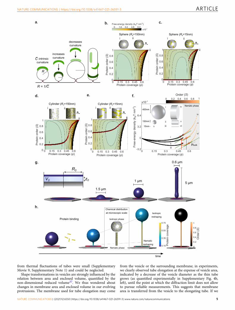
order as given by a nematic order parameter S, and membranecurvature. This theory accounts for the entropic and stericinteractions between proteins and for their bending elasticity,focusing on the scaffolding effect. We discuss the mappingbetween different mechanisms coupling protein coverage tomembrane curvature for Amphiphysin (scaffolding, effect ofinsertions, bulky disordered domains) and detail our model inSupplementary Note 1, where we specifically quantify the role ofcrowding of disordered domains by adapting a model for thecoupling between such domains and curvature31 to the presentcontext, finding that the effect is small.
Reshaping occurs through an isotropic-to-nematic transition.On flat membranes and for elliptical particles of the size andaspect ratio of Amphiphysin, the theory recovers a classicalentropically-controlled discontinuous isotropic-to-nematic tran-sition during which the system abruptly changes from low to highorder as protein coverage increases above ϕ � 0:5, in agreementwith previous results in 3D32, Supplementary Fig. 3a1. We thenexamined the protein free-energy landscape on curved surfaces,where the elastic curvature energy of proteins depends on theirintrinsic curvature and on the curvature of the surface along theprotein long direction (Fig. 2a). On spherical surfaces andaccording to the theory, this energy landscape coincides with thatof the flat membrane with a bias proportional to ϕ times thebending energy of proteins on the curved surface. Thus, theminimum energy paths as density increases (red dots inFig. 2b–e) and hence the abrupt isotropic-to-nematic transitionpersists regardless of sphere radius, Fig. 2b, c, noting that on acomplete sphere the nematic phase necessarily involves defects27.On cylindrical surfaces, however, curvature is anisotropic and theenergy landscape is fundamentally modified according to ourtheory, as proteins can lower their free energy by orienting alonga direction of favorable curvature. The competition betweenprotein bending and entropy results in a continuous isotropic-to-nematic transition (Fig. 2d) and a significant degree of orienta-tional order even at low coverage when the tube curvature iscomparable to that of the protein (Fig. 2e). The model thuspredicts how the nematic ordering of the curved and elongatedmembrane depends on coverage, curvature, and curvatureanisotropy.
We then studied whether the model predicted the experimen-tally observed coexistence of thin tubes (which according to thetheory should have higher coverage and order) and larger spheres(which should have lower coverage and isotropic organization).We examined the energy landscape along the minimizing paths(red dots) for spheres and tubes of varying radius (Fig. 2f). Sincethe slope of these curves is the chemical potential of proteins onthe membrane, which tends to equilibrate with the fixed chemicalpotential of dissolved proteins in the medium, points of chemicalcoexistence are characterized by a common slope (red circles).This figure shows the largely non-unique combinations ofgeometry and membrane coverage compatible with coexistencein chemical equilibrium between higher-coverage nematic phaseson cylinders and lower-coverage isotropic phases on spheres,supporting plausibility of such coexistence in the dynamicalstructures.
Shape, however, is also a dynamical variable and the selectionof protein organization and shape requires the two-way interplaybetween the chemical-free energy Fprot and the elastic free energyof the membrane Fmem. To account for this and for the out-of-equilibrium nature of our experiments, we self-consistentlycoupled a parametrization of the mean-field energy densityfunctional theory used above with a continuum model for lipidmembrane reshaping and hydrodynamics33–35. The combined
model accounts for both energies, Fprot þ Fmem, for the dynamicsof protein adsorption from a bulk reservoir, for the diffusion ofproteins on the surface, and for the membrane dissipationassociated with shape changes31 (see Supplementary Note 1, for adiscussion of the model, its implementation and its parameters).Focusing on a single membrane protrusion in mechanicalequilibrium (tubular or spherical, Fig. 2g) off a supported bilayercircular patch in the absence of proteins, this model predicts thedynamics of membrane shape, ϕ, and S following a suddenincrease of dissolved protein concentration in the medium(Fig. 2h). In all simulations, we fixed the chemical potential ofproteins in the medium to account for the dissolved proteinreservoir. Once the system is driven out-of-equilibrium, we mustchoose a mechanical ensemble controlling the ability of thesimulated system to exchange membrane area and enclosedvolume with its surroundings during the dynamics. To cover theexperimental conditions, we considered a reference mechanicalensemble, used everywhere unless explicitly stated, and tested therobustness of our results by further considering a broad range ofensembles with varying ease of membrane and volume exchange.For membrane exchange, we interpolated between fixed tension(allowing for membrane exchange) and fixed projected mem-brane area (no membrane exchange). For water exchange, weconsidered adhesion potentials with different strengths and afixed pressure difference ensemble. Soft potentials allow forchanges in the distance between the adhered part of themembrane and the substrate, and hence for enclosed volumeexchange, as does the fixed pressure ensemble, whereas protru-sion volume was nearly fixed by considering very stiff adhesionpotentials (see Supplementary Note 1).
Dynamic reshaping of buds and tubes. We then comparedmodel predictions with the experimental setup, by monitoring thereshaping of the buds or tubes formed after pSLB compressionand upon subsequent injection of Amphiphysin at variousnominal bulk concentrations. First, we examined how themechanically-formed buds were reshaped in time as Amphi-physin binding occurs. In both our experiments and simulations,we systematically observed the growth of a thin tube emergingfrom the base of the bud, connecting the bud to the supportedbilayer (Fig. 3a, and Supplementary Movies 8, 9, and 10). Suchbud elongation from its neck also occurred upon exposure to0.3 μM of non-fluorescently labeled Amphiphysin (Supplemen-tary Fig. 4a and Supplementary Movie 11). According to ourmodel, this elongation is due to a dynamic and progressivetransition between two coexisting states of the membrane-proteinsystem, one with low protein coverage, isotropic organization,and low curvature (spherical and flat parts) and another with highcoverage, high nematic order, and high anisotropic curvature(thin elongating necks), Figs. 2h, 3a, and Supplementary Movie 8.The model predicts that the curvature of thin tubes is comparableto the intrinsic curvature of proteins, about 15 nm−1, Supple-mentary Fig. 2b. This transition is driven by the lower bendingenergy of proteins at the thin neck, which outweighs both theentropic penalty of a local protein enrichment and nematicorganization (Fig. 2f) and the higher membrane curvature energyof a tube relative to a larger vesicle. Our simulations also showedthat protein delivery to the enriched region overwhelminglyoccurs by adsorption from the bulk rather than from membranediffusion from neighboring regions (Supplementary Note 1). Ourmodel does not account for thermal fluctuations of shape, whichhave been shown to play an important role in reshaping by BARproteins of initially planar membranes36. Here, reshaping isdirected by membrane templates, particularly membrane necks.In this case, estimated protein coverage fluctuations resulting
ARTICLE NATURE COMMUNICATIONS | https://doi.org/10.1038/s41467-021-26591-3
4 NATURE COMMUNICATIONS | (2021) 12:6550 | https://doi.org/10.1038/s41467-021-26591-3 | www.nature.com/naturecommunications
from thermal fluctuations of tubes were small (SupplementaryMovie 9, Supplementary Note 1) and could be neglected.
Shape transformations in vesicles are strongly influenced by therelation between area and enclosed volume, quantified by thenon-dimensional reduced volume37. We thus wondered aboutchanges in membrane area and enclosed volume in our evolvingprotrusions. The membrane used for tube elongation may come
from the vesicle or the surrounding membrane; in experiments,we clearly observed tube elongation at the expense of vesicle area,indicated by a decrease of the vesicle diameter as the thin tubegrows (as quantified experimentally in Supplementary Fig. 4b,left), until the point at which the diffraction limit does not allowto pursue reliable measurements. This suggests that membranearea is transferred from the vesicle to the elongating tube. If we
b.
Sphere (Rs=15nm)
0
0.2
0.4
0.6
0.8
1
i
Prot
ein
orde
r (S
)
Protein coverage ( )
Sphere (Rs=150nm)
0.2
0.4
0.6
0.8
1
i
Prot
ein
orde
r (S
)
Protein coverage ( )
0 3.2 4.81.6Free-energy density (kBT nm-2)
x10-16.4
iii
ii
i ii
ii
c.
Protein binding
d.
h.
time
0.4 0.80 0.6 10.2Order (S)
-3.2
0
3.2
6.4
i
ii
iii
iv
v
vi
Nematic phase
i
iiiii
iv
v
vi
x10-1
0
0.2
0.4
0.6
0.8
1
Cylinder (Rc=150nm)
Prot
ein
orde
r (S
)
Nematicreshaping
Isotropicreshaping
0.4
0.8
0
0.6
1
0.2
Ord
er (S
)
0.15
0.55
0.35
0.75
Prot
ein
cove
rage
f.
g.
0
0.2
0.4
0.6
0.8
1
Cylinder (Rc=15nm)
ii
i
Prot
ein
orde
r (S
)
Protein coverage ( )
ii
iii iii
Isotropic phase
Nematic phase
Chemical distribution at microscopic scale
i
ii
Free
-ene
rgy
dens
ity (k
BT
nm-2
)
1.5 µm
1 µm 5 µm
0.6 µm
R0
z0V0
n n400nm
150nm
15nm
n n
µbulk
a.
increases curvature
decreases curvature
:intrinsiccurvature
R = 1/C̅
C̅
R
051.0 54.03.00 0.6 51.0 54.03.00 0.6
Protein coverage ( )51.0 54.03.00 0.6 51.0 54.03.00 0.6
Protein coverage ( )51.0 54.03.00 0.6
e.
NATURE COMMUNICATIONS | https://doi.org/10.1038/s41467-021-26591-3 ARTICLE
NATURE COMMUNICATIONS | (2021) 12:6550 | https://doi.org/10.1038/s41467-021-26591-3 | www.nature.com/naturecommunications 5

compare a bud with an elongated tube with the same area, thetube would have a much lower enclosed volume. This suggeststhat this transformation requires significant enclosed volumeexchange, explaining why simulations with our referenceensemble did not lead to significant vesicle shrinking duringtube elongation, Fig. 3a and Supplementary Movie 8, butsimulations with an ensemble fixing the membrane area of theprotrusion and enabling easy water exchange replicated thisaspect of the membrane reshaping (Supplementary Fig. 4b, right,Supplementary Movie 12).
Then, we considered the reshaping dynamics of tubes, whichwere frequently formed upon compression. In this case, wesystematically found both in experiments and simulations thattube reshaping was initiated by the formation of a sequence ofpearls (Fig. 3b and Supplementary Movies 13–15). Tube pearlingalso occurred upon exposure to 0.3 μM of non-fluorescentlylabeled Amphiphysin (Supplementary Fig. 4c and SupplementaryMovie 16). Subsequently, the pearled tubes transformed intopearls connected by thin tubes. This configuration was stable forlong time (though such reshaped tubes collapsed on themselves,likely due to the related loss of tension3,38—this phase is bestobserved in the movie, see Supplementary Movie 14). Necksprogressively elongated, eventually making pearls disappear andtransforming the structure into a long thin tube.
To understand this process, we noticed that in our simulationspearling occurred at low and nearly uniform protein coveragewith nearly isotropic organization, Figs. 2d, 3b, and hence thistransformation can be ascribed to a previously described pearlinginstability in the presence of sufficiently large isotropic anduniform spontaneous curvature31,39,40. Indeed, pearling insimulations occurred even without nematic order, SupplementaryFig. 4d. Although this first step is independent of molecularalignment, it triggers nematic order, as pearling generates severalthin necks along the tube. These necks nucleate regions of highanisotropic curvature, high coverage and nematic order coexistingwith low-curvature spheres with low-coverage and isotropicmolecular arrangement and subsequent tube elongation betweenthe pearls. If the protein chemical potential is high enough, thenecks progressively elongate into tubes while the spheresprogressively disappear. We witness here a striking process inwhich a large tube (compared with the N-BAR dimer intrinsiccurvature) is reshaped by Amphiphysin at low coverage due to anisotropic rearrangement of the proteins, giving rise to nucleationpoints of thin tubular necks promoting further protein enrich-ment and nematic ordering.
Physical parameters governing membrane reshaping. We thenaddressed the time-scale of the reshaping process. For vesiclesexposed to Amphiphysin, we measured tube elongation rates
between 20 and 75 nm/s at 0.25 and 0.35 μM nominal con-centrations, and from 365 to 550 nm/s at 0.5 μM. These rates weremuch smaller, about two orders of magnitude, than those pre-dicted by our simulations, Supplementary Fig. 4b. To addressthese large differences in time-scale between simulations andexperiments, we turned to the dynamics of the injection process,not accounted for in our model. Since our protein injectionmethod was systematic (Supplementary Fig. 5a), we modeledprotein delivery through bulk diffusion in the medium from theinjection point to the close vicinity of the SLB. We estimated thetime evolution of protein concentration to which the SLB iseffectively exposed by solving a diffusion equation, readily pro-viding a mapping between time and concentration at the SLBdepending on the bulk nominal concentration (SupplementaryFigs. 5a and 6a). According to this analysis, diffusion in the bulkis the slowest process as compared to adsorption, membranediffusion, and membrane mechanical reshaping (see Supple-mentary Note 1), and thus it controls reshaping and explainsquantitatively the very different times at which a given state isobserved for different nominal concentrations (SupplementaryFigs. 5c and 6c). It also explains that in our experiment, thehigher the bulk nominal concentration, the faster reshapingoccurred (for buds, Supplementary Fig. 5b, c and SupplementaryMovies 9 and 10, for tubes, see Supplementary Fig. 6b, c andSupplementary Movies 13 and 14). When we performed simu-lations as a succession of quasi-equilibrium states at increasingconcentrations up to the nominal concentration (SupplementaryFigs. 5d and 6d), we found the same reshaping mechanisms as inour dynamical simulations of buds and tubes where the nominalconcentration was applied instantaneously (Fig. 3a, b). Morespecifically, for buds, both approaches exhibited the nucleationand elongation of a tubular nematic and protein-rich phase at thebud neck, and for thick tubes they both proceeded by a pearlinginstability at low-coverage low-order protein states followed bynucleation and growth of tubular nematic and protein-richphases at the necks between the pearls. Hence, we can interpretour experimental observations as quasi-equilibrium states at agiven dissolved protein chemical potential in the close vicinity ofthe SLB.
We further tested the robustness of the reshaping mechanismsidentified above by varying the mechanical ensemble governingexchange of membrane area and enclosed fluid volume inprotrusions as they deform (see Supplementary Note 1).Supplementary Fig. 7 displays snapshots of the shape of thebuds or tubes at their onset of reshaping or after furtherelongation at similar bulk concentrations to allow for comparisonand for a selection of the tested ensembles. We found that,although the thresholds for reshaping and the reshapingprogression at a given bulk concentration slightly depended on
Fig. 2 Theoretical and computational modeling. a Schematic diagram of a BAR domain interacting with a lipid membrane. Protein elastic energy dependson surface curvature and protein orientation. For cylindrical surface, curvature is maximal (dark green) and minimal (light green) along perpendiculardirections. b–e Landscape of free-energy density per unit area Fprot according to our mean-field density functional theory (Section 4.1 of SupplementaryNote 1) depending on protein coverage Φ, nematic alignment S, and the shape and size of the underlying membrane (sphere or cylinder as illustrated ontop of each plot, where we have generated microscopic realizations of molecular organization consistent with coverage and orientational order of themean-field theory using a Monte Carlo algorithm). Red dots denote states of equilibrium alignments S for a given protein coverage ϕ, i.e., minimizers of thefree energy along vertical profiles, depicting the transition from isotropic (i) to nematic phase (ii–iii). The white region in the energy landscape is forbiddendue to steric protein interactions. b, c Discontinuous transitions for protein alignment on isotropically curved membranes. d, e Continuous transitions foranisotropically curved membrane. The intrinsic protein radius of curvature is 1
�C¼ 15 nm (see Supplementary Note 1 for other model parameters). f Free-
energy density profiles for spheres and cylinders of different sizes along the equilibrium paths. The chemical potential of proteins is the slope of thesecurves. All points marked with red circles have the same chemical potential at the tangent points µb and hence are in chemical equilibrium. g Membraneprotrusions obtained by lateral compression of an adhered membrane patch of radius R0 interacting with a substrate with a potential U(z) and for variousamounts of enclosed volume V0, see Supplementary Note 1. h Schematic of reshaping dynamics involving membrane relaxation, and protein binding,diffusion, and ordering.
ARTICLE NATURE COMMUNICATIONS | https://doi.org/10.1038/s41467-021-26591-3
6 NATURE COMMUNICATIONS | (2021) 12:6550 | https://doi.org/10.1038/s41467-021-26591-3 | www.nature.com/naturecommunications

Fig. 3 Dynamics of membrane reshaping. a Results of simulations (top) and experiments (bottom) showing bud reshaping with time in response toAmphiphysin. Scale bar, 5 μm. b Results of simulations (up) and experiments (down) showing tube reshaping with time in response to Amphiphysin. Thesesimulations were performed using the reference mechanical ensemble described in Supplementary Note 1. Scale bar, 5 μm. c Examples of Amphiphysinfluorescence intensities in buds incubated at two different concentrations. Bud elongation times are marked with an arrow. Source data are provided as aSource data file. d Examples of Amphiphysin fluorescence intensities in tubes incubated at two different concentrations. Tube pearling times are markedwith an arrow. Source data are provided as a Source data file. e Ratios of protein coverage on tubes versus buds, normalized to the values measured for thelipid bilayer. Left: experimental values (n= 15), right: theoretical concentration ratios ϕt
ϕvfor a 1 µm diameter bud, exposed to different bulk concentrations.
Data are shown as mean ± s.d. and source data are provided as a Source data file. f Left: Initial and final states of pressurized caps (obtained from anhypoosmotic shock) upon incubation with Amphiphysin. At 2.5 µM concentration, lysis of the caps can be observed. Scale bars, 5 μm. Right: Modelprediction in pressurized caps of about 1.5 µm in diameter in radius exposed to different Amphiphysin concentrations. States in the pink shaded area areprone to membrane lysis.
NATURE COMMUNICATIONS | https://doi.org/10.1038/s41467-021-26591-3 ARTICLE
NATURE COMMUNICATIONS | (2021) 12:6550 | https://doi.org/10.1038/s41467-021-26591-3 | www.nature.com/naturecommunications 7

the mechanical constraints, the fundamental mechanisms ofreshaping regions were consistent irrespective of the mechanicalensemble. Those consisted in nucleation (either at pre-existingnecks for buds or at necks generated by the pearling transition intubes) and in the elongation of highly curved tubes with high-coverage and nematic order, coexisting with low-curvature, low-coverage, and isotropically organized regions. The only exception,not observed in our experiments, was the reshaping of tubes inthe specific condition of no membrane exchange and very easyvolume exchange by fixing pressure across the membrane, inwhich case the high-curvature high-coverage nematic tubularstate was reached by progressive elongation and thinning,Supplementary Fig. 8.
We also varied the size of the protrusions to capture theheterogeneity in sizes and shapes experimentally obtained, withbud diameters ranging from 0.5 to 1.5 µm, and tube lengths from2 to 5 µm, and a diameter of 600 nm as discussed inSupplementary Note 1, section 5. We consistently found thatthe fundamental features of the previously described reshapingprocess were independent of protrusion dimensions (Supple-mentary Fig. 7, Supplementary Movies 17 and 18).
Finally, as the model predicts coverage as the key parameterthat controls reshaping, we explored the coverage at which theonset of tubulation for buds, or pearling for tubes, occurred.Experimentally, we plotted the protein binding curves on thebuds or on the tubes obtained at different bulk nominalconcentrations, by displaying the mean intensity of the proteinfluorescence on buds over time (Fig. 3c, d). The onset ofreshaping occurred faster at higher concentration, however, theystarted at a comparable intensity level regardless of the nominalbulk concentration. Then, we developed a protocol to obtain anestimate of protein coverage from fluorescence levels. Weperformed a classical calibration of the protein fluorescenceversus coverage41, and included a geometrical correction, bytaking as a reference the fluorescence from the lipid channel (seeSupplementary Fig. 9a). This enabled us to correct for theincrease of fluorescence due to integration of a fluorescence froma 3D structure when taking 2D images. It also corrects for the lossof signal since we are not exactly focusing on the bilayer plane,but a bit above to resolve better the 3D geometry of the movingtemplates. We fitted the data with an exponential curve, andaveraged the coverage values obtained at bud elongation and tubepearling. As a result, initiation of bud elongation or tube pearlingoccurred at respectively 0.44+/− 0.097 and ~0.34+/− 0.08coverage (Supplementary Fig. 9b). Then, we analyzed thecorresponding theoretical predictions for the onset of reshapingas a function of the mechanical ensemble and size, SupplementaryFig. 9c. Theoretical predictions mildly depended on the mechan-ical ensemble and size, but generally matched experimentalresults well for bud elongation. In the case of tube pearling theywere slightly lower, likely due to the experimental difficulty ofprecisely capturing the onset of pearling (in contrast to budelongation, which is a much more obvious event). Besidescoverage, another hallmark of this isotropic-nematic coexistenceis a protein enrichment on the tube relative to the vesicle. Weestimated this experimentally (Supplementary Fig. 10a) andmeasured approximately a two-fold higher protein concentrationin tubular versus bud regions (Fig. 3e). For a wide range of buddiameters and protein concentrations, our simulations predicteda comparable enrichment (Fig. 3e and Supplementary Fig. 10b).Our finding is in good agreement with a related study4 whereenrichment ranging from 1.8 to 5 were reported on tubes pulledfrom a giant vesicle.
Taken together, our results show the complete path throughwhich low curvature spherical or tubular templates exposed toBAR proteins evolve toward uniformly thin and protein-rich
nematic tubes. This process involves a non-homogeneous,mechanochemical transition involving a low-curvature, low-coverage spherical state with isotropic molecular (phase I)organization and a high-curvature, high-coverage nematic tubularstate (phase II). Indeed, at low to moderate protein coverages,heterogeneous intermediates are formed, exhibiting mixturesphases I and II. In fact, phase II nucleates in a phase I matrix, andpropagates the phase boundaries until reaching a homogeneousphase II at full protein coverage. This occurs in a dynamic processwhere curvature sensing and generation are integrated within thesame framework.
Reshaping is hindered by high tension. As an additional case,we explored the behavior of the system when shape changes arenot allowed. To this end, we generated shallow spherical capprotrusions, which develop when hypo-osmotic shocks are gen-erated both in vitro and in cells6,7. In this case, the cap templatesadopt a spherical shape and are pressurized, unlike the budspreviously obtained by bilayer compression. Therefore, themembrane needs to accommodate a significant excess volume ofliquid with little excess membrane area, leading to a structureunder significant tension6 where shape changes are very difficult.Accordingly, shallow spherical caps formed by a hypo-osmoticshock in our experimental system were not visibly reshaped byAmphiphysin even at significant concentrations (Fig. 3f andSupplementary Movie 19). Moreover, the cap teared and col-lapsed upon exposure to higher Amphiphysin concentrations(Fig. 3f and Supplementary Movie 20). Our model predicts thatupon exposure of such shallow caps to BAR proteins, shapechanges are negligible. The model does not explicitly describetearing but predicts membrane tension. As protein concentrationincreases, tension in the membrane sharply increases, potentiallyleading to membrane tearing21,42 (Fig. 3f).
Cell compression triggers membrane tubulation by Amphi-physin. Beyond the specifics of the reshaping process, animportant conclusion from this study is that the mechanicalgeneration of membrane structures acts as a catalyzer of mem-brane reshaping by BAR-domain proteins. Indeed, compressedmembranes exhibited a wide range of reshaping behaviors(Fig. 3), whereas non-mechanically stimulated membranesexposed to the same Amphiphysin concentration did not reshapein any clear way (Supplementary Fig. 2e and SupplementaryMovie 6). This suggests the interesting possibility that cells couldharness the mechanically-induced formation of membraneinvaginations7 to trigger BAR-mediated responses, therebyenabling mechanosensing mechanisms. To explore this possibi-lity, we cultured dermal fibroblasts (DF) and overexpressed GFP-Amphiphysin, which is well known to trigger spontaneousmembrane tubulation20. Then, we stretched and subsequentlycompressed the cells using a previously described protocol7.Upon compression, cells formed dot-like membrane folds termed“reservoirs” (Fig. 4a), analogous to the membrane structuresobserved in vitro in Figs. 1 and 3. Amphiphysin-containingmembrane tubes formed before, during, and after stretch. How-ever, their number decreased during the stretch phase, likely dueto increased membrane tension (Fig. 4c). Upon release of thestretch, tube formation strongly increased, reaching values wellabove the initial non-stretched condition (Fig. 4c and Supple-mentary Movie 21). Further, tubes formed upon de-stretchnucleated close to reservoir locations (Fig. 4b). We measured theelongation rates of these tubes and they ranged from 200 to350 nm/s, comparable with the elongation rates found in thepSLB experiments for bud elongation. Though Amphiphysinoverexpression presumably leads to concentrations above
ARTICLE NATURE COMMUNICATIONS | https://doi.org/10.1038/s41467-021-26591-3
8 NATURE COMMUNICATIONS | (2021) 12:6550 | https://doi.org/10.1038/s41467-021-26591-3 | www.nature.com/naturecommunications

physiological levels, these results clearly show that mechanicalcompression of cells can stimulate not only BAR-proteinrecruitment (and possible ensuing signaling cascades) but alsoBAR-mediated membrane tubulation, abruptly affecting plasmamembrane shape.
DiscussionCurvature sensing and membrane reshaping properties of BARproteins have been extensively studied on highly curved tubes (upto 100 nm in diameter) mostly in equilibrium4,43–45, but thedynamics of the process and its dependence on the initial tem-plate were unexplored. In this work, we present a charged syn-thetic lipid bilayer system that stores sufficient bilayer area andallows for significant bilayer stretch. With stretch release, lipidsaccommodated in curved structures generate templates for bilayerreshaping upon Amphiphysin binding. Our system is com-plementary to other in vitro systems, that typically consider eithertubes pulled under tension,3,4 or free-standing liposomes or tubesuncoupled from any external lipid reservoir46,47. Instead, oursystem generates a heterogeneous population of shapes at lowtension, curvature, and concentration, a highly relevant scenarioin cells, and evaluates BAR protein reshaping dynamically. Ouraccompanying model provides a mechanistic explanation of thisdynamic reshaping, which conforms a very rich process withmany intermediate steps, including non-homogeneous phaseseparation between isotropic and nematic phases, and with majorreshaping processes occurring at low coverage and curvature.This behavior emerges naturally from the fundamental physics of
membrane mechanics and its mechanochemical interactions withcurved proteins, generating a non-trivial feedback betweenmembrane mechanical stimulation and subsequent response.Beyond the physics of the process, such feedback could poten-tially be used in the many cellular processes involving membranereshaping. Indeed, the physiological role of BAR proteins at lowconcentration is mostly studied in the context of BAR proteinsensing of highly curved structures, but many cell studies haveshown BAR proteins acting on lower curvature structures48,where reshaping is expected to occur out of equilibrium. Thisstudy provides a mechanistic framework to understand how BARprotein remodeling in such a context may occur. This may berelevant in well-studied processes such as endocytosis, but also inemerging roles of BAR proteins in maintenance of cell polarity14,response to osmotic changes49, or build-up of caveolarstructures12,13. Our results also open the door to unexploredscenarios involving reshaping of low tension, low-curvaturemembranes obtained under mechanical constraints.
MethodsProtein expression and purification. The plasmid containing full-length humanAmphiphysin 1 (FL-hAMPH), pGEX-Amphiphysin1, was a kind gift from Pr. DeCamilli, Yale University. The plasmid codes for the FL-hAMPH preceded by aGlutathione S-Transferase (GST-Tag) and a cleavage site recognized by prescissionprotein. The plasmid was transformed in Escherichia coli RosettaTM (DE3) pLysScells (Novagen). Selected colonies were grown in luria broth supplemented with25 μg/ml chloramphenicol and 25 μg/ml kanamycin at 37 °C until an OD between0.6 and 0.8 was reached. Protein expression was induced by 1 mM isopropyl-β-D-thiogalactopyranosid (IPTG) overnight at 25 °C. Cells were pelleted for 30 min at3,315 × g, pellet was resuspended in lysis buffer (10 mM phosphate buffer saline pH7.3, supplemented with cOmplete protease inihibitor, EDTA free (Roche) and
b. c.
reservoir formation
relaxedstrained
protein tubulation reservoir and protein tubes resorption
0srelaxed
84s 180s
gfp
amph
iphy
sin
mem
bran
e
gfp amphiphysinmembrane overlay
a.
Fig. 4 Mechanical stretch in cells triggers Amphiphysin-mediated tubulation. a Representative images (in both membrane and Amphiphysin channels)of a cell before, during, and after stretch release. b Detail of membrane and Amphiphysin channels during tubulation. c Quantification of the number ofAmphiphysin tubes at rest, during stretch, and once stretch is released (n= 22, Friedman test (two-tailed)). Data are shown as mean ± s.d., and sourcedata are provided as a Source data file. Scale bars, 5 μm.
NATURE COMMUNICATIONS | https://doi.org/10.1038/s41467-021-26591-3 ARTICLE
NATURE COMMUNICATIONS | (2021) 12:6550 | https://doi.org/10.1038/s41467-021-26591-3 | www.nature.com/naturecommunications 9

1 mM Phenylmethylsulfonyl fluoride (Sigma)). Cells were lysed (5 pulses of 30 ssonication with 30 s rest), incubated for 20 min on ice with 5 μg/ml DNase, andcentrifuged at 75,000 × g for 45 min. The supernatant was collected and incubatedwith 2 column volume (for 20 mL supernatant) of Gluthatione Sepharose 4B (GEHealthcare) for 1 h 30 min on a rotating wheel. The beads were subsequentlywashed with phosphate buffer saline buffer, pH 7.3 before exchanging to thecleavage buffer (50 mM Tris-base, 150 mM NaCl, 1 mM EDTA, 1 mM DTT, pH7.0). Sixty units of Prescision protease (BioRad Laboratories) were added to thebeads and cleavage of the GST-Tag was allowed for 1 h at room temperaturefollowed by an overnight incubation at 4 °C on a rotating wheel. The flow throughwas recovered, and contained cleaved Amphiphysin that was further purified bysize exclusion chromatography in a Superdex 75 26/60 in 10 mM PBS, pH 7.5,1 mM DTT. Two fractions were obtained, both containing Amphiphysin accordingto the SDS-page gel, but the second fraction of smaller size was taken and con-centrated for further use. The purity and identity of the product was established byHPLC and mass spectrometry (BioSuite pPhenyl 1000RPC 2.0 × 75 mm coupled toa LCT-Premier Waters from GE Healthcare). Neutravidin was from Thermofisher.Proteins (Amphiphysin and Neutravidin) were coupled to an Alexa Fluor® 488TFP ester according to the manufacturer protocol and the resulting protein-alexa488 was concentrated again. Adsorption was measured in a Nanodrop at 280 nm toobtain protein concentration and at 488 nm to obtain fluorophore concentration.This gave an average amount of fluorophore per protein of 3 per Amphiphysindimers and 1 per Neutravidin protein. Amphiphysin was frozen and kept at−80 °C, experiments were performed with freshly unfrozen samples. Proteinintegrity was verified by SDS-page of the unfrozen samples.
Preparation of stretchable membranes. Stretchable polydimethylsiloxane (Syl-gard Silicone Elastomer Kit, Dow Corning) membranes were prepared as pre-viously described7. Briefly, a mix of 10:1 base to crosslinker ratio was spun for1 min at 500 rpm and cured at 65 °C overnight on plastic supports. Once poly-merized, membranes were peeled off and assembled onto a metal ring that cansubsequently be assembled in the stretch device.
Patterned supported lipid bilayer (pSLB) formation on PDMS membrane.pSLBs were prepared by combining 1,2-dioleoyl-sn-glycero-3-phosphocoline(DOPC), 1,2-dioleoyl-snglycero-3-phospho(1′-rac-glycerol) (sodium salt) (DOPS),and 1,2-dioleoyl-sn-glycero-3-phosphate (sodium salt) (DOPA), 1,2-dipalmitoyl-sn-glycero-3-phosphoethanolamine-N-(lissamine rhodamine B sulfonyl) (ammo-nium salt) (LissRhod-DPPE). 1.25 mg of total lipids in a DOPC:DOPS:DOPA 3:2:1proportion, with 0.5% mol LissRhod-DPPE were dissolved in chloroform. Additionof 1,2-dioleoyl-sn-3-phosphoethanolamine (DOPE) as an alternative to DOPA inthe pSLB did not allow for a fluid bilayer formation. For control experiments,biotinylated pSLBs were prepared with 1.25 mg DOPC with 0.5% mol LissRhod-DPPE and 5% mol 1,2-dipalmitoyl-sn-glycero-3-phosphoethanolamine-N-(capbiotinyl) (sodium salt) (16:0 Biotinyl Cap PE). We consistently used the same acylchains in the lipid composition in order to minimize asymmetry between the twoleaflets. The solvent was evaporated for a minimum of 4 h. The lipid film wasimmediately hydrated with 750 μL of PBS, pH 7.5 (final concentration of 1.6 mg/ml) at room temperature. After gentle vortexing, a solution of giant multilamellarvesicles was obtained. Large unilamellar vesicles (LUVs) were prepared bymechanical extrusion using the Avanti extruder set. The lipid suspension wasextruded repeatedly (15 times) through a polycarbonate membrane (Whatman®Nuclepore™ Track-Etched Membranes diam. 19 mm, pore size 0.05 μm). The meandiameter of the LUVs was verified by Dynamic Light Scattering (Zetasizer Nano-series S, Malvern instruments). LUVs were always prepared freshly the previousday of the experiment.
To prepare the pSLB, a TEM grid (G200H-Cu, Aname) was placed in themiddle of the PDMS membrane ring. The membrane was subsequently plasmacleaned in a Harrick oxygen plasma cleaner using the following parameter:constant flow of oxygen between 0.4 and 0.6 mbar, high power, and exposure timebetween 15 and 60 s. A small 6 mm inner diameter ring was simultaneously plasmacleaned and bonded around the TEM grid. Then, the TEM grid was removed andthe liposome solution was deposited and confined inside the thin bonded ring, withsubsequent incubation for 1 h at room temperature. LUVs were then extensivelywashed with PBS buffer pH 7.5. The membrane was mounted in the stretchingdevice placed in the microscope.
FRAP of the pSLB. Patterned Supported Lipid Bilayers (pSLB) were obtained asdescribed above on PDMS membranes, and the ring-containing membranes weremounted under an upright epifluorescence microscope (Nikon Ni, with Hama-matsu Orca Flash 4.0, v2). Images of pSLBs, obtained with either 15 or 30 s plasmacleaning, were acquired with a 60x water dipping objective (NIR Apo 60X/WD 2.8,Nikon) and an Orca R2 camera. A small linear region of the pSLB was frapped byrepeatedly scanning and focusing 180 fs pulses generated by a fiber laser (Femto-Power, Fianium) with central wavelength at 1064 nm at 20 MHz. A set of galvomirrors (Thorlabs) and a telescope before the port of the microscope allowed toposition and move (oscillations at 400 Hz) the diffraction-limited spot at a desiredplace on the bilayer. Once bleached, fluorescence recovery was monitored for5 min. Time-lapse imaging during the pSLB photobleaching and its recovery after
photobleaching was done with a home-made software (Labview 2011). Recovery ofthe intensity of the bleached lines were plotted either for the full line, or byseparating the line into a left and a right area and assess whether the recovery wassymmetric (Supplementary Fig. 1).
Sucrose-loaded assay and negative-stain transmission electron microscopy.Sucrose-loaded vesicles were prepared as previously described in the literature24,using a mixture of DOPC, DOPS, and DOPE lipids in a 1:2:1 ratio. Lipids wereevaporated and subsequently rehydrated with PBS buffer pH 7.5, containing 0.3 Msucrose. A solution of 0.6 mM lipids of vesicles was incubated for 20 min with40 μM of Amphiphysin (non-fluorescent) at 37 °C. The solution was incubated ona copper grid (G200H-Cu+ Formvar, Aname), previously activated (with 5 minUV) and subsequently stained with 2% neutral phosphotungstic acid. Grids wereimaged in a JEOL 1010 80 kV TEM microscope, and recorded with the AnalySISsoftware.
Mechanical/osmotic stimulation of the pSLB, protein injection, and liveimaging. Membrane-containing rings were mounted in the stretch system aspreviously described7. Image acquisition of cells and pSLBs were acquired with a60x objective (NIR Apo 60X/WD 2.8, Nikon) in an inverted microscope (NikonEclipse Ti) with a spinning disk confocal unit (CSU-W1, Yokogawa), a ZylasCMOS camera (Andor) and using the Micromanager software. The bilayer wasstretched slowly for 120 s and the strain, obtained through the measurement of thehexagon extension, was between 5 and 8%. After 120 s stretch, the bilayer wasslowly released for 300 s. At release and upon tube appearance, images wereacquired every sec in two different channels collecting each fluorophore emissionsignal. Given the 3D structure of the tubes and buds, manual focusing enabled toimage these lipid templates over time, slightly above the bilayer plane. Threemicroliters of an Amphiphysin or Neutravidin stock solution (of a concentrationdepending on the desired end concentration but always in the same buffer as theone covering the pSLB to avoid any osmotic perturbation) was gently micro-injected in the buffer droplet hydrating the pSLB. End concentration ranged from50 nM to 5 μM. In some instances, the non-fluorescent protein was used to reachhigh concentrations. For the controls of tube behavior in absence of protein, noinjection was performed. To modify osmolarity, the pSLB was exposed to mediummixed with de-ionized water and after pressurized cap formation, protein wasinjected in the same conditions as above. Osmolarity was adjusted to that of thebuffer hydrating the pSLB.
Supported lipid bilayer (SLB) formation on glass coverslips. SLBs on glasscoverslips used for the calibration in the quantitative fluorescence microscopy wereobtained as previously described50. Glass coverslips were cleaned by immersion in5:1:1 solution of H2O:NH4:H2O2 at 65 °C for 20 min and were dried under astream of N2 gas. GMVs were obtained as previously but with different lipidmixtures. To obtain SLBs with 0.1 to 0.5% of protein-like fluorophores, two LUV-stock solutions were prepared, either DOPC only, or DOPC with 0.5% 1,2-dioleoyl-sn-glycero-3-phosphoethanolamine-N-(TopFluor® AF488) (ammonium salt).Lipid films were rehydrated in 150 mM NaCl and 10 mM Tris, pH 7.4, to a finalconcentration of 3 mg/mL. GMVs were extruded as previously described to obtainLUVs. Small rings of 6 mm diameter of PDMS were bonded as described beforeusing plasma cleaning of both substrates, forming a small chamber on top of thecoverslip. Coverslips were activated by cleaning with oxygen plasma (Harrick) in aconstant flow mode (pressure 0.6 and at high power) for 20 min. The two LUV-stock solutions were diluted in fusion buffer (300 mM NaCl, 10 mM Tris, 10 mMMgCl2) to 0.5 mg/mL solutions at different ratios to obtain a set of solutions from 0to 0.5% TopFluor-AF488. SLBs of the different fluorophore ratios were obtained byincubating the diluted solutions in the glass coverslips chambers, immediately afterthe plasma cleaning process, for 1 h at room temperature. Liposomes wereextensively rinsed with the fusion buffer and subsequently milli-Q water.
Imaging of the SLBs, liposome, and protein solutions on glass for quantitativefluorescence microscopy. SLBs on glass were imaged in the same condition as thepSLB on PDMS. For the AF-488 enriched SLB, the exposure time and laser powerwere the same as for the protein channel. For the LissRhod-DPPE enriched SLB,parameters were the same as for the lipid channel. Background for the AF-488enriched SLB was obtained by focusing on a LissRhod-DPPE enriched bilayer andrecording an image in the 488 nm channel. The opposite was done for LissRhod-DPPE enriched background. Fluorescence image of protein solutions at differentconcentrations, from 0 to 0.75 μM, and of LissRhod-DPPE enriched LUV solutions(from 0 to 0.1%) were recorded with the same settings as for the pSLB proteinchannel.
Cell culture and transfection. Normal Human Dermal Fibroblasts derived froman adult donor (NHDF-Ad, Lonza, CC-2511) were cultured using Dulbecco’smodified Eagle medium (DMEM, Thermofisher Scientific, 41965-039) supple-mented with 10% FBS (Thermofisher Scientific, 10270-106), 1% Insulin-Transferrin-Selenium (Thermofisher Scientific, 41400045) and 1% penicillin-streptomycin (Thermofischer Scientific, 10378-016). Cell cultures were routinelychecked for mycoplasma. CO2-independent media was prepared by using CO2-
ARTICLE NATURE COMMUNICATIONS | https://doi.org/10.1038/s41467-021-26591-3
10 NATURE COMMUNICATIONS | (2021) 12:6550 | https://doi.org/10.1038/s41467-021-26591-3 | www.nature.com/naturecommunications

independent DMEM (Thermofischer Scientific, 18045-054) supplemented with10% FBS, 1% penicillin-streptomycin, 1.5% HEPES 1M, and 2% L-Glutamine(Thermofischer Scientific, 25030-024). One day before experiments, cells were co-transfected with the membrane-targeting plasmid peGFP-mem and the Amph1-pmCherryN1. Transfection was performed using the Neon transfection deviceaccording to the manufacturer’s instructions (Invitrogen). peGFP-mem was a kindgift from Pr. F. Tebar. and contained the N‐terminal amino acids of GAP‐4351,which has a signal for post‐translational palmitoylation of cysteines 3 and 4 thattargets fusion protein to cellular membrane, coupled to a monomeric eGFPfluorescent protein. Amph1-pmCherryN1 was a kind gift of Pr. De Camilli andcontained the full-length Amphiphysin 1 coupled to a mCherry fluorophore.
Mechanical stimulation of the cells and live imaging. Cell mechanical stimulationwas done as previously described7. Briefly, a 150 μL droplet of a 10 μg/mL fibronectinsolution (Sigma) was deposited in the center of the membrane mounted in the ring.After overnight incubation at 4 °C, the fibronectin solution was rinsed, cells were seededon the fibronectin-coated membranes and allowed to attach during 30–90min. Thenring-containing membranes were mounted in the stretch system previously described7.Cell images were acquired with a 60x water dipping objective (NIR Apo 60X/WD 2.8,Nikon) and an Orca Flash 4.0 camera (Hamamatsu), in an upright epifluorescencemicroscope with the Metamorph software. Cells were always imaged in two differentchannels collecting each fluorophore emission signal, every 3 s. They were imaged for2min at rest, 3min in the 6% stretched state (nominal stretch of the PDMS substrate),and 3min during the release of the stretch.
QuantificationsDiameter of tubes expelled by Amphiphysin from sucrose-loaded vesicles using TEMimages. The diameter of the lipid tube reshaped by amphiphysin was measuredusing the TEM images from the sucrose-loaded assay. Diameters at one or twoplaces of tubes expelled from the vesicles were measured manually on seven dif-ferent high magnification images (*60k) of two independent experiments. Themean diameter was computed from these measurements.
Binding curves of the protein to the buds and tubes. Stacks of the acquired imageswere prepared in Fiji. A stack containing a single lipid object (tube or bud) wasisolated from the timelapse stacks obtained in protein channel, as well as a stack ofa small area of the pSLB close to the object. Objects were automatically thresholdedin Cell Profiler and their mean florescence intensity was extracted. After back-ground correction, the fluorescence intensity was plotted over time for each object.
Protein enrichment on the reshaped tube. The raw intensities of the elongated tubeswere measured as explained above (in the tube diameter section), for both lipid andprotein channels, at the same timepoint. The raw intensity of the bud in bothchannels was also measured assuming a spherical shape. We define the tube versusbud enrichment in both channels by the ratio between the mean intensities of thetube and bud. Mean intensities are calculated by dividing the raw intensities by thearea of the tube or bud, which is the same in both channels. In the case of the lipidimage, no enrichment is assumed. We thus normalize the protein enrichment valuewith that of the lipid which makes our measurement independent of geometry. Seealso Supplementary Fig. 9a.
Estimation of protein coverage. To estimate the coverage of tubes and buds withAmphiphysin, we first prepared flat membrane bilayers containing 0.5% LissRhodamine fluorophore, and measured their average fluorescence intensity perunit area. Then, tubes or buds in experiments were identified as described in the“binding curve method”, and their average fluorescence intensity in the lipidchannel was also calculated. By calculating a ratio between both values, we obtain ageometrical correction factor. Due to the 3D shapes of tubes and buds, thisaccounts for loss of signal if not all fluorescence is collected in the confocal slice, orgain of signal due to integration of fluorescence due to the 3D object.
Then, we prepared flat membrane bilayers, but labeled with the samefluorophore used for Amphiphysin, AF-488. By measuring fluorescence intensitiesas a function of AF-488 concentration, we obtained a calibration curve between thefluorescence signal and fluorophore concentration, as previously described41,50.Finally, we measured the average fluorescence intensity of tubes and buds in theAmphiphysin channel, and used the calibration curve and the geometricalcorrection factor to estimate an Amphiphysin dimer concentration (accounting forthe number of fluorophores per dimer). After assuming a dimer area of 58 nm2
(same area as in our simulations, close to the one classically used4), we finallyobtain a coverage estimation (see also Supplementary Fig. 7a) of the protein on thetube or bud at each timepoint. We then fit the data with an exponential curve usingthe equation: Coverage ¼ Cmax ð1� e�ktÞ to the experimental evolution with timeof each analyzed structure, and take the coverage value at which reshaping begins.Then, we calculated the mean and standard deviation of all points.
Quantification of Amphiphysin tubulation in the cell experiments. In movies ofAmphiphysin over-expressing cells, time slots of 90 s before, during, and after stretchwere analyzed. The number of tubulations appearing in each one of the slots was
manually counted having as reference the timepoint of formation of the structure. Thegraph and statistics were generated using the Graphpad prism software.
Quantification of the elongation rate. For both tubes elongating from buds in vitroor tube elongating in the cellular plasma membrane, the elongation rate wasobtained by plotting the length of the tube (increasing with time) at different timepoints. The slope of the fit to a linear curve directly gives the elongation rate.
Reporting summary. Further information on research design is available in the NatureResearch Reporting Summary linked to this article.
Data availabilityThe authors declare that all quantifications supporting the findings of this study areavailable within the paper and its Supplementary Information files, and are provided as aSource data file. Other data, such as raw and processed microscopy images, as well as allany other data generated in this study, are available from the corresponding authors onrequest. Source data are provided with this paper.
Code availabilityThe computer codes used for simulations are available as Supplementary Software 1.
Received: 2 November 2020; Accepted: 11 October 2021;
References1. Frost, A., Unger, V. M. & De Camilli, P. The BAR domain superfamily:
membrane-molding macromolecules. Cell 137, 191–196 (2009).2. Mim, C. et al. Structural basis of membrane bending by the N-BAR protein
endophilin. Cell 149, 137–145 (2012).3. Shi, Z. & Baumgart, T. Membrane tension and peripheral protein density
mediate membrane shape transitions. Nat. Commun. 6, 1–8 (2015).4. Sorre, B. et al. Nature of curvature coupling of amphiphysin with membranes
depends on its bound density. Proc. Natl Acad. Sci. USA 109, 173–178 (2012).5. Lou, H. Y., Zhao, W., Zeng, Y. & Cui, B. The role of membrane curvature in
nanoscale topography-induced intracellular signaling. Acc. Chem. Res. 51,1046–1053 (2018).
6. Staykova, M., Arroyo, M., Rahimi, M. & Stone, H. A. Confined bilayerspassively regulate shape and stress. Phys. Rev. Lett. 110, 1–5 (2013).
7. Kosmalska, A. J. et al. Physical principles of membrane remodelling duringcell mechanoadaptation. Nat. Commun. 6, 1–11 (2015).
8. Boucrot, E. et al. Endophilin marks and controls a clathrin-independentendocytic pathway. Nature 517, 460–465 (2015).
9. Renard, H. F. et al. Endophilin-A2 functions in membrane scission in clathrin-independent endocytosis. Nature 517, 493–496 (2015).
10. Simunovic, M., Voth, G. A., Callan-Jones, A. & Bassereau, P. When physicstakes over: BAR proteins and membrane curvature. Trends Cell Biol. 25,780–792 (2015).
11. Pickett, J. Bending around the BAR. Nat. Rev. Mol. Cell Biol. 8, 514 (2007).12. Hansen, C. G., Howard, G. & Nichols, B. J. Pacsin 2 is recruited to caveolae
and functions in caveolar biogenesis. J. Cell Sci. 124, 2777–2785 (2011).13. Echarri, A. et al. An Abl-FBP17 mechanosensing system couples local plasma
membrane curvature and stress fiber remodeling during mechanoadaptation.Nat. Commun. 10, 1–16 (2019).
14. Tsujita, K., Takenawa, T. & Itoh, T. Feedback regulation between plasmamembrane tension and membrane-bending proteins organizes cell polarityduring leading edge formation. Nat. Cell Biol. 17, 749–758 (2015).
15. Galic, M. et al. Dynamic recruitment of the curvature-sensitive proteinArhGAP44 to nanoscale membrane deformations limits exploratory filopodiainitiation in neurons. Elife 3, e03116 (2014).
16. Li, X. et al. A nanostructure platform for live-cell manipulation of membranecurvature. Nat. Protoc. 14, 1772–1802 (2019).
17. Hsieh, W. T. et al. Curvature sorting of peripheral proteins on solid-supportedwavy membranes. Langmuir 28, 12838–12843 (2012).
18. Galic, M. et al. External push and internal pull forces recruit curvature-sensingN-BAR domain proteins to the plasma membrane. Nat. Cell Biol. 14, 874–881(2012).
19. Lenz, P., Ajo-Franklin, C. M. & Boxer, S. G. Patterned supported lipid bilayersand monolayers on poly(dimethylsiloxane). Langmuir 20, 11092–11099(2004).
20. Peter, B. J. et al. BAR domains as sensors of membrane curvature: theamphiphysin BAR structure. Science 303, 495–499 (2004).
21. Simunovic, M. et al. Protein-mediated transformation of lipid vesicles intotubular networks. Biophys. J. 105, 711–719 (2013).
NATURE COMMUNICATIONS | https://doi.org/10.1038/s41467-021-26591-3 ARTICLE
NATURE COMMUNICATIONS | (2021) 12:6550 | https://doi.org/10.1038/s41467-021-26591-3 | www.nature.com/naturecommunications 11

22. Bhatia, V. K. et al. Amphipathic motifs in BAR domains are essential formembrane curvature sensing. EMBO J. 28, 3303–3314 (2009).
23. Isas, J. M., Ambroso, M. R., Hegde, P. B., Langen, J. & Langen, R. Tubulationby amphiphysin requires concentration-dependent switching from wedging toscaffolding. Structure 23, 873–881 (2015).
24. Takei, K., Slepnev, V. I., Haucke, V. & De Camilli, P. Functional partnershipbetween amphiphysin and dynamin in clathrin-mediated endocytosis. Nat.Cell Biol. 1, 33–39 (1999).
25. Zimmerberg, J. & McLaughlin, S. Membrane curvature: how BAR domainsbend bilayers. Curr. Biol. 14, R250–R252 (2004).
26. Takei, K. et al. Generation of coated intermediates of clathrin-mediatedendocytosis on protein-free liposomes. Cell 94, 131–141 (1998).
27. Noguchi, H. Membrane tubule formation by banana-shaped proteins with orwithout transient network structure. Sci. Rep. 6, 1–8 (2016).
28. Ramakrishnan, N., Sunil Kumar, P. B. & Ipsen, J. H. Membrane-mediatedaggregation of curvature-inducing nematogens and membrane tubulation.Biophys. J. 104, 1018–1028 (2013).
29. Bonazzi, F. & Weikl, T. R. Membrane morphologies induced by Arc-shapedscaffolds are determined by Arc angle and coverage. Biophys. J. 116,1239–1247 (2019).
30. Tozzi, C., Walani, N., Le Roux, A. L., Roca-Cusachs, P. & Arroyo, M. A theoryof ordering of elongated and curved proteins on membranes driven by densityand curvature. Soft Matter 17, 3367–3379 (2021).
31. Tozzi, C., Walani, N. & Arroyo, M. Out-of-equilibrium mechanochemistryand self-organization of fluid membranes interacting with curved proteins. N.J. Phys. 21, 090034 (2019).
32. Nascimento, E. S., Palffy-Muhoray, P., Taylor, J. M., Virga, E. G. & Zheng, X.Density functional theory for dense nematic liquid crystals with stericinteractions. Phys. Rev. E 96, 022704 (2017).
33. Arroyo, M., Walani, N., Torres-Sánchez, A. & Kaurin, D. in CISMInternational Centre for Mechanical Sciences, Courses and Lectures Vol. 577,287–332 (2018).
34. Arroyo, M. & Desimone, A. Relaxation dynamics of fluid membranes. Phys.Rev. E - Stat. Nonlinear, Soft Matter Phys. 79, 031915 (2009).
35. Torres-Sanchez, A., Millan, D. & Arroyo, M. Modelling fluid deformable surfaceswith an emphasis on biological interfaces. J. Fluid Mech. 872, 218–271 (2019).
36. Simunovic, M., Srivastava, A. & Voth, G. A. Linear aggregation of proteins onthe membrane as a prelude to membrane remodeling. Proc. Natl Acad. Sci.USA 110, 20396–20401 (2013).
37. Seifert, U., Berndl, K. & Lipowsky, R. Shape transformations of vesicles: phasediagram for spontaneous- curvature and bilayer-coupling models. Phys. Rev. A44, 1182–1202 (1991).
38. Simunovic, M. & Voth, G. A. Membrane tension controls the assembly ofcurvature-generating proteins. Nat. Commun. 6, 1–8 (2015).
39. Campelo, F. & Hernández-Machado, A. Model for curvature-driven pearlinginstability in membranes. Phys. Rev. Lett. 99, 088101 (2007).
40. Tsafrir, I. et al. Pearling instabilities of membrane tubes with anchoredpolymers. Phys. Rev. Lett. 86, 1138–1141 (2001).
41. Prévost, C., Tsai, F. C., Bassereau, P. & Simunovic, M. Pulling membranenanotubes from giant unilamellar vesicles. J. Vis. Exp. 2017, 1–2 (2017).
42. Ayton, G. S. et al. New insights into BAR domain-induced membraneremodeling. Biophys. J. 97, 1616–1625 (2009).
43. Simunovic, M. et al. How curvature-generating proteins build scaffolds onmembrane nanotubes. Proc. Natl Acad. Sci. USA 113, 11226–11231 (2016).
44. Simunovic, M., Prévost, C., Andrew, C. J. & Bassereau, P. Physical basis ofsome membrane shaping mechanisms. Philos. Trans. R. Soc. A: Math., Phys.Eng. Sci. 374, 20160034 (2016).
45. Bassereau, P. et al. The 2018 biomembrane curvature and remodelingroadmap. J. Phys. D. Appl. Phys. 51, 343001 (2018).
46. Larsen, J. B. et al. How membrane geometry regulates protein sortingindependently of mean curvature. ACS Cent. Sci. 6, 1159–1168 (2020).
47. Zeno, W. F. et al. Synergy between intrinsically disordered domains and structuredproteins amplifies membrane curvature sensing. Nat. Commun. 9, 1–14 (2018).
48. Le Roux, A. L., Quiroga, X., Walani, N., Arroyo, M. & Roca-Cusachs, P. Theplasma membrane as a mechanochemical transducer. Philos. Trans. R. Soc. BBiol. Sci. 374, 20180221 (2019).
49. Vidal-Quadras, M. et al. Endocytic turnover of Rab8 controls cell polarization.J. Cell Sci. 130, 1147–1157 (2017).
50. Bennett, M. et al. Molecular clutch drives cell response to surface viscosity.Proc. Natl Acad. Sci. USA 115, 1192–1197 (2018).
51. Vidal-Quadras, M. et al. Rac1 and calmodulin interactions modulate dynamicsof ARF6-dependent endocytosis. Traffic 12, 1879–1896 (2011).
AcknowledgementsThis work was supported by the Spanish Ministry of Science and Innovation(PGC2018-099645-B-I00 to X.T., PID2019-110949GB-I00 to M.A., BFU2016-79916-P and PID2019-110298GB-I00 to P.R.-C.), the Spanish Ministry of Economy andCompetitiveness/FEDER (BES-2016-078220 to C.T., the European Commission(H2020-FETPROACT-01-2016-731957), the European Research Council (Adv-883739 to X.T., CoG-681434 to M.A.), the Generalitat de Catalunya (2017-SGR-1602to X.T. and P.R.-C., 2017-SGR-1278 to M.A.), the prize “ICREA Academia” forexcellence in research to P.R.-C. and to M.A., Fundació la Marató de TV3 (201936-30-31 and 201903-30-31-32), and “la Caixa” Foundation (Agreement LCF/PR/HR20/52400004). IBEC and CIMNE are recipients of a Severo Ochoa Award ofExcellence from the MINECO. We would like to thank all the members of P. Roca-Cusachs, X. Trepat, and M. Arroyo laboratories for technical assistance and dis-cussions. We thank M. Pons, X. Menino, M.G. Parajo, M.-A. Rodriguez, N. Castro,R. Sunyer, I. Granero, V. González, the Unitat de Criomicroscòpia Electrònica(Centres Científics i Tecnològics de la Universitat de Barcelona, CCiTUB), and theMicroFabSpace and Microscopy Characterization Facility, Unit 7 of ICTS “NAN-BIOSIS” from CIBER-BBN at IBEC, for their excellent technical assistance.
Author contributionsA.L.L.R., C.T., N.W., M.A., and P.R.-C. conceived the study; A.L.L.R, X.Q., M.S., X.T.,and P.R.-C. designed the experiments, C.T., N.-W., and M.A. designed the simulation;A.L.L.R., X.Q., D.Z., and M.S. performed the experiments; C.T. and N.-W. performed thesimulation. A.L.L.R., X.Q., and P.R.-C. analyzed the experiments; A.L.L.R., C.T., N-.W.,and M.A. analyzed the simulation; and A.L.L.R., M.A., and P.R.-C. wrote the manuscript.All authors commented on the manuscript and contributed to it.
Competing interestsThe authors declare no competing interests.
Additional informationSupplementary information The online version contains supplementary materialavailable at https://doi.org/10.1038/s41467-021-26591-3.
Correspondence and requests for materials should be addressed to Anabel-Lise Le Roux,Marino Arroyo or Pere Roca-Cusachs.
Peer review information Nature Communications thanks the anonymous reviewers fortheir contribution to the peer review of this work. Peer reviewer reports are available.
Reprints and permission information is available at http://www.nature.com/reprints
Publisher’s note Springer Nature remains neutral with regard to jurisdictional claims inpublished maps and institutional affiliations.
Open Access This article is licensed under a Creative CommonsAttribution 4.0 International License, which permits use, sharing,
adaptation, distribution and reproduction in any medium or format, as long as you giveappropriate credit to the original author(s) and the source, provide a link to the CreativeCommons license, and indicate if changes were made. The images or other third partymaterial in this article are included in the article’s Creative Commons license, unlessindicated otherwise in a credit line to the material. If material is not included in thearticle’s Creative Commons license and your intended use is not permitted by statutoryregulation or exceeds the permitted use, you will need to obtain permission directly fromthe copyright holder. To view a copy of this license, visit http://creativecommons.org/licenses/by/4.0/.
© The Author(s) 2021
ARTICLE NATURE COMMUNICATIONS | https://doi.org/10.1038/s41467-021-26591-3
12 NATURE COMMUNICATIONS | (2021) 12:6550 | https://doi.org/10.1038/s41467-021-26591-3 | www.nature.com/naturecommunications