dxa and frax: case based review of interpretation and .... laura ryan.pdfdxa and frax: case‐based...
TRANSCRIPT

DXA and FRAX: Case‐based Review of Interpretation and
Optimal UseLaura E. Ryan, M.D.
Division of Endocrinology, Diabetes and Metabolism
The Ohio State University

Before we begin . . .
1 2 3 4 5
13%
20% 20%
30%
17%
1. Over time, a change in bone density of 2.5% or more is clinically significant
2. There is no difference in bone density between the right or left hip based upon handedness or dominance
3. If there is arthritis affecting all lumbar vertebrae except one, it is acceptable to make a diagnosis of osteoporosis or osteopenia using one vertebra
4. The forearm bone density is a useful tool for monitoring change in bone density after initiation of therapy
5. Ward’s area is an acceptable area of bone density upon which to
make a diagnosis

Monday morning – Mrs. Johnson
• 67yo WF new to your practice has just had her first bone density and comes in to discuss it with you
• She has never had a fracture, but she has lost 1.5” in ht since her youth
• Does not have a family history of hip fracture• She has never smoked, never required steroids• Went through a natural menopause at age 52, took HT for 4 years, then stopped
• She does take calcium + vitamin D twice a day and has for years; walks 2 miles, 3 days per week

The report of her dual energy bone densitometry test states that her T‐score at her lumbar spine is ‐1.5, her total hip is ‐0.8 and her femoral neck is ‐0.7. You decide to:
1 2 3 4
23%
13%
37%
27%
1. Start anti‐resorptive therapy
2. Place her information into the FRAX fracture calculation tool to get a sense of her risk for fracture
3. Call the radiology center for images of her bone density test
4. Therapy is not indicated at this time: continue calcium + D and repeat DXA in 2 years

Is her T‐score really ‐1.5? Is that really reflectiveof her fracture risk?

Dual Energy x‐ray absorptiometry DXA
• Uses an x‐ray beam that produces two different photoelectric peaks to be able to separate bone from soft tissue
• Different types (Hologic, Lunar, Delphi, Norland) use different sources of x‐rays– One of the many aspects of different machines that makes each one unique and not exactly comparable
• The radiation exposure from one DXA is about 1/30 that of a CXR:– Daily background exposure: 6‐7Sv– Central DXA 2‐6Sv– CXR 50‐150Sv

Which sites to use?
• All sites are basically equally effective at predicting fracture at their own site
• Currently ISCD recommends limiting the use of WHO criteria for diagnosis to the PA spine, total femur and femoral neck
• Proximal or 1/3 or 33% forearm can be used in cases of suspicion of cortical bone loss or as an additional site of osteoporosis diagnosis when the spine or either hip is not useful

Uses of BMD by DXA• Diagnosis of Osteopenia or Osteoporosis
– Postmenopausal women– Glucocorticoid use– Metabolic bone disease– Osteopenia on plain radiograph– Previous fragility fracture or loss of height– **Previous fracture more predictive of future fracture than DXA
• Prognosis – fracture risk assessment• Monitor therapeutic response

Diagnosis – T‐scoreWHO criteria
• Normal ‐ > ‐1• Osteopenia ‐ <‐1 and >‐2.5• Osteopororsis ‐ <‐2.5• “Severe” Osteoporosis ‐ <‐2.5 + Hx Fx
• **Osteoporosis is also diagnosed in patients with a history of fragility fracture, regardless of BMD

Basically normal LS DXA:L1‐L3 – U or Y shapedL4 – H shapedL5 – “I on its side”
Increase from L1 to L2; Increase from L2 to L3; also increase in L3 to L4, though this increase is often <50% seen in other increments and occasionallyyou may see a slight decline

Total femur does not include hip joint
Should see just a small portion of lessertrochanter ‐ this gives the lowest femoral neck T‐score.To achieve this positioning, internallyrotate the femur 15‐20 degrees
Total femur includes femoral neck, Ward’s area, trochanteric region and the shaft

‐84% have 5 Lumbar vertebrae withlowest rib on T12‐7% have 5 LS vertebrae with lowest rib on T11‐<2% have 6 lumbar vertebrae*
Borenstein PE, Peterson RR. Am J Phys Antoropo 1996;25:139‐146.

Femoral neck box should notencroach into the gr. trochanterarea by more than 1mm

Leg dominance does not affect total hip BMD
There is debate: single v. dual hip examsOne study of 2372 women concluded the dxwould have changed in only 1.2% of cases.
Nice to have data on both hips in case of futurefracture or instrumentation
Petley GW et al, Osteoporosis Int 2000;11:675-679

Serial Monitoring of BMD
• Must look at the regions of interest of the two tests being compared to make sure they are the same
• Compare bone density by comparing BMD in gm/cm2: • (current BMD – previous BMD)/previous BMD x 100• You must know the least significant change of each site for
your machine, measured from the precision error – different for every machine and affected by multiple variables– For example, the OSU Hologic Discovery A machine has a LSC at the
lumbar spine of 3% and at the total hip of 4%– Any change less than these is not considered clinically significant
• The radiologist for your local DXA should be able to provide for you the LSC
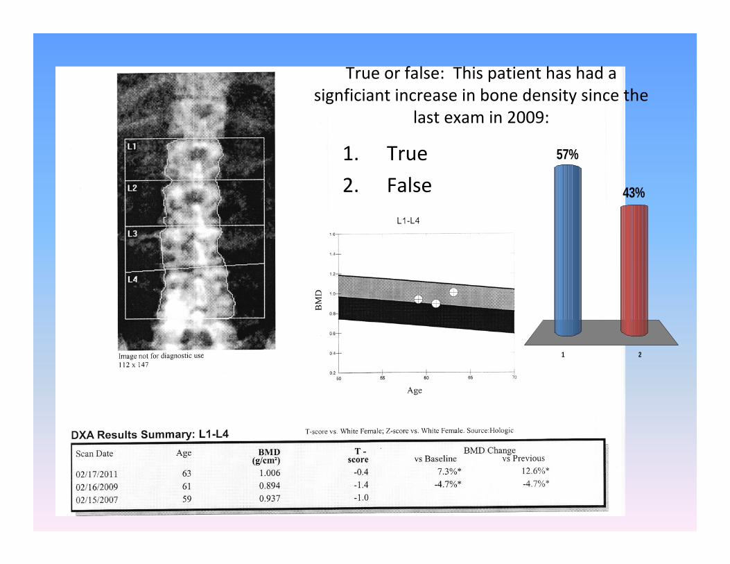
True or false: This patient has had a signficiant increase in bone density since the
last exam in 2009:
1 2
43%
57%1. True
2. False

Serial Monitoring of BMD
• Must look at the regions of interest of the two tests being compared to make sure they are the same
• Compare bone density by comparing BMD in gm/cm2: • (current BMD – previous BMD)/previous BMD x 100• You must know the least significant change of each site for
your machine, measured from the precision error – different for every machine and affected by multiple variables– For example, the OSU Hologic Discovery A machine as a LSC at the
lumbar spine of 3% and at the total hip of 4%– Any change less than these is not considered clinically significant
• The radiologist for your local DXA should be able to provide for you the LSC

2011 2009
What do you do with this discrepancy?Which LS assignment is correct?

When do you exclude vertebrae?
• ISCD position states that you should use as many vertebrae as possible to improve accuracy
• Exclude vertebrae that are >1 SD different from adjacent vertebra
• Can’t use fewer than 2 vertebrae
• In this example, you would report L1‐L3 and exclude L4
• 60% of women age 60yo will have degenerative changes of the spine
‐2.2
‐2.6
‐2.1
‐0.8
T‐scores:

What do you decide to do with Mrs. Johnson’s lumbar spine bone density?
1 2 3 4
17% 17%
37%
30%
1. Read all 4 vertebrae – she has osteopenia
2. There are not enough quality vertebrae to measure – examine the forearm instead
3. Exclude L1 and L4 – read L2 – L3
4. Order bone density by quantitative CT

Scoliosis is also common with advancing age.It can falsely increase bone density.
Would you exclude any of these vertebrae?
Scoliosis
-2.4
-2.9
-3.5
-3.2

Clearly one vertebrae is differentfrom the others –Would you exclude it?

A vertebral compression fracture canlook like this –Most common sites: T7‐T9 and T12‐L2
L2 should be excluded from analysis.
Consideration of plain film appropriateDx of fracture trumps T‐score ‐2.3!

63yo WF with BrCAHeight: 66.5 inWeight: 263.0 lb
Total Hip T‐score is ‐2.6
Impact of obesity on LS BMD

Same person, same day
First BMD: 0.994 g/cm2
Second BMD: 1.043 g/cm2
Change: +4.9%

What about the Z‐score?
• This pt clearly has osteoporosis
• Z‐score indicates accelerated loss
• Z‐score less than 2SD from peers concerning for secondary causes of bone loss
• Z‐score >2SD most likely due to OA –could also signify sclerosis

‐Most useful site in the forearm is the “proximal”or 1/3 or 33% forearm‐composed almost entirely of cortical bone‐useful in primary hyperparathyroidism‐changes very little over time, even in responseto treatment – not recommended as a site forserial measurement

Tuesday morning, 8:30
• Your 60yo female patient presents for f/u regarding her osteoporosis
• Colles fracture 6 years ago after slipping on wet grass and falling from standing height– Baseline LS DXA t‐score ‐2.7; Total hip T‐score ‐2.2
• She took bisphosphonate from 2005 – 2009, with improvement in bone density and no further fractures – you started her on a drug ‘holiday’ in 2009 – now here for f/u
• Over the past 2 years she’s done well, no falls or fractures
• Repeat bone density reveals– LS T‐score ‐2.4 – 6% increase since 2005; stable since 2009
– TH T‐score ‐1.8 – 4% increase since 2005, stable since 2009

Bottom of the bone density report gives a FRAX score of major osteoporotic fracture risk 21%, hip fracture risk 2.5% ‐ ‘consider therapy’. You decide to:
1 2 3 4
25% 25%25%25%1. Restart bisphosphonates due to her high FRAX score
2. Start denosumab, given the FRAX score
3. Repeat bone density in 2 years, continue calcium and vitamin D supplementation
4. Order markers of bone turnover to give a better sense of her current risk for fracture

National Osteoporosis Foundation guidelines for therapeutic management
• A hip or vertebral (clinical or morphometric) fracture• T-score < -2.5 at the femoral neck or spine after
appropriate evaluation to exclude secondary causes• Low bone mass (T-score between -1.0 and -2.5 at the
FN, TH or LS) AND a 10-year probably of a hip fracture >3% or of a major osteoporotic fracture of >20% based upon FRAX
• Clinician’s judgement and/or patient preference may indicate treatment for people with 10-year fracture probabilities above or below these levels
2008 NOF Clinician’s Guide to Prevention & Treatment of Osteoporosis,www.nof.org/professionals/clinical-guidelines
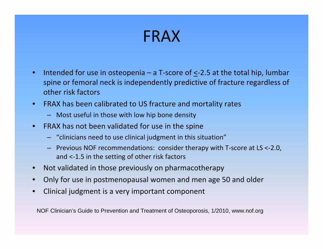
FRAX
• Intended for use in osteopenia – a T‐score of <‐2.5 at the total hip, lumbar spine or femoral neck is independently predictive of fracture regardless of other risk factors
• FRAX has been calibrated to US fracture and mortality rates– Most useful in those with low hip bone density
• FRAX has not been validated for use in the spine– “clinicians need to use clinical judgment in this situation”
– Previous NOF recommendations: consider therapy with T‐score at LS <‐2.0, and <‐1.5 in the setting of other risk factors
• Not validated in those previously on pharmacotherapy
• Only for use in postmenopausal women and men age 50 and older
• Clinical judgment is a very important component
NOF Clinician’s Guide to Prevention and Treatment of Osteoporosis, 1/2010, www.nof.org

FRAX Fracture Calculation Tool
http://www.shef.ac.uk/FRAX/tool.jsp
NOF TreatmentGuidelines:
10 year probabilityFor major Osteoporotic Fx:20%
10 year prob forHip fracture:3%

FRAX Issues• What about glucocorticoids?
– FRAX tends to underestimate fracture risk with glucocorticoid doses over 7.5mg prednisone equivalent
– Overestimates fx risk in doses 2.5mg or less
– Underestimates risk in those on high dose inhaled
• Isolated low bone mass at the lumbar spine– only use of the femoral neck is recommended at this time
– Clinical judgement in isolated osteopenia of the spine
• Duration and dose of tobacco smoking is important
• Parental non‐hip fracture may confer risk onto your patient, but cannot be quantified using FRAX
• ** All underscoring the importance of clinical judgment

Cancellous vs. Cortical discrepancy55yo patient with breast cancer

A patient with osteopenia presents with three metatarsal fractures over the past year incurred while jogging. Which is true regarding the use of FRAX in this patient?
1 2 3 4
25% 25%25%25%
1. FRAX can be helpful to provide guidance in deciding whether to treat this patient.
2. FRAX is not needed – this patient has already had low trauma fractures.
3. Metatarsal fractures should not be considered under the heading “previous fracture”
4. I would start bisphosphonate therapy and use FRAX later in conjunction with 2 year f/u DXA

How else can we use FRAX?
• 53yo female presents for follow up on recent testing – she had a heel ultrasound at a health fair and was told she “has the bones of an 80 year old”!
• Her mother had a hip fracture at the age of 86
• She has never smoked cigarettes, never required glucocorticoids, has not had any height loss from her youth
• She suffered a fibula fracture after stepping into a hole in a field while walking
• You order a DXA:– LS T‐score ‐2.1
– TH T‐score ‐1.8
– FN T‐score ‐2.0

Other uses for FRAX:
FRAX is helpful for reassurance, too!

Summary• DXA remains the test of choice for evaluating fracture risk, but start to
insist on evaluating the images of each DXA you order
• Not infrequently, LS vertebra of interest are malpositioned
• Arthritis impacts, and likely falsely elevates, bone density of the spine up to 60% of the time in people >60yo.
• The least amount of vertebra that you can make a diagnosis upon is two
• The total hip, femoral neck and lumbar spine bone densities are most predictive of future fracture
• FRAX is a helpful tool in the setting of low bone density (osteopenia).
• FRAX cannot be used in someone who has a prior history of pharmacotherapy.
• Clinical judgment continues to play a significant role, even in the use of FRAX.
• FRAX can be a helpful tool to provide reassurance to your patient with low risk of fracture.