drug discovery tools for immunotoxicology research
TRANSCRIPT

Drug Discovery Tools for Immunotoxicology Research

Table of Contents
Immunotoxicology . . . . . . . . . . . . . . . . . . . . . . . . . . . . . . . . . . . . . . . . . . . . . . . . . . . . . . . . . . . . . . . . . . . . . . . . . . . . . . . . . .3
Immunotoxicology Overview . . . . . . . . . . . . . . . . . . . . . . . . . . . . . . . . . . . . . . . . . . . . . . . . . . . . . . . . . . . . . . . . . . . .3-5
Immunosuppression . . . . . . . . . . . . . . . . . . . . . . . . . . . . . . . . . . . . . . . . . . . . . . . . . . . . . . . . . . . . . . . . . . . . . . . . . . .3
Clinical Chemistry . . . . . . . . . . . . . . . . . . . . . . . . . . . . . . . . . . . . . . . . . . . . . . . . . . . . . . . . . . . . . . . . . . . . . . . . .3
Blood Cellular Levels . . . . . . . . . . . . . . . . . . . . . . . . . . . . . . . . . . . . . . . . . . . . . . . . . . . . . . . . . . . . . . . . . . . . . . .3
Cell Phenotyping . . . . . . . . . . . . . . . . . . . . . . . . . . . . . . . . . . . . . . . . . . . . . . . . . . . . . . . . . . . . . . . . . . . . . . . . . .3
Immune Function Studies . . . . . . . . . . . . . . . . . . . . . . . . . . . . . . . . . . . . . . . . . . . . . . . . . . . . . . . . . . . . . . . . . . .3
Immunogenicity . . . . . . . . . . . . . . . . . . . . . . . . . . . . . . . . . . . . . . . . . . . . . . . . . . . . . . . . . . . . . . . . . . . . . . . . . . . . . .4
Hypersensitivity . . . . . . . . . . . . . . . . . . . . . . . . . . . . . . . . . . . . . . . . . . . . . . . . . . . . . . . . . . . . . . . . . . . . . . . . . . . . . .4
Type I . . . . . . . . . . . . . . . . . . . . . . . . . . . . . . . . . . . . . . . . . . . . . . . . . . . . . . . . . . . . . . . . . . . . . . . . . . . . . . . . . . .4
Type II/III . . . . . . . . . . . . . . . . . . . . . . . . . . . . . . . . . . . . . . . . . . . . . . . . . . . . . . . . . . . . . . . . . . . . . . . . . . . . . . . . .4
Type IV . . . . . . . . . . . . . . . . . . . . . . . . . . . . . . . . . . . . . . . . . . . . . . . . . . . . . . . . . . . . . . . . . . . . . . . . . . . . . . . . . .5
Pseudoallergy . . . . . . . . . . . . . . . . . . . . . . . . . . . . . . . . . . . . . . . . . . . . . . . . . . . . . . . . . . . . . . . . . . . . . . . . . . . .5
Autoimmunity . . . . . . . . . . . . . . . . . . . . . . . . . . . . . . . . . . . . . . . . . . . . . . . . . . . . . . . . . . . . . . . . . . . . . . . . . . . . . . .5
Adverse Immunostimulation . . . . . . . . . . . . . . . . . . . . . . . . . . . . . . . . . . . . . . . . . . . . . . . . . . . . . . . . . . . . . . . . . . . .5
References . . . . . . . . . . . . . . . . . . . . . . . . . . . . . . . . . . . . . . . . . . . . . . . . . . . . . . . . . . . . . . . . . . . . . . . . . . . . . . . . . . . . . . . .5
Drug Discovery Tools . . . . . . . . . . . . . . . . . . . . . . . . . . . . . . . . . . . . . . . . . . . . . . . . . . . . . . . . . . . . . . . . . . . . . . . . . . . . . . . .6
Micronucleus Analysis Assay . . . . . . . . . . . . . . . . . . . . . . . . . . . . . . . . . . . . . . . . . . . . . . . . . . . . . . . . . . . . . . . . . . . . . . .6
BrdU Assay . . . . . . . . . . . . . . . . . . . . . . . . . . . . . . . . . . . . . . . . . . . . . . . . . . . . . . . . . . . . . . . . . . . . . . . . . . . . . . . . . . . . .6
Rat Immunophenotyping . . . . . . . . . . . . . . . . . . . . . . . . . . . . . . . . . . . . . . . . . . . . . . . . . . . . . . . . . . . . . . . . . . . . . . . . .7
Dog Immunophenotyping . . . . . . . . . . . . . . . . . . . . . . . . . . . . . . . . . . . . . . . . . . . . . . . . . . . . . . . . . . . . . . . . . . . . . . . .9
Non-Human Primate Immunophenotyping . . . . . . . . . . . . . . . . . . . . . . . . . . . . . . . . . . . . . . . . . . . . . . . . . . . . . . . . .10
BD Cytometric Bead Array (BD CBA) . . . . . . . . . . . . . . . . . . . . . . . . . . . . . . . . . . . . . . . . . . . . . . . . . . . . . . . . . . . . . . .13
Reagents for Immunotoxicology Research . . . . . . . . . . . . . . . . . . . . . . . . . . . . . . . . . . . . . . . . . . . . . . . . . . . . . . . . . . . . . .15
Micronucleus Analysis Assay . . . . . . . . . . . . . . . . . . . . . . . . . . . . . . . . . . . . . . . . . . . . . . . . . . . . . . . . . . . . . . . . . . . . . .15
BrdU Assay . . . . . . . . . . . . . . . . . . . . . . . . . . . . . . . . . . . . . . . . . . . . . . . . . . . . . . . . . . . . . . . . . . . . . . . . . . . . . . . . . . . .15
Rat Reagents . . . . . . . . . . . . . . . . . . . . . . . . . . . . . . . . . . . . . . . . . . . . . . . . . . . . . . . . . . . . . . . . . . . . . . . . . . . . . . . . . .15
Dog Reagents . . . . . . . . . . . . . . . . . . . . . . . . . . . . . . . . . . . . . . . . . . . . . . . . . . . . . . . . . . . . . . . . . . . . . . . . . . . . . . . . .22
Non-Human Primate Reagents . . . . . . . . . . . . . . . . . . . . . . . . . . . . . . . . . . . . . . . . . . . . . . . . . . . . . . . . . . . . . . . . . . . .25
2 www.bdbiosciences.com
For Research Use Only. Not for use in diagnostic or therapeutic procedures. Purchase does not include or carry any right to resell or transfer this product either as a stand-alone product or as a component of another product. Any use of this product other than the permitted use without the express written authorization of Becton Dickinson and Company is strictly prohibited.BD, BD Logo and all other trademarks are the property of Becton, Dickinson and Company. ©2003 BD
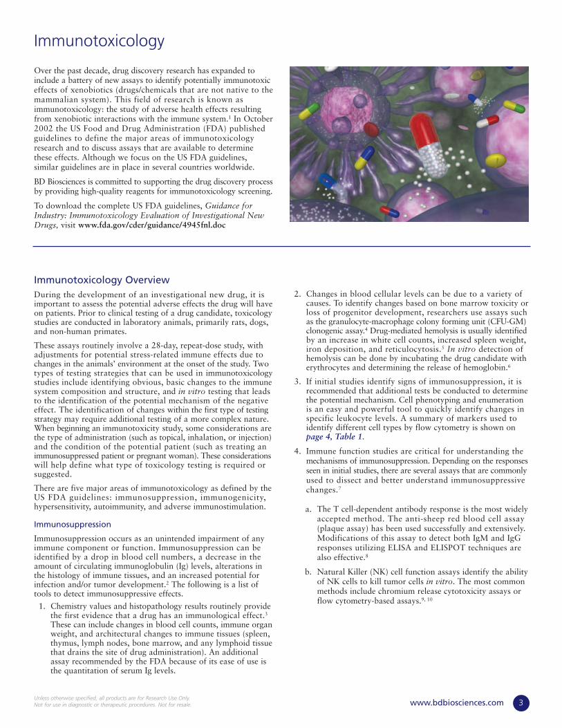
Over the past decade, drug discovery research has expanded toinclude a battery of new assays to identify potentially immunotoxiceffects of xenobiotics (drugs/chemicals that are not native to themammalian system). This field of research is known asimmunotoxicology: the study of adverse health effects resultingfrom xenobiotic interactions with the immune system.1 In October2002 the US Food and Drug Administration (FDA) publishedguidelines to define the major areas of immunotoxicologyresearch and to discuss assays that are available to determinethese effects. Although we focus on the US FDA guidelines,similar guidelines are in place in several countries worldwide.
BD Biosciences is committed to supporting the drug discovery processby providing high-quality reagents for immunotoxicology screening.
To download the complete US FDA guidelines, Guidance forIndustry: Immunotoxicology Evaluation of Investigational NewDrugs, visit www.fda.gov/cder/guidance/4945fnl.doc
Immunotoxicology OverviewDuring the development of an investigational new drug, it isimportant to assess the potential adverse effects the drug will haveon patients. Prior to clinical testing of a drug candidate, toxicologystudies are conducted in laboratory animals, primarily rats, dogs,and non-human primates.
These assays routinely involve a 28-day, repeat-dose study, withadjustments for potential stress-related immune effects due tochanges in the animals’ environment at the onset of the study. Twotypes of testing strategies that can be used in immunotoxicologystudies include identifying obvious, basic changes to the immunesystem composition and structure, and in vitro testing that leadsto the identification of the potential mechanism of the negativeeffect. The identification of changes within the first type of testingstrategy may require additional testing of a more complex nature.When beginning an immunotoxicity study, some considerations arethe type of administration (such as topical, inhalation, or injection)and the condition of the potential patient (such as treating animmunosuppressed patient or pregnant woman). These considerationswill help define what type of toxicology testing is required orsuggested.
There are five major areas of immunotoxicology as defined by theUS FDA guidelines: immunosuppression, immunogenicity,hypersensitivity, autoimmunity, and adverse immunostimulation.
Immunosuppression
Immunosuppression occurs as an unintended impairment of anyimmune component or function. Immunosuppression can beidentified by a drop in blood cell numbers, a decrease in theamount of circulating immunoglobulin (Ig) levels, alterations inthe histology of immune tissues, and an increased potential forinfection and/or tumor development.2 The following is a list oftools to detect immunosuppressive effects.
1. Chemistry values and histopathology results routinely providethe first evidence that a drug has an immunological effect.3
These can include changes in blood cell counts, immune organweight, and architectural changes to immune tissues (spleen,thymus, lymph nodes, bone marrow, and any lymphoid tissuethat drains the site of drug administration). An additionalassay recommended by the FDA because of its ease of use isthe quantitation of serum Ig levels.
2. Changes in blood cellular levels can be due to a variety ofcauses. To identify changes based on bone marrow toxicity orloss of progenitor development, researchers use assays suchas the granulocyte-macrophage colony forming unit (CFU-GM)clonogenic assay.4 Drug-mediated hemolysis is usually identifiedby an increase in white cell counts, increased spleen weight,iron deposition, and reticulocytosis.5 In vitro detection ofhemolysis can be done by incubating the drug candidate witherythrocytes and determining the release of hemoglobin.6
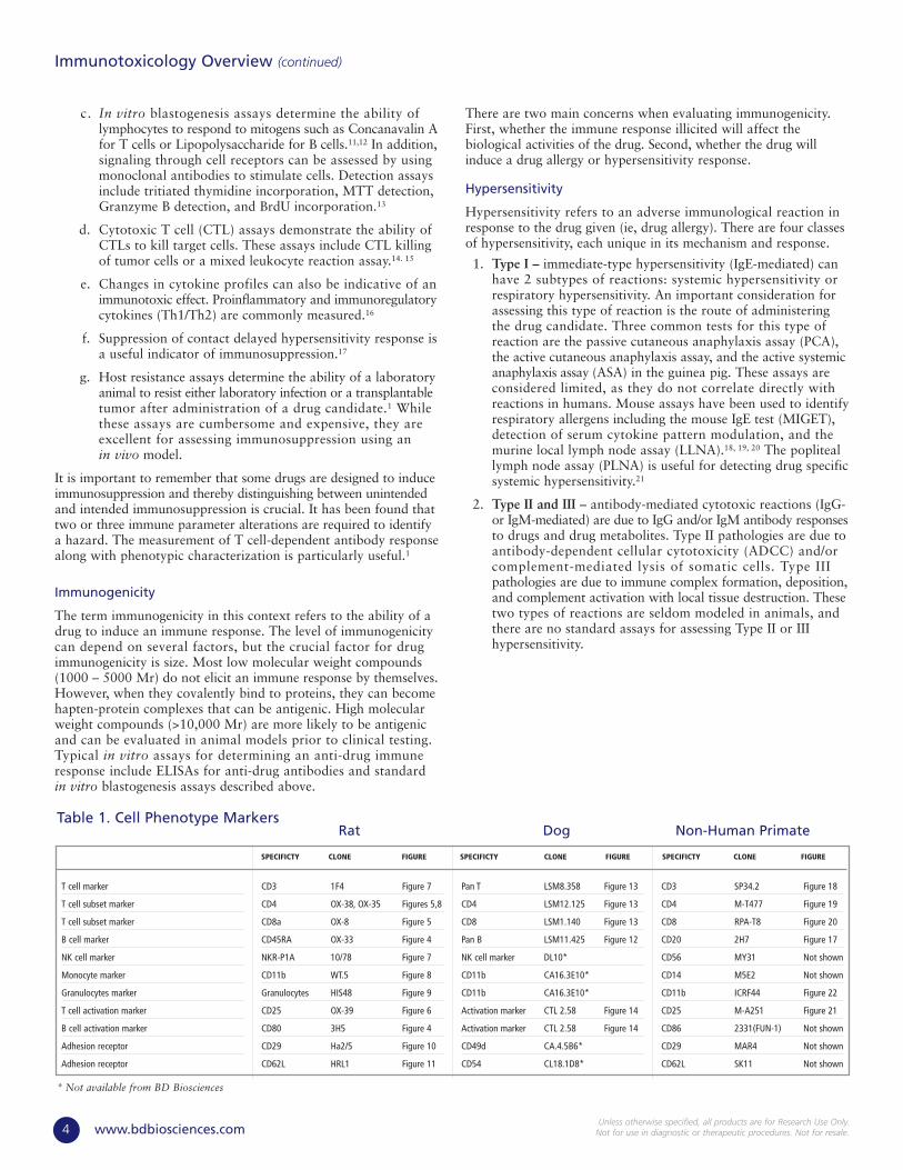
3. If initial studies identify signs of immunosuppression, it isrecommended that additional tests be conducted to determinethe potential mechanism. Cell phenotyping and enumerationis an easy and powerful tool to quickly identify changes inspecific leukocyte levels. A summary of markers used toidentify different cell types by flow cytometry is shown onpage 4, Table 1.
4. Immune function studies are critical for understanding themechanisms of immunosuppression. Depending on the responsesseen in initial studies, there are several assays that are commonlyused to dissect and better understand immunosuppressivechanges.7
a. The T cell-dependent antibody response is the most widelyaccepted method. The anti-sheep red blood cell assay(plaque assay) has been used successfully and extensively.Modifications of this assay to detect both IgM and IgGresponses utilizing ELISA and ELISPOT techniques arealso effective.8
b. Natural Killer (NK) cell function assays identify the abilityof NK cells to kill tumor cells in vitro. The most commonmethods include chromium release cytotoxicity assays orflow cytometry-based assays.9, 10
Immunotoxicology
3www.bdbiosciences.comUnless otherwise specified, all products are for Research Use Only.Not for use in diagnostic or therapeutic procedures. Not for resale.
c. In vitro blastogenesis assays determine the ability oflymphocytes to respond to mitogens such as Concanavalin Afor T cells or Lipopolysaccharide for B cells.11,12 In addition,signaling through cell receptors can be assessed by usingmonoclonal antibodies to stimulate cells. Detection assaysinclude tritiated thymidine incorporation, MTT detection,Granzyme B detection, and BrdU incorporation.13
d. Cytotoxic T cell (CTL) assays demonstrate the ability ofCTLs to kill target cells. These assays include CTL killingof tumor cells or a mixed leukocyte reaction assay.14. 15
e. Changes in cytokine profiles can also be indicative of animmunotoxic effect. Proinflammatory and immunoregulatorycytokines (Th1/Th2) are commonly measured.16
f. Suppression of contact delayed hypersensitivity response isa useful indicator of immunosuppression.17
g. Host resistance assays determine the ability of a laboratoryanimal to resist either laboratory infection or a transplantabletumor after administration of a drug candidate.1 Whilethese assays are cumbersome and expensive, they areexcellent for assessing immunosuppression using an in vivo model.
It is important to remember that some drugs are designed to induceimmunosuppression and thereby distinguishing between unintendedand intended immunosuppression is crucial. It has been found thattwo or three immune parameter alterations are required to identifya hazard. The measurement of T cell-dependent antibody responsealong with phenotypic characterization is particularly useful.1
Immunogenicity
The term immunogenicity in this context refers to the ability of adrug to induce an immune response. The level of immunogenicitycan depend on several factors, but the crucial factor for drugimmunogenicity is size. Most low molecular weight compounds(1000 – 5000 Mr) do not elicit an immune response by themselves.However, when they covalently bind to proteins, they can becomehapten-protein complexes that can be antigenic. High molecularweight compounds (>10,000 Mr) are more likely to be antigenicand can be evaluated in animal models prior to clinical testing.Typical in vitro assays for determining an anti-drug immuneresponse include ELISAs for anti-drug antibodies and standardin vitro blastogenesis assays described above.
There are two main concerns when evaluating immunogenicity.First, whether the immune response illicited will affect thebiological activities of the drug. Second, whether the drug willinduce a drug allergy or hypersensitivity response.
Hypersensitivity
Hypersensitivity refers to an adverse immunological reaction inresponse to the drug given (ie, drug allergy). There are four classesof hypersensitivity, each unique in its mechanism and response.
1. Type I – immediate-type hypersensitivity (IgE-mediated) canhave 2 subtypes of reactions: systemic hypersensitivity orrespiratory hypersensitivity. An important consideration forassessing this type of reaction is the route of administeringthe drug candidate. Three common tests for this type ofreaction are the passive cutaneous anaphylaxis assay (PCA),the active cutaneous anaphylaxis assay, and the active systemicanaphylaxis assay (ASA) in the guinea pig. These assays areconsidered limited, as they do not correlate directly withreactions in humans. Mouse assays have been used to identifyrespiratory allergens including the mouse IgE test (MIGET),detection of serum cytokine pattern modulation, and themurine local lymph node assay (LLNA).18, 19, 20 The popliteallymph node assay (PLNA) is useful for detecting drug specificsystemic hypersensitivity.21
2. Type II and III – antibody-mediated cytotoxic reactions (IgG-or IgM-mediated) are due to IgG and/or IgM antibody responsesto drugs and drug metabolites. Type II pathologies are due toantibody-dependent cellular cytotoxicity (ADCC) and/orcomplement-mediated lysis of somatic cells. Type IIIpathologies are due to immune complex formation, deposition,and complement activation with local tissue destruction. Thesetwo types of reactions are seldom modeled in animals, andthere are no standard assays for assessing Type II or IIIhypersensitivity.
Immunotoxicology Overview (continued)
4 www.bdbiosciences.comUnless otherwise specified, all products are for Research Use Only.
Not for use in diagnostic or therapeutic procedures. Not for resale.
Table 1. Cell Phenotype Markers
T cell marker
T cell subset marker
T cell subset marker
B cell marker
NK cell marker
Monocyte marker
Granulocytes marker
T cell activation marker
B cell activation marker
Adhesion receptor
Adhesion receptor
CD3 1F4 Figure 7
CD4 OX-38, OX-35 Figures 5,8
CD8a OX-8 Figure 5
CD45RA OX-33 Figure 4
NKR-P1A 10/78 Figure 7
CD11b WT.5 Figure 8
Granulocytes HIS48 Figure 9
CD25 OX-39 Figure 6
CD80 3H5 Figure 4
CD29 Ha2/5 Figure 10
CD62L HRL1 Figure 11
Pan T LSM8.358 Figure 13
CD4 LSM12.125 Figure 13
CD8 LSM1.140 Figure 13
Pan B LSM11.425 Figure 12
NK cell marker DL10*
CD11b CA16.3E10*
CD11b CA16.3E10*
Activation marker CTL 2.58 Figure 14
Activation marker CTL 2.58 Figure 14
CD49d CA.4.5B6*
CD54 CL18.1D8*
CD3 SP34.2 Figure 18
CD4 M-T477 Figure 19
CD8 RPA-T8 Figure 20
CD20 2H7 Figure 17
CD56 MY31 Not shown
CD14 M5E2 Not shown
CD11b ICRF44 Figure 22
CD25 M-A251 Figure 21
CD86 2331(FUN-1) Not shown
CD29 MAR4 Not shown
CD62L SK11 Not shown
SPECIFICTY CLONE FIGURE SPECIFICTY CLONE FIGURE SPECIFICTY CLONE FIGURE
Rat Dog Non-Human Primate
* Not available from BD Biosciences

3. Type IV – delayed-type hypersensitivity (T lymphocyte-mediated) occurs as a skin reaction. Nonclinical studies toassess type IV responses have classically been conducted inguinea pigs using sensitization and challenge. Several FDA-accepted tests have been developed to assess a drug’s ability toinduce sensitization, including the Buehler assay, the guineapig maximization test, the split adjuvant technique, and theDraize test. Additionally, tests have been developed in mice,such as the mouse ear swelling test (MEST), that yield resultssimilar to the guinea pig assays.22 To detect the inductionof delayed-type hypersensitivity reactions, one techniquein particular has been used extensively with drugs knownas sensitizers. This assay, the LLNA, can be used as analternative to the guinea pig assays.23
4. Pseudoallergic (Anaphylactoid) reactions occur when theinflammatory or anaphylactic systems are activated, but notin response to a specific antigen. Anaphylactoid reactions aredifferent from anaphylaxis (IgE-mediated) and can bedifferentiated by mast cell histamine release and activation of the complement system.24 Detection of pseudoallergicresponses includes assays to detect serum complementproteins by ELISA or other related methods.
5www.bdbiosciences.comUnless otherwise specified, all products are for Research Use Only.Not for use in diagnostic or therapeutic procedures. Not for resale.
Autoimmunity
Autoimmunity occurs when the immune system fails to distinguishbetween self and non-self, producing antibodies directed againstits own tissue and activating an immune response to self antigens.This type of a response can cause tissue damage, immune complexdeposition, inappropriate activation of cells, and hypersensitivityreactions. Autoimmune reactions can be organ-specific or non-specific with local or systemic responses.
No standard assays have been validated for determining potentialautoimmune reactions. The most commonly used method is thePLNA. 21, 25 Adaptations of the LLNA have also been described toassess potential autoimmune reactions.23
Adverse Immunostimulation
Adverse immunostimulation refers to unregulated and unintendedactivation of some component of the immune system. This causeschronic inflammation and an infiltration of leukocytes in tissues.26
This type of reaction is not commonly seen with administration ofa drug candidate. In fact, chemicals with this type of activity areoften evaluated as potential adjuvants. There are no standardizedmethods for assessing this type of drug effect.
References
1. Toxicologic Pathology, 2002. 30(1): p. 54-58.
2. Toxicology Letters, 1998. 102-103: p. 267-270.
3. Toxicology, 1994. 86(3): p. 187-212.
4. Cell Biol Toxicol, 2001. 17(2): p. 95-105.
5. Toxic Responses of The Blood. Casarett & Doull's Toxicology. 2001, NewYork: McGraw-Hill. 389-417.
6. Red blood cell lysis (Basic Protocol 4). Current Protocols in Toxicology.Vol. 1. 1999, New York: John Wiley & Sons. 2.4.4.
7. Toxicology Letters, 1998. 102-103: p. 247-255.
8. Toxicology, 2000. 156(1): p. 1-11.
9. Journal of Immunological Methods, 2001. 253: p. 177-187.
10. Cytometry, 2000. 41: p. 289-297.
11. Fundam Appl Toxicol, 1993. 21(4): p. 535-545.
12. Toxicology, 1995. 96(2): p. 147-156.
13. Methods, 1999. 19: p. 28-35.
14. Toxicology, 1998. 127(1-3): p. 223-232.
15. Toxicology, 1997. 119: p. 95-101.
16. Methods, 1999. 19: p. 17-27.
17. Journal of Immunology, 2000. 165: p. 2374-2381.
18. Fundamental and Applied Toxicology, 1996. 33: p. 1-10.
19. Toxicology, 2001. 158: p. 51-58.
20. Toxicology and Applied Pharmacology, 1997. 145: p. 218-229.
21. Toxicology, 2001. 158: p. 65-69.
22. Toxicology and Applied Pharmacology, 1986. 84: p. 93-114.
23. Toxicology, 2001. 158: p. 59-64.
24. Crit Rev. Therapeutic Drug Carrier Syst, 2001. 18(6): p. 567-606.
25. Environ Health Perspect, 1999. 107: p. 673-677.
26. Eur J Nucl Med, 2001. 28(9): p. 1384-1393.

The micronucleus assay is an established method for studying in vivo chromosomal damage.It is based on the observation that displaced chromatin, resulting from chromosome lossor breakage, may fail to be incorporated into daughter nuclei as a cell divides. The resulting“micronucleus” is found in the cytoplasm. Elevations in the frequency of micronuclei (MN)are indicative of genotoxic activity.
The µicroFlow® and µicroFlowPLUS Micronucleus Analysis Kits are designed to measureMN events in peripheral blood cells of mice or rats. During erythropoiesis, an erythroblastexpels its main nucleus to become a reticulocyte (RET), while the MN remain in the cytoplasm.The newly formed RET is then released from the bone marrow into the circulatingbloodstream, where it develops into a normochromatic erythrocyte (NCE). µicroFlow®and µicroFlowPLUS are based on a RET-specific cell surface marker, CD71, whichdifferentially labels immature erythrocytes. When fixed peripheral blood cells areappropriately treated, NCEs and RETs with and without MN are easily resolved andquantified by flow cytometry. These kits measure the frequency of each cell population of interest (%RET, %MN-RET, %MN-NCE).µicroFlow® is a registered trademark of Litron Laboratories
Rat Immunophenotyping
6 www.bdbiosciences.comUnless otherwise specified, all products are for Research Use Only.
Not for use in diagnostic or therapeutic procedures. Not for resale.
µicroFlow® and µicroFlowPLUS Micronucleus Analysis Kits for Quantitative Analysisof Micronuclei by Flow Cytometry
BrdU Analysis for Detection of Proliferating Cells
The immunofluorescent staining of incorporated bromodeoxyuridine (BrdU) and flowcytometric analysis provide a high resolution technique to determine the frequency andnature of individual cells that have synthesized DNA. In this method, BrdU (an analog ofthe DNA precursor, thymidine) is incorporated into newly synthesized DNA by cells enteringand progressing through the S (DNA synthesis) phase of the cell cycle. The incorporatedBrdU is stained with specific anti-BrdU fluorescent antibodies. The levels of cell-associatedBrdU are then measured by flow cytometry.
Propidium Iodide
Reticulocytes
Parasitized RBCs
Nucleated
100 101 102 103 104
FITC
an
ti-C
D71
(O
X-2
6)
100
101
102
103
104A.
PE a
nti
-IL-
4 (O
X-3
1)
APC
an
ti-B
RD
U (
3D4)
APC anti-BRDU (3D4) APC anti-BRDU (3D4) APC anti-BRDU (3D4)7AAD
FITC
an
ti-I
FN-γ
(DB
1)
PE a
nti
-CD
25 (
OX
-39)
100 101 102 103 104100
101
102
103
104
0 200 400 600 800100
101
102
103
104
100 101 102 103 104100
101
102
103
104
100 101 102 103 104100
101
102
103
104
A. B. D.C.
B.
Figure 1B. Mouse erythrocytes stained withµicroflow reagent.
Figure 1A. FITC and PI staining of rodenterythrocytes for flow cytometric analysis
Figure 2. Multicolor flow cytometric analysis of proliferating (BrdU positive) rat cells that produce IFN-γ. Spleen cells from a rat were primed in vitrowith anti-CD3 and restimulated with PMA and ionomycin in the presence of a protein transport inhibitor (to promote intracellular cytokine accumulation).During the final 45 minutes of culture, the cells were labeled with 20 µM BrdU. The cells were harvested and stained with FITC anti-IFN-γ (DB1) (A),APC anti-BrdU (3D4), 7-AAD (B), PE anti-CD25 (OX-39) (C), and in a separate well PE anti-IL-4 (OX-31) (D).
Drug Discovery Tools
7www.bdbiosciences.comUnless otherwise specified, all products are for Research Use Only.Not for use in diagnostic or therapeutic procedures. Not for resale.
B lymphocytes
T lymphocytes
FITC anti-CD45RA (OX-33)100 101 102 103 104
PE a
nti
-CD
80 (
3H5)
100
101
102
103
104B.
anti-Marginal Zone B Cell marker (HIS57) on paraffin-embedded spleen
B.
100 101 102 103 1040
40
80
120
160
200
Cel
l Nu
mb
er
FITC anti-CD25 (0X-39)
anti-CD4 (OX-38) on frozen spleen
C.
FITC anti-CD4 (OX-35)100 101 102 103 104
APC
an
dt-
CD
3 (1
F4)
100
101
102
103
104D.
anti-CD8a (OX-8) on paraffin-embedded spleen
A.
100 101 102 103 104100
101
102
103
104
FITC anti-CD8a (OX-8)
APC
an
ti-C
D3
(1F4
)
B.
FITC anti-IgM (G53-239)100 101 102 103 104
PE a
nti
-CD
45R
A (
OX
-33)
100
101
102
103
104A.
100 101 102 103 104100
101
102
103
104
FITC anti-CD45RA (OX-33)
PE a
nti
-CD
80 (
3H5)
A.
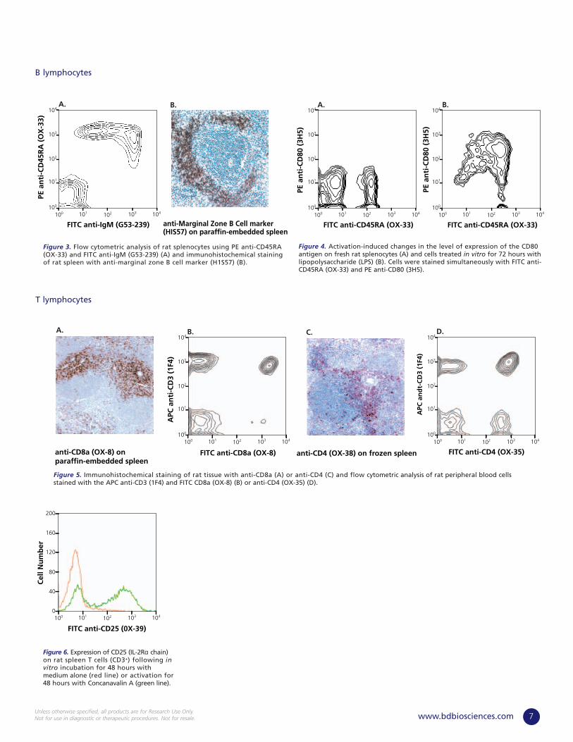
Figure 4. Activation-induced changes in the level of expression of the CD80antigen on fresh rat splenocytes (A) and cells treated in vitro for 72 hours withlipopolysaccharide (LPS) (B). Cells were stained simultaneously with FITC anti-CD45RA (OX-33) and PE anti-CD80 (3H5).
Figure 3. Flow cytometric analysis of rat splenocytes using PE anti-CD45RA(OX-33) and FITC anti-IgM (G53-239) (A) and immunohistochemical stainingof rat spleen with anti-marginal zone B cell marker (H1S57) (B).
Figure 6. Expression of CD25 (IL-2Rα chain)on rat spleen T cells (CD3+) following invitro incubation for 48 hours withmedium alone (red line) or activation for48 hours with Concanavalin A (green line).
Figure 5. Immunohistochemical staining of rat tissue with anti-CD8a (A) or anti-CD4 (C) and flow cytometric analysis of rat peripheral blood cellsstained with the APC anti-CD3 (1F4) and FITC CD8a (OX-8) (B) or anti-CD4 (OX-35) (D).

Rat Immunophenotyping (continued)
8 www.bdbiosciences.comUnless otherwise specified, all products are for Research Use Only.
Not for use in diagnostic or therapeutic procedures. Not for resale.
100 101 102 103 104100
101
102
103
104
FITC anti-CD3 (1F4)
PE a
nti
-NK
R-P
1A (
10/7
8)
NK-T Cells
NK Cells
100 101 102 103 104
APC
an
ti-C
D3
(1F4
)
PE anti-CD4 (OX-35)
CD4- T cellsCD4+ T cells
CD3- CD4+Monocytes
100
101
102
103
104A.
FITC anti-CD11b (WT.5)100 101 102 103 104
Cel
l Nu
mb
er
0
10
20
40
50
30
B.
Forward Scatter0 50 100 150 250
Sid
e Sc
atte
r
0
50
100
150
250
200
200
Granulocytes
Lymphocytes/ Monocytes
A.
APC anti-Granulocyte Marker (HIS48)100 101 102 103 104
Cel
l Nu
mb
er
0
60
120
240
300
180
B.
anti-CD29 (Ha2/5)
A.
100 101 102 103 104100
101
102
103
104
FITC anti-CD29 (Ha2/5)
PE a
nti
-CD
3 (1
F4)
B.
100 101 102 103 104100
101
102
103
104
FITC anti-CD62L (HRL1)
PE a
nti
-CD
3 (1
F4)
Figure 7. Rat peripheral blood leukocytesstained with PE anti-NKR-P1A (10/78) andFITC anti-CD3 (1F4).
Figure 11. Flow cytometric analysis of ratsplenocytes stained with PE anti-CD3 (1F4)and FITC anti-CD62L (HRL1).
Figure 8. Flow cytometric analysis of rat peripheral blood monocytes characterized bythe expression of CD4 (OX-35) and lack of CD3 (1F4) (A).The CD3– CD4+ subset wasanalyzed using FITC anti-CD11b (WT.5) (B).
Figure 9. Flow cytometric analysis of rat granulocytes, characterized by a high side scatterprofile (A). Gated granulocytes were stained with APC anti-granulocyte marker (HIS48) (B).
Figure 10. Immunohistochemical staining of frozen rat spleen with anti-CD29(Ha2/5) (A) and flow cytometric analysis of rat splenocytes stained with PE anti-CD3 (1F4) and FITC anti-CD29 (Ha2/4) (B).
NK Cells
Granulocytes
Adhesion Molecules
Monocytes
Dog Immunophenotyping
9www.bdbiosciences.comUnless otherwise specified, all products are for Research Use Only.Not for use in diagnostic or therapeutic procedures. Not for resale.
A.
PE anti-B cells (LSM11.425)100 101 102 103 104
Sid
e Sc
atte
r
0
250
500
750
1000
A.
Sid
e Sc
atte
r
Sid
e Sc
atte
r
0
250
500
750
1000
0
250
500
750
1000
FITC anti-CD8 (LSM1.140)
C.
FITC anti-Pan T cells (LSM8.358)100 101 102 103 104100 101 102 103 104
B.
Sid
e Sc
atte
r
0
250
500
750
1000
FITC anti-CD4 (LSM12.125)100 101 102 103 104
FITC anti-Activation Marker (CTL2.58)100
0
80
160
240
320
101 102 103 104
Cel
l Nu
mb
er
0 50 100 150 2500
80
160
240
400
200
320
Purified anti-CD44 (69.55)
Cel
l Nu
mb
er
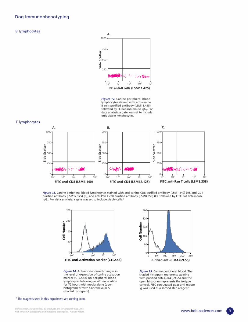
Figure 14. Activation-induced changes inthe level of expression of canine activationmarker (CTL2.58) on peripheral bloodlymphocytes following in vitro incubationfor 72 hours with media alone (openhistogram) or with Concanavalin A(shaded histogram).
Figure 12. Canine peripheral bloodlymphocytes stained with anti-canine B cells purified antibody (LSM11.425),followed by PE Rat anti-mouse IgG1. Fordata analysis, a gate was set to includeonly viable lymphocytes.
Figure 13. Canine peripheral blood lymphocytes stained with anti-canine CD8 purified antibody (LSM1.140) (A), anti-CD4purified antibody (LSM12.125) (B), and anti-Pan T cell purified antibody (LSM8.853) (C), followed by FITC Rat anti-mouseIgG1. For data analysis, a gate was set to include viable cells.*
Figure 15. Canine peripheral blood. Theshaded histogram represents stainingwith purified anti-CD44 (69.55) and theopen histogram represents the isotypecontrol. FITC-conjugated goat anti-mouseIg was used as a second-step reagent.
B lymphocytes
T lymphocytes
* The reagents used in this experiment are coming soon.

Baboon Rhesus Cynomolgus
Non-Human Primate Immunophenotyping
10 www.bdbiosciences.comUnless otherwise specified, all products are for Research Use Only.
Not for use in diagnostic or therapeutic procedures. Not for resale.
100 101 102 103 104
Cel
l Nu
mb
er
0
60
120
240
300
180
100 101 102 103 104
Cel
l Nu
mb
er
0
60
120
240
300
180
100 101 102 103 104
Cel
l Nu
mb
er
0
60
120
240
300
180
Baboon Rhesus Cynomolgus
100 101 102 103 104
Cel
l Nu
mb
er
0
60
120
240
300
180
100 101 102 103 104
Cel
l Nu
mb
er
0
60
120
240
300
180
100 101 102 103 104
Cel
l Nu
mb
er
0
60
120
240
300
180
Baboon Rhesus
0 50 100 150 250
Sid
e Sc
atte
r
0
50
100
150
250
200
200
0 50 100 150 250Si
de
Scat
ter
0
50
100
150
250
200
200
Figure 18. Flow cytometric analysis of CD3 expression on peripheral blood lymphocytes of non-human primates.
Figure 17. Flow cytometric analysis of CD20 expression on peripheral blood leukocytes of non-human primates.
Figure 16. Flow cytometric analysis of CD45 expression on peripheral blood leukocytes ofnon-human primates.
T lymphocytes
B lymphocytes
PerCP anti-CD3 (SP34.2) PerCP anti-CD3 (SP34.2)
PE-Cy5 anti-CD45 (Tü116) PE-Cy5 anti-CD45 (Tü116)
PerCP anti-CD3 (SP34.2)
PE anti-CD20 (2H7) PE anti-CD20 (2H7) PE anti-CD20 (2H7)

11www.bdbiosciences.comUnless otherwise specified, all products are for Research Use Only.Not for use in diagnostic or therapeutic procedures. Not for resale.
Baboon Rhesus Cynomolgus
100 101 102 103 104
Cel
l Nu
mb
er
0
60
120
240
300
180
100 101 102 103 104C
ell N
um
ber
0
60
120
240
300
180
100 101 102 103 104
Cel
l Nu
mb
er
0
60
120
240
300
180
Baboon Rhesus Cynomolgus
100 101 102 103 104
Cel
l Nu
mb
er
0
60
120
240
300
180
100 101 102 103 104
Cel
l Nu
mb
er
0
60
120
240
300
180
100 101 102 103 104
Cel
l Nu
mb
er
0
60
120
240
300
180
Baboon Rhesus Cynomolgus
100 101 102 103 104
Cel
l Nu
mb
er
0
60
120
240
300
180
100 101 102 103 104
Cel
l Nu
mb
er
0
60
120
240
300
180
100 101 102 103 104
Cel
l Nu
mb
er
0
60
120
240
300
180
Figure 19. Flow cytometric analysis of CD4 expression on peripheral blood leukocytes of non-human primates.
Figure 20. Flow cytometric analysis of CD8 expression on peripheral blood leukocytes of non-human primates.
Figure 21. Flow cytometric analysis of CD25 expression on peripheral blood mononuclear cells of non-human primates (thin black line:isotype control, green line: unstimulated PBMCs, red line: Concavalin A-stimulated PBMCs).
T lymphocytes (continued)
PE anti-CD8 (RPA-T8) PE anti-CD8 (RPA-T8) PE anti-CD8 (RPA-T8)
PE anti-CD25 (M-A251) PE anti-CD25 (M-A251) PE anti-CD25 (M-A251)
FITC anti-CD4 (M-T477) FITC anti-CD4 (M-T477) FITC anti-CD4 (M-T477)

Non-Human Primate Immunophenotyping (continued)
12 www.bdbiosciences.comUnless otherwise specified, all products are for Research Use Only.
Not for use in diagnostic or therapeutic procedures. Not for resale.
Baboon Rhesus Cynomolgus
100 101 102 103 104100
101
102
103
104
100 101 102 103 104100
101
102
103
104
100 101 102 103 104100
101
102
103
104
Baboon Rhesus Cynomolgus
100 101 102 103 104100
101
102
103
104
100 101 102 103 104100
101
102
103
104
100 101 102 103 104100
101
102
103
104
Figure 23. Flow cytometric analysis of CD69 and TNF expression in peripheral blood mononuclear cells of non-human primates.
Figure 24. Flow cytometric analysis of CD69 and IFN-γ expression in stimulated peripheral blood lymphocytes of non-human primates.
Baboon Rhesus
100 101 102 103 104
Cel
l Nu
mb
er
0
60
120
240
300
180
100 101 102 103 104
Cel
l Nu
mb
er
0
60
120
240
300
180
Figure 22. Flow cytometric analysis of CD11b expression on peripheral blood granulocytesof non-human primates.
Granulocytes
Cytokines
Purified anti-CD11b (ICRF44) Purified anti-CD11b (ICRF44)
FITC anti-TNF (Mab11)
PE a
nti
-CD
69 (
FN50
)
PE a
nti
-CD
69 (
FN50
)
PE a
nti
-CD
69 (
FN50
)
PE a
nti
-CD
69 (
FN50
)
PE a
nti
-CD
69 (
FN50
)
PE a
nti
-CD
69 (
FN50
)
FITC anti-TNF (Mab11) FITC anti-TNF (Mab11)
FITC anti-IFN-γ (4S.B3) FITC anti-IFN-γ (4S.B3) FITC anti-IFN-γ (4S.B3)
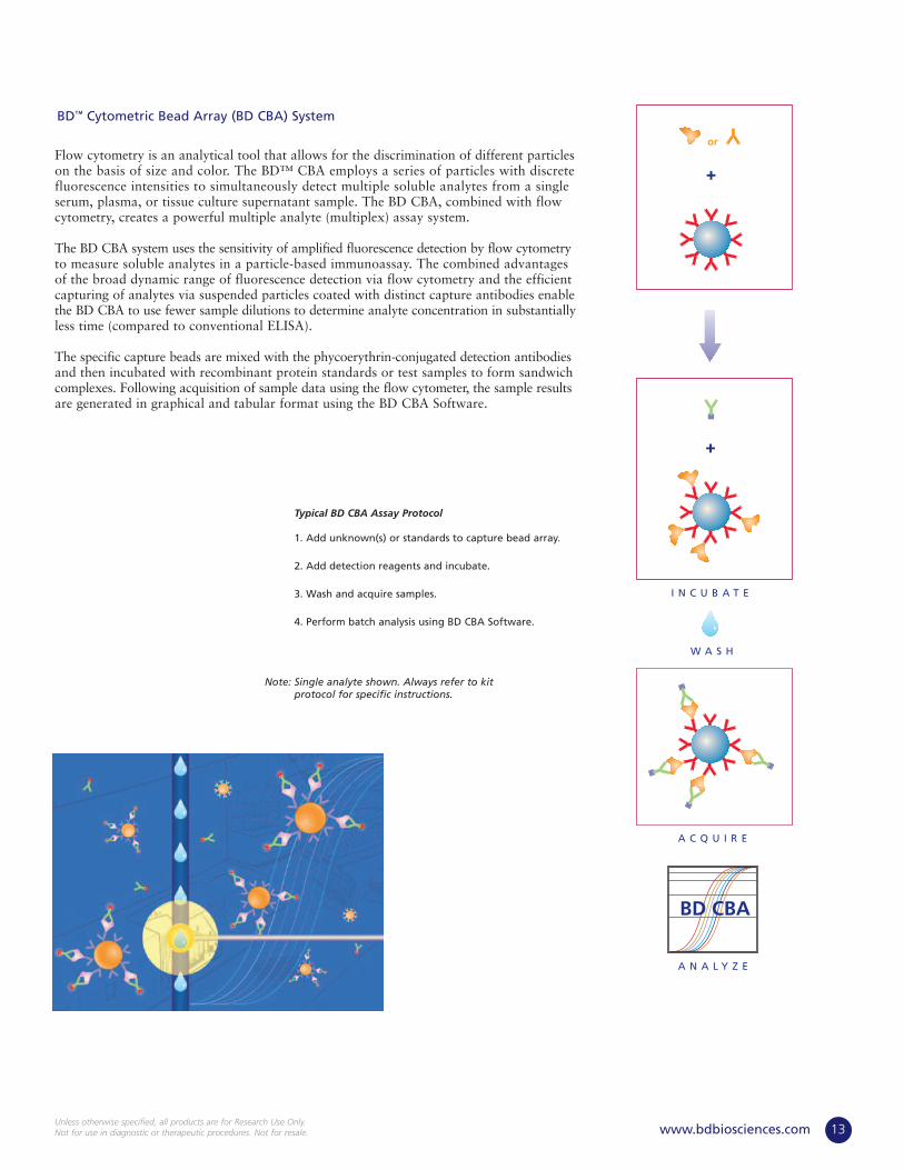
Flow cytometry is an analytical tool that allows for the discrimination of different particleson the basis of size and color. The BD™ CBA employs a series of particles with discretefluorescence intensities to simultaneously detect multiple soluble analytes from a singleserum, plasma, or tissue culture supernatant sample. The BD CBA, combined with flowcytometry, creates a powerful multiple analyte (multiplex) assay system.
The BD CBA system uses the sensitivity of amplified fluorescence detection by flow cytometryto measure soluble analytes in a particle-based immunoassay. The combined advantagesof the broad dynamic range of fluorescence detection via flow cytometry and the efficientcapturing of analytes via suspended particles coated with distinct capture antibodies enablethe BD CBA to use fewer sample dilutions to determine analyte concentration in substantiallyless time (compared to conventional ELISA).
The specific capture beads are mixed with the phycoerythrin-conjugated detection antibodiesand then incubated with recombinant protein standards or test samples to form sandwichcomplexes. Following acquisition of sample data using the flow cytometer, the sample resultsare generated in graphical and tabular format using the BD CBA Software.
Typical BD CBA Assay Protocol
1. Add unknown(s) or standards to capture bead array.
2. Add detection reagents and incubate.
3. Wash and acquire samples.
4. Perform batch analysis using BD CBA Software.
Note: Single analyte shown. Always refer to kitprotocol for specific instructions.
13www.bdbiosciences.comUnless otherwise specified, all products are for Research Use Only.Not for use in diagnostic or therapeutic procedures. Not for resale.
+
or
+
W A S H
I N C U B A T E
A N A L Y Z E
A C Q U I R E
BD CBA
BD™ Cytometric Bead Array (BD CBA) System

Figure 25. Representative Non-Human PrimateReactivity. Cynomolgus macaque serum tested usingthe BD CBA Human Inflammation Kit.
Many BD CBA customers have determined that they are able to detect positive signals for rhesus and cynomolgus macaque sampleswhen using the BD CBA Human Th1/Th2 Cytokine Kit. The BD CBA results have been confirmed by ELISA using the BD CBA antibodypairs with activated cell culture samples from both rhesus and cynomolgus macaques. The cross-reactivity of BD CBA Human assayswith non-human primate (NHP) analytes have not yet been normalized to native or recombinant NHP proteins, so direct quantitation is not yet available (Table 2, Figure 25).
Non-Human Primate (continued)
14 www.bdbiosciences.comUnless otherwise specified, all products are for Research Use Only.
Not for use in diagnostic or therapeutic procedures. Not for resale.
100 101 102 103 104100
101
102
103
104
Measured Cytokine (PE)
FL3-
Hei
gh
t
Interleukin-8
Interleukin-6
Human Chemokine Kit I CXCL8/IL-8, CCL5/RANTES, CCL2/MCP-1 552990
Human Th1/Th2 Cytokine Kit Interleukin (IL)-4, IL-5, TNF, IFN-γ 550749
Human Th1/Th2 Cytokine Kit II Interleukin (IL)-4, IL-6, TNF, IFN-γ 551809
Human Inflammation Kit I Interleukin (IL)-8, IL-6, TNF 551811
(IL-1β and IL-12p70 not yet tested)
BD Cytometric Bead Array (BD CBA) Kits
Table 2. BD CBA Cross-Reactivity with Non-Human Primate Cytokines and Chemokines
DESCRIPTION RHESUS AND CYNOMOLGUS CROSS-REACTIVITY CAT. NO.
BD Cytometric Bead Array System

BD BrdU Flow Kit FCM 50 tests 559619
BD BrdU APC Flow Kit FCM 50 tests 552598 ■
NEW
Rat µicroflow Basic Kit Rat FCM 60 tests 552729
Rat µicroflow Plus Kit Rat FCM 60 tests 552731
Mouse µicroflow Basic Kit Ms FCM 60 tests 552728
Mouse µicroflow Plus Kit Ms FCM 60 tests 552730
Micronucleus Analysis Assay Reagents
DESCRIPTION REACT APPS SIZE CAT. NO.
BrdU Assay Reagents
CD2 (LFA-2) Rat OX-34 Mouse IgG2a, κ FCM, IHC(F), IHC(Fr), IP Purified 0.5 mg 554826
FCM FITC 0.5 mg 554827
CD3 Rat 1F4 Mouse (BALB/c) IgM, κ Cyt, FCM, IP Purified 0.5 mg 556970
FCM FITC 0.5 mg 557354
FCM PE 0.1 mg 550353
FCM APC 0.1 mg 557030
G4.18 Mouse (BALB/c) IgG3, κ Block, Cost, Cyt, FCM NA/LE 0.5 mg 554829
Cost, Cyt, FCM, IP Purified 0.1 mg 559974
Cost, Cyt, FCM, IP Purified 0.5 mg 554830
IHC(F), IHC(Fr), IHC(Zn) Purified 1 ml 550295
FCM Biotin 0.5 mg 554831
FCM FITC 0.1 mg 559975
FCM FITC 0.5 mg 554832
FCM PE 0.2 mg 554833
G4.18/G1 Mouse (BALB/c) IgG1, κ Cost, Cyt, FCM, IP Purified 0.5 mg inquire ■
FCM PE 0.2 mg inquire ■
CD4 Rat OX-35 Mouse (BALB/c) IgG2a, κ FCM, IP Purified 0.5 mg 554835
IHC(Fr) Purified 1 ml 550296
FCM Biotin 0.5 mg 554836
FCM FITC 0.5 mg 554837
FCM PE 0.2 mg 554838
FCM PE-Cy5 0.1 mg 554839
FCM APC 0.1 mg 550057
OX-38 Mouse (BALB/c) IgG2a, κ FCM, IP Purified 0.5 mg 554841
IHC(Fr) Purified 1 ml 550297
FCM FITC 0.5 mg 554843
FCM PE 0.1 mg 551397
CD5 Rat OX-19 Mouse IgG1, κ FCM, IHC(Fr), IHC(Zn), IP Purified 0.5 mg 554849
FCM FITC 0.1 mg 551449 ■
FCM PE 0.2 mg 554851
Rat ReagentsAntibodies to Rat Leukocytes and Related Cells
DESCRIPTION REACT CLONE ISOTYPE APPS FORMAT SIZE CAT. NO.
15www.bdbiosciences.comUnless otherwise specified, all products are for Research Use Only.Not for use in diagnostic or therapeutic procedures. Not for resale.
NEW
DESCRIPTION APPS SIZE CAT. NO.NEW
Reagents for Immunotoxicology Research

CD6 Rat OX-52 Mouse (BALB/c) IgG2a, κ FCM, IHC(Fr), IP Purified 0.1 mg 550979
FCM FITC 0.5 mg 554904
CD8a Rat G28 Mouse (BALB/c) IgG2a, κ FCM, IP Purified 0.1 mg 559977
FCM FITC 0.1 mg 559978
OX-8 Mouse (BALB/c) IgG1, κ FCM, IP, WB Purified 0.5 mg 554854
IHC(F), IHC(Fr), IHC(Zn) Purified 1 ml 550298
FCM, WB Biotin 0.5 mg 554855
FCM FITC 0.5 mg 554856
FCM PE 0.1 mg 559976
FCM PE 0.2 mg 554857
FCM PerCP 0.1 mg 558824
CD8b Rat 341 Mouse (BALB/c) IgG1, κ FCM, IHC(Fr), IHC(Zn), Purified 0.5 mg 554971IP, WB
FCM Biotin 0.1 mg 550970 ■
FCM FITC 0.5 mg 554973
CD9 Rat RPM.7 Mouse IgG3, κ FCM, IP, WB Purified 0.1 mg 551808
FCM FITC 0.1 mg 551806
CD11a (Integrin αL chain, Rat WT.1 Mouse (BALB/c) IgG2a, κ FCM, IHC(Fr), IP Purified 0.1 mg 559979LFA-1 α chain)
FCM FITC 0.5 mg 554985
FCM PE 0.1 mg 550972
CD11b (Integrin αM chain, Mac-1 α chain) Rat WT.5 Mouse (BALB/c) IgA, κ FCM, IHC(Fr), IP, WB Purified 0.5 mg 554980
FCM Biotin 0.5 mg 554981
FCM FITC 0.5 mg 554982
CD11b/c Rat OX-42 Mouse (BALB/c) IgG2a, κ Block, FCM NA/LE 0.5 mg 554858
FCM, IP Purified 0.5 mg 554859
IHC(Fr), IHC(Zn) Purified 1 ml 550299
FCM FITC 0.5 mg 554861
FCM PE 0.2 mg 554862
CD18 (Integrin β2 chain) Rat WT.3 Mouse (BALB/c) IgG1, κ Block, FCM NA/LE 0.5 mg 554976
FCM, IHC(Fr), IHC(Zn), IP Purified 0.5 mg 554977
FCM FITC 0.5 mg 554979
CD24 Rat HIS50 Mouse (BALB/c) IgM, κ FCM, IF, IHC(Fr), WB Purified 0.1 mg 551133
CD25 (IL-2R α chain) Rat OX-39 Mouse (BALB/c) IgG1, κ FCM, IHC(Fr), IP Purified 0.1 mg 559980
FCM Biotin 0.1 mg 559981
FCM FITC 0.5 mg 554865
FCM PE 0.2 mg 554866
CD26 Rat OX-61 Mouse (BALB/c) IgG2a, κ FCM, IHC(Fr), IP, WB Purified 0.1 mg 559639
FCM PE 0.1 mg 559641
CD27 Ms, Rat LG.3A10 Armenian Hamster IgG1, κ Cost, FCM NA/LE 0.5 mg 553777
FCM Biotin 0.1 mg 558753
FCM, IC/FCM PE 0.1 mg 558754
CD28 Rat JJ316 Mouse (BALB/c) IgG1, κ Cost, FCM, IP NA/LE 0.5 mg 554992
JJ319 Mouse (BALB/c) IgG1, κ Cost, FCM NA/LE 0.5 mg 554993
Cost, FCM, IP Purified 0.1 mg 559982
FCM FITC 0.1 mg 550973
FCM PE 0.1 mg 559984
Rat Reagents (continued)
Antibodies to Rat Leukocytes and Related Cells
16 www.bdbiosciences.comUnless otherwise specified, all products are for Research Use Only.
Not for use in diagnostic or therapeutic procedures. Not for resale.
DESCRIPTION REACT CLONE ISOTYPE APPS FORMAT SIZE CAT. NO.
Reagents for Immunotoxicology Research (continued)
NEW

17www.bdbiosciences.comUnless otherwise specified, all products are for Research Use Only.Not for use in diagnostic or therapeutic procedures. Not for resale.
CD29 (Integrin β1 chain) Ms, Rat Ha2/5 Armenian Hamster IgM, κ Block, FCM NA/LE 0.5 mg 555002
FCM, IP Purified 0.5 mg 555003
FCM Biotin 0.5 mg 555004
FCM FITC 0.5 mg 555005
HMβ1-1 Armenian Hamster IgG2, λ Block, FCM, IP Purified 0.5 mg 553837
IHC(Fr) Purified 1 ml 550530
CD31 (PECAM-1) Pig, Rat TLD-3A12 Mouse (BALB/c) IgG1, κ Block, FCM NA/LE 0.5 mg 555024
ELISA, FCM, IP Purified 0.5 mg 555025
IHC(Fr), IHC(Zn) Purified 1 ml 550300
FCM Biotin 0.5 mg 555026
FCM PE 0.1 mg 555027
CD32 (FcγII Receptor) Rat D34-485 Mouse (BALB/c) IgG1, κ Block, FCM NA/LE 0.5 mg 550273
Block, FCM, IHC(Fr), Purified 0.1 mg 550270IHC(Zn), WB
Block, FCM, IHC(Fr), Purified 0.5 mg 550271IHC(Zn), WB
FCM FITC 0.5 mg 550272
CD36 (Scavenger Receptor) Rat CRF D-2712 Mouse IgA, κ FCM, ICC Purified 0.1 mg 552554
CD40 Ms, Rat HM40-3 Armenian Hamster IgM, κ Block, Cost, FCM NA/LE 0.5 mg 553721
FCM Purified 0.1 mg 553722
FCM FITC 0.5 mg 553723
CD42d (Platelet Glycoprotein V) Rat RPM.4 Mouse (BALB/c) IgG2a, κ FCM, IP Purified 0.5 mg 557239
FCM FITC 0.1 mg 551805
Ms, Rat 1C2 Armenian Hamster IgG3, κ FCM, IP, IHC (Fr) Purified .05 mg 552992
CD43 (Leukosialin) Rat HIS17 Mouse (BALB/c) IgG1, κ FCM, IHC(F), IHC(Fr), Purified 0.5 mg 554867IHC(Zn)
FCM Biotin 0.5 mg 554868
CD44H (Pgp-1, H-CAM) Rat OX-49 Mouse (BALB/c) IgG2a, κ FCM, IHC(F), IHC(Fr), Purified 0.5 mg 554869IHC(Zn), IP
FCM FITC 0.1 mg 550974
CD45 (Leukocyte Common Antigen) Rat OX-1 Mouse (BALB/c) IgG1, κ FCM, IP Purified 0.5 mg 554875
IHC(F)*, IHC(Fr), Purified 1 ml 550566IHC(Zn)
FCM Biotin 0.5 mg 554876
FCM FITC 0.5 mg 554877
FCM PE 0.2 mg 554878
FCM PE-Cy5 0.1 mg 559135
CD45.1 (RT7.1 of LCA) Rat NDS58 Rat (AS) IgG2b, κ FCM FITC 0.1 mg 550802
CD45.2 (RT7.2 of LCA) Rat HIS41 Mouse (BALB/c) IgG1, κ FCM, IHC(Fr) Purified 0.1 mg 559985
FCM FITC 0.1 mg 559986
CD45R Rat HIS24 Mouse (BALB/c) IgG2b, κ FCM, WB Purified 0.5 mg 554879
IHC(F), IHC(Fr), IHC(Zn) Purified 1 ml 550301
FCM FITC 0.5 mg 554880
FCM PE 0.2 mg 554881
CD45RA Rat OX-33 Mouse (BALB/c) IgG1, κ FCM Purified 0.5 mg 554882
IHC(Fr), IHC(Zn) Purified 1 ml 550567
FCM FITC 0.5 mg 554883
FCM PE 0.1 mg 551402
FCM PE 0.2 mg 554884
FCM PE-Cy5 0.1 mg 557015
CD45RC Rat OX-22 Mouse (BALB/c) IgG1, κ FCM Purified 0.1 mg 551451 ■
FCM FITC 0.5 mg 554887
FCM PE 0.2 mg 554888
Rat Reagents (continued)
Antibodies to Rat Leukocytes and Related Cells
DESCRIPTION REACT CLONE ISOTYPE APPS FORMAT SIZE CAT. NO.NEW

Reagents for Immunotoxicology Research (continued)
CD48 (OX-45 antigen) Rat OX-45 Mouse (BALB/c) IgG1, κ FCM Purified 0.1 mg 552011 ■
CD49a (Integrin α1 chain) Ms, Rat Ha31/8 Armenian Hamster IgG2, λ Block, FCM NA/LE 0.5 mg 555000
FCM, IP Purified 0.5 mg 555001
IHC(Fr) Purified 1 ml 550568
CD49b (Integrin α2 chain) Ms, Rat Ha1/29 Armenian Hamster IgG2, λ FCM, IHC(Fr) Purified 0.1 mg 559987
FCM, IHC(Fr) Purified 0.5 mg 554998
FCM FITC 0.5 mg 554999
CD49c (Integrin α3 chain) Dog, Ms, Rat 42 Mouse IgG1 IF, WB Purified 50 µg 611044
IF, WB Purified 150 µg 611045
CD49d (Integrin α4 chain) Rat MRα4-1 Mouse (BALB/c) IgG2a, κ FCM, IP Purified 0.5 mg 553348
FCM FITC 0.1 mg 557457
CD49e (Integrin α5 chain) Ms, Rat HMα5-1 Armenian Hamster IgG1, κ FCM, IP Purified 0.5 mg 553350
CD53 Rat OX-44 Mouse (BALB/c) IgG1, κ FCM, IHC(Fr), IHC(Zn), IP Purified 0.1 mg 551452 ■
FCM FITC 0.1 mg 551804
CD54 (ICAM-1) Rat 1A29 Mouse (BALB/c) IgG1, κ Block, FCM NA/LE 0.5 mg 554966
FCM, IP Purified 0.5 mg 554967
IHC(F)*, IHC(Fr), Purified 1 ml 550302IHC(Zn)
FCM Biotin 0.5 mg 554968
FCM FITC 0.5 mg 554969
FCM PE 0.2 mg 554970
CD59 (MAC inhibitor) Rat TH9 Mouse (BALB/c) IgG1, κ FCM FITC 0.1 mg 550976 ■
CD61 (Integrin β3 chain) Ms, Rat 2C9.G2 Armenian Hamster IgG1, κ Block, FCM NA/LE 0.5 mg 553343
FCM Purified 0.5 mg 553344
IHC(Fr) Purified 1 ml 550541
FCM Biotin 0.5 mg 553345
FCM FITC 0.5 mg 553346
FCM PE 0.2 mg 553347
Rat F11 Mouse (BALB/c) IgG1, κ Block, FCM, IP NA/LE 0.5 mg 554950
FCM, IP Purified 0.5 mg 554951
IHC(Fr), IHC(Zn) Purified 1 ml 550569
FCM FITC 0.5 mg 554952
CD62L (L-selectin, LECAM-1) Rat HRL1 Armenian Hamster IgG2, λ FCM Purified 0.1 mg 559988
FCM Biotin 0.1 mg 559989
FCM FITC 0.5 mg 554963
FCM PE 0.1 mg 551398
HRL2 Armenian Hamster IgG1, κ ELISA, FCM, IHC(Fr) Purified 0.1 mg 559990
CD62P (P-selectin) Dog, Hu, Ms, Polyclonal Rabbit IgG ELISA, FCM, IHC(Fr), IP, Purified 0.1 mg 553716Rat WB
CD63 (ME491) Rat AD1 Mouse (BALB/c) IgG1, κ FCM, IHC, IP Purified 0.1 mg 551458 ■
CD71 (Transferrin Receptor) Rat OX-26 Mouse (BALB/c) IgG2a, κ FCM, IHC(Fr), IP Purified 0.5 mg 554889
FCM FITC 0.5 mg 554890
FCM PE 0.2 mg 554891
CD73 (Ecto-5’-nucleotidase) Rat 5F/B9 Mouse (BALB/c) IgG1, κ Cost, FCM NA/LE 0.5 mg 551122
FCM, IF, IHC(Fr), Purified 0.1 mg 551123IHC(Zn), IP
FCM PE 0.1 mg 551124
CD80 (B7-1) Rat 3H5 Mouse (BALB/c) IgG1, κ FCM, IHC(Fr), IP Purified 0.5 mg 555012
FCM Biotin 0.5 mg 555013
FCM PE 0.2 mg 555014
Rat Reagents (continued)
Antibodies to Rat Leukocytes and Related Cells
18 www.bdbiosciences.comUnless otherwise specified, all products are for Research Use Only.
Not for use in diagnostic or therapeutic procedures. Not for resale.
DESCRIPTION REACT CLONE ISOTYPE APPS FORMAT SIZE CAT. NO.NEW

*IHC application not routinely tested by BD Biosciences Pharmingen.
19www.bdbiosciences.comUnless otherwise specified, all products are for Research Use Only.Not for use in diagnostic or therapeutic procedures. Not for resale.
CD81 (TAPA-1) Ms, Rat Eat2 Armenian Hamster IgG1, κ FCM, IP, WB Purified 0.5 mg 559517
FCM Biotin 0.5 mg 559518
FCM PE 0.1 mg 559519
CD86 (B7-2) Rat 24F Mouse (BALB/c) IgG1, κ Block, FCM NA/LE 0.5 mg 555015
FCM, IHC(Fr), IP Purified 0.5 mg 555016
FCM Biotin 0.5 mg 555017
FCM FITC 0.5 mg 555018
FCM PE 0.1 mg 551396
CD90.1 (Thy-1.1) Ms, Rat HIS51 Mouse (BALB/c) IgG2a, κ FCM Purified 0.5 mg 554892
IHC(F), IHC(Fr), IHC(Zn) Purified 1 ml 550570
FCM Biotin 0.5 mg 554893
FCM FITC 0.5 mg 554894
G Pig, Ms, OX-7 Mouse (BALB/c) IgG1, κ Cost, FCM, IP, WB Purified 0.5 mg 554895Rab, Rat
IHC(F), IHC(Fr), IHC(Zn) Purified 1 ml 550571
FCM Biotin 0.5 mg 554896
FCM FITC 0.5 mg 554897
FCM PE 0.1 mg 551401
FCM PE 0.2 mg 554898
FCM PerCP 0.1 mg 557266
CD95 (Fas) Chick, Dog, 13 Mouse IgG1 IF, IHC, WB Purified 50 µg 610197*Hu, Ms, Rat
CD106 (VCAM-1) Rat MR106 Mouse IgG1, κ FCM, IHC(Fr), IP, WB Purified 0.5 mg 559165
FCM PE 0.1 mg 559229
CD122 (IL-2 Receptor β chain) Rat L316 Mouse (BALB/c) IgG1, κ FCM Biotin 0.5 mg 557282
FCM FITC 0.1 mg 551803
CD134 (OX-40 Antigen) Rat OX-40 Mouse (BALB/c) IgG2b, κ FCM Biotin 0.1 mg 550977
FCM FITC 0.5 mg 554848
CD140b (PDGF Receptor β chain) Chick, Dog, 28 Mouse IgG2b IF, IHC, IP, WB Purified 50 µg 610113*Hu, Ms, Rat
IF, IHC, IP, WB Purified 150 µg 610114*
CD147 Rat OX-47 Mouse IgG1, κ FCM Purified 0.1 mg 552012 ■
CD161a (NKR-P1A) Rat 10/78 Mouse IgG1, κ FCM, IP Purified 0.5 mg 555006
IHC(Fr), IHC(Zn) Purified 1 ml 550306
FCM Biotin 0.1 mg 550978
FCM FITC 0.5 mg 555008
FCM PE 0.2 mg 555009
CD172 (SIRP) Rat OX-41 Mouse IgG2a,κ FCM, IHC, IP, WB Purified 0.1 mg 552297 ■
FCM PE 0.1 mg 552298 ■
CD178 (Fas Ligand, CD95 Ligand) Dog, Hu, Ms, 33 Mouse IgG1 IF, IP, WB Purified 50 µg 610410Rat
IF, IP, WB Purified 150 µg 610411
Ms, Rat MFL4 Armenian Hamster IgG3, κ Block, FCM NA/LE 0.5 mg 555021
FCM Purified 0.5 mg 555022
FCM Biotin 0.5 mg 556998
CD200 (OX-2 Antigen) Rat OX-2 Mouse IgG1, κ Block, FCM, IHC, IP Purified 0.1 mg 552457 ■
CD200 (OX-2 Receptor) Rat OX-102 Mouse (BALB/c) IgG1, κ FCM, IP, WB Purified 0.1 mg 552469 ■
β2 Microglobulin Rat TLD-3H12B Mouse (BALB/c) IgG1, κ FCM, IHC, WB Purified 0.5 mg 558765
FCM FITC 0.1 mg 551807 ■
Rat Reagents (continued)
Antibodies to Rat Leukocytes and Related Cells
DESCRIPTION REACT CLONE ISOTYPE APPS FORMAT SIZE CAT. NO.NEW

Reagents for Immunotoxicology Research (continued)
C1qRp Rat LOV8 Mouse IgG1, λ FCM, IP Purified 0.1 mg 552294 ■
FCM PE 0.1 mg 552295 ■
Crry/p65 Rat 512 Mouse (BALB/c) IgG1, κ FCM, WB Purified 0.5 mg 554991
Dendritic Cells Rat OX-62 Mouse (BALB/c) IgG1, κ FCM, IP, WB Purified 0.5 mg 555010
IHC(F)*, IHC(Fr), Purified 1 ml 550303IHC(Zn)
Erythroid Cells Rat HIS49 Mouse (BALB/c) IgM, κ FCM, IHC(Fr) Purified 0.1 mg 550961
FCM Biotin 0.1 mg 550962
Granulocytes Rat HIS48 Mouse IgM, κ FCM Purified 0.5 mg 554905
IHC(F), IHC(Fr), IHC(Zn) Purified 1 ml 550304
FCM Biotin 0.5 mg 554906
FCM FITC 0.5 mg 554907
RP-1 Mouse (BALB/c) IgG2a, κ FCM, IP Purified 0.5 mg 550000
FCM Biotin 0.5 mg 550001
FCM PE 0.1 mg 550002
RP-3 Mouse (BALB/c) IgM, κ Cyt, FCM NA/LE 0.5 mg 550055
FCM Purified 0.5 mg 559999
High Affinity IgE Receptor (FcεRI) Rat BC4 Mouse (BALB/c) IgG1, κ FCM, IP Purified 0.1 mg 551469 ■
Ki-67 Hu, Ms, Pig, B56 Mouse IgG1, κ IC/FCM FITC Set 100 tests 556026Rat
IC/FCM PE Set 100 tests 556027
Ly49 Inhibitory Receptor 2 Rat STOK2 Rat (DA) IgG2a, κ Block, FCM, IP Purified 0.1 mg 552296 ■
Macrophage Activator (RMA) Rat anti-RMA Mouse IgG1, κ FCM, IHC(Fr), IP Purified 0.5 mg 555020
Macrophage Subset Rat HIS36 Mouse IgG2a, κ FCM Purified 0.5 mg 554900
IHC(Fr), IHC(Zn) Purified 1 ml 550573
FCM PE 0.2 mg 554901
MAdCAM-1 Rat OST2 Mouse IgG1, κ FCM, IP Purified 0.1 mg 550014
IHC(Fr), IHC(Zn) Purified 1 ml 550309
Marginal Zone B Cells Rat HIS57 Mouse IgG1, κ FCM, IHC(F), IHC(Fr) Purified 0.1 mg 559960
FCM Biotin 0.1 mg 559962
FCM FITC 0.5 mg 559963
Mast Cells Rat AR32AA4 (aka Mouse (BALB/c) IgG1, κ FCM Purified 0.1 mg 551770 ■
AA4)
Mononuclear Phagocyte Rat 1C7 Mouse IgG1, κ FCM Purified 0.5 mg 554954
IHC(F), IHC(Fr), IHC(Zn) Purified 1 ml 550305
FCM Biotin 0.1 mg 559992
Myeloid Lineage Rat OX-82 Mouse (BALB/c) IgG1, κ FCM Purified 0.1 mg 552130 ■
OX-40 Ligand (OX-40L) Rat ATM-2 Mouse (BALB/c) IgG1, κ FCM Purified 0.5 mg 559957
FCM Biotin 0.5 mg 559959
RT6.1 Rat P4/16 Rat IgG2b κ FCM, WB Purified 0.1 mg 552725 ■
RT6.2 Rat GY1/12 Rat IgG2c, κ FCM, WB Purified 0.1 mg 552471 ■
Rat Reagents (continued)
Antibodies to Rat Leukocytes and Related Cells
20 www.bdbiosciences.comUnless otherwise specified, all products are for Research Use Only.
Not for use in diagnostic or therapeutic procedures. Not for resale.
DESCRIPTION REACT CLONE ISOTYPE APPS FORMAT SIZE CAT. NO.NEW

RT1A Rat OX-18 Mouse (BALB/c) IgG1, κ FCM, IHC(Fr), IP Purified 0.5 mg 554917
FCM, IHC(Fr) Biotin 0.1 mg 550980
FCM FITC 0.5 mg 554919
FCM PE 0.1 mg 559993
RT1Aa,b Rat C3 Rat (LOU/cN) IgG2b, κ FCM Purified 0.5 mg 554932
FCM, IHC(Fr) Biotin 0.1 mg 559994
FCM FITC 0.1 mg 550981
RT1Aa,b,l Rat B5 Rat (LOU/cN) IgM, κ FCM Purified 0.1 mg 559995
FCM FITC 0.1 mg 559996
RT1B Ms, Rat OX-6 Mouse (BALB/c) IgG1, κ FCM, IP Purified 0.5 mg 554926
IHC(F), IHC(F)*, Purified 1 ml 550574IHC(Fr), IHC(Zn)
FCM Biotin 0.5 mg 554927
FCM FITC 0.5 mg 554928
FCM PE 0.2 mg 554929
FCM PerCP 0.1 mg 557016
RT1D Ms, Rat 14-4-4S Mouse (C3H.SW) IgG2a, κ Cyt, FCM, IHC(Fr), IP Purified 0.1 mg 558734
FCM, IHC(Fr) Biotin 0.1 mg 558735
FCM FITC 0.5 mg 553543
FCM PE 0.1 mg 553544
RT1D Rat OX-17 Mouse (BALB/c) IgG1, κ FCM FITC 0.1 mg 550982
Antibodies to Rat Major Histocompatibility Complex (MHC) Antigens
DESCRIPTION REACT CLONE ISOTYPE APPS FORMAT SIZE CAT. NO.
αβ TCR Rat R73 Mouse (BALB/c) IgG1, κ Cost, Cyt, FCM NA/LE 0.5 mg 554910
Cost, Cyt, FCM, IP, WB Purified 0.5 mg 554911
IHC(F), IHC(Fr), IHC(Zn) Purified 1 ml 550307
FCM Biotin 0.1 mg 551801 ■
FCM FITC 0.5 mg 554913
FCM PE 0.2 mg 554914
FCM PerCP 0.1 mg 557019
γδTCR Rat V65 Mouse (BALB/c) IgG1, κ Cost, Cyt, FCM, IF, IP Purified 0.5 mg 554956
IHC(Fr) Purified 1 ml 550308
FCM Biotin 0.1 mg 559997
FCM FITC 0.5 mg 554958
FCM PE 0.1 mg 551802
Vα 4 TCR Rat G99 Mouse IgG1, κ Cost, FCM, IHC(Fr) Purified 0.5 mg 554989
Vβ 3.3 TCR Rat C-A11 Mouse (BALB/c) IgG2a, κ FCM Biotin 0.1 mg 550731
Vβ 8.2l and Vβ 8.4a TCR Rat R78 Mouse (BALB/c) IgG1, κ Cost, Cyt, FCM, IHC(Fr) Purified 0.5 mg 554940
FCM FITC 0.1 mg 559998
Vβ 13 TCR Rat 18B1 Mouse (BALB/c) IgG1, κ FCM Biotin 0.1 mg 550721
Vβ 16 TCR Rat HIS42 Mouse (BALB/c) IgG2b, κ Cost, FCM, IHC(Fr) Purified 0.5 mg 554915
Antibodies to Rat T-Cell Receptors (TCR)
DESCRIPTION REACT CLONE ISOTYPE APPS FORMAT SIZE CAT. NO.
21www.bdbiosciences.comUnless otherwise specified, all products are for Research Use Only.Not for use in diagnostic or therapeutic procedures. Not for resale.
NEW
NEW

Rat IFN-γ Set 31.3-2000 pg/ml ELISA Reagents for 20 Plates 558861
Rat IL-2 Set 31.3-2000 pg/ml ELISA Reagents for 20 Plates 557044
Rat IL-4 Set 1.6-100 pg/ml ELISA Reagents for 20 Plates 555198
Rat IL-6 Set 78-5000 pg/ml ELISA Reagents for 20 Plates 550319
Rat IL-10 Set 15.6-1000 pg/ml ELISA Reagents for 20 Plates 555134
Rat MCP-1 Set 31.3-2000 pg/ml ELISA Reagents for 20 Plates 555130
Rat TNF Set 31.3-2000 pg/ml ELISA Reagents for 20 Plates 558870
BD OptEIA™ ELISA Sets
DESCRIPTION ASSAY RANGE APPS SIZE CAT. NO.
Rat TNF Kit 31.3-2000 pg/ml 13 pg/ml ELISA 2 plates 550734
BD OptEIA™ ELISA Kits
DESCRIPTION ASSAY RANGE SENSITIVITY APPS SIZE CAT. NO.
CD11a (LFA-1) Dog, Hu, Rab HI111 Mouse IgG1, κ FA, FCM, IHC(Fr) NA/LE 0.5 mg 555381
FCM Purified 0.1 mg 555382
IHC(Fr) Purified 1 ml 550373
FCM FITC 100 tests 555383
FCM PE 100 tests 555384
FCM PE 50 tests 550851
FCM PE-Cy5 100 tests 551131 ■
FCM APC 100 tests 559875
FCM APC 50 tests 550852
CD14 Dog, Hu M5E2 Mouse IgG2a, κ FCM NA/LE 0.5 mg 555395
FCM Purified 0.1 mg 557152
FCM, IHC(Fr) Purified 0.1 mg 555396
IHC(Fr), IHC(Zn) Purified 1 ml 550376
FCM FITC 100 tests 555397
FCM FITC 50 tests 557153
FCM PE 100 tests 555398
FCM PE 50 tests 557154
FCM APC 100 tests 555399
FCM PerCP-Cy5.5 0.1 mg 550787
CD34 Dog 1H6 Mouse (BALB/c) IgG1, κ FCM PE 0.1 mg 559369
2E9 Mouse (BALB/c) IgG1, κ FCM Biotin 0.1 mg 550427
DogAntibodies to Dog Leukocytes
DESCRIPTION REACT CLONE ISOTYPE APPS FORMAT SIZE CAT. NO.
Reagents for Immunotoxicology Research (continued)
22 www.bdbiosciences.comUnless otherwise specified, all products are for Research Use Only.
Not for use in diagnostic or therapeutic procedures. Not for resale.
NEW
NEW
NEW

23www.bdbiosciences.comUnless otherwise specified, all products are for Research Use Only.Not for use in diagnostic or therapeutic procedures. Not for resale.
CD44 (Pgp-1, Ly-24) Dog 69-S5 Mouse (BALB/c) IgG1, κ FCM, IF Purified 0.1 mg 559964
Dog, Hu, Ms, Pig IM7 Rat IgG2b, κ Block, Cyt, FCM NA/LE 0.5 mg 553130
Cyt, ELISA, FCM, IP Purified 0.5 mg 553131
IHC(F)*, IHC(Fr), Purified 1 ml 550538IHC(Zn)
FCM Biotin 0.5 mg 553132
FCM FITC 0.5 mg 553133
FCM PE 0.1 mg 553134
FCM PE-Cy5 0.1 mg 553135
FCM APC 0.1 mg 559250
CD49c (Integrin α3 chain) Dog, Ms, Rat 42 Mouse IgG1 IF, WB Purified 50 µg 611044
IF, WB Purified 150 µg 611045
CD49d (Integrin α4 chain) Dog, Hu 9F10 Mouse IgG1, κ FCM NA/LE 0.5 mg 555501
FCM Purified 0.1 mg 555502
FCM PE 100 tests 555503
FCM PE 50 tests 556635
FCM PE-Cy5 100 tests 559880
FCM APC 100 tests 559881
CD49f (Integrin α6 chain) Dog, Hu, Ms, Pig GoH3 Rat (Sprague-Dawley) FCM, IHC(Fr), IP Purified 0.1 mg 555734IgG2a, κ
FCM FITC 100 tests 555735
FCM PE 100 tests 555736
FCM PE-Cy5 100 tests 551129 ■
FCM APC 100 tests 551140 ■
CD58 (LFA-3) Dog, Hu 1C3 Mouse IgG2a, κ FCM Purified 0.1 mg 555919
FCM FITC 100 tests 555920
FCM PE 100 tests 555921
FCM PE-Cy5 100 tests 551399
CD61 Dog, Hu VI-PL2 Mouse IgG1, κ FCM Purified 0.1 mg 555752
FCM FITC 100 tests 555753
FCM FITC 50 tests 557291
FCM PE 100 tests 555754
FCM PE 50 tests 557290
CD62P (P-selectin) Dog, Hu, Ms, Rat Polyclonal Rabbit IgG ELISA, FCM, Purified 0.1 mg 553716IP, WB
CD80 (B7-1) Dog, Ms 16-10A1 Armenian Hamster IgG2, κ FCM, IHC(Fr), IP Purified 0.5 mg 553766
FCM Biotin 0.5 mg 553767
FCM FITC 0.5 mg 553768
FCM PE 0.2 mg 553769
Dog, Ms 1G10 Rat IgG2a, κ FCM Purified 0.5 mg 553368
CD81 Dog, Hu, Rab JS-81 Mouse IgG1, κ FCM Purified 0.1 mg 555675
FCM FITC 100 tests 551108
FCM PE 100 tests 555676
FCM PE-Cy5 100 tests 551078 ■
FCM APC 100 tests 551112 ■
CD94 Dog, Hu HP-3D9 Mouse IgG1, κ FCM, IHC(Fr) Purified 0.1 mg 555887
FCM FITC 100 tests 555888
FCM PE 100 tests 555889
FCM APC 100 tests 559876
Dog (continued)
Antibodies to Dog Leukocytes
DESCRIPTION REACT CLONE ISOTYPE APPS FORMAT SIZE CAT. NO.NEW

Reagents for Immunotoxicology Research (continued)
*IHC application not routinely tested by BD Biosciences Pharmingen.
CD95 (Fas/APO-1) Dog, Hu, Pig DX2 Mouse IgG1, κ FA, FCM NA/LE 0.5 mg 555670
FCM, IHC(Fr) Purified 0.1 mg 555671
FCM Biotin 100 tests 555672
FCM FITC 100 tests 555673
FCM PE 100 tests 555674
FCM PE-Cy5 100 tests 559773
FCM APC 100 tests 558814
CD117 (c-kit) Dog, Hu, Rab YB5.B8 Mouse IgG1, κ FCM Purified 0.1 mg 555713
FCM PE 0.1 mg 555714
FCM PE-Cy5 0.1 mg 559879
FCM APC 0.1 mg 550412
CD132 (common γ chain) Dog, Hu TUGh4 Rat IgG2b, κ FA, FCM NA/LE 0.5 mg 555895
FCM Purified 0.5 mg 555896
FCM Biotin 0.5 mg 555897
FCM PE 0.2 mg 555898
CD140b (PDGF Receptor β chain) Chick, Dog, Hu, 28 Mouse IgG2b IF, IHC, IP, WB Purified 50 µg 610113*Ms, Rat
IF, IHC, IP, WB Purified 150 µg 610114*
CD178 (Fas Ligand, CD95 Dog, Hu, Ms, Rat 33 Mouse IgG1 IF, IP, WB Purified 50 µg 610410Ligand)
IF, IP, WB Purified 150 µg 610411
HLA-DR Dog, Hu G46-6 Mouse IgG2a, κ FCM NA/LE 0.5 mg 555809
FCM Purified 0.1 mg 555810
IHC(Fr) Purified 1 ml 550409
FCM FITC 100 tests 555811
FCM FITC 50 tests 556643
FCM PE 100 tests 555812
FCM PE 50 tests 556644
FCM PE-Cy5 100 tests 555813
FCM APC 100 tests 559866
Dog, Hu, Rab TÜ36 Mouse IgG2b, κ FCM Purified 0.1 mg 555559
FCM FITC 100 tests 555560
FCM PE 100 tests 555561
FCM PE-Cy5 100 tests 551375
FCM APC 100 tests 559868
SLA-DR Bov, Cat, Dog, 1F12 Mouse (BALB/c) IgG2b, κ FCM, IP, WB Purified 0.1 mg 551537 ■
G Pig, Gt,Hs, Hu,Mink, Pig, Sheep
VAP-1 Dog, Ms, Rat 54 Mouse IgG1 WB Purified 50 µg 611190
WB Purified 150 µg 611191
Dog (continued)
Antibodies to Dog Leukocytes
24 www.bdbiosciences.comUnless otherwise specified, all products are for Research Use Only.
Not for use in diagnostic or therapeutic procedures. Not for resale.
DESCRIPTION REACT CLONE ISOTYPE APPS FORMAT SIZE CAT. NO.NEW

25www.bdbiosciences.comUnless otherwise specified, all products are for Research Use Only.Not for use in diagnostic or therapeutic procedures. Not for resale.
CCR6 Cyno, Rhe 11A9 Mouse IgG2a, κ FCM PE 50 tests 551773 ■
CD2 Bab, Cyno, Rhe RPA-2.10 Mouse IgG1,κ FCM, IHC(Fr) Purified 0.1 mg 556607
FCM FITC 50 tests 556608
FCM PE 50 tests 556609
CD3 Bab, Cyno, Rhe SP34 Mouse IgG3, λ FA, FCM NA/LE 0.5 mg 557052
FA, FCM Purified 0.1 mg 556610
FA, FCM FITC 50 tests 556611
FA, FCM PE 50 tests 556612
SP34-2 Mouse IgG1, κ FCM Purified 0.1 mg 551916 ■
FCM PE 50 tests 552127 ■
CD3/CD16/CD45 Rhe SP34, 3G8, TÜ116 FCM FITC, PE, PerCP 50 tests 551067
CD3/CD20/CD45 Rhe SP34, 2H7, TÜ116 FCM FITC, PE, PerCP 50 tests 551068
CD3/CD4/CD45 Rhe SP34, M-T477, TÜ116 FCM FITC, PE, PerCP 50 tests 551070
CD3/CD8/CD45 Rhe SP34, RPA-T8, TÜ116 FCM FITC, PE, PerCP 50 tests 551069
CD4 Bab, Cyno, Rhe L200 Mouse IgG1, κ FCM Purified 0.1 mg 550625
FCM FITC 50 tests 550628
FCM PE 50 tests 550630
FCM PerCP 50 tests 550631
M-T477 Mouse IgG2a FCM Purified 0.1 mg 556614
FCM FITC 50 tests 556615
FCM PE 50 tests 556616
CD4/CCR5 Cyno, Rhe M-T477, 3A9 FCM FITC, PE 50 tests 551466 ■
CD4/CXCR4 Bab, Cyno, Rhe M-T477, 12G5 FCM FITC, PE 50 tests 551465 ■
CD6 Bab, Cyno, Rhe M-T605 Mouse IgG1, κ FCM FITC 50 tests 559491
FCM PE 50 tests 559492
CD8 Bab, Cyno, Rhe RPA-T8 Mouse IgG1, κ FCM Purified 0.1 mg 557084
FCM FITC 50 tests 557085
CD8/CCR5 Bab, Cyno, Rhe RPA-T8, 3A9 FCM FITC, PE 50 tests 551470 ■
CD8/CXCR4 Bab, Cyno, Rhe RPA-T8, 12G5 FCM FITC, PE 50 tests 551468 ■
CD9 Bab, Cyno, Rhe M-L13 Mouse IgG1, κ FCM, IHC(Fr), WB Purified 0.1 mg 559456
FCM FITC 50 tests 559457
CD10 Bab, Cyno, Rhe HI10a Mouse IgG1, κ FCM PE 50 tests 557143
CD11a/LFA-1 Bab, Cyno, Rhe HI111 Mouse IgG1, κ FCM PE 50 tests 550851
FCM APC 50 tests 550852
CD11b/Mac-1 (CR3) Bab, Cyno, Rhe ICRF44 Mouse IgG1, κ FCM Purified 0.1 mg 557320
FCM PE 50 tests 557321
CD14 Cyno, Rhe M5E2 Mouse IgG2a, κ FCM Purified 0.1 mg 557152
FCM FITC 50 tests 557153
FCM PE 50 tests 557154
CD16 Bab, Cyno, Rhe 3G8 Mouse IgG1, κ FCM Purified 0.1 mg 556617
FCM FITC 50 tests 556618
FCM PE 50 tests 556619
CD18 (Integrin β2 chain, Cyno, Rhe 6.7 Mouse IgG1, κ FCM FITC 50 tests 557156CR3/CR4)
FCM PE 50 tests 557157
CD20 Bab, Cyno, Rhe 2H7 Mouse IgG2b, κ FCM Purified 0.1 mg 556631
FCM FITC 50 tests 556632
FCM PE 50 tests 556633
CD21 Bab, Cyno, Rhe B-ly4 Mouse IgG1, κ FCM PE 50 tests 557327
CD25 Bab, Cyno, Rhe M-A251 Mouse IgG1, κ FCM PE 50 tests 557138
Non-Human Primate (NHP)Antibodies for Non-Human Primate (NHP) Research
DESCRIPTION REACT CLONE ISOTYPE APPS FORMAT SIZE CAT. NO.NEW

Reagents for Immunotoxicology Research (continued)
CD27 Bab, Cyno, Rhe M-T271 Mouse IgG1, κ FCM Purified 0.1 mg 557328
FCM FITC 50 tests 557329
FCM PE 50 tests 557330
CD28 Bab, Cyno, Rhe CD28.2 Mouse IgG1, κ FCM Purified 0.1 mg 556620
FCM FITC 50 tests 556621
FCM PE 50 tests 556622
CD29 Bab, Rhe MAR4 Mouse IgG1, κ FCM Purified 0.1 mg 557331
FCM PE 50 tests 557332
CD31 Bab, Cyno, Rhe WM59 Mouse IgG1, κ FCM FITC 50 tests 557508
CD32 Bab, Rhe FLI8.26 (2003) Mouse IgG2b, κ FCM Purified 0.1 mg 557333
FCM FITC 50 tests 557334
CD34 Cyno, Rhe 563 Mouse IgG1, κ FCM PE 50 tests 550619
CD40 Bab, Cyno, Rhe 5C3 Mouse IgG1, κ FCM Purified 0.1 mg 556623
FCM FITC 50 tests 556624
FCM PE 50 tests 558835
CD41a Bab, Cyno, Rhe HIP8 Mouse IgG1, κ FCM Purified 0.1 mg 557295
FCM FITC 50 tests 557296
FCM PE 50 tests 557297
CD41b Bab, Cyno, Rhe HIP2 Mouse IgG3, κ FCM Purified 0.1 mg 557293
FCM FITC 50 tests 557294
CD42a Bab, Cyno, Rhe ALMA.16 Mouse IgG1, κ FCM Purified 0.1 mg 559458
FCM FITC 50 tests 550472
CD45 Cyno, Rhe TÜ116 Mouse IgG1, κ FCM PE 50 tests 557059
FCM PE-Cy5 50 tests 557075
FCM PerCP 50 tests 557513
FCM PerCP-Cy5.5 50 tests 552724
CD45RA Bab, Cyno, Rhe 5H9 Mouse IgG1, κ FCM Purified 0.1 mg 556625
FCM FITC 50 tests 556626
FCM PE 50 tests 556627
CD49b Bab, Cyno, Rhe AK-7 Mouse IgG1, κ FCM FITC 50 tests 557338
CD49d (Integrin α4 chain) Bab, Cyno, Rhe 9F10 Mouse IgG1, κ FCM Purified 0.1 mg 556634
FCM PE 50 tests 556635
CD49f Bab, Cyno, Rhe GoH3 Mouse IgG1, κ FCM FITC 50 tests 557510
FCM PE 50 tests 557511
CD56 Bab, Cyno, Rhe MY31 Mouse IgG1, κ FCM PE 50 tests 556647
CD59 Bab, Cyno, Rhe p282 (H19) Mouse IgG2a, κ FCM PE 50 tests 557141 ■
CD61 Bab, Cyno, Rhe VI-PL2 Mouse IgG1, κ FCM FITC 50 tests 557291
CD62L Cyno, Rhe SK11 Mouse IgG2a, κ FCM PE 50 tests 557341 ■
CD62P (P-Selectin) Bab, Cyno, Rhe AC1.2 Mouse IgG1 FCM FITC 50 tests 550866
FCM PE 50 tests 550561
CD63 Bab, Cyno, Rhe H5C6 Mouse IgG1, κ FCM Purified 0.1 mg 557304
FCM FITC 50 tests 550759
CD69 Bab, Cyno, Rhe FN50 Mouse IgG1, κ FCM FITC 50 tests 557049
FCM PE 50 tests 557050
CD83 Cyno, Rhe HB15e Mouse IgG1, κ FCM PE 50 tests 550634
CD86 Bab, Cyno, Rhe 2331 (FUN-1) Mouse IgG1, κ FCM FITC 50 tests 557343
FCM PE 50 tests 557344
CD95 Bab, Cyno, Rhe DX2 Mouse IgG1, κ FCM Purified 0.1 mg 556639
FCM FITC 50 tests 556640
FCM PE 50 tests 556641
Non-Human Primate (NHP) (continued)
Antibodies for Non-Human Primate (NHP) Research
26 www.bdbiosciences.comUnless otherwise specified, all products are for Research Use Only.
Not for use in diagnostic or therapeutic procedures. Not for resale.
DESCRIPTION REACT CLONE ISOTYPE APPS FORMAT SIZE CAT. NO.NEW

27www.bdbiosciences.comUnless otherwise specified, all products are for Research Use Only.Not for use in diagnostic or therapeutic procedures. Not for resale.
CD122 Bab, Cyno, Rhe Mikβ2 Mouse IgG2a, κ FCM Purified 0.1 mg 557322
FCM PE 0.1 mg 557323
CD130 Bab, Cyno, Rhe AM64 Mouse IgG1, κ FCM Purified 0.1 mg 557324
FCM PE 50 tests 557325
CDw137 Bab, Cyno, Rhe 4B4-1 Mouse IgG1, κ FCM PE 50 tests 557303
CD152 Bab, Cyno, Rhe BNI3 Mouse IgG2a, κ FCM Purified 0.1 mg 557300
FCM PE 50 tests 557301
CD154 Bab, Cyno, Rhe TRAP1 Mouse IgG1, κ FCM Purified 0.1 mg 557298
FCM FITC 50 tests 558988
FCM PE 50 tests 557299
CD162 Bab, Cyno, Rhe KPL-1 Mouse IgG1, κ FCM Purified 0.1 mg 557502
FCM PE 50 tests 557503
CD183 (CXCR3) Cyno, Rhe 1C6/CXCR3 Mouse IgG1 FCM PE 50 tests 550633
CD184 (CXCR4, Fusin) Bab, Cyno, Rhe 12G5 Mouse IgG2a, κ FCM PE 50 tests 557145
CD195 (CCR5) Cyno, Rhe 3A9 Mouse IgG2a FCM PE 50 tests 550632
HLA-A,B,C Bab, Cyno, Rhe G46-2.6 Mouse IgG1, κ FCM Purified 0.1 mg 557347
FCM FITC 50 tests 557348
FCM PE 50 tests 557349
HLA-DR Bab, Cyno, Rhe G46-6 Mouse IgG2a, κ FCM Purified 0.1 mg 556642
FCM FITC 50 tests 556643
FCM PE 50 tests 556644
FCM PerCP-Cy5.5 50 tests 552764 ■
HLA-DR,P,Q Bab, Cyno, Rhe TÜ39 Mouse IgG2a, κ FCM FITC 50 tests 550853
IFN-γ Bab, Cyno, Rhe 4S.B3 Mouse IgG1, κ IC/FCM PE 50 tests 557074
IC/FCM APC 50 tests 551385 ■
IL-2 Bab, Cyno, Rhe MQ1-17H12 Rat IgG2a IC/FCM Purified 0.1 mg 558881
IC/FCM FITC 50 tests 559361
IC/FCM APC 50 tests 551383 ■
IL-16 Bab, Cyno, Rhe 14.1 Mouse IgG2a, κ IC/FCM PE 50 tests 551471 ■
APC 50 tests 551472 ■
MCP-1 (monocyte Cyno, Rhe 5D3-F7 Mouse IgG1, κ FCM PE 50 tests 557066 ■
chemoattractant protein 1)
FCM APC 50 tests 551532 ■
MIP-1α (macrophage Cyno, Rhe 11A3 Mouse IgG2a,κ FCM APC 50 tests 551533 ■
inflammatory protein-1α)
Pan Leukocyte DO58-1283 Mouse IgG1 FCM Purified 0.1 mg 552566 ■
Red Blood Cells (RBC) Bab E34-1678 Mouse IgM FCM Purified 0.1 mg 551350 ■
FCM FITC 50 tests 551351 ■
E34-731 Mouse IgG1, κ FCM Purified 0.1 mg 551299 ■
FCM FITC 50 tests 551300 ■
FCM PE 50 tests 551301 ■
TNF Bab, Cyno, Rhe Mab11 Mouse IgG1, κ IC/FCM Purified 0.1 mg 558882
IC/FCM PE 50 tests 557068
Non-Human Primate (NHP) (continued)
Antibodies for Non-Human Primate (NHP) Research
DESCRIPTION REACT CLONE ISOTYPE APPS FORMAT SIZE CAT. NO.NEW

PRESORTEDSTANDARD
U.S. POSTAGEPAID
San Leandro, CAPermit No.169
Argentina/Paraguay/UruguayTel 54.11.4551.7100 x106Fax 54.11.4551.7400
Australia/New ZealandAustraliaTel 61.2.8875.7000Fax [email protected] ZealandTel 64.9.574.2468Fax [email protected]
AustriaSCIENTIFIC SUPPORT
Tel 43.1.706.36.60.44Fax [email protected] SERVICE
Tel 43.1.706.36.60.0Fax 43.1.706.36.60.11
Belgium Tel 32.53.720.600Fax [email protected] SERVICE
Tel 32.53.720.550Fax [email protected]
BrazilTel 55.11.5185.9995Fax 55.11.5185.9895
Central America/CaribbeanTel 506.290.7318Fax 506.290.7331
ChileTel 56.2 460.0380 x21Fax 56.2 460.0306
ChinaTel 8610.6418.1608Fax 8610.6418.1610
ColombiaTel 57.1.572.4060 x244Fax 57.1.244.1363
DenmarkTel 45.43.43.45.66Fax [email protected]
East AfricaTel 254.2.341157Fax [email protected]
Eastern EuropeTel 49.6221.305.161Fax [email protected]
EgyptTel 202.268.0181Fax 202.266.7562
FinlandTel 358.9.88.70.7832Fax [email protected]
FranceTel 33.4.76.68.36.40Fax 33.4.76.68.35.06SCIENTIFIC SUPPORT
Tel 33.4.76.68.34.25Fax 33.4.76.68.55.71CUSTOMER SERVICE
Tel 33.4.76.68.37.32Fax 33.4.76.68.35.06
GermanySCIENTIFIC SUPPORT
Tel 49.6221.305.525Fax [email protected] SERVICE
Tel 49.6221.305.551Fax [email protected]
GreeceTel 30.210.940.77.41Fax 30.210.940.77.40
Hong KongTel 852.2575.8668Fax 852.2803.5320
HungarySzerenaTel 36.1.345.7090Fax 36.1.345.7093
IndiaTel 91.124.638.3566.77/3219Fax 91.124.638.3225
IndonesiaTel 62.21.577.1920Fax 62.21.577.1925
ItalyTel 39.02.48.240.1Fax 39.02.48.20.33.36
JapanFujisawa Pharmaceutical Co., Ltd.(Reagents from ImmunocytometrySystems & Pharmingen)Tel 81.6.6206.7890Fax 81.6.6206.7934
KoreaTel 822.3404.3700Fax 822.557.4048
MalaysiaTel 603.7725.5517Fax 603.7725.4772
MexicoTel 52.55.5999.8296Fax 52.55.5999.8288
Middle EastTel 971.4.337.95.25Fax [email protected]
Netherlands Tel 31.20.582.94.24Fax [email protected] SERVICE
Tel 31.20.582.94.20Fax [email protected]
North AfricaTel 33.4.76.68.35.03Fax [email protected]
NorwayImmunocytometry Systems &PharmingenLaborel S/ATel 47.23.05.19.30Fax 47.22.63.07.51ClontechTel 47.22.95.59.59Fax 47.22.95.59.40
Peru/Bolivia/EcuadorTel 51.1.430.0323Fax 51.1.430.1077
PhilippinesTel 632.807.6073Fax 632.850.1998
PolandTel 48.22.651.53.00Fax 48.22.651.79.24
PortugalEnzifarmaTel 351.21.422.01.00Fax 351.21.422.01.10
Saudi ArabiaTel 966.1.26.00.805/806Fax 966.1.26.00.804
South AfricaTel 27.11.807.15 31Fax 27.11.807.19 53
SpainImmunocytometry Systems &PharmingenSCIENTIFIC SUPPORT
Tel 34.91.848.81.77Fax 34.91.848.81.05CUSTOMER SERVICE
Tel 34.902.27.17.27Fax 34.91.848.81.04ClontechTel 34.91.848.81.85Fax 34.91.848.81.04
SwedenTel 46.8.775.51.10Fax [email protected]
SwitzerlandSCIENTIFIC SUPPORT
Tel 41.61.485.22.91Fax [email protected] SERVICE
Tel 41.61.485.22.22Fax [email protected]
TaiwanTel 8862.2722.5660Fax 8862.2725.1768
ThailandTel 662.643.1374Fax 662.643.1381
TurkeyTel 90.212.222.87.77Fax 90.212.222.87.76
UKTel 44.1865.78.16.88Fax 44.1865.78.16.27SCIENTIFIC SUPPORT
Tel 44.1865.78.15.66
VenezuelaTel 58.212.443.6411 x248Fax 58.212.442.4477
West AfricaSobidisTel 225.20.33.40.32Fax 225.20.33.40.28
03-7900030-3-A1
BD Biosciences10975 Torreyana Rd.San Diego, CA 92121-1106Toll free 877.232.8995 Tel 858.812.8800 Fax 858.812.8888
Asia PacificBD SingaporeTel 65.6861.0633Fax 65.6860.1590
United StatesBD BiosciencesClontechFax 650.354.0775Discovery LabwareFax 978.901.7493Immunocytometry SystemsFax 408.954.2347PharmingenFax 858.812.8888Customer/Technical ServiceToll free 877.232.8995www.bdbiosciences.com
JapanNippon Becton DickinsonToll free 0120.8555.90Tel 81.24.593.5405Fax 81.24.593.5761
Clontech Company(Clontech Products)Tel 81.3.5324.9609Fax 81.3.5324.9637
EuropeBelgiumTel 32.53.720.211Fax [email protected]
CanadaBD BiosciencesToll free 888.259.0187Tel 905.542.8028Fax [email protected]
For Research Use Only. Not for use in diagnostic or therapeutic procedures. Not for resale.BD, BD Logo and all other trademarks are the property of Becton, Dickinson and Company. ©2003 BD