dr. m.g.r. medical universityrepository-tnmgrmu.ac.in/1441/1/200400709vasuki.pdf · 2017-07-11 ·...
TRANSCRIPT

Epidemiological and Bacteriological profile of Hospital Acquired Pneumonia with special reference to
Klebsiella pneumoniae and its characterization by Antibiogram and Klebocin typing
Dissertation submitted in
Partial fulfillment of the regulations required for the award of
M.D. DEGREE
in
MICROBIOLOGY – BRANCH IV
The Tamil Nadu
Dr. M.G.R. Medical University
Chennai
March -2009
ee
e
CERTIFICATE
This is to certify that the dissertation, entitled
“Epidemiological and Bacteriological profile of Hospital Acquired
Pneumonia with special reference to Klebsiella pneumoniae and its
characterization by Antibiogram and Klebocin typing” submitted to
The TN Dr. M.G.R. Medical University, in partial fulfillment of regulations
required for the award of M.D. Degree in Microbiology - Branch IV is a record
of original research work done by Dr.V.Vasuki at the Department of
Microbiology, Coimbatore Medical College Hospital during the period from
May 2007 to April 2008 under my guidance and supervision and the
conclusions reached in this study are her own.
Dean Signature of the Guide
Coimbatore Medical College
Professor & Head
Department of Microbiology
Coimbatore Medical College

v
B

DECLARATION
I, Dr.V.Vasuki solemnly declare that the dissertation, entitled
“Epidemiological and Bacteriological profile of Hospital Acquired
Pneumonia with special reference to Klebsiella pneumoniae and its
characterization by Antibiogram and Klebocin typing” submitted to
The TN Dr. M.G.R. Medical University, in partial fulfillment of regulations
required for the award of M.D. Degree in Microbiology –Branch IV, was done
by me at Coimbatore Medical College Hospital during the period from May
2007 to April 2008 under the guidance and supervision of
Dr.Anbu.N.Aravazhi, M.D., Professor and Head of the Department of
Microbiology. I have not submitted this dissertation on any previous occasion
to any University for the award of any degree.
Place:
Date: Dr.V.VASUKI

ACKNOWLEDGEMENT
I am very thankful to our honorable Dean Dr. V. Kumaran, M.S., M.Ch.,
Coimbatore Medical College, Coimbatore for permitting me to carryout this
study.
I owe a special dept of gratitude to the honored teacher
Dr.Anbu.N.Aravazhi, M.D., Professor and Head of the Department of
Microbiology, Coimbatore Medical College for his valuable guidance and
constant encouragement given to me throughout this study.
I express my sincere thanks to Dr.K.Rajendran, B.Sc., M.D., Additional
Professor, Department of Microbiology, Coimbatore Medical College for his
inspiration and advice to complete this work.
I am very thankful to Dr. K. Umakanthan M.D., Professor and Head of
the Department of Medicine, Coimbatore Medical College for having allowed
me to do the research work in Intensive Medical Care Unit and his support.
I am also very grateful to Dr. S. Veerakesari M.D., Professor of
Medicine, Chief Medical Officer, IMCU, Coimbatore Medical College for his
valuable guidance in diagnosis of cases and collection of samples.
I express my sincere thanks to Dr. M. Bhaskar M.D., for his valuable
suggestions and guidance during the earlier part of this work.
My thanks are due to Dr. P.Sankar M.D., Assistant professor of
Department of Microbiology for his valuble suggestions to carry out this study.
My sincere thanks are also due to Dr. N. Bharathi Santhose M.D., and
Dr.S.Deepa M.D., Tutors of Department of Microbiology for their
encouragement and help to complete this study.
I would like to thank all my Co-PGs for their kind support and Co-
operation in this work.
I am very thankful to all Laboratory Technicians for their help
extended by them to me.
I wish to extend my thanks to all Staff Members of IMCU for their
timely help and kind Co-operation during this study.
I would like to express my thanks to my family members, without their
constant support I could not complete this study.
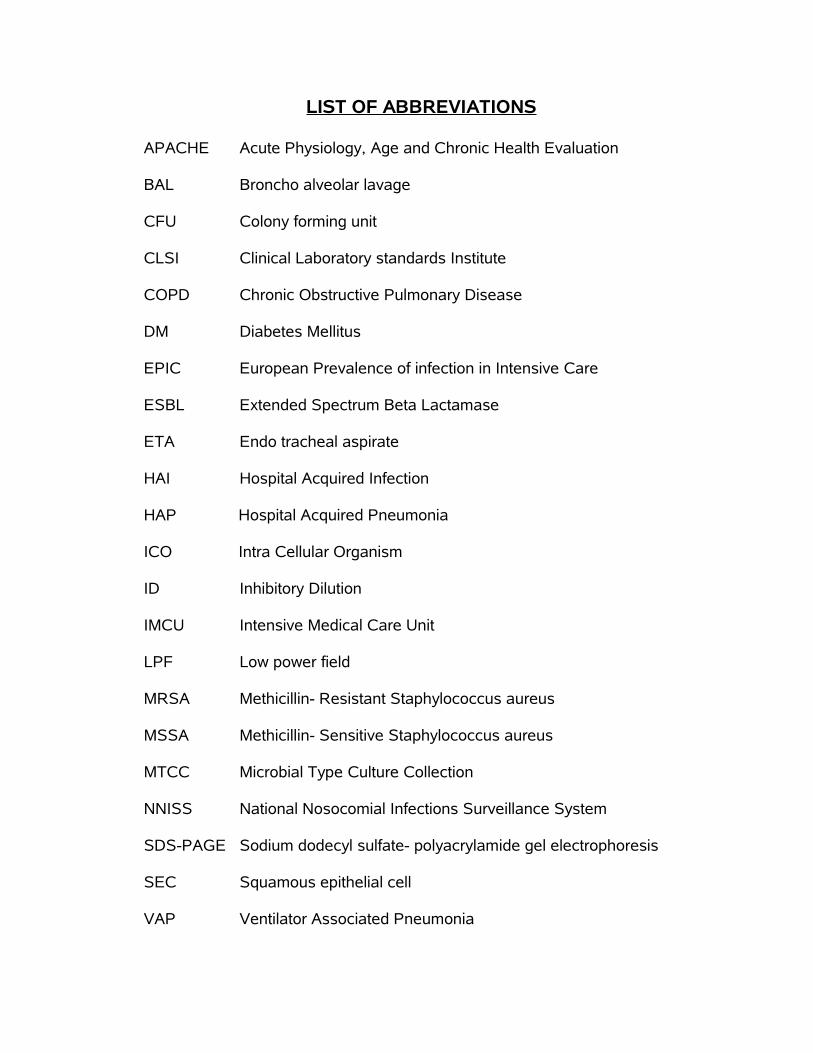
LIST OF ABBREVIATIONS
APACHE Acute Physiology, Age and Chronic Health Evaluation
BAL Broncho alveolar lavage
CFU Colony forming unit
CLSI Clinical Laboratory standards Institute
COPD Chronic Obstructive Pulmonary Disease
DM Diabetes Mellitus
EPIC European Prevalence of infection in Intensive Care
ESBL Extended Spectrum Beta Lactamase
ETA Endo tracheal aspirate
HAI Hospital Acquired Infection
HAP Hospital Acquired Pneumonia
ICO Intra Cellular Organism
ID Inhibitory Dilution
IMCU Intensive Medical Care Unit
LPF Low power field
MRSA Methicillin- Resistant Staphylococcus aureus
MSSA Methicillin- Sensitive Staphylococcus aureus
MTCC Microbial Type Culture Collection
NNISS National Nosocomial Infections Surveillance System
SDS-PAGE Sodium dodecyl sulfate- polyacrylamide gel electrophoresis
SEC Squamous epithelial cell
VAP Ventilator Associated Pneumonia

CONTENTS
S. No. Page No.
1. INTRODUCTION 1
2. AIM AND OBJECTIVES 4
3. REVIEW OF LITERATURE 5
4. MATERIALS AND METHODS 29
5. RESULTS 46
6. DISCUSSION 56
7. SUMMARY 65
8. CONCLUSION 68
9. REFERENCES
10.APPENDICES
i. LIST OF TABLES
ii. LIST OF COLOUR PLATES
iii. PROFORMA
iv. WORKSHEET

INTRODUCTION
Hospital acquired infections continue to be an important cause of
morbidity and mortality among hospitalized patients. 1, 24 The critically ill
patient is at particular risk of developing ICU acquired infection, with the
lungs being especially vulnerable. 2
Hospital acquired pneumonia (HAP) is currently the second most
common hospital infection accounting for 13 to 18 percent of all nosocomial
infections, with estimates of associated mortality ranging from 20 to 50
percent. 2
The majority of cases of HAP occur outside of ICUs. However the
highest risk is in patients on mechanical ventilation. Estimates of incidence
range from 4 to 7 episodes per 1000 hospitalizations. 3 Intubated patients
may have rates of pneumonia 7 to 21- fold higher than patients without a
respiratory therapy device. 5 Infection rates are twice as high in large
teaching hospitals as compared with smaller institutions. 5
HAP results in a significant increase in the cost of care of
hospitalized patients. 3 Its development prolongs a patient’s stay in the
ICU 3, 4 and most of the extra cost is due to an increased length of hospital
stay. 3, 19
The causes of HAP are varied and differ across different patient
populations and different types of ICUs. 4 Hospital acquired bacterial

pneumonia is frequently polymicrobial with gram negative bacilli
predominating. 5, 22 This varied presentation underscores the need for the
intensivists treating the patients with HAP to have a clear knowledge of the
ambient microbiological flora in their ICU.4
Delayed administration of adequate antibiotic therapy is linked to an
increased mortality rate. 6 Hence, the focus of initial antibiotic therapy
should provide rapid antibiotic coverage for all likely pathogens. The
antibiotic spectrum may then be focused or narrowed based on the results
of cultures. A guideline-based approach using the local hospital or ICU
antibiogram may help in appropriate and adequate initial therapy and hence
reduce the overall use of antibiotics and the associated selection pressure
for multi drug resistant organisms.6
Bacteria belonging to the genus Klebsiella frequently cause human
nosocomial infections. In particular, the medically most important Klebsiella
species K.pneumoniae, accounts for a significant proportion of HAP.
Because of their ability to spread rapidly in the hospital environment, these
bacteria tend to cause nosocomial outbreaks. Hospital outbreaks of Multi
Drug Resistant Klebsiella species are often caused by newer type strains,
the so called Extended Spectrum Beta lactamase (ESBL) producers. The
incidence of ESBL producing strains among clinical Klebsiella isolates has
been steadily increasing over the past years. 7

Epidemiological typing is useful in determining the extent of an
outbreak and in elucidating the sources and spread of infection. 8
Combination of Klebocin typing and antibiogram suggest an important tool
for epidemiological studies. 9
Though HAP is widely analyzed by many researchers, not much is known
about the incidence and bacteriological profile i.e., only few studies are
being published by them. This study is conducted prospectively to evaluate
the clinical and bacteriological profile of HAP in ICU patients. It may
increase the awareness of clinicians about the need to reduce the morbidity
and mortality by coming to know about the various pathogens causing HAP
and its sensitivity and/or resistance to various antibiotics. The clinicians
need to establish a suitable antibiotic policy by working out local ICU
antibiogram charts. This study is also done to find out the prevalence of
ESBL producers in HAP. Klebocin typing in association with antibiogram
may help to find out the source of Hospital Acquired Infections.

THE OVERALL AIM OF THE STUDY:
To analyze the incidence, epidemiology, antimicrobial susceptibility
pattern of isolates from HAP patients in IMCU and klebocin typing of
Klebsiella pneumoniae isolated in HAP.
OBJECTIVES:
1. To study the incidence of Hospital Acquired Pneumonia (HAP)
among patients admitted in IMCU.
2. To evaluate the Clinical and Bacteriological profile of HAP.
3. To determine the antibiotic susceptibility pattern of the all bacteria
isolated.
4. To find out the prevalence of ESBL producers in HAP.
5. To evaluate the role of Klebocin typing in epidemiological typing of
K.pneumoniae strains.
6. To detect the source of infection in IMCU using antibiogram and
klebocin typing.

REVIEW OF LITERATURE
Hospital Acquired Pneumonia (HAP) or Nosocomial pneumonia is
defined as pneumonia that occurs at least 48 hours after a patient has been
admitted to the hospital and that was not incubating at the time of
admission. Ventilator- Associated Pneumonia (VAP) is a type of HAP in
patients receiving mechanical ventilation that arises at least 48 hours after
endo tracheal intubation. 11
Epidemiology:
Accurate information concerning the epidemiology of HAP is lacking,
as there is no universally accepted criteria for its diagnosis. 1 HAP is the
second most frequent nosocomial infection. 2 In the year 2006 Glasgow et al
reported that in United Kingdom, HAP accounted for approximately 25% of
hospital infections and was as common as that of the UTI. 12 In various
other studies conducted by different authors in United States HAP
accounted for approximately 15% of all Hospital- related infections and had
been reported as the second most common HAI next to UTI. 2, 10, 30, 31 In the
year 2007 Muhammad et al reported that HAP accounted for 21% of all HAI
in two medical ICUs of a public tertiary care hospital, Karachi and showed
that HAP was the third most common HAI next to UTI (44.6%) and blood
stream infection (27%). 22
The incidence of HAP differs in different Intensive Care Units. 22
Factors like patient population, criteria used for diagnosis, length of ICU
stay and prior exposure to antibiotics may influence the incidence of
HAP.1,18 According to European Prevalence of Infection in Intensive Care
study (EPIC) by Vincent et al involving over 4500 patients, the incidence of
HAP in ICU was 46.9%. 25 Chevret et al from France reported in their
multicenter prospective study on 996 patients admitted in ICUs that the
incidence of HAP was 8.9%. 29 Alp et al from Netherlands reported that
the incidence was 6.8% in ICU patients in the year 2004. 32 In US the
incidence of HAP in ICU patient ranged between 7.8 to 68 percent. 2, 10, 31
Sopena et al from Spain reported that the incidence of HAP was 36.4%
during the year 2005. 30 Berba et al from Philadelphia reported that
incidence was 28.2% among ICU patients. 33
The incidence of HAP is age dependent. HAP is most common in
elderly patient aged >60 years. However patients of any age may be
affected. No racial and sexual predilection exists. 12, 20 Muhammad et al
from Karachi in their study divided the patients admitted in ICU into six age
groups and reported that the highest incidence was seen in patients of 41 to
60 years of age. 22 Berba et al from Philadelphia reported that 30% of the
patients were more than 60 years of age. 33
HAP is the leading cause of death from infections that are acquired in
the hospital. 2 Heyland et al from France showed that the patients with HAP
stayed in the ICU for 4.3 days longer and had a trend toward an increase in
risk of death. 26 Mortality rate for HAP ranges between 24 and 50%. 4, 28
Celis et al from Spain reported that the mortality rate was 36.6% among
HAP patients in a 1000- bed teaching hospital. 27 Sopena et al from Spain
in the year 2005 reported that the mortality rate was 26% among HAP
patients. 30 Alp et al from Netherlands reported that the mortality rate was
65% in ICU patients with HAP in the year 2004. 32
Time is an important epidemiological variable and risk factor for
specific pathogens & outcomes in patient with HAP & VAP. 11 Early-Onset
HAP is defined as pneumonia occurring within the first 4 days of
hospitalization, usually having a better prognosis. Late-Onset HAP is
defined as pneumonia occurring after five or more days of hospitalization,
which are more likely to be caused by MDR pathogens and are associated
with increased patient mortality & morbidity. 11
Indian scenario:
Merchant et al from Mumbai in year 1998 reported that the incidence
of HAP among IMCU patients was 16.7% and the mortality rate was 40% in
HAP. 19 Trivedi et al from Mumbai in the year 2000 reported that the
incidence of HAP in IMCU was 9.38% with the mortality rate of 21.3%; in
the same year another study from Mumbai by Tullu et al in Pediatric
Intensive Care Unit showed the incidence of HAP as 27.54% with the
mortality rate of 47.37%. 16, 17
Mukhopadhyay et al from Lucknow in the year 2003 reported the
incidence of HAP in ICU patients as 53.9% and the mortality rate as 47.3%.
18 Pawar et al from New Delhi in the year 2003 reported the incidence of
HAP as 2.6% among ICU patients with the mortality rate of 16%. 14 Rakshit
et al from Mumbai in the year 2005 reported the incidence as 47% and the
mortality rate as 37%. 13 Dey et al from Manipal in the year 2007 reported
that the incidence of HAP being 45.4%.15
Risk factors for the development of HAP:
There are several risk factors for the development of HAP like
Mechanical ventilation, Reintubation, Age > 60 years, Severity of illness,
Acute or chronic lung disease, Excessive sedation, Enteral nutrition, Supine
body positions, Glasgow coma scale < 9, Use of Muscle relaxants,
Cigarette smoking, Administration of antacids or histamine type 2
antagonists, Emergency surgery, Prior Antibiotic use, Duration of ICU or
Hospital stay. 1, 21, 37
Intubation and mechanical ventilation increase the risk of HAP from 6-
to 21- fold and up to 28% of patients receiving mechanical ventilation will
develop this complication. 1 Many studies on HAP showed that the
occurrence of HAP was high in the ICUs mainly because of high utilization
of invasive procedures like mechanical ventilation. 36 Rakshit et al
reported that the significant risk factors for development of HAP were
prolonged duration of mechanical ventilation, higher APACHE III scores on

admission signifying severe illness and reintubation. 13 Mukhopadhyay et
al from Lucknow in the year 2003 reported unplanned or failed extubation
followed by re-intubation as a significant risk factor for the development of
pneumonia. It is probable that aspiration of infected upper airway
secretions occurs at the time of reintubation. Dey et al from Manipal in the
year 2007 reported that reintubation was a definitive risk factor for the
development of HAP. 15
Pawar et al reported that the risk factors for HAP were COPD,
emergency surgery, reintubation, coma, steroid treatment, enteral feedings
and prior antibiotics in their study. 14 Stress ulcer prophylaxis is routinely
used in the critically ill. The use of H2 blockers is associated with a change
in the acidity of the gastric juices that favors bacterial colonization with
Gram negative bacteria. 33
Celis et al reported that factors significantly predisposing to HAP were
tracheal intubations, depressed level of consciousness, underlying chronic
lung diseases, thoracic or upper abdominal surgery and age older than 70
years. 27 Enteral feeding via a nasogastric tube promotes gastro-
esophageal reflux and is also associated with an increase in gastric pH and
colonization of the stomach with aerobic Gram negative bacilli. 33
Mukhopadhyay et al reported in their study that the rate of acquisition of
HAP increased along with the duration of stay in the ICU. 18
Pathogenesis of HAP:
For HAP to occur, the delicate balance between host defenses and
microbial propensity for colonization and invasion must shift in favor of the
ability of the pathogens to persist and invade the lower respiratory tract.
Sources of pathogens for HAP include healthcare devices, the
environment (Air, water, equipment, and fomites), and can occur with
transfer of microorganisms between the patient and staff or other patient. A
number of host- and treatment- related colonization factors, such as the
severity of the patient’s underlying disease, prior surgery, exposure to
antibiotics, other medication and exposure to invasive respiratory devices
and equipment are important in the pathogenesis of HAP and VAP. 11
Inhalation, aspiration and hematogenous spread are the three main
mechanisms by which pathogens reach the lungs. Inhalation or direct
inoculation of pathogens into the lower airway, hematogenous spread from
infected intravenous catheters, and bacterial translocation from the GIT
lumen are uncommon pathogenic mechanisms.
Aspiration of oropharyngeal pathogens or leakage of secretions containing
bacteria around the endotracheal tube cuff is the primary routes of bacterial
entry into the LRT. 20
Infected biofilm in the endotracheal tube, with subsequent
embolization to distal airways, may be important in the pathogenesis of VAP.
The stomach and sinuses may be potential reservoirs of nosocomial
pathogens that contribute to bacterial colonization of the oropharynx, but
their contribution is controversial which may vary by the population at risk,
and may be decreasing with the changing natural history and management of
HAP. 11, 20
Microbiology of HAP:
HAP is caused by a wide spectrum of bacterial pathogens, may be
polymicrobial and rarely due to viral or fungal pathogens. Common
pathogens include aerobic gram-negative bacilli such as P.aeruginosa,
Klebsiella pneumoniae, Escherichia coli, Acinetobacter species,
Streptococcus pneumoniae and Haemophilus influenzae. Serratia,
Legionella pneumophila , Stenotrophomonas maltophilia and Burkholderia
cepacia are uncommon bacteria causing HAP.
Fungal pathogens isolated from HAP are Candida species and
Aspergillus fumigatus. Influenza, Para influenza, Adenovirus, Measles and
Respiratory Syncytial Virus are viral pathogens causing HAP. 11, 20 The
bacteriology of nonventilated patients was similar to that of ventilated
patients, including infection with pathogens such as methicillin- resistant
Staphylococcus aureus (MRSA), P.aeruginosa, Acinetobacter species and
K.pneumoniae.11

Chastre et al from Paris in the year 2002 reported that the
predominant organisms responsible for HAP being Staphylococcus aureus,
Pseudomonas aeruginosa and Enterobacteriaceae. 28
Leroy et al from France in the year 2002 reported that the main
organisms were P.aeruginosa (31.2%), Enterobacteriaceae (20.8%),
Staphylococcus aureus (18.8%) with 33% Methicillin- resistant strain,
Haemophilus influenzae (6.5%), Streptococcus pneumoniae (5.8%),
Acinetobacter spp. (5.8%) and Stenotrophomonas maltophilia (5.2%). 35
Alp et al from Netherlands in the year 2004 studied that the most
commonly isolated pathogens were Acinetobacter baumanii (29.6%), P.
aeruginosa (20.6%) and Klebsiella pneumoniae (14.4%) among HAP
patients. 32
Tullu et al from Mumbai in the year 2000 studied the bacteriology of
HAP in Pediatric Intensive Care Unit and reported that the organisms
commonly isolated being E. coli (34.4%), Klebsiella pneumoniae (30.2%),
Pseudomonas (11.5%), Proteus (11.5%) and Acinetobacter (5.2%). 17
Mukhopadhyay et al from Sanjay Gandhi Post Graduate Institute
Medical Sciences, Lucknow, in the year 2002 reported that Gram negative
organisms were isolated more frequently than Gram positive cocci in
patients with HAP; Pseudomonas aeruginosa was the most frequent isolate
followed by E.coli and Klebsiella pneumoniae. 18

Pawar et al in the year 2003 from New Delhi reported that the most
common pathogens isolated from HAP cases being Pseudomonas
aeruginosa, E.coli, Klebsiella pneumoniae, Staphylococcus species and
Acinetobacter species in that order. 14
Rakshit et al from Mumbai reported that most common offending
organism isolated in cases with VAP was P.aeruginosa followed by
Klebsiella and E. coli in the year 2005. 13
Rajasekhar et al from Nizam’s Institute of Medical Sciences,
Hyderabad, in the year 2006 reported that Acinetobacter baumanii and
Klebsiella pneumoniae were commonest isolates among HAP patients. 23
Dey and Bairy in the year 2007 from Manipal reported that
Acinetobacter species (48.94%) was the commonest organism followed by
Pseudomonas aeruginosa (25.53%), Klebsiella pneumoniae (12.77%), E.
coli (10.64%) and Serratia marcescens (2.13%).15
Klebsiella pneumoniae – An important pathogen in HAP:
The Genus Klebsiella is defined as containing gram-negative, non-
motile, usually encapsulated rod-shaped, lactose fermenting bacteria of the
family Enterobacteriaceae which produce lysine decarboxylase but not
ornithine decarboxylase and are generally positive in the Voges-Proskauer
test.

Epidemiology:
Klebsiella species are ubiquitous in nature. The two common
habitats of Klebsiella are the environment such as surface water, sewage,
soil & plants and the mucosal surfaces of mammals such as humans,
horses or swine. In humans Klebsiella pneumoniae is present as a
commensal in the nasopharynx with carrier rate ranging between 1 and 6%
and in the intestinal tract with the detection rate in the stool samples
ranging from 5 to 38%. It is rarely found on the human skin. 7
In the hospital environment colonization rates increase in direct
proportion to the length of stay. Tulsi et al reported that colonization of
intestine, umbilical stump, throat, skin and external ear with Klebsiella
pneumoniae increased from 10 percent on admission to 26 percent on day
3 and 39 percent on day 6 in a Neonatal Intensive Care Unit. 69 Reported
carrier rates in hospitalized patients are 77% in stool, 19% in pharynx and
42% on the hands of patients.
The significance of increased colonization was illustrated by the
observation that the attack rate of Klebsiella nosocomial infection in patients
having colonization of Klebsiella was four times as high as for patients
without colonization. 7 Even hospital personnel have elevated rates of
Klebsiella colonization. The principal reservoirs of K. pneumoniae in the
hospital setting are medical equipment (contaminated due to faulty hygienic

procedures), blood products, GIT of patients and hands and pharynx of
hospital personnel. 7
Incidence of Klebsiella pneumoniae in HAI:
As an opportunistic pathogen, K.pneumoniae primarily attack immuno
compromised individuals who are hospitalized and suffer from severe
underlying diseases such as DM or COPD. 7 Table 1 show the percentage
of Hospital acquired Infections caused by Klebsiella pneumoniae and the
rank of Klebsiella pneumoniae compared to all other bacterial pathogens.
Table 1.HAI caused by K.pneumoniae: 7
Infection % of infections
caused by
Klebsiella
Rank (Compared to
all other bacterial
pathogens)UTI 6 – 17 5 – 7
Pneumonia 7 – 14 2 – 4
Septicemia 4 – 15 3 – 8
Wound infections 2 – 4 6 – 11
Nosocomial infections
in ICU patients
4 – 17 4 – 9
Neonatal septicemia 3 – 20 2 – 8
Lockhart S R et al from Chicago reported that during their 12-year
study period from 1993 to 2004, 74,394 gram-negative bacillus isolates
recovered from intensive care unit (ICU) patients in United States hospitals

and Klebsiella pneumoniae accounted for 14.2% of the total HAI. 76 Aly et
al from Egypt in the year 2008 reported that the incidence of Klebsiella
pneumoniae in nosocomial infections in a medical – surgical Intensive Care
Unit was 11%. 77
Gaynes et al and the National Nosocomial Infections Surveillance
System (NNISS), Centers for Disease Control and Prevention, Atlanta
reported in the year 2005 that the incidence of Klebsiella pneumoniae in
nosocomial pneumonia was 8.4%, in urinary tract infection 4.6%, in blood
stream infection 4.5% and in surgical site infection 2.7%. 78 Muhammad in
the year 2007 from Karachi reported that the incidence Klebsiella
pneumoniae in nosocomial infections in Intensive Care Unit was 17.4%. 22
Pal R B et al from Mumbai reported that Klebsiella pneumoniae accounted
for 26.66% of nosocomial infections in Intensive Care Unit. 9
Incidence of Klebsiella pneumoniae in HAP:
In most of the studies, K.pneumoniae was the second most
common pathogen causing HAP next to P.aeruginosa. Merchant et al from
Mumbai in year 1998 reported that the incidence of Klebsiella pneumoniae
in HAP among IMCU patients was 34% and it was the second most
common pathogen next to P. aeruginosa (44%). 19
Tullu et al from Mumbai in the year 2000 in their study of bacteriology
of HAP in Pediatric Intensive Care Unit reported the incidence of Klebsiella
pneumoniae as 30.2%. 17
Mukhopadhyay et al Sanjay Gandhi Post Graduate Institute Medical
Sciences, Lucknow, in the year 2002 reported that Klebsiella pneumoniae
had been isolated as a pathogen from 16.8% of HAP patients.18
Pawar et al in the year 2003 from New Delhi reported that the
incidence of Klebsiella pneumoniae was 9.5% among HAP patients. 14
Rakshit et al from Mumbai in the year 2005 reported that the
incidence of Klebsiella pneumoniae in VAP was 21.8% and it was the
second most common pathogen next to P.aeruginosa. 13
Dey and Bairy in the year 2007 from Manipal reported that 12.77% of
HAP was caused by Klebsiella pneumoniae. 15
Rajasekhar et al from Nizam’s Institute of Medical Sciences,
Hyderabad, in the year 2006 reported Klebsiella pneumoniae accounted
18.18% of total isolates among HAP patients. 23

Pathogenicity Factors of Klebsiella: 7
1. Capsular Antigens:
The capsular repeating subunits consisting of four to six sugars and
very often uronic acids (as negatively charged components) can be
classified into 77 serological types. The capsular material forms thick
bundles of fibrillous structures covering the bacterial surface in massive
layers. This protects the bacterium from phagocytosis by
polymorphonuclear granulocytes and prevents killing of the bacteria by
bactericidal serum factors. The molecular mechanism presumably consists
of inhibiting the activation or uptake of complement components, especially
C3b.
2. Pili (Fimbriae):
These are nonflagellar, filamentous projections on the bacterial
surface which are up to 10µm long and have a diameter of 1 to11 nm. They
consist of polymeric globular protein subunits (pilin) with a molecular mass
of 15 to 26 kDa.
Type1 (common) pili:
This adhesin is designated as mannose-sensitive hemagglutinin
(MSHA), located on the fimbrial shaft. The role of these pili is mainly for
binding of the bacteria to mucus or to epithelial cells of the urogenital,

respiratory and intestinal tracts. Adherence of bacteria to cells of the
repiratory tract leads to impairment of colonization resistance in the upper
airways with a subsequent proliferation of facultative pathogenic bacteria.
This impairment may result in the development of pneumonia especially in
patients undergoing long-term mechanical ventilation.
Type 3 pili: mannose-resistant, Klebsiella-like hemagglutinin (MR/K-
HA).Strains of K.pneumoniae expressing type 3 pili adhere to endothelial
cells, epithelia of the respiratory tract and uroepithelieal cells.
Other types of pili are KPF-28 Fimbria which has been found in the majority
of K.pneumoniae strains producing CAZ-5/SHV-4 type ESBL, CF29K
(Non Fimbrial) adhesin belonging to the K88 adhesin family and
Aggregative adhesin composed of capsule-like extra cellular material.
3. Serum Resistance and Lipopolysaccharide:
Capsular polysaccharides (CPS) may cover and mask the underlying LPS
and exhibit a surface structure that does not activate complement. The O
side chains of the LPS may reach through the capsule layer and be
exposed to the exterior milieu. C 3b is subsequently deposited onto LPS
molecules. Since it is fixed preferentially to the longest O-polysaccharide
side chains, C3b is far away from the bacterial cell membrane. Thus the
formation of the lytic membrane attack complex (C5b-C9) is prevented and
subsequent membrane damage and cell death do not take place.

4. Siderophores:
These are high-affinity, low-molecular-weight iron chelators that are
capable of competitively taking up iron bound to host proteins. Two
different chemical groups of siderophores are Phenolate-type siderophores:
Enterobactin (also known as enterochelin) and Hydroxamate-type
siderophores: Aerobactin.
Although the production of cytotoxins, enterotoxins and hemolysin has
been described, these features probably play a rather minor role in
Klebsiella.
Epidemiological typing of Klebsiella:
From an epidemiological point of view it is often necessary to
determine the clonality of the strains. A useful and effective typing system
should be (1) standardized, (2) reproducible, (3) sensitive, (4) stable, (5)
available, (6) inexpensive and (7) field tested in conjunction with
epidemiologic investigation. This is very useful in endemic and epidemic
nosocomial outbreaks of Klebsiella infections to improve the management
of such outbreaks. Klebsiella marker systems include determination of
antibiotic susceptibility patterns, biotype, serotype, bacteriophage
susceptibility, Bacteriocin susceptibility, and plasmid content, size and
endonuclease fragment size. 47, 48, 50

Biotyping:
Biotyping based on an extended panel of biochemical and culture
test is certainly the most practicable method of typing for laboratories that
are epidemiologically not optimally equipped. Biotyping can be carried out
by using macro tube tests alone or by combining a commercially available
miniaturized system such as the API 20E system with additional macro tube
tests. The drawbacks of this typing method are the large number of
reactions to be tested, the often long cultivation times, poor reproducibility
and poor sensitivity. Therefore biotyping of Klebsiella species is not very
suitable as an epidemiological tool. 7, 47
Serotyping:
Of 82 capsule antigens described, 77 types form the basis for an
internationally recognized capsule antigen scheme. Capsule typing shows
good reproducibility and is capable of differentiating most clinical isolates. 7
The drawbacks of this method are the large number of serological cross-
reactions that occur among the 77 capsule types, the serotyping procedure
is cumbersome because of the time needed to perform the test, it is
susceptible to subjective interpretations because of weak reactions that are
not always easy to interpret, since anti-capsule antisera are not
commercially available, this technique is practiced mostly in specialized
laboratories. 7, 49
Although 12 different O-antigen types of Klebsiella have also been
described, they are difficult to classify because their determination is
hampered by the heat-stable capsules. 7
Phage Typing:
The phage reaction is easily read and the reproducibility of the
method is acceptable. This technique shows a relatively poor typing rate of
19 to 67% and as a single typing method phage typing is not very sensitive.
7, 47
Bacteriocin typing:
Bacteriocins are bactericidal substances usually proteins
produced by bacteria to inhibit the growth of other bacteria usually
members of the same species. 63 In year 1988 James from UK reported
that klebocin production was encoded by a 5.5 kb plasmid and SDS- PAGE
klebocin gave rise to two polypeptides, one of 85 kDa and the other of 11
kDa. 56 An isolate can be characterized either by its ability to inhibit
specific indicator strains or by its sensitivity to bacteriocins synthesized by a
set of producer strains. Since the synthesis of bacteriocins is not frequent
enough in Klebsiella, the latter technique has become the method of choice
for Bacteriocin typing of organisms belonging to this genus. This method
has proven superior for typing of clinical and environmental Klebsiella
strains as well as of nosocomial outbreaks of Klebsiella. 45, 57

Bacteriocin susceptibility typing is easier and cheaper. Depending on
the producer strains used, between 67% and 96% of strains are typable. 47
Israil in year 1981 reported that klebocin typing was a quite simple, reliable
method and did not imply any special requirements. The number of strains
typable was higher than of those typable by phages and the typability was
85% in his study. 46 The same author in year 1980 reported that the
typability was 88.9%. 52 Podschun and Ullman in the year 1996 studied
that 96% of strains were typable by klebocin typing. 58 They developed the
modification of the scrape and point method for klebocin typing. 53
Hall used a set of 10 klebocin producing strains to type 630 clinical
isolates of Klebsiella. The typability was 77% by streak and point method. A
major problem in his study was the lack of reproducibility among large
numbers of weak reactions encountered. 59 Bauerfeind et al showed that
the typability was 96.3% using streak and point method. 62 Buffenmyer et
al in the year 1976 developed standard procedures for the klebocin
production, storage, klebocin titration and the utilization of klebocins for
typing and reported that the typability was 67% in their study. 61
In the year 1997 Pal et al from Mumbai reported that the typability was
71.62% using six producer strains by spot inoculation method. In view of
the high induction of klebocins by mitomycin C, storage stability at -20 o C
and the simplicity of the technique, they stated spot inoculation method as
more appropriate typing method. 9
Chhibber et al from Panjab University, Chandigarh in the year 1998
reported that by using six klebocin producer strains the typability was
72.8%. 60 They studied the effect of various inducing agents on klebocin
production by Klebsiella pneumoniae. A significant level of klebocin was
detected only after induction. The highest level of klebocin was achieved
with mitomycin C followed by rifampicin and polymyxin B. 54 They also
purified and characterized the klebocin and reported that chemical analysis
of the purified preparation showed it to be a protein and was sensitive to
digestion by various proteolytic enzymes. 55
Malik et al from Aligarh Muslim University in the year 2003 reported
that the typability of klebocin typing was 83.3% and reproducibility was
73.3%. 43 Aggarwal et al in year 2003 from Government Medical College,
Amritsar reported that klebocin typability in their study was 73.5%. 44
Molecular typing methods:
The procedures like plasmid profiles, ribotyping, multilocus enzyme
analysis by polyacrylamide gel electrophoresis and pulsed-field gel
electrophoresis which are technically more complicated, vary from laboratory
to laboratory and lack standardization making it difficult to compare them. 7, 47
ESBL producing Klebsiella pneumoniae:

Especially feared are epidemic hospital infections caused by
multi drug resistant strains. In the 1970s, these strains were chiefly amino
glycoside-resistant Klebsiella strains. Since 1982 strains that produce
ESBLs which render them resistant to extended-spectrum cephalosporins
have evolved. 7 Today ESBL producing Klebsiella strains are emerging and
spreading in the community as well as hospital settings. 41 In Europe the β-
lactamases of ceftazidime-resistant Klebsiella strains are commonly of the
SHV-type whereas TEM-10 and TEM-12 are more prevalent in the United
States. 7
Feizabadi et al reported that production of ESBL was detected in
72.8% of nosocomial Klebsiella pneumoniae isolates during their study from
2006 to 2007 in Tehran hospitals. 74
Gonlugur et al in the year 2004 Turkish University Hospital reported
that the incidence of ESBL producing Klebsiella pneumoniae among
respiratory isolates was 12.2%. 39
Hosoglu et al in the year 2007 reported that 68.3% of Klebsiella
pneumoniae isolates in Turkish hospitals were detected as ESBL
producers. 72
Asian HAP working group in 2008 reported the overall incidence of
ESBL producing Klebsiella pneumoniae as 0.9 to 40% of cases of HAP. 80
Verhamme et al in the year 2007 from Netherlands reported that the

incidence of ESBL producing Klebsiella pneumoniae was highest in Late-
onset HAP (100%). 79 In the year 2007 Vitkauskiene et al reported that the
incidence of ESBL producing Klebsiella pneumoniae among HAP patients
was 88.9%. 84
Indian studies on ESBL producing Klebsiella pneumoniae:
In India, ESBL producing strains of K.pneumoniae have emerged as a
challenge in hospitalized as well as community based patients. 65 High
prevalence of ESBL producing Klebsiella strains has been reported by
various groups. In 1997, from Nagpur 17 out of 66 Klebsiella isolates
showed ESBL production. 86 In the year 2002, 68 percent of Gram
negative bacteria were found to ESBL producers in a study from New Delhi
in which 80 percent of Klebsiella were ESBL producers. 87
In 2004 two other studies from Delhi showed 70.6 and 12.6 percent
Klebsiella isolates to be ESBL producers respectively. 88, 89
In Lucknow the ESBL production were seen in 58% of Klebsiella
isolates from neonatal septicemia during 2004-2005. 64 Dey and Bairy in
the year 2007 from Manipal reported that 100 percent of K. pneumoniae
isolates from VAP cases were ESBL producers. 15
Source detection in Hospital Acquired Klebsiella infections:
In the year 1997 Pal et al from Mumbai reported that by combination
of klebocin types and antibiogram of clinical isolates, the source of infection
could be traced in 86.84% cases. The sources of infection were ICU sites
and staff in 60.52% cases, 6.31% cases as endogenous flora and rest was
unknown. 9
Malik et al in the year 2003 reported that Klebocin typing and
antibiogram of the environmental isolates of Klebsiella pneumoniae showed
the same pattern as was observed in patient isolates and concluded these
isolates as the source of infection. 43 Cooke et al reported that the sources
of Klebsiella infections in hospital patients could be found out using
Serotyping and bacteriocin typing. 51 Tulsi et al detected the source of
Klebsiella pneumoniae from ward environments using klebocin typing. 69
Clinical outcome of HAP:
Rakshit et al from Mumbai in the year 2005 reported that 37% of HAP
patients succumbed while being treated in Medical Critical Care Unit. Five
patients were more than 60 years old and none of them could survive. The
mortality was significantly higher in older age group and in patients with co
morbid illnesses. Significantly higher survival rates were found in cases
that had early and planned tracheostomy, as compared with those who
needed reintubations. 13 Trivedi et al from Mumbai in the year 2000

reported that high mortality was associated with habits like smoking
(33.3%), age group over 60 years (27.3%), presence of comorbid illness
like DM and COPD (38.5%), complications like ARDS (61.3%) or sepsis
with end organ failure (73.7%) and need of intubation (36.2%) or
mechanical ventilation (40.55). 16
Rajasekhar et al from Hyderabad in the year 2006 reported that the
clinical condition of 87% cases in Early onset HAP and 33% cases in Late
onset HAP improved significantly with the modification of antibiotic therapy
made after the results of cultures of ETA samples. The over all mortality in
their study was 18%. 23 Berba R et al in their study showed that the mean
duration of hospitalization was almost doubled when HAP occurred. The
mortality rates were more than three times higher among HAP patients than
among non -HAP patients. 35% of deaths among HAP patients occurred
within 48 hours from the time the HAP diagnosis was made. 33

MATERIALS & METHODS
This prospective study was conducted over a period of one year
among patients admitted in Intensive Medical Care Unit (IMCU) of
Coimbatore Medical College & Hospital, Tamil Nadu. The study population
comprised all patients admitted to the IMCU from May 1, 2007 to April 30,
2008. Approval was obtained from the Ethical Committee prior to
conducting the study and informed consent from all patients under study
was also obtained.
HAP was diagnosed based on standard diagnostic criteria adapted by
the Centers for Disease Control and Prevention for the diagnosis of
pneumonia if signs of pneumonia occurred after 48 hours following IMCU
admission. 34 HAP was considered when new or progressive chest
radiographical infiltrates occurred ≥48 hours after hospital admission in
conjunction with the following clinical criteria:
At least one of the following:
1. Fever (> 38° C/ 100.4° F) with no other recognized cause
2. Leukocytosis (≥ 12,000 WBC/mm3) or leucopenia (< 4,000 WBC/
mm3)
3. For adults ≥ 70 years old, altered mental status with no other
recognized cause
4.

And
At least two of the following:
1. New onset of purulent sputum / change in the character of the
sputum / increased respiratory secretions / increased suctioning
requirements.
2. New Onset or worsening cough / dyspnea / tachypnea
(Respiratory Rate > 25 breaths / min)
3. Rales or bronchial breath sounds
4. Worsening gas exchange : O2 desaturations [Pa O2 / Fi O2 ≤ 240 ] /
increased O2 requirements / increased ventilation demand
The following cases were excluded from the study:
1. Patients who died within 48 hours from the time of admission to the
IMCU
2. Patients discharged or went home against medical advice within 48
hours of admission
3. Patients who were diagnosed to have pneumonia during the time of or
within 48 hours of admission (Pneumonia in these cases were
presumed to have developed from a previous hospital admission or
community)

Data collection:
Data collection began from the time of admission to the IMCU and
continued until the occurrence of HAP, death or discharge from IMCU
whichever occurred first.
On IMCU admission name, age, sex, address, date of admission,
diagnosis on admission, underlying illness, presence of immuno
compromised state (DM, Malignancies and AIDS), history of smoking and
alcoholism were recorded. A thorough general & systemic examination of
the patient was also done.
When HAP occurred, the time of onset from hospital admission,
temperature, chest radio graphical involvement and leukocyte count were
recorded. Intervention – related variables including need for supplemented
O2 & device used, need for mechanical ventilation, suctioning devices used,
naso gastric tube placement, stress ulcer prophylaxis, steroids, sedatives
and antibiotics actually given for at least 48 hrs were also recorded.
The data on the hospitalization outcome including length of hospital
stay and discharged versus mortality was also determined.
Specimens collected:
Sputum
Bronchoscopic aspirate (BAL)
Endotracheal aspirate (ETA)
Blood
For source detection:
Swabs from the patient’s environment (i.e. IMCU sites such
as wall, floor, trolley, suction tubing, bed & curtains) as well as swabs from
the IMCU staff members (i.e. skin, nasal & throat swabs) were collected
monthly.
Specimen collection & Transport:
Collection of Sputum:
The patient was instructed to rinse mouth with water before collecting
sputum sample. Early morning sputum samples were collected in sterile
wide mouthed containers fitted with screw capped lids.
Collection of BAL: 1, 35
A trained respiratory therapist collected the specimen every time. The
tip of the flexible Fibre Optic Bronchoscope (FOB) was positioned close to
the segmental area corresponding to radiographic infiltrates. Three aliquots
of 50 ml sterile 0.9% Normal saline were instilled. After the injection of each
aliquot, gentle aspiration was done through the suction channel. The
aspirates pooled in a sterile container were submitted to the laboratory
immediately for microscopy & microbiological analysis.
Collection of ETA: 15
The ETA was collected using a 22-inch Ramson’s 12F suction
catheter with a mucous extractor, which was gently introduced through the
ET tube for a distance of approximately 25-26 cm. Gentle aspiration was

then performed without instilling saline and the catheter was withdrawn
from the ET tube. After the catheter was withdrawn, 2ml of sterile 0.9%
Normal saline was injected into it with a sterile syringe to flush the exudates
into a sterile container for collection. ETA samples were immediately taken
to the laboratory for processing.
Collection of Blood sample:
After cleansing the site for venepuncture with Betadine and 70%
Alcohol, about 5ml of Blood was collected and added to 50ml of sterile
Brain Heart Infusion (BHI) broth in Blood culture bottles. All the specimens
were sent to the laboratory immediately after collection.
Collection of swabs from IMCU sites:
Swabs were collected during the time of infection from IMCU sites and
staff. Moistened cotton wool swabs were used for collecting samples from
the wall, floor and trolley. The swabs were placed in Nutrient Broth and
incubated at 37o C overnight. After sub culturing on plates, the isolates were
identified by standard laboratory techniques. The samples from bed clothes
and curtains were collected by sweep plate method. The Petri dish
containing culture medium was removed from its lid and rubbed over the
fabrics. The colonies were identified after incubation by standard laboratory
techniques.

Safety precautions:
• All samples were considered potentially infectious and leak proof
containers were used for collection and transportation of the samples.
• Biological safety cabinet class II was used for carrying out all
procedures and protective wears like mask, gloves etc was used.
• Disinfection of the containers by treating with 2.5% Sodium
Hypochlorite solution / autoclaving was followed.
Processing of specimens:
Direct Microscopy:
The BAL, ETA and sputum samples were subjected to Gram staining
and Potassium Hydroxide (KOH) mount using standard laboratory
techniques 66, 68 to assess the quality of the samples for further processing.
Culture Procedures:
The samples were mechanically liquefied and homogenized by
vortexing for 1 min and then serially diluted in 0.9% sterile saline solution
with final dilutions of 10-2, 10-3 and 10-4. The diluted samples were inoculated
onto Blood Agar plate (BAP) with 10% sheep blood, Chocolate Agar plate
with 10% sheep blood (CAP) and Mac Conkey Agar plate and Sabourauds
Dextrose Agar plate (SDA) by using 4mm Nichrome wire loop (Himedia,
Mumbai) which holds 0.01ml of solution for quantitative culture.
All plates were incubated overnight at 37° C and CAP at 37° C with
5% CO2 and one SDA plate was kept at room temperature.

All plates were checked for growth overnight and then after 24 & 48
hrs of incubation. SDA plates were checked for any growth daily for the first
week and twice a week up to four week. Growth of any bacterial isolate
below the threshold (Table 2) was assumed to be due to colonization or
contamination. All the bacterial pathogens with colony count above the
diagnostic threshold were identified by colony morphology, microscopy and
detailed biochemical testings using standard laboratory techniques. 42, 66
Table2. Criteria for the assessment of a good quality respiratory
sample in HAP. 35, 67
ETA BAL SPUTUM
Neutrophils >25/ LPF 77- 82% >25/ LPF
SEC <10/LPF <1% <10/ LPF
ICO No data ≥5% No data
Quantitative culture threshold
(cfu / ml )
≥105-106 ≥104 ≥105-106
Antimicrobial susceptibility test:
Antimicrobial susceptibility test of the bacterial isolates was
performed on Mueller Hinton Agar (Hi-media, Mumbai) plates by Kirby
Bauer’s disc diffusion method and antibiotic sensitivity pattern studied
according to Clinical Laboratory Standards Institute (CLSI).

Gram negative bacilli were tested for the following antimicrobials (Hi-
media, Mumbai):
Antimicrobial agent Disc content
Gentamicin 10 µg
Amikacin 30 µg
Amoxycillin/Clavulanic acid 20/10 µg
Cotrimoxazole 25 µg
Cephalexin 30 µg
Cefotaxime 30 µg
Ceftazidime 30 µg
Ceftriaxone 30 µg
Cefpodoxime 10 µg
Aztreonam 30 µg
Cefepime 30 µg
Ciprofloxacin 5 µg
Ofloxacin 5 µg
Gatifloxacin 5 µg
Imipenem 10 µg
Gram positive cocci were tested for the following antimicrobials (Hi-
media, Mumbai):
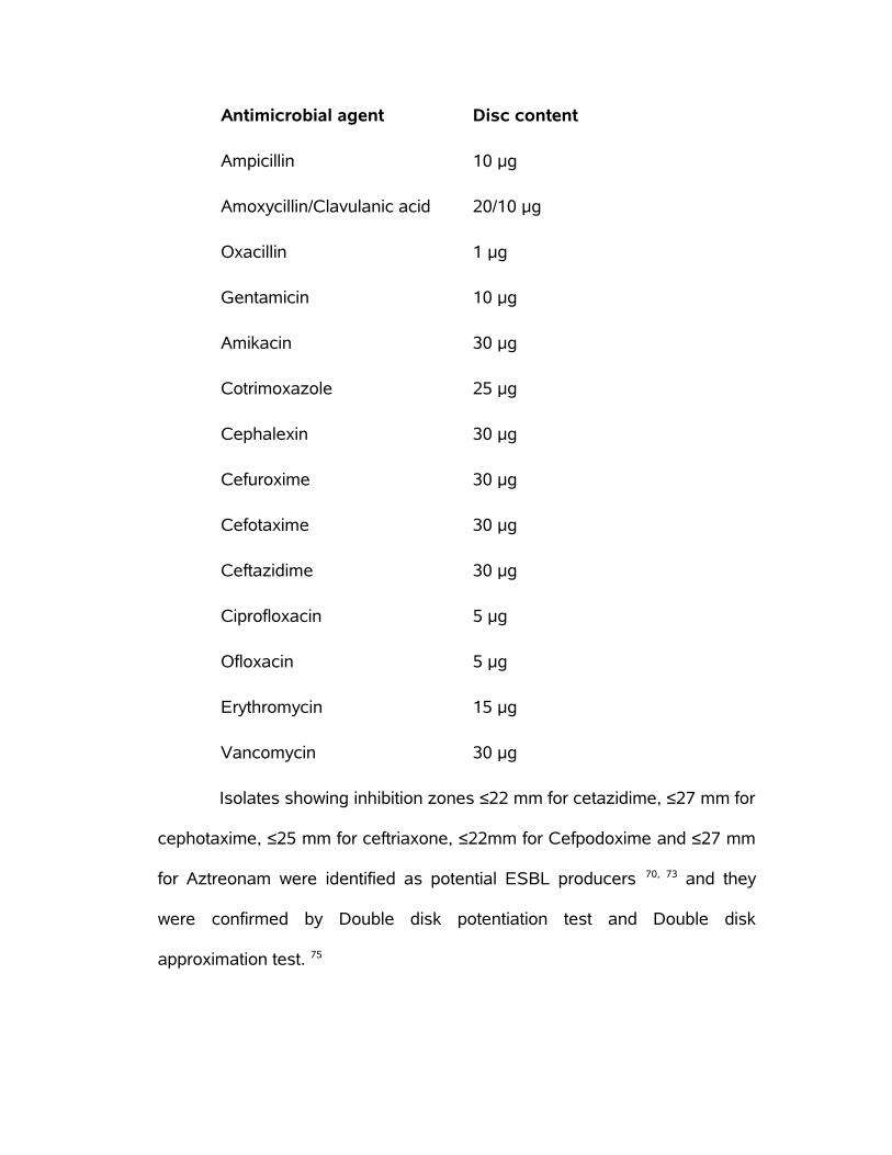
Antimicrobial agent Disc content
Ampicillin 10 µg
Amoxycillin/Clavulanic acid 20/10 µg
Oxacillin 1 µg
Gentamicin 10 µg
Amikacin 30 µg
Cotrimoxazole 25 µg
Cephalexin 30 µg
Cefuroxime 30 µg
Cefotaxime 30 µg
Ceftazidime 30 µg
Ciprofloxacin 5 µg
Ofloxacin 5 µg
Erythromycin 15 µg
Vancomycin 30 µg
Isolates showing inhibition zones ≤22 mm for cetazidime, ≤27 mm for
cephotaxime, ≤25 mm for ceftriaxone, ≤22mm for Cefpodoxime and ≤27 mm
for Aztreonam were identified as potential ESBL producers 70, 73 and they
were confirmed by Double disk potentiation test and Double disk
approximation test. 75

Double disk potentiation test: 85
Ceftazidime (30mcg, Himedia, Mumbai) and Ceftazidime -Clavulanic
acid (30/10mcg, Himedia, Mumbai) discs were placed onto Muellar Hinton
Agar (MHA) plates inoculated with a suspension (adjusted to 0.5 McFarland
turbidity standard) made from an overnight agar plate of the test strain.
Plates were then incubated at 35o C overnight. Regardless of the zone
diameter, ≥5 mm increase in a zone diameter for Ceftazidime -Clavulanic
acid disc than disc with Ceftazidime alone confirmed the presence of ESBL
producers.
Double Disk approximation test: 40, 71
A disc of Amoxicillin-Clavulanic acid (20/10mcg, Himedia, Mumbai)
and a disc of Ceftazidime (30mcg, Himedia, Mumbai) were kept 30mm
apart from center to center on Muellar Hinton Agar (MHA) plates inoculated
with a suspension (adjusted to 0.5 McFarland turbidity standard) made from
an overnight agar plate of the test strain. Plates were then incubated at 35o
C overnight.
Enhancement of the zone of inhibition around the Ceftazidime disc
towards the Clavulanic acid disc was interpreted as positive for ESBL
production.
All cultures of Klebsiella pneumoniae were reconfirmed by colony
morphology on MacConkey agar, reaction in TSI agar, the absence of
motility, absence of Indole production, Negative Methyl Red test, Positive
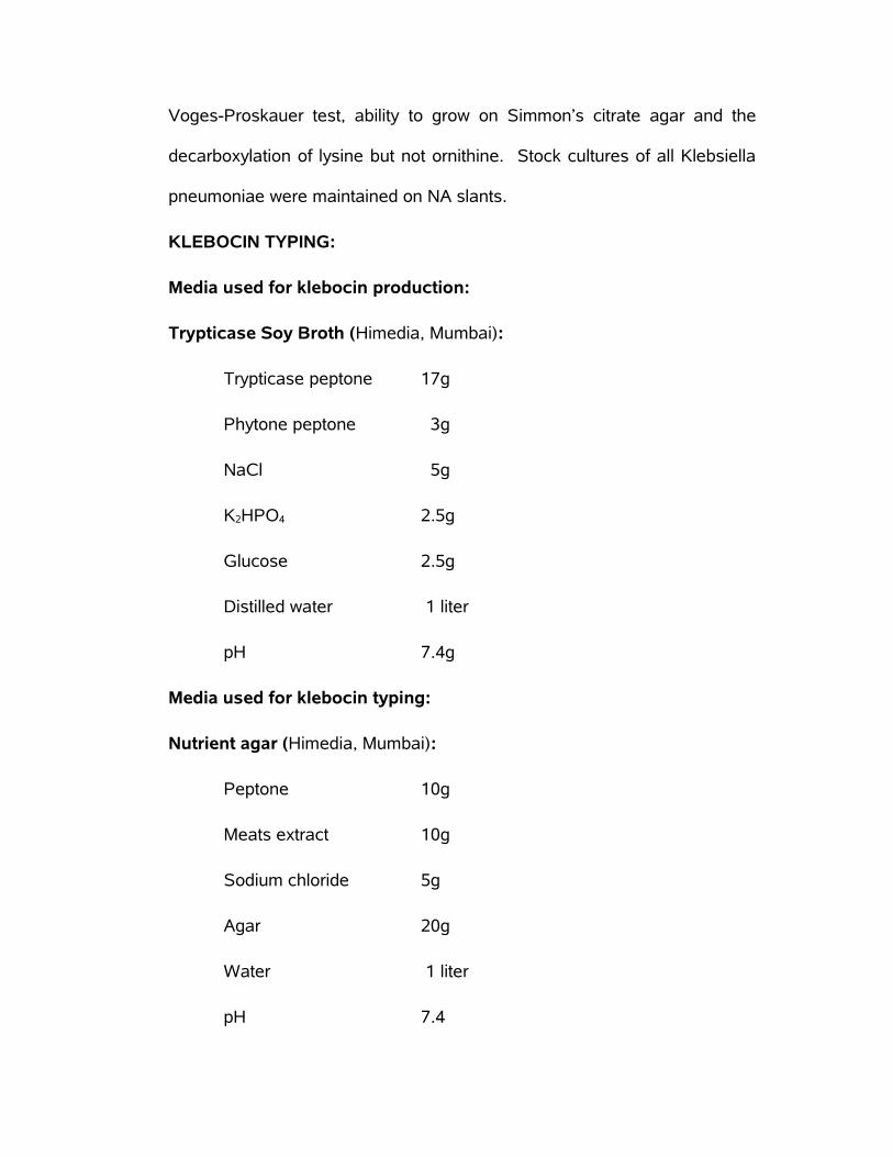
Voges-Proskauer test, ability to grow on Simmon’s citrate agar and the
decarboxylation of lysine but not ornithine. Stock cultures of all Klebsiella
pneumoniae were maintained on NA slants.
KLEBOCIN TYPING:
Media used for klebocin production:
Trypticase Soy Broth (Himedia, Mumbai):
Trypticase peptone 17g
Phytone peptone 3g
NaCl 5g
K2HPO4 2.5g
Glucose 2.5g
Distilled water 1 liter
pH 7.4g
Media used for klebocin typing:
Nutrient agar (Himedia, Mumbai):
Peptone 10g
Meats extract 10g
Sodium chloride 5g
Agar 20g
Water 1 liter
pH 7.4
Klebsiella stains:
Four Klebocin producer strains, MTCC 109, MTCC 432, MTCC 618
and MTCC 39 were kindly supplied by Institute of Microbial Technology,
Sector 39-A, Chandigarh - 160 036, India in the form of freeze dried
cultures in ampoules. Two more klebocin producers (L2 and L1) were
included in the producer strain set. These last two laboratory strains were
selected for typing set using standard procedures as described by Hall et al,
59 Bauerfeind et al 62 and Dykes et al. 63 Collectively, the set of producer
strains used in this study comprised of six klebocin producers as follows:
MTCC 109 (producer 1), MTCC 432 (producer 2), MTCC 618 (producer 3),
MTCC 39 (producer 4), L2 (producer 5) and L1 (producer 6).
The indicator strain isolated in our laboratory, sensitive to all six
klebocins was used as a positive control in klebocin typing.
Recovery of freeze dried cultures from ampoule:
The intactness and the identification label of the ampoule were
checked initially. The ampoule was marked with a file transversely over the
cotton-wool plug. The tip of a glass rod was heated red-hot in a Bunsen
flame and applied firmly to the file mark on the ampoule, which could crack at
that point. Air was allowed to enter via the plug; then carefully the drawn-out
end of the ampoule and the plug were put into a container of disinfectant, for
subsequent autoclaving. With aseptic precautions, a small amount of sterile
Nutrient Broth was added to the ampoule with a sterile Pasteur pipette,
expressing and taking up the broth several times to bring the contents of the

ampoule into suspension. The suspended material in the broth was seeded
into Trypticase Soy Broth to ensure recovery of the organism after
appropriate incubation.
Induction of klebocin production: 9
To each broth culture (about 0.7ml) of producer strains, 3.3 ml
Trypticase Soy Broth (TSB) and 1 ml of mitomycin C (0.5mcg/ml) was
added and incubated at 37o C for 5 hours with intermittent shaking.
The cells were killed by addition of 0.25 ml chloroform. The
suspension was centrifuged at 3000 rpm for 10 min in cold centrifuge.
Supernatant containing respective klebocins was aliquoted in screw cap
vials and stored at -20o C. The purified preparations of klebocins were
subjected to SDS-PAGE which showed them to be proteins having two
peptides, one of 85kDa and the other of 11kDa.
Klebocin titration: 61
The klebocins were serially diluted eightfold in Nutrient Broth,
ranging from 1:8 to 1:512. With micropipette 0.025ml of each dilution was
delivered onto an NA (2% agar) plates (15 by 100 mm) streaked with cotton
tipped applicator that had been moistened in a culture of the Indicator
strain. This culture was prepared by sub culturing from a work stock culture
into 2ml of Nutrient Broth and incubating at 37o C for 6 hours. The plates
were incubated overnight at 37o C.
The highest Dilution of klebocin yielding completely clear inhibition of
the indicator strain was defined as the Inhibitory Dilution (ID). Control spots

of chloroform treated TSB containing 1µg of mitomycin C per ml showed no
inhibition of the indicator strain.
Typing procedure: 9, 61
The test strains were inoculated by a straight needle from NA slants
into 2 ml of Nutrient broth and incubated at 37o C for six hours. These test
cultures were swabbed over a nutrient agar plates and 12µl of each
klebocin was spot inoculated on marked sectors with a micropipette. The
plates were incubated overnight at 37o C.
A positive control plate swabbed with a six hours culture of the
indicator strain was included with every typing.
The readings were made by grading the inhibition seen, using a
scale of no reaction to 4+.
1+ - a partial inhibition with confluent growth.
2+ - a partial inhibition showing patches of semi confluent
growth or more than 10 colonies.
3+ - a clear zone containing no more than 10 distinct colonies.
4+ - a completely clear zone of inhibition.
All reactions that were 1+ or greater were designated as being
positive, and those that were less than 1+ were designated as negative.
The negative and positive reactions to six klebocins were recorded, using a
mnemonics system proposed by Farmer (1976).

The six klebocins were divided into three pairs. Each combination of
reactions for a given pair was assigned a mnemonic notation (Table 3).
Thus the klebocin type for a given isolate is reported as a mnemonic
composed of three numbers
Table 3. Mnemonic system for reporting klebocin sensitivity patterns:61
a Inhibition of isolate by both klebocins.
b Inhibition of isolate by first klebocin and no inhibition by second
klebocin.
Reproducibility of klebocin typing:
The reproducibility was determined by using the formula: R = Nr / N,
where Nr is the number of isolates assigned the same type on repeat
testing and N is the number of isolates tested. 43
Table 4. Examples of klebocin sensitivity pattern using mnemonic
system:
Producer
Strains
Example 1 (Type 211) Example 2 (Type 314)
Reaction Mnemonic
notation
Reaction Mnemonic
notationMTCC 109
MTCC 432
+
−2
−
+3

MTCC 618
MTCC 39
+
+
1
+
+
1
L2
L1
+
+
1
−
−
4

RESULTS
During the one-year study period, among 2658 patients admitted to
the IMCU, only 2454 cases were followed and included in this study. The
remaining 204 cases were excluded. (118 died within 48hrs of admission, 86
were discharged or went home against advice).
Among 2454 patients, 64% of patients (n=1570) were males and 36%
(n=884) were females. The mean age was 59.96 with the range of 15 to 89 yrs
old. 35% of the patients were more than 60yrs of age. Out of 2454 cases,
253(10.3%) patients developed HAP. The highest incidence of HAP (55.73%)
was observed in the age group more than 60 years. (Ref table 5, 6)
The primary reason for IMCU admission was due to neurological
events (31.1%), cardiac and pulmonary emergencies (26.0%), acute infections
(12.%5), poisoning (5.3%), envenomation (0.5%), etc and the incidence of
HAP was greater in patients with diseases requiring prolonged mechanical
ventilation and in patients with those diseases that predispose to pulmonary
infection such as sepsis and prolonged stay in IMCU.
Out of 1352 patients on mechanical ventilation, 62.0% of patients
(n=157) developed HAP and only 38.0% of patients (n=96) developed HAP
out of 1102 non ventilated patients. (Ref table 7)
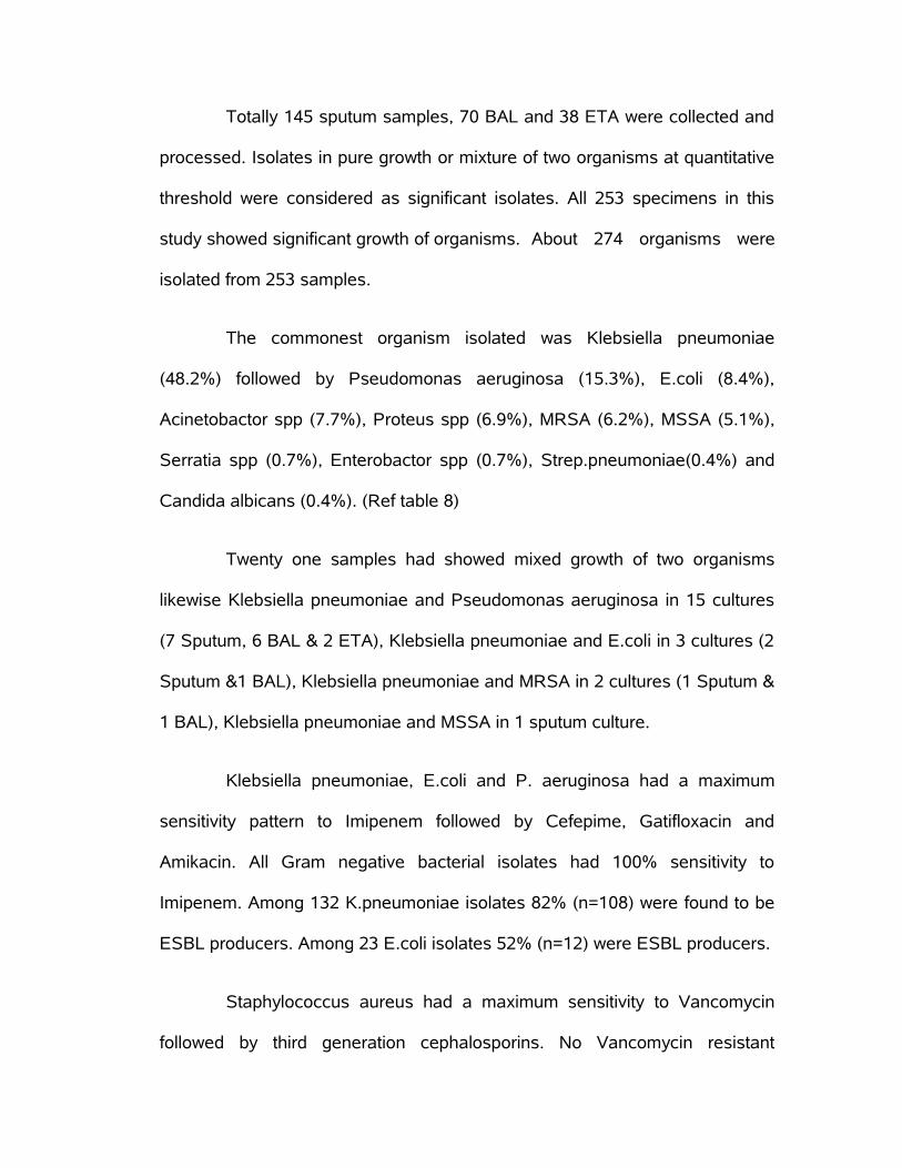
Totally 145 sputum samples, 70 BAL and 38 ETA were collected and
processed. Isolates in pure growth or mixture of two organisms at quantitative
threshold were considered as significant isolates. All 253 specimens in this
study showed significant growth of organisms. About 274 organisms were
isolated from 253 samples.
The commonest organism isolated was Klebsiella pneumoniae
(48.2%) followed by Pseudomonas aeruginosa (15.3%), E.coli (8.4%),
Acinetobactor spp (7.7%), Proteus spp (6.9%), MRSA (6.2%), MSSA (5.1%),
Serratia spp (0.7%), Enterobactor spp (0.7%), Strep.pneumoniae(0.4%) and
Candida albicans (0.4%). (Ref table 8)
Twenty one samples had showed mixed growth of two organisms
likewise Klebsiella pneumoniae and Pseudomonas aeruginosa in 15 cultures
(7 Sputum, 6 BAL & 2 ETA), Klebsiella pneumoniae and E.coli in 3 cultures (2
Sputum &1 BAL), Klebsiella pneumoniae and MRSA in 2 cultures (1 Sputum &
1 BAL), Klebsiella pneumoniae and MSSA in 1 sputum culture.
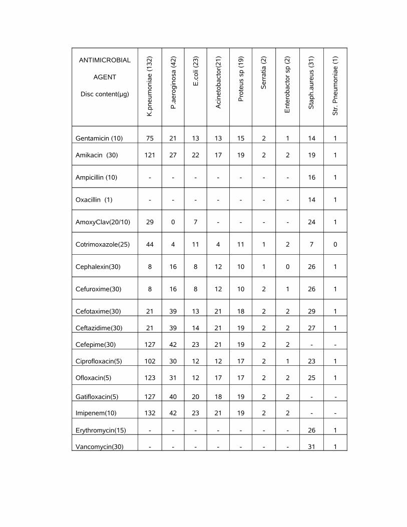
Klebsiella pneumoniae, E.coli and P. aeruginosa had a maximum
sensitivity pattern to Imipenem followed by Cefepime, Gatifloxacin and
Amikacin. All Gram negative bacterial isolates had 100% sensitivity to
Imipenem. Among 132 K.pneumoniae isolates 82% (n=108) were found to be
ESBL producers. Among 23 E.coli isolates 52% (n=12) were ESBL producers.
Staphylococcus aureus had a maximum sensitivity to Vancomycin
followed by third generation cephalosporins. No Vancomycin resistant
Staph.aureus was detected. 54.84% (n=17) of Staph.aureus were Methicillin
Resistant strains. (Ref table 9)
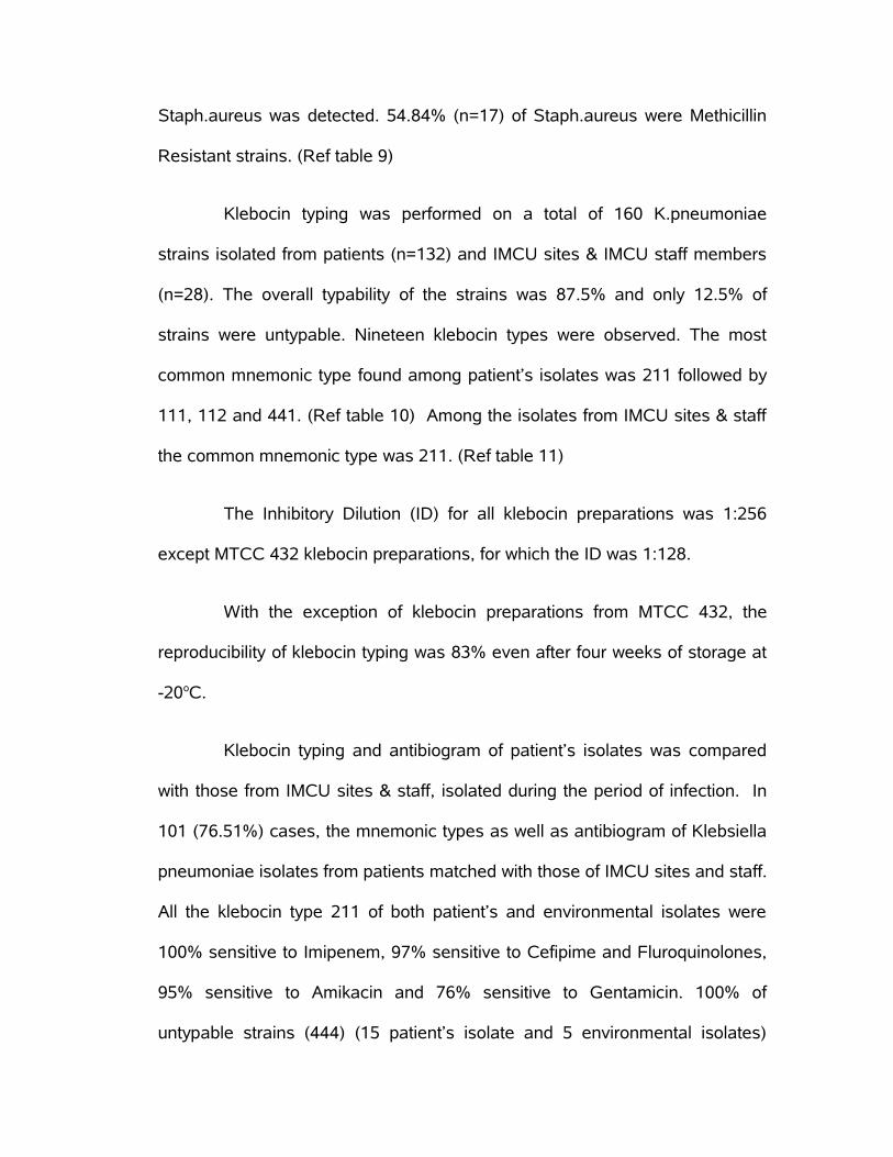
Klebocin typing was performed on a total of 160 K.pneumoniae
strains isolated from patients (n=132) and IMCU sites & IMCU staff members
(n=28). The overall typability of the strains was 87.5% and only 12.5% of
strains were untypable. Nineteen klebocin types were observed. The most
common mnemonic type found among patient’s isolates was 211 followed by
111, 112 and 441. (Ref table 10) Among the isolates from IMCU sites & staff
the common mnemonic type was 211. (Ref table 11)
The Inhibitory Dilution (ID) for all klebocin preparations was 1:256
except MTCC 432 klebocin preparations, for which the ID was 1:128.
With the exception of klebocin preparations from MTCC 432, the
reproducibility of klebocin typing was 83% even after four weeks of storage at
-20oC.
Klebocin typing and antibiogram of patient’s isolates was compared
with those from IMCU sites & staff, isolated during the period of infection. In
101 (76.51%) cases, the mnemonic types as well as antibiogram of Klebsiella
pneumoniae isolates from patients matched with those of IMCU sites and staff.
All the klebocin type 211 of both patient’s and environmental isolates were
100% sensitive to Imipenem, 97% sensitive to Cefipime and Fluroquinolones,
95% sensitive to Amikacin and 76% sensitive to Gentamicin. 100% of
untypable strains (444) (15 patient’s isolate and 5 environmental isolates)

were sensitive to Imipenem, 93% sensitive to Amikacin, Cefipime, Ofloxacin
and Gatifloxacin, 80% to Ciprofloxacin. The klebocin type 111 which was
isolated from suction tube had similar antibiotic pattern like that of Patient’s
isolates (n=9). All klebocin type 111 were resistant to Cotrimoxazole,
Cephalexin, Cefuroxime, Cefotaxime, Ceftazidime and Ceftriaxone and 100%
sensitive to Imipenem. This antibiotic sensitivity pattern was similar to that of
klebocin type 112 (1 from throat of the IMCU staff and 7 from HAP patients).
(Ref table 12)
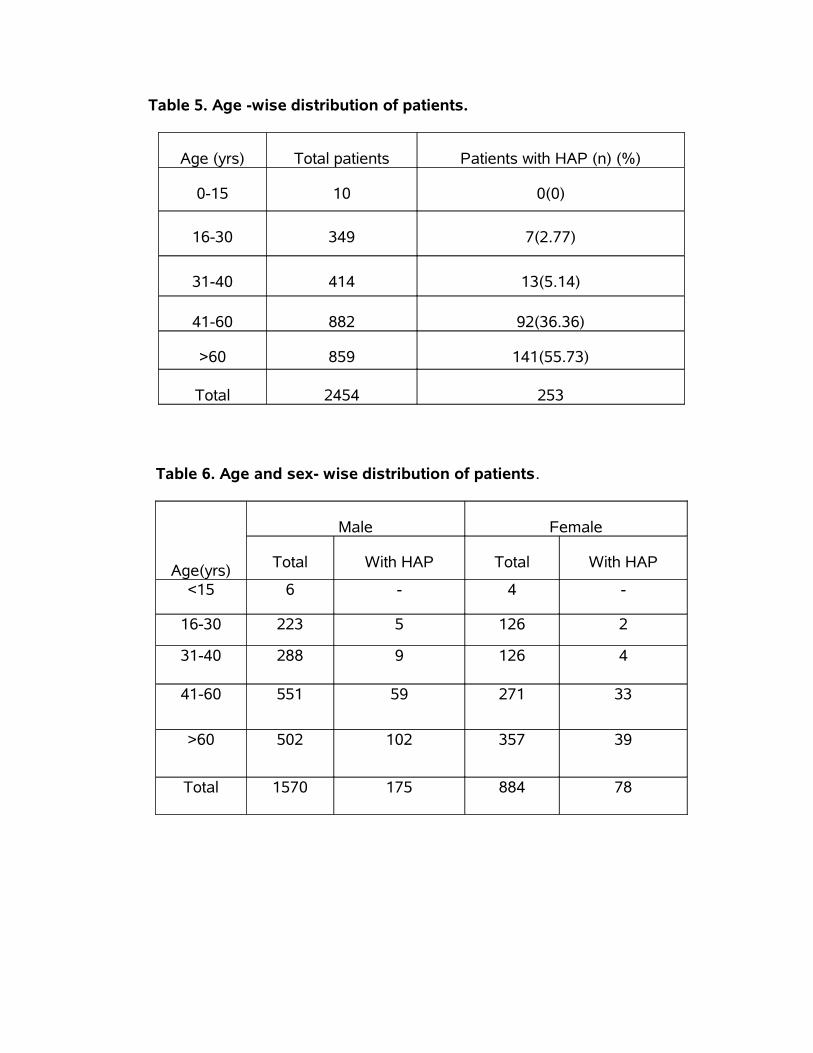
Table 5. Age -wise distribution of patients.
Age (yrs) Total patients Patients with HAP (n) (%)
0-15 10 0(0)
16-30 349 7(2.77)
31-40 414 13(5.14)
41-60 882 92(36.36)
>60 859 141(55.73)
Total 2454 253
Table 6. Age and sex- wise distribution of patients.
Age(yrs)
Male Female
Total With HAP Total With HAP
<15 6 - 4 -
16-30 223 5 126 2
31-40 288 9 126 4
41-60 551 59 271 33
>60 502 102 357 39
Total 1570 175 884 78

Table 7. Influence of mechanical ventilation on HAP.
Ventilator Total Patients with HAP (%)
Yes 1352 157(62.0)
No 1102 96(38.0)
Total 2454 253
Table 8. Pathogens isolated from HAP patients.
Pathogens isolated No of isolates (n=274) (%)
K.pneumoniae 132 (48.2)
P.aeruginosa 42 (15.2)
E.coli 23 (8.4)
Proteus spp 19 (6.9)
Acinetobactor spp. 21 (7.7)
Serratia 2 (0.7)
Enterobactor spp. 2 (0.7)
MSSA 14 (5.1)
MRSA 17 (6.2)
Strep. pneumoniae 1 (0.4)
Candida albicans 1 (0.4)
Table 9. Antimicrobial susceptibility pattern of Bacterial isolates.
ANTIMICROBIAL
AGENT
Disc content(µg)
K.p
neu
mo
nia
e (
132
)
P.a
ero
gin
osa
(4
2)
E.c
oli
(23
)
Aci
neto
ba
cto
r(2
1)
Pro
teu
s sp
(1
9)
Se
rra
tia (
2)
Ent
ero
ba
cto
r sp
(2
)
Sta
ph.a
ure
us
(31
)
Str
. P
neu
mo
nia
e (
1)
Gentamicin (10) 75 21 13 13 15 2 1 14 1
Amikacin (30) 121 27 22 17 19 2 2 19 1
Ampicillin (10) - - - - - - - 16 1
Oxacillin (1) - - - - - - - 14 1
AmoxyClav(20/10) 29 0 7 - - - - 24 1
Cotrimoxazole(25) 44 4 11 4 11 1 2 7 0
Cephalexin(30) 8 16 8 12 10 1 0 26 1
Cefuroxime(30) 8 16 8 12 10 2 1 26 1
Cefotaxime(30) 21 39 13 21 18 2 2 29 1
Ceftazidime(30) 21 39 14 21 19 2 2 27 1
Cefepime(30) 127 42 23 21 19 2 2 - -
Ciprofloxacin(5) 102 30 12 12 17 2 1 23 1
Ofloxacin(5) 123 31 12 17 17 2 2 25 1
Gatifloxacin(5) 127 40 20 18 19 2 2 - -
Imipenem(10) 132 42 23 21 19 2 2 - -
Erythromycin(15) - - - - - - - 26 1
Vancomycin(30) - - - - - - - 31 1

Table 10. Mnemonic types of Klebsiella pneumoniae isolates.
Mnemonic types Patient’s strains IMCU sites
IMCU Staff
Total no. of strains(%)
211 70 12 9 91(56.8%)
444 15 2 3 20(12.5%)
111 9 1 − 10(6.2%)
112 7 − 1 8(5.0%)
441 6 − − 6(3.7%)
311 4 − − 4(2.5%)
241 3 − − 3(1.8%)
314 3 − − 3(1.8%)
414 2 − − 2(1.2%)
341 2 − − 2(1.2%)
243 2 − − 2(1.2%)
312 2 − − 2(1.2%)
221 1 − − 1(0.6%)
231 1 − − 1(0.6%)
141 1 − − 1(0.6%)
332 1 − − 1(0.6%)
411 1 − − 1(0.6%)
412 1 − − 1(0.6%)
244 1 − − 1(0.6%)
Total no. of strains 132 15 13 160
Typability 88.64% 86.66% 76.92% 87.5%
60
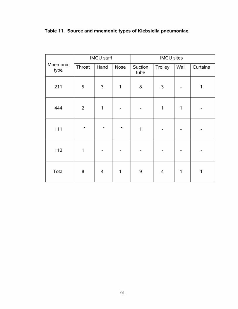
Table 11. Source and mnemonic types of Klebsiella pneumoniae.
Mnemonic type
IMCU staff IMCU sites
Throat Hand Nose Suction tube
Trolley Wall Curtains
211 5 3 1 8 3 - 1
444 2 1 - - 1 1 -
111 - - - 1 - - -
112 1 - - - - - -
Total 8 4 1 9 4 1 1
61
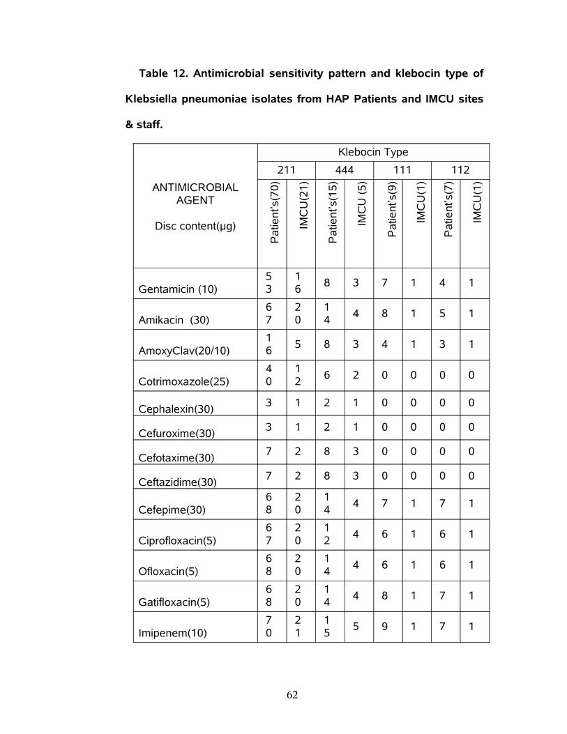
Table 12. Antimicrobial sensitivity pattern and klebocin type of
Klebsiella pneumoniae isolates from HAP Patients and IMCU sites
& staff.
ANTIMICROBIAL AGENT
Disc content(µg)
Klebocin Type
211 444 111 112
Pa
tien
t’s(7
0)
IMC
U(2
1)
Pa
tien
t’s(1
5)
IMC
U (
5)
Pa
tien
t’s(9
)
IMC
U(1
)
Pa
tien
t’s(7
)
IMC
U(1
)
Gentamicin (10)53
16
8 3 7 1 4 1
Amikacin (30)67
20
14
4 8 1 5 1
AmoxyClav(20/10)16
5 8 3 4 1 3 1
Cotrimoxazole(25)40
12
6 2 0 0 0 0
Cephalexin(30)3 1 2 1 0 0 0 0
Cefuroxime(30)3 1 2 1 0 0 0 0
Cefotaxime(30)7 2 8 3 0 0 0 0
Ceftazidime(30)7 2 8 3 0 0 0 0
Cefepime(30)68
20
14
4 7 1 7 1
Ciprofloxacin(5)67
20
12
4 6 1 6 1
Ofloxacin(5)68
20
14
4 6 1 6 1
Gatifloxacin(5)68
20
14
4 8 1 7 1
Imipenem(10)70
21
15
5 9 1 7 1
62
DISCUSSION
. The present study showed that the incidence of HAP was 10.3%
(n=253) out of 2454 cases admitted in IMCU, Coimbatore Medical College
Hospital over a period of one year.
This incidence was lower than the study by Mukhopadhyay et al
from Lucknow (53.9%), 18 Rakshit et al from Mumbai (47%), 13 Vincent et al
from Europe (46.9%), 25 Dey et al from Manipal (45.4%), 15 Sopena et al
from Spain (36.4%), 30 Berba et al from Philadelphia (28.2%) 33 and
Merchant et al from Mumbai (16.7%). 19 This was higher than the incidence
reported by Chevret et al from France (8.9%), 29 Alp et al from
Netherlands(6.8%), 32 Trivedi et al from Mumbai (9.38%)16 and Pawar et al
from New Delhi (2.6%). 14
It is possible that our incidence rate may be an over estimate of the
HAP in the hospital because of the nature of the clinical criteria used.
Studies based solely on clinical criteria alone are criticized because of the
non-specificity of parameters like fever, leukocytosis and infiltrates on the
chest radiographs. However, the stringent steps followed to make a
diagnosis of HAP in this study and the close monitoring before and after the
diagnosis of HAP occurred should make our estimate very close to the true
HAP incidence. It is unlikely that a true HAP case would have been missed
because we did quantitative culture of all specimens (BAL, Sputum and
ETA) to discriminate between the true pathogen and the contaminant using
the diagnostic threshold for each specimen.
63
High occurrence (55.73%) of HAP among the age group more than
60 years was observed in the present study. This could be due to the bulk of
the study population in this study was more 60 years of age group. This was
in accordance with an earlier study by Berba et al. 33 Age more than 60
years is one of the known risk factor for the development of HAP as reported
in previous studies. 1, 27 But Muhammad et al reported that highest incidence
was among 41 to 60 years of age group. 22
The present study showed that the incidence of HAP was high
among male patients than females. This finding was similar to the study by
Mukhopadhyay et al from Lucknow. 18 But Berba et al showed that the male
sex had a protective effect against the development of HAP. 33 Dey et al
reported that gender had no significant role in the development of HAP. 15
In the present study the incidence of HAP was greater in patients
with diseases requiring prolonged mechanical ventilation like OP poisoning
(15.0%) and considerably low in patients with diseases which presumably,
had unaffected lungs before admission to IMCU like snake bite (0.4%). These
findings were similar to the previous studies. 13, 15
HAP developed in 157 out of the 1352 patients (62.0%) receiving
mechanical ventilation but in only 96 out of 1102 patients (38.0%) with no
mechanical ventilation. This incidence of VAP (62.0%) was higher than the
study by Muhammad et al (30.7%) 22 and lower than the study by Alp et al
who showed that 75.5% of all patients with HAP were VAP. 32
64
Mechanical ventilation is a definitive risk factor for developing HAP
that has been shown previously by many studies 15, 18 and this study also
shows the significance of that risk factor causing HAP.
Klebsiella pneumoniae (48.2%) was the commonest organism
isolated in this study. Most of the previous studies reported Pseudomonas
aeruginosa as the commonest isolate from HAP patients in IMCU. 35, 14, 13, 18
But Pseudomonas aeruginosa was the second common organism in the
present study. Acinetobacter spp (7.7%) was the fourth common isolate in
this study. Dey et al, Rajasekhar et al and Alp et al reported that
Acinetobacter spp as the commonest organism in their study. 15, 23, 32 E. coli
was the commonest organism in the study by Tullu et al. 17 It was third
commonest organism in the present study. These findings indicate that the
causative pathogens always vary in different setups. The present study
suggests that the colonization rate for Klebsiella pneumoniae may be higher
in IMCU.
The rate of polymicrobial infection was found to be only 8.3% in this
study which was lower than the study by Mukhopadhyay et al (16.3%) 18 and
Singhal et al (12.3%). 83 The lower rate of colonization of IMCU environment
by more than one type of organisms may be the reason for the lower
incidence of polymicrobial infection.
65

Antimicrobial resistance among Gram negative bacilli is increasing
worldwide and is of particular concern in the Intensive Care Unit setting. A
direct correlation has been shown between resistance of Gram negative
bacilli and patient mortality, cost of patient care and length of stay in the
hospital. 76 In a study by Kaul et al about the Gram- negative bacterial
antibiotic susceptibility patterns in IMCU, Christian Medical College, Vellore
showed that Klebsiella resistance to cefotaxime and ceftazidime ranged from
25-50% and 14-91%, while E.coli resistance to these antibiotics ranged from
50-70% and 50-80% respectively. 81 In this study Klebsiella resistance to
cefotaxime and ceftazidime was 84% and E.coli resistance to these
antibiotics were 43% and 39% respectively. The resistance of K.pneumoniae
and E.coli to third generation cephalosporins was higher in this study.
All isolates of Acinetobacter, Serratia and Enterobactor were
sensitive to third generation cephalosporins. 92% of Pseudomonas
aeruginosa and 94% of Proteus were sensitive to cefotaxime and ceftazidime.
These findings were similar to the study by Kaul et al from Christian Medical
College, Vellore, who reported that in Pseudomonas aeruginosa and the
other non-fermenting gram-negative bacteria (NFGNB) Ceftazidime
resistance decreased. Among Aminoglycosides, most of the GNB were
sensitive to Amikacin than Gentamicin. Highest sensitivity rates were
detected for Gatifloxacin than Ciprofloxacin and Ofloxacin among Quinolones.
These findings were similar to previous studies on antimicrobial resistance
among gram-negative bacteria by many authors. 21, 22, 23, 39, 64
66

All Gram negative bacilli isolated in this study had a maximum
sensitivity pattern to Imipenem and Cefepime. This was similar to the study by
Lockhart et al about antimicrobial resistance among Gram-Negative Bacilli
causing infections in Intensive Care Unit patients in the United States
between 1993 and 2004 76 and Rakshit et al from Mumbai. 13
Gram-negative bacilli producing ESBL appear to be on the rise in
Asian countries and pose a serious problem in pulmonary infections. 80 In the
present study the occurrence of ESBL production among K.pneumoniae and
E.coli were 82% and 52% respectively. For Klebsiella pneumoniae this finding
was higher than the study by Feizabadi et al (72.8%), 74 Gonlugur et al
(12.2%), 39 Hosoglu et al (68.3%), 72 Asian HAP Working group (0.9% to
40%) 80 and lower than the study by Verhamme et al (100%), 79 Vitkauskiene
et al (88.9%), 84 Dey et al (100%). 15 For E.coli this finding was higher
than the study by Gonlugur et al (20.8%), 39 Asian HAP Working group (2.3%
to 40%) 80 and lower than the study by Hosoglu et al (74.6%), 72 Dey et al
(100%). 15
In this study ESBLs were predominantly present among K.
pneumoniae compared to E. coli. Our findings are similar to that of most of
the studies in Europe and USA 38 and the Indian studies by Jain et al 64 and
Shanmuganathan et al. 65 But studies by Gonlugur et al, 39 Hosoglu et al 72
and Kumar et al 82 showed higher incidence of ESBLs among E.coli than
K.pneumoniae. The indiscriminate use of third-generation cephalosporins has
been proposed as a reason for the rise of ESBL producing strains in India. 80
67
Among the Staphylococcus aureus isolates in this study, the
occurrence of Methicillin resistance was observed in 54.84%. This was
higher than the study by Leroy et al (33%) 35 and lower than the study by
Mukhopadhyay et al (59.45%) 18 and Rakshit et al (100%). 13 No Vancomycin
resistant Staphylococcus aureus was detected in the present study.
Epidemiological investigation of nosocomial infections is greatly
facilitated by the use of a precise typing system for the organism involved.
Our study about the hospital acquired pneumonia in IMCU suggests a high
prevalence of Klebsiella pneumoniae accounting for 48.2% of total cases. In
the present study, we have used Klebocin typing and antibiotic sensitivity
profiles as the typing methods for the detection of source of Klebsiella
infections in our IMCU. Although not comparable to molecular methods in
discriminatory power, antibiogram and klebocin typing are simpler and
relatively economical method of bacteriological typing.
In the present study all Klebsiella pneumoniae isolates were
subjected to klebocin typing by spot inoculation method as described by Pal
et al who done the klebocin typing in their study by spot inoculation method
which was described by Chugh et al with some modifications. Using the spot
inoculation method, we observed 87.5% typability, whereas Buffenmyer et
al, 61 Chibber et al, 60 Malik et al, 43 Aggarwal et al 44 and Pal et al 9 reported
67%, 72.8%, 83.3%, 73.5% and 71.62% typability respectively. Bauernfeind
et al ,62 Hall, 59 Israil 46 and Podschun et al 58 have reported 96.3%, 77%,
68
85% and 96% typability respectively using streak and point method for
klebocin typing.
Despite the relatively higher typability observed in streak and point
method, we followed the spot inoculation method. In view of the high
induction of klebocins by mitomycin C, storage stability at -20oC and simplicity
of the technique, spot inoculation method is more appropriate typing method.9
Nineteen klebocin types were observed in the present study using six
klebocin producer strains. Pal et al reported that twenty eight klebocin types
had been observed in their study using six klebocin producer strains 9
whereas Malik et al observed four patterns of klebocin types in their study
with six klebocin producer strains. 43
We observed the klebocin types 211 (56.8%), 111 (6.2%), 112
(5.0%) and 441 (3.7%) were the most common types in our study. According
to Buffenmyer et al and other previous studies type 444 was considered as
untypable and these strains accounted for 12.5% of the total strains studied.
Pal et al reported types 111 (50%), 211 (5.59%), 312 (3.08%) and 112
(2.31%) as the commonest klebocin types in their study and 28.37% strains
could not be typed. 9
Malik et al found type 312 (43.4%) as the commonest followed by
type 334 (23.4%), type 112 (16.6%) while remaining 16.6% strains could not
be typed.
69

Chibber et al reported the predominant klebocin types as 244
(14.3%), 313 (13.7%) and 113 (7.6%). 60 Aggarwal et al observed the
commonest klebocin types as 111, 313 and 113. Probably there is variation in
the prevalence of various klebocin types in different topographical areas and
institutions. Moreover the set of producer strains used in the present study
was different from other studies in which they had used a set of producer
strains standardized by them in their own setups. It is therefore difficult to
compare our typing patterns with other studies.
In the present study, we found that by combination of klebocin types
and antibiogram of K.pneumoniae isolates, the source of infection could be
traced in 76.51% cases. In a study on klebocin typing by Chibber et al 60 no
significant correlation was observed between the source of isolation and the
klebocin type whereas Pal et al 9 observed when the klebocin typing was
used in association with antibiogram, in 86.84% cases probable source of
infection could be detected.
Despite the importance of the findings presented in this study, there
are some limitations that should be taken into consideration when assessing
our conclusions. First, although for the diagnosis of HAP we did quantitative
culture of Sputum, BAL and ETA, yet the results from this study need to be
validated by comparison to gold standards such as histology of lung tissue.
70
Last, in the present study the reproducibility of the klebocin typing
was 83%. This was higher than the previous study by Malik et al who showed
in their study that the reproducibility of the klebocin typing was 73.3%. 43 This
is of major concern because a useful and effective epidemiological typing
system should have high reproducibility. In our study the reproducibility may
be a major problem with the klebocin typing procedure. This difficulty was
primarily due to the sensitivity of the test strains to MTCC 432 klebocin and
was presumably associated with their low titers. This problem was a serious
shortcoming since, as shown in Table 10 the commonest type in our study
was 211. This pattern of typing was due to lack of sensitivity of the clinical
isolates to MTCC 432 klebocin (producer 2). Replacing the producer strain of
this klebocin with the strain that yields high-titer preparations may help in
achieving acceptable reproducibility. Such strains may be obtained by
screening Klebsiella pneumoniae isolates for klebocin activity or testing
producer strains of other investigators.
In spite of these problems, this study proposed the klebocin typing in
association with antibiogram as a tool for epidemiological typing of Klebsiella
pneumoniae isolates in the laboratories where the facilities to perform
genomic based molecular typing are not available. The advantages of
klebocin typing include (i) the ready availability of required materials in most
bacteriological laboratories, (ii) the ability to perform the typing procedure
within the usual work day, and (iii) the ability to store the klebocins for future
use.
71
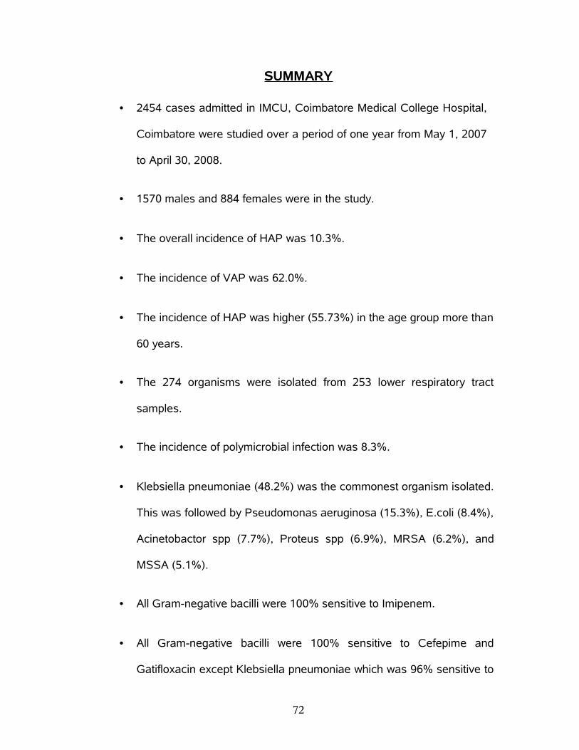
SUMMARY
• 2454 cases admitted in IMCU, Coimbatore Medical College Hospital,
Coimbatore were studied over a period of one year from May 1, 2007
to April 30, 2008.
• 1570 males and 884 females were in the study.
• The overall incidence of HAP was 10.3%.
• The incidence of VAP was 62.0%.
• The incidence of HAP was higher (55.73%) in the age group more than
60 years.
• The 274 organisms were isolated from 253 lower respiratory tract
samples.
• The incidence of polymicrobial infection was 8.3%.
• Klebsiella pneumoniae (48.2%) was the commonest organism isolated.
This was followed by Pseudomonas aeruginosa (15.3%), E.coli (8.4%),
Acinetobactor spp (7.7%), Proteus spp (6.9%), MRSA (6.2%), and
MSSA (5.1%).
• All Gram-negative bacilli were 100% sensitive to Imipenem.
• All Gram-negative bacilli were 100% sensitive to Cefepime and
Gatifloxacin except Klebsiella pneumoniae which was 96% sensitive to
72
Cefepime and Gatifloxacin, Pseudomonas aeruginosa which was 95%
sensitive to Gatifloxacin and E.coli which was 87% sensitive to
Gatifloxacin.
• 92% of Pseudomonas aeruginosa and 94% of Proteus were sensitive
to third generation cephalosporins.
• The screening for the ESBL producing GNB was done with
Cetazidime, Cephotaxime, Ceftriaxone, Cefpodoxime and Aztreonam
by Kirby Bauer’s disc diffusion method.
• The confirmation of ESBL production was done by Double disk
potentiation test and Double disk approximation test.
• 82% of K.pneumoniae and 52% of E.coli were extended spectrum
beta-lactamase producing strains.
• Staphylococcus aureus was 100% sensitive to Vancomycin.
• 54.84% of Staphylococcus aureus were Methicillin Resistant strains.
• Number of Klebsiella pneumoniae isolated from IMCU sites and Staff
was 28.
• Induction of klebocin production from six producer strains was done
with mitomycin C.
• Presence of klebocins was confirmed by SDS-PAGE.
73

• Klebocin typing was done for a total of 160 K.pneumoniae isolated
from HAP patients and IMCU sites & staff.
• Nineteen klebocin types were observed using six klebocin producer
strains.
• The typability of klebocin typing was 87.5% by the spot inoculation
method.
• 211 (56.8%) was the commonest klebocin type followed by 111 (6.2%),
112 (5.0%) and 441 (3.7%).
• The untypable strain (444) accounted for 12.5%.
• The reproducibility of the klebocin typing was 83%.
• The source of Klebsiella infection was detected in 86.84% of cases
using klebocin typing and antibiogram.
• The commonest source of Klebsiella infection in our IMCU were
suction tube (32%), throat of the IMCU staff (29 %), hands of the IMCU
staff (14%) and trolley (14%).
74

CONCLUSION
This one year prospective study on 2454 cases admitted in IMCU,
Coimbatore Medical College Hospital was conducted in order to study the
incidence, the Clinical and Bacteriological profile of HAP, the antibiotic
susceptibility pattern of the bacterial isolates, the role of Klebocin typing in
epidemiological typing of K.pneumoniae strains and the source of infection
in IMCU using antibiogram and klebocin typing.
The incidence of HAP was 10.3% with the occurrence being most in
the age group more than 60 years. Mechanical ventilation was a important
risk factor for the development of HAP. Klebsiella pneumoniae, Pseudomonas
aeruginosa, E.coli, Acinetobacter, Proteus spp, and MRSA were the most
common bacterial isolates.
The most effective antibiotics against the GNB isolated from HAP
patients were Imipenem followed by Cefepime and Gatifloxacin. The antibiotic
susceptibility pattern of the isolates will help the clinicians to choose the
appropriate antimicrobial agents for prophylactic as well as treatment
purposes.
When klebocin typing was used in association with antibiogram, in
86.84% cases of nosocomial Klebsiella infection probable source of infection
could be detected. Thus a combination of these two typing methods poses a
contribution in epidemiological studies.
75

RESULTS
During the one-year study period, among 2658 patients admitted to
the IMCU, only 2454 cases were followed and included in this study. The
remaining 204 cases were excluded. (118 died within 48hrs of admission, 86
were discharged or went home against advice).
Among 2454 patients, 64% of patients (n=1570) were males and
36% (n=884) were females. The mean age was 59.96 with the range of 15 to
89 yrs old. 35% of the patients were more than 60yrs of age. Out of 2454
cases, 253(10.3%) patients developed HAP. The highest incidence of HAP
(55.73%) was observed in the age group more than 60 years. (Ref table 5, 6)
The primary reason for IMCU admission was due to neurological
events (31.1%), cardiac and pulmonary emergencies (26.0%), acute
infections (12.%5), poisoning (5.3%), envenomation (0.5%), etc and the
incidence of HAP was greater in patients with diseases requiring prolonged
mechanical ventilation and in patients with those diseases that predispose to
pulmonary infection such as sepsis and prolonged stay in IMCU.
Out of 1352 patients on mechanical ventilation, 62.0% of patients
(n=157) developed HAP and only 38.0% of patients (n=96) developed HAP
out of 1102 non ventilated patients. (Ref table 7)
76

Totally 145 sputum samples, 70 BAL and 38 ETA were collected and
processed. Isolates in pure growth or mixture of two organisms at quantitative
threshold were considered as significant isolates. All 253 specimens in this
study showed significant growth of organisms. About 274 organisms were
isolated from 253 samples.
The commonest organism isolated was Klebsiella pneumoniae
(48.2%) followed by Pseudomonas aeruginosa (15.3%), E.coli (8.4%),
Acinetobactor spp (7.7%), Proteus spp (6.9%), MRSA (6.2%), MSSA (5.1%),
Serratia spp (0.7%), Enterobactor spp (0.7%), Strep.pneumoniae(0.4%) and
Candida albicans (0.4%). (Ref table 8)
Twenty one samples had showed mixed growth of two organisms
likewise Klebsiella pneumoniae and Pseudomonas aeruginosa in 15 cultures
(7 Sputum, 6 BAL & 2 ETA), Klebsiella pneumoniae and E.coli in 3 cultures (2
Sputum &1 BAL), Klebsiella pneumoniae and MRSA in 2 cultures (1 Sputum
& 1 BAL), Klebsiella pneumoniae and MSSA in 1 sputum culture.
Klebsiella pneumoniae, E.coli and P. aeruginosa had a maximum
sensitivity pattern to Imipenem followed by Cefepime, Gatifloxacin and
Amikacin. All Gram negative bacterial isolates had 100% sensitivity to
Imipenem. Among 132 K.pneumoniae isolates 82% (n=108) were found to be
ESBL producers. Among 23 E.coli isolates 52% (n=12) were ESBL
producers.
77

Staphylococcus aureus had a maximum sensitivity to Vancomycin
followed by third generation cephalosporins. No Vancomycin resistant
Staph.aureus was detected. 54.84% (n=17) of Staph.aureus were Methicillin
Resistant strains. (Ref table 9)
Klebocin typing was performed on a total of 160 K.pneumoniae
strains isolated from patients (n=132) and IMCU sites & IMCU staff members
(n=28). The overall typability of the strains was 87.5% and only 12.5% of
strains were untypable. Nineteen klebocin types were observed. The most
common mnemonic type found among patient’s isolates was 211 followed by
111, 112 and 441. (Ref table 10) Among the isolates from IMCU sites & staff
the common mnemonic type was 211. (Ref table 11)
The Inhibitory Dilution (ID) for all klebocin preparations was 1:256
except MTCC 432 klebocin preparations, for which the ID was 1:128.
With the exception of klebocin preparations from MTCC 432, the
reproducibility of klebocin typing was 83% even after four weeks of storage at
-20oC.
Klebocin typing and antibiogram of patient’s isolates was compared
with those from IMCU sites & staff, isolated during the period of infection. In
101 (76.51%) cases, the mnemonic types as well as antibiogram of Klebsiella
pneumoniae isolates from patients matched with those of IMCU sites and
staff. All the klebocin type 211 of both patient’s and environmental isolates
were 100% sensitive to Imipenem, 97% sensitive to Cefipime and
78
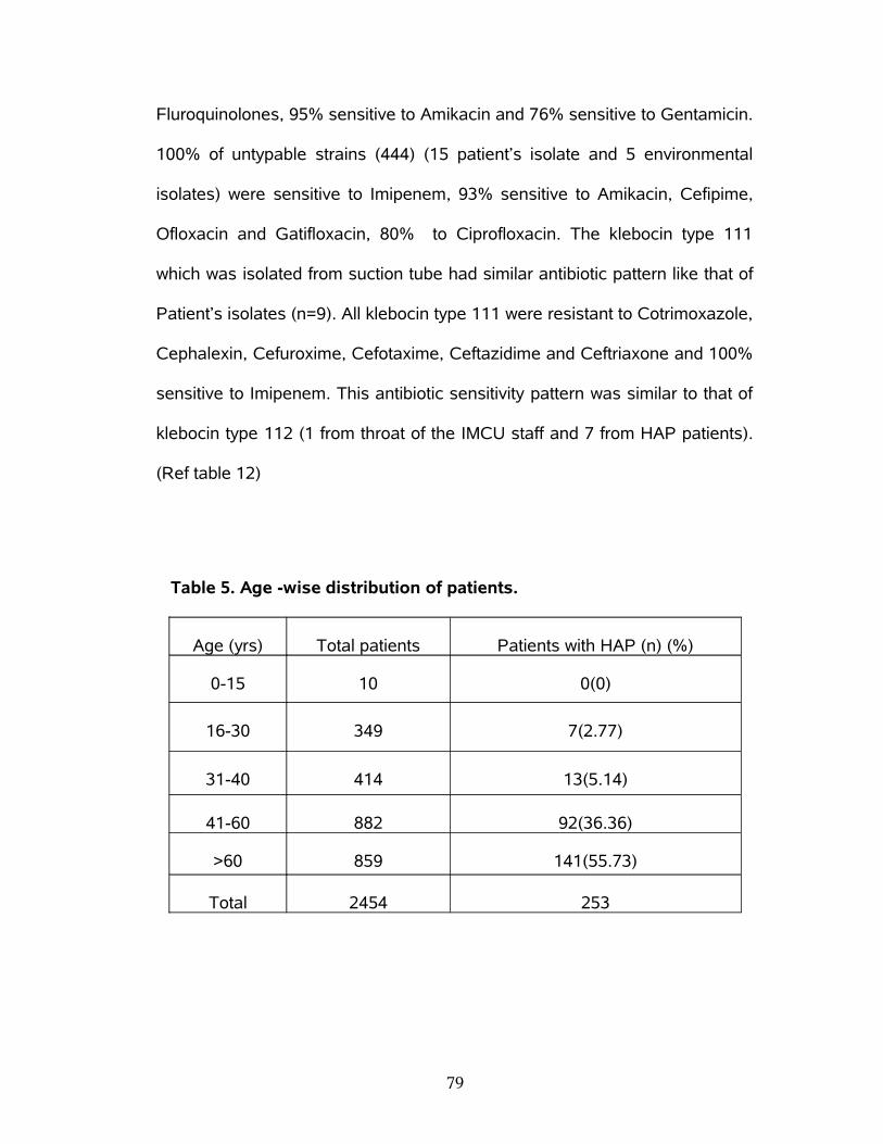
Fluroquinolones, 95% sensitive to Amikacin and 76% sensitive to Gentamicin.
100% of untypable strains (444) (15 patient’s isolate and 5 environmental
isolates) were sensitive to Imipenem, 93% sensitive to Amikacin, Cefipime,
Ofloxacin and Gatifloxacin, 80% to Ciprofloxacin. The klebocin type 111
which was isolated from suction tube had similar antibiotic pattern like that of
Patient’s isolates (n=9). All klebocin type 111 were resistant to Cotrimoxazole,
Cephalexin, Cefuroxime, Cefotaxime, Ceftazidime and Ceftriaxone and 100%
sensitive to Imipenem. This antibiotic sensitivity pattern was similar to that of
klebocin type 112 (1 from throat of the IMCU staff and 7 from HAP patients).
(Ref table 12)
Table 5. Age -wise distribution of patients.
Age (yrs) Total patients Patients with HAP (n) (%)
0-15 10 0(0)
16-30 349 7(2.77)
31-40 414 13(5.14)
41-60 882 92(36.36)
>60 859 141(55.73)
Total 2454 253
79
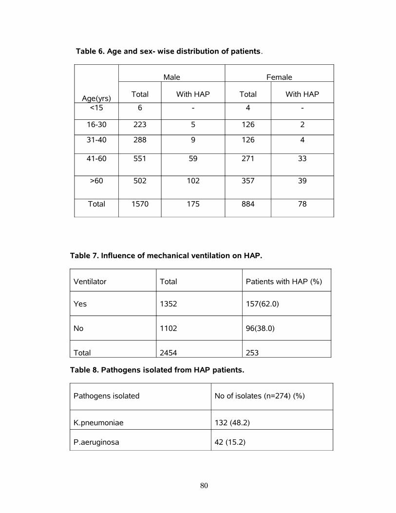
Table 6. Age and sex- wise distribution of patients.
Age(yrs)
Male Female
Total With HAP Total With HAP
<15 6 - 4 -
16-30 223 5 126 2
31-40 288 9 126 4
41-60 551 59 271 33
>60 502 102 357 39
Total 1570 175 884 78
Table 7. Influence of mechanical ventilation on HAP.
Ventilator Total Patients with HAP (%)
Yes 1352 157(62.0)
No 1102 96(38.0)
Total 2454 253
Table 8. Pathogens isolated from HAP patients.
Pathogens isolated No of isolates (n=274) (%)
K.pneumoniae 132 (48.2)
P.aeruginosa 42 (15.2)
80
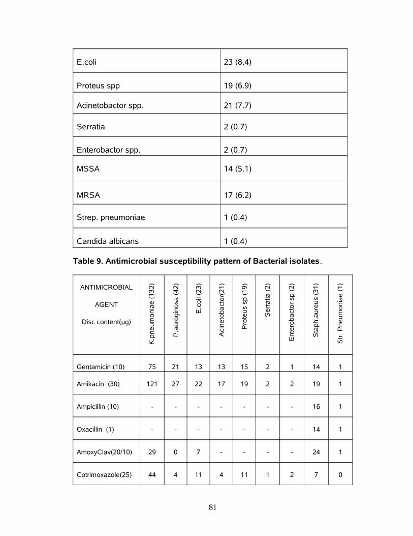
E.coli 23 (8.4)
Proteus spp 19 (6.9)
Acinetobactor spp. 21 (7.7)
Serratia 2 (0.7)
Enterobactor spp. 2 (0.7)
MSSA 14 (5.1)
MRSA 17 (6.2)
Strep. pneumoniae 1 (0.4)
Candida albicans 1 (0.4)
Table 9. Antimicrobial susceptibility pattern of Bacterial isolates.
ANTIMICROBIAL
AGENT
Disc content(µg)
K.p
neu
mo
nia
e (
132
)
P.a
ero
gin
osa
(4
2)
E.c
oli
(23
)
Aci
neto
ba
cto
r(2
1)
Pro
teu
s sp
(1
9)
Se
rra
tia (
2)
Ent
ero
ba
cto
r sp
(2
)
Sta
ph.a
ure
us
(31
)
Str
. P
neu
mo
nia
e (
1)
Gentamicin (10) 75 21 13 13 15 2 1 14 1
Amikacin (30) 121 27 22 17 19 2 2 19 1
Ampicillin (10) - - - - - - - 16 1
Oxacillin (1) - - - - - - - 14 1
AmoxyClav(20/10) 29 0 7 - - - - 24 1
Cotrimoxazole(25) 44 4 11 4 11 1 2 7 0
81
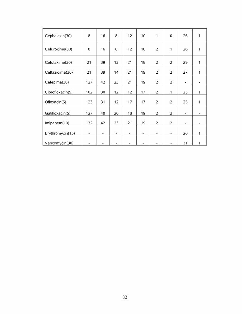
Cephalexin(30) 8 16 8 12 10 1 0 26 1
Cefuroxime(30) 8 16 8 12 10 2 1 26 1
Cefotaxime(30) 21 39 13 21 18 2 2 29 1
Ceftazidime(30) 21 39 14 21 19 2 2 27 1
Cefepime(30) 127 42 23 21 19 2 2 - -
Ciprofloxacin(5) 102 30 12 12 17 2 1 23 1
Ofloxacin(5) 123 31 12 17 17 2 2 25 1
Gatifloxacin(5) 127 40 20 18 19 2 2 - -
Imipenem(10) 132 42 23 21 19 2 2 - -
Erythromycin(15) - - - - - - - 26 1
Vancomycin(30) - - - - - - - 31 1
82

Table 10. Mnemonic types of Klebsiella pneumoniae isolates.
Mnemonic types Patient’s strains IMCU sites
IMCU Staff
Total no. of strains(%)
211 70 12 9 91(56.8%)
444 15 2 3 20(12.5%)
111 9 1 − 10(6.2%)
112 7 − 1 8(5.0%)
441 6 − − 6(3.7%)
311 4 − − 4(2.5%)
241 3 − − 3(1.8%)
314 3 − − 3(1.8%)
414 2 − − 2(1.2%)
341 2 − − 2(1.2%)
243 2 − − 2(1.2%)
312 2 − − 2(1.2%)
221 1 − − 1(0.6%)
231 1 − − 1(0.6%)
141 1 − − 1(0.6%)
332 1 − − 1(0.6%)
411 1 − − 1(0.6%)
412 1 − − 1(0.6%)
244 1 − − 1(0.6%)
Total no. of strains 132 15 13 160
Typability 88.64% 86.66% 76.92% 87.5%
8

Table 11. Source and mnemonic types of Klebsiella pneumoniae.
Mnemonic type
IMCU staff IMCU sites
Throat Hand Nose Suction tube
Trolley Wall Curtains
211 5 3 1 8 3 - 1
444 2 1 - - 1 1 -
111 - - - 1 - - -
112 1 - - - - - -
Total 8 4 1 9 4 1 1
8

Table 12. Antimicrobial sensitivity pattern and klebocin type of
Klebsiella pneumoniae isolates from HAP Patients and IMCU sites
& staff.
ANTIMICROBIAL AGENT
Disc content(µg)
Klebocin Type
211 444 111 112
Pa
tien
t’s(7
0)
IMC
U(2
1)
Pa
tien
t’s(1
5)
IMC
U (
5)
Pa
tien
t’s(9
)
IMC
U(1
)
Pa
tien
t’s(7
)
IMC
U(1
)
Gentamicin (10)53
16
8 3 7 1 4 1
Amikacin (30)67
20
14
4 8 1 5 1
AmoxyClav(20/10)16
5 8 3 4 1 3 1
Cotrimoxazole(25)40
12
6 2 0 0 0 0
Cephalexin(30)3 1 2 1 0 0 0 0
Cefuroxime(30)3 1 2 1 0 0 0 0
Cefotaxime(30)7 2 8 3 0 0 0 0
Ceftazidime(30)7 2 8 3 0 0 0 0
Cefepime(30)68
20
14
4 7 1 7 1
Ciprofloxacin(5)67
20
12
4 6 1 6 1
Ofloxacin(5)68
20
14
4 6 1 6 1
Gatifloxacin(5)68
20
14
4 8 1 7 1
8

Imipenem(10)70
21
15
5 9 1 7 1
8

DISCUSSION
. The present study showed that the incidence of HAP was 10.3%
(n=253) out of 2454 cases admitted in IMCU, Coimbatore Medical College
Hospital over a period of one year.
This incidence was lower than the study by Mukhopadhyay et al
from Lucknow (53.9%), 18 Rakshit et al from Mumbai (47%), 13 Vincent et al
from Europe (46.9%), 25 Dey et al from Manipal (45.4%), 15 Sopena et al
from Spain (36.4%), 30 Berba et al from Philadelphia (28.2%) 33 and
Merchant et al from Mumbai (16.7%). 19 This was higher than the incidence
reported by Chevret et al from France (8.9%), 29 Alp et al from
Netherlands(6.8%), 32 Trivedi et al from Mumbai (9.38%)16 and Pawar et al
from New Delhi (2.6%). 14
It is possible that our incidence rate may be an over estimate of the
HAP in the hospital because of the nature of the clinical criteria used.
Studies based solely on clinical criteria alone are criticized because of the
non-specificity of parameters like fever, leukocytosis and infiltrates on the
chest radiographs. However, the stringent steps followed to make a
diagnosis of HAP in this study and the close monitoring before and after the
diagnosis of HAP occurred should make our estimate very close to the true
HAP incidence. It is unlikely that a true HAP case would have been missed
because we did quantitative culture of all specimens (BAL, Sputum and
8

ETA) to discriminate between the true pathogen and the contaminant using
the diagnostic threshold for each specimen.
High occurrence (55.73%) of HAP among the age group more than
60 years was observed in the present study. This could be due to the bulk of
the study population in this study was more 60 years of age group. This was
in accordance with an earlier study by Berba et al. 33 Age more than 60
years is one of the known risk factor for the development of HAP as reported
in previous studies. 1, 27 But Muhammad et al reported that highest incidence
was among 41 to 60 years of age group. 22
The present study showed that the incidence of HAP was high
among male patients than females. This finding was similar to the study by
Mukhopadhyay et al from Lucknow. 18 But Berba et al showed that the male
sex had a protective effect against the development of HAP. 33 Dey et al
reported that gender had no significant role in the development of HAP. 15
In the present study the incidence of HAP was greater in patients
with diseases requiring prolonged mechanical ventilation like OP poisoning
(15.0%) and considerably low in patients with diseases which presumably,
had unaffected lungs before admission to IMCU like snake bite (0.4%). These
findings were similar to the previous studies. 13, 15
HAP developed in 157 out of the 1352 patients (62.0%) receiving
mechanical ventilation but in only 96 out of 1102 patients (38.0%) with no
mechanical ventilation. This incidence of VAP (62.0%) was higher than the
8

study by Muhammad et al (30.7%) 22 and lower than the study by Alp et al
who showed that 75.5% of all patients with HAP were VAP. 32
Mechanical ventilation is a definitive risk factor for developing HAP
that has been shown previously by many studies 15, 18 and this study also
shows the significance of that risk factor causing HAP.
Klebsiella pneumoniae (48.2%) was the commonest organism
isolated in this study. Most of the previous studies reported Pseudomonas
aeruginosa as the commonest isolate from HAP patients in IMCU. 35, 14, 13, 18
But Pseudomonas aeruginosa was the second common organism in the
present study. Acinetobacter spp (7.7%) was the fourth common isolate in
this study. Dey et al, Rajasekhar et al and Alp et al reported that
Acinetobacter spp as the commonest organism in their study. 15, 23, 32 E. coli
was the commonest organism in the study by Tullu et al. 17 It was third
commonest organism in the present study. These findings indicate that the
causative pathogens always vary in different setups. The present study
suggests that the colonization rate for Klebsiella pneumoniae may be higher
in IMCU.
The rate of polymicrobial infection was found to be only 8.3% in this
study which was lower than the study by Mukhopadhyay et al (16.3%) 18 and
Singhal et al (12.3%). 83 The lower rate of colonization of IMCU environment
8
by more than one type of organisms may be the reason for the lower
incidence of polymicrobial infection.
Antimicrobial resistance among Gram negative bacilli is increasing
worldwide and is of particular concern in the Intensive Care Unit setting. A
direct correlation has been shown between resistance of Gram negative
bacilli and patient mortality, cost of patient care and length of stay in the
hospital. 76 In a study by Kaul et al about the Gram- negative bacterial
antibiotic susceptibility patterns in IMCU, Christian Medical College, Vellore
showed that Klebsiella resistance to cefotaxime and ceftazidime ranged from
25-50% and 14-91%, while E.coli resistance to these antibiotics ranged from
50-70% and 50-80% respectively. 81 In this study Klebsiella resistance to
cefotaxime and ceftazidime was 84% and E.coli resistance to these
antibiotics were 43% and 39% respectively. The resistance of K.pneumoniae
and E.coli to third generation cephalosporins was higher in this study.
All isolates of Acinetobacter, Serratia and Enterobactor were
sensitive to third generation cephalosporins. 92% of Pseudomonas
aeruginosa and 94% of Proteus were sensitive to cefotaxime and ceftazidime.
These findings were similar to the study by Kaul et al from Christian Medical
College, Vellore, who reported that in Pseudomonas aeruginosa and the
other non-fermenting gram-negative bacteria (NFGNB) Ceftazidime
resistance decreased. Among Aminoglycosides, most of the GNB were
9
sensitive to Amikacin than Gentamicin. Highest sensitivity rates were
detected for Gatifloxacin than Ciprofloxacin and Ofloxacin among Quinolones.
These findings were similar to previous studies on antimicrobial resistance
among gram-negative bacteria by many authors. 21, 22, 23, 39, 64
All Gram negative bacilli isolated in this study had a maximum
sensitivity pattern to Imipenem and Cefepime. This was similar to the study by
Lockhart et al about antimicrobial resistance among Gram-Negative Bacilli
causing infections in Intensive Care Unit patients in the United States
between 1993 and 2004 76 and Rakshit et al from Mumbai. 13
Gram-negative bacilli producing ESBL appear to be on the rise in
Asian countries and pose a serious problem in pulmonary infections. 80 In the
present study the occurrence of ESBL production among K.pneumoniae and
E.coli were 82% and 52% respectively. For Klebsiella pneumoniae this finding
was higher than the study by Feizabadi et al (72.8%), 74 Gonlugur et al
(12.2%), 39 Hosoglu et al (68.3%), 72 Asian HAP Working group (0.9% to
40%) 80 and lower than the study by Verhamme et al (100%), 79 Vitkauskiene
et al (88.9%), 84 Dey et al (100%). 15 For E.coli this finding was higher
than the study by Gonlugur et al (20.8%), 39 Asian HAP Working group (2.3%
to 40%) 80 and lower than the study by Hosoglu et al (74.6%), 72 Dey et al
(100%). 15
In this study ESBLs were predominantly present among K.
pneumoniae compared to E. coli. Our findings are similar to that of most of
9

the studies in Europe and USA 38 and the Indian studies by Jain et al 64 and
Shanmuganathan et al. 65 But studies by Gonlugur et al, 39 Hosoglu et al 72
and Kumar et al 82 showed higher incidence of ESBLs among E.coli than
K.pneumoniae. The indiscriminate use of third-generation cephalosporins has
been proposed as a reason for the rise of ESBL producing strains in India. 80
Among the Staphylococcus aureus isolates in this study, the
occurrence of Methicillin resistance was observed in 54.84%. This was
higher than the study by Leroy et al (33%) 35 and lower than the study by
Mukhopadhyay et al (59.45%) 18 and Rakshit et al (100%). 13 No Vancomycin
resistant Staphylococcus aureus was detected in the present study.
Epidemiological investigation of nosocomial infections is greatly
facilitated by the use of a precise typing system for the organism involved.
Our study about the hospital acquired pneumonia in IMCU suggests a high
prevalence of Klebsiella pneumoniae accounting for 48.2% of total cases. In
the present study, we have used Klebocin typing and antibiotic sensitivity
profiles as the typing methods for the detection of source of Klebsiella
infections in our IMCU. Although not comparable to molecular methods in
discriminatory power, antibiogram and klebocin typing are simpler and
relatively economical method of bacteriological typing.
In the present study all Klebsiella pneumoniae isolates were
subjected to klebocin typing by spot inoculation method as described by Pal
et al who done the klebocin typing in their study by spot inoculation method
9
which was described by Chugh et al with some modifications. Using the spot
inoculation method, we observed 87.5% typability, whereas Buffenmyer et
al, 61 Chibber et al, 60 Malik et al, 43 Aggarwal et al 44 and Pal et al 9 reported
67%, 72.8%, 83.3%, 73.5% and 71.62% typability respectively. Bauernfeind
et al ,62 Hall, 59 Israil 46 and Podschun et al 58 have reported 96.3%, 77%,
85% and 96% typability respectively using streak and point method for
klebocin typing.
Despite the relatively higher typability observed in streak and point
method, we followed the spot inoculation method. In view of the high
induction of klebocins by mitomycin C, storage stability at -20oC and simplicity
of the technique, spot inoculation method is more appropriate typing method.9
Nineteen klebocin types were observed in the present study using six
klebocin producer strains. Pal et al reported that twenty eight klebocin types
had been observed in their study using six klebocin producer strains 9
whereas Malik et al observed four patterns of klebocin types in their study
with six klebocin producer strains. 43
We observed the klebocin types 211 (56.8%), 111 (6.2%), 112
(5.0%) and 441 (3.7%) were the most common types in our study. According
to Buffenmyer et al and other previous studies type 444 was considered as
untypable and these strains accounted for 12.5% of the total strains studied.
Pal et al reported types 111 (50%), 211 (5.59%), 312 (3.08%) and 112
9

(2.31%) as the commonest klebocin types in their study and 28.37% strains
could not be typed. 9
Malik et al found type 312 (43.4%) as the commonest followed by
type 334 (23.4%), type 112 (16.6%) while remaining 16.6% strains could not
be typed.
Chibber et al reported the predominant klebocin types as 244
(14.3%), 313 (13.7%) and 113 (7.6%). 60 Aggarwal et al observed the
commonest klebocin types as 111, 313 and 113. Probably there is variation in
the prevalence of various klebocin types in different topographical areas and
institutions. Moreover the set of producer strains used in the present study
was different from other studies in which they had used a set of producer
strains standardized by them in their own setups. It is therefore difficult to
compare our typing patterns with other studies.
In the present study, we found that by combination of klebocin types
and antibiogram of K.pneumoniae isolates, the source of infection could be
traced in 76.51% cases. In a study on klebocin typing by Chibber et al 60 no
significant correlation was observed between the source of isolation and the
klebocin type whereas Pal et al 9 observed when the klebocin typing was
used in association with antibiogram, in 86.84% cases probable source of
infection could be detected.
Despite the importance of the findings presented in this study, there
are some limitations that should be taken into consideration when assessing
9
our conclusions. First, although for the diagnosis of HAP we did quantitative
culture of Sputum, BAL and ETA, yet the results from this study need to be
validated by comparison to gold standards such as histology of lung tissue.
Last, in the present study the reproducibility of the klebocin typing
was 83%. This was higher than the previous study by Malik et al who showed
in their study that the reproducibility of the klebocin typing was 73.3%. 43 This
is of major concern because a useful and effective epidemiological typing
system should have high reproducibility. In our study the reproducibility may
be a major problem with the klebocin typing procedure. This difficulty was
primarily due to the sensitivity of the test strains to MTCC 432 klebocin and
was presumably associated with their low titers. This problem was a serious
shortcoming since, as shown in Table 10 the commonest type in our study
was 211. This pattern of typing was due to lack of sensitivity of the clinical
isolates to MTCC 432 klebocin (producer 2). Replacing the producer strain of
this klebocin with the strain that yields high-titer preparations may help in
achieving acceptable reproducibility. Such strains may be obtained by
screening Klebsiella pneumoniae isolates for klebocin activity or testing
producer strains of other investigators.
In spite of these problems, this study proposed the klebocin typing in
association with antibiogram as a tool for epidemiological typing of Klebsiella
pneumoniae isolates in the laboratories where the facilities to perform
9
genomic based molecular typing are not available. The advantages of
klebocin typing include (i) the ready availability of required materials in most
bacteriological laboratories, (ii) the ability to perform the typing procedure
within the usual work day, and (iii) the ability to store the klebocins for future
use.
9

REFERENCES
11. Hunter JD. Ventilator Associated Pneumonia. J Postgrad Med 2006; 82: 172-178.
12. Hoffken G, Niederman MS. Nosocomial pneumonia; the importance of a de-escalating strategy for antibiotic treatment of pneumonia in the ICU. Chest 2002; 122:2183-2196.
13. Thomas M File, Jr, MD. Hospital-acquired (nosocomial) pneumonia in adults. Up To Date. Verson 15.1: September 29, 2006.
14.Kimberly A. Davis. Ventilator-Associated pneumonia: A Review. J Intensive Care Med. 2006; 21:211-226.
15. Robert C Read. Bacterial infections of the lower respiratory tract. Topley & Wilson’s. 10th edition, volume-1.2005:640.
16. Porzecanski I, Bowton DL. Diagnosis and treatment of ventilator associated pneumonia. Chest 2006; 130: 597-604.
17.Podschun R, Ullmann U. Klebsiella spp. as Nosocomial Pathogens: Epidemiology, Taxonomy, Typing Methods, and Pathogenicity factors. J. Clin. Microbiol. 1998; 11:589-603.
18. Parker MT. HAI: Guidelines to laboratory methods, WHO Regional Publication European series No.4. P.35, 1978, WHO, Copenhagen.
19.Pal RB, Rebello JS, Banker DD, Jain PK. Klebocin Typing of Klebsiella species isolated from nosocomial infections in Intensive Care Unit. J Postgrad Med. 1997; 43: 98-101.
20.Mehlhorn A, Anderson J, Balcer H. Review of Hospital-Acquired, Ventilator-Associated, and Health Care-Associated Pneumonia in Immunocompetent Adults. US Pharm. 2006; 7: HS-30-36.
21. American Thoracic Society; Infectious Diseases Society of America. Guidelines for the Management of Adults with Hospital Acquired, Ventilator Associated and Health Care Associated Pneumonia. Am J Respir Crit Care Med 2005; 171:388-416.
22.Sundaram R, Infirmary V, Glasgow. Nosocomial Pneumonia. Anaesthesia UK. Oct.2006.
9

23. Rakshit P, Nagar VS, DeshPande AK. Incidence, Clinical outcome and risk stratification of ventilator-Associated Pneumonia- a Prospective cohort study. Indian J Crit Care Med 2005; 9: 211-216.
24.Pawar M, Mehta Y, Trehan N, Kulkarni V. Ventilator-Associated Pneumonia: Incidence, risk factors, outcome, and microbiology. J Cardio thorac Vasc. 2003; 17:22-28.
25.Dey A, Bairy I. Incidence of multi drug-resistant organisms causing Ventilator-Associated pneumonia in a tertiary care hospital: A nine month’s prospective study. Ann Thorac Med 2007; 2:52-57.
26.Trivedi TH, Shejale SB, Yeolekar ME. Nosocomial Pneumonia in medical Intensive Care Unit. J Assoc Physicians India.2000 Nov; 48(11): 1070-3.
27.Tullu MS, Deshmukh CT, Baveja SM. Bacterial Nosocomial Pneumonia in Paediatric Intensive Care Unit. J Postgrad Med 2000; 46:18-22.
28. Mukhopadhyay C, Anudida B, Ayyagari A. Role of mechanical ventilation & development of multi drug resistant organisms in hospital acquired pneumonia. Indian J Med Res.2003 Dec; 118:229-235.
29. Merchant M, Karnad DR, Kanbur AA. Incidence of nosocomial pneumonia in a Medical Intensive Care Unit and general medical ward patients in a public hospital in Bombay, India. J Hosp Infect. 1998 Jun; 39(2):143-8.
30.Cunha BA: Nosocomial Pneumonia. Up To Date 2006, May.
31.Joseph P. Lynch III. Hospital-Acquired Pneumonia: Risk Factors, Microbiology, and Treatment. Chest 2001; 119: 373-384.
32. Muhammad F.R, Yasmin H, Menon AR, et al. Pattern of Nosocomial Infection in two Intensive Care Unit of a tertiary care hospital in Karachi. JC PSP 2007; Vol. 17(3): 136-139.
33. Rajasekhar T, Anuradha K, Suhasini T, Lakshmi V. The role of quantitative cultures of non-bronchoscopic samples in Ventilator-Associated Pneumonia. Indian J Med Microbiol 2006; 24:107-113.
34. Mehta RM, Niederman MS. Nosocomial Pneumonia in the Intensive Care Unit: Controversies and Dilemmas. J Intensive Care Med.2003; Vol.18, No.4, 175-188.
35. Vincent JL, Bihari DJ, Suter PM, et al. The Prevalence of nosocomial infection in Intensive Care Units in Europe: Results of the European Prevalence of Infection in Intensive Care (EPIC) study. EPIC International Advisory Committee. JAMA 1995; 274: 639-644.
9

36. Heyland DK, Cook DJ, Griffith L, et al. The Attributable Morbidity and Mortality of Ventilator-Associated Pneumonia in the Critically Ill Patient. Am. J. Respir. Crit. Care Med., 1999 April; 159(4): 1249-1256.
37.Celis R, Torres A, Gatell JM, et al. Nosocomial Pneumonia: A Multivariate analysis of risk and prognosis. Chest 1988; 93:318-324.
38. Chastre J, Fagon JY. Ventilator-Associated Pneumonia. Am J Respir Crit Care Med 2002; 165:867-903.
39.Chevret S, Hemmer M, Carlet J, Langer M. Incidence and risk factors of pneumonia acquired in Intensive Care Units. Results from a multicentre prospective study on 996 patients. European Cooperative Group on Nosocomial Pneumonia. Intensive Care Med 1993; 19 (5):256-64.
40.Sopena N, Sabira M. Multicentre study of hospital acquired pneumonia in non-ICU Patients. Chest 2005 Jan; 127(1): 213-19.
41.John Roark. Fighting Nosocomial Pneumonia. Infection Control Today. 05/2003.
42.Alp E, Guven M, Yildiz O, et al. Incidence, risk factors and mortality of nosocomial pneumonia in Intensive Care Units: A Prospective study. Ann Clin Microbiol Antimicrobials 2004, 3:17.
43. Berba R, Alejandria M,Rosacos J, et al. Incidence, Risk factors and Outcome of Hospital Acquired Pneumonia in Critically Ill Patients at the Philippine General Hospital. Phil J Microbiol Infect Dis1999; 28(2):29-38.
44. Centers for Disease Control and Prevention: Guidelines for Prevention of Nosocomial Pneumonia. MMWR Recomm Rep 1997,46(RR-1):1-79.
45.Leroy O, Giradie P, Yazdanpanah Y, et al. Hospital Acquired Pneumonia: microbiological data and potential adequacy of anti microbial regimens. Eur Respir J 2002; 20: 432-439.
46.Merck Mannual. Hospital-Acquired Pneumonia. Nov 2005.47.Emori TG, Gaynes RP. An overview of nosocomial infections, including
the role of the microbiology laboratory. Clin Microbiol Rev. Oct 1993; 6(4): 428-442.
48.Jacoby GA, Mediros AA. More extended-spectrum beta-lactamases. Antimicrob Agents Chemother 1991; 35:1697-704.
9

49.Gonlugur U, Bakici MZ, Akkurt I, Efeoglu T. Antibiotic susceptibility patterns among respiratory isolates of Gram Negative Becilli in a Turkish University hospital. BMC Microbiol. 2004; 4:32.
50. Chaudhary U, Aggarwal R. Extended Spectrum β-Lactamases (ESBL) - An emerging threat to clinical therapeutics. Indian J Med Microbiol 2004; 22(2): 75-80.
51. Oncul O. Antimicrobial resistance pattern of Extented Spectrum β-Lactamase producing Klebsiella spp. Indian J Med Res 2007; 125:699-700.
52. Winn WC, Allen SD, Koneman EW, et al. Taxonomy of the Enterobacteriaceae. Koneman’s Colour Atlas and Textbook of Diagnostic Microbiology. Sixth Edition 2006.
53.Malik A, Hesani SE, Shahid M, Khan HM, Ahmad AJ. Nosocomial Klebsiella infection in neonates in a tertiary care hospital: Protein profile by SDS- PAGE and klebocin typing as epidemiological markers. Indian J Med Microbiol 2003; 21(2): 82-86.
54.Aggarwal A, Khanna S, Arora U. Characterization, biotyping, antibiogram and klebocin typing of Klebsiella with special reference to Klebsiella oxytoca. Indian J Med Sci 2003; 57(2): 68-70.
55.Bauernfeind A, Petermuller C, Schneider R. Bacteriocins as tools in analysis of Nosocomial Klebsiella pneumoniae infections. J Clin Microbiol 1981; 14(1):15-19.
56. Israil AM. The significance of bacteriocin typing of Klebsiella strains. Zentralbl Bakteriol Mikrobiol Hyg[A]. 1981; 250(1-2): 63-71.
57.Rubin SJ. Klebsiella marker systems. Infect Control. 1985 Feb; 6(2): 59-63.
58.Aber RC, Mackel DC. Epidemiological typing of nosocomial microorganisms. Am J Med. 1981 Apr; 70(4): 899-905.
59. Edmondson AS, Cooke EM. The development and assessment of a bacteriocin typing method for Klebsiella. J Hyg (Lond). 1979 Apr; 82(2): 207-23.
60.Farmer JJ. Conventional typing methods. J Hosp Infect. 1988 Feb; 11(A): 309-14.
1

61.Cooke EM, Brayson JC, Edmondson AS, Hall D. An investigation into the incidence and sources of Klebsiella infections in hospital patients. J Hyg (Lond). 1979 Jun; 82(2): 473-80.
62. Israil AM. Studies concerning the sensitivity of Klebsiella strains to a set of Klebsiella bacteriocins. Zentralbl Bakteriol A. 1980; 248(1): 91-8.
63.Podschun R, Ullman U. Bacteriocin typing of environmental Klebsiella isolates. Zentralbl Hyg Umweltmed. 1993 Nov; 195(1): 22-26.
64. Chhibber S, Vedehra DV. Effect of various inducing agents on bacteriocin (klebocin) production by Klebsiella pneumoniae. Folia Microbiol (Praha). 1987; 32(2): 137-41.
65.Chhibber S, Vedehra DV. Purification and characterization of a bacteriocin from Klebsiella pneumoniae 158. J Gen Microbiol. 1986 Apr; 132(4): 1051-4.
66.James R. Molecular cloning and purification of klebicin B. J Gen Microbiol. 1988 Sep; 134(9): 2525-33.
67. Pokhil SI, Kosobutskii LA, Ovcharenko BM, et al. The bacteriocin typing of Klebsiella pneumoniae strains. Mikrobiol Zh. 1991 Nov-Dec; 53(6): 88-93.
68.Podschun R, Ullman U. Bacteriocin typing of Klebsiella spp. Isolated from different sources. Zentralbl Hyg Umweltmed. 1996 Feb; 198(3): 258-64.
69.Hall FA. Bacteriocin typing of Klebsiella spp. J Clin Path. 1971; 24: 712-716.
70. Chhibber S, Goel A, Kapoor N, Saxena M, Vadehra DV. Bacteriocin (klebocin) typing of clinical isolates of Klebsiella pneumoniae. Euro J Epidemiol 1988; 4(1):115-118
71. Buffenmyer CL, Rycheck RR, Yee RB. Bacteriocin (klebocin) sensitivity typing of Klebsiella. J Clin Microbiol 1976; 4(3):239-244.
72.Bauerfeind A, Petermuller C. Typing of Enterobacter spp. By bateriocin susceptibility and its use in epidemiological analysis.J Clin Microbiol 1984; 20(1): 70-73.
73. Dykes GA, Hastings JW. Selection and fitness in Bacteriocin- producing bacteria. Proc R Soc Lond B1997; 264:683-687.
74. Jain A, Mondal R. Prevalence and antimicrobial resistance pattern of extended spectrum β- lactamase producing Klebsiella spp isolated from cases of neonatal septicaemia. Indian J Med Res 2007; 125:89-94.
1

75.Shanmuganathan C et al. Learning from an outbreak: ESBL- the essential points. Indian J Med Microbiol 2004; 22(4):255-257.
76. Forbes BA, Sahm DF, Weissfeld AS. Bailey & Scott’s Diagnostic Microbiology. 12th edi. 2007.
77.Baselski VS, Wunderink RG. Bronchoscopic diagnosis of Pneumonia. Clin Microbiol Rev. 1994; 7.4: 533-558.
78. Collee JG, Fraser AG, Marmion BP, Simmons A. Mackie & McCartney practical medical microbiology. 14th edi.2006.
79. Tulsi, Chugh D, Siham AG, et al. Colonization and infections of neonates by Klebsiella pneumoniae in an intensive care unit.
80. Jacoby GA, Han P. Detection of Extended Spectrum β-Lactamases in clinical isolates of Klebsiella pneumoniae and Escherichia coli. J Clin Microbiol.1996; 34.4: 908-911.
81. Vercauteren E, Descheemaeker P, Ieven M, Sanders CC, Goosens H. Comparison of screening methods for detection of Extended Spectrum β-Lactamases and their prevalence among blood isolates of Escherichia coli and Klebsiella spp. in a Belgian Teaching Hospital. J Clin Microbiol 1997; 35(9):2191-2197.
82. Hosoglu S, Gundes S, Kolayli F, et al. Extended spectrum beta- lactamases in ceftazidime- resistant E.coli and Klebsiella pneumoniae isolates in Turkish hospitals. Indian J Med Microbiol 2007; 25:346-50.
83. Rodrigues C, Joshi P, Jani SH, Alphonse M, Radhakrishnan R, Mehta A. Detection of –Lactamases in nosocomial gram negative clinical isolates. Indian J Med Microbiol 2004; 22: 247-50.
84. Feizabadi M, Yeganeh MS, Nili F, Mirsalehian AA, Mirafshar SM. PFGE analysis of MDR isolates of Klebsiella pneumoniae cultured from patients at Tehran hospitals. Euro society of Clin Microbiol Infect Dis. 2008 April; p1683.
85.Gupta V. An update on newer β-Lactamases. Indian J Med Res 2007; 126:417-427.
86. Lockhart SR, Abramson MA, Beekmann SE, Gallagher G, Riedel S, Diekema DJ, et al. Antimicrobial resistance among Gram-Negative Bacilli
1
causing infections in Intensive Care Unit patients in the United States between 1993 and 2004. J Clin Microbiol 2007; 45(10):3352-3359.
87. Aly NY, Al-Mousa HH, Al Asar el SM. Nosocomial infections in a medical-surgical intensive care unit. Med Princ Pract 2008;17(5):373-7.
88. Gaynes R, Edwards JR. The National Nosocomial Infections surveillance System. Overview of nosocomial infections caused by Gram-Negative Bacilli. J Clin Infect Dis 2005; 41:848-54.
89. Verhamme KMC, DeCooster W, Roo LD, Beenhouwer HD, Nollet G, Verbeke J, et al. Pathogens in Early-onset and Late-onset Intensive Care Unit- Acquired Pneumonia. J Infect Control Hosp Epidemiol 2007; 28(4):389-397.
90.Lagamayo EN. Antimicrobial resistance in major pathogens of hospital- acquired pneumonia in Asian countries. Am J Infect Control 2008; 36:S101-8.
91. Kaul S, Brahmadathan KN, Jagannati M, Sundarsanam TD, Pitchamuthu K, Abraham OC, John G. One year trends in the gram-negative bacterial antibiotic susceptibility patterns in a medical intensive care unit in south India. Indian J med Microbiol 2007; 25:230-5.
92. Kumar MS, Lakshmi V, Rajagopalan R. Occurrence of extened spectrum beta- lactamases among Enterobacteriaceae spp. Isolated at a tertiary care institute. Indian J Med Microbiol 2006; 24:208-11.
93. Singhal R, Mohanty S, Sood S, Das B, Kapil A. Profile of bacterial isolates from patients with ventilator associated pneumonias in a tertiary care hospital in India. Indian J Med Res 2005; 121:63-64.
94. Vitkauskiene A et al. Relationship between isolation of extened spectrum
beta- lactamase producing Klebsiella pneumoniae and course of hospital- acquired pneumonia. Medicina 2007; 43(10):778-83.
95.Parasakthi N. Consensus guidelines for the management of infections by ESBL- producing bacteria. 2001.
96.Hansotia JB, Agarwal V, Pathak AA, Saoji AM. Extended spectrum beta-lactamase mediated resistance to third generation cephalosporins in Klebsiella pneumoniae in Nagpur, central India. Indian J Med Res 1997; 105:158-61.
1

97. Mathur P, Kapil A, Das B, Dhawan B. Prevalence of extended spectrum beta-lactamase producing Gram negative bacteria in a tertiary care hospital. Indian J Med Res 2002; 115:153-7.
98.Grover SS, Sharma M, Pasha ST, Singh G, Lal S. Antimicrobial susceptibility pattern and prevalence of extended spectrum beta-lactamase producing strains of Klebsiella pneumoniae from a major hospital in New Delhi. J Commun Dis 2004; 36:17-26.
99. Datta P, Thakur A, Mishra B, Gupta V. Prevalence of clinical strains resistant to various beta-lactamases in a tertiary care hospital in India. Jpn J Infect Dis 2004; 57:146-9.
1