dr mervat aboulmaaty - epsegypt.com electrophysiology mer… · dr mervat aboulmaaty ... dr rehab...
TRANSCRIPT


Dr Mervat Aboulmaaty prof. of Cardiology Ain Shams University
Dr Rehab Hamdy
Lecturer of Cardiology Elazhar University 2014
Basic electrophysiology

The normal conductive system of the heart
SAN
AVN
His

Objectives
General objectives of EP-study
Assess the integrity or function of the
Conductive System
Induce tachycardia & study its mechanism
RF ablation
Sinus node function AVNN function

EPS procedure
Vascular access (Venous – arterial)
Electrode catheter (mapping – pacing)
Catheter position (Cath Lab)
Multichannel recording system + programmable stimulator
3D Electroanatomical Mapping

Electrode catheter
Bipolar intracardiac recording (localized leectrical activity- depolarization of tissue)
Connecting port
1 4 3 2 1
Dist. Prox.

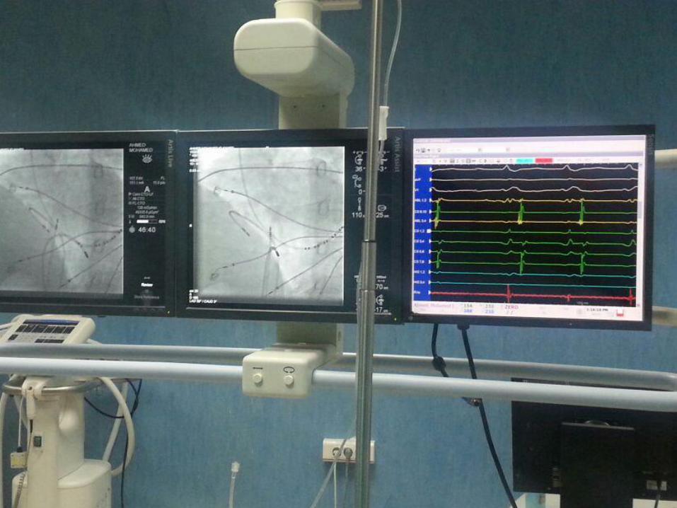
EP Lab = Cath Room + EPS machine + Ablator
+/- 3D mapping

Floroا
Stimulator
EPS Machine RF ablator & Infusion Pump
جهاز القسطرة

Display screen: The first page is 12 lead surface ECG

second page :intracardiac ECG (local electrograms)
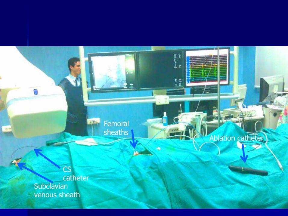
Ablation catheter
Femoral sheaths
Subclavian venous sheath
CS catheter

Ablator
Indifferent electrode
Ablation catheter
Temp: time: Impedence:

Catheter position
SVT VT
HRA SAN HRA
His AV cond His
RVA RVA +/- LVA
CS SCV/IVC
Recording from RA, RV, His , CS

Catheter Placement and recording
HRA
CS
HIS
RV
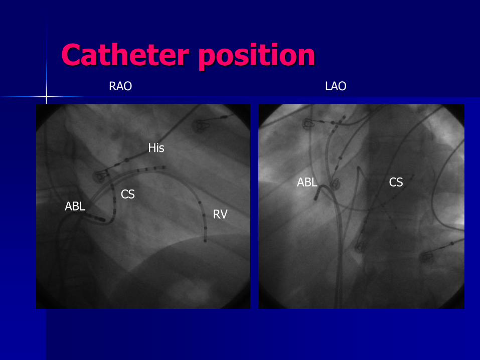
Catheter position RAO LAO
CS
His
RV
CS ABL
ABL

Catheters projection in RAO and LAO views Cs catheter (deflectable) inserted through IVC

LAO 20 Pole mapping catheter catheter (HALLO) and long sheaths for stability
Halo catheter
mapping the RA
CS
His
ABL
Long Sheath
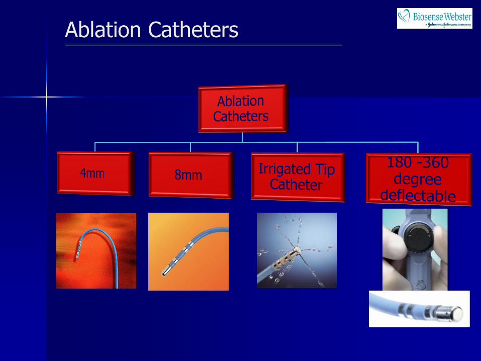
Ablation Catheters
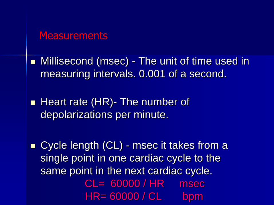
Millisecond (msec) - The unit of time used in
measuring intervals. 0.001 of a second.
Heart rate (HR)- The number of
depolarizations per minute.
Cycle length (CL) - msec it takes from a
single point in one cardiac cycle to the
same point in the next cardiac cycle.
CL= 60000 / HR msec
HR= 60000 / CL bpm
Measurements
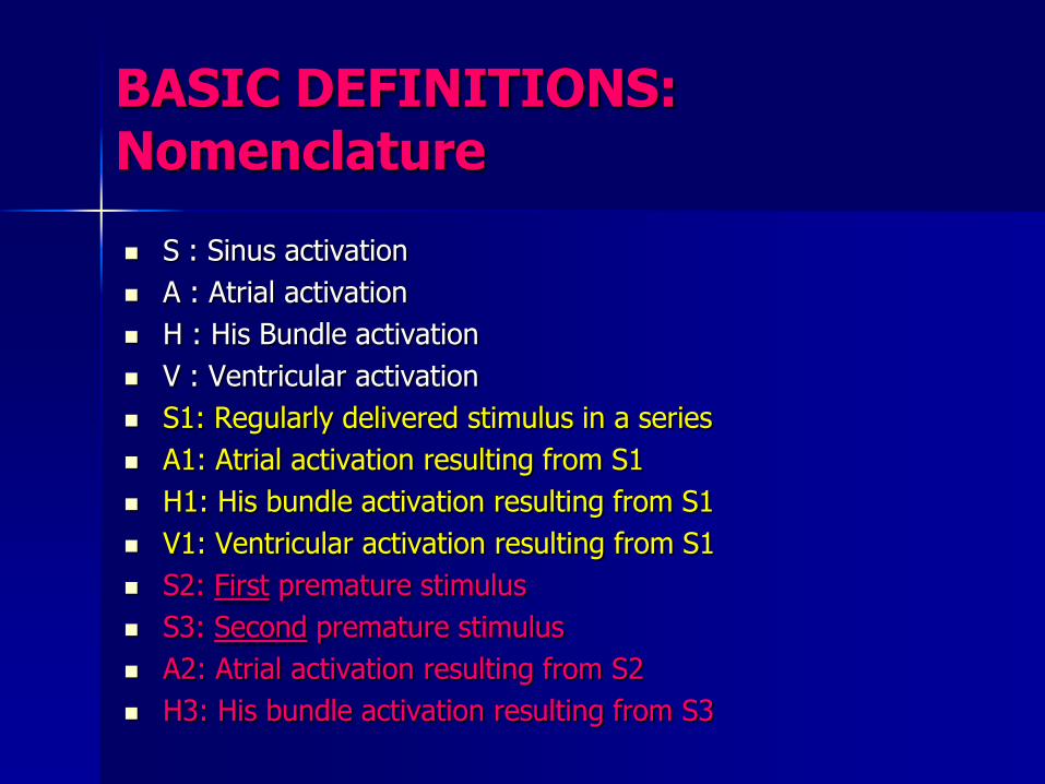
BASIC DEFINITIONS: Nomenclature
S : Sinus activation
A : Atrial activation
H : His Bundle activation
V : Ventricular activation
S1: Regularly delivered stimulus in a series
A1: Atrial activation resulting from S1
H1: His bundle activation resulting from S1
V1: Ventricular activation resulting from S1
S2: First premature stimulus
S3: Second premature stimulus
A2: Atrial activation resulting from S2
H3: His bundle activation resulting from S3

Refractory Period
Effective refractory period (ERP) is the longest S1-S2 that fails to depolarize the chamber being paced or the AV node.
Functional refractory period (FRP) is the shortest S1-S2 that depolarizes the chamber being paced or the AV node.

Catheter Placement – High Right Atrium (HRA)

Catheter Placement – Right Ventricular Apex (RVA)

Catheter Placement – His Bundle
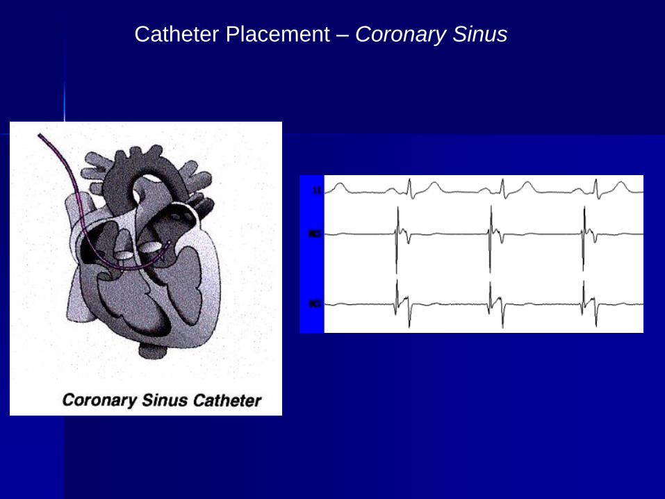
Catheter Placement – Coronary Sinus

baseline cond. intervals
SCL= interval bet. 2 successive A waves
AH interval ( 50—120 msec) AVN
HV interval (35 – 55 msec) His-RV/QRS
His deflection (cond. Via His bundle)

Basic intracardiac recordings

Basic intracardiac measurements
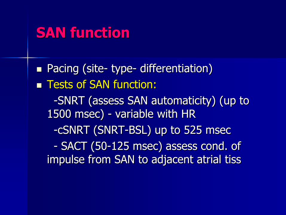
SAN function
Pacing (site- type- differentiation)
Tests of SAN function:
-SNRT (assess SAN automaticity) (up to 1500 msec) - variable with HR
-cSNRT (SNRT-BSL) up to 525 msec
- SACT (50-125 msec) assess cond. of impulse from SAN to adjacent atrial tiss

SAN function
S1
A 550
SCL= 450 msec SNRT= 550 msec CSNRT= 150 msec
400
Time = 30 sec
HRA
Recovery interval

SAN function Sino Atrial Conduction Time
S1 A
Idea: reset the SAN – Narula method
580
Recovery interval= time of paced beat penetrates & resets SAN+SCL+time for spont. Beat to exit SAN = SCL+ 2 SACT SACT=580-450/2 = 65 msec
8 paced beats

Tests of AVN function
Antegrade 1:1 AV conduction
Antegrade WCL
Retrograde 1:1 VA conduction
Retrograde WCL
AVN/ERP
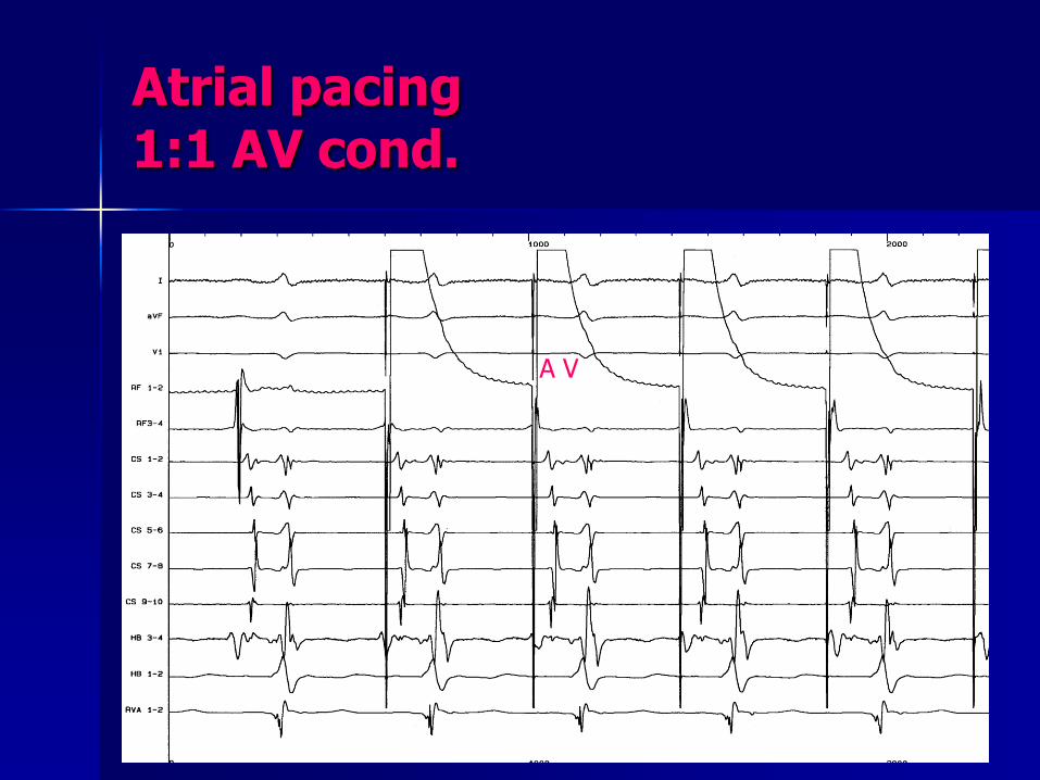
Atrial pacing 1:1 AV cond.
A V

Atrial pacing Weckebach AV block
A
V
A
V
A A
H
V
HIS
HIS
RA
R V

Basic ICECG Basic study
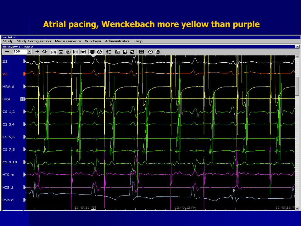
Atrial pacing, Wenckebach more yellow than purple

Atrial Extrastimulation To measure AVN/ERP

Atrial Extrastimulation to measure AVN/ERP
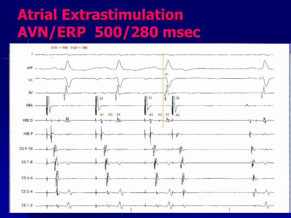
Atrial Extrastimulation AVN/ERP 500/280 msec

V pacing To measure retrograde AVN or detect AP
A V V A

1:1 VA cond.
PCL= 300 msec
V A
Retrograde sequence of activation
Concentric
V A V A
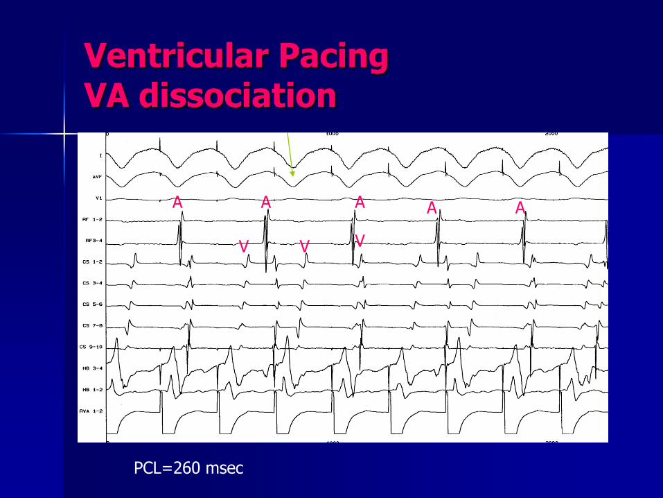
Ventricular Pacing VA dissociation
PCL=260 msec
A A
V V V
A A A
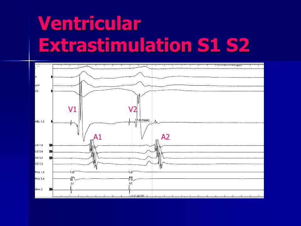
Ventricular Extrastimulation S1 S2
V1 V2
A1 A2
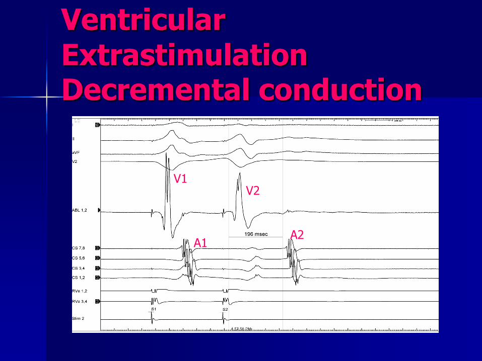
Ventricular Extrastimulation Decremental conduction
V1 V2
A2 A1

Ventricular Extrastimulation : ERP of retrograde limb of AVN
A1
S2 No V V1

Ventricular Tachycardia Study Induction by Extra stimulus and termination by Overdrive Pacing
48
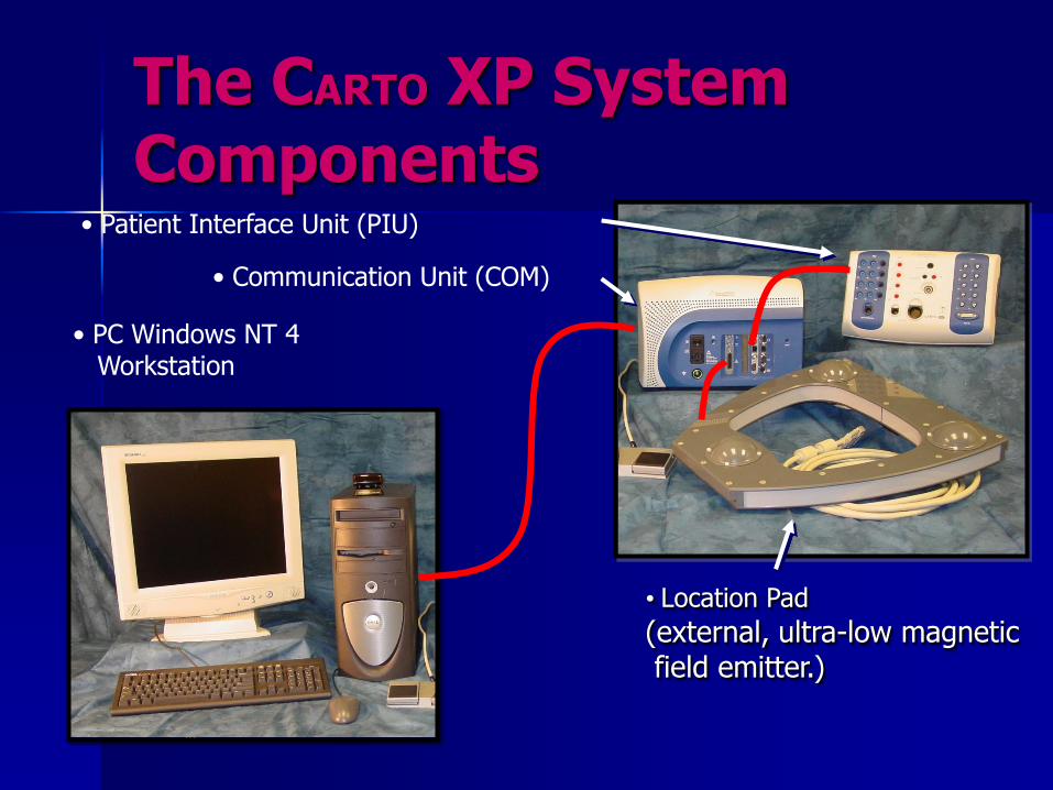
The CARTO XP System Components
• Location Pad
(external, ultra-low magnetic field emitter.)
• Patient Interface Unit (PIU)
• PC Windows NT 4 Workstation
• Communication Unit (COM)

49
Location Pad
An External Ultra-Low Magnetic Field Emitter

50
Catheter Tip Location
D1
D2
D3
The Triangulation Principle
The Fields possess Temporal and spatial distinguishing characteristics
51
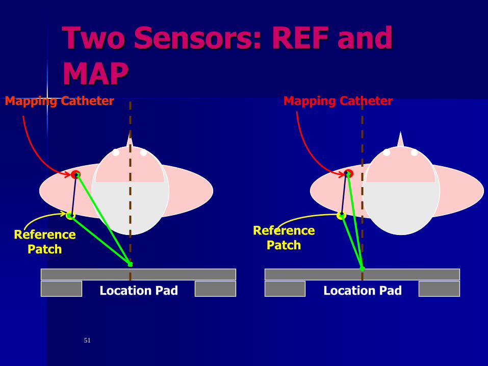
Location Pad Location Pad
Mapping Catheter
Reference Patch
Mapping Catheter
Reference Patch
Two Sensors: REF and MAP
52
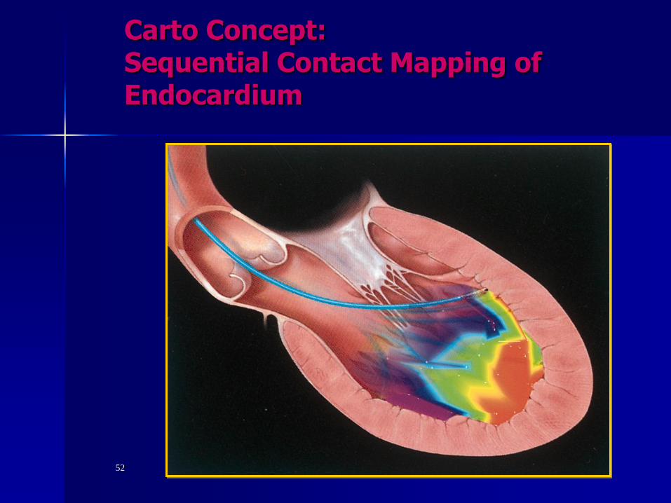
Carto Concept: Sequential Contact Mapping of Endocardium

What is new ??!! Patient with LVOT VT .CARTO 3 Mapping

Welcome to Egypt Ain Sokhna and Hurghada
