dr. maya menon asst. prof srm medical college …. prof srm medical college prof. shantamani prof...
TRANSCRIPT

Dr. Maya MenonAsst. Prof SRM Medical College
Prof. Shantamani
Prof .Tasneem

Mrs. X 24 yrs, Primi unbooked admitted on 20/12/11 ,38W2D POG with H/O headache and pedal edema for 3 days.No other imminent signs/symptomsMarried since 10 monthsAll trimesters uneventful

O/E pt anemic,facial puffiness+, pedal edema+BP-170/90 mm HgP/A – uterus term, cephalic,relaxed
FHS good.
Investigations for preeclampsia sent
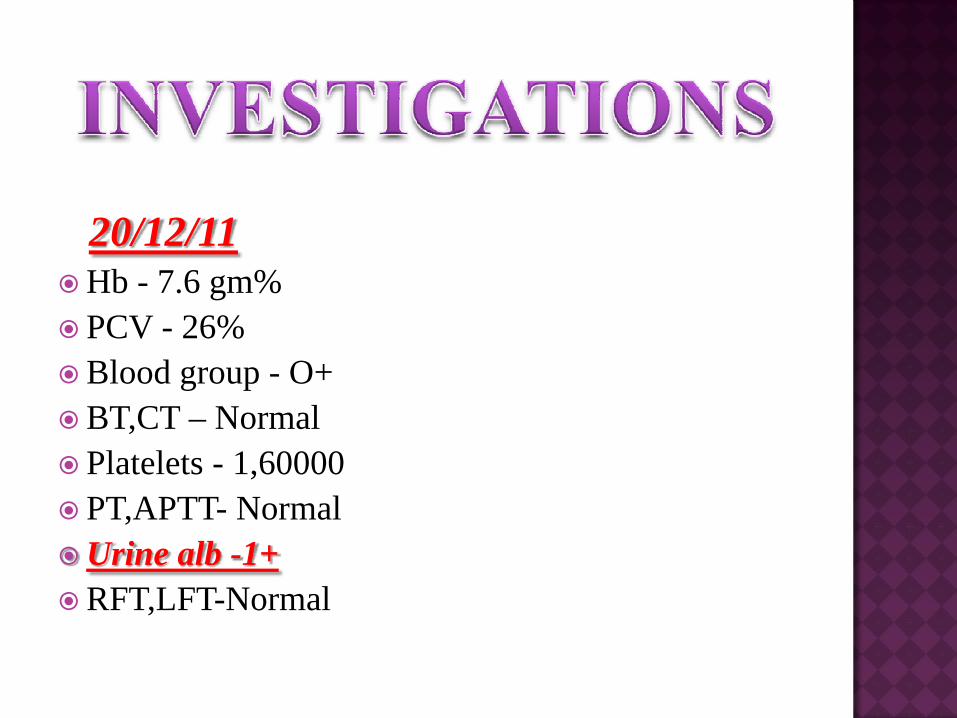
20/12/11Hb - 7.6 gm% PCV - 26%Blood group - O+BT,CT – NormalPlatelets - 1,60000PT,APTT- NormalUrine alb -1+RFT,LFT-Normal

USG 20/12/11
SLF,cephalic,GA-37-38 wks,placenta-anterior,liquor-adequate.
Fundoscopy – normal.
CTG – reactive.

Pt started on antihypertensives
T.Alpha methyldopa 500 mg 8 hrlyT.nifedepine 10 mg 12 hrly
1 unit blood transfused on 21/12/11 since Hb-7.6gm%
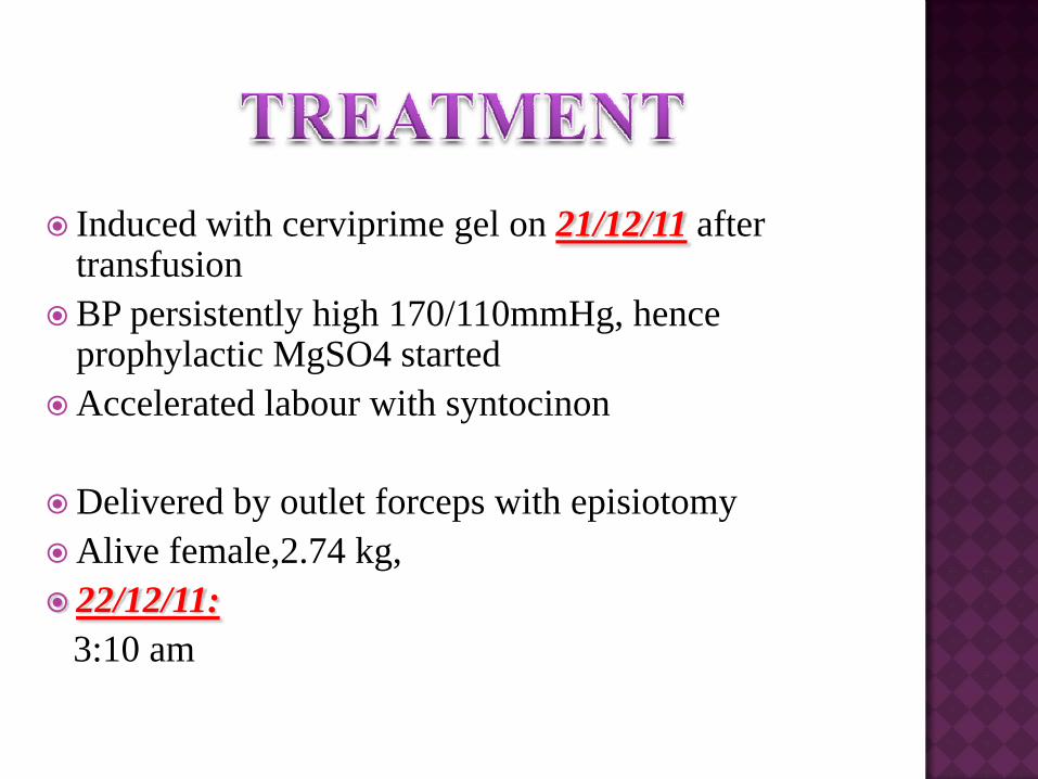
Induced with cerviprime gel on 21/12/11 after transfusionBP persistently high 170/110mmHg, hence prophylactic MgSO4 startedAccelerated labour with syntocinon
Delivered by outlet forceps with episiotomyAlive female,2.74 kg,22/12/11:3:10 am

Immediate postpartum-BP 140/90-130/80 mm HgPt on T. Alpha methyldopa 250 mg bd2nd unit blood transfused postnatally. MgSO4 continued,Antibiotic continued.
23.12.11
DAP opinion for uncontrolled BPAldomet 500 mg 8 hrly,Depin SR bd

24.12.11DAP opinion-Labetalol 100 mg bd ,alprazolam 2.5 mg.
26.12.11 4th post natal day.Pt C/o giddiness,headache,blurring of visionBP persistently high-160/110 mmHg.
Cardiologist opinion-IV Labetalol 20 mg over 2mins,Rpt 40mg after 20 mins BP checked every 5 mins .BP is normal IV dose tapered.
Oral Labetalol 200 mg bd increased to 400mg bd.Inj Clexane- 0.6mg sc suspecting C V T.

27.12.11
Ophthalmology opinion:
Visual acuity-limited to finger counting in both eyes,pupils were equal & reactive no relative afferent pupillary defect.
Colour vision & Amsler Grid test -normal.
Fundoscopy-Tortuous vessels with optic disc oedema.

Neurologist opinion:Examination unremarkable
CT Brain:Ill defined hypointense lesion in the bilateral cortical white matter in occipital region
MRI:T1 weighted images show hypointense & T2weighted images show .hyperintense areas-subcortical white matter.bilateral vasogenic oedema-detected in post cerebral circulation–postparietal & temporal & occipital lobe.
Diagnosis:Posterior reversible encephalopathy syndrome.

Supportive,symptomatic Rx,Antihypertensive-IV Labetalol 20 mg-40mg, oral Labetalol 200 mg bd increased to 400mg bd.Alpha methyl dopa 500mg 6 hrly .
Patient recovered within 48hrs.Discharged with oral antihypertensive drugs.
MRI – Normal study(after 6wks).

Posterior reversible encephalopathy syndrome (PRES) is a well-recognized, clinical neuroradiological entity characterized by
Transitory neurological disturbancesIncluding altered mental status, seizures, headache and ataxia.Blurred vision,scotoma & visual hallucination with acute or subacute onset PRES

A neuroimaging study usually shows transient edema primarily in the cortex and subcortical white matter of the parieto-occipital lobes and other cerebral structures,such as basal ganglia, frontal lobes, cerebellumand brainstem, can also be involved

Toxemia of Pregancy-Eclampsia & PreeclampsiaImmunosuppressive therapies with cyclosporineMultiorgan dysfunction syndrome,Autoimmuneconditions -SLE,Sclreoderma,High dose of Cisplatin,gemcitabineI V Immunoglobulin - Guillain Barresyndrome, tumor lysis syndrome.Hypomagnesemia,Hypocholestremia,Hypercale-mia.

Hypertension causes a failure of cerebral autoregulation causing an injury to capillary network & thus hyperperfusion causing vasogenic cerebral oedema
Endothelial dysfunction causes vasoconstriction & hypoperfusion causing ischemia & vasogenic cerebral oedema.
Two theories –Breakdown of blood brain barrier causes vasogenic cerebral oedema which is evident in neuroimaging.

PRES is reversible with appropriate treatment Neurotoxic manifestation result from cerebral oedema.PRES shown to progress from reversible vasogenicoedema to irreversible ischemic damage if appropriate treatment not initiated.Ischemic damage can cause irreversible neurologic sequelae – epiplesy & death.Posterior brain at greater risk for autoregulationbreakdown because it is less innervated rendering it less able to adjust to blood pressure fluctuations.Failure of autoregulation results in vasogenicoedema.

OPHTHALMOLOGY:Loss of visionBlurring of visionScotomaVisual hallucination
NEUROLOGY:HeadacheAphasiaFacial numbnessSeizureAtaxia

General examination,Neurological examination Opthalomology examination-Formal Goldman visual testing- ScotomaVisual symptoms – loss of vision, blurring of vision , visual hallucination.Color vision & Amsler Grid testing .Fundoscopy –tortuous vessels with /without optic disc oedema.CT brain-areas of low attenuation seen in posterior cerebral hemisphere.MRI-T1 weighted images hypointense,T2 weighted images hyperintense in subcortical white matter in posterior parietal & temporal & occiptal lobe.MRA & MRV – normal.


Supportive,Symptomatic treatment,Treat underlying cause,I.V anti hypertensive drugs –labetolol,hydralazine,oral nifedepine,Alpha methyl dopa, Anticonvulsants-started given for 3 months.

Severe /abrupt hypertension cause failure of cerebral auto regulation causing injury to capillary network &endothelialdysfunction –BBB-vasogenic cerebral oedema .Symptoms-headache,blurring vision,seizure.CT-low attenuationMRI-T2 hyperintensity in occipital lobe/parietal lobe.Rx-I V antihypertensive,Anticonvulsants,treat underlying cause.Early diagnosis & rapid management complete recovery saves life.
Thrombosis of cortical,cerebral veins,venous sinus,dural sinus thrombosis-cytotoxic cerebral oedema –intracranial hypertension-hematoma.Symptoms -headache,blurring vision,seizure.CT-Empty Delta sign –heamorrhagic infarct.MRV –Filling defect in dural sinus.MRI-Thrombus appear as isotense /hypotense.Rx-Heparin 5-7days,Warfarin 3 months,Anticonvulsants,treat underlying cause.Surgical evacuation-clot/decompressive craniectomy.


PRES is a reversible condition but prompt recognition required occurs in severe /abrupt hypertension.MRI –Diagnostic tool.Monitoring BP and / or discontinuing eventually provocative immunosuppressive drugs & adequately treating seizures.Lowering BP is mandatory . Mean arterial BP -105-125 mmHg.Parenteral –Labetalol /hydralazineOraly nifedipine/labetalol/alpha methyl dopaComplete clinical recovery is seen within 24-48 hrs & abnormal imaging is seen to normalise within 6 wks.

1Pula JH, Eggenberger E. Posterior Reversible Encephalopathic Syndrome. CurrOpin Ophthalmol 2008;19:479-84.
2.Bartynski WS. Posterior Reversible Encephalopathy Syndrome, Part 1: Fundamental Imaging and Clinical Features. AJNR Am J Neuroradiol2008;29:1036-42.
3.Bartynski WS. Posterior Reversible Encephalopathy Syndrome, Part 2: Controversies Surrounding Pathophysiology of Vasogenic Edema. AJNR Am J Neuroradiol 2008;29:1043-9.
4.Moratalla MB. Posterior reversible encephalopathy syndrome. Emerg Med J 2010;27:547.5
Roth C, Ferbert A. Posterior Reversible Encephalopathy Syndrome: Is there a difference between Pregnant and Non-Pregnant Patients? Eur Neurol 2009;62:142-9.
