dr. archana rani associate professor department of anatomy kgmu up, lucknow 2.12.2014
TRANSCRIPT
Dr. Archana RaniAssociate Professor
Department of AnatomyKGMU UP, Lucknow
2.12.2014
CARTILAGE
Modified connective tissueForms skeletal basis of some parts of bodyMatrix is firm giving it the characteristic
consistencyResists compressionAvascular (nutrients diffuse through matrix)Perichondrium is rich in blood vessels
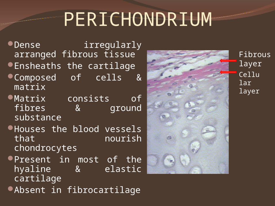
PERICHONDRIUMDense irregularly
arranged fibrous tissue Ensheaths the cartilageComposed of cells &
matrixMatrix consists of fibres
& ground substanceHouses the blood vessels
that nourish chondrocytes
Present in most of the hyaline & elastic cartilage
Absent in fibrocartilage
Fibrous layer
Cellular layer
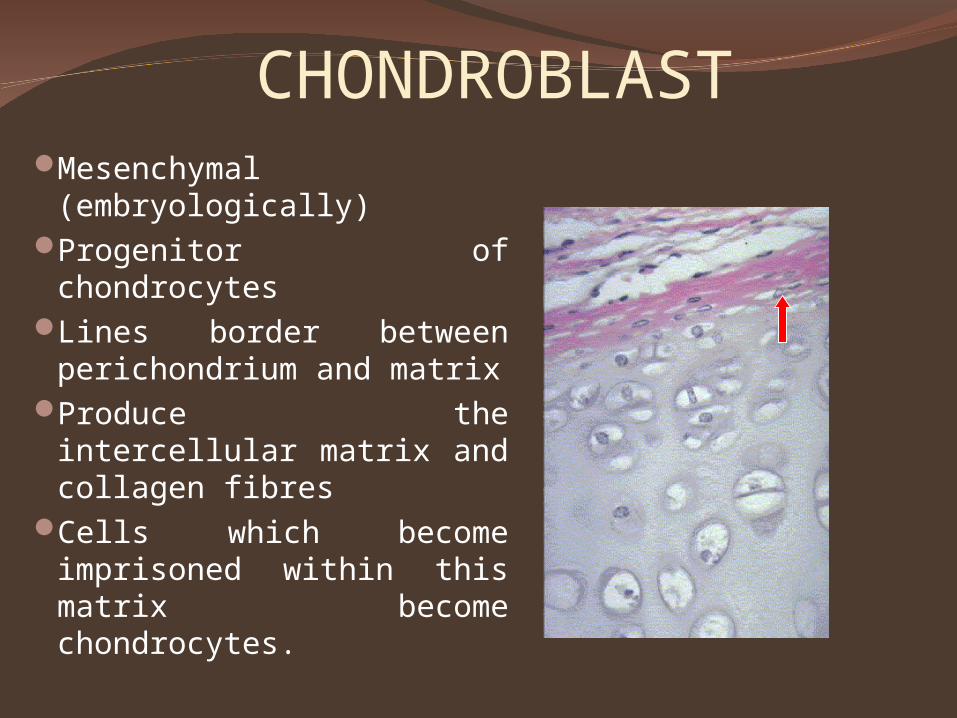
CHONDROBLASTMesenchymal
(embryologically)Progenitor of
chondrocytesLines border between
perichondrium and matrixProduce the intercellular
matrix and collagen fibresCells which become
imprisoned within this matrix become chondrocytes.
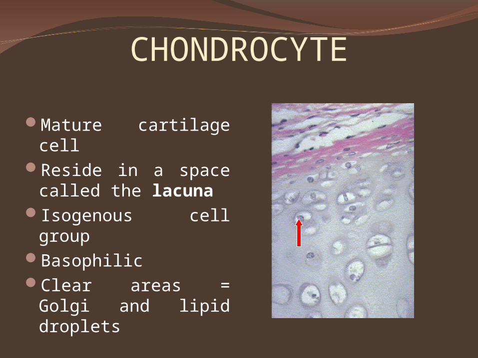
CHONDROCYTE
Mature cartilage cellReside in a space
called the lacunaIsogenous cell groupBasophilicClear areas = Golgi
and lipid droplets
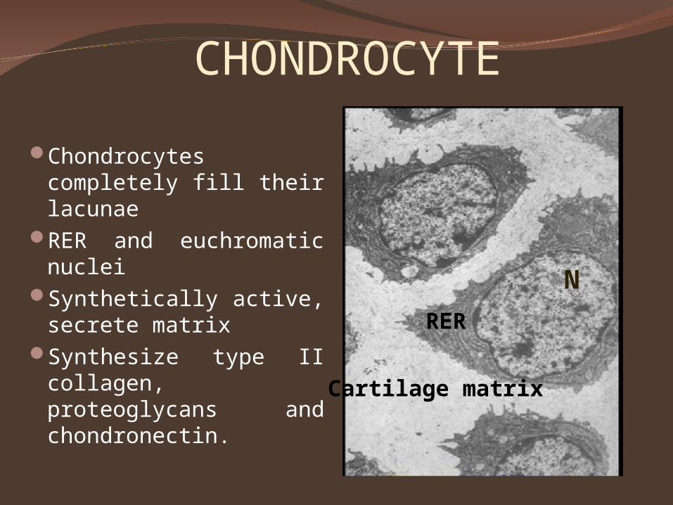
CHONDROCYTE
Chondrocytes completely fill their lacunae
RER and euchromatic nuclei
Synthetically active, secrete matrix
Synthesize type II collagen, proteoglycans and chondronectin.
Cartilage matrix
RER
N
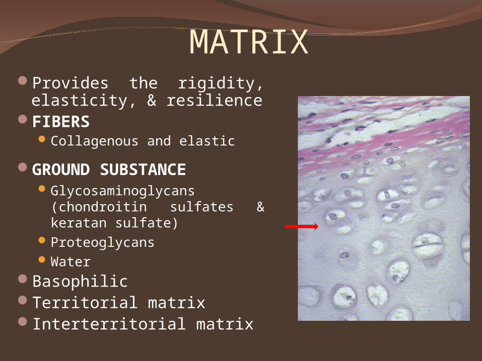
MATRIXProvides the rigidity,
elasticity, & resilienceFIBERS
Collagenous and elastic
GROUND SUBSTANCEGlycosaminoglycans
(chondroitin sulfates & keratan sulfate)
ProteoglycansWater
BasophilicTerritorial matrixInterterritorial matrix
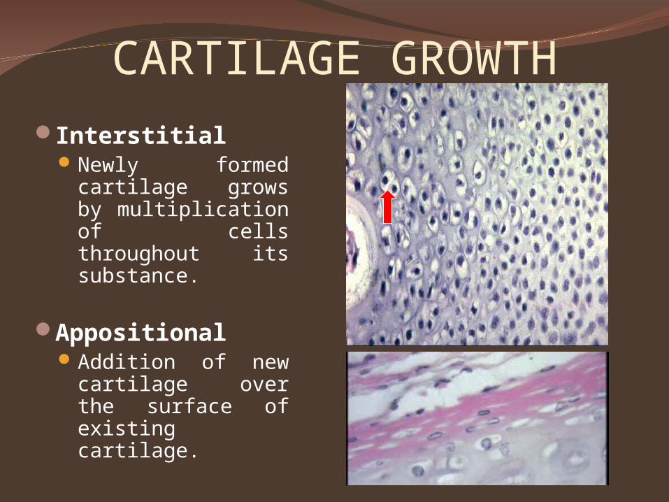
CARTILAGE GROWTH
InterstitialNewly formed
cartilage grows by multiplication of cells throughout its substance.
AppositionalAddition of new
cartilage over the surface of existing cartilage.
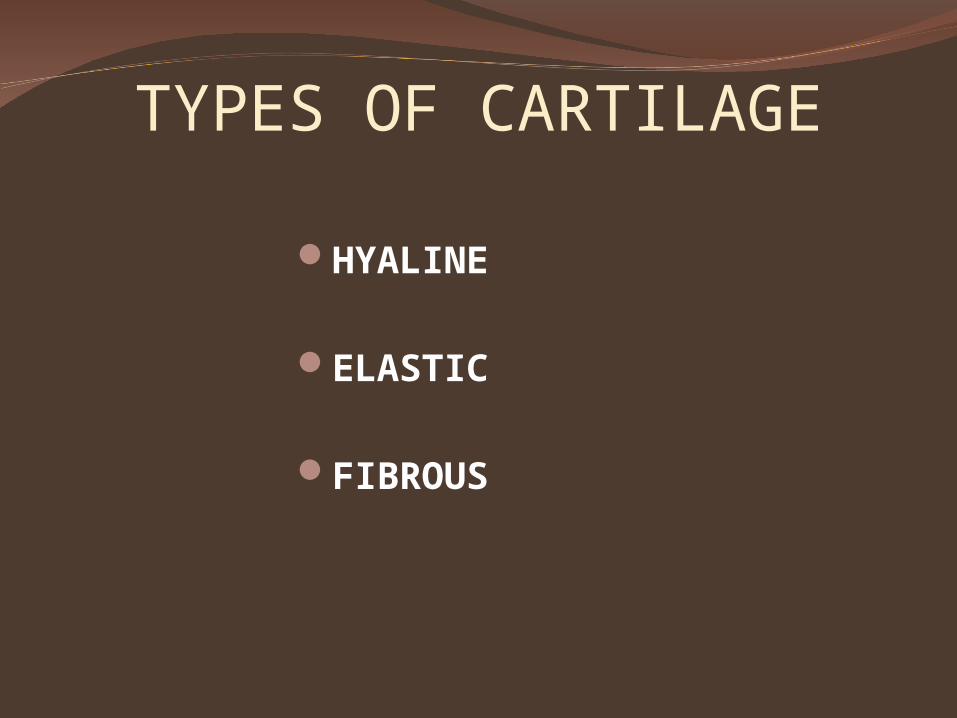
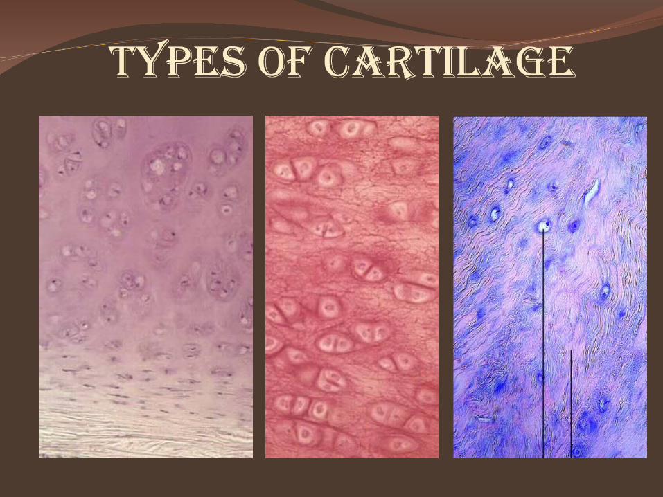
TYPES OF CARTILAGE
HYALINE
ELASTIC
FIBROUS
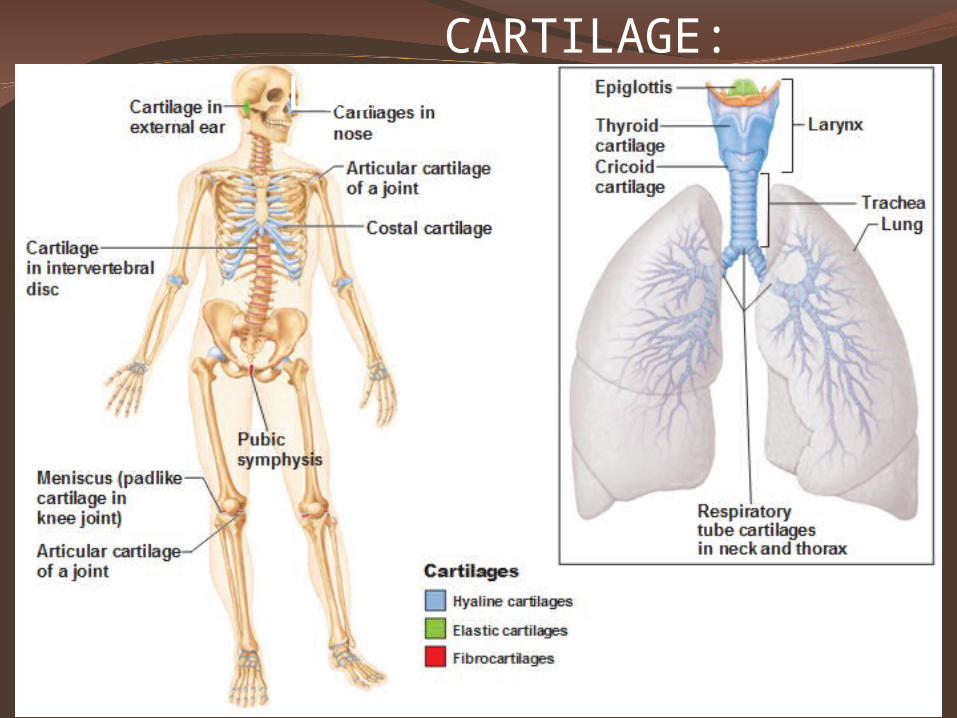
CARTILAGE: LOCATIONS
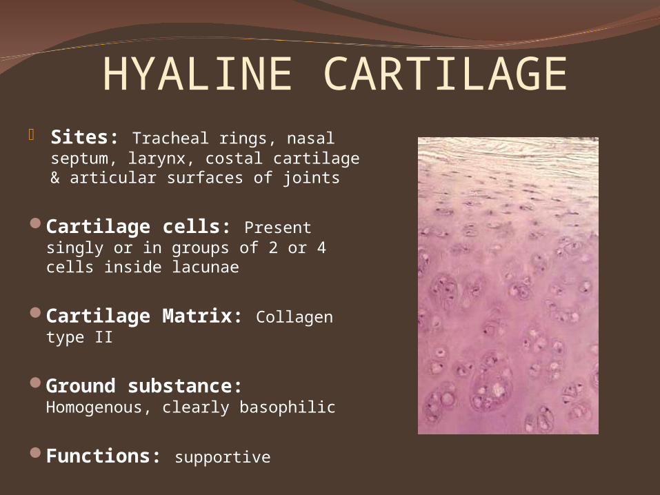
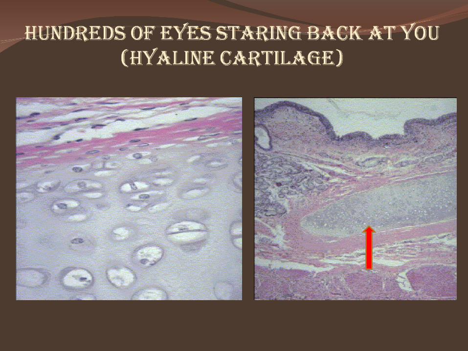
HYALINE CARTILAGE Sites: Tracheal rings, nasal
septum, larynx, costal cartilage & articular surfaces of joints
Cartilage cells: Present singly or in groups of 2 or 4 cells inside lacunae
Cartilage Matrix: Collagen type II
Ground substance: Homogenous, clearly basophilic
Functions: supportive
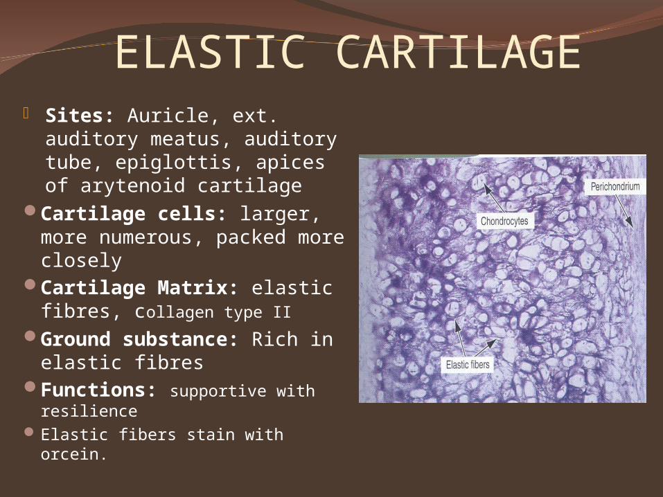
ELASTIC CARTILAGE Sites: Auricle, ext.
auditory meatus, auditory tube, epiglottis, apices of arytenoid cartilage
Cartilage cells: larger, more numerous, packed more closely
Cartilage Matrix: elastic fibres, collagen type II
Ground substance: Rich in elastic fibres
Functions: supportive with resilience
Elastic fibers stain with orcein.
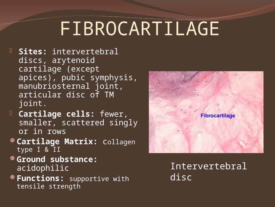
FIBROCARTILAGE Sites: intervertebral
discs, arytenoid cartilage (except apices), pubic symphysis, manubriosternal joint, articular disc of TM joint.
Cartilage cells: fewer, smaller, scattered singly or in rows
Cartilage Matrix: collagen type I & II
Ground substance: acidophilic
Functions: supportive with tensile strength
Intervertebral disc
Clinical application
Osteoarthritis
Pseudoachondroplasia
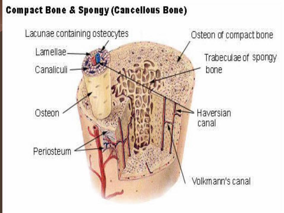
bonE
Modified connective tissueHighly vascular mineralized connective
tissue consisting of cells and dense intercellular organic matrix impregnated with inorganic salts.
Provide support & protection to the vital organs
Forms skeletal framework
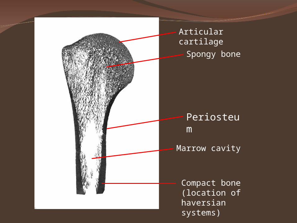
Articular cartilage
Spongy bone
Marrow cavity
Compact bone (location of haversian systems)
Periosteum
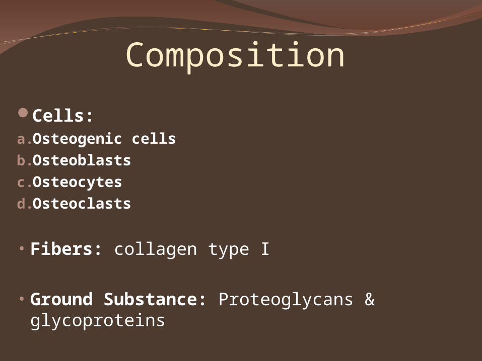
Composition
Cells: a.Osteogenic cellsb.Osteoblastsc.Osteocytesd.Osteoclasts
• Fibers: collagen type I
• Ground Substance: Proteoglycans & glycoproteins
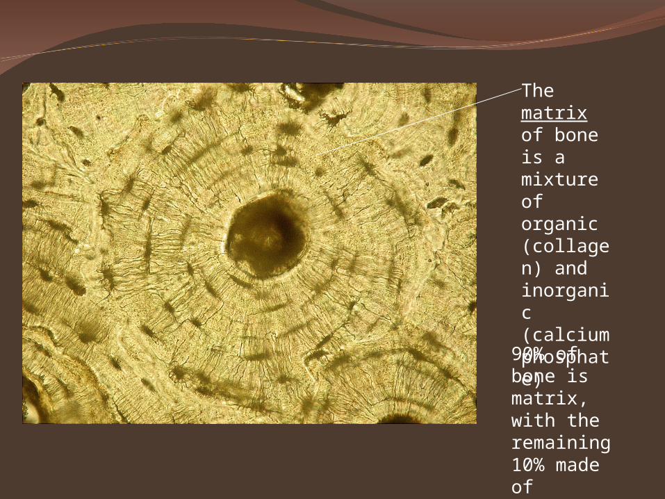
The matrix of bone is a mixture of organic (collagen) and inorganic (calcium phosphate)
90% of bone is matrix, with the remaining 10% made of osteocytes.
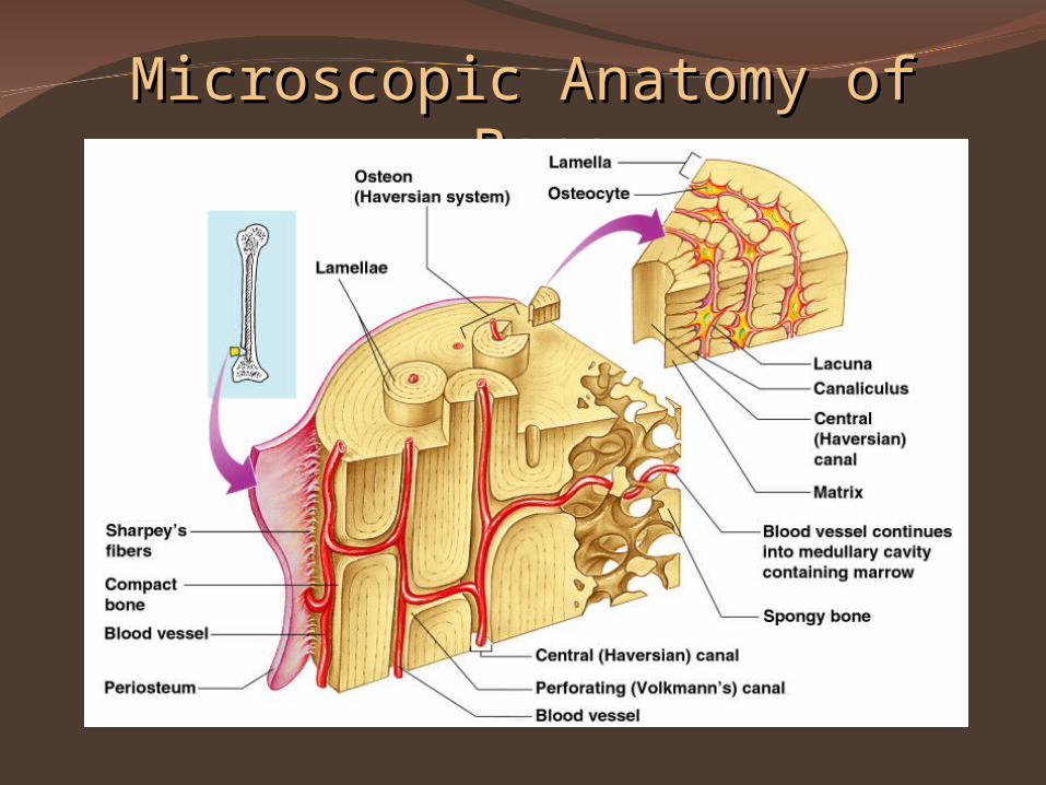
Microscopic Anatomy of BoneMicroscopic Anatomy of Bone
Figure 5.3
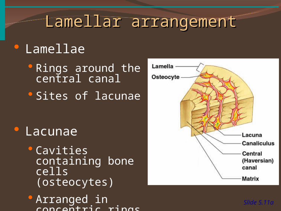
Lamellar arrangementLamellar arrangement
Slide 5.11a
Lamellae Rings around the
central canal
Sites of lacunae
Lacunae Cavities containing
bone cells (osteocytes)
Arranged in concentric rings
Figure 5.3
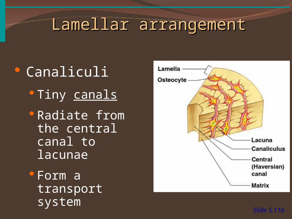
Slide 5.11b
Canaliculi
Tiny canals
Radiate from the central canal to lacunae
Form a transport system
Figure 5.3
Lamellar arrangementLamellar arrangement
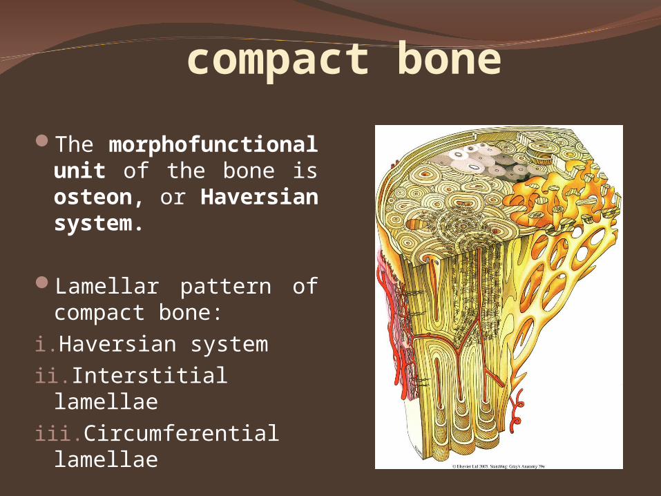
compact bone
The morphofunctional unit of the bone is osteon, or Haversian system.
Lamellar pattern of compact bone:
i. Haversian system ii.Interstitial lamellaeiii.Circumferential
lamellae
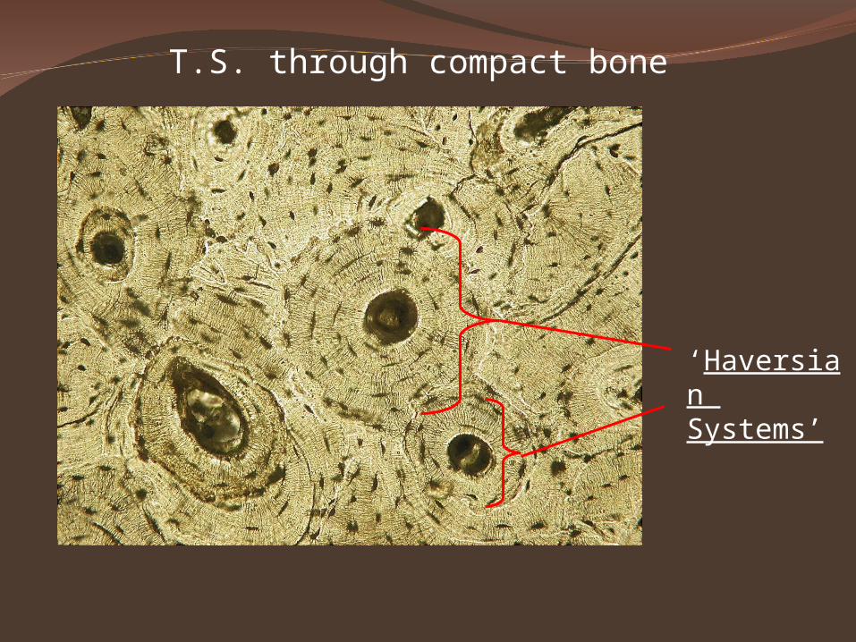
T.S. through compact bone
‘Haversian Systems’
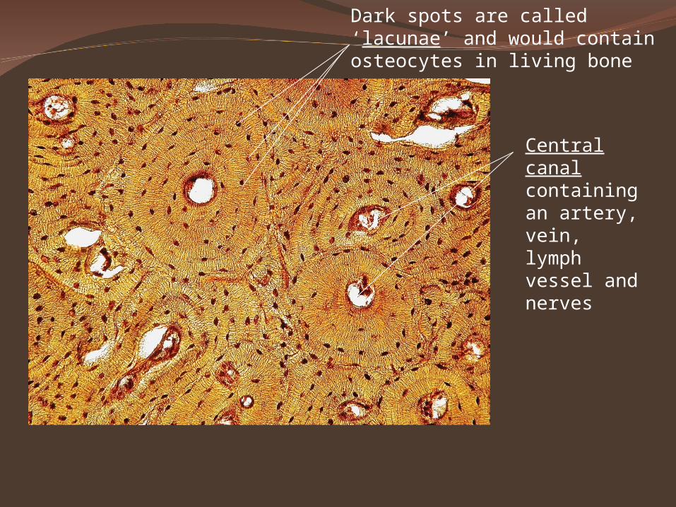
Dark spots are called ‘lacunae’ and would contain osteocytes in living bone
Central canal containing an artery, vein, lymph vessel and nerves
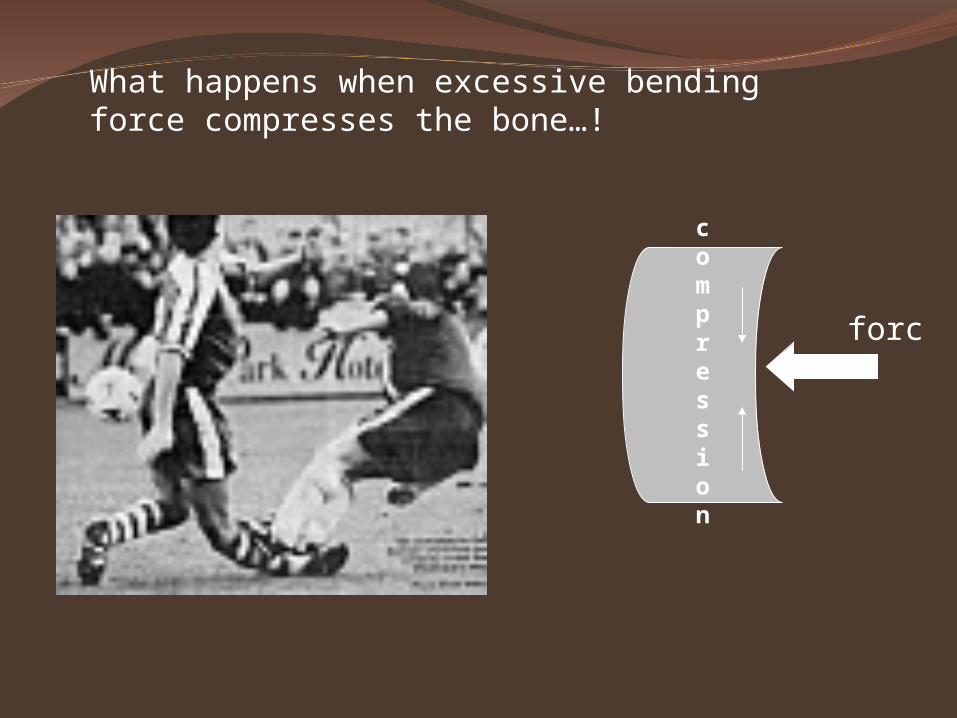
What happens when excessive bending force compresses the bone…!
compression
force
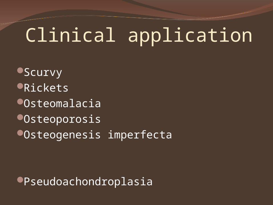
Clinical application
ScurvyRicketsOsteomalaciaOsteoporosisOsteogenesis imperfecta
Pseudoachondroplasia
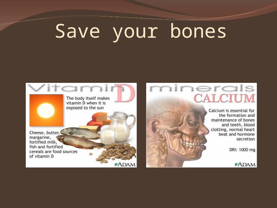
Save your bones
Take home message
Aerobic exercisesLow impact, weight bearing
exercisesResistance exercises
References
1. diFiore’s Atlas of Histology with functional Correlations, 12th Edition.
2. Essentials of Anatomy for Dentistry Students,1st Edition.
3. Textbook of Histology, 3rd Edition.
MCQ
• Fibrous cartilage is present in:1.Auricle2.Nose3.Tracheal rings 4.Intervertebral discs
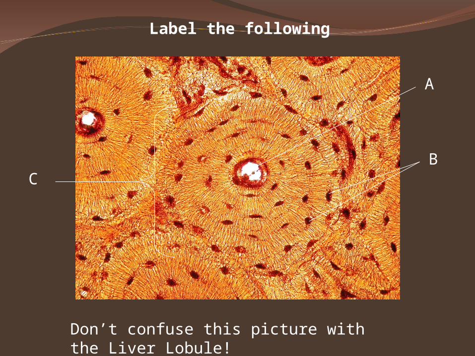
Label the following
A
BC
Don’t confuse this picture with the Liver Lobule!
MCQ• Elastic cartilage is present in:1.Apices of arytenoid cartilage2.Epiphysis3.Tracheal rings 4.Temporomandibular joints
MCQ• Hyaline cartilage is present in:1.Tracheal rings 2.External auditory meatus3.Semilunar cartilages of knee joint4.Intervertebral discs
MCQ
The blood vessels and nerves go inside the compact bone through:
1. Haversian canal2. Volkman’s canal3. Canaliculi4. Interstitial lamellae