dppiv drug discovery kit - cloud object storage | store ... 05/22/2014 bml-ak499 1 dppiv drug...
TRANSCRIPT
26-G Keewaydin Drive, Salem, NH 03079│P: (800) 592-5726│F: (603) 898-6854│[email protected]│www.alpco.com
DPPIV Drug Discovery Kit
Designed to screen DPPIV inhibitors.
For Research Use Only. Not For Use In Diagnostic Procedures.
Catalog Number: 74-DPPHU-E01
Size: 96 Wells
Version: 05/22/2014 – ALPCO April 23, 2015

Revision: 05/22/2014 BML-AK499 1
DPPIV Drug Discovery Kit –BML- AK499
BACKGROUND
DPPIV (DPP4, CD26) is a member of the class of proteases known as prolyl peptidases, which cleave proteins after proline residues1. DPPIV, a serine dipeptidyl peptidase, cleaves the N-terminal X-Ala or X-Pro from target polypeptides, such as chemokines (e.g. CXCL11) and peptide hormones (e.g., glucagon-like peptide-1, GLP1)1-3. DPPIV possesses a transmembrane region and a very short cytoplasmic domain, but is often cleaved and released as a soluble, circulating form4. It is found as a dimer with itself or with FAP (fibroblast activation protein-α, seprase), another prolyl peptidase1. It also has non-peptidase functions: through its interaction with adenosine deaminase (ADA) and extracellular matrix components, it influences T-cell activation and proliferation2,5,6. It is thought to play roles in diabetes, cancer, and autoimmune diseases, making it a target for drug discovery7-11.
The DPPIV Drug Discovery Kit is a complete assay system designed to screen DPPIV inhibitors, providing enough material to perform at least 96 assays. The kit contains both a chromogenic substrate (H-Gly-Pro-pNA; Km=114 µM) and a fluorogenic substrate (H-Gly-Pro-AMC; Km=50 µM). Cleavage of the p-nitroaniline (pNA) from the chromogenic substrate increases absorbance at 405 nm. The fluorimetric assay is based on the cleavage of 7-amino-4-methylcoumarin (AMC) moiety from the C-terminus of the peptide substrate, which increases its fluorescence intensity at 460 nm. The kit is useful to screen inhibitors of DPPIV, a potential therapeutic target. A DPPIV inhibitor, P32/98 (KI=130 nM12), is included for use as a control.
Other DPP enzymes are available for specificity profiling. Contact Enzo Life Sciences or go to www.enzolifesciences.com.
REFERENCES:
1. J.S. Rosenblum and J.W. Kozarich Curr. Opin. Chem. Biol. 2003 7 496 2. R. Thoma et al. Structure 2003 11 947 3. H. Shibuya-Saruta et al. J. Clin. Lab. Anal. 1996 10 435 4. D.A. Pereira et al. Braz. J. Med. Biol. Res. 2003 36 567 5. I. Ben-Shooshan et al. Biochem. Biophys. Acta 2002 1587 21 6. K. Aertgeerts et al. Protein Sci. 2004 13 145 7. A. E. Weber J. Med. Chem. 2004 47 4135 8. B. Pro and N.H. Dang Histol. Histopathol. 2004 19 1345 9. U. Aytac and N.H. Dang Curr. Drug Targets Immune Endocr. Metabol. Disord. 2004 4 11 10. H. Gotoh et al. Clin. Chem. 1988 34 2499 11. D.J. Augeri et al. J. Med. Chem. 2005 48 5025 12. J.A. Pospisilik et al. Diabetes 2003 52 741
Revision: 05/22/2014 BML-AK499 2
PLEASE READ ENTIRE BOOKLET BEFORE PROCEEDING WITH THE ASSAY. CAREFULLY NOTE THE HANDLING AND STORAGE CONDITIONS OF EACH KIT COMPONENT. PLEASE CONTACT ENZO LIFE SCIENCES TECHNICAL SERVICES FOR ASSISTANCE IF NECESSARY.
COMPONENTS OF BML-AK499 KIT
BML-SE434-9090 DPPIV ENZYME (HUMAN, RECOMBINANT)
FORM: Recombinant soluble human DPPIV. One U=1µmole/min@37°C, 100 µM H-Gly-Pro-pNA.
STORAGE: -70°C; AVOID FREEZE/THAW CYCLES
QUANTITY: 35 mU
BML-P188-9090 pNA SUBSTRATE (H-Gly-Pro-pNA; MW=328.8)
FORM: 10 mM in DMSO
STORAGE: -70°C
QUANTITY: 150 µl
BML-KI106-0001 pNA CALIBRATION STANDARD (p-nitroaniline; MW=138)
FORM: 50 µM in assay buffer.
STORAGE: -70°C
QUANTITY: 1 ml
BML-P189-9090 AMC SUBSTRATE (H-Gly-Pro-AMC; MW=410.3)
FORM: 0.5 mM in DMSO
STORAGE: -70°C
QUANTITY: 150 µl
BML-KI107-0001 AMC CALIBRATION STANDARD
(7-amino-4-methylcoumarin; MW=175)
FORM: 30 µM in assay buffer.
STORAGE: -70°C
QUANTITY: 1 ml

Revision: 05/22/2014 BML-AK499 3
BML-PI142-9090 INHIBITOR (P32/98; MW=260.4)
FORM: 1 mM in DMSO
STORAGE: -70°C
QUANTITY: 20 µl
BML-KI342-0020 ASSAY BUFFER
(50 mM Tris, pH 7.5)
FORM: Liquid in screw-cap plastic bottle
STORAGE: -70°C
QUANTITY: 20 ml
80-2407 ½-VOLUME CLEAR & NBS WHITE MICROPLATE MICROPLATES – 1 EACH
STORAGE: Room temperature.
OTHER MATERIALS REQUIRED
Microplate reader capable of measuring A405 to 3-decimal accuracy, or fluorescence at wavelengths of approximately 380nm (excitation)/ 460nm (emission)
Pipettes or multi-channel pipettes capable of pipetting 10-1000 µl accurately (note: dilution of reagents can be made to increase the minimal volume to >10 µl).
Ice bucket to keep reagents cold until use.
SUGGESTED EXPERIMENTAL METHODS
Note on storage: Store all components except the microplates (room temperature) at -70°C for the highest stability. The DPPIV enzyme should be handled carefully in order to retain maximal enzymatic activity. It is stable, in diluted or concentrated form, for several hours on ice. As supplied, DPPIV enzyme is stable for at least 5 freeze/thaw cycles. To minimize the number of freeze/thaw cycles, aliquot the DPPIV into separate tubes and store at -70°C. Do not maintain diluted components at reaction temperature (e.g. 37°C) for an extended period of time prior to running the assay.

Revision: 05/22/2014 BML-AK499 4
To start assay:
1. Defrost kit components and hold on ice until use. Thaw and store DMSO components (substrates, inhibitor) at room temperature, preferably in a dark place. Briefly centrifuge all vials. Minimize the time that any kit component is thawed.
2. Dilute inhibitor (P32/98) 1/10 in assay buffer. Example: Add 2 µl inhibitor to 18 µl assay buffer, in a separate tube.
3. For colorimetric assay (at A405nm, using clear microplate): Dilute substrate (H-Gly-Pro-pNA) 1/50 in assay buffer (50 µl is needed per well). Example: Add 5 µl substrate to 245 µl assay buffer, in a separate tube.
4. For fluorimetric assay (at Ex:380 nm/Em:460 nm, using white microplate), dilute substrate (H-Gly-Pro-AMC) 1/50 in assay buffer (50 µl is needed per well). Example: Add 5 µl substrate to 245 µl assay buffer, in a separate tube.
5. Add assay buffer to each desired well of microplate so that the total assay volume will be 100 µl. See Table 1 for examples.
6. Allow microplate and diluted assay components to equilibrate to assay temperature (e.g. 37°C).
7. After a brief thawing, quickly centrifuge the vial of DPPIV (BML-SE434-9090) to bring contents to bottom of tube. Dilute enough DPPIV in assay buffer to a stock concentration of 17.3µU/µL to produce required quantity for the experiment (the specific activity (SA) for the given lot is printed on the vial of SE434-9090).
8. Add DPPIV to the “Control”, “Inhibitor” and “Test Sample” wells, please note that the final amount of DPPIV per well is to be 26mU. DO NOT ADD DPPIV TO BLANKS!
9. Add 10 µl P32/98 inhibitor (diluted in step 2) to the “Inhibitor” well only! Final inhibitor concentration is 10 µM.
10. Add desired volume of test sample(s) to appropriate well(s). See Table 1. 11. Incubate plate for 10 min at reaction temperature (or as desired) to allow
inhibitor/enzyme interaction.
12. Start assay by the addition of 50 µl H-Gly-Pro-pNA substrate (diluted in step 3) or 50 µl H-Gly-Pro-AMC substrate (diluted in step 4), which have been equilibrated to reaction temperature (e.g. 37°C). Enough of each substrate is provided to perform 96 assays at 100 µM for the chromogenic substrate or 5 µM for the fluorogenic substrate.
13. Read plate continuously, at A405nm for the pNA substrate, or Ex:380 nm/Em:460 nm for the AMC substrate, in a microplate reader. For example, record data at 1 min. intervals for a total of 10 to 60 min.
14. Perform data analysis (see below).
NOTE: Retain microplate for future use of unused wells.
Revision: 05/22/2014 BML-AK499 5
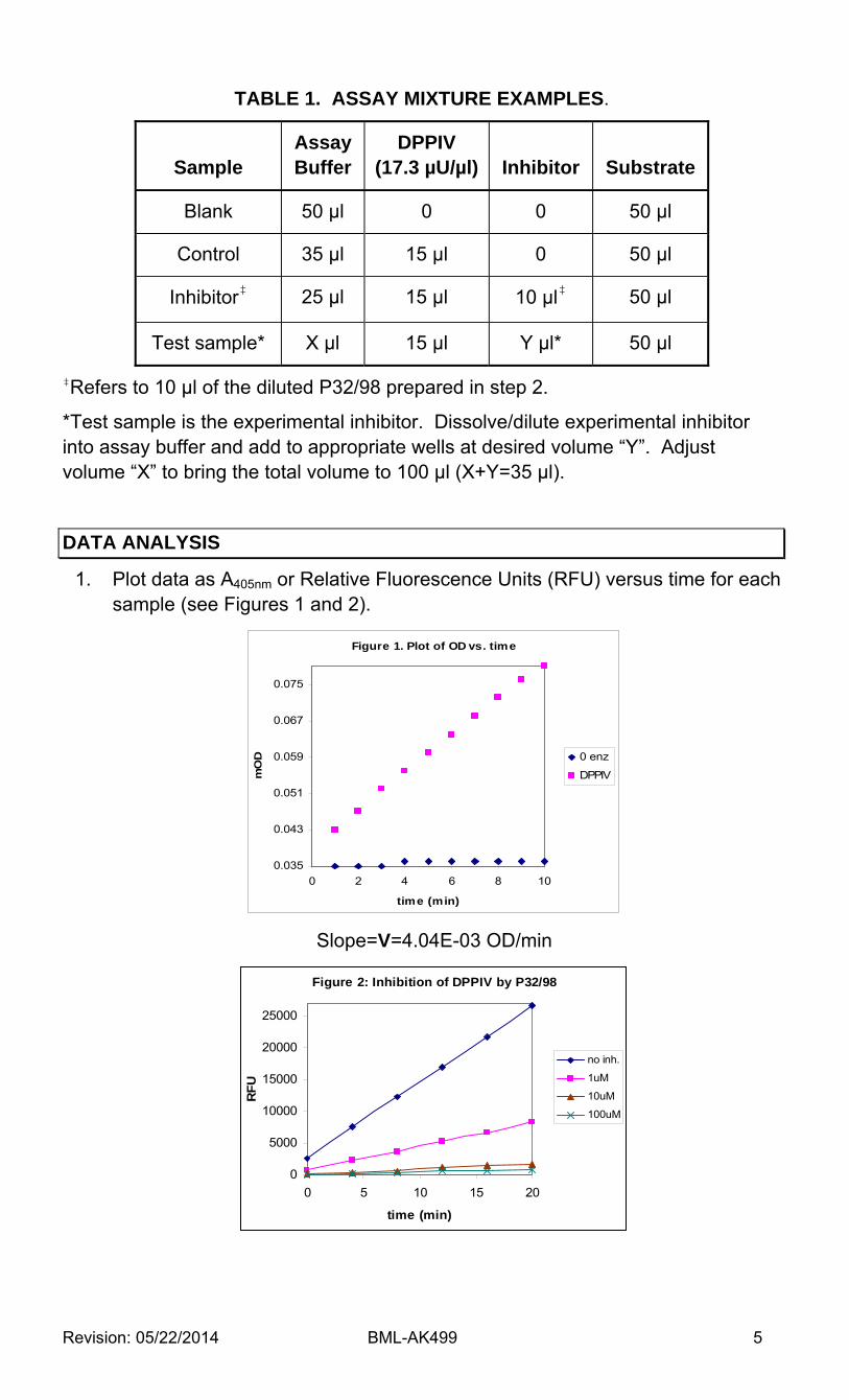
TABLE 1. ASSAY MIXTURE EXAMPLES.
Sample Assay Buffer
DPPIV (17.3 µU/µl) Inhibitor Substrate
Blank 50 µl 0 0 50 µl
Control 35 µl 15 µl 0 50 µl
Inhibitor‡ 25 µl 15 µl 10 µl‡ 50 µl
Test sample* X µl 15 µl Y µl* 50 µl
‡Refers to 10 µl of the diluted P32/98 prepared in step 2.
*Test sample is the experimental inhibitor. Dissolve/dilute experimental inhibitor into assay buffer and add to appropriate wells at desired volume “Y”. Adjust volume “X” to bring the total volume to 100 µl (X+Y=35 µl).
DATA ANALYSIS
1. Plot data as A405nm or Relative Fluorescence Units (RFU) versus time for each sample (see Figures 1 and 2).
Slope=V=4.04E-03 OD/min
Figure 1. Plot of OD vs. time
0.035
0.043
0.051
0.059
0.067
0.075
0 2 4 6 8 10
time (min)
mO
D 0 enz
DPPIV
Figure 2: Inhibition of DPPIV by P32/98
0
5000
10000
15000
20000
25000
0 5 10 15 20
time (min)
RF
U
no inh.
1uM
10uM
100uM

Revision: 05/22/2014 BML-AK499 6
2. Determine the time range over which the reaction is linear. Typically, 0 to 15 min works well.
3. Obtain a “best fit” line for the data points and determine the slope. 4. Average the slopes of duplicate samples. 5. If the blank has a significant slope, subtract this number from the slopes for
all samples. 6. To find % remaining activity in presence of inhibitor:
% activity remaining (with inhibitor)=
(slope of + inhibitor sample/control slope) x 100
See Figure 3.
7. To find the activity of the samples expressed as pmol substrate/min using chromogenic substrate:
Determine microplate reader conversion factor:
a) Add 100 µl calibration standard (p-nitroaniline; 50 µM concentration) to 2 wells of the clear microplate. In Tris buffer the extinction coefficient for p-nitroaniline at 405 nm is ~9700 M-1cm-1 (R. Lottenberg et al., 1983; Biochim.Biophys.Acta 742:558). Typically, 100 µl of the 50 µM standard, in a ½ volume well, produces an A405
of about 0.3. Alternatively, build a pNA standard curve and use the slope (µM/OD) as the conversion factor.
b) Determine the average A405nm using 100 µl assay buffer as a blank. c) Calculate the conversion factor.
Conversion factor (µM/OD) =50/average A405 from step b)
d) Calculate the activity as pmol/min:
activity (pmol/min) = slope (OD/min) x conversion factor(µM/OD) x assay vol (µl)
Figure 3: Percent Inhibition of DPPIV by P32/98
0
20
40
60
80
100
120
control 1uM 10uM 100uM
% A
ctiv
ity

Revision: 05/22/2014 BML-AK499 7
Example calculation for activity with colorimetric substrate:
conversion factor = 50 µM/0.294 OD = 170 µM/OD
activity of a control sample =
4.04E-03 (OD/min) x 170(µM/OD) x 100(µl) = 68 pmol/min
See Figure 4 for example of kinetic determination.
Km=114 µM
Vmax=3.41 pmol/sec
kcat/Km=7.66 x 105 M-1s-1
Figure 4. Kinetics of H-Gly-Pro-pNA cleavage by DPPIV, 0.13 mU/well; 37°C. Rates were obtained from the slopes of the initial, linear portion of plots of A405 vs. time. Curve and kinetic parameters derived from a non-linear least squares fit to
the Michaelis-Menten equation (Marquadt algorithm).
Revision: 05/22/2014 BML-AK499 8
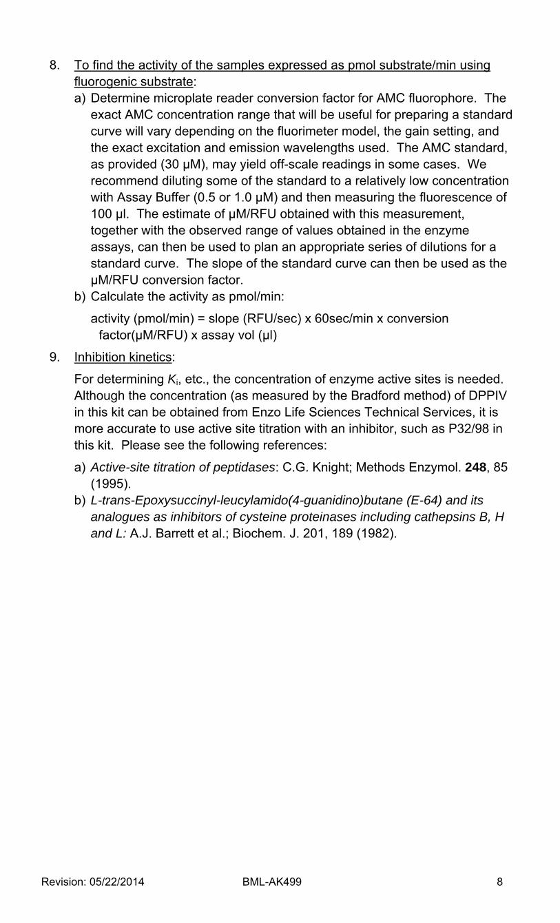
8. To find the activity of the samples expressed as pmol substrate/min using fluorogenic substrate: a) Determine microplate reader conversion factor for AMC fluorophore. The
exact AMC concentration range that will be useful for preparing a standard curve will vary depending on the fluorimeter model, the gain setting, and the exact excitation and emission wavelengths used. The AMC standard, as provided (30 µM), may yield off-scale readings in some cases. We recommend diluting some of the standard to a relatively low concentration with Assay Buffer (0.5 or 1.0 µM) and then measuring the fluorescence of 100 µl. The estimate of µM/RFU obtained with this measurement, together with the observed range of values obtained in the enzyme assays, can then be used to plan an appropriate series of dilutions for a standard curve. The slope of the standard curve can then be used as the µM/RFU conversion factor.
b) Calculate the activity as pmol/min:
activity (pmol/min) = slope (RFU/sec) x 60sec/min x conversion factor(µM/RFU) x assay vol (µl)
9. Inhibition kinetics:
For determining Ki, etc., the concentration of enzyme active sites is needed. Although the concentration (as measured by the Bradford method) of DPPIV in this kit can be obtained from Enzo Life Sciences Technical Services, it is more accurate to use active site titration with an inhibitor, such as P32/98 in this kit. Please see the following references:
a) Active-site titration of peptidases: C.G. Knight; Methods Enzymol. 248, 85 (1995).
b) L-trans-Epoxysuccinyl-leucylamido(4-guanidino)butane (E-64) and its analogues as inhibitors of cysteine proteinases including cathepsins B, H and L: A.J. Barrett et al.; Biochem. J. 201, 189 (1982).

Revision: 05/22/2014 BML-AK499 10
USE FOR RESEARCH PURPOSES ONLY
Unless otherwise specified expressly on the packaging, all products sold hereunder are
intended for and may be used for research purposes only and may not be used for food,
drug, cosmetic or household use or for the diagnosis or treatment of human beings.
Purchase does not include any right or license to use, develop or otherwise exploit these
products commercially. Any commercial use, development or exploitation of these
products or development using these products without the express written authorization
of Enzo Life Sciences, Inc. is strictly prohibited. Buyer assumes all risk and liability for the
use and/or results obtained by the use of the products covered by this invoice whether
used singularly or in combination with other products.
LIMITED WARRANTY; DISCLAIMER OF WARRANTIES
These products are offered under a limited warranty. The products are guaranteed to
meet all appropriate specifications described in the package insert at the time of
shipment. Enzo Life Sciences’ sole obligation is to replace the product to the extent of the
purchasing price. All claims must be made to Enzo Life Sciences, Inc., within five (5) days of
receipt of order. THIS WARRANTY IS EXPRESSLY IN LIEU OF ANY OTHER WARRANTIES OR
LIABILITIES, EXPRESS OR IMPLIED, INCLUDING WARRANTIES OF MERCHANTABILITY,
FITNESS FOR A PARTICULAR PURPOSE, AND NONINFRINGEMENT OF THE PATENT OR
OTHER INTELLECTUAL PROPERTY RIGHTS OF OTHERS, AND ALL SUCH WARRANTIES (AND
ANY OTHER WARRANTIES IMPLIED BY LAW) ARE EXPRESSLY DISCLAIMED.
TRADEMARKS AND PATENTS
Several Enzo Life Sciences products and product applications are covered by US and
foreign patents and patents pending.
Global Headquarters Enzo Life Sciences Inc. Enzo Life Sciences (ELS) AG 10 Executive Blvd Industriestrasse 17, Postfach Farmingdale, NY 11735 CH‐4415 Lause / Switzerland (p) 1‐800‐942‐0430 (p) +41/0 61 926 89 89 (f) 1‐631‐694‐7501 (f) +41/0 61 926 89 79 (e) info‐[email protected] (e) info‐[email protected] Please visit our website at www.enzolifesciences.com for additional contact information.
www.enzolifesciences.com Enabling Discovery in Life Science®