doxorubicin inhibits dnmt1 resulting in conditional...
TRANSCRIPT

MOL 002634
1
Title page
Doxorubicin inhibits DNMT1 resulting in conditional apoptosis
Tomoki Yokochi# and Keith D. Robertson*,@
Epigenetic Gene Regulation and Cancer Section, Bldg.41-B715, National Cancer Institute,
National Institutes of Health, Bethesda, MD 20892, USA
Molecular Pharmacology Fast Forward. Published on August 31, 2004 as doi:10.1124/mol.104.002634
Copyright 2004 by the American Society for Pharmacology and Experimental Therapeutics.
This article has not been copyedited and formatted. The final version may differ from this version.Molecular Pharmacology Fast Forward. Published on August 31, 2004 as DOI: 10.1124/mol.104.002634
at ASPE
T Journals on February 28, 2019
molpharm
.aspetjournals.orgD
ownloaded from
MOL 002634
2
Running title page
Running title: Inhibition of DNMT1 by a DNA intercalating drug doxorubicin
* To whom correspondence should be addressed at Keith D. Robertson, Ph.D.
Department of Biochemistry & Molecular Biology, University of Florida, Box 100245,
Gainesville, FL 32610-0254
Phone: 352-392-1810
Fax: 352-392-2953
E-mail: [email protected]
Manuscript information
The number of text pages: 20
The number of tables: 0
The number of figures: 4
The number of references: 30
The number of words in the Abstract: 191
The number of words in the Introduction: 614
The number of words in the Discussion: 555
Abbreviations:
AdoMet, S-adenosyl-L-methionine; AdoHcy, S-adenosyl-L-homocysteine; 5-AdC, 5-aza-2'-
deoxycytidine.
This article has not been copyedited and formatted. The final version may differ from this version.Molecular Pharmacology Fast Forward. Published on August 31, 2004 as DOI: 10.1124/mol.104.002634
at ASPE
T Journals on February 28, 2019
molpharm
.aspetjournals.orgD
ownloaded from
MOL 002634
3
Abstract
Chemotherapy utilizing DNA intercalators is one of the most successful approaches to cancer
treatment. Although DNA intercalators are believed to inhibit DNA polymerases and
topoisomerases resulting in the induction of apoptosis in tumor cells, other factors potentially
inhibited by the anthracycline antibiotics remain to be elucidated. Here we show that the
enzymatic activity of DNMT1, the primary DNA methyltransferase in mammalian cells, is
inhibited by DNA intercalators such as doxorubicin in an in vitro assay. Enzymatic analyses
indicate that doxorubicin inhibits the catalytic activity of DNMT1 via DNA intercalation. We
also found that apoptosis was induced in DNMT1+/+ HCT116 cells by only a limited range of
doxorubicin dose, meaning that apoptotic cell death is "conditional" with respect to the
concentration of the DNA intercalating drug. Interestingly, conditional apoptosis is not
observed in human colorectal cancer cells lacking DNMT1 but can be induced in DNMT1-/-
cells by transfection of a plasmid expressing DNMT1. Our results suggest that DNMT1 is
one of the major targets of doxorubicin resulting in drug-induced apoptosis in human cancer
cells. We propose that expression levels of DNMT1 in tumor cells may affect the
effectiveness of doxorubicin in chemotherapy.
This article has not been copyedited and formatted. The final version may differ from this version.Molecular Pharmacology Fast Forward. Published on August 31, 2004 as DOI: 10.1124/mol.104.002634
at ASPE
T Journals on February 28, 2019
molpharm
.aspetjournals.orgD
ownloaded from
MOL 002634
4
Introduction
DNA intercalating agents, such as amsacrine (m-AMSA), actinomycin, mitoxantrone, and
doxorubicin, have been employed as anti-cancer drugs and are in routine clinical use as
chemotherapeutic agents (Brana et al., 2001). DNA intercalators share common structural
motifs - the presence of planar polyaromatic systems that bind by insertion between DNA
base-pairs. Doxorubicin (Adriamycin), an anthracycline-based DNA intercalator, is one of
the most typical anticancer drugs commonly employed in the clinic. It has been well accepted
that the anti-tumor activity of doxorubicin is due to the formation of a cleavable complex of
topoisomerase II, resulting in apoptosis (Hickman, 1992; Kiechle and Zhang, 2002).
Doxorubicin is indicated in the treatment of a broad spectrum of solid tumors (e.g. breast,
bladder, endometrium, thyroid, lung, ovary, stomach, and sarcomas of the bone) and in the
treatment of lymphoma, as well as acute lymphoblastic and myeloblastic leukemias (Carter,
1975). One of the most important and clinically relevant side effects of doxorubicin is the
induction of cardiomyopathy (Lenaz and Page, 1976). A number of mechanisms have been
proposed to explain this effect of doxorubicin, including oxidative stress (Myers et al., 1977),
the induction of mitochondrial damage (Wallace, 2003), and changes in gene expression in
cardiac myocytes and muscle cells in general (Boucek et al., 1999; Kurabayashi et al., 1994).
Therefore, doxorubicin likely impacts on the activity of other important proteins or pathways
and these need to be elucidated to better understand and to take advantage of its anti-tumor
activity.
DNA methyltransferase 1 (DNMT1) is the primary enzyme responsible for maintenance of
DNA methylation on genomic DNA (Bestor et al., 1988; Pradhan et al., 1999; Yoder et al.,
1996; Yokochi and Robertson, 2002). Disruption of DNMT1 function causes chromosome
instability and dysregulation of transcription, and ultimately leads to apoptotic cell death
This article has not been copyedited and formatted. The final version may differ from this version.Molecular Pharmacology Fast Forward. Published on August 31, 2004 as DOI: 10.1124/mol.104.002634
at ASPE
T Journals on February 28, 2019
molpharm
.aspetjournals.orgD
ownloaded from
MOL 002634
5
(Ehrlich, 2002; Jones and Baylin, 2002; Robertson, 2001). Cre-mediated deletion of the
Dnmt1 gene resulted in demethylation of genomic DNA in cultured murine fibroblasts and
led to p53-dependent cell death (Jackson-Grusby et al., 2001), suggesting that loss of
DNMT1 induces apoptosis. However, other studies have shown that deletion of the DNMT1
gene is compatible with cell viability in the HCT116 cell line (Rhee et al., 2002; Rhee et al.,
2000). The DNMT1-/- HCT116 cells exhibited normal morphology except for a slightly
slower growth rate compared to the parental or DNMT1+/- cells (Rhee et al., 2000), indicating
that loss of DNMT1 did not induce apoptosis in this cell line. Therefore, it was of great
interest to determine whether DNMT1 is associated with apoptosis mediated by doxorubicin.
Previous work has suggested that DNA methyltransferase activity in crude cell extracts could
be inhibited by DNA intercalators (Adams and Rinaldi, 1987), however the molecular
mechanism of intercalator-dependent inhibition of DNMT1 functions in vitro has not been
reported.
In this report, we first investigated the effect of the inhibition of DNMT1 activity by DNA
intercalating drugs including doxorubicin in an in vitro assay system. Enzymatic studies
demonstrate that doxorubicin is a potent inhibitor of DNMT1 activity via DNA intercalation.
We also examined doxorubicin-induced cytotoxicity utilizing the HCT116 human colorectal
cancer cell line as a model system. Our data show that doxorubicin treatment of HCT116
cells results in massive cell killing by apoptosis at only a particular drug dose (referred to as
conditional apoptosis). Lower, and remarkably, higher doses of doxorubicin do not result in
significant apoptosis induction. This conditional apoptosis is absent in cells lacking the major
DNA methyltransferase DNMT1. However, DNMT1-/- HCT116 cells can be made apoptosis-
inducible by reintroduction of DNMT1. These results suggest for the first time that DNMT1
This article has not been copyedited and formatted. The final version may differ from this version.Molecular Pharmacology Fast Forward. Published on August 31, 2004 as DOI: 10.1124/mol.104.002634
at ASPE
T Journals on February 28, 2019
molpharm
.aspetjournals.orgD
ownloaded from

MOL 002634
6
is one of the important targets for doxorubicin and that interaction of DNMT1 and
doxorubicin may contribute to the induction of apoptosis in cancer cells.
Materials and Methods
Recombinant protein preparation and DNA methyltransferase assay. Expression and
purification of recombinant DNMT1 has been described previously (Yokochi and Robertson,
2002). All experiments in a given panel in Fig. 1 were performed with the same enzyme
preparations. DNA methyltransferase activity of DNMT1 was measured using the DNMT-
magnetic beads assay (Yokochi and Robertson, 2002). A typical methylation reaction (40 µl)
contained 125 nM DNA oligonucleotides (hemimethylated DNA conjugated with a biotin
molecule, 34 base-pairs (Yokochi and Robertson, 2002)), 30 nM DNMT1, and 600 nM
tritium-labeled AdoMet (Amersham Bioscience, 1 mCi/ml) in reaction buffer (50 mM Tris,
pH 8.0, 5 mM EDTA, 10% glycerol, 10 mM 2-mercaptoethanol, 0.5 mM
phenylmethylsulfonylfluoride). Experiments were done independently at least three times and
the mean values are shown. Radioactive materials were purchased from Amersham
Bioscience. General chemicals were purchased from Sigma, Invitrogen, and Roche.
Cell culture, drug treatment, and transient transfection. HCT116 and DNMT1-/- HCT116
cells were cultured in McCoy's 5A medium supplemented with 10% fetal bovine serum and 2
mM L-glutamine. Trypsinized cells were plated onto 100 mm (10 ml medium) and 35 mm (2
ml medium) diameter dishes and standard 96-well flat bottom plates (100 µl medium) and
incubated for 24 hours prior to drug treatment. Since growth rates of HCT116 cells and its
knockout derivative differ slightly (Rhee et al., 2000), initial cell numbers were adjusted so
that the cell concentrations were typically 1×10⁵ cells/ml at the time of drug addition.
DNMT1-/- HCT116 cells were transfected with GFP or GFP-DNMT1 expression vectors
This article has not been copyedited and formatted. The final version may differ from this version.Molecular Pharmacology Fast Forward. Published on August 31, 2004 as DOI: 10.1124/mol.104.002634
at ASPE
T Journals on February 28, 2019
molpharm
.aspetjournals.orgD
ownloaded from
MOL 002634
7
using TransIT-LT1 transfection reagent (Mirus) according to the manufacturer's instructions.
Each experiment was repeated three times independently, in which at least 10 cells
expressing green fluorescence were examined and were confirmed to be consistent in terms
of doxorubicin sensitivity.
Cell viability assay, Trypan Blue staining, and apoptosis-related assays. MTS (3-(4,5-
dimethylthiazol-2-yl)-5-(3-carboxymethoxyphenyl)-2-4-sulfophenyl)-2H-tetrazolium, inner
salt) assay and Caspase-Glo 3/7 assay (Promega) were performed in accordance with the
manufacturer's instructions. A fixed concentration of cells (1×10⁵ cells/ml at the time of drug
addition) was treated with various concentrations of drugs, and the cell viability or caspase
activity were measured after 48 h. Parallel reactions were carried out without cells and this
value was subtracted from the experimental values. Results were expressed as the mean of
four independent experiments and error bars represent the standard deviation (S.D.). DNA
fragmentation in cells treated with doxorubicin was detected as described (Yeung, 2002). For
Trypan Blue staining, cells in 2 ml of medium were treated with various concentrations of
doxorubicin for 48 hours, trypsinized, resuspended in fresh medium, and then an equal
volume of Trypan Blue stain was added. The number of live and dead cells was counted on
pictures taken with a microscopy system with a digital camera (Nikon). Results were
expressed as the mean of four independent experiments.
Western blotting and methyl acceptance assay. The DNMT1 antipeptide antibody has been
previously described (Yokochi and Robertson, 2002). The PCNA and β-actin antibodies were
purchased from Santa Cruz Biotechnology. Genomic DNA was extracted from HCT116 cells
that were treated with various concentrations of doxorubicin as follows (0, 1×10⁻¹⁰, 1×10⁻⁹,
1×10⁻⁸, 1×10⁻⁷, 2×10⁻⁷, 5×10⁻⁷, 1×10⁻⁶, 2×10⁻⁶, 5×10⁻⁶, 1×10⁻⁵, 2×10⁻⁵, 5×10⁻⁵, 1×10⁻⁴ M) for
This article has not been copyedited and formatted. The final version may differ from this version.Molecular Pharmacology Fast Forward. Published on August 31, 2004 as DOI: 10.1124/mol.104.002634
at ASPE
T Journals on February 28, 2019
molpharm
.aspetjournals.orgD
ownloaded from
MOL 002634
8
48 hours. Methylation acceptance capability of genomic DNA was measured by in vitro
methylation reaction utilizing bacterial CpG methylase SssI (New England Biolabs). A
reaction solution contained genomic DNA (2 µg), SssI (1 µl), and 900 nM tritium-labeled
AdoMet in reaction buffer (50 mM Tris, pH 8.0, 5 mM EDTA, 10% glycerol, 10 mM 2-
mercaptoethanol, 0.5 mM phenylmethylsulfonylfluoride). Following ethanol precipitation,
tritium incorporation into genomic DNA was measured by a liquid scintillation counter.
Results were presented as the mean of three independent experiments. S.D. was shown as
error bars.
Results
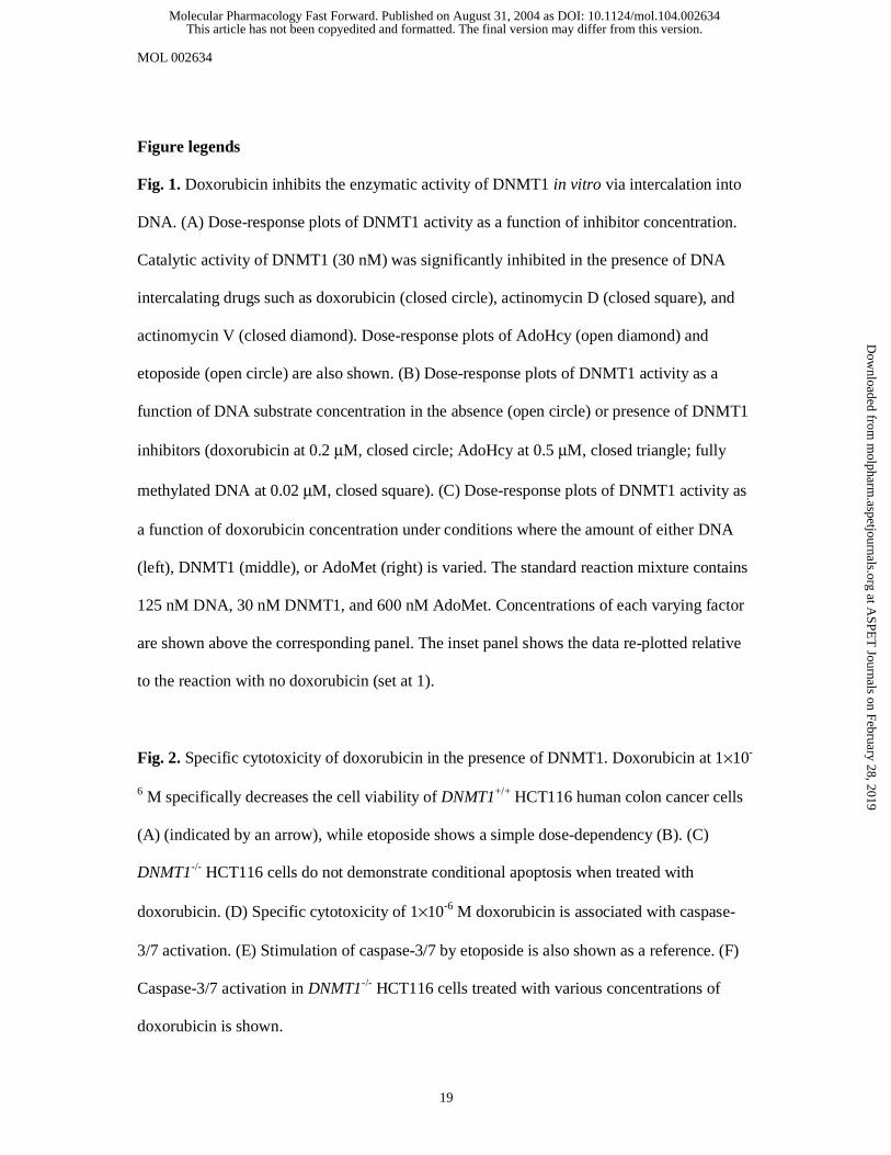
Doxorubicin inhibits the enzymatic activity of DNMT1 in vitro via intercalation into
DNA. We employed an in vitro enzymatic assay system (Yokochi and Robertson, 2002),
utilizing highly purified recombinant DNMT1 to investigate the effects of DNA intercalators
on DNMT1 catalytic activity. Doxorubicin, as well as other DNA intercalating agents,
significantly inhibited DNMT1 activity in vitro (Fig. 1A). We next compared dose-response
plots of DNMT1 activity against the DNA substrate in the presence of a fixed concentration
of doxorubicin, S-adenosyl-L-homocysteine (AdoHcy), or methylated DNA as enzymatic
inhibitors. Both AdoHcy and methylated DNA are product inhibitors of DNMT1 and they
require direct binding to the enzyme to inhibit its catalytic activity. Consistent with the
velocity equations for an enzymatic reaction under the effect of an inhibitor (Copeland, 2000;
Segel, 1993), DNMT1 activity yielded hyperbolic curves in the presence of either AdoHcy or
methylated DNA (Fig. 1B). In contrast, DNMT1 inhibition yielded a sigmoidal curve under
the effect of doxorubicin inhibition, suggesting that the inhibitory mechanism by doxorubicin
might not be explained by a simple “inhibitor-enzyme” interaction model (Copeland, 2000).
To identify which factor (DNMT1, DNA, or AdoMet) interacts with doxorubicin to inhibit
This article has not been copyedited and formatted. The final version may differ from this version.Molecular Pharmacology Fast Forward. Published on August 31, 2004 as DOI: 10.1124/mol.104.002634
at ASPE
T Journals on February 28, 2019
molpharm
.aspetjournals.orgD
ownloaded from

MOL 002634
9
the DNMT1 catalytic reaction, we employed conditions in which one of the three factors was
varied to examine whether it would alter the doxorubicin-dose dependency of DNMT1
activity on the inhibition plots (Fig. 1C). The slope in the relative comparison plot of the
inhibition curves was significantly affected only when the amount of DNA was varied (Fig.
1C, left panel, inset), indicating that the inhibitory effect of doxorubicin is dose dependent
with respect to DNA. These results suggest that the formation of a drug-DNA complex is
important for the inhibition of DNMT1 by doxorubicin and exclude the possibility that
doxorubicin binds directly to DNMT1 to cause inhibition. Thus, we conclude that
doxorubicin inhibits DNMT1 activity via DNA intercalation.
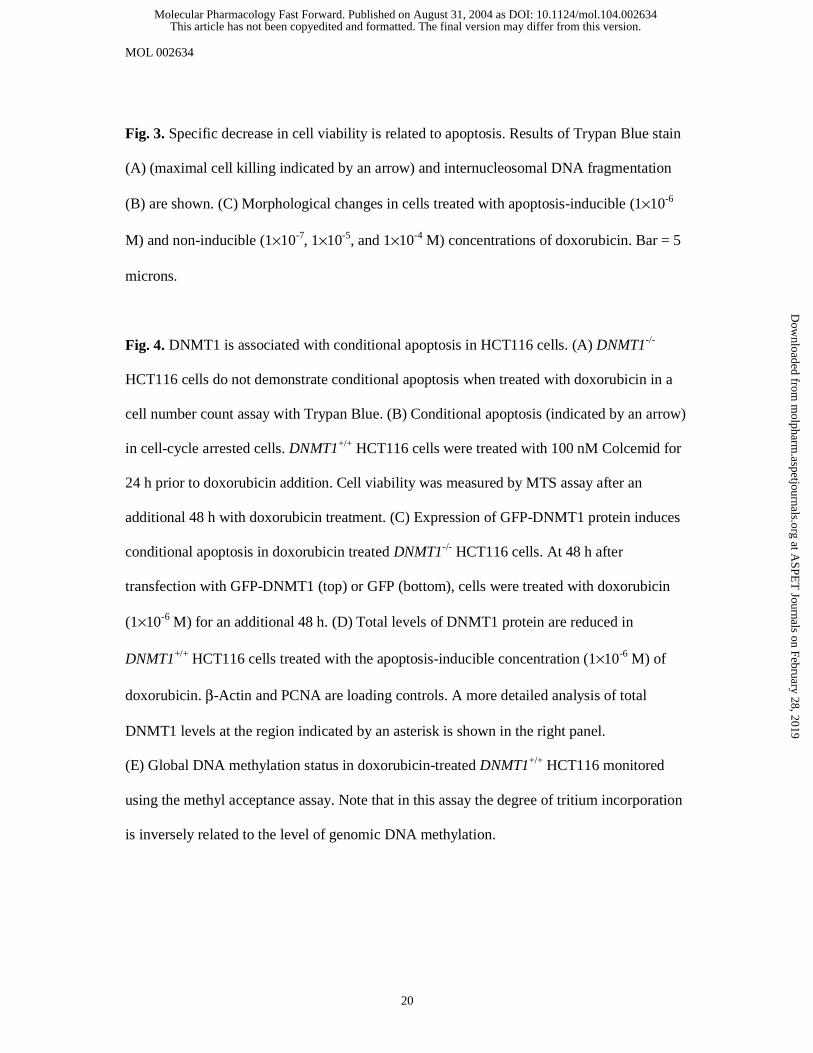
Doxorubicin demonstrates a specific cytotoxicity in the presence of DNMT1. We next
sought to examine the possible consequence of the inhibition of DNMT1 by doxorubicin in
human cancer cells. As a reference, we utilized a different type of anticancer drug, etoposide
(VP-16-213). Etoposide is a non-DNA intercalating drug that inhibits topoisomerase II
specifically resulting in p53-dependent apoptosis (Ross et al., 1984) but does not inhibit
DNMT1 in vitro (Fig. 1A). A fixed number of HCT116 human colorectal carcinoma cells
(DNMT1+/+ and DNMT1-/-) were treated with various concentrations of anticancer agents
(doxorubicin and etoposide) for 48 hours, and the cell viability was measured by MTS assay.
A particular dose (1×10⁻⁶ M) of doxorubicin specifically decreased cell viability (Fig. 2A),
while etoposide yielded a simple dose-dependent decrease in cell viability (Fig. 2B).
Interestingly, the DNMT1-/- HCT116 cells (Rhee et al., 2000) did not demonstrate the specific
cytotoxicity following doxorubicin treatment (Fig. 2C). Similar specific cytotoxicity was
observed with other DNA intercalators such as actinomycin D and actinomycin V in
DNMT1+/+ HCT116 cells, and in other cell lines such as HeLa and 293 with doxorubicin (data
not shown). The large peak of caspase-3/7 activation at 1×10⁻⁶ M doxorubicin (Fig. 2D,
This article has not been copyedited and formatted. The final version may differ from this version.Molecular Pharmacology Fast Forward. Published on August 31, 2004 as DOI: 10.1124/mol.104.002634
at ASPE
T Journals on February 28, 2019
molpharm
.aspetjournals.orgD
ownloaded from
MOL 002634
10
etoposide control in Fig. 2E) is consistent with the decrease in cell viability (Fig. 2A),
suggesting that the cytotoxicity at 1×10⁻⁶ M doxorubicin is due to apoptotic cell death. A
slight activation of caspase-3/7 was observed (∼two-fold) in a broad, but non-specific range
of doxorubicin doses between 1×10⁻⁷ M and 1×10⁻⁵ M in DNMT1-/- cells (Fig. 2F). This is
consistent with previous findings that the other major targets of doxorubicin are DNA
polymerases and topoisomerases, and that the inhibition of these DNA-binding proteins with
doxorubicin leads to apoptosis (Hickman, 1992; Kiechle and Zhang, 2002).
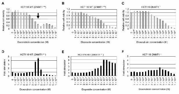
Apoptosis is induced by only a particular concentration of doxorubicin. To further
confirm that apoptosis is associated with the decreasing cell viability induced by doxorubicin
in DNMT1+/+ HCT116 cells, the numbers of live and dead cells were counted after Trypan
Blue staining (Fig. 3A). A large fraction of cells died under the treatment with 1×10⁻⁶ M
doxorubicin, suggesting that the decrease in cell viability is related to massive cell killing.
Ten-fold higher (1×10⁻⁵ M) or ten-fold lower (1×10⁻⁷ M) doses of doxorubicin, however, did
not lead to significant cell death. DNA laddering analysis also supports this notion (Fig. 3B).
The improved DNA laddering assay described by Yeung (Yeung, 2002) clearly demonstrated
nucleosomal fragmentation, which is a well documented characteristic of apoptosis, at the
1×10⁻⁶ M doxorubicin dose, while random genomic DNA digestion occurred at higher doses
(1×10⁻⁵ M and above). Cells treated with 1×10⁻⁶ M doxorubicin appeared shrunken and
developed blebs on their cell surface, typical morphological hallmarks of apoptosis
(Budihardjo et al., 1999) (Fig. 3C). Our results therefore suggest that apoptosis induced by
doxorubicin is "conditional", meaning that only a particular concentration of drug specifically
induces apoptosis in DNMT1+/+ HCT116 cells, but higher or lower concentrations do not. It
should be noted that, at the highest dose (1×10⁻⁴ M) of doxorubicin, cells blackened, swelled,
and burst (Fig. 3C), and showed neither caspase-3/7 activation (Fig. 2F) nor DNA laddering
This article has not been copyedited and formatted. The final version may differ from this version.Molecular Pharmacology Fast Forward. Published on August 31, 2004 as DOI: 10.1124/mol.104.002634
at ASPE
T Journals on February 28, 2019
molpharm
.aspetjournals.orgD
ownloaded from
MOL 002634
11
(Fig. 3B). These characteristics are consistent with cell death due to necrosis (Kroemer et al.,
1998).
Conditional apoptosis induced by doxorubicin depends on DNMT1 in HCT116 cells.
Consistent with the results of cell viability (Fig. 2C) and caspase activity (Fig. 2F), DNMT1-/-
HCT116 cells yielded a simple dose-dependency with regard to doxorubicin concentration in
the Trypan Blue staining assay (Fig. 4A). Since DNMT1-/- HCT116 cells grow more slowly
than DNMT1+/+ HCT116 cells, differences between these two cell lines may be explained by
the difference of cell growth rates. To rule out this possibility, the cell cycle of DNMT1+/+
HCT116 cells was first arrested by other growth inhibitors (e.g. Colcemid (Fig. 4B),
nocodazole, hydroxyurea, and aphidicolin (data not shown)). Doxorubicin-mediated
conditional apoptosis was induced even in these growth-arrested cells (Fig. 4B). This result
suggests that the difference of doxorubicin sensitivity between DNMT1+/+ and DNMT1-/- cells
is not related to cell growth rates and that cell proliferation and DNA replication are not
essential for conditional apoptosis. To further confirm the direct contribution of DNMT1 to
conditional apoptosis, transient transfection of DNMT1-/- HCT116 cells was performed with
either empty GFP (green fluorescent protein) expression vector or GFP-tagged DNMT1
expression vector. After 48 hours of transfection, cells were treated with 1×10⁻⁶ M
doxorubicin (the dose inducing apoptosis in DNMT1+/+ cells, but not in DNMT1-/- cells) for an
additional 48 hours. Expression of GFP-DNMT1 caused apoptosis in DNMT1-/- HCT116
cells following treatment with 1×10⁻⁶ M doxorubicin (Fig. 4C, top panels), whereas
expression of GFP alone did not (Fig. 4C, bottom panels). Total protein levels of DNMT1 in
DNMT1+/+ HCT116 cells were greatly reduced following treatment with 1×10⁻⁶ M
doxorubicin (Fig. 4D), further supporting the notion that DNMT1 in cells is related to
This article has not been copyedited and formatted. The final version may differ from this version.Molecular Pharmacology Fast Forward. Published on August 31, 2004 as DOI: 10.1124/mol.104.002634
at ASPE
T Journals on February 28, 2019
molpharm
.aspetjournals.orgD
ownloaded from
MOL 002634
12
conditional apoptosis induced by doxorubicin. Taken together, we conclude that DNMT1
contributes to the conditional apoptosis in HCT116 cells.
Changes in global methylation status of genomic DNA are not associated with
conditional apoptosis. Results presented in Fig. 3A from the Trypan blue cell counting assay
clearly indicated that one of the effects of doxorubicin was to cause cell cycle arrest. This
effect occurred over a relatively wider range of drug concentration than the conditional
apoptosis effect, beginning at 1×10⁻⁷ M doxorubicin. Since demethylation due to inhibition of
DNMT1 is believed to occur by a passive mechanism, which requires active cell division to
dilute out the methylated parental DNA strands, it was not clear whether doxorubicin
treatment and conditional apoptosis would be associated with detectable genomic
demethylation. In order to determine this, global DNA methylation levels in doxorubicin-
treated DNMT1+/+ HCT116 were examined using the methyl-acceptance assay. Results of
this assay (Fig. 4E) revealed that the cellular DNA methylation status of HCT116 cells was
not significantly altered following treatment with 1×10⁻⁶ M doxorubicin. Although there
appeared to be some hypomethylation occurring at very high doses of doxorubicin, the
significance of such results is difficult to ascertain because cells are undergoing high levels of
cell death by necrosis under these conditions.
Discussion
Two non-exclusive molecular mechanisms have been proposed as the trigger of apoptosis
induction by DNA methyltransferase inhibitors: genomic demethylation and enzyme-
mediated DNA damage. Loss of genomic methylation as the consequence of DNA
methyltransferase inhibition causes dysregulation of gene expression and apoptosis.
Depletion of Dnmt1 from Xenopus embryos causes embryonic lethality and inappropriate
This article has not been copyedited and formatted. The final version may differ from this version.Molecular Pharmacology Fast Forward. Published on August 31, 2004 as DOI: 10.1124/mol.104.002634
at ASPE
T Journals on February 28, 2019
molpharm
.aspetjournals.orgD
ownloaded from

MOL 002634
13
gene expression (Stancheva and Meehan, 2000). Furthermore, Cre-lox mediated depletion of
Dnmt1 in mouse fibroblasts caused loss of Dnmt1 protein, genomic hypomethylation, and
induction of p53-dependent apoptosis (Jackson-Grusby et al., 2001). Since cytosine
methylation within the promoter regions of genes can cause transcriptional silencing,
demethylation may activate the expression of genes that in turn activate p53. In this report,
however, we found that conditional apoptosis was induced only when DNMT1 protein and
1×10⁻⁶ M doxorubicin coexist. Therefore, the latter mechanism - loss of genomic methylation
- may not represent the most plausible cause of conditional apoptosis. An alternative
mechanism is that catalytically inactive DNMT1 bound to doxorubicin-treated DNA may be
perceived by the cell as a form of DNA damage. The cytotoxic effects of 5-aza-2'-
deoxycytidine (5-AdC) are believed to be mediated through the irreversible covalent binding
of DNA methyltransferase to 5-AdC-substituted DNA, which is then recognized as DNA
damage by the cell, resulting in apoptosis (Juttermann et al., 1994). Indeed, it has been shown
that 5-AdC treatment causes p53 activation through a traditional DNA damage response
pathway, which is consistent with this model (Karpf et al., 2001). Other studies have shown
that the introduction of DNMT1-/- DNMT3B-/- double mutations into HCT116 cells did not
cause apoptosis, even though these cells exhibited marked genomic hypomethylation (a
roughly 95% reduction in methylated cytidine content) (Rhee et al., 2002). In addition, we
demonstrated that doxorubicin could induce conditional apoptosis only in DNMT1+/+
HCT116 cells, but not in DNMT1-/- HCT116 (this work) or DNMT1-/- DNMT3B-/- HCT116
(data not shown) cells. These results suggest that DNMT1 itself, rather than the secondary
demethylation of genomic DNA, is the primary mediator of drug-induced cytotoxicity.
Total soluble DNMT1 protein levels were decreased specifically in DNMT1+/+ HCT116 cells
treated with the apoptosis-inducible concentration of doxorubicin (Fig. 4D). The reduced
This article has not been copyedited and formatted. The final version may differ from this version.Molecular Pharmacology Fast Forward. Published on August 31, 2004 as DOI: 10.1124/mol.104.002634
at ASPE
T Journals on February 28, 2019
molpharm
.aspetjournals.orgD
ownloaded from

MOL 002634
14
DNMT1 protein levels may be due to the formation of an irreversible DNMT1-DNA-
doxorubicin complex such that the DNMT1 becomes trapped in an insoluble chromatin
fraction during protein extraction. Although there is no direct evidence at this time, we
speculate that doxorubicin may stabilize the covalent link between DNMT1 and DNA. The
DNMT1-DNA complex could be recognized as DNA damage resulting in apoptosis. The
degradation or removal of the complex may be facilitated by the cellular DNA repair
machinery. Analysis of these, and other possibilities, will be the subject of future work.
It has been thought that the anti-tumor activity of DNA intercalators, including doxorubicin,
is closely related to DNA cleavage depending on topoisomerase II and inhibition of DNA
replication (Brana et al., 2001; Hickman, 1992; Kiechle and Zhang, 2002). Our data indicate
that DNMT1 is also required for conditional apoptosis induced by a particular concentration
of doxorubicin, suggesting that DNA methyltransferase is one of the targets of doxorubicin
for apoptosis induction in cancer cells. We propose that the expression levels of DNMT1 in
tumor cells may be important criteria that should be taken into account to evaluate the
selective cytotoxicity of the drug (Pratt et al., 1994) and to determine the optimal dose
regimen.
Acknowledgments
We would like to thank Drs. B. Vogelstein for cell lines; and T. Karpova and the fluorescence
imaging facility at NCI/NIH for technical assistance.
This article has not been copyedited and formatted. The final version may differ from this version.Molecular Pharmacology Fast Forward. Published on August 31, 2004 as DOI: 10.1124/mol.104.002634
at ASPE
T Journals on February 28, 2019
molpharm
.aspetjournals.orgD
ownloaded from
MOL 002634
15
References
Adams RL and Rinaldi A (1987) Effect of echinomycin on DNA methylation. FEBS Lett
215(2):266-268.
Bestor T, Laudano A, Mattaliano R and Ingram V (1988) Cloning and sequencing of a cDNA
encoding DNA methyltransferase of mouse cells. The carboxyl-terminal domain of
the mammalian enzymes is related to bacterial restriction methyltransferases. J Mol
Biol 203(4):971-983.
Boucek RJ, Jr., Miracle A, Anderson M, Engelman R, Atkinson J and Dodd DA (1999)
Persistent effects of doxorubicin on cardiac gene expression. J Mol Cell Cardiol
31(8):1435-1446.
Brana MF, Cacho M, Gradillas A, de Pascual-Teresa B and Ramos A (2001) Intercalators as
anticancer drugs. Curr Pharm Des 7(17):1745-1780.
Budihardjo I, Oliver H, Lutter M, Luo X and Wang X (1999) Biochemical pathways of
caspase activation during apoptosis. Annu Rev Cell Dev Biol 15:269-290.
Carter SK (1975) Adriamycin-a review. J Natl Cancer Inst 55(6):1265-1274.
Copeland RA (2000) Enzymes. Wiley-VCH, New York.
Ehrlich M (2002) DNA methylation in cancer: too much, but also too little. Oncogene
21(35):5400-5413.
Hickman JA (1992) Apoptosis induced by anticancer drugs. Cancer Metastasis Rev
11(2):121-139.
Jackson-Grusby L, Beard C, Possemato R, Tudor M, Fambrough D, Csankovszki G,
Dausman J, Lee P, Wilson C, Lander E and Jaenisch R (2001) Loss of genomic
methylation causes p53-dependent apoptosis and epigenetic deregulation. Nat Genet
27(1):31-39.
This article has not been copyedited and formatted. The final version may differ from this version.Molecular Pharmacology Fast Forward. Published on August 31, 2004 as DOI: 10.1124/mol.104.002634
at ASPE
T Journals on February 28, 2019
molpharm
.aspetjournals.orgD
ownloaded from
MOL 002634
16
Jones PA and Baylin SB (2002) The fundamental role of epigenetic events in cancer. Nat Rev
Genet 3(6):415-428.
Juttermann R, Li E and Jaenisch R (1994) Toxicity of 5-aza-2'-deoxycytidine to mammalian
cells is mediated primarily by covalent trapping of DNA methyltransferase rather than
DNA demethylation. Proc Natl Acad Sci USA 91(25):11797-11801.
Karpf AR, Moore BC, Ririe TO and Jones DA (2001) Activation of the p53 DNA damage
response pathway after inhibition of DNA methyltransferase by 5-aza-2'-
deoxycytidine. Mol Pharmacol 59(4):751-757.
Kiechle FL and Zhang X (2002) Apoptosis: biochemical aspects and clinical implications.
Clin Chim Acta 326(1-2):27-45.
Kroemer G, Dallaporta B and Resche-Rigon M (1998) The mitochondrial death/life regulator
in apoptosis and necrosis. Annu Rev Physiol 60:619-642.
Kurabayashi M, Jeyaseelan R and Kedes L (1994) Doxorubicin represses the function of the
myogenic helix-loop-helix transcription factor MyoD. Involvement of Id gene
induction. J Biol Chem 269(8):6031-6039.
Lenaz L and Page JA (1976) Cardiotoxicity of adriamycin and related anthracyclines. Cancer
Treat Rev 3(3):111-120.
Myers CE, McGuire WP, Liss RH, Ifrim I, Grotzinger K and Young RC (1977) Adriamycin:
the role of lipid peroxidation in cardiac toxicity and tumor response. Science
197(4299):165-167.
Pradhan S, Bacolla A, Wells RD and Roberts RJ (1999) Recombinant human DNA (cytosine-
5) methyltransferase. I. Expression, purification, and comparison of de novo and
maintenance methylation. J Biol Chem 274(46):33002-33010.
Pratt WB, Ruddon RW, Ensminger WD and Maybaum J (1994) The anticancer drugs.
Oxford University Press, New York.
This article has not been copyedited and formatted. The final version may differ from this version.Molecular Pharmacology Fast Forward. Published on August 31, 2004 as DOI: 10.1124/mol.104.002634
at ASPE
T Journals on February 28, 2019
molpharm
.aspetjournals.orgD
ownloaded from
MOL 002634
17
Rhee I, Bachman KE, Park BH, Jair KW, Yen RW, Schuebel KE, Cui H, Feinberg AP,
Lengauer C, Kinzler KW, Baylin SB and Vogelstein B (2002) DNMT1 and DNMT3b
cooperate to silence genes in human cancer cells. Nature 416(6880):552-556.
Rhee I, Jair KW, Yen RW, Lengauer C, Herman JG, Kinzler KW, Vogelstein B, Baylin SB
and Schuebel KE (2000) CpG methylation is maintained in human cancer cells
lacking DNMT1. Nature 404(6781):1003-1007.
Robertson KD (2001) DNA methylation, methyltransferases, and cancer. Oncogene
20(24):3139-3155.
Ross W, Rowe T, Glisson B, Yalowich J and Liu L (1984) Role of topoisomerase II in
mediating epipodophyllotoxin-induced DNA cleavage. Cancer Res 44(12 Pt 1):5857-
5860.
Segel IH (1993) Enzyme kinetics. Wiley, New York.
Stancheva I and Meehan RR (2000) Transient depletion of xDnmt1 leads to premature gene
activation in Xenopus embryos. Genes Dev 14(3):313-327.
Wallace KB (2003) Doxorubicin-induced cardiac mitochondrionopathy. Pharmacol Toxicol
93(3):105-115.
Yeung MC (2002) Accelerated apoptotic DNA laddering protocol. Biotechniques 33(4):734,
736.
Yoder JA, Yen RW, Vertino PM, Bestor TH and Baylin SB (1996) New 5' regions of the
murine and human genes for DNA (cytosine-5)-methyltransferase. J Biol Chem
271(49):31092-31097.
Yokochi T and Robertson KD (2002) Preferential methylation of unmethylated DNA by
mammalian de novo DNA methyltransferase Dnmt3a. J Biol Chem 277(14):11735-
11745.
This article has not been copyedited and formatted. The final version may differ from this version.Molecular Pharmacology Fast Forward. Published on August 31, 2004 as DOI: 10.1124/mol.104.002634
at ASPE
T Journals on February 28, 2019
molpharm
.aspetjournals.orgD
ownloaded from

MOL 002634
18
Footnotes
T.Y. acknowledges support from the Rett Syndrome Research Foundation. This work was
supported by funds from the National Cancer Institute (1K22CA84535-01, K.D.R.).
# Current address:
Tomoki Yokochi, Ph.D.
Department of Biochemistry & Molecular Biology, SUNY Upstate Medical University, 750
East Adams St., Syracuse, NY 13210
E-mail: [email protected]
@ Current address:
Keith D. Robertson, Ph.D.
Department of Biochemistry & Molecular Biology, University of Florida, Box 100245,
Gainesville, FL 32610-0254
Phone: 352-392-1810
Fax: 352-392-2953
E-mail: [email protected]
The name of person who received the reprint requests:
Keith D. Robertson, Ph.D.
Department of Biochemistry & Molecular Biology, University of Florida, Box 100245,
Gainesville, FL 32610-0254
This article has not been copyedited and formatted. The final version may differ from this version.Molecular Pharmacology Fast Forward. Published on August 31, 2004 as DOI: 10.1124/mol.104.002634
at ASPE
T Journals on February 28, 2019
molpharm
.aspetjournals.orgD
ownloaded from
MOL 002634
19
Figure legends
Fig. 1. Doxorubicin inhibits the enzymatic activity of DNMT1 in vitro via intercalation into
DNA. (A) Dose-response plots of DNMT1 activity as a function of inhibitor concentration.
Catalytic activity of DNMT1 (30 nM) was significantly inhibited in the presence of DNA
intercalating drugs such as doxorubicin (closed circle), actinomycin D (closed square), and
actinomycin V (closed diamond). Dose-response plots of AdoHcy (open diamond) and
etoposide (open circle) are also shown. (B) Dose-response plots of DNMT1 activity as a
function of DNA substrate concentration in the absence (open circle) or presence of DNMT1
inhibitors (doxorubicin at 0.2 µM, closed circle; AdoHcy at 0.5 µM, closed triangle; fully
methylated DNA at 0.02 µM, closed square). (C) Dose-response plots of DNMT1 activity as
a function of doxorubicin concentration under conditions where the amount of either DNA
(left), DNMT1 (middle), or AdoMet (right) is varied. The standard reaction mixture contains
125 nM DNA, 30 nM DNMT1, and 600 nM AdoMet. Concentrations of each varying factor
are shown above the corresponding panel. The inset panel shows the data re-plotted relative
to the reaction with no doxorubicin (set at 1).
Fig. 2. Specific cytotoxicity of doxorubicin in the presence of DNMT1. Doxorubicin at 1×10-
6 M specifically decreases the cell viability of DNMT1+/+ HCT116 human colon cancer cells
(A) (indicated by an arrow), while etoposide shows a simple dose-dependency (B). (C)
DNMT1-/- HCT116 cells do not demonstrate conditional apoptosis when treated with
doxorubicin. (D) Specific cytotoxicity of 1×10⁻⁶ M doxorubicin is associated with caspase-
3/7 activation. (E) Stimulation of caspase-3/7 by etoposide is also shown as a reference. (F)
Caspase-3/7 activation in DNMT1-/- HCT116 cells treated with various concentrations of
doxorubicin is shown.
This article has not been copyedited and formatted. The final version may differ from this version.Molecular Pharmacology Fast Forward. Published on August 31, 2004 as DOI: 10.1124/mol.104.002634
at ASPE
T Journals on February 28, 2019
molpharm
.aspetjournals.orgD
ownloaded from
MOL 002634
20
Fig. 3. Specific decrease in cell viability is related to apoptosis. Results of Trypan Blue stain
(A) (maximal cell killing indicated by an arrow) and internucleosomal DNA fragmentation
(B) are shown. (C) Morphological changes in cells treated with apoptosis-inducible (1×10⁻⁶
M) and non-inducible (1×10⁻⁷, 1×10⁻⁵, and 1×10⁻⁴ M) concentrations of doxorubicin. Bar = 5
microns.
Fig. 4. DNMT1 is associated with conditional apoptosis in HCT116 cells. (A) DNMT1-/-
HCT116 cells do not demonstrate conditional apoptosis when treated with doxorubicin in a
cell number count assay with Trypan Blue. (B) Conditional apoptosis (indicated by an arrow)
in cell-cycle arrested cells. DNMT1+/+ HCT116 cells were treated with 100 nM Colcemid for
24 h prior to doxorubicin addition. Cell viability was measured by MTS assay after an
additional 48 h with doxorubicin treatment. (C) Expression of GFP-DNMT1 protein induces
conditional apoptosis in doxorubicin treated DNMT1-/- HCT116 cells. At 48 h after
transfection with GFP-DNMT1 (top) or GFP (bottom), cells were treated with doxorubicin
(1×10⁻⁶ M) for an additional 48 h. (D) Total levels of DNMT1 protein are reduced in
DNMT1+/+ HCT116 cells treated with the apoptosis-inducible concentration (1×10⁻⁶ M) of
doxorubicin. β-Actin and PCNA are loading controls. A more detailed analysis of total
DNMT1 levels at the region indicated by an asterisk is shown in the right panel.
(E) Global DNA methylation status in doxorubicin-treated DNMT1+/+ HCT116 monitored
using the methyl acceptance assay. Note that in this assay the degree of tritium incorporation
is inversely related to the level of genomic DNA methylation.
This article has not been copyedited and formatted. The final version may differ from this version.Molecular Pharmacology Fast Forward. Published on August 31, 2004 as DOI: 10.1124/mol.104.002634
at ASPE
T Journals on February 28, 2019
molpharm
.aspetjournals.orgD
ownloaded from

This article has not been copyedited and formatted. The final version may differ from this version.Molecular Pharmacology Fast Forward. Published on August 31, 2004 as DOI: 10.1124/mol.104.002634
at ASPE
T Journals on February 28, 2019
molpharm
.aspetjournals.orgD
ownloaded from
This article has not been copyedited and formatted. The final version may differ from this version.Molecular Pharmacology Fast Forward. Published on August 31, 2004 as DOI: 10.1124/mol.104.002634
at ASPE
T Journals on February 28, 2019
molpharm
.aspetjournals.orgD
ownloaded from
This article has not been copyedited and formatted. The final version may differ from this version.Molecular Pharmacology Fast Forward. Published on August 31, 2004 as DOI: 10.1124/mol.104.002634
at ASPE
T Journals on February 28, 2019
molpharm
.aspetjournals.orgD
ownloaded from

This article has not been copyedited and formatted. The final version may differ from this version.Molecular Pharmacology Fast Forward. Published on August 31, 2004 as DOI: 10.1124/mol.104.002634
at ASPE
T Journals on February 28, 2019
molpharm
.aspetjournals.orgD
ownloaded from